Embed Size (px)
Citation preview

Disorders of metabolism purine and pyrimidine; porfyrie
Blanka Stibůrková
Institute of Rheumatology
Institute of Inherited Metabolic Disorders 1.LF UK
19. 10. 2015 Pathobiochemistry
Exam questions
• disorders of uric acid metabolism • disorders of purines/pyrimidines metabolism • porphyrias
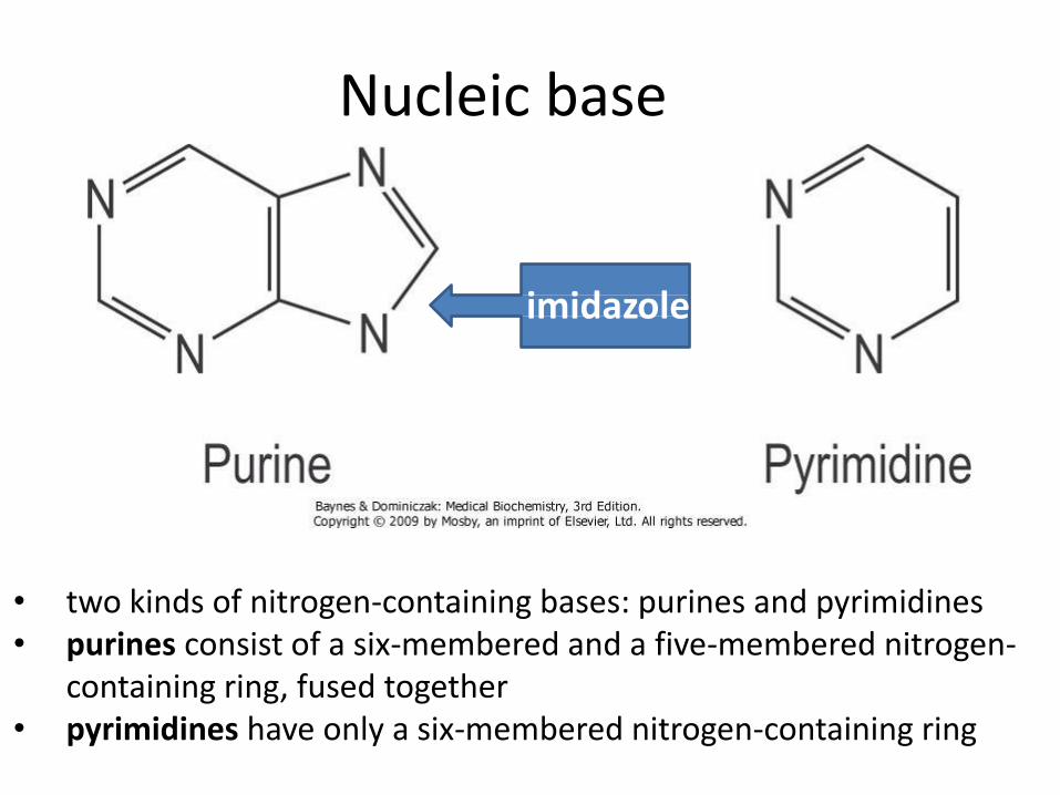
Nucleic base
imidazole
• two kinds of nitrogen-containing bases: purines and pyrimidines • purines consist of a six-membered and a five-membered nitrogen-
containing ring, fused together • pyrimidines have only a six-membered nitrogen-containing ring
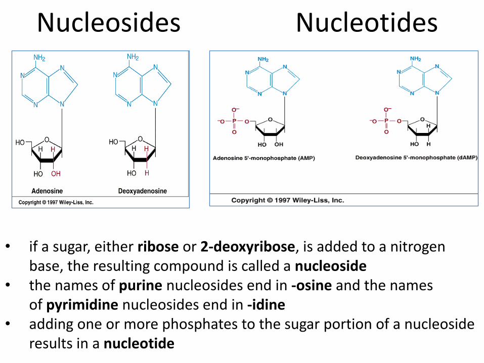
Nucleosides Nucleotides
• if a sugar, either ribose or 2-deoxyribose, is added to a nitrogen base, the resulting compound is called a nucleoside
• the names of purine nucleosides end in -osine and the names of pyrimidine nucleosides end in -idine
• adding one or more phosphates to the sugar portion of a nucleoside results in a nucleotide
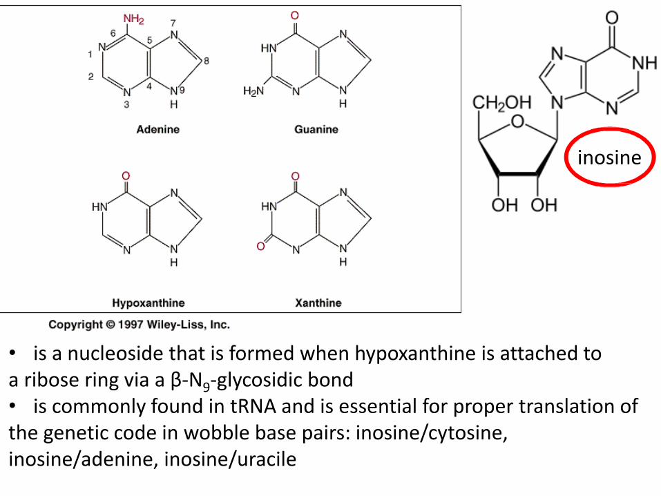
• is a nucleoside that is formed when hypoxanthine is attached to a ribose ring via a β-N9-glycosidic bond • is commonly found in tRNA and is essential for proper translation of the genetic code in wobble base pairs: inosine/cytosine, inosine/adenine, inosine/uracile
inosine

uridine
• is a glycosylated pyrimidine-analog containing uracil attached to a ribose ring via a β-N1-glycosidic bond • uridine is found in RNA and not DNA • deoxyuridin (idoxuridine, trifluridine): antiviral drugs → they are similar enough to be incorporated as part of DNA replication, but they possess side groups on the uracil component
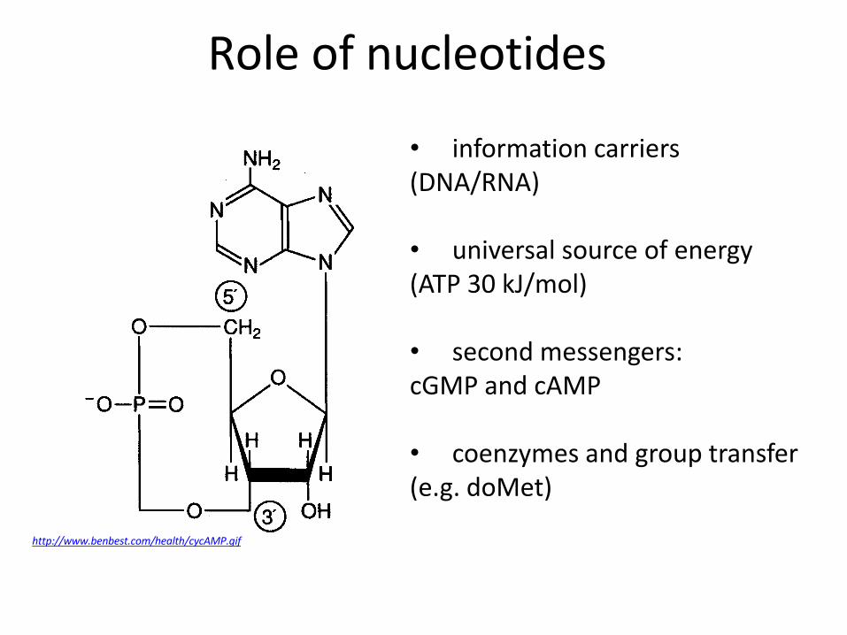
Role of nucleotides
• information carriers (DNA/RNA)
• universal source of energy (ATP 30 kJ/mol)
• second messengers: cGMP and cAMP • coenzymes and group transfer (e.g. doMet)
http://www.benbest.com/health/cycAMP.gif
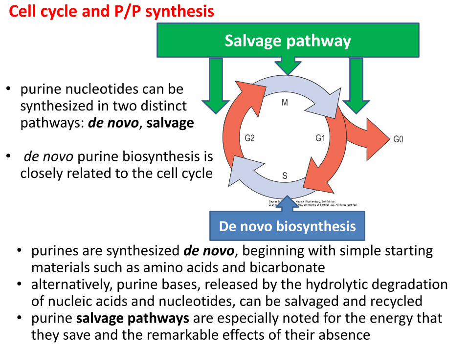
Cell cycle and P/P synthesis
Recyklace („salvage pathway“)
De novo biosynthesa
• purines are synthesized de novo, beginning with simple starting materials such as amino acids and bicarbonate
• alternatively, purine bases, released by the hydrolytic degradation of nucleic acids and nucleotides, can be salvaged and recycled
• purine salvage pathways are especially noted for the energy that they save and the remarkable effects of their absence
Salvage pathway
De novo biosynthesis
• purine nucleotides can be synthesized in two distinct pathways: de novo, salvage
• de novo purine biosynthesis is closely related to the cell cycle
• salwage pathway is not sufficient to meet total body requirements and so some de novo synthesis is essential
• de novo synthesis of purines is most active in liver
• non-hepatic tissues generally have limited or even no de novo synthesis
• pyrimidine synthesis occurs in a variety of tissues
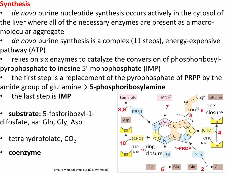
Synthesis • de novo purine nucleotide synthesis occurs actively in the cytosol of the liver where all of the necessary enzymes are present as a macro-molecular aggregate • de novo purine synthesis is a complex (11 steps), energy-expensive pathway (ATP) • relies on six enzymes to catalyze the conversion of phosphoribosyl-pyrophosphate to inosine 5′-monophosphate (IMP) • the first step is a replacement of the pyrophosphate of PRPP by the amide group of glutamine→ 5-phosphoribosylamine • the last step is IMP
• substrate: 5-fosforibozyl-1- difosfate, aa: Gln, Gly, Asp • tetrahydrofolate, CO2
• coenzyme
Tůma P: Metabolismus purinů a pyrimidinů
ring closure
ring closure
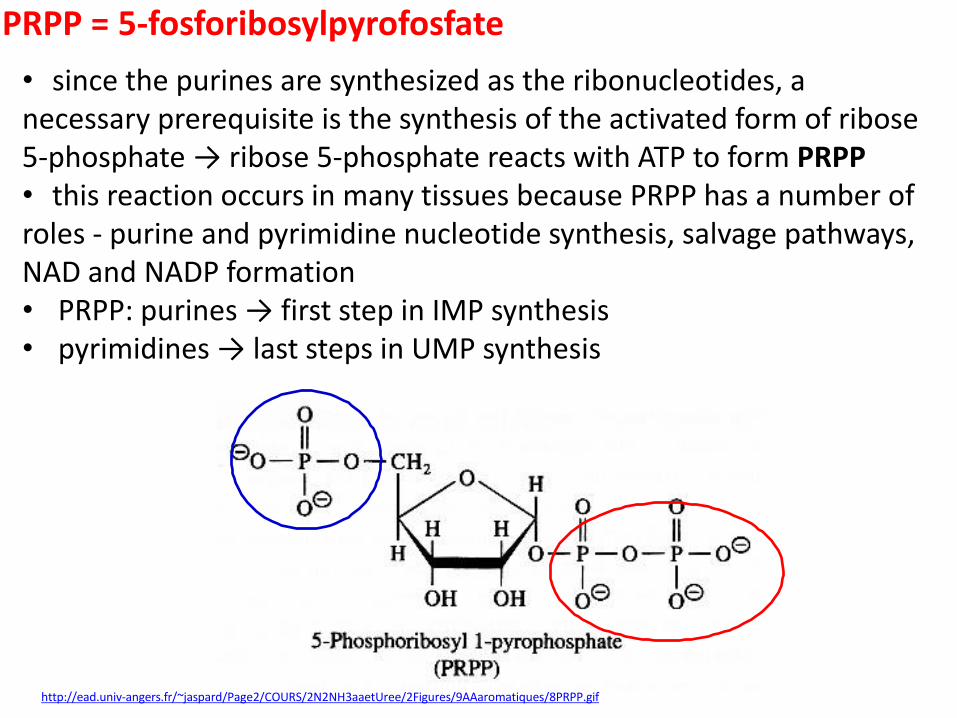
PRPP = 5-fosforibosylpyrofosfate
http://ead.univ-angers.fr/~jaspard/Page2/COURS/2N2NH3aaetUree/2Figures/9AAaromatiques/8PRPP.gif
• since the purines are synthesized as the ribonucleotides, a necessary prerequisite is the synthesis of the activated form of ribose 5-phosphate → ribose 5-phosphate reacts with ATP to form PRPP • this reaction occurs in many tissues because PRPP has a number of roles - purine and pyrimidine nucleotide synthesis, salvage pathways, NAD and NADP formation • PRPP: purines → first step in IMP synthesis • pyrimidines → last steps in UMP synthesis
Synthesis of pyrimidine • since pyrimidine are simpler than purines → synthesis simpler • begins with carbamoyl phosphate synthesized in the cytosol of those
tissues capable of making pyrimidines (highest in spleen, thymus, gastrointestinal tract and testes)
• Toxoplasma gondii: is a ubiquitous protozoan parasite that is responsible for severe congenital birth defects and fatal toxoplasmic encephalitis in immunocompromized people
• T. gondii has a fragmented pathway for salvaging pyrimidine nucleobases derived from the parasite or host cell, and this limited pyrimidine salvage capacity is funnelled exclusively through uracil • the first enzyme of the de novo pathway, carbamoyl-phosphate synthetase, is different from mammals → targeting of the T. gondii de novo pyrimidine synthesis pathway is one potential approach to developing more-effective vaccination strategies based on live attenuated strains with defined genetic disruptions
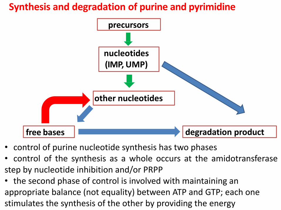
Synthesis and degradation of purine and pyrimidine
precursors
nucleotides (IMP, UMP)
free bases
other nucleotides
degradation product
• control of purine nucleotide synthesis has two phases • control of the synthesis as a whole occurs at the amidotransferase step by nucleotide inhibition and/or PRPP • the second phase of control is involved with maintaining an appropriate balance (not equality) between ATP and GTP; each one stimulates the synthesis of the other by providing the energy
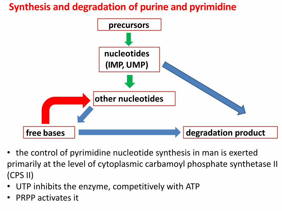
Synthesis and degradation of purine and pyrimidine
precursors
nucleotides (IMP, UMP)
free bases
other nucleotides
degradation product
• the control of pyrimidine nucleotide synthesis in man is exerted primarily at the level of cytoplasmic carbamoyl phosphate synthetase II (CPS II) • UTP inhibits the enzyme, competitively with ATP • PRPP activates it
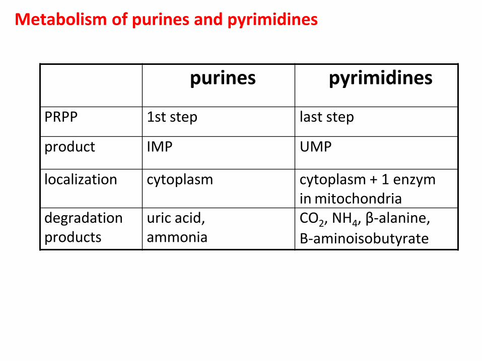
Metabolism of purines and pyrimidines
purines pyrimidines
PRPP 1st step last step
product IMP UMP
localization cytoplasm cytoplasm + 1 enzym in mitochondria
degradation products
uric acid, ammonia
CO2, NH4, β-alanine,
Β-aminoisobutyrate
http://arthritis-research.com/content/figures/ar1909-2.gif
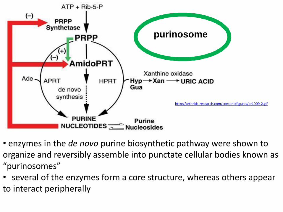
purinosome
• enzymes in the de novo purine biosynthetic pathway were shown to organize and reversibly assemble into punctate cellular bodies known as “purinosomes” • several of the enzymes form a core structure, whereas others appear to interact peripherally
Catabolism of purine and pyrimidine • most nucleic acids in the cell are associated with protein • dietary nucleoprotein is degraded by pancreatic enzymes and tissue nucleoprotein by lysosomal enzymes • after dissociation of the protein and nucleic acid, the protein is metabolized like any other protein • the nucleic acids are hydrolyzed randomly by nucleases to yield a mixture of polynucleotides → these are further cleaved by phosphodiesterases to a mixture of the mononucleotides • the nucleotides are hydrolyzed by nucleotidases to give the nucleosides and Pi → this is probably the end product in the intestine with the nucleosides being the primary form absorbed • in at least some tissues, the nucleosides undergo phosphorolysis with nucleoside phosphorylases to yield the base and ribose 1-P (or deoxyribose 1-P) • the purine and pyrimidine bases released are either degraded or salvaged for reincorporation into nucleotides
Catabolism
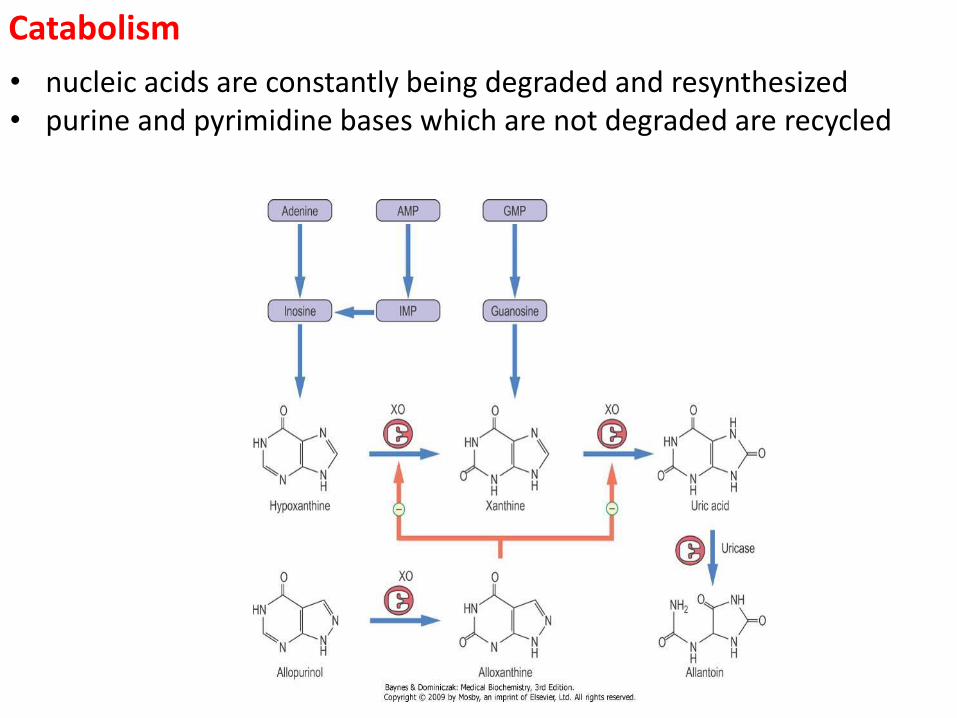
• nucleic acids are constantly being degraded and resynthesized • purine and pyrimidine bases which are not degraded are recycled
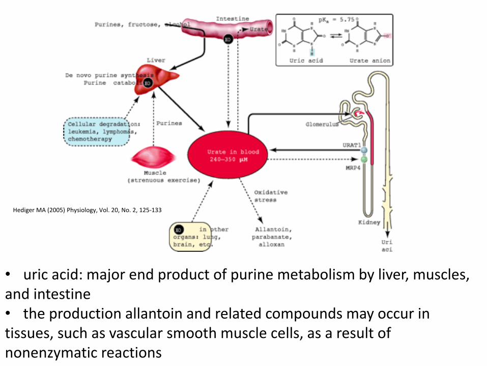
• uric acid: major end product of purine metabolism by liver, muscles, and intestine • the production allantoin and related compounds may occur in tissues, such as vascular smooth muscle cells, as a result of nonenzymatic reactions
Hediger MA (2005) Physiology, Vol. 20, No. 2, 125-133
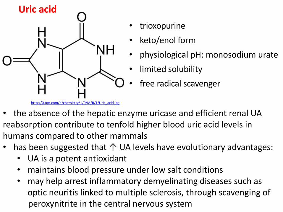
Uric acid
• trioxopurine
• keto/enol form
• physiological pH: monosodium urate
• limited solubility
• free radical scavenger
http://0.tqn.com/d/chemistry/1/0/M/R/1/Uric_acid.jpg
• the absence of the hepatic enzyme uricase and efficient renal UA reabsorption contribute to tenfold higher blood uric acid levels in humans compared to other mammals • has been suggested that ↑ UA levels have evolutionary advantages:
• UA is a potent antioxidant • maintains blood pressure under low salt conditions • may help arrest inflammatory demyelinating diseases such as
optic neuritis linked to multiple sclerosis, through scavenging of peroxynitrite in the central nervous system
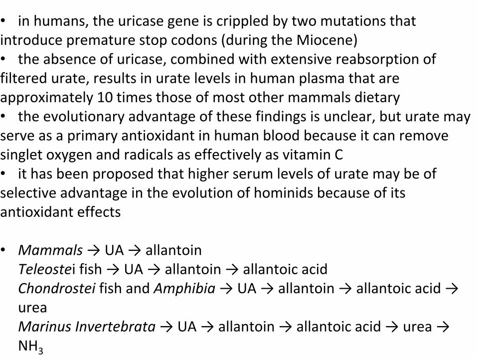
• in humans, the uricase gene is crippled by two mutations that introduce premature stop codons (during the Miocene) • the absence of uricase, combined with extensive reabsorption of filtered urate, results in urate levels in human plasma that are approximately 10 times those of most other mammals dietary • the evolutionary advantage of these findings is unclear, but urate may serve as a primary antioxidant in human blood because it can remove singlet oxygen and radicals as effectively as vitamin C • it has been proposed that higher serum levels of urate may be of selective advantage in the evolution of hominids because of its antioxidant effects
• Mammals → UA → allantoin
Teleostei fish → UA → allantoin → allantoic acid Chondrostei fish and Amphibia → UA → allantoin → allantoic acid → urea Marinus Invertebrata → UA → allantoin → allantoic acid → urea → NH3
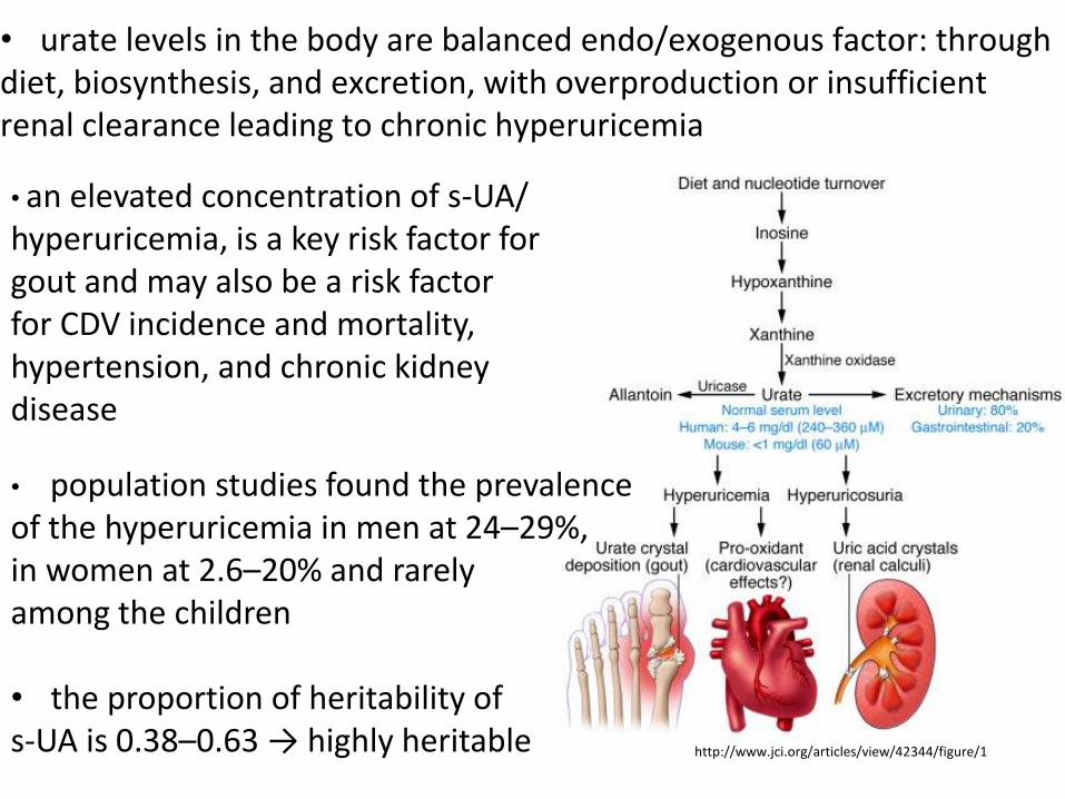
• urate levels in the body are balanced endo/exogenous factor: through diet, biosynthesis, and excretion, with overproduction or insufficient renal clearance leading to chronic hyperuricemia • an elevated concentration of s-UA/ hyperuricemia, is a key risk factor for gout and may also be a risk factor for CDV incidence and mortality, hypertension, and chronic kidney disease
• population studies found the prevalence of the hyperuricemia in men at 24–29%, in women at 2.6–20% and rarely among the children • the proportion of heritability of s-UA is 0.38–0.63 → highly heritable http://www.jci.org/articles/view/42344/figure/1
• level of s-UA: man < 420 μmol/l, children and woman < 340 μmol/l
• allantoin: only non-enzymatic processes with reactive oxygen species will give rise to allantoin, which is thus a suitable biomarker to measure oxidative stress in chronic illnesses and senescence
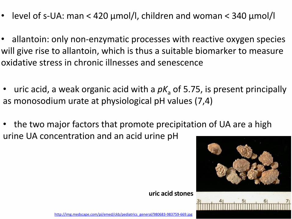
uric acid stones
http://img.medscape.com/pi/emed/ckb/pediatrics_general/980683-983759-669.jpg
• uric acid, a weak organic acid with a pKa of 5.75, is present principally as monosodium urate at physiological pH values (7,4)
• the two major factors that promote precipitation of UA are a high urine UA concentration and an acid urine pH
Uric acid • UA is a powerful scavenger of peroxyl radicals, hydroxyl radicals and singlet oxygen in human biological fluids
• UA accounts for up to 60% of plasma antioxidative capacity and presumably protects not only erythrocytes, but also DNA-contained in long-lived T and B lymphocytes and macrophages
• UA has been hypothesized to protect against oxidative stress, a prominent contributor to dopaminergic neuron degeneration in Parkinson’s disease → studies have evaluated a potential association between s-UA and the risk of developing Parkinson’s disease, finding a lower risk among individuals with higher levels of s-UA
Roch-Ramel F. et al. (1994) Am J Physiol, 266(5 Pt 2):F797-805; Enomoto A., Enou H. (2005) Clin Exp Nephrol 9:195-205
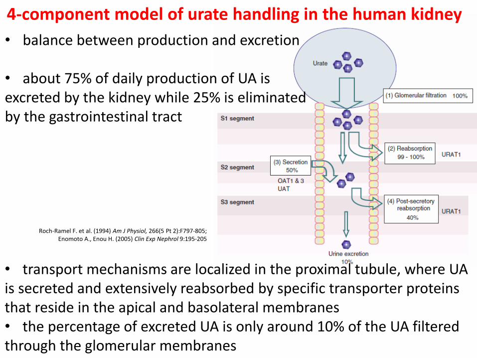
4-component model of urate handling in the human kidney
• transport mechanisms are localized in the proximal tubule, where UA is secreted and extensively reabsorbed by specific transporter proteins that reside in the apical and basolateral membranes • the percentage of excreted UA is only around 10% of the UA filtered through the glomerular membranes
• balance between production and excretion
• about 75% of daily production of UA is excreted by the kidney while 25% is eliminated by the gastrointestinal tract
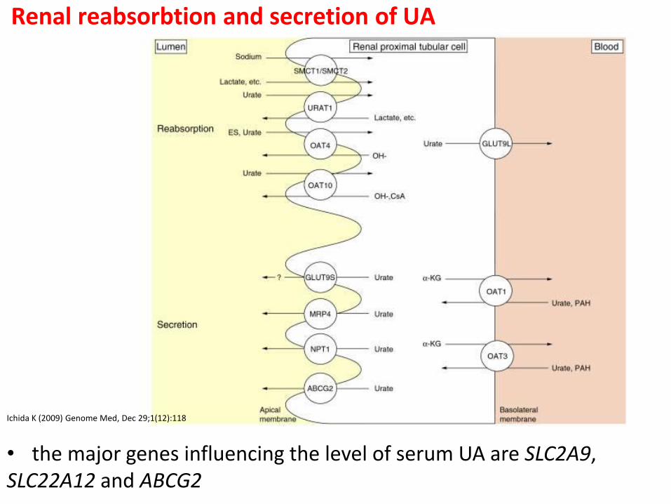
Renal reabsorbtion and secretion of UA
Ichida K (2009) Genome Med, Dec 29;1(12):118
• the major genes influencing the level of serum UA are SLC2A9, SLC22A12 and ABCG2
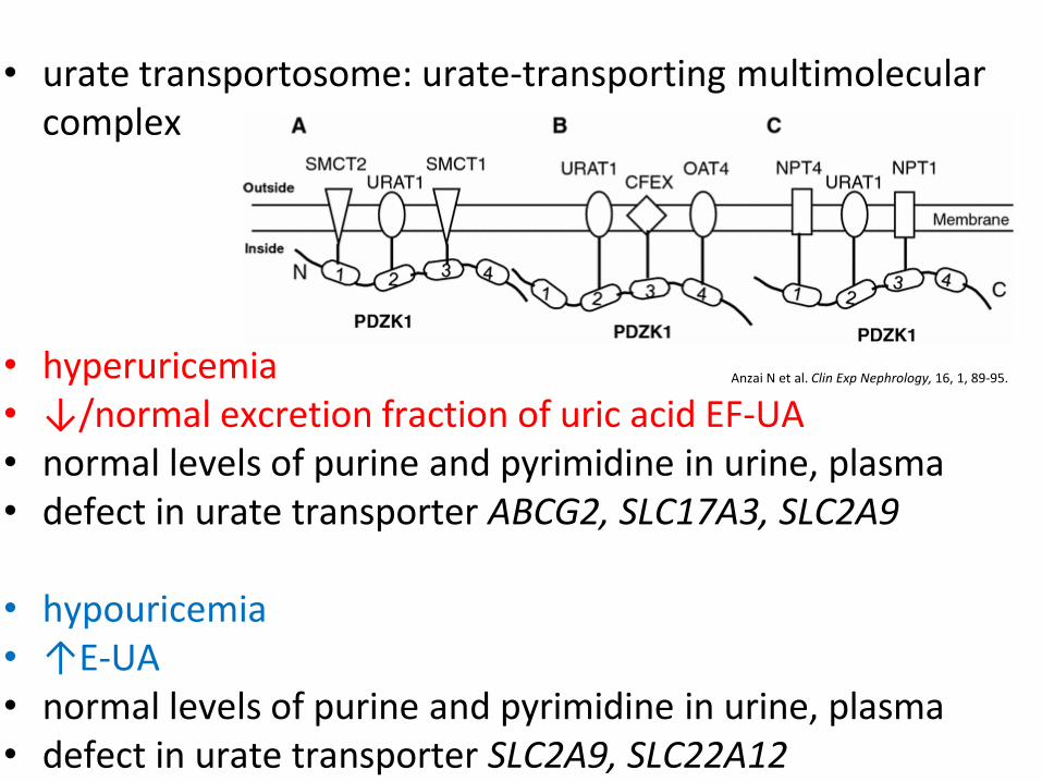
• urate transportosome: urate-transporting multimolecular
complex
• hyperuricemia • ↓/normal excretion fraction of uric acid EF-UA
• normal levels of purine and pyrimidine in urine, plasma • defect in urate transporter ABCG2, SLC17A3, SLC2A9
• hypouricemia • ↑E-UA • normal levels of purine and pyrimidine in urine, plasma • defect in urate transporter SLC2A9, SLC22A12
Anzai N et al. Clin Exp Nephrology, 16, 1, 89-95.
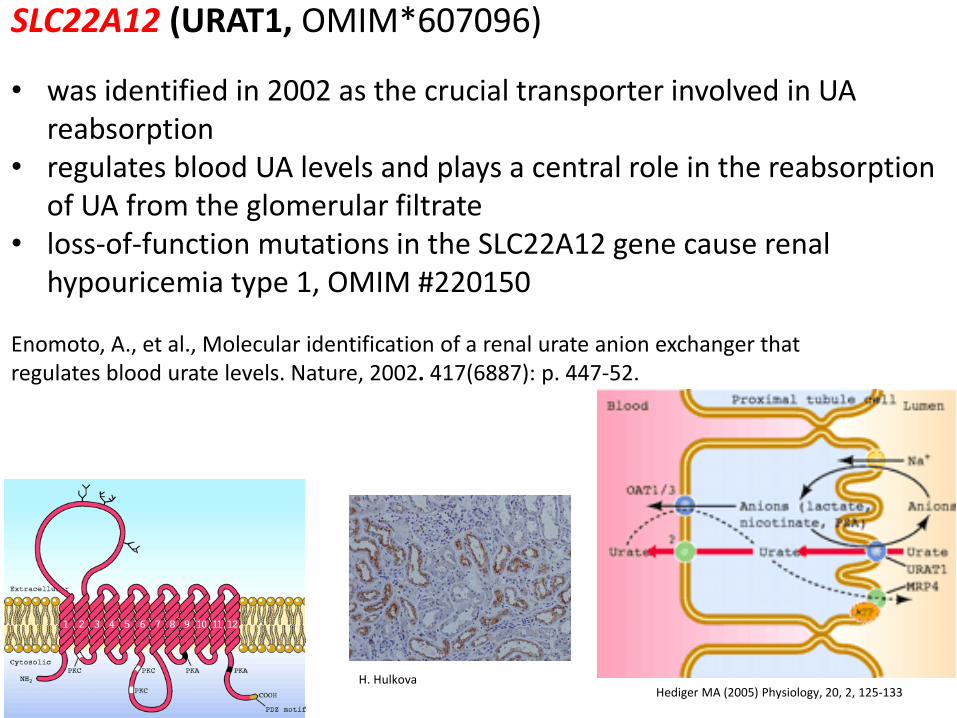
SLC22A12 (URAT1, OMIM*607096)
• was identified in 2002 as the crucial transporter involved in UA reabsorption
• regulates blood UA levels and plays a central role in the reabsorption of UA from the glomerular filtrate
• loss-of-function mutations in the SLC22A12 gene cause renal hypouricemia type 1, OMIM #220150
Enomoto, A., et al., Molecular identification of a renal urate anion exchanger that regulates blood urate levels. Nature, 2002. 417(6887): p. 447-52.
Hediger MA (2005) Physiology, 20, 2, 125-133 H. Hulkova
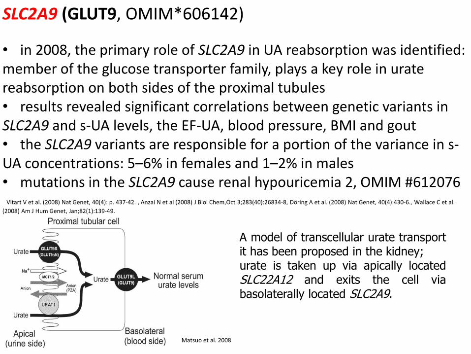
A model of transcellular urate transport it has been proposed in the kidney; urate is taken up via apically located SLC22A12 and exits the cell via basolaterally located SLC2A9.
SLC2A9 (GLUT9, OMIM*606142)
• in 2008, the primary role of SLC2A9 in UA reabsorption was identified: member of the glucose transporter family, plays a key role in urate reabsorption on both sides of the proximal tubules • results revealed significant correlations between genetic variants in SLC2A9 and s-UA levels, the EF-UA, blood pressure, BMI and gout • the SLC2A9 variants are responsible for a portion of the variance in s-UA concentrations: 5–6% in females and 1–2% in males • mutations in the SLC2A9 cause renal hypouricemia 2, OMIM #612076 Vitart V et al. (2008) Nat Genet, 40(4): p. 437-42. , Anzai N et al (2008) J Biol Chem,Oct 3;283(40):26834-8, Döring A et al. (2008) Nat Genet, 40(4):430-6., Wallace C et al.
(2008) Am J Hum Genet, Jan;82(1):139-49.
Matsuo et al. 2008
• membrane transporter belonging to the ATP-binding cassette (ABC) superfamily of membrane transporters
ABCG2 (ABCG2, OMIM*603756)
• initially found to be a xenobiotic transporter that plays a role in the multidrug resistance phenotype of a specific human breast cancer and has since been shown to confer multidrug resistance in several cancer cells by actively exporting a wide variety of drugs across the plasma membrane • a high capacity transporter for UA excretion in the kidney, liver, and gut protein→ to mediate renal urate secretion as a urate efflux transporter in the (luminal) brush-border membrane of proximal tubule cells • variant c.421C.A (rs2231142, p.Q141K) results in a reduction of the urate transport rate by 53% compared with that for the wild-type ABCG2 and causes approximately 10% of the hyperuricemia and gout cases in Caucasians
• our ever increasing understanding of renal urate transport has enabled significant new developments in medications targeting hyperuricemia
• Lesinurad (RDEA594) is an active metabolite of RDEA806, which is a non-nucleoside reverse-transcriptase inhibitor for URAT1 → it is an oral uricosuric agent candidate for the treatment of hyperuricemia in patients with gout and has completed phase III
HYPOURICEMIA
• serum urate levels 2 mg/dl (119µmol/l) • decreased uric acid production • decreased renal tubular urate reabsorption • secondary reduction in uric acid biosynthesis hepatic failure • acquired causes of the Fanconi renotubular syndrome and its variants • drugs XDH inhibitor, uricosuric agents, coumarin anticoagulants (warfarin)… • nutritional deficiencies vitamines B12, C, D… • inherited disorders - deficiency of purine metabolism, RHUC
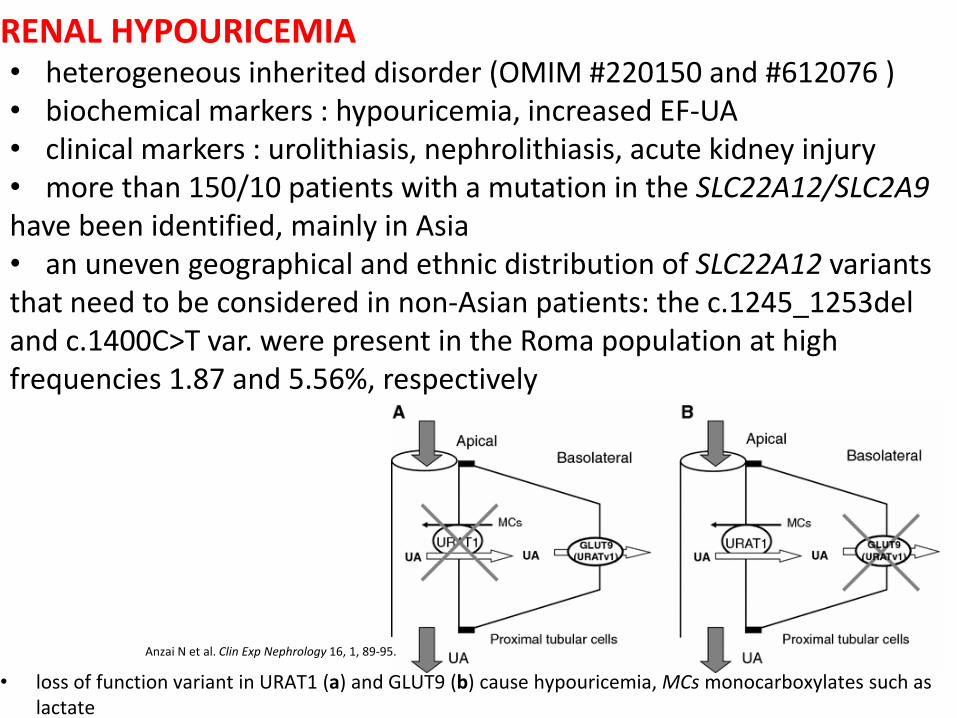
RENAL HYPOURICEMIA
• loss of function variant in URAT1 (a) and GLUT9 (b) cause hypouricemia, MCs monocarboxylates such as lactate
• heterogeneous inherited disorder (OMIM #220150 and #612076 ) • biochemical markers : hypouricemia, increased EF-UA • clinical markers : urolithiasis, nephrolithiasis, acute kidney injury • more than 150/10 patients with a mutation in the SLC22A12/SLC2A9 have been identified, mainly in Asia • an uneven geographical and ethnic distribution of SLC22A12 variants that need to be considered in non-Asian patients: the c.1245_1253del and c.1400C>T var. were present in the Roma population at high frequencies 1.87 and 5.56%, respectively
Anzai N et al. Clin Exp Nephrology 16, 1, 89-95.
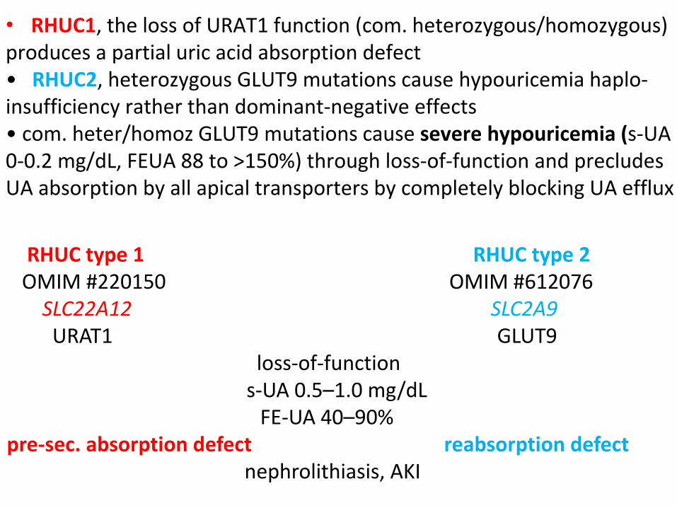
• RHUC1, the loss of URAT1 function (com. heterozygous/homozygous) produces a partial uric acid absorption defect • RHUC2, heterozygous GLUT9 mutations cause hypouricemia haplo-insufficiency rather than dominant-negative effects • com. heter/homoz GLUT9 mutations cause severe hypouricemia (s-UA 0-0.2 mg/dL, FEUA 88 to >150%) through loss-of-function and precludes UA absorption by all apical transporters by completely blocking UA efflux
RHUC type 1 RHUC type 2 OMIM #220150 OMIM #612076 SLC22A12 SLC2A9 URAT1 GLUT9
loss-of-function s-UA 0.5–1.0 mg/dL
FE-UA 40–90% pre-sec. absorption defect reabsorption defect nephrolithiasis, AKI
• primary hyperuricemia → hyperuricemia is considered primary when it exists in the absence of coexisting diseases or drugs that alter uric acid production or excretion • purine metabolic disorders • kidney disorders • increased production of uric acid from purine
• secondary hyperuricemia → this refers to excessive urate production or diminished renal clearance occurring as a consequence of another disease, drug, dietary product, or toxin
• certain cancers, or chemotherapy agents may cause an increased turnover rate of cell death
• kidney disease • medications • endocrine or metabolic conditions -certain forms of diabetes, or
acidosis can cause hyperuricemia • etc
• 90% of patients with primary gout have a disorder of UA excretion
• uromodulin-associated kidney disease (UAKD) is a dominant heritable renal disease in humans which is caused by mutations in the uromodulin (UMOD) gene and characterized by heterogeneous clinical appearance • UAKD summarizes several clinically defined diseases including medullary cystic kidney disease type 2 (OMIM#603860), familial juvenile hyperuricemic nephropathy (OMIM #162000) and glomerulocystic kidney disease (OMIM #609886) • AD, characterized by elevated s-UA concentrations due to a low EF-UA, defective urinary concentrating ability, interstitial nephropathy, and progression to end-stage renal failure→ hyperuricemia/gout is mostly the first clinical sign which is diagnosed in UAKD patients • deficiency in urate transporter
• AD, AR : SLC2A9, SLC17A3, ABCG2
• ~ 10% of hyperuricemia/gout cases are due to overproduction
• enzymopathy of purine metabolism (deficiency of HPRT, superactivity of PRPS1)
• deficiency of glucose 6-phosphatase in glycogen storage disese type I → hyperuricemia results from a combination of increased generation and decreased excretion of UA, which is generated when increased amounts of G6P are metabolized via the pentose phosphate pathway → UA competes with lactic acid and other organic acids for renal excretion in the urine
http://scienceblogs.com/moleculeoftheday/images/gout-cartoon.jpg
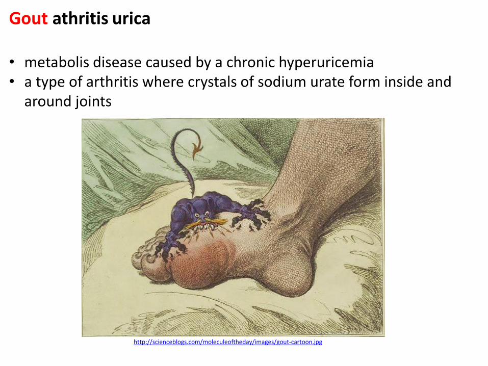
Gout athritis urica • metabolis disease caused by a chronic hyperuricemia • a type of arthritis where crystals of sodium urate form inside and
around joints
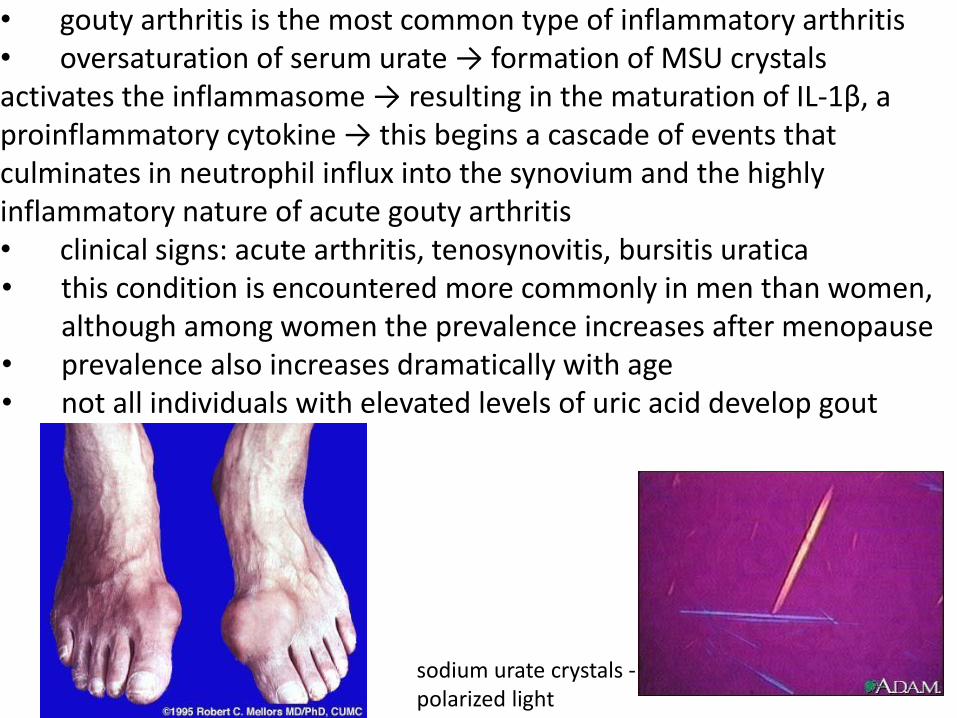
• gouty arthritis is the most common type of inflammatory arthritis • oversaturation of serum urate → formation of MSU crystals activates the inflammasome → resulting in the maturation of IL-1β, a proinflammatory cytokine → this begins a cascade of events that culminates in neutrophil influx into the synovium and the highly inflammatory nature of acute gouty arthritis • clinical signs: acute arthritis, tenosynovitis, bursitis uratica • this condition is encountered more commonly in men than women,
although among women the prevalence increases after menopause • prevalence also increases dramatically with age • not all individuals with elevated levels of uric acid develop gout
sodium urate crystals - polarized light
O2 + H
2O H
2O
2
Hypoxanthin
H2O
2
Xanthin
O2 + H
2O
GuaninXanthinoxidasa Guanindeaminasa
NH4
+ H2O
Xanthinoxidasa
N
N
N
N
O
O
H
H
O
H
H
Močová kyselina uric acid
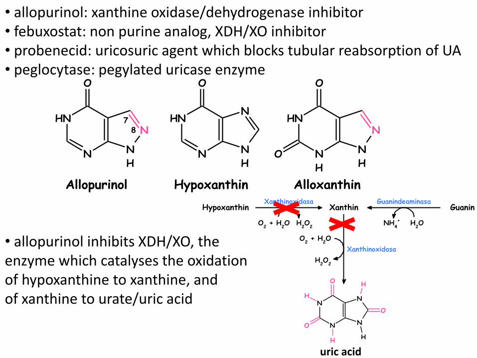
• allopurinol: xanthine oxidase/dehydrogenase inhibitor • febuxostat: non purine analog, XDH/XO inhibitor • probenecid: uricosuric agent which blocks tubular reabsorption of UA • peglocytase: pegylated uricase enzyme
• allopurinol inhibits XDH/XO, the enzyme which catalyses the oxidation of hypoxanthine to xanthine, and of xanthine to urate/uric acid
Hypoxanthin Alloxanthin
N
H
NH
N
N
O
N
H
NH
N
H
N
O
O
Allopurinol
N
H
NH
N
N
O
78
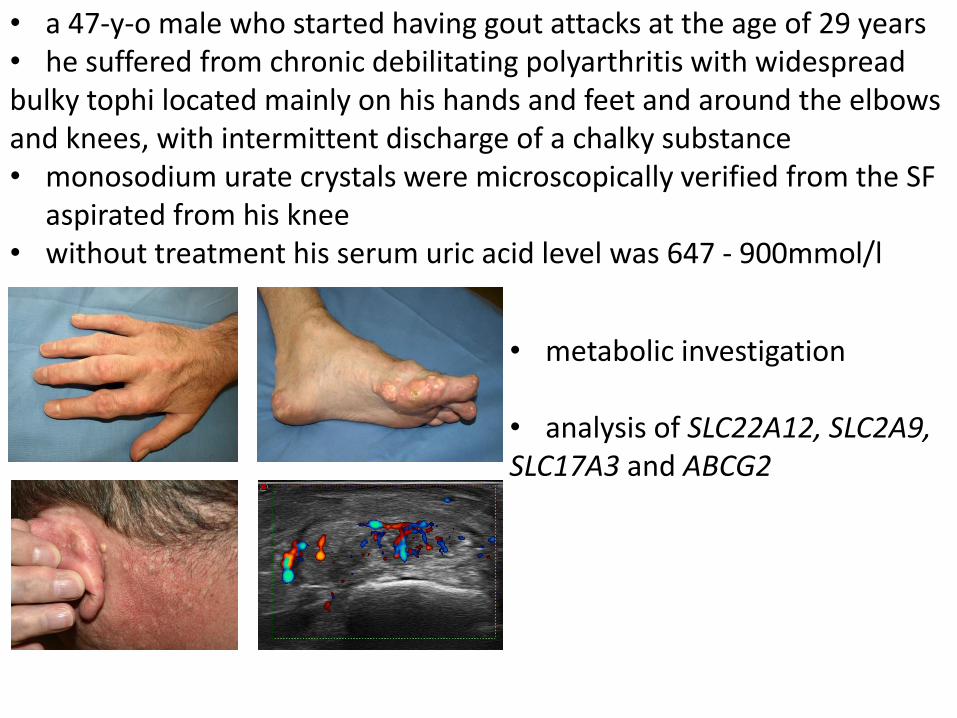
• metabolic investigation
• analysis of SLC22A12, SLC2A9, SLC17A3 and ABCG2
• a 47-y-o male who started having gout attacks at the age of 29 years • he suffered from chronic debilitating polyarthritis with widespread bulky tophi located mainly on his hands and feet and around the elbows and knees, with intermittent discharge of a chalky substance • monosodium urate crystals were microscopically verified from the SF
aspirated from his knee • without treatment his serum uric acid level was 647 - 900mmol/l
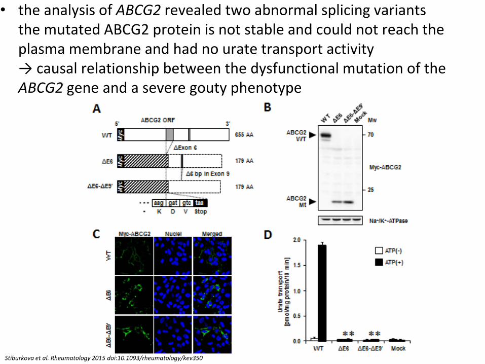
• the analysis of ABCG2 revealed two abnormal splicing variants the mutated ABCG2 protein is not stable and could not reach the plasma membrane and had no urate transport activity → causal relationship between the dysfunctional mutation of the ABCG2 gene and a severe gouty phenotype
Stiburkova et al. Rheumatology 2015 doi:10.1093/rheumatology/kev350
Inherited metabolic disorders of purine metabolism
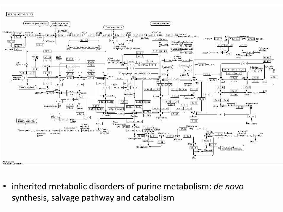
• inherited metabolic disorders of purine metabolism: de novo synthesis, salvage pathway and catabolism
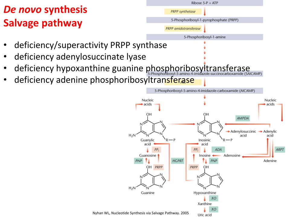
De novo synthesis Salvage pathway
• deficiency/superactivity PRPP synthase • deficiency adenylosuccinate lyase • deficiency hypoxanthine guanine phosphoribosyltransferase • deficiency adenine phosphoribosyltransferase
Nyhan WL, Nucleotide Synthesis via Salvage Pathway. 2005
Purinnukleosidfosforylasa
(PNP)
N
N
N
N
NH2
Ribosa-5-fosfát
N
N
N
N
O
Ribosa-5-fosfát
H
N
N
N
N
O
Ribosa-5-fosfát
O
H
H
N
N
N
H
N
O
Ribosa-5-fosfát
NH2
H
AMP IMP XMP GMP
H2O
Pi
Nukleotidasa
AMP deaminasa
H2O NH
4
+
AdenosinAdenosindeaminasa
H2O NH
4
+
H2O
Pi
Nukleotidasa
Inosin
Ribosa-1-P
Purinnukleosidfosforylasa
(PNP)
Hypoxanthin
Pi
H2O
Pi
Nukleotidasa
Xanthosin
Ribosa-1-P
Purinnukleosidfosforylasa
(PNP)
Xanthin
Pi
H2O
Pi
Nukleotidasa
Guanosin
Ribosa-1-P
Guanin
Pi
Xanthinoxidasa Guanindeaminasa
O2 + H
2O H
2O
2NH
4
+ H2O
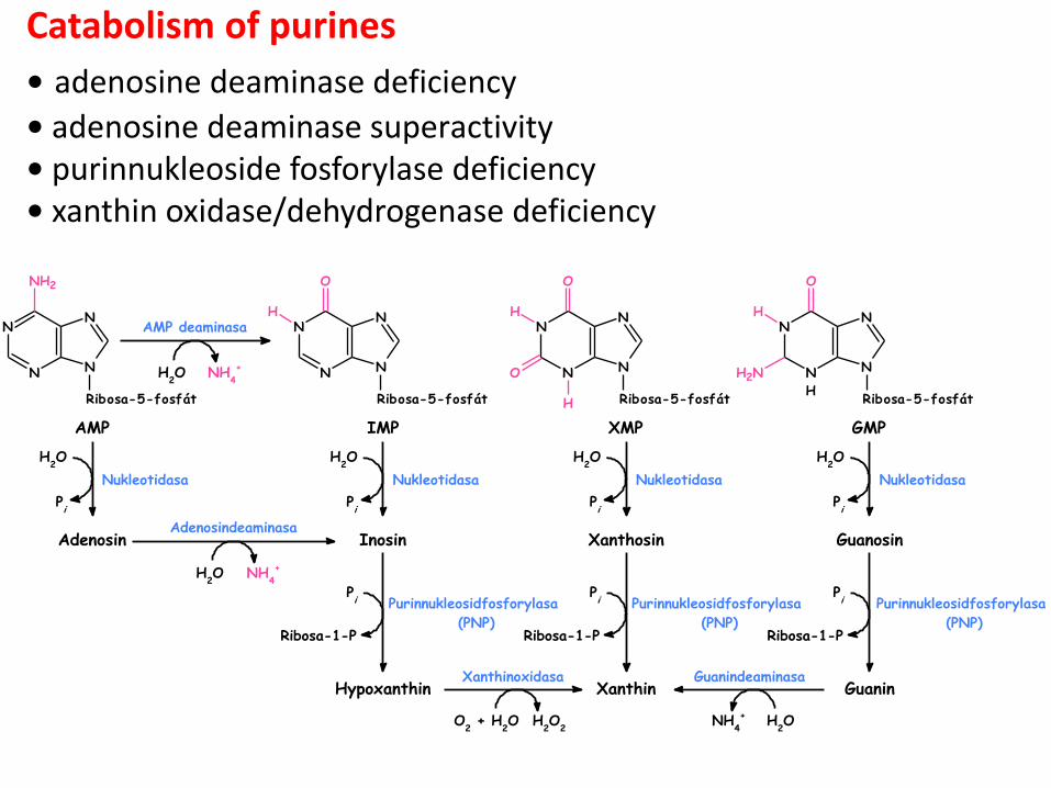
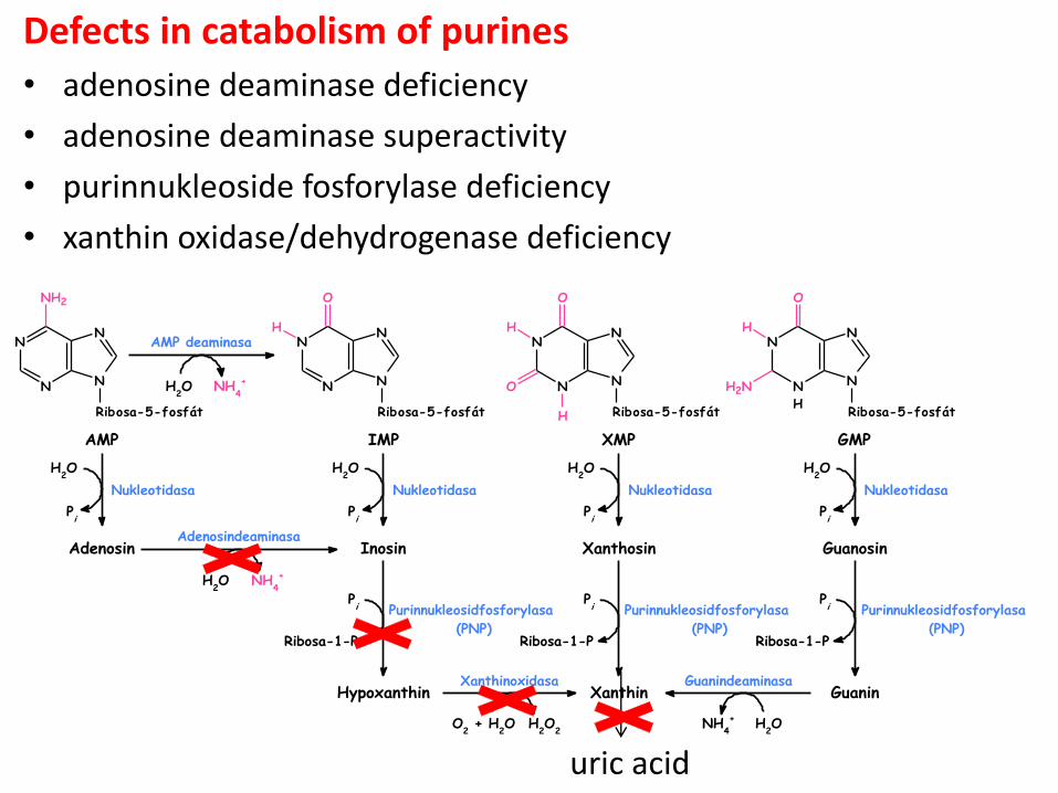
Catabolism of purines
• adenosine deaminase deficiency
• adenosine deaminase superactivity • purinnukleoside fosforylase deficiency • xanthin oxidase/dehydrogenase deficiency
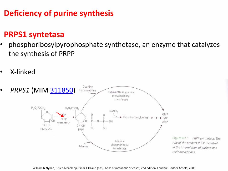
Deficiency of purine synthesis PRPS1 syntetasa • phosphoribosylpyrophosphate synthetase, an enzyme that catalyzes
the synthesis of PRPP • X-linked • PRPS1 (MIM 311850)
William N Nyhan, Bruce A Barshop, Pinar T Ozand (eds). Atlas of metabolic diseases, 2nd edition. London: Hodder Arnold, 2005
PRPS1 superactivity • regulatory defects and ↑ expression of PRS-I, normal kinetic enzyme properties • 30 patients, 7 point mutation in PRPS1 has been identified • variable neurological findings, neurological symptoms, sensorineural hearing loss, PM • severe superactivity phenotypes with purine overproduction accompanied by neuropathy, PRPS1 is not truly “superactive” because it does not have a higher Vmax than the normal protein, but rather it lacks regulatory control of its • milder phenotype: ↑ transcript levels, but no genetic defects have been identified, ↑ expression, that manifest as purine overproduction without any neuropathy • overexpression has been shown to be accompanied by raised intracellular levels of PRPP in all cell types examined, i.e., fibroblasts, lymphoblasts, and erythrocytes • ↑ de novo purine synthesis causes accelerated nucleotide degradation to UA

WN Nyhan et al. Atlas of metabolic diseases, 2nd edition. London: Hodder Arnold, 2005
Charcot-Marie-Tooth disease-5 (MIM 311070) • part of the spectrum of PRPS1-related disorders, is characterized by peripheral neuropathy, early-onset (prelingual) bilateral profound sensorineural hearing loss, and optic neuropathy • the onset of peripheral neuropathy is between ages five and 12 y X-linked nonsyndromic sensorineural deafness (MIM 304500) • vestibular and cochlea hair cells in early developing and postnatal mice and in the spiral ganglion cells in mice at P6, a role in inner ear development and maintenance Arts syndrome (MIM 301835) • mental retardation, early-onset hypotonia, ataxia, delayed motor development, hearing impairment, and optic atrophyneurological problems and immune dysfunction PRPS-I superactivity (MIM 300661)
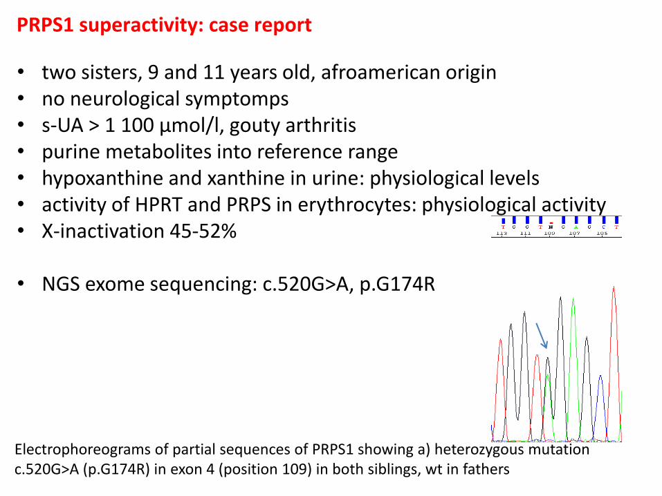
PRPS1 superactivity: case report
Electrophoreograms of partial sequences of PRPS1 showing a) heterozygous mutation c.520G>A (p.G174R) in exon 4 (position 109) in both siblings, wt in fathers
• two sisters, 9 and 11 years old, afroamerican origin • no neurological symptomps • s-UA > 1 100 µmol/l, gouty arthritis • purine metabolites into reference range • hypoxanthine and xanthine in urine: physiological levels • activity of HPRT and PRPS in erythrocytes: physiological activity • X-inactivation 45-52%
• NGS exome sequencing: c.520G>A, p.G174R
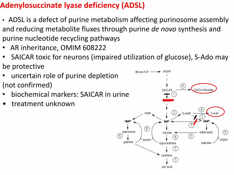
Adenylosuccinate lyase deficiency (ADSL)
• ADSL is a defect of purine metabolism affecting purinosome assembly and reducing metabolite fluxes through purine de novo synthesis and purine nucleotide recycling pathways • AR inheritance, OMIM 608222 • SAICAR toxic for neurons (impaired utilization of glucose), S-Ado may be protective • uncertain role of purine depletion (not confirmed) • biochemical markers: SAICAR in urine • treatment unknown
• ADSL covers a continuous clinical spectrum with three major forms • fatal neonatal • severe (type I) • mild to moderate form (type II)
• clinical variability is found, even in patients from the same family • onset is generally between birth and early childhood • cases ranging from fatal neonatal encephalopathy (presenting with
hypokinesia, intractable seizures and respiratory failure) to mild intellectual disability have been reported
• intellectual disability is found in all patients, epilepsy of various types in most, and autistic features in about one third (failure to make eye contact, hypersensitivity to noise and light, repetitive behavior, agitation, temper tantrums, autoaggression and self-mutilation)
• prenatal manifestations are also reported: impaired intrauterine growth, microcephaly, fetal hypokinesia, and loss of fetal heart rate variability
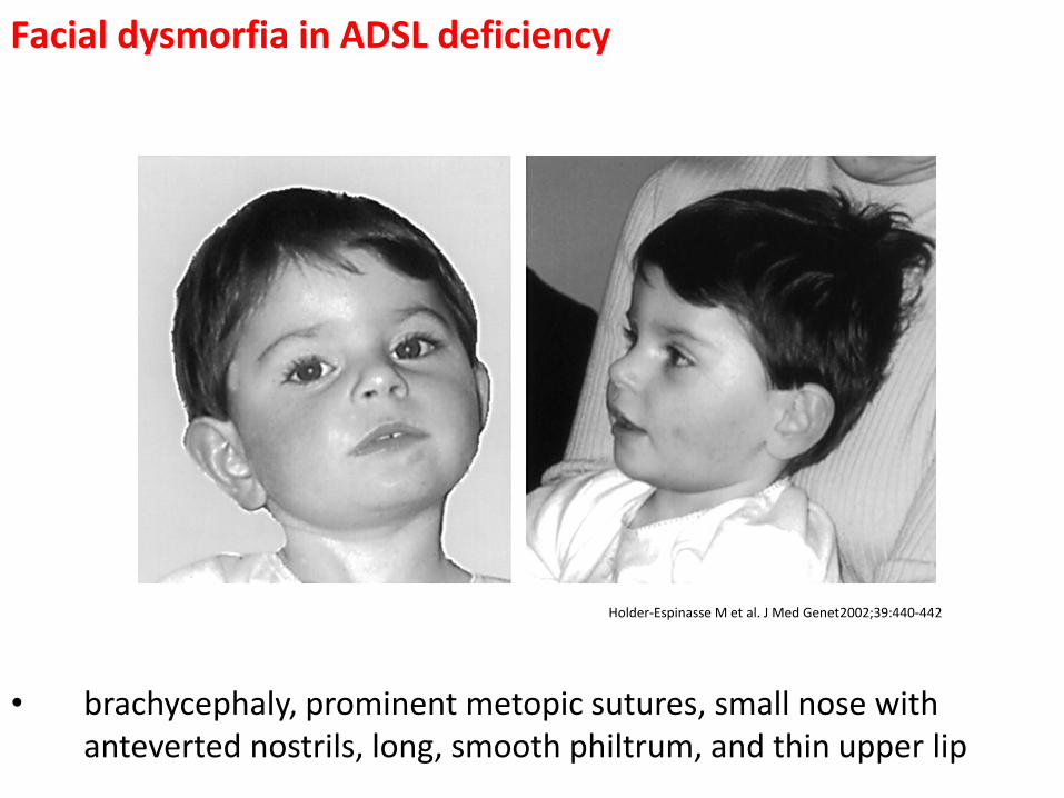
Facial dysmorfia in ADSL deficiency
Holder-Espinasse M et al. J Med Genet 2002;39:440-442
• brachycephaly, prominent metopic sutures, small nose with anteverted nostrils, long, smooth philtrum, and thin upper lip
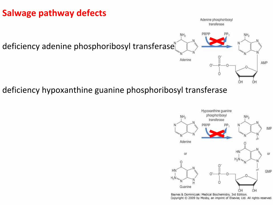
Salwage pathway defects
deficiency adenine phosphoribosyl transferase deficiency hypoxanthine guanine phosphoribosyl transferase
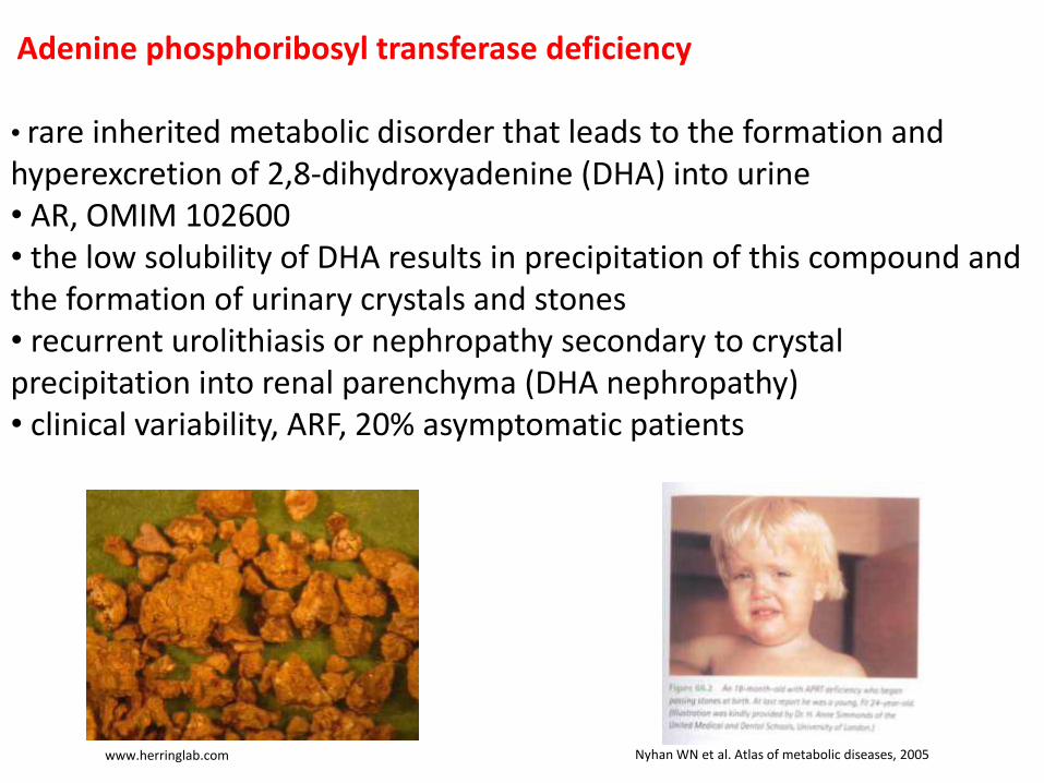
Adenine phosphoribosyl transferase deficiency • rare inherited metabolic disorder that leads to the formation and hyperexcretion of 2,8-dihydroxyadenine (DHA) into urine • AR, OMIM 102600 • the low solubility of DHA results in precipitation of this compound and the formation of urinary crystals and stones • recurrent urolithiasis or nephropathy secondary to crystal precipitation into renal parenchyma (DHA nephropathy) • clinical variability, ARF, 20% asymptomatic patients
Nyhan WN et al. Atlas of metabolic diseases, 2005 www.herringlab.com
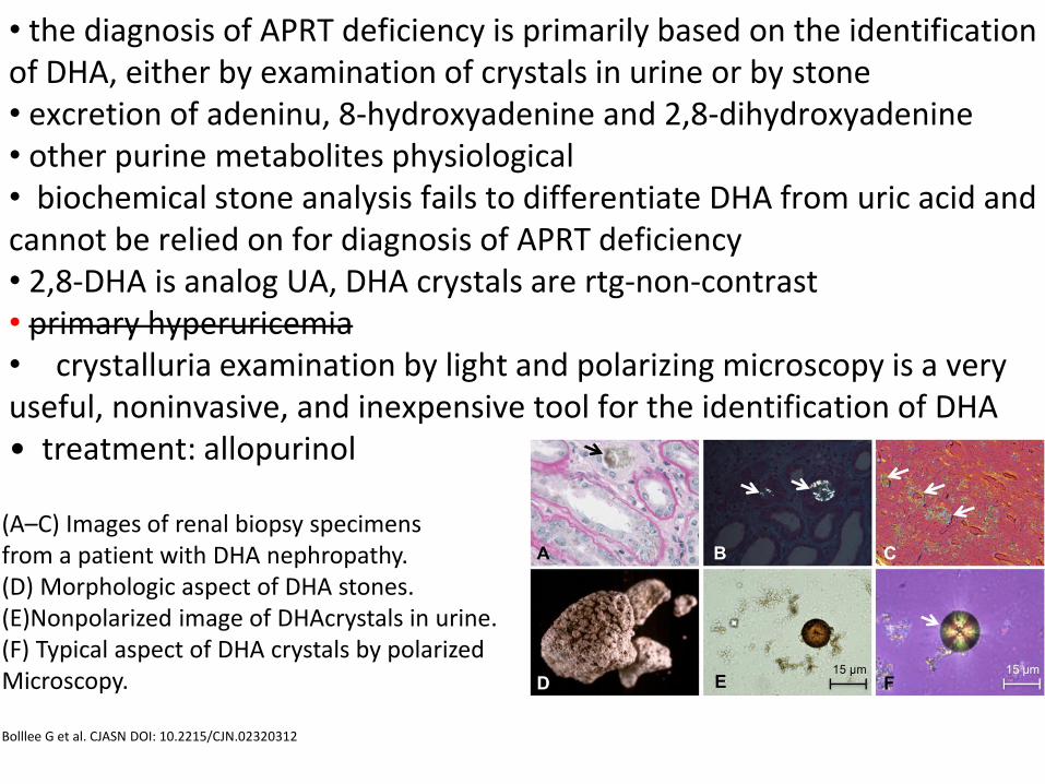
• the diagnosis of APRT deficiency is primarily based on the identification of DHA, either by examination of crystals in urine or by stone • excretion of adeninu, 8-hydroxyadenine and 2,8-dihydroxyadenine • other purine metabolites physiological • biochemical stone analysis fails to differentiate DHA from uric acid and cannot be relied on for diagnosis of APRT deficiency • 2,8-DHA is analog UA, DHA crystals are rtg-non-contrast • primary hyperuricemia • crystalluria examination by light and polarizing microscopy is a very useful, noninvasive, and inexpensive tool for the identification of DHA • treatment: allopurinol
(A–C) Images of renal biopsy specimens from a patient with DHA nephropathy. (D) Morphologic aspect of DHA stones. (E)Nonpolarized image of DHAcrystals in urine. (F) Typical aspect of DHA crystals by polarized Microscopy. Bolllee G et al. CJASN DOI: 10.2215/CJN.02320312
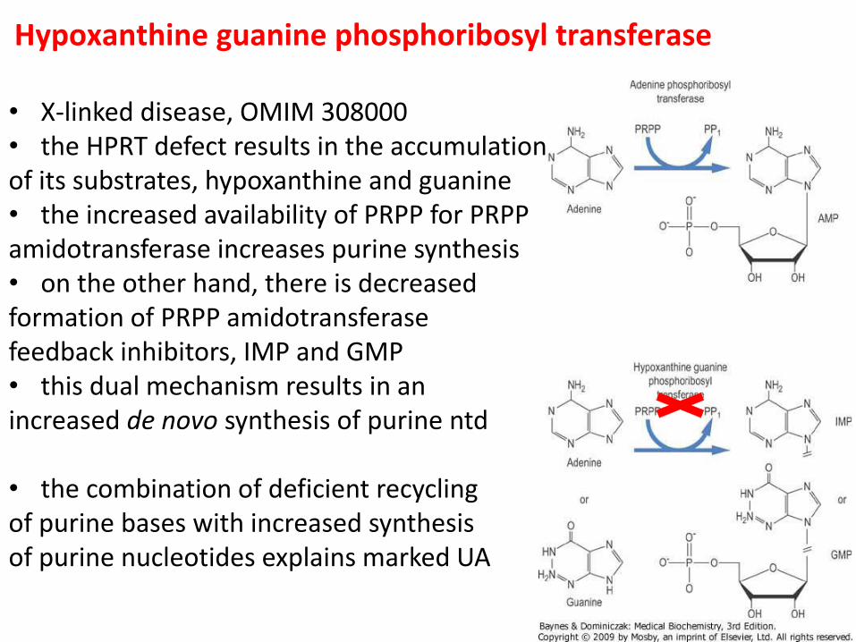
Hypoxanthine guanine phosphoribosyl transferase
• X-linked disease, OMIM 308000 • the HPRT defect results in the accumulation of its substrates, hypoxanthine and guanine • the increased availability of PRPP for PRPP amidotransferase increases purine synthesis • on the other hand, there is decreased formation of PRPP amidotransferase feedback inhibitors, IMP and GMP • this dual mechanism results in an increased de novo synthesis of purine ntd • the combination of deficient recycling of purine bases with increased synthesis of purine nucleotides explains marked UA
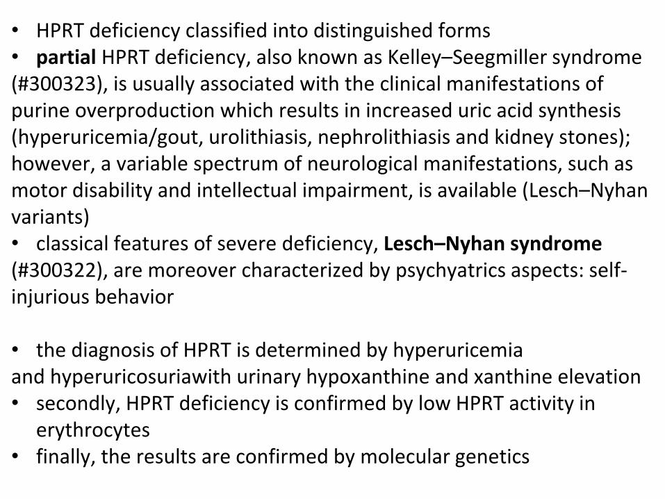
• HPRT deficiency classified into distinguished forms • partial HPRT deficiency, also known as Kelley–Seegmiller syndrome (#300323), is usually associated with the clinical manifestations of purine overproduction which results in increased uric acid synthesis (hyperuricemia/gout, urolithiasis, nephrolithiasis and kidney stones); however, a variable spectrum of neurological manifestations, such as motor disability and intellectual impairment, is available (Lesch–Nyhan variants) • classical features of severe deficiency, Lesch–Nyhan syndrome (#300322), are moreover characterized by psychyatrics aspects: self-injurious behavior
• the diagnosis of HPRT is determined by hyperuricemia and hyperuricosuriawith urinary hypoxanthine and xanthine elevation • secondly, HPRT deficiency is confirmed by low HPRT activity in
erythrocytes • finally, the results are confirmed by molecular genetics
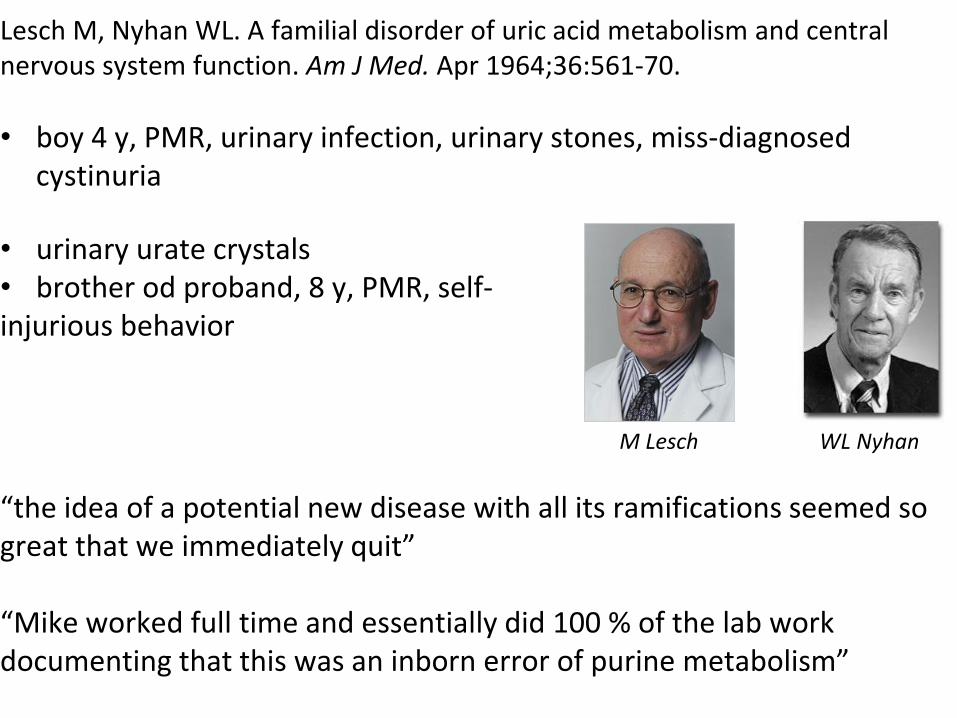
Lesch M, Nyhan WL. A familial disorder of uric acid metabolism and central nervous system function. Am J Med. Apr 1964;36:561-70.
• boy 4 y, PMR, urinary infection, urinary stones, miss-diagnosed cystinuria
• urinary urate crystals • brother od proband, 8 y, PMR, self- injurious behavior
“the idea of a potential new disease with all its ramifications seemed so great that we immediately quit” “Mike worked full time and essentially did 100 % of the lab work documenting that this was an inborn error of purine metabolism”
M Lesch WL Nyhan
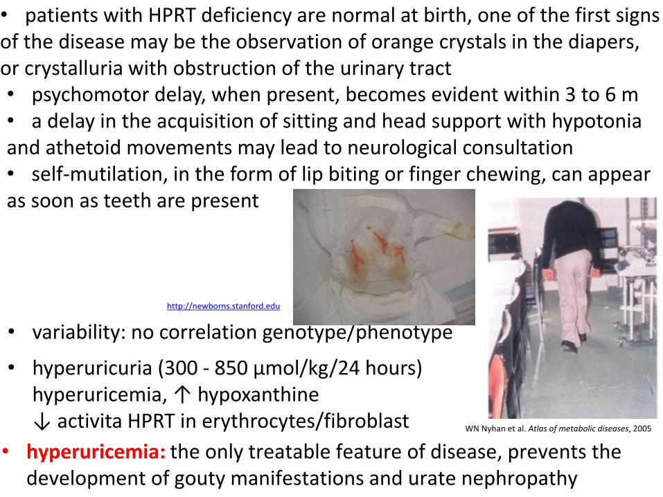
• patients with HPRT deficiency are normal at birth, one of the first signs of the disease may be the observation of orange crystals in the diapers, or crystalluria with obstruction of the urinary tract
• hyperuricemia: the only treatable feature of disease, prevents the development of gouty manifestations and urate nephropathy
WN Nyhan et al. Atlas of metabolic diseases, 2005
• variability: no correlation genotype/phenotype
http://newborns.stanford.edu
• hyperuricuria (300 - 850 µmol/kg/24 hours) hyperuricemia, ↑ hypoxanthine ↓ activita HPRT in erythrocytes/fibroblast
• psychomotor delay, when present, becomes evident within 3 to 6 m • a delay in the acquisition of sitting and head support with hypotonia and athetoid movements may lead to neurological consultation • self-mutilation, in the form of lip biting or finger chewing, can appear as soon as teeth are present
• various theories for neurological anormalities incl. purines depletion, possibly secondary dopamin synthesis defect (decreased DOPA-decarboxylase)
• HPRT deficiency shows an X-linked recessive inheritance pattern; thus, Lesch–Nyhan syndrome occurs almost exclusively in males • female carriers are usually asymptomatic; fewer than ten clinically affected females were described previously • carrier diagnosis is an important issue for most families with HPRT deficiency and carrier status cannot be confirmed by biochemical and enzymatic methods in most of the cases • accurate carrier diagnosis can be performed by molecular genetics; however, in about 5% of patients, no molecular defect is found
• treatment controlling UA overproduction with allopurinol or febuxostat is available • however, allopurinol has not usually been considered to cause
behavioral and neurological symptoms
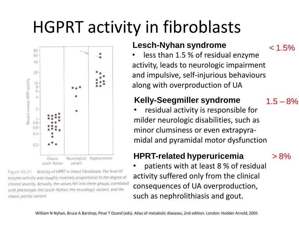
HGPRT activity in fibroblasts
William N Nyhan, Bruce A Barshop, Pinar T Ozand (eds). Atlas of metabolic diseases, 2nd edition. London: Hodder Arnold, 2005
Lesch-Nyhan syndrome • less than 1.5 % of residual enzyme activity, leads to neurologic impairment and impulsive, self-injurious behaviours along with overproduction of UA
< 1.5%
Kelly-Seegmiller syndrome • residual activity is responsible for milder neurologic disabilities, such as minor clumsiness or even extrapyra-midal and pyramidal motor dysfunction
1.5 – 8%
HPRT-related hyperuricemia • patients with at least 8 % of residual activity suffered only from the clinical consequences of UA overproduction, such as nephrolithiasis and gout.
> 8%
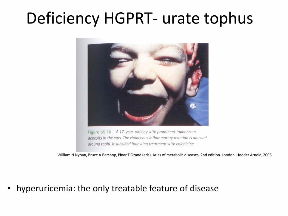
Deficiency HGPRT- urate tophus
William N Nyhan, Bruce A Barshop, Pinar T Ozand (eds). Atlas of metabolic diseases, 2nd edition. London: Hodder Arnold, 2005
• hyperuricemia: the only treatable feature of disease

http://www.dent.ucla.edu/pic/visitors/teethloss/images/PreFig9.jpg
• compulsive self-injurious behaviour is the most striking feature of Lesch-Nyhan syndrome and is only present in patients with the complete enzyme defect, although some Lesch-Nyhan patients never show auto-destructive behaviour
• the patients begin to bite their lips, tongue or fingers and, without restrictions, important auto-mutilating lesions can develop
• the mutilation is not the result of a lack of sensation (the patients feel pain and are relieved when protected from themselves) and recently it has been ascribed to an obsessive-compulsive behaviour
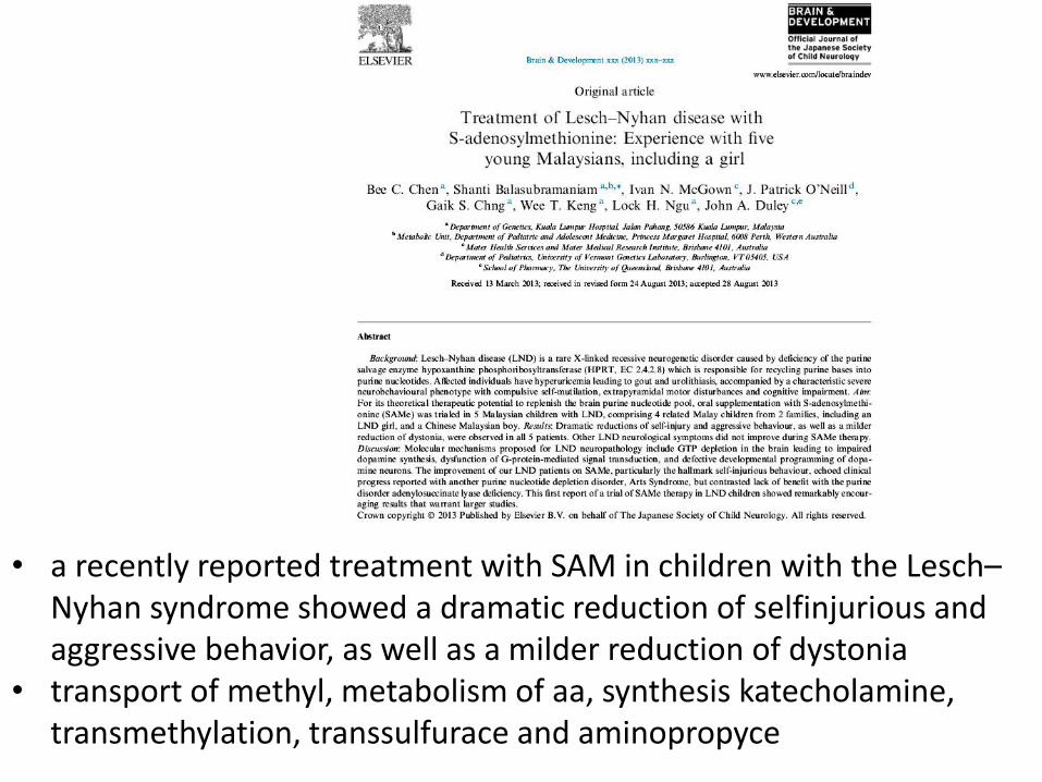
• a recently reported treatment with SAM in children with the Lesch–Nyhan syndrome showed a dramatic reduction of selfinjurious and aggressive behavior, as well as a milder reduction of dystonia
• transport of methyl, metabolism of aa, synthesis katecholamine, transmethylation, transsulfurace and aminopropyce
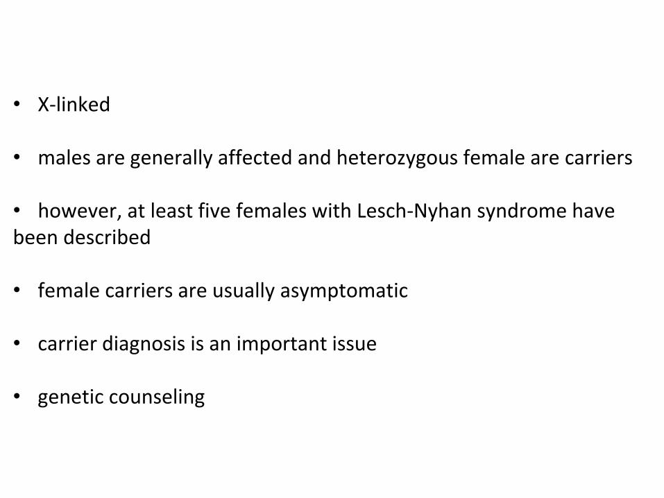
• X-linked
• males are generally affected and heterozygous female are carriers
• however, at least five females with Lesch-Nyhan syndrome have been described
• female carriers are usually asymptomatic
• carrier diagnosis is an important issue
• genetic counseling
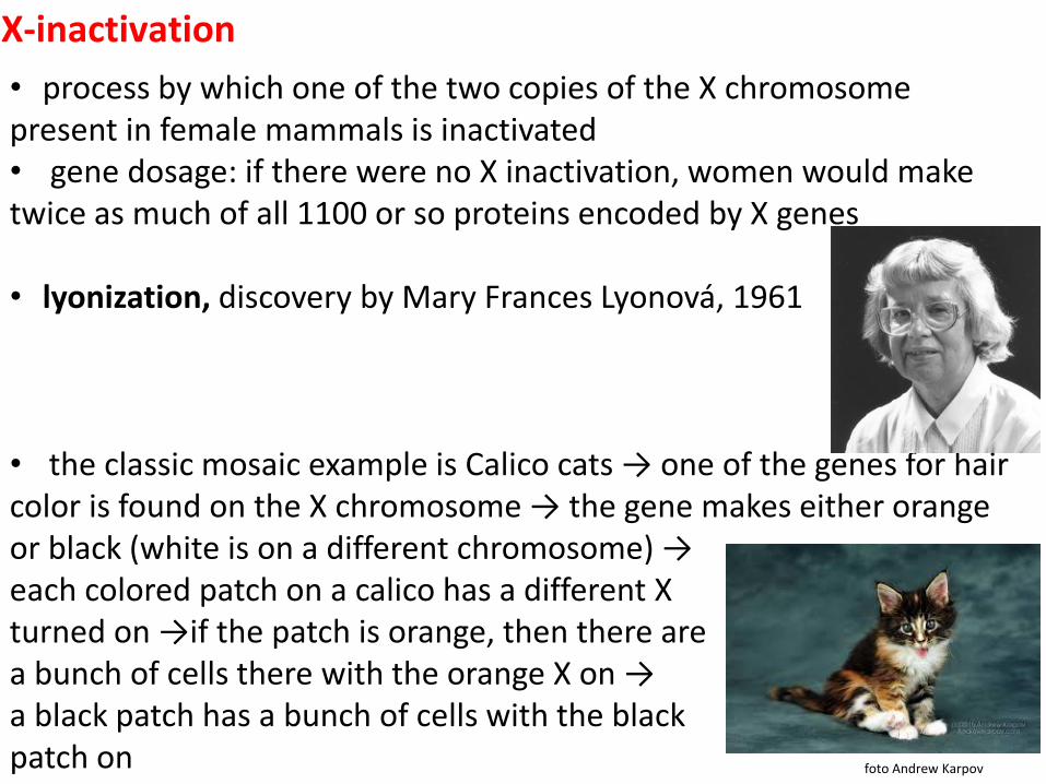
X-inactivation
• process by which one of the two copies of the X chromosome present in female mammals is inactivated • gene dosage: if there were no X inactivation, women would make twice as much of all 1100 or so proteins encoded by X genes
• lyonization, discovery by Mary Frances Lyonová, 1961
• the classic mosaic example is Calico cats → one of the genes for hair color is found on the X chromosome → the gene makes either orange or black (white is on a different chromosome) → each colored patch on a calico has a different X turned on →if the patch is orange, then there are a bunch of cells there with the orange X on → a black patch has a bunch of cells with the black patch on foto Andrew Karpov

X-inactivation
foto K. Barták
• the Barr body has long been recognized as the cytological manifesta-tion of the inactive X chromosome (Xi) in interphase nuclei, 1949 • a useful histological method for sex determination • in humans with more than one X chromosome, the number of Barr bodies visible at interphase is always one fewer than the total number of X chromosomes → Klinefelter syndrome (47,XXY) have a single Barr body, whereas Turner syndrome (45,X) have no Barr body • this happens early in embryonic development at random in mammals (100 cells, 16. day) • X inactivation is controlled by a unique locus, the X inactivation center (Xic), which is essential for the developmentally regulated initiation and spread of inactivation along the X chromosome • chromatin, covalent modification of histone, cytosine methylation • 10-15% of genes escape of inactivation → XIST (X-inactive specific transcript) • inactivation is random and stable
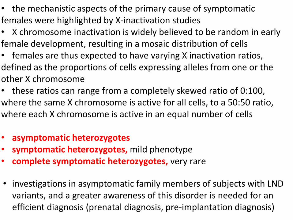
• the mechanistic aspects of the primary cause of symptomatic females were highlighted by X-inactivation studies • X chromosome inactivation is widely believed to be random in early female development, resulting in a mosaic distribution of cells • females are thus expected to have varying X inactivation ratios, defined as the proportions of cells expressing alleles from one or the other X chromosome • these ratios can range from a completely skewed ratio of 0:100, where the same X chromosome is active for all cells, to a 50:50 ratio, where each X chromosome is active in an equal number of cells
• asymptomatic heterozygotes • symptomatic heterozygotes, mild phenotype • complete symptomatic heterozygotes, very rare
• investigations in asymptomatic family members of subjects with LND variants, and a greater awareness of this disorder is needed for an efficient diagnosis (prenatal diagnosis, pre-implantation diagnosis)
• II.4: 56-year old man with a 10-year history of recurrent renal colic caused by radiolucent nephrolithiasis, proteinuria, chronic polyarticular arthritis, and polyneuropathy, was hospitalized because of clinical symptoms of nephrotic syndrome. • large tophi were present on the right elbow→ the examination of a tophus aspirate confirmed the presence of urate crystals • hyperuricaemia (612mol/l), renal function was normal
• II.1: 48-year old man with 7-year history of chronic tophaceous gout was frequently hospitalized because of recurrent acute attacks of arthritis in his first metatarsophalangeals, heels, knees, elbows, carpal and distal interphalangeal joints • he had experienced one episode of renal colic, caused by radiolucent
nephrolithiasis 25 years ago • advanced chronic renal insufficiency • no neurologic disability was observed s-UA>800 μmol/l • erosive bone changes in the interphalangeal and metatarsopha-
langeal joints, wrists, and knees → tophaceous gout was diagnosed
HPRT deficiency: case report 1
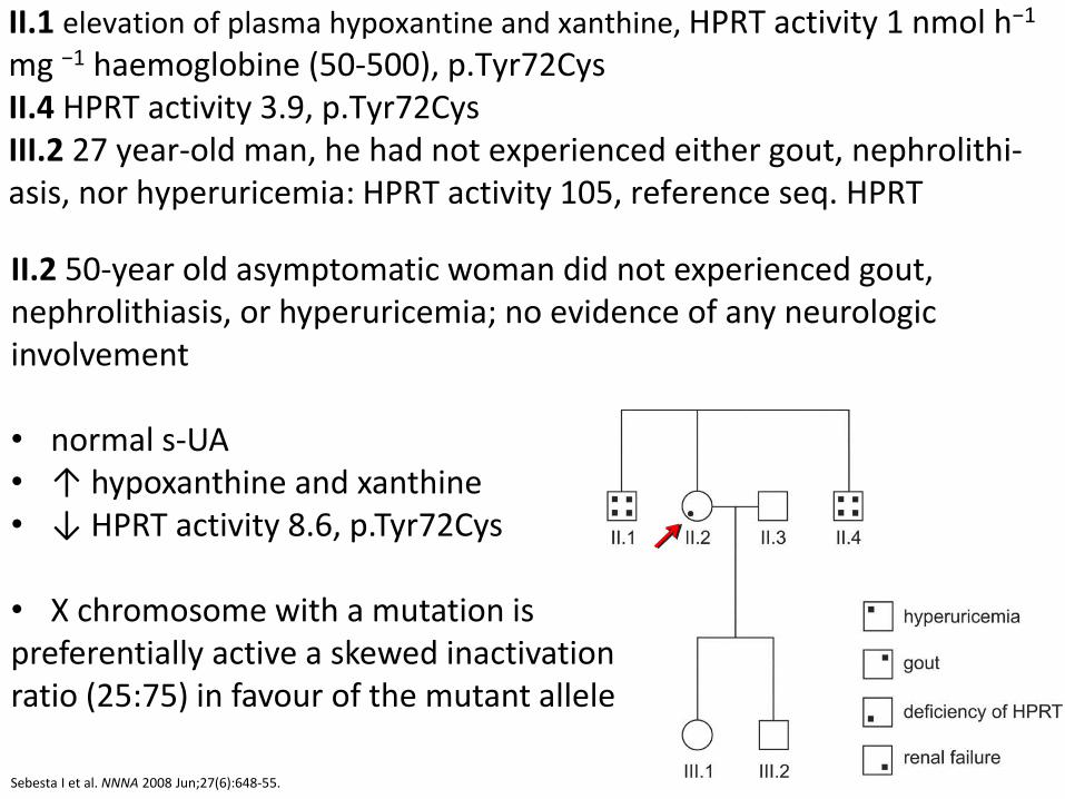
II.1 elevation of plasma hypoxantine and xanthine, HPRT activity 1 nmol h−1 mg −1 haemoglobine (50-500), p.Tyr72Cys II.4 HPRT activity 3.9, p.Tyr72Cys III.2 27 year-old man, he had not experienced either gout, nephrolithi-asis, nor hyperuricemia: HPRT activity 105, reference seq. HPRT
Sebesta I et al. NNNA 2008 Jun;27(6):648-55.
II.2 50-year old asymptomatic woman did not experienced gout, nephrolithiasis, or hyperuricemia; no evidence of any neurologic involvement • normal s-UA • ↑ hypoxanthine and xanthine • ↓ HPRT activity 8.6, p.Tyr72Cys
• X chromosome with a mutation is preferentially active a skewed inactivation ratio (25:75) in favour of the mutant allele
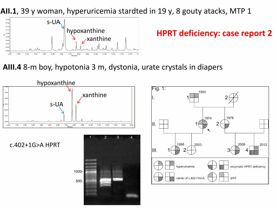
AII.1, 39 y woman, hyperuricemia stardted in 19 y, 8 gouty atacks, MTP 1
AIII.4 8-m boy, hypotonia 3 m, dystonia, urate crystals in diapers
s-UA
hypoxanthine xanthine
s-UA
hypoxanthine
xanthine
c.402+1G>A HPRT
HPRT deficiency: case report 2
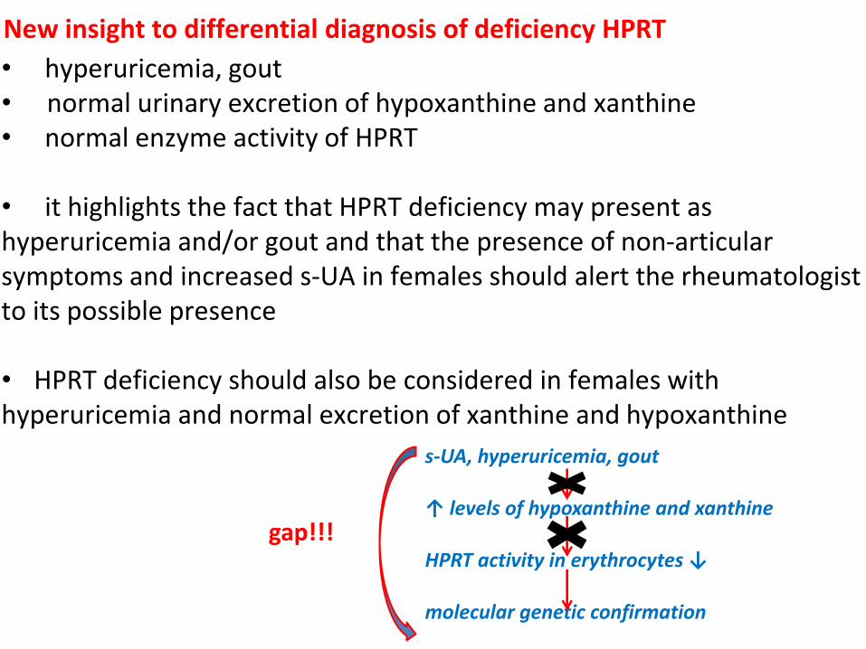
• hyperuricemia, gout • normal urinary excretion of hypoxanthine and xanthine • normal enzyme activity of HPRT
• it highlights the fact that HPRT deficiency may present as hyperuricemia and/or gout and that the presence of non-articular symptoms and increased s-UA in females should alert the rheumatologist to its possible presence
• HPRT deficiency should also be considered in females with hyperuricemia and normal excretion of xanthine and hypoxanthine
New insight to differential diagnosis of deficiency HPRT
s-UA, hyperuricemia, gout ↑ levels of hypoxanthine and xanthine HPRT activity in erythrocytes ↓ molecular genetic confirmation
gap!!!

familiar hyperuricemia and/or gout, hyperuricemia and/or gout namely in children and young woman, are an important
clue and should always be further investigated
Purinnukleosidfosforylasa
(PNP)
N
N
N
N
NH2
Ribosa-5-fosfát
N
N
N
N
O
Ribosa-5-fosfát
H
N
N
N
N
O
Ribosa-5-fosfát
O
H
H
N
N
N
H
N
O
Ribosa-5-fosfát
NH2
H
AMP IMP XMP GMP
H2O
Pi
Nukleotidasa
AMP deaminasa
H2O NH
4
+
AdenosinAdenosindeaminasa
H2O NH
4
+
H2O
Pi
Nukleotidasa
Inosin
Ribosa-1-P
Purinnukleosidfosforylasa
(PNP)
Hypoxanthin
Pi
H2O
Pi
Nukleotidasa
Xanthosin
Ribosa-1-P
Purinnukleosidfosforylasa
(PNP)
Xanthin
Pi
H2O
Pi
Nukleotidasa
Guanosin
Ribosa-1-P
Guanin
Pi
Xanthinoxidasa Guanindeaminasa
O2 + H
2O H
2O
2NH
4
+ H2O
uric acid
Defects in catabolism of purines
• adenosine deaminase deficiency
• adenosine deaminase superactivity
• purinnukleoside fosforylase deficiency
• xanthin oxidase/dehydrogenase deficiency
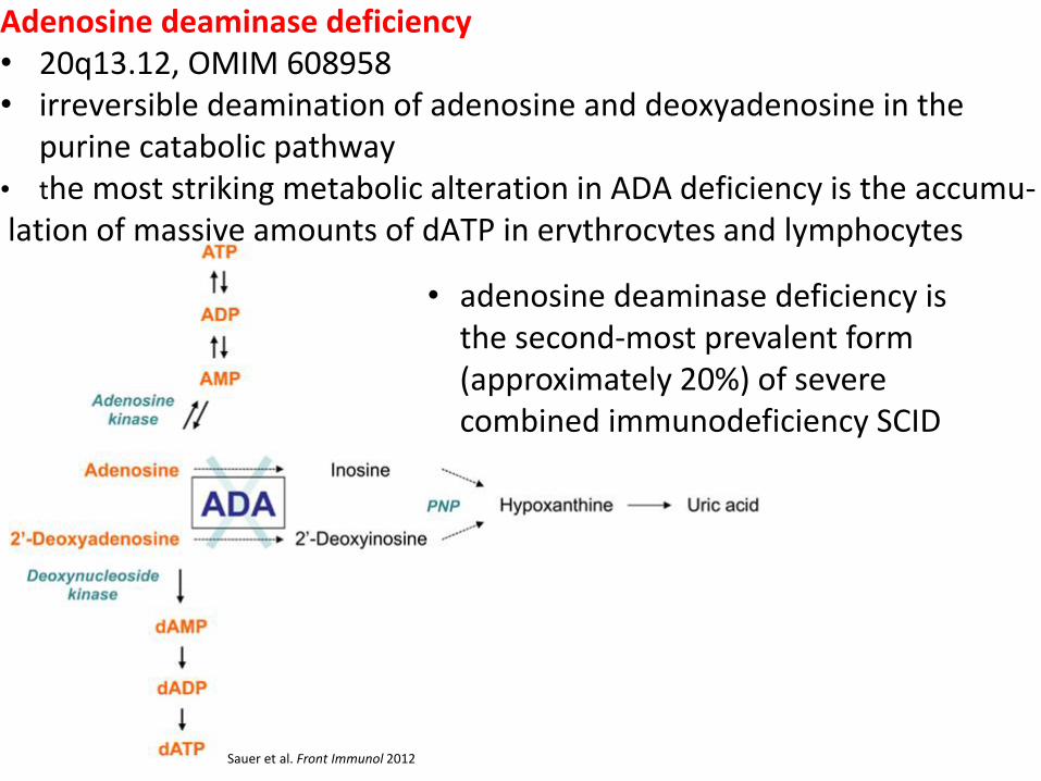
Adenosine deaminase deficiency • 20q13.12, OMIM 608958 • irreversible deamination of adenosine and deoxyadenosine in the
purine catabolic pathway • the most striking metabolic alteration in ADA deficiency is the accumu- lation of massive amounts of dATP in erythrocytes and lymphocytes
Sauer et al. Front Immunol 2012
• adenosine deaminase deficiency is the second-most prevalent form (approximately 20%) of severe combined immunodeficiency SCID
Adenosine deaminase deficiency • ↑ adenosine and deoxyadenosine → uric acid stones • ↑ plasma adenine • elevated adenosine levels, as occurring in ADA deficiency contribute to apoptosis and block in the differentiation of thymocytes, causing severe T lymphopenia in humans • ↑ deoxyadenosine and dATP in lymphocytes, inhibition ribonucleotide reductase essential for synthesis of DNA • lymphopenia and attrition of immune function over time are the two findings common to all presentations of ADA deficiency • rapidly fatal course due to infections with fungal, viral, and opportu-nistic agents are characteristic of early onset forms of ADA def → w/m • variable progressive neurological symptoms (movement disoders,
spasticity) • currently available therapeutic options include bone marrow transplantation, enzyme replacement therapy with bovine ADA, or hematopoietic stem cell gene therapy
O2 + H
2O H
2O
2
Hypoxanthin
H2O
2
Xanthin
O2 + H
2O
GuaninXanthinoxidasa Guanindeaminasa
NH4
+ H2O
Xanthinoxidasa
N
N
N
N
O
O
H
H
O
H
H
Močová kyselina
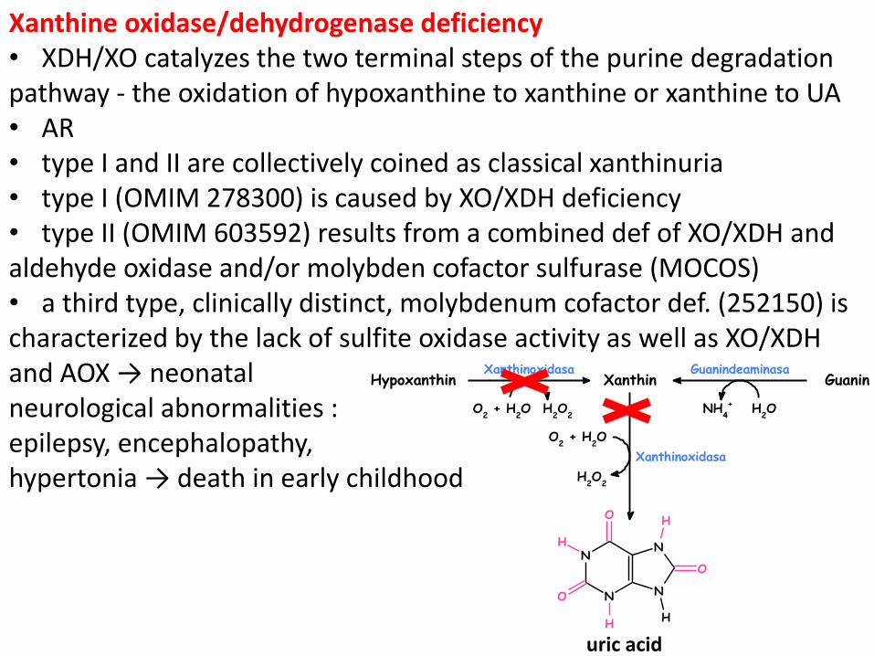
Xanthine oxidase/dehydrogenase deficiency • XDH/XO catalyzes the two terminal steps of the purine degradation pathway - the oxidation of hypoxanthine to xanthine or xanthine to UA • AR • type I and II are collectively coined as classical xanthinuria • type I (OMIM 278300) is caused by XO/XDH deficiency • type II (OMIM 603592) results from a combined def of XO/XDH and aldehyde oxidase and/or molybden cofactor sulfurase (MOCOS) • a third type, clinically distinct, molybdenum cofactor def. (252150) is characterized by the lack of sulfite oxidase activity as well as XO/XDH and AOX → neonatal neurological abnormalities : epilepsy, encephalopathy, hypertonia → death in early childhood
uric acid
http://www.tamilspider.com/attachments/Resources/3322-71129-xanthine.jpg
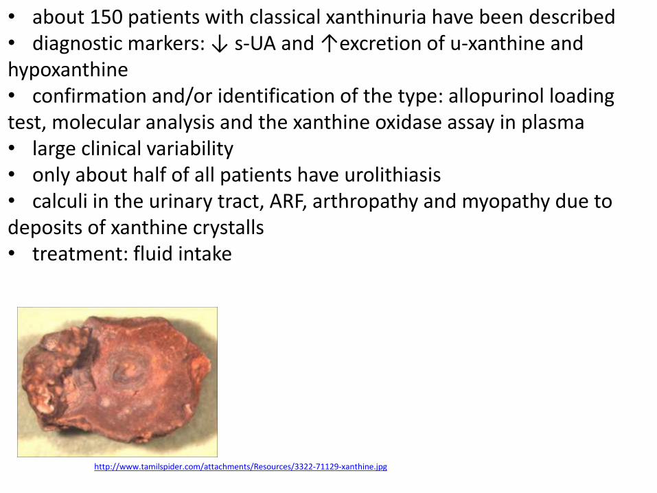
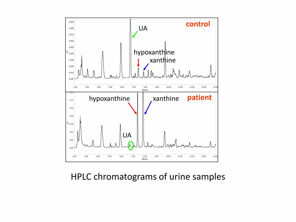
• about 150 patients with classical xanthinuria have been described • diagnostic markers: ↓ s-UA and ↑excretion of u-xanthine and hypoxanthine • confirmation and/or identification of the type: allopurinol loading test, molecular analysis and the xanthine oxidase assay in plasma • large clinical variability • only about half of all patients have urolithiasis • calculi in the urinary tract, ARF, arthropathy and myopathy due to deposits of xanthine crystalls • treatment: fluid intake
AU
0.00
0.02
0.04
0.06
0.08
0.10
0.12
0.14
Minutes
2.00 3.00 4.00 5.00 6.00 7.00 8.00 9.00 10.00 11.00 12.00 13.00 14.00
AU
0.000
0.010
0.020
0.030
0.040
0.050
0.060
0.070
0.080
0.090
Minutes
2.00 3.00 4.00 5.00 6.00 7.00 8.00 9.00 10.00 11.00 12.00 13.00 14.00
xanthine
UA
hypoxanthine
xanthine
UA
hypoxanthine
control
patient
HPLC chromatograms of urine samples
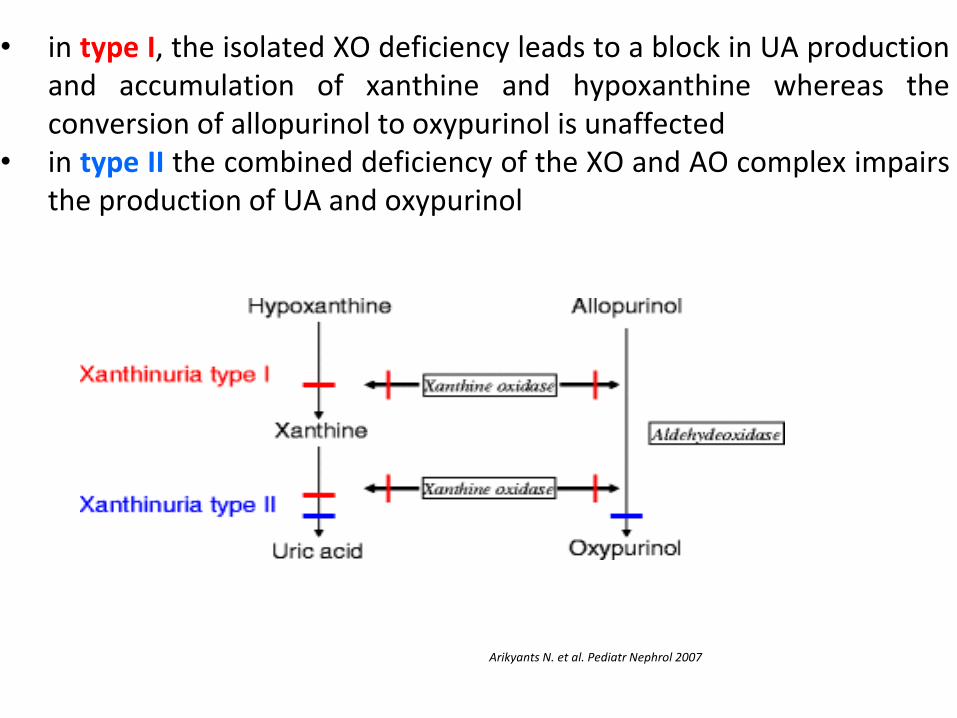
• in type I, the isolated XO deficiency leads to a block in UA production and accumulation of xanthine and hypoxanthine whereas the conversion of allopurinol to oxypurinol is unaffected
• in type II the combined deficiency of the XO and AO complex impairs the production of UA and oxypurinol
Arikyants N. et al. Pediatr Nephrol 2007

XDH II
XDH I
• sample of plasma before the administration of allopurinol→ then 300/150 mg of allopurinol orally in one dose → after one hour, a second plasma sample → HPLC
• the allopurinol loading test was performed in order to distinguish the type of xanthinuria (I or II)
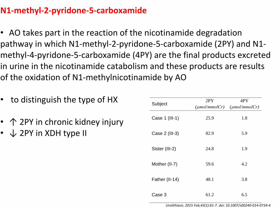
N1-methyl-2-pyridone-5-carboxamide • AO takes part in the reaction of the nicotinamide degradation pathway in which N1-methyl-2-pyridone-5-carboxamide (2PY) and N1-methyl-4-pyridone-5-carboxamide (4PY) are the final products excreted in urine in the nicotinamide catabolism and these products are results of the oxidation of N1-methylnicotinamide by AO • to distinguish the type of HX
• ↑ 2PY in chronic kidney injury • ↓ 2PY in XDH type II
Subject 2PY
(µmol/mmolCr)
4PY
(µmol/mmolCr)
Case 1 (III-1) 25.9 1.8
Case 2 (III-3) 82.9 5.9
Sister (III-2) 24.8 1.9
Mother (II-7) 59.6 4.2
Father (II-14) 48.1 3.8
Case 3 61.2 6.5
Urolithiasis. 2015 Feb;43(1):61-7. doi: 10.1007/s00240-014-0734-4
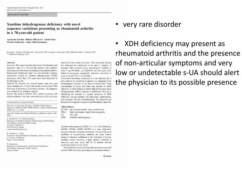
• very rare disorder
• XDH deficiency may present as rheumatoid arthritis and the presence of non-articular symptoms and very low or undetectable s-UA should alert the physician to its possible presence
Inherited metabolic disorders of pyrimidine metabolism
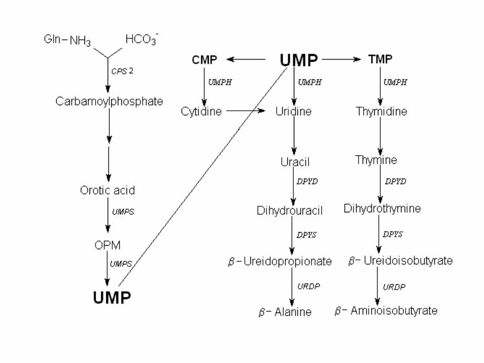
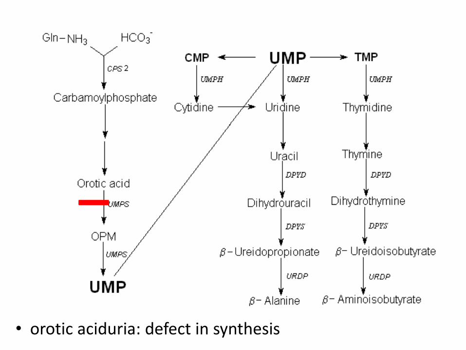
• orotic aciduria: defect in synthesis
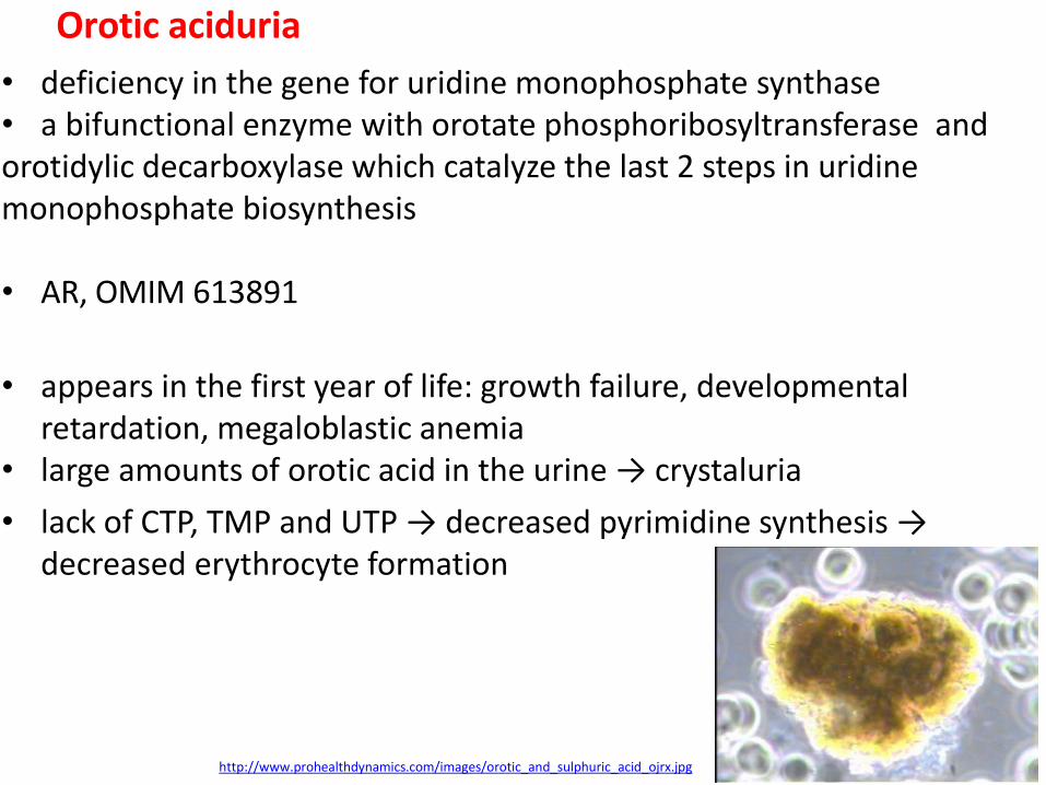
Orotic aciduria
• deficiency in the gene for uridine monophosphate synthase • a bifunctional enzyme with orotate phosphoribosyltransferase and orotidylic decarboxylase which catalyze the last 2 steps in uridine monophosphate biosynthesis • AR, OMIM 613891
• appears in the first year of life: growth failure, developmental retardation, megaloblastic anemia
• large amounts of orotic acid in the urine → crystaluria
• lack of CTP, TMP and UTP → decreased pyrimidine synthesis → decreased erythrocyte formation
http://www.prohealthdynamics.com/images/orotic_and_sulphuric_acid_ojrx.jpg
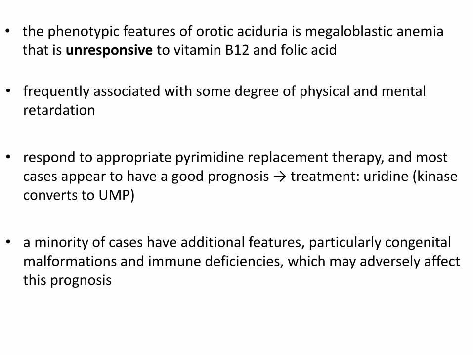
• the phenotypic features of orotic aciduria is megaloblastic anemia
that is unresponsive to vitamin B12 and folic acid
• frequently associated with some degree of physical and mental retardation
• respond to appropriate pyrimidine replacement therapy, and most cases appear to have a good prognosis → treatment: uridine (kinase converts to UMP)
• a minority of cases have additional features, particularly congenital malformations and immune deficiencies, which may adversely affect this prognosis
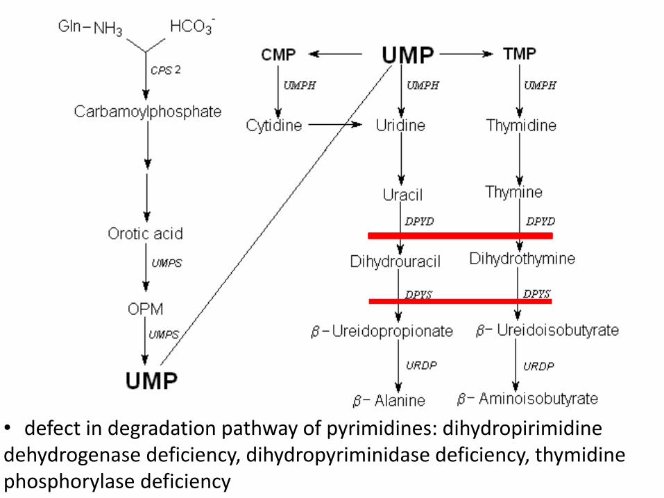
• defect in degradation pathway of pyrimidines: dihydropirimidine dehydrogenase deficiency, dihydropyriminidase deficiency, thymidine phosphorylase deficiency
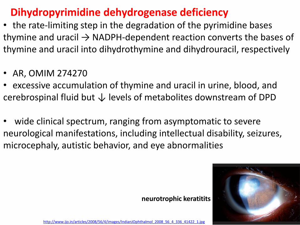
Dihydropyrimidine dehydrogenase deficiency
http://www.ijo.in/articles/2008/56/4/images/IndianJOphthalmol_2008_56_4_336_41422_1.jpg
neurotrophic keratitits
• the rate-limiting step in the degradation of the pyrimidine bases thymine and uracil → NADPH-dependent reaction converts the bases of thymine and uracil into dihydrothymine and dihydrouracil, respectively
• AR, OMIM 274270 • excessive accumulation of thymine and uracil in urine, blood, and cerebrospinal fluid but ↓ levels of metabolites downstream of DPD
• wide clinical spectrum, ranging from asymptomatic to severe neurological manifestations, including intellectual disability, seizures, microcephaly, autistic behavior, and eye abnormalities
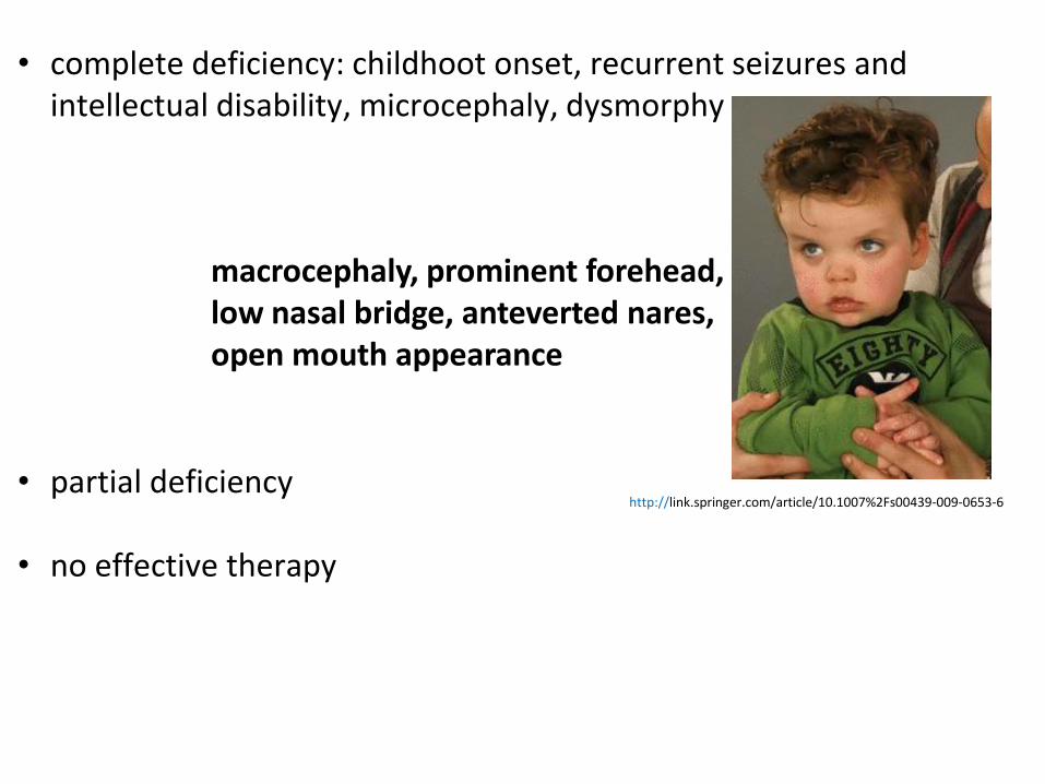
• complete deficiency: childhoot onset, recurrent seizures and
intellectual disability, microcephaly, dysmorphy
• partial deficiency
• no effective therapy
macrocephaly, prominent forehead, low nasal bridge, anteverted nares, open mouth appearance
http://link.springer.com/article/10.1007%2Fs00439-009-0653-6
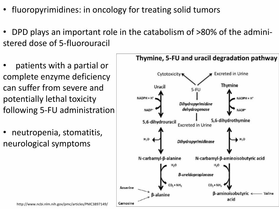
• fluoropyrimidines: in oncology for treating solid tumors
• DPD plays an important role in the catabolism of >80% of the admini- stered dose of 5-fluorouracil • patients with a partial or complete enzyme deficiency can suffer from severe and potentially lethal toxicity following 5-FU administration • neutropenia, stomatitis, neurological symptoms
http://www.ncbi.nlm.nih.gov/pmc/articles/PMC3897149/
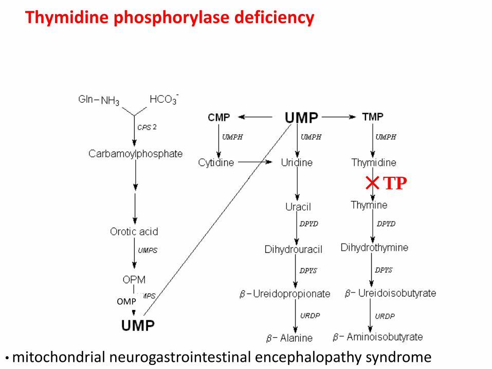
Thymidine phosphorylase deficiency
OMP
TP
• mitochondrial neurogastrointestinal encephalopathy syndrome
Mitochondrial neurogastrointestinal encephalopathy syndrome (MNGIE)
• multisystem disorder, AR, OMIM 603041 • start in 1st to 5th decade (60% pacients before 20 y) • progressive degeneration of the muscles of the gastrointestinal tract causing gastrointestinal dysmotility, weakness of extra-ocular muscles (ophthalmoparesis), degeneration of peripheral nerves causing altered sensation and weakness the distal arms and legs, and cachexia • the specific symptoms associated with MNGIE variable: vomiting, nausea, diarrhea, reflux, abdominal pain … • progressive GIT dysmotility
• ↑ plasma and urine thymidine and deoxyuridine concentration
• lactic acidemia, lactic acidosis
• thymidine phosphorylase enzyme activity in leukocytes is less than 10% of the control mean
• molecular genetic testing of the thymidine phosphorylase gene, detects pathogenic variants in approximately 100% of affected individuals
• no genetype/phenotype correlations
• treatment is supportive
• alel patients have peripheral neuropathy → the neuropathy is demyelinating
• the segmental demyelination is hypothesized to be caused by the uneven distribution of mtDNA abnormalities along the nerve
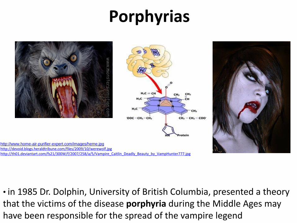
Porphyrias
http://www.home-air-purifier-expert.com/images/heme.jpg http://devoid.blogs.heraldtribune.com/files/2009/10/werewolf.jpg http://th01.deviantart.com/fs21/300W/f/2007/258/a/5/Vampire_Caitlin_Deadly_Beauty_by_VampHunter777.jpg
• in 1985 Dr. Dolphin, University of British Columbia, presented a theory that the victims of the disease porphyria during the Middle Ages may have been responsible for the spread of the vampire legend
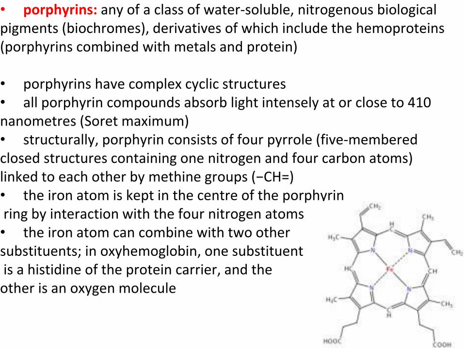
• porphyrins: any of a class of water-soluble, nitrogenous biological pigments (biochromes), derivatives of which include the hemoproteins (porphyrins combined with metals and protein)
• porphyrins have complex cyclic structures • all porphyrin compounds absorb light intensely at or close to 410 nanometres (Soret maximum) • structurally, porphyrin consists of four pyrrole (five-membered closed structures containing one nitrogen and four carbon atoms) linked to each other by methine groups (−CH=) • the iron atom is kept in the centre of the porphyrin ring by interaction with the four nitrogen atoms • the iron atom can combine with two other substituents; in oxyhemoglobin, one substituent is a histidine of the protein carrier, and the other is an oxygen molecule

Heme - structure
http://www.home-air-purifier-expert.com/images/heme.jpg
• heme is the prosthetic group of hemoglobin, myoglobin, and the cytochromes: Fe2+ contained in the centre of a four pyrrolic groups joined together by methine bridges
• the ring contains a large number of conjugated double bonds, which allows the molecule to absorb light in the visible part of the spectrum
• the iron atom and the attached protein chain modify the wavelength of the absorption and gives hemoglobin its characteristic colour
• the heme ion serves as a source or sink of electrons during electron transfer or redox chemistry
Hemoproteins
• hemes are most commonly recognized as components of hemoglobin, but are also found in a number of other biologically important hemoproteins
• myoglobin • cytochrome (Fe2+ ↔Fe3+) • catalase (responsible for the degradation of hydrogen peroxide) • endothelial nitric oxide • etc…
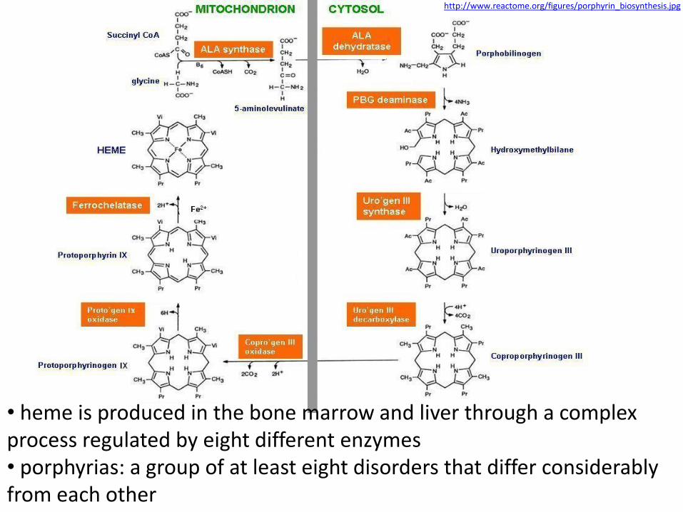
http://www.reactome.org/figures/porphyrin_biosynthesis.jpg
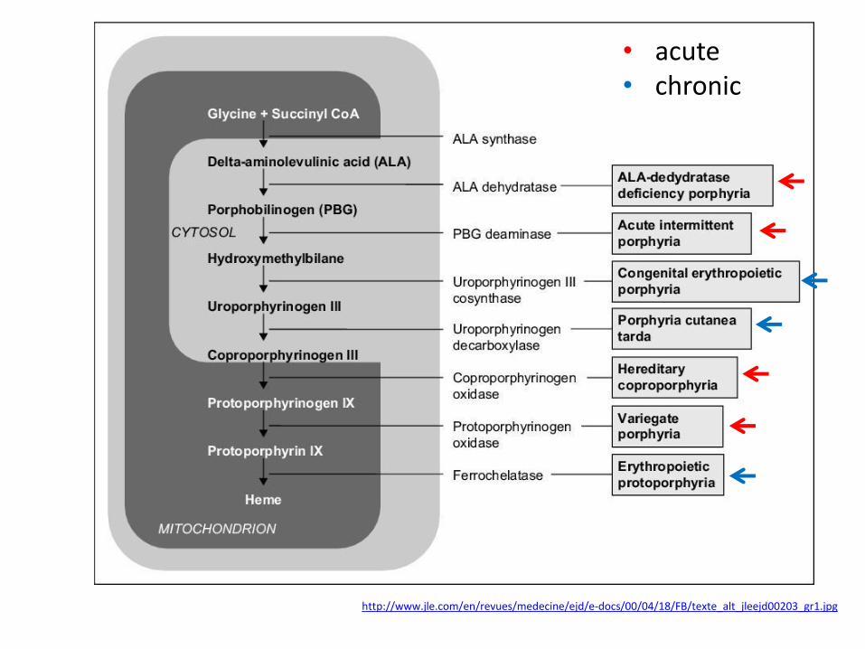
• heme is produced in the bone marrow and liver through a complex process regulated by eight different enzymes • porphyrias: a group of at least eight disorders that differ considerably from each other
Porphyrias
• genetic diseases resulting in decreased activity of one of the enzymes involved in heme synthesis • the terms porphyria are derived from the Greek word porphyrus, meaning purple → urine from some porphyria patients may be reddish in color due to the presence of excess porphyrins and related substances in the urine, and the urine may darken after exposure to light • symptoms vary depending on the enzyme, the severity of the deficiency and whether heme synthesis is affected primarily in liver or in developing erythrocytes • a common feature in all porphyrias is the accumulation of porphyrins or porphyrin precursors
• often AD inheritance, some AR inheritance • usually onset in adulthood • common manifestation only after exposure (fasting, menses, drugs, sunlight)
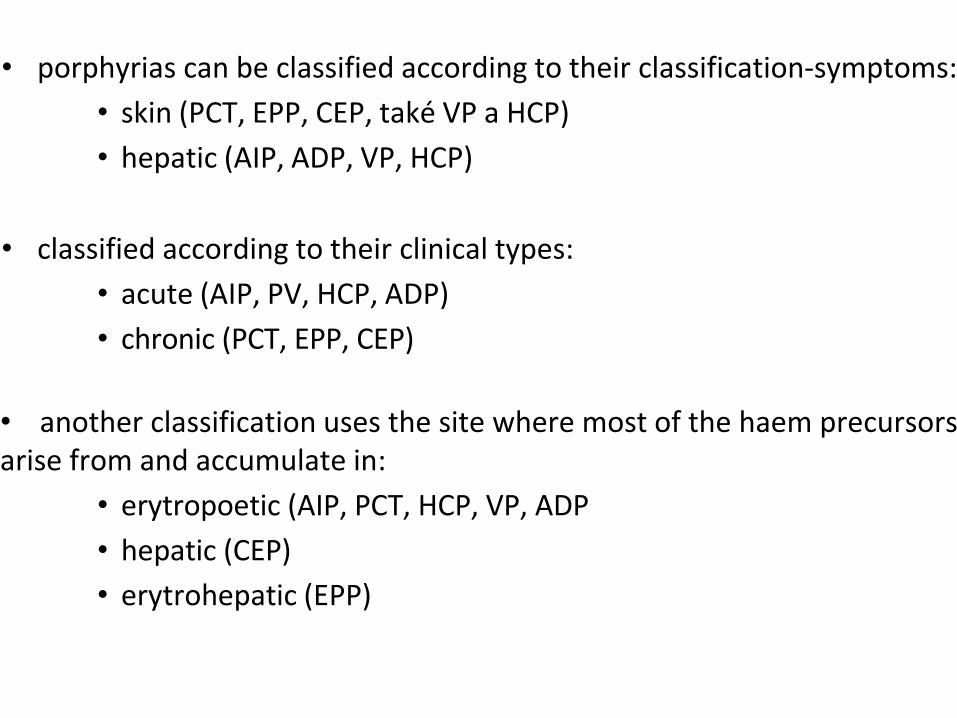
• porphyrias can be classified according to their classification-symptoms:
• skin (PCT, EPP, CEP, také VP a HCP)
• hepatic (AIP, ADP, VP, HCP)
• classified according to their clinical types:
• acute (AIP, PV, HCP, ADP)
• chronic (PCT, EPP, CEP)
• another classification uses the site where most of the haem precursors arise from and accumulate in:
• erytropoetic (AIP, PCT, HCP, VP, ADP
• hepatic (CEP)
• erytrohepatic (EPP)
http://www.jle.com/en/revues/medecine/ejd/e-docs/00/04/18/FB/texte_alt_jleejd00203_gr1.jpg
• acute • chronic
http://terrycomeau.com/Porphyria/Porphyria.htm
http://www.pathguy.com/lectures/erythrodontia.jpg
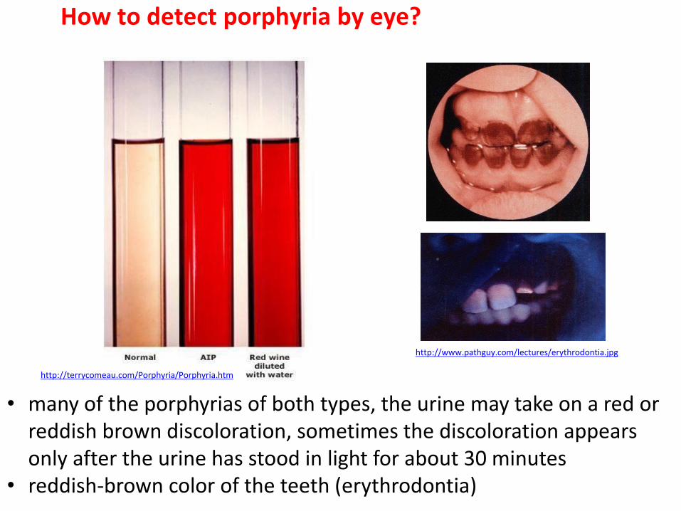
How to detect porphyria by eye?
• many of the porphyrias of both types, the urine may take on a red or reddish brown discoloration, sometimes the discoloration appears only after the urine has stood in light for about 30 minutes
• reddish-brown color of the teeth (erythrodontia)
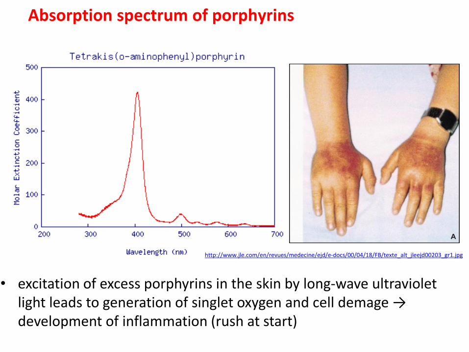
Absorption spectrum of porphyrins
• excitation of excess porphyrins in the skin by long-wave ultraviolet light leads to generation of singlet oxygen and cell demage → development of inflammation (rush at start)
http://www.jle.com/en/revues/medecine/ejd/e-docs/00/04/18/FB/texte_alt_jleejd00203_gr1.jpg

Porphyrias producing acute neuropsychiatric features • frequency and severity of attacks vary widely between people • between attacks the patient is usually healthy • acute attacks are precipitated by metabolic, hormonal and environmental factors that induce hepatic delta-aminolaevulinic acid synthase (ALA synthase) activity • this increased activity causes the haem precursors delta-amino-laevulinic acid and porphobilinogen to increase→ because of the reduced activity of one of the different enzymes needed to convert these precursors further (depending on which porphyria is present), there is pathological accumulation • rarely an acute attack may be life-threatening !!!
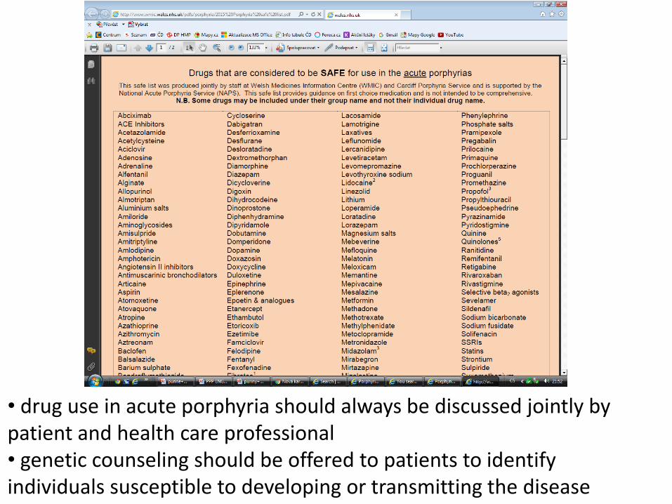
• drug use in acute porphyria should always be discussed jointly by patient and health care professional • genetic counseling should be offered to patients to identify individuals susceptible to developing or transmitting the disease
Acute hepatic porphyria
• sub-group of porphyrias characterized by the occurrence of neuro-visceral attacks with or without cutaneous manifestations • all acute hepatic porphyrias can be accompanied by neuro-visceral attacks that appear as intense abdominal pain (in 85-95% of cases) over one to two weeks, neurological symptoms (muscular weakness, sensory loss or convulsions) and psychological symptoms (irritability, anxiety, auditory or visual hallucinations, mental confusion)
• acute intermittent porphyria (the most common)
• variagate porphyria
• hereditary coproporphyria
• hereditary deficit of delta-aminolevulinic acid dehydratase (extremely rare)

Hepatic – acute - induced porphyrias
• acute intermittent porphyria
• porphyria variegata
• hereditary koproporphyria
• Doose porphyria (AR)
• inheritance – AD
• in addition to genetic risks, environmental factors may trigger the development of signs and symptoms in some types of porphyriatrigger from certain drugs (barbiturates or sulfonamide antibiotics or, less often, birth control pills, or some drugs that affect the mind or behavior, known as psychoactive drugs), chemicals, dieting or fasting, smoking, physical stress (such as infections or other illnesses), liver disease, emotional stress, alcohol use, menstrual hormones, sun exposure …
• approximately 80% of individuals who carry a gene mutation for acute intermittent porphyria, variegate porphyria, and hereditary copro-porphyria remain asymptomatic, and others may have only one or a few acute attacks throughout life
• in most of these cases the levels of delta-aminolevulinic acid (ALA), porphobilinogen, and porphyrins in urine, serum, and feces are normal • additionally, most patients with ALA-dehydratase deficiency porphyria, who may have less than 5% of normal ALA dehydratase activity, also remain asymptomatic for most of their lives • severe neuropathic abdominal pain, the most frequent symptom, is diffuse rather than localized and is often accompanied by nausea, vomiting, distention, constipation, and sometimes diarrhea • other symptoms include insomnia (often an early symptom), heart palpitations, seizures (sometimes due to hyponatremia), restlessness, hallucinations, and other acute psychiatric symptoms
Chronic porphyrias
• the disease can manifest in adulthood (porphyria cutanea tarda) or in childhood (hepatoerythropoietic porphyria)
• patients present with cutaneous lesions (fragility, bullae, scars) on the surface of skin exposed to the sun (hands, face) and, unlike in cases of acute hepatic porphyrias (see this term), don't present with acute neuro-visceral attacks
• the acquired forms of the diseases may be triggered by risk factors (alcohol, hepatitis C, estrogen, iron overload)
Neurological symptoms in porphyrias
• peripheral NS increased activity of sympathicus: tachycardia, hypertension abdominal pain-nonlocalised but also colic parestesias peripheral neuropathy- muscle weaknes
• central NS aggitation psychotic episodes
• mechanisms: synaptic function interference (GABA vs. ALA similarity), heme depletion (NOS, Trp pyrrolase)
• AD, deficiency of porphobilinogen deaminase (PBG-D; the third enzyme in the heme biosynthesis pathway) that leads to an accumulation of the precursors of porphyrins in the liver (delta-aminolevulinic acid, ALA and porphobilinogen, PBG) • AIP is the most frequent and the most severe form of the acute hepatic porphyrias • manifests after puberty (20 – 40y) and preferentially affects women 2:1 • occurrence of neuro-visceral attacks without cutaneous manifestations, attacks can persist for several days and that repeat over several weeks, manifest as intense abdominal pain (>95% of cases) and neurological and/or psychological symptoms • psychological symptoms are variable: irritability, emotionality, depression, considerable anxiety and, more rarely, auditory and visual hallucinations, disorientation, mental confusion • neurological manifestations can affect the central nervous system as much as the peripheral nervous system (myalgia, paresis,…)
Acute intermittent porphyria (AIP)
• diagnosis: the observation of urine that is pink orred after exposure to light evokes the diagnosis of the disease • the evidence of elevated concentrations of delta-aminolevulinic acid, and porphobilinogen in the urine and residual PBG-D activity in 50% of red blood cells (not always found during attacks) • confirmed by the identification of a causal mutation of the HMBS gene • when an acute attack is confirmed, urgent treatment with an injection of human hemin and/or perfusion of carbohydrates is required • management includes the suppression of triggering factors, relief from pain (opiates), vomiting and anxiety, and the prevention of attacks (by avoiding triggering factors, particularly drugs)
http://terrycomeau.com/Porphyria/Porphyria.htm
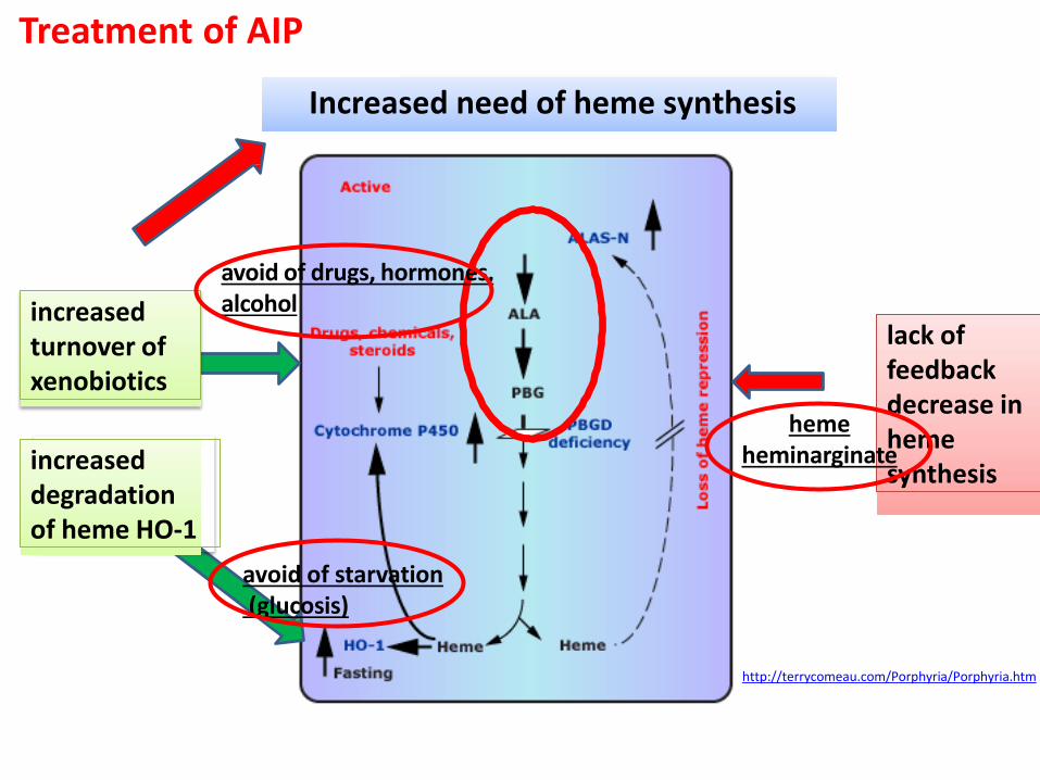
lack of feedback decrease in heme synthesis increased
degradation of heme HO-1
Increased need of heme synthesis
increased turnover of xenobiotics
Treatment of AIP
avoid of drugs, hormones, alcohol
avoid of starvation (glucosis)
heme heminarginate
Hereditary coproporphyria (HCP) • AD, coproporphyrinogen oxidase (CPOX) • clinical presentation similar to that of AIP, except that some patients (about 20%) develop blistering photosensitivity resulting in cutaneous lesions that resemble those in PCT, neuro-psychiatric symptoms • attacks are generally milder than those seen in patients with AIP • urinary delta-aminolevulinic acid and porphobilinogen ↑, especially during acute attacks, but generally to a lesser degree than in AIP • the diagnostic finding is a significant increase in urine PBG and coproporphyrin, plasma porphyrin levels are usually normal but may be increased in patients with skin lesions • treatment, complications, and preventive measures for HCP are the same as for AIP
Porphyria variegata
• AD, a deficiency of protoporphyrinogen oxidase (PPOX) • the term "variegate" refers to the fact that this porphyria can present with both neurologic and/or cutaneous symptoms • the presenting signs and symptoms during acute attacks are identical to those in AIP though generally milder • recommendations for treatment and management are the same as AIP • blistering skin lesions with sun exposure are much more common than in HCP, and are indistinguishable from those of PCT and may be chronic→ there is no remedy for VP photosensitivity other than use of protective clothing and avoidance of prolonged sun exposure • urine delta-aminolevulinic acid and porphobilinogen are ↑ during attacks, but as in HCP, these may increase to a lesser degree and decrease more rapidly than in AIP • in contrast to AIP and HCP, plasma porphyrins are frequently increased in VP and display a distinctive fluorescence peak at ~626nm
Doss porhyria
• AR, δ-Aminolevulinic Acid Dehydratase Porphyria (ADP) • ADP is the least common of all the porphyrias with less than 10 cases • all of the reported cases have been males, in contrast to the other acute porphyrias which are more prevalent in females • a severe deficiency of the enzyme δ-aminolevulinic acid dehydratase causes an increase of 5'-aminolevulinic acid in the liver, other tissues, blood plasma, and urine • urine coproporphyrin and erythrocyte protoporphyrin are increased • treatment is the same as in the other acute porphyrias
Porphyria cutanea tarda • the most common type of porphyria • PCT is due to a deficiency of the uroporphyrinogen decarboxylase • the development of symptoms requires the enzyme deficiency in the liver to be less than 20% of normal activity • liver disease is common, 35% of people develop cirrhosis and 7-24% develop liver cancer • skin damage occurs because excess porphyrins produced in the liver are transported by the blood to the skin • PCT has several common precipitating factors → these factors include excess iron in the liver, moderate or heavy alcohol use, smoking, taking estrogens, and infection with hepatitis C virus • unlike other porphyrias, most cases of PCT are acquired (referred to as sporadic or Type I PCT), and not inherited → this is secondary to a UROD inhibitor, uroporphomethene, which is generated within the liver • iron overload of mild to moderate degree is present in all PCT cases and is required for the generation of this UROD
http://www.jle.com/en/revues/medecine/ejd/e-docs/00/04/18/FB/texte_alt_jleejd00203_gr1.jpg
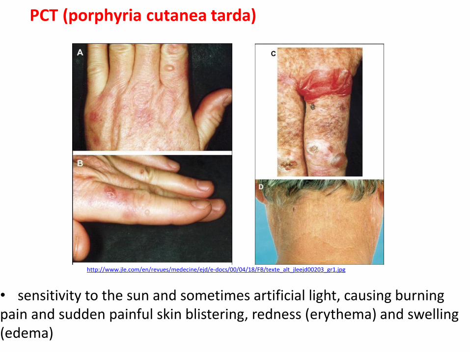
PCT (porphyria cutanea tarda)
• sensitivity to the sun and sometimes artificial light, causing burning pain and sudden painful skin blistering, redness (erythema) and swelling (edema)
• diagnosis is based on the measurement of concentrations of porphyrins in urine, stools and blood - with a predominance of uro- porphyrinogen and heptacarboxylporphyrin • fluorescence of plasma porphyrins at 619-620 nm
• the evidence of a deficiency of URO-D in red blood cells and the identification of a causal mutation of the URO-D gene allow a confirmed diagnosis
• PCT is the most readily treated porphyria: phlebotomy, in which a almost half a liter of blood is removed, is the most widely recommended treatment → the excess iron is gradually removed, the activity of uroporphyrinogen decarboxylase in the liver returns toward normal, and porphyrin levels in the liver and blood fall gradually • low doses of chloroquine or hydroxychloroquine are also effective in treating PCT → remove excess porphyrins from the liver • avoid sun exposure as much as possible

Congenital Erythropoietic Porphyria (CEP)
• AR, extremely rare, Günther disease, uroporphyrinogen III synthase • severe cutaneous photosensitivity at birth or in early infancy with blistering and increased friability of the skin over light-exposed areas, hemolytic anemia is common and can range from mild to severe, with some affected individuals requiring chronic blood transfusions • the first manifestation is often pink to dark red discoloration of the urine • the diagnosis of CEP is supported by the biochemical findings of markedly ↓d uroporphyrinogen-synthase activity in erythrocytes and/or markedly increased levels of urinary uroporphyrin and coproporphyrin
• only available cure for CEP is a bone marrow transplant
• ß-carotene
http://www.pathguy.com/lectures/erythrodontia.jpg
http://www.jle.com/en/revues/medecine/ejd/e-docs/00/04/18/FB/texte_alt_jleejd00203_gr1.jpg
Erythropoietic protoporphyria (EPP)
• AD (OMIM*612386), AR (OMIM#177000), defect of ferrochelatase • X-linked, delta-aminolevulinic acid synthase-2 gene, OMIM #300752 • protoporphyrin accumulates first in the bone marrow, and then in red blood cells, plasma and sometimes the liver • early onset in childhood • diagnosed: elevated levels of protoporphyrin IX in erythrocytes and plasma • photosensitivity and erythrodontia → swelling, burning, itching, and redness of the skin may appear during or after exposure to sunlight, including sunlight that passes through window glass • hemolysis and liver abnormalities- ca hepatis • neurological abnormalities rarely • therapy: transfusion, beta-caroten, transplant • EPP patients should also not use any drug or anesthetic which causes cholestasis (slowing down bile flow)
Thank you
• disorders of uric acid metabolism
• disorders of purines/pyrimidines metabolism
• porphyrias


![PURINE AND PYRIMIDINE METABOLISM IN MAN VII · 2007. 7. 13. · Pyrrolo[3,2-D]pyrimidines, a New Class of Purine Nucleoside Phosphyorylase (PNP) Inhibitors as Potential T-cell Selective](https://img.pdfslide.us/doc/110x75/60a9988442e1294207469220/purine-and-pyrimidine-metabolism-in-man-vii-2007-7-13-pyrrolo32-dpyrimidines.jpg)