Embed Size (px)
Citation preview
DISEASES OF THE CONJUNCTIVA AYESHA S ABDULLAH 21.09.2013
LEARNING OUTCOMES
By the end of this lecture the students would be able to;1. Classify diseases of the conjunctiva2. Identify the common symptoms and signs of conjunctival
diseases3. Classify diseases of the conjunctiva4. Identify the common symptoms and signs of conjunctival
diseases5. Enlist the causes & risk factors of conjunctivitis.6. Differentiate between bacterial, viral, chlamydial and allergic
conjunctivitis on the basis of clinical presentation.7. Describe the associated complications, treatment and
prevention strategies for each type of conjunctivitis.8. Identify Pterygium on photographs, describe its pathogenesis,
complications and treatment.
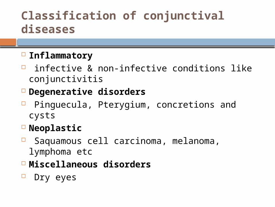
Classification of conjunctival diseases
Inflammatory infective & non-infective conditions like
conjunctivitis Degenerative disorders Pinguecula, Pterygium, concretions and cysts Neoplastic Saquamous cell carcinoma, melanoma,
lymphoma etc Miscellaneous disorders Dry eyes
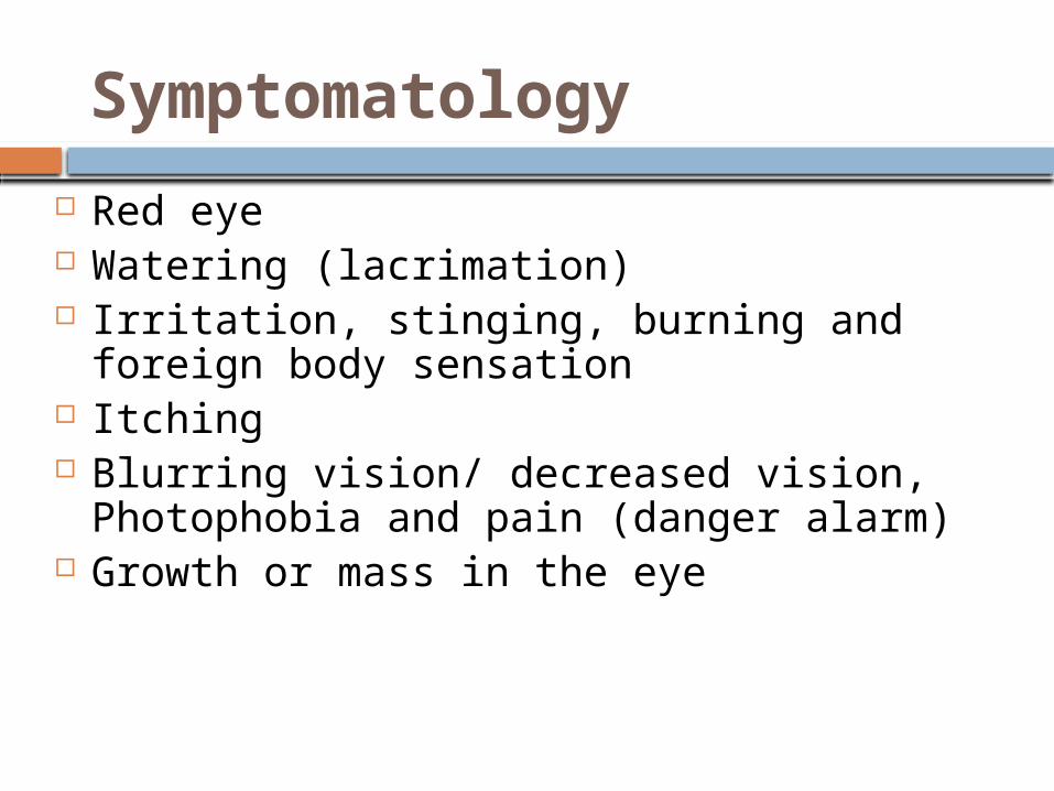
Symptomatology
Red eye Watering (lacrimation) Irritation, stinging, burning and foreign body
sensation Itching Blurring vision/ decreased vision,
Photophobia and pain (danger alarm) Growth or mass in the eye
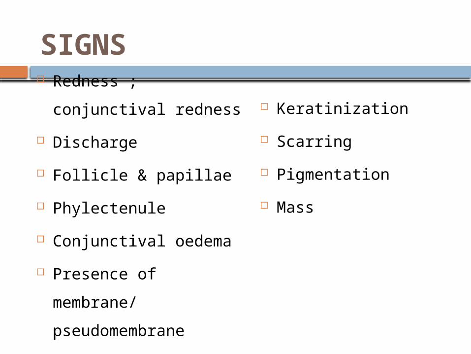
SIGNS Redness ; conjunctival
redness
Discharge
Follicle & papillae
Phylectenule
Conjunctival oedema
Presence of membrane/
pseudomembrane
Subconjunctival
haemorrhage
Lymphadenopathy
Keratinization
Scarring
Pigmentation
Mass
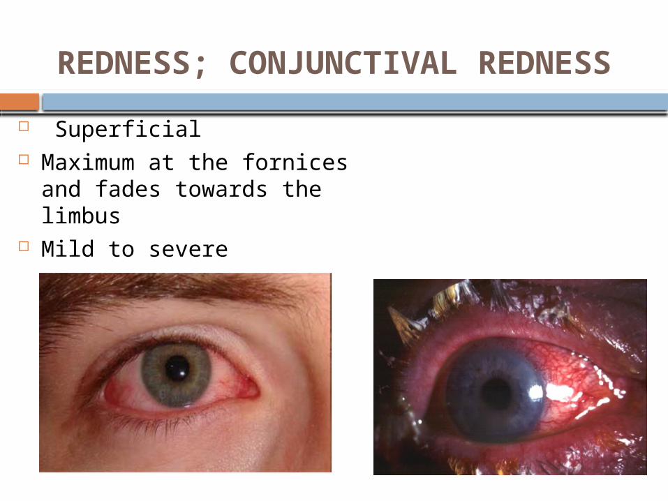
REDNESS; CONJUNCTIVAL REDNESS
Superficial Maximum at the fornices and
fades towards the limbus Mild to severe
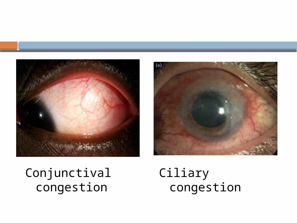
Conjunctival congestion
Ciliary congestion
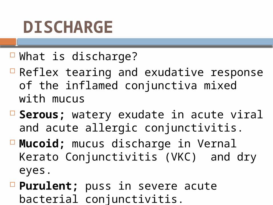
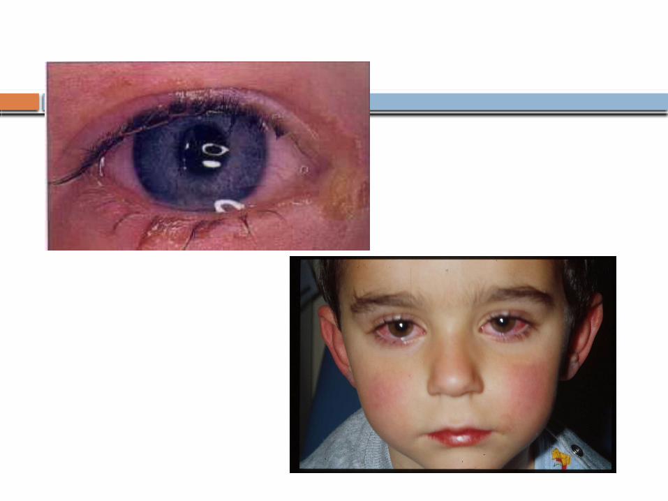
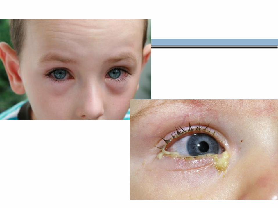
DISCHARGE
What is discharge? Reflex tearing and exudative response of
the inflamed conjunctiva mixed with mucus Serous; watery exudate in acute viral and
acute allergic conjunctivitis. Mucoid; mucus discharge in Vernal Kerato
Conjunctivitis (VKC) and dry eyes. Purulent; puss in severe acute bacterial
conjunctivitis. Mucopurulent; puss plus mucus in mild
bacterial conjunctivitis and Chlamydial conjunctivitis.
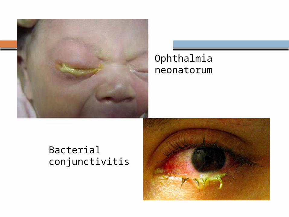
Ophthalmia neonatorum
Bacterial conjunctivitis
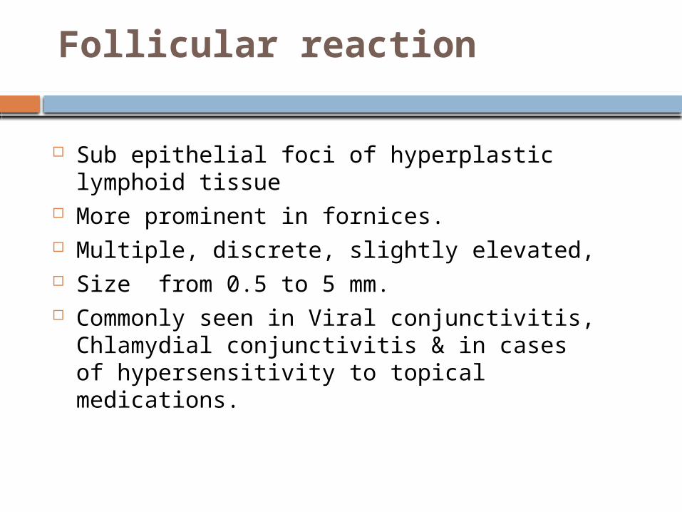
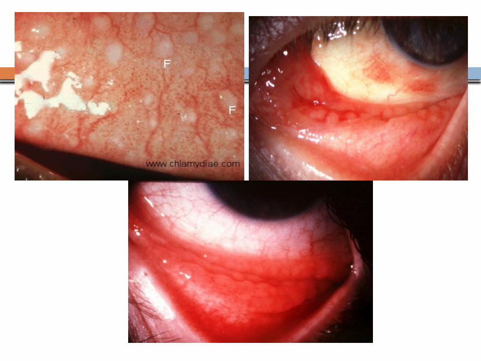
Follicular reaction
Sub epithelial foci of hyperplastic lymphoid tissue
More prominent in fornices. Multiple, discrete, slightly elevated, Size from 0.5 to 5 mm. Commonly seen in Viral conjunctivitis,
Chlamydial conjunctivitis & in cases of hypersensitivity to topical medications.
15
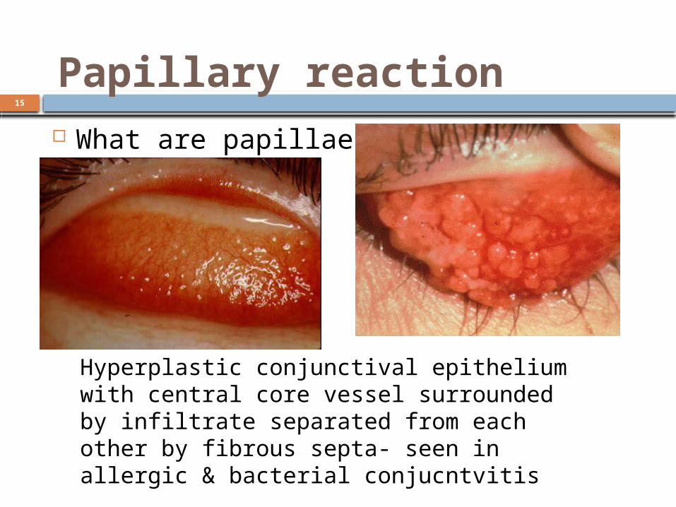
What are papillae?
Papillary reaction
Hyperplastic conjunctival epithelium with central core vessel surrounded by infiltrate separated from each other by fibrous septa- seen in allergic & bacterial conjucntvitis
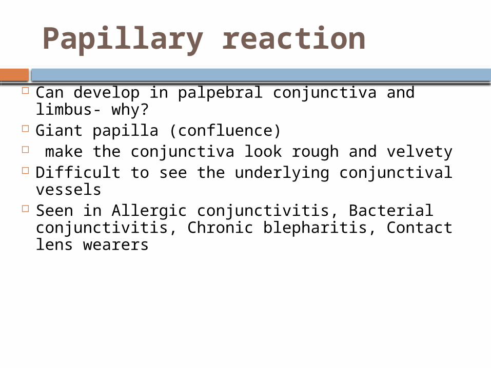
Papillary reaction
Can develop in palpebral conjunctiva and limbus- why?
Giant papilla (confluence) make the conjunctiva look rough and velvety Difficult to see the underlying conjunctival vessels Seen in Allergic conjunctivitis, Bacterial
conjunctivitis, Chronic blepharitis, Contact lens wearers
17
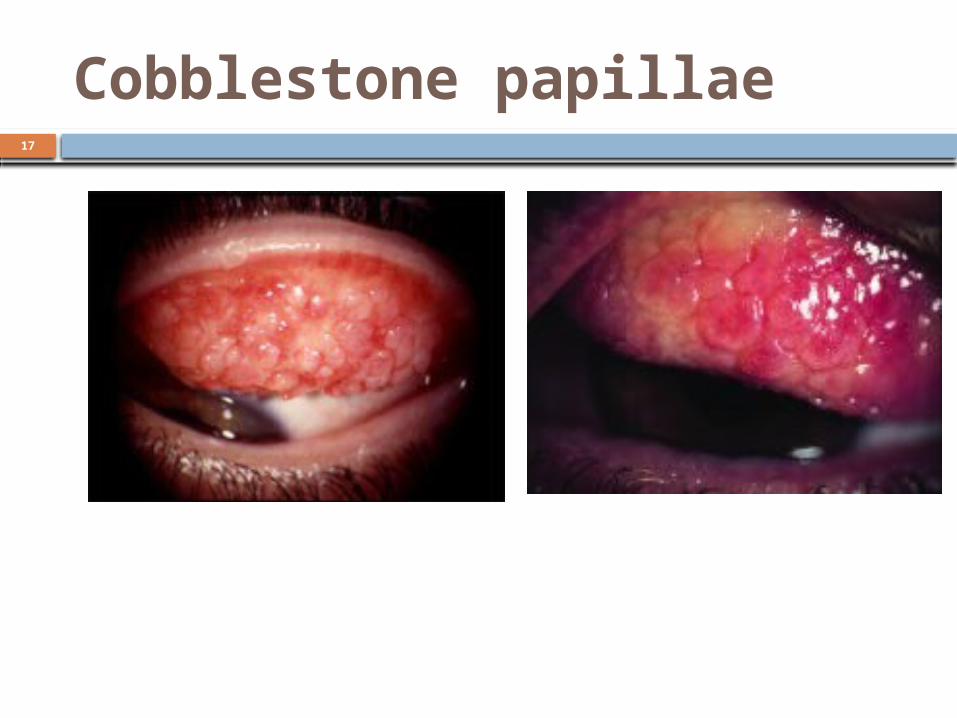
Cobblestone papillae
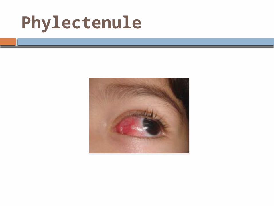
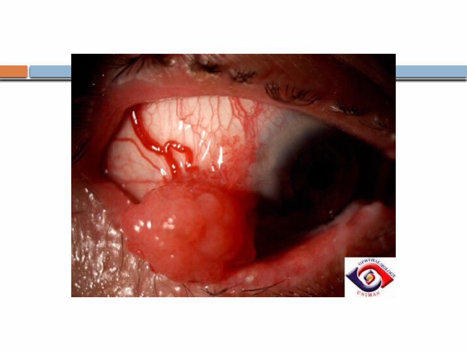
Phylectenule
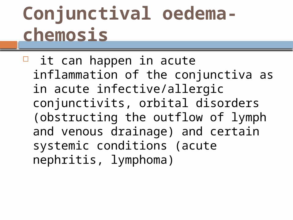
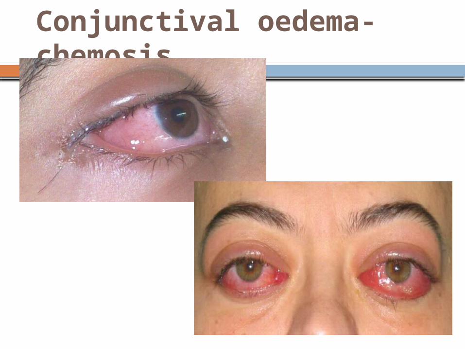
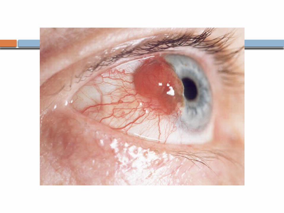
Conjunctival oedema- chemosis it can happen in acute inflammation of
the conjunctiva as in acute infective/allergic conjunctivits, orbital disorders (obstructing the outflow of lymph and venous drainage) and certain systemic conditions (acute nephritis, lymphoma)
Conjunctival oedema- chemosis
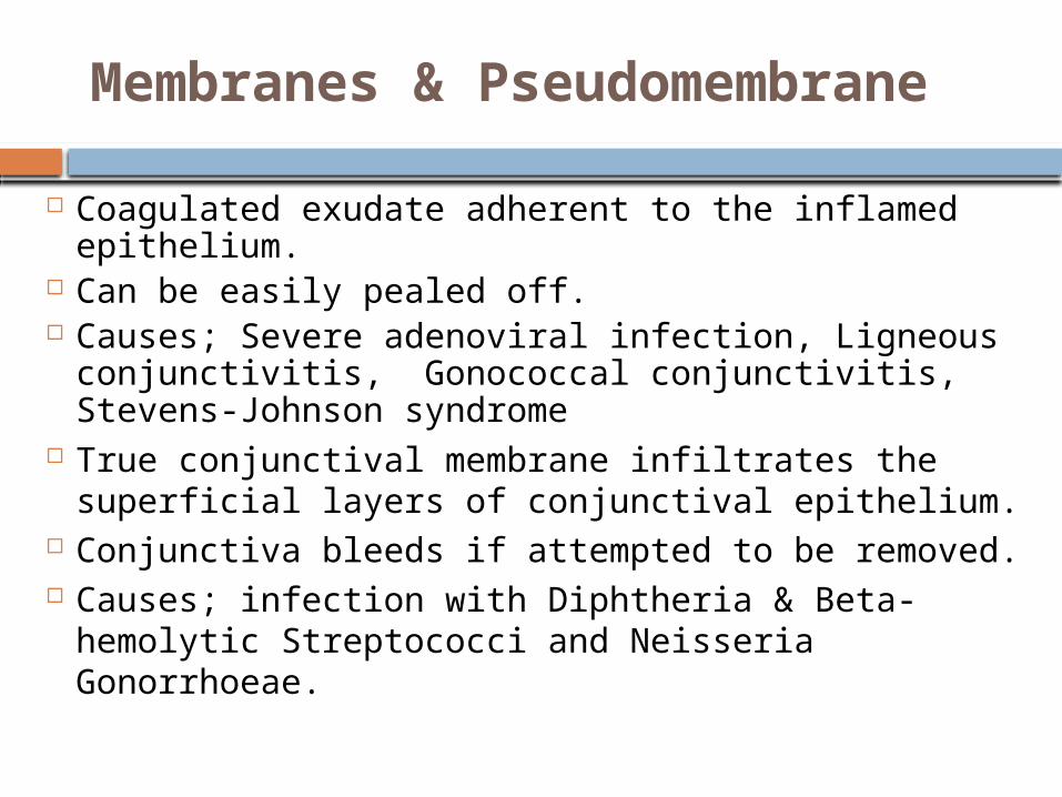
Membranes & Pseudomembrane
Coagulated exudate adherent to the inflamed epithelium.
Can be easily pealed off. Causes; Severe adenoviral infection, Ligneous
conjunctivitis, Gonococcal conjunctivitis, Stevens-Johnson syndrome
True conjunctival membrane infiltrates the superficial layers of conjunctival epithelium.
Conjunctiva bleeds if attempted to be removed. Causes; infection with Diphtheria & Beta-
hemolytic Streptococci and Neisseria Gonorrhoeae.
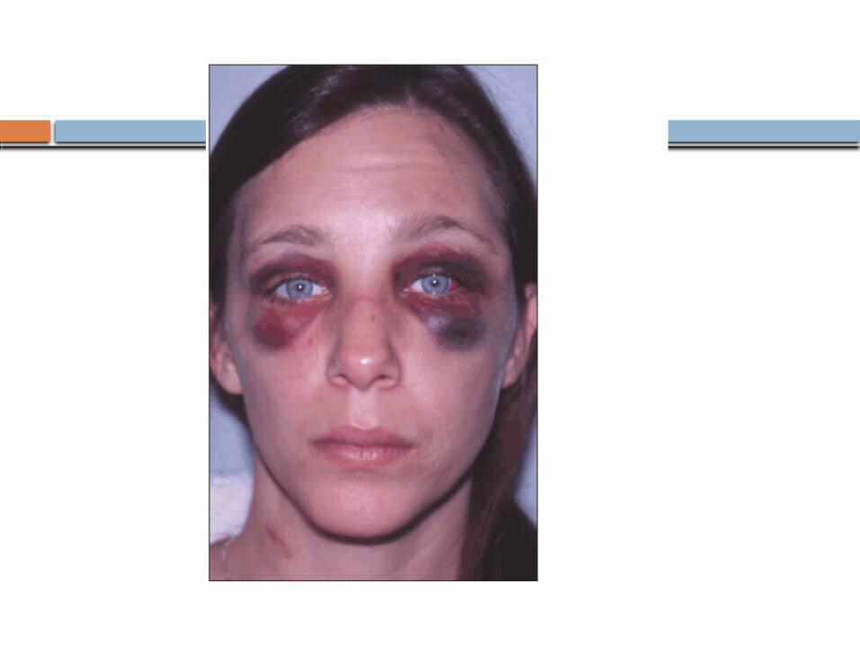
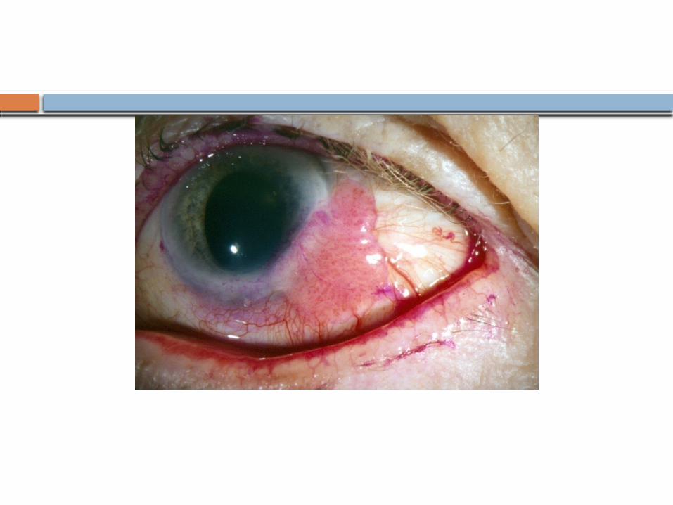
Subconjunctival Haemorrhage Can happen in severe cases of viral or
bacterial conjunctivitis Trauma Haemotological disorders (bleeding
disorders, leukaemias) Fracture base of the skull
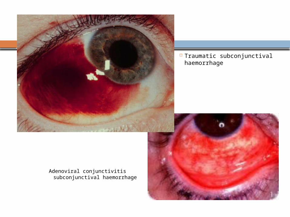
Traumatic subconjunctival haemorrhage
Adenoviral conjunctivitis subconjunctival haemorrhage
Lymphadenopathy
Pre auricular and sub mandibular.
In ; Viral infection, Chlamydial infection,
Severe bacterial infections, Parinaud
oculoglandular syndrome.
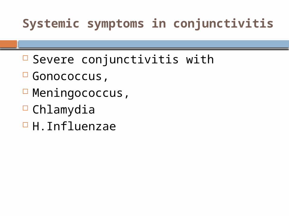
Systemic symptoms in conjunctivitis
Severe conjunctivitis with Gonococcus, Meningococcus, Chlamydia H.Influenzae
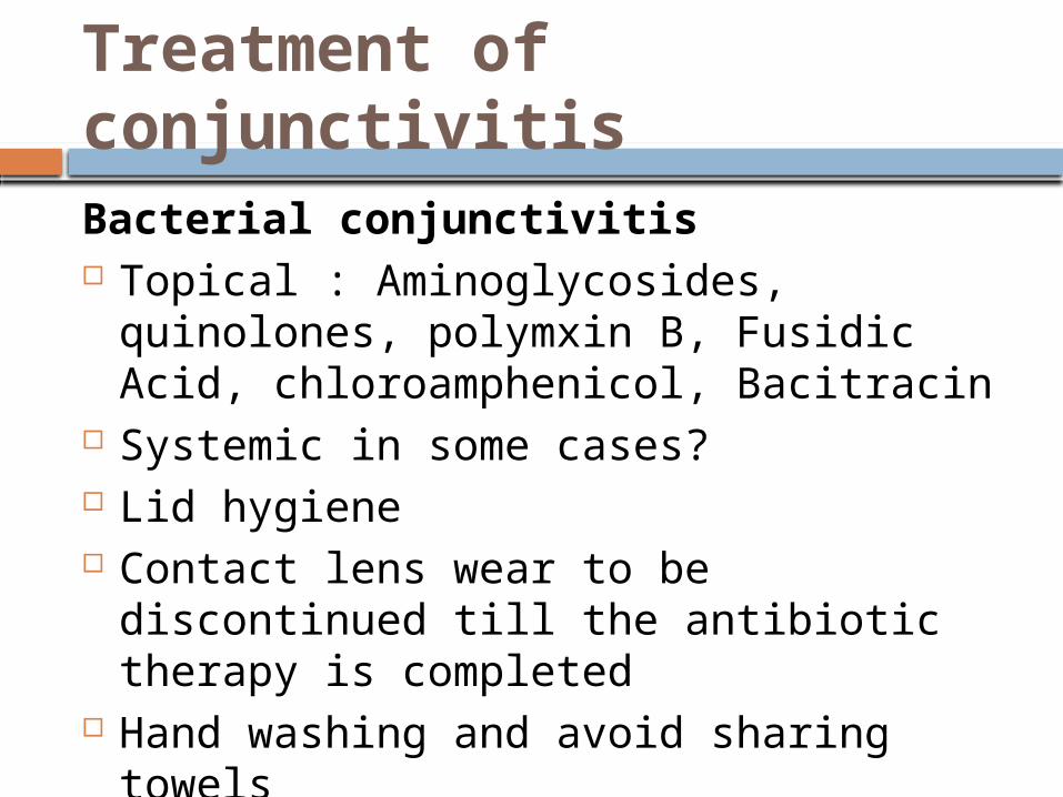
Treatment of conjunctivitisBacterial conjunctivitis Topical : Aminoglycosides, quinolones,
polymxin B, Fusidic Acid, chloroamphenicol, Bacitracin
Systemic in some cases? Lid hygiene Contact lens wear to be discontinued till
the antibiotic therapy is completed Hand washing and avoid sharing towels
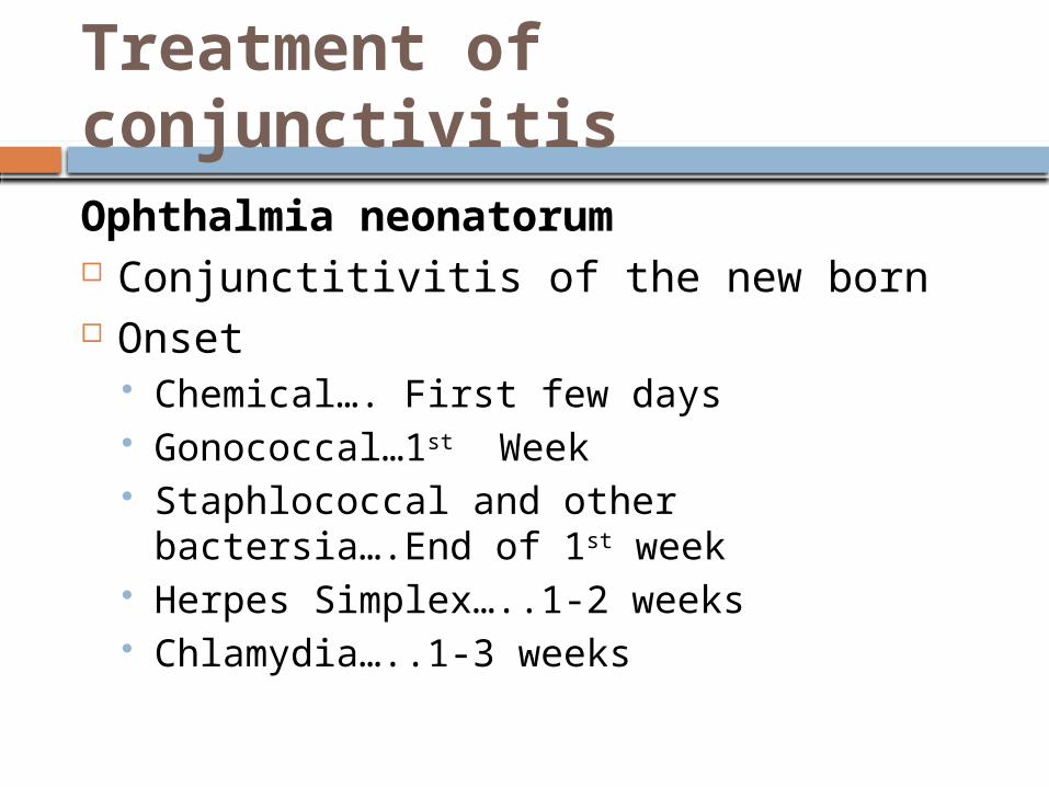
Ophthalmia neonatorum Conjunctitivitis of the new born Onset
Chemical…. First few days Gonococcal…1st Week Staphlococcal and other bactersia….End of
1st week Herpes Simplex…..1-2 weeks Chlamydia…..1-3 weeks
Treatment of conjunctivitis
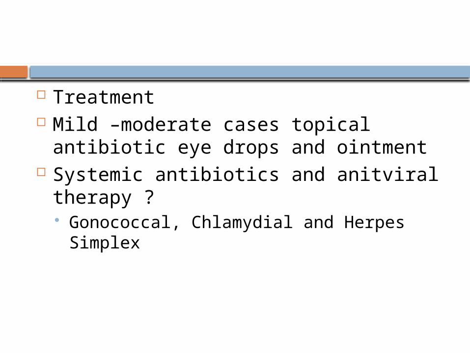
Treatment Mild –moderate cases topical antibiotic
eye drops and ointment Systemic antibiotics and anitviral
therapy ? Gonococcal, Chlamydial and Herpes
Simplex
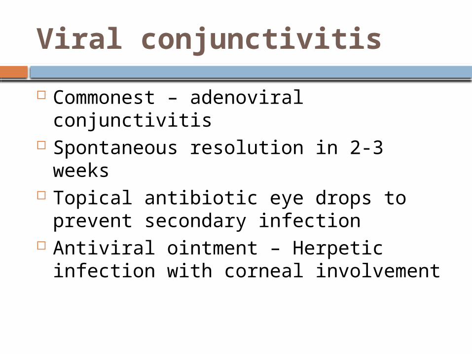
Viral conjunctivitis
Commonest – adenoviral conjunctivitis Spontaneous resolution in 2-3 weeks Topical antibiotic eye drops to prevent
secondary infection Antiviral ointment – Herpetic infection
with corneal involvement
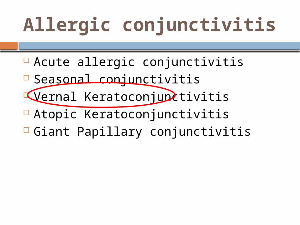
Allergic conjunctivitis
Acute allergic conjunctivitis Seasonal conjunctivitis Vernal Keratoconjunctivitis Atopic Keratoconjunctivitis Giant Papillary conjunctivitis
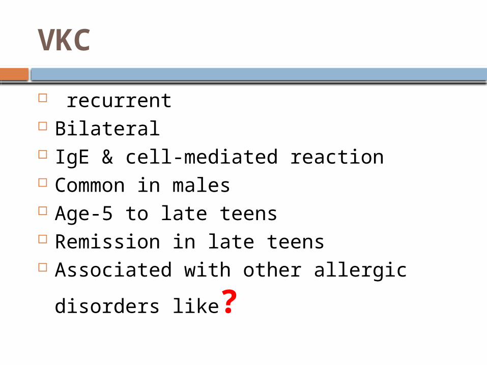
VKC
recurrent Bilateral IgE & cell-mediated reaction Common in males Age-5 to late teens Remission in late teens Associated with other allergic disorders
like?
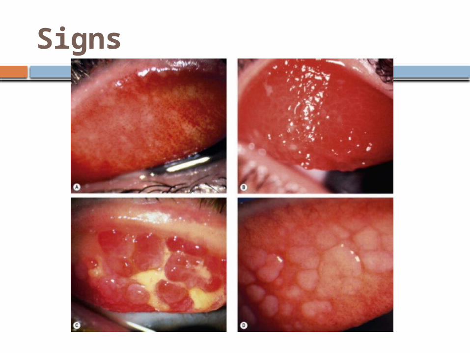
Signs
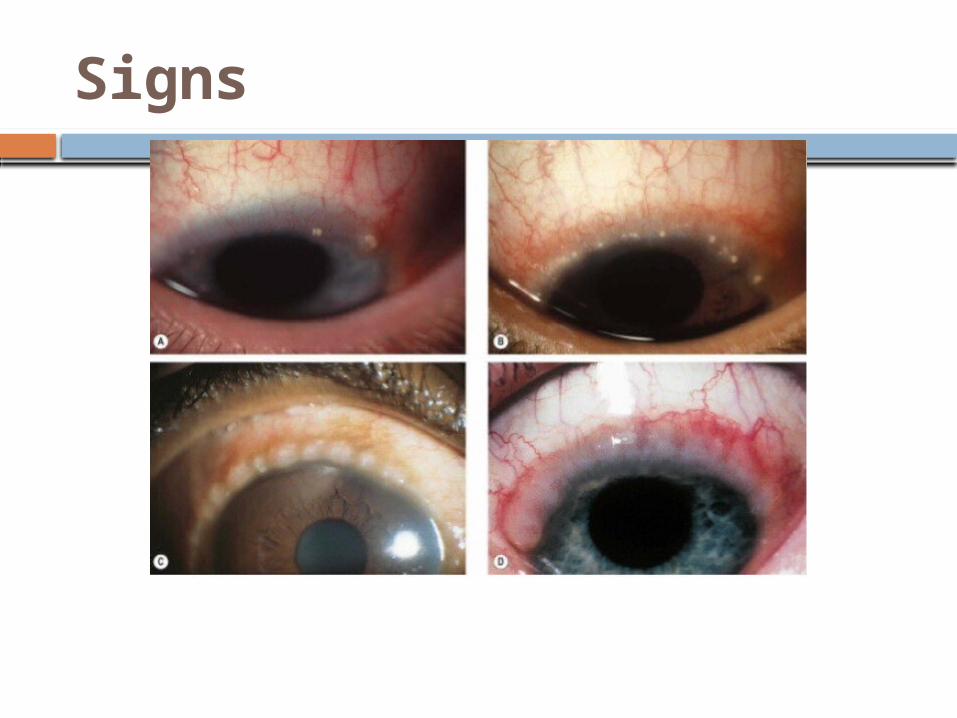
Signs
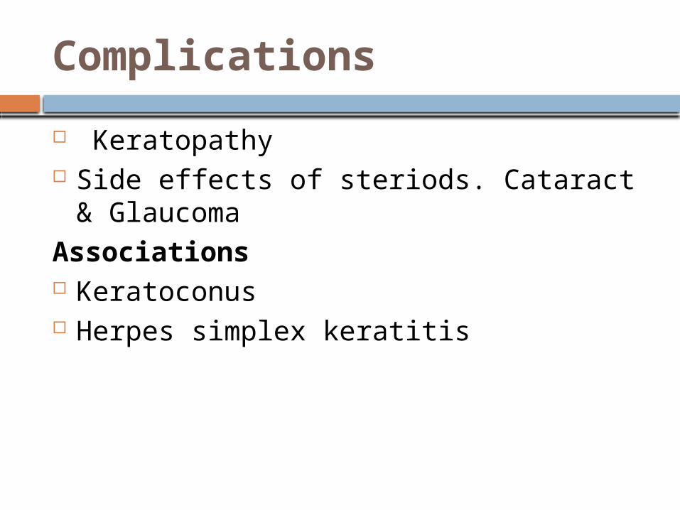
Complications
Keratopathy Side effects of steriods. Cataract &
GlaucomaAssociations Keratoconus Herpes simplex keratitis
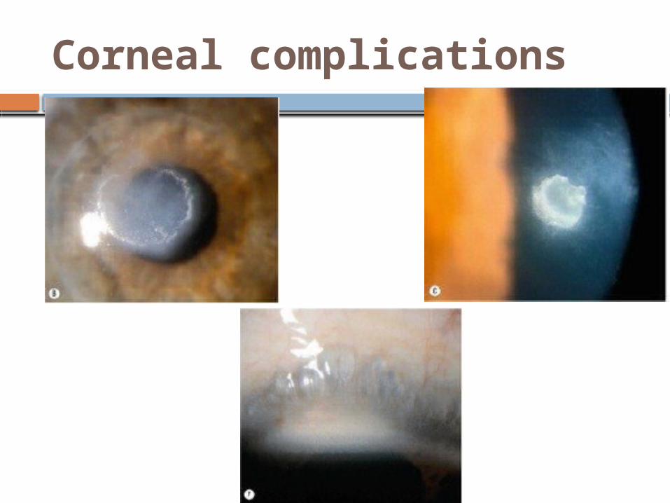
Corneal complications
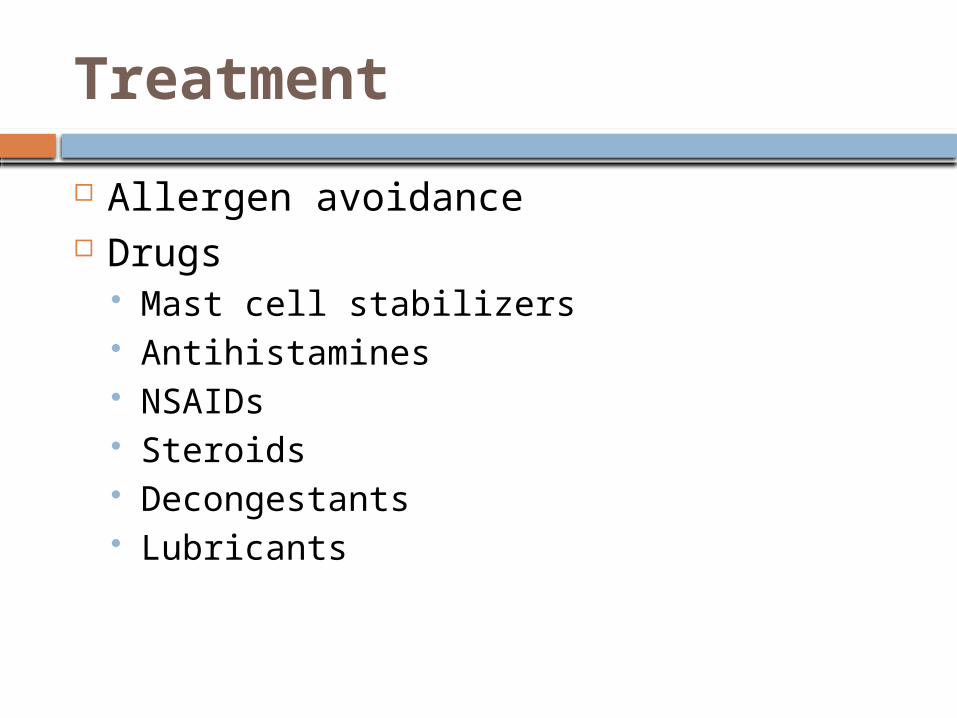
Treatment
Allergen avoidance Drugs
Mast cell stabilizers Antihistamines NSAIDs Steroids Decongestants Lubricants
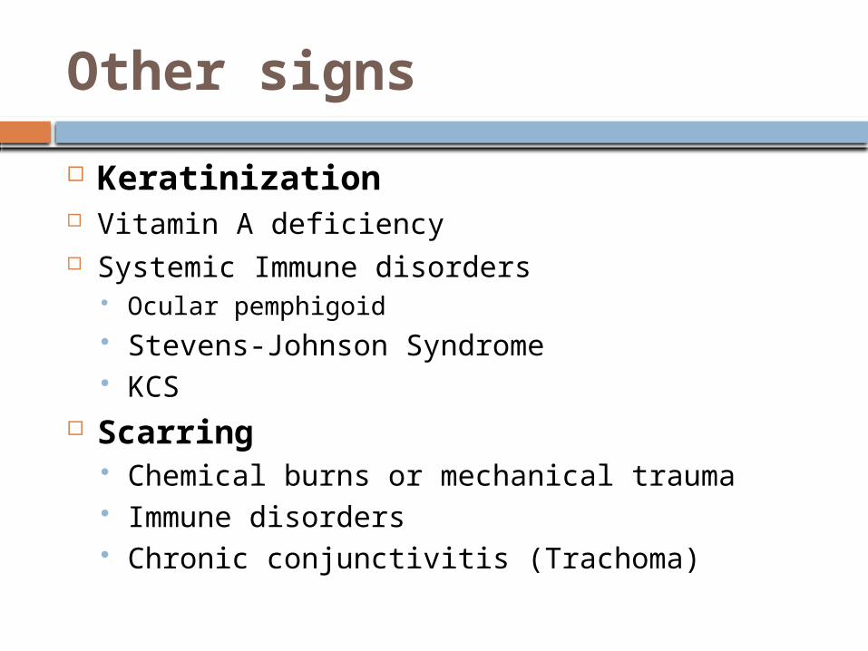
Other signs
Keratinization Vitamin A deficiency Systemic Immune disorders
Ocular pemphigoid Stevens-Johnson Syndrome KCS
Scarring Chemical burns or mechanical trauma Immune disorders Chronic conjunctivitis (Trachoma)
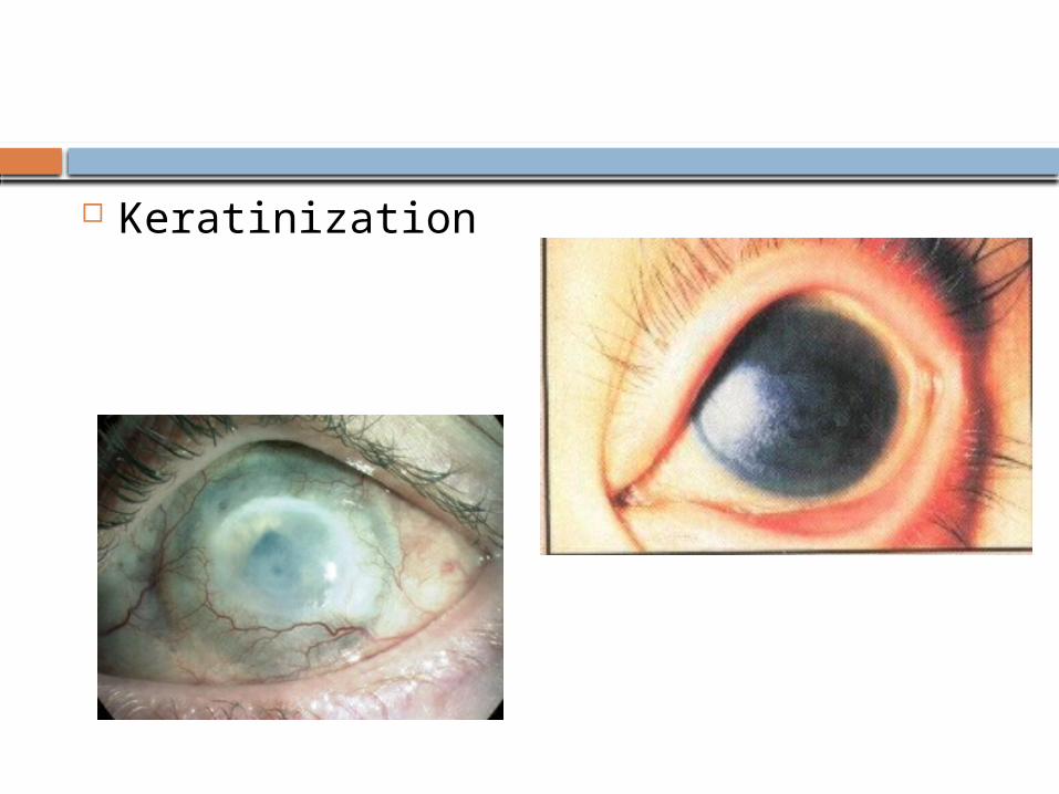
Keratinization
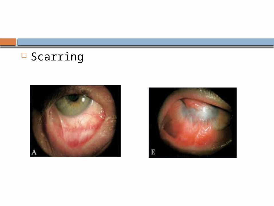
Scarring
Conjucntival Growth /mass Benign ; cysts, pterygium, lipodermoid Malignant ; melanoma, squamous cell
carcinoma and others
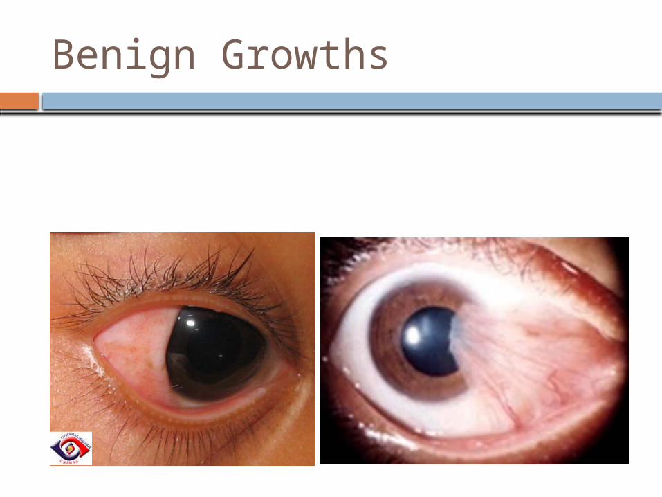
Benign Growths
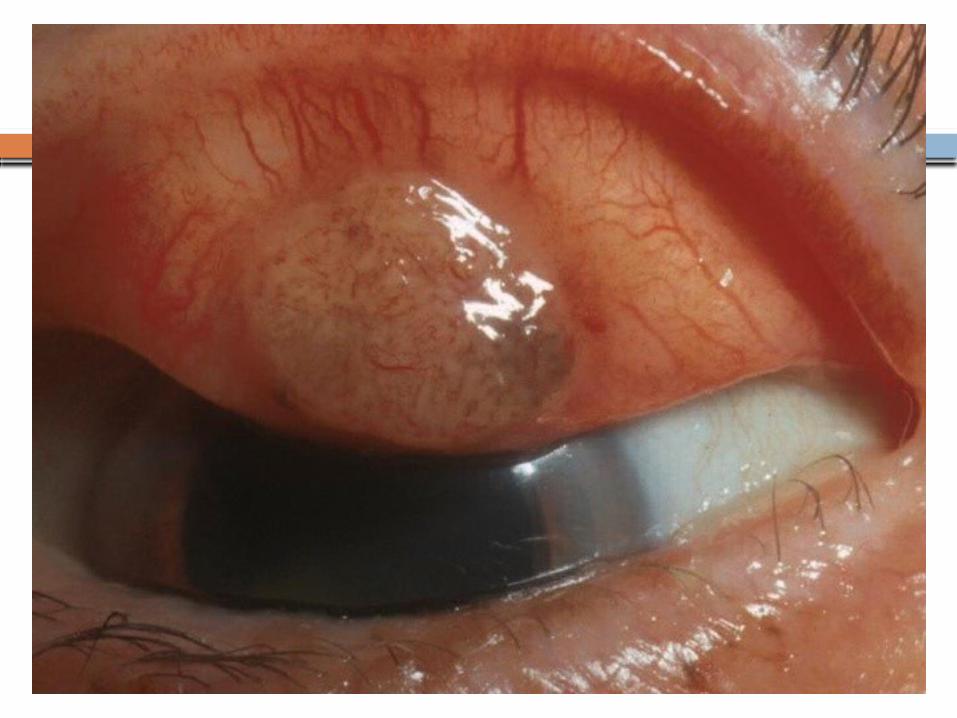
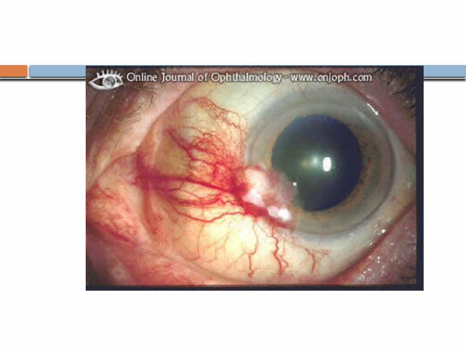
Pterygium
A degenerative condition Triangular, fibrovascular connective tissue
overgrowth of bulbar conjunctiva onto the cornea usually on the nasal side
Can reduce vision through producing Astigmatism and corneal opacity
Many treatment modalities have been tried but so far the best option with least recurrence rate is
?
Laboratory Investigations
Indications
Severe purulent conjunctivitis Follicular conjunctivitis: viral vs
chlamydial Conjunctival inflammation Neonatal conjunctivitis
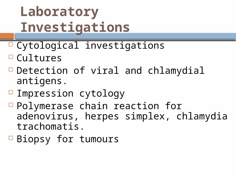
Laboratory Investigations
Cytological investigations Cultures Detection of viral and chlamydial antigens. Impression cytology Polymerase chain reaction for adenovirus,
herpes simplex, chlamydia trachomatis. Biopsy for tumours
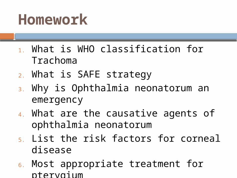
Homework
1. What is WHO classification for Trachoma
2. What is SAFE strategy3. Why is Ophthalmia neonatorum an
emergency4. What are the causative agents of
ophthalmia neonatorum5. List the risk factors for corneal disease6. Most appropriate treatment for
pterygium
Homework-Ans
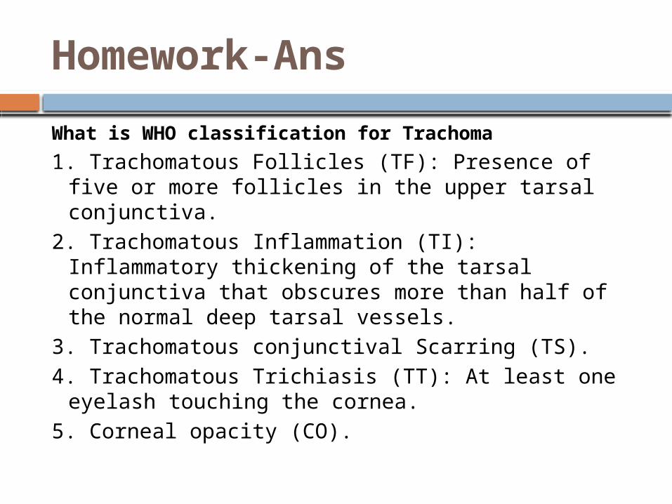
What is WHO classification for Trachoma
1. Trachomatous Follicles (TF): Presence of five or more follicles in the upper tarsal conjunctiva.
2. Trachomatous Inflammation (TI): Inflammatory thickening of the tarsal conjunctiva that obscures more than half of the normal deep tarsal vessels.
3. Trachomatous conjunctival Scarring (TS).4. Trachomatous Trichiasis (TT): At least one
eyelash touching the cornea.5. Corneal opacity (CO).
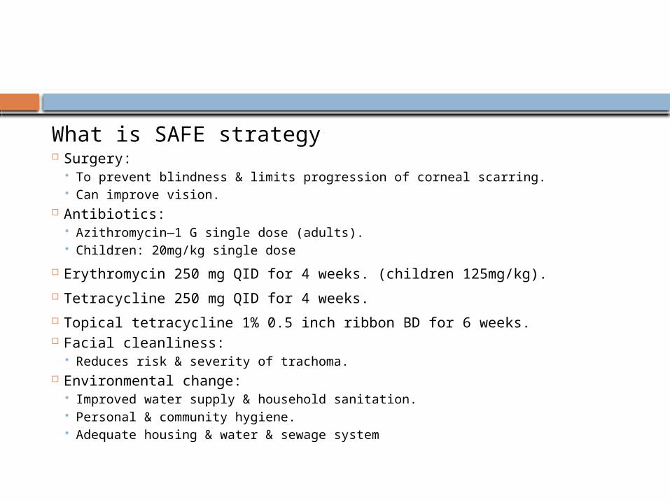
What is SAFE strategy Surgery:
To prevent blindness & limits progression of corneal scarring. Can improve vision.
Antibiotics: Azithromycin—1 G single dose (adults). Children: 20mg/kg single dose
Erythromycin 250 mg QID for 4 weeks. (children 125mg/kg).
Tetracycline 250 mg QID for 4 weeks.
Topical tetracycline 1% 0.5 inch ribbon BD for 6 weeks. Facial cleanliness:
Reduces risk & severity of trachoma. Environmental change:
Improved water supply & household sanitation. Personal & community hygiene. Adequate housing & water & sewage system
Why is Ophthalmia neonatorum an emergencyIt is considered as an ophthalmic emergency because with the immature immune system and ocular surface of the newborn the infection can result in corneal ulceration, perforation and systemic consequences. The complications that the baby can develop are; Corneal ulceration & scarringBlindness infections like
Otitis Rhinitis Pneumonitis
Death If untreated, corneal ulceration may occur in N gonorrhoeae infection and rapidly
progress to corneal perforation. When not immediately treated, Pseudomonas infection may lead to
endophthalmitis and subsequent death. Pneumonia, rhinitis and otitis has been reported with chlamydial conjunctivitis. HSV keratoconjunctivitis can cause corneal scarring and ulceration. Additionally,
disseminated HSV infection often includes central nervous system involvement
What are the causative agents of ophthalmia neonatorum Staphylococcus Pneumoniae S. Aureus Chlamydia Trachomatis Neisseiria Gonorrhoea H.influenzae Enterobacteriaceae Herpes Simplex
Chemical like silver nitrate/disinfectants used at birth
List the risk factors for corneal disease
Ocular surface diseases like lid problems (trichiasis, entropion , ectropion), lacrimal diseases ( CDC, dry eyes)
Systemic problems like Immunocompromised states & malnutrition (VAD)
Most appropriate Treatment for pterygium
Excision with conjunctival autograft


![National Library of Serbia...feronasa] bulbar conjunctiva [61 According to some references, pans of conjunctiva higher goblet cell density are Inferonasal bulbar conjunctiva, tarsal](https://img.pdfslide.us/doc/110x75/6084bbb33561423ad20313c4/national-library-of-feronasa-bulbar-conjunctiva-61-according-to-some-references.jpg)