Embed Size (px)
Citation preview

Diseases of the Circulatory System
University of San Francisco
Dr. M. Maag©2003 Margaret Maag

2
Class 8-9 Objectives Upon completion of this lesson, the student will be
able to describe the effects of aging on the cardiovascular system. investigate risk factors associated with CVD. formulate nursing interventions for CVD. determine the clinical manifestations of left and right heart
failure. discuss the etiology and manifestations of myocarditis and
pericarditis. identify the prevalence of atrial and ventricular arrhythmias.
3
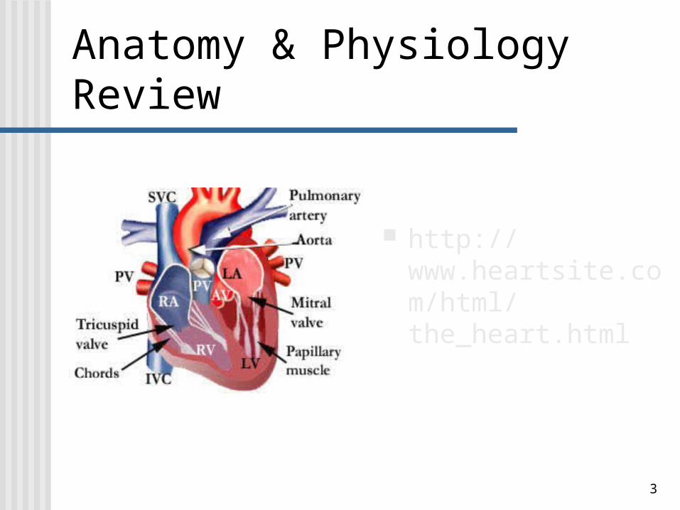
Anatomy & Physiology Review
http://www.heartsite.com/html/the_heart.html
4
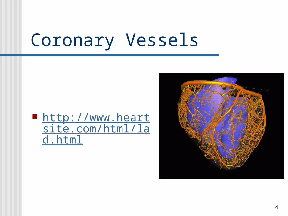
Coronary Vessels
http://www.heartsite.com/html/lad.html

5
Cardiac Conduction http://www.heartsite.c
om/html/electrical_activity.html

6
Review
How does preload, afterload & contractility influence stroke volume and determine cardiac output?
Preload: pressure created in the left ventricle at the end of diastole.
Afterload is the pressure or resistance to blood flow out of the left ventricle.
Contractility is the force of the ventricular contraction in order to eject the stroke volume. The ability of the muscle fibers to shorten (Hartshorn et al., 1997).

7
Review
Cardiac Output (HR X SV) Stroke volume: volume of blood ejected during
systole Normal adult C.O. = 5 L/min.
How does vascular resistance affect hemo dynamics?
Arterial pressure The MAP is the average pressure in arteries
Blood pressure CO times TPR

8
Arteriosclerosis
A chronic disease of the arterial system Thickening of the arterial walls and loss of
elasticity Causes a narrowing of the tunica intima that
can result in Hypertension Decreased tissue perfusion Aneurysms

9
AtherosclerosisPlaque Formation
Damaged endothelium Fatty streak formation Fibrous plaque develops Complicated lesion

10
Atherosclerosis(“fat scar” or “atheromas”)
Pathological process of arterial wall damage & occlusion of the artery with plaque formation
Plaque: accumulated monocytes & lipids at inflammatory sites along the tunica intima
Response to an arterial wall injury An inflammatory response affecting the aorta,
coronary arteries, and medium-sized arteries Leading cause of death from MI and CVAs
• 3/4 of deaths are r/t CVD

11
Atherosclerosis Risk factors
Total serum cholesterol > 240 mg/dL• LDL cholesterol > 160 mg/dL
Obesity Smoking DM Hypertension Decreased HDL Decreased estrogen levels Sedentary life style

12
Atherosclerosis Patho: local vasospasm and thrombus formation
alter the hemodynamics causing a change in flow and pressure real risk is the vulnerability to rupture thrombosis formation with subtotal or total vessel occlusion can cause angina or MI
Clinical symptoms: especially seen in the coronary, femoral, popliteal, dorsalis pedis & cerebral vessels < venous return; no pulses; skin pale & cool to
touch,distal to obstruction; paresthesia

13
Peripheral Artery Disease
Buerger’s disease, also known as “thromboangiitis obliterans” Inflammatory disease of peripheral arteries Affects the small and medium arteries and
veins of upper and lower extremities High association with tobacco use http://www.nytimes.com/2003/06/10/health/10B
ROD.html

14
Peripheral Artery Disease Raynaud Phenomenon
Local vasospasm of the small arteries• secondary to systemic diseases
• Scleroderma, pulmonary hypertension, malignancy
Raynaud Disease Primary vasospastic disorder the digit turns white, blue, red
• pain, numbness & cold sensation may be present

15
DVT
Deep Vein Thrombosis Asymptomatic, however associated with risk
factors• Venous stasis: immobility, age, left heart failure• Vessel damage: trauma, IV medications• > Coagulation: pregnancy, oral contraception,
some cancers, coagulation disorders Prevention: ambulation following surgery!
16
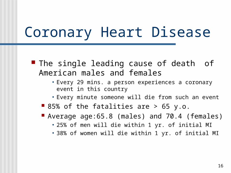
Coronary Heart Disease
The single leading cause of death of American males and females
• Every 29 mins. a person experiences a coronary event in this country
• Every minute someone will die from such an event
85% of the fatalities are > 65 y.o. Average age:65.8 (males) and 70.4 (females)
• 25% of men will die within 1 yr. of initial MI• 38% of women will die within 1 yr. of initial MI

17
Coronary Heart Disease Dyslipidemia
Disorders of lipoprotein metabolism, may be manifested by
• > total serum cholesterol• > LDL and triglycerides • < HDL cholesterol concentration
Causal relationship between > cholesterol levels and CHD.
• Cholesterol lowering Rx reduces lipid content of atherosclerotic plaque (e.g. Simvastatin)

18
Coronary Heart Disease Hyperhomocysteine
Due to a genetic lack of the enzyme that breaks down homocysteine
And/or a nutritional lack of folate, cobalamin, or pyridoxine
• < levels of folic acid, B12, B6 hampers the natural breakdown of homocysteines
Causes the arteries to narrow and harden Check serum levels Encourage a diet rich in folate and B vitamins

19
Hypertension“a cause of pump failure”
Blood pressure is the pressure exerted by the blood on the arterial walls and is a reflection of the ventricles as pumps (Hartshorn, 1997).
Mean average: 120/80
Hypertenison: consistent BP > 140/90 (adults) • staged according to severity • 140/90: watch out for children and people with
poor diet & lack of exercise (“suspicious”)

20
Hypertension
Primary: “essential” = unknown cause = 90-95% of cases At risk:
• ASHD, > age, obesity, > lipids,> glucose levels, ETOH abuse hypertension accelerates atherosclerosis & vice versa 41.4% of white females (55-64) are hypertensive 63% of black females (55-64) are hypertensive 50 million Americans (6 and older) have HBP 1 in 5 Americans has HBP Mortality: 1999 (males: 40% & females: 60%) Cigarette smoking increases risk of atherosclerosis Genetic and environmental factors

21
Hypertension
Secondary: specific to a disorder renal disease or renal artery stenosis neoplasia: Wilm’s Tumor phenochromacytoma: adrenal medulla tumor pregnancy: “eclampsia” > protein diets (> lipid levels)

22
HypertensionClinical Symptoms
Etiology: problem with C.O. & vessel resistance Patho: > large artery stiffness; backward flow of blood as
it meets > resistance Signs & Symptoms:
“Silent killer” : no signs and symptoms Some: headache, epistaxis, or orthostatic hypotension
Target organs will begin to deteriorate• cardiac failure, left ventricular hypertrophy, CVA,
PVD, renal failure, retinopathy

23
Hypertension
Prevention: BP screening; modify risk factors Tx: Early pharmacological intervention
Diuretics: Lasix, Dyazide, Spironolactone (aldosterone-receptor blocker)
Vasodilators to < peripheral resistance: Minipress or Cardizem
Ace inhibitors to interrupt RAAS: Captopril ,Vasotec• Drastically reduces the incidence of CVAs
> BP & left ventricular hypertrophy > mortality rates in both genders

24
Cerebral Vascular Accident Atherosclerotic brain infarction
Most common type of complete stroke• 61% of all “strokes” (excluding TIAs)• Approximately 4,600,000 stroke survivors • Stroke rate < by 13% from ‘89 to ‘99
• However the actual number of deaths rose 8.6%
• More common in men vs. women, except in the later years of life
• 1999 stroke mortality• male: 64,485
• female: 102,881
25
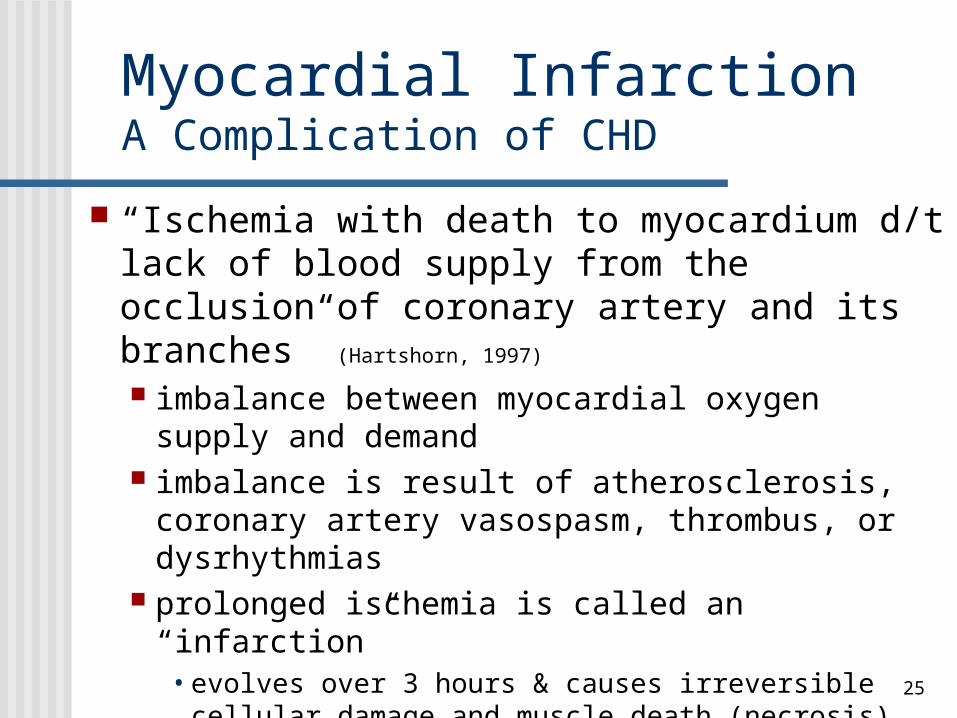
Myocardial InfarctionA Complication of CHD
“Ischemia with death to myocardium d/t lack of blood supply from the occlusion of coronary artery and its branches” (Hartshorn, 1997)
imbalance between myocardial oxygen supply and demand
imbalance is result of atherosclerosis, coronary artery vasospasm, thrombus, or dysrhythmias
prolonged ischemia is called an “infarction”• evolves over 3 hours & causes irreversible cellular
damage and muscle death (necrosis)

26
Myocardial Ischemia Angina Pectoris
• 6,400,000 Americans experience angina d/t a lack of blood flow to the heart
• 2,400,000 males and 4,000,000 females
Stable angina-• Predictable chest discomfort on exertion or under mental or
emotional stress• Generally substernal and confused with indigestion, pain in jaw,
neck, and/or shoulder• Pain is relieved with rest/nitroglycerin
Prinzmetal angina: unpredictable chest pain at rest or during sleep patterns
Silent Ischemia: no symptoms except ischemia

27
Myocardial Ischemia Leads to dysrhythmias, heart failure, sudden death ECG changes: ST depression, T wave inversion ,
and ST segment elevation Infarction leads to cell death & irreversible damage Clinical presentation: angina, vasovagal reflexes,
cool, pale, diaphoretic ECG changes: p.1015
• ischemia (st depression)• Zone of injury r/t (st elevation)• Zone of infarction/necrosis (> q wave)

28
Myocardial InfarctionClinical Symptoms
> Cardiac Enzymes d/t myocardial ischemia CK: “creatine kinase” onset = 2-6 hrs after MI LDH: “lactate dehydrogenase” = 12 hrs after MI AST: “asparate transaminase” + 6-8 hrs after MI Troponin: protein marker for early detection of MI
MI: 20 - 60% are “silent” skin is cool, clammy, pale, & diaphoretic Color of skin is dusky, ashen, hyperthermic SOB, dyspnea, tachypnea, hypotension, anxious, denial, depression, “impending doom or death”, nausea,
vomiting

29
Myocardial InfarctionSites Anterior Left Ventricle
involves LAD (40 - 50%) Inferior/Posterior wall of Left Ventricle & Right
Ventricle involves RCA (30-40%)
Lateral wall of Left Ventricle involves LCA (15 - 20%)

30
Arrhythmias
12 lead ECG is useful in identifying dysrhythmias Pattern of electrical current identifys location of
ischemia, myocardial injury & detection and confirmation of an infarct
Anginal attack: T-wave inversion; ST-segment depression; minor ST elevation when ischemia is severe and progressive
MI: Q waves; PVCs; V-tach; V-fib; Atrial flutter; atrial fib; Bundle Branch Block; second or third-degree block; sinus tach or bradycardia

31
Valvular Dysfunction Stenosis- constricted
Rheumatic heart disease Congenital Calcification
Regurgitant - insufficiency or incompetence The valve leaflets fail to shut completely Stimulates chamber dilation and myocardial
hypertrophy• Compensatory mechanism to increase the pumping of the
heart
32
Cardiac Failure Inability of the heart to contract with adequate rate & force
to pump enough blood to meet the metabolic needs of the body’s tissues circulatory failure (hypo-perfusion)
Forward effects: C. O. systolic dysfunction: inability of left heart to pump blood into
circulation results in a decreased systemic blood pressure compensation: alerts the RAAS & SNS
Backward effects: emptying of Left Ventricle diastolic dysfunction: d/t volume overload of L.V. volume in pulmonary veins & capillary bed impaired gas exchange

33
Congestive Heart Failure Left sided: failure of the left ventricle to pump the
blood received from the R side of the heart pulmonary circuit becomes congested with
blood MOST common cause is MI; Systemic
Hypertension; Cardiomyopathy S&S: Dyspnea, SaO2 decreases, RR increases,
DOE, pink-tinged sputum, acute anxiety fatigue, weakness, dizziness d/t SaO2 & C.O.,
othopnea, “Cardiac Asthma” (wheezing), crackles, ashen skin color, ck. electrolytes

34
Right heart failure Output of the right ventricle is less than the input
from the systemic venous circuit congested venous circuit ensues: poor forward flow major cause of RHF is LHF results in > central venous pressure or hepatomegaly COPD, ARDS, Cystic Fibrosis
S&S: dependent peripheral edema, enlarged spleen & liver fluid retention ascites pleural effusions, JVD, engorged venous & portal systems

35
Cardiac Inflammation
Myocarditis: forms scar tissue inflammation & injury of myocardium without ischemia caused by an infection with virus or bacterial protein that triggers
an autoimmune attack on myocardial cells CMV, HIV, Hep B, coxsackievirus TB, B-hemolytic strep, slamonella, lyme disease fungi: candidiasis, histoplasmosis, chalmydia
S&S: flu like symptoms; fatigue; dyspnea; chest pain, IDC (idiopathic
dilated cardiomyopathy), cardiac death decreases ejection fraction (15%)

36
Cardiac Inflammation
Pericarditis Inflammation of pericardial sac layers Trauma, viral, neoplasms,MI, flu, iatrogenic At risk: renal failure, radiation therapy, drugs or post-
surgical open heart Pre-load is compromised d/t inflammation
S&S: fever, severe chest pain upon deep inspiration,
pericardial effusion, pericardial friction rubs; cardiac tamponade with pulsus paradoxus : < systolic
BP during inspiration
37
References
American Heart Association. 2002 Heart and Stroke Statistical Update. Dallas, Tex.: American Heart Association; 2001.
Bullock, B. A. & Reet, L. H. (2000). Focus on Pathophysiology. Philadelphia: Lippincott.
Corwin, E.J.(2000).Handbook of pathophysiology. Baltimore: Lippincott.
Hansen, M. (1998). Pathophysiology: Foundations of disease and clinical intervention. Philadelphia: Saunders.
Hartshorn, J. C., Sole, M. L., & Lamborn, M. L. (1997).Introduction to critical care nursing.Philadelphia: Saunders.
Huether, S. E., & McCance, K. L. (2002). Pathophysiology. St. Louis: Mosby.
http://www.heartsite.com Illustrations in this presentation used are from HeartSite.com