Embed Size (px)
Citation preview

59
Clinical PRACTICE DEVELOPMENT
Wounds UK, 2008, Vol 4, No 3
Disease progression in venous and lymphovenous disease: the need for early identification and management
John Timmons, Janice Bianchi
John Timmons is Editor of Wounds UK and Clinical Nurse Specialist, Department of Tissue Viability, Aberdeen Royal Infirmary, Janice Bianchi is Lecturer, Glasgow Caledonian University, Glasgow
Oedema can exist as a result of many pathological disease processes. In patients with lymphovenous disease there is evidence that disease progresses from relatively minor signs and symptoms to more severe debilitating conditions if left untreated. When early signs are recognised and treatment is implemented at an early stage, it may be possible to prevent or slow the progression of disease. This article describes the use of a disease progression tool that helps identify early signs of disease so that early treatments can be initiated. It also looks at patient assessment and treatment options.
Patients with venous and lymphovenous disease, if allowed to progress untreated, will develop
gradually more severe signs and symptoms over time. If the early signs and stages of disease are assessed appropriately, treatment can commence at an earlier stage and may be able to stop the progression to more serious disease.
A disease progression tool has been created through a collaborative project with Activa Healthcare, Wounds UK and a team of clinical experts in the UK which allows clinicians using the chart (Figure 1) to recognise the most common skin changes seen in the various stages of venous and lymphatic disease. The chart will assist the clinician in carrying out a full patient assessment
are shown in Table 1. The table also outlines the stages of disease and the treatments which may be employed to manage the condition at each stage which will be discussed in more depth later. Skin changes and the development of chronic oedemaIn the early stages of disease patients may present with spider veins and mild swelling and as the disease progresses, the skin changes become more apparent and more severe.
Skin changes occur along the continuum of oedema. There are accompanying signs which practitioners should be aware of in order to initiate treatment and to allow timely referral to an appropriate specialist. The symptoms featured in the disease progression model will now be described in more depth.
Signs and symptoms of early diseaseSpider veinsSpider veins occur in early stages of venous disease when there is mild venous hypertension.
Mild swelling, aching, heavy legs Individuals with venous disease may notice swelling or aching in the legs if they have been standing for long periods. Exercise and leg elevation can ease symptoms. This can also occur in post-thrombotic limbs.
KEY WORDSChronic oedemaLymphatic diseaseVenous diseaseDisease progression Skin changes
and making treatment decisions, enabling disease progression to be slowed down or managed appropriately.
The idea behind the disease progression tool is to allow nurses at any level to identify the signs and symptoms of chronic oedema at each stage of the disease. Depending on the stage of the oedema, treatment may vary from simple interventions such as low-level compression hosiery and skin care to more involved treatments such as manual lymphatic drainage and bandaging techniques.
This article will explore the concept of disease progression in chronic oedema and in particular venous and lymphovenous disease, and provide guidance on patient assessment and how to use the disease progression tool to improve patient care and initiate a faster response to the disease.
The disease progression toolFigure 1 presents a potential pathway for development of disease, showing increasing severity and the need for more specialised care to treat progressive oedema. It shows the possible signs and symptoms of disease which may lead to a positive clinical diagnosis from which correct treatment can be applied.
Pictures of patients with the varying symptoms of the progressing disease
OedemaNRC.indd 1 5/9/08 13:02:36

60
Clinical PRACTICE DEVELOPMENTClinical PRACTICE DEVELOPMENT
Wounds UK, 2008, Vol 4, No 3
Figure 1. A new tool for identifying venous and lymphatic disease development.
OedemaNRC.indd 30 5/9/08 13:02:38

62
Clinical PRACTICE DEVELOPMENTClinical PRACTICE DEVELOPMENT
Wounds UK, 2008, Vol 4, No 3
Slight bulging veinsBulging veins signify potential valve incompetency within the venous system and resultant back fl ow. This swelling may disappear once the limb is elevated.
Ankle fl are (corona phlebectatica)Ankle fl are consists of a myriad of tiny vein branches that are so fi ne and so numerous that individual veins can be diffi cult to delineate. These veins give the skin a red-pink hue that blanches upon fi nger pressure. Once pressure is released, the pink colour returns immediately. This condition may be an early marker for eventual ulceration (Ruckley et al, 2002).
Dermatitis (eczema)Dermatitis is a pruritic, epidermal and dermal infl ammatory reaction of the skin. Although there can be many exogenous and endogeous causes, the
main cause in patients with venous disease are venous dermatitis and contact dermatitis. Contact dermatitis can be either caused by an irritant or an allergic reaction.
Venous dermatitisVenous dermatitis is also known as varicose eczema and venous stasis dermatitis. The reason this condition occurs is poorly understood, however underlying hypoxia due to the presence of pericapillary fi brin cuffs or arteriovenous shunts, leading to poor perfusion has been proposed (Romanelli and Romanelli, 2007).
Irritant contact dermatitis This condition can be a complication of venous dermatitis in patients with venous disease. The condition is caused by exposure to an irritant substance causing breakdown in the barrier function of the skin.
Allergic contact dermatitis Allergic contact dermatitis is also referred to as contact sensitivity. An allergen sensitises the skin through contact and when the allergen is encountered again, sensitised lymphocytes are reactivated via the skin and circulation, resulting in a reaction in the skin seen clinically as eczema (Sibbald and Cameron, 2001). This type of eczema is particularly common in patients with venous dermatitis and chronic venous leg ulcers.
Varicose veins Veins have thinner walls than arteries. When there is hypertension in the superfi cial veins they dilate and become distorted leading to varicose veins. This may be an indication of hypertension in the deep veins.
Hyperpigmentation (haemosiderin staining)Hyperpigmentation is manifest by brown staining around the gaiter area
Clinical appearance
Signs/symptoms Ankle fl areSpider veins
Venous dermatitis Open venous leg ulcer
Venous dermatitis with mild swelling
Bariatric oedema with hyperkeratosis
Stage of disease Early venous skin changes
Established venous disease/lymphatic disease
Established venous diseaseNo obvious lymphatic involvement
Early lymphovenous disease
Mid-term disease
Possible diagnosis Venous diseaseVenous insuffi ciency
Venous diseaseLymphovenousdisease
Venous disease with ulceration
Lymphovenous disease
Venous incompetence
Investigations Full assessmentABPIPulse oximetry
Full assessmentABPI Pulse oximetry LymphoscintigraphyDuplex scan
Full assessmentABPIPulse oximetryDuplex scan
Full assessmentABPI/Pulse OximetryLymphoscintigraphyDuplex scan
Full assessmentABPI/pulse oximetryDuplex scan
Treatment phase and suggestedtreatments
Early treatmentSkin careMild compression
Preventive treatmentSteroid therapyEmollientsCompression bandaging
Preventive treatmentWound dressingsCompression bandagingHosiery
Preventive treatment Steroid therapyEmollientsCompression bandagingHosiery
Preventive treatmentSteroid therapyEmollientsCompression bandagingHosiery
Table 1. Examples of presentations of skin changes, disease stage, possible diagnosis and treatment options.
OedemaNRC.indd 32 5/9/08 13:02:42

Clinical PRACTICE DEVELOPMENTClinical PRACTICE DEVELOPMENT
63Wounds UK, 2008, Vol 4, No 3
Clinical Appearance
Signs/symptoms Swelling, ulceration Cardiac oedema Venous changes
LipodermatosclerosisHyperkeratosis
Advanced skin changesHyperkeratosisSkin foldsSwollen feet
PapillomatosisHyperkeratosisSevere swellingLymphangiomata
Stage of disease Mild to moderate disease
Advanced disease Advanced disease Advanced disease
Possible diagnosis Cardiac oedema Lymphovenous disease due to dependency oedema
Primary lymphoedemaAdvanced lymphovenous disease
Advanced lymphovenous disease
Investigations Full assessmentCardiac and renal assessmentVascular assessment
Full assessmentABPI/pulse oximetryVascular assessmentMedical assessment
Full assessmentVascular assessmentLymphoscintigraphyMedical assessment
Full assessmentVascular assessmentLymphoscintigraphyMedical assessment
Treatment phase and suggested treatments
Staged introduction of mild compressionEmollientsCleansing
Intensive treatmentEmollient therapyCompression bandagingCompression hosiery Medical management
Intensive treatment Cleansing EmollientsMLDFull leg compression bandaging
Intensive treatment CleansingThick emollientsMLDFull leg compression bandaging
Table 1 continued…
of the lower leg and is common in patients with venous hypertension. It is caused by the accumulation of hemosiderin (a by-product of the breakdown of red blood cells) in the interstitial space.
Atrophie blanche Atrophie blanche is a poorly understood condition manifested by avascular or poorly vascularised areas of skin often studded with dot-like capillaries. There is no clearly defined diagnosis, as atrophie blanche can be seen in many types of vascular conditions such as vasculitic, venous disease and ar terial disease.
Symptoms of mid-term diseaseIndurationThe presence of induration indicates that the oedema present has progressed to the extent that indentations in the surface of the skin
can be noticed. In normal limbs there is rarely visible induration present and so oedema is indicated when this symptom is present.
Open ulcerationAn open ulcer is an indication that there may be underlying venous disease and/or arterial disease. There is also a risk that it could be a carcinoma. Accurate assessment and history will determine the root cause of the ulcer and treatment will be dependent on the aetiology. In almost 70% of cases there is venous disease present and there is a risk of progression of the disease if left untreated. Wound care will depend on tissue types, exudate levels and infection status. Local wound care protocols should be followed.
Severe distorted veins Severe varicose veins are large visible veins which appear bulbous and protrude from the skin on various
sites on the lower limbs. These may be associated with pain and they are a defi nite indication that there is a failure within the venous system. Patients with history of severe varicose veins may be susceptible to developing lymphovenous disease.
Chronic oedema Oedema of the limb is deemed to be chronic if it has been present for more than three months. As the condition progresses, the state of the skin will change from initial soft pitting oedema and become fibrosed, thicker and may have other signs such as hyperkeratosis and lipodermatosclerosis.
HyperkeratosisHyperkeratosis is an increased thickening of the stratum corneum resulting in thickened, scaly skin. This condition can become severe which can make treatment more difficult.
OedemaNRC.indd 33 5/9/08 13:02:47

Clinical PRACTICE DEVELOPMENTClinical PRACTICE DEVELOPMENTClinical PRACTICE DEVELOPMENT
64 Wounds UK, 2008, Vol 4, No 3 65Wounds UK, 2008, Vol 4, No 3
Signs and symptoms of long-standing diseaseSkin foldsIn very swollen limbs the extra fluid will cause stretching of the skin and in severe cases it will overhang in a pendulous fashion.
Stemmer’s signPatients with chronic oedema are likely to have a positive Stemmer’s sign (the inability to pinch the loose skin at the base of the second toe).
PapillomatosisThis is the appearance of papules or nodules which protrude from the skin giving a cobblestone appearance. This is often apparent in advanced disease.
Lymphangiomata Are small projections which may appear like blisters which are actually dilated lymphatic capillaries present in the dermis.
LymphorrhoeaIs the term given to lymphatic fluid leaking from the superficial lymphatic system which is commonly referred to as ‘wet legs’. This can be extremely uncomfortable and distressing for the patient, and although present in advanced disease, it can also be noted in patients with acute cardiac and renal failure.
Acute and chronic lipodermatosclerosis (LDS) LDS is often associated with venous insufficiency. It is characterised by skin induration and hyperpigmentation (Kirsner et al, 1993). Prolonged venous hypertension leads to an increase in size and permeability of the dermal capillaries in the area and subsequently blood macromolecules may leak into the dermis (Romanelli and Romanelli, 2007).
Acute LDS is described as tender and painful erythematous skin. This can be misdiagnosed as cellulitis.
Chronic LDS can cause the leg to change shape to resemble an inverted champagne bottle (narrow ankle with a full calf). The skin tends to be scaly with brown hyperpigmentation and indurated areas. Chronic LDS is associated with an increased risk of venous ulceration.
CellulitisCellulitis, sometimes known as erysipelas, is a bacterial-induced infection of the skin and soft tissue which is characterised by pain, erythema and swelling. The type of bacteria which causes the cellulitis may vary but commonly will be streptococcal in origin (Timmons, 2005). Patients with swollen limbs and other skin changes are particularly prone to cyclical infection and will require appropriate antibiotic therapy.
Healed ulcer Patients with healed ulcers are likely to have experienced venous disease in the past and are at risk of developing ulceration in the future unless preventive measures are taken which include ABPI monitoring, compression hosiery, emollient therapy and regular reassessment.
Assessment of the patientThe presence of oedema at any stage of venous and lymphatic disease if left unmanaged can potentially accelerate disease development and will certainly impact on the patient’s quality of life. Therefore, the primary assessment of a patient with chronic oedema at any stage involves an in-depth look at the patient’s history, a patient interview, physical examination and investigations (Billingham, 2007). Once the patient history has been taken and the skin is being assessed, the disease progression model can be applied.
The history of the patient should include a full medical history including concurrent illnesses, and details of any recent or past surgery. A patient’s drug history may reveal that they are taking medications, such as steroids, which may also be responsible for causing oedema. It will also help to highlight any cardiac problems which may be important as poor cardiac status can impact on the presence of oedema. History of other problems such as venous disease, renal disease and arthritis can also give clues as to the cause of the oedema and/or skin changes. In addition to the medical history, the history of the patient’s skin changes and swelling should be taken. This will allow the practitioner to find out how the swelling started. Key questions to aid investigations are listed in Table 2.
The patient’s skin will deteriorate over time if left untreated regardless of the root cause of the oedema. It is these changes which can give vital clues as to the duration of the condition and also which condition may be the root cause of the oedema. It is also important to find out what can influence the swelling and whether it reduces at certain times of the day and whether the swelling affects the trunk. The tool can help here by showing which stage of disease or how far along the process the patient has reached, for example in early disease, prompt identification can lead to prompt treatment and preventive measures.
Pain can also present in patients with oedema and this may be related to swelling and/or ulceration of the limb. There are a number of complicating factors which can influence the type and severity of pain experienced including vascular disease, DVT and cellulitis. A history of the pain and what affects it will allow treatments to be used which deal with the pain both through analgesia and through symptom control. Billingham (2007) recommends the McGill (Melzack, 1975) pain score as a useful method of pain assessment as it is an in-depth tool which allows a full exploration of the issues and helps to establish a more holistic ‘diagnosis’ of the pain by examining sensory, affective and evaluative aspects.
Interview Due to the complex nature and variety of causes of oedema there is a need to discuss all issues with the patient, not simply the physical problems but those of social, socioeconomic, psychological issues and their home environment.
Regardless of the age and cause of the oedema, there is potential for the patient to display signs of anxiety and depression caused by the loss of self-esteem relating to the physical appearance of the disease.
Physical examination This is necessary in order to assess the extent of the disease and reveal potential underlying causes.
The affected limb or limbs should be fully exposed to allow the change in shape to be accurately assessed. Palpation of
OedemaNRC.indd 34 5/9/08 13:02:49

Clinical PRACTICE DEVELOPMENTClinical PRACTICE DEVELOPMENT Clinical PRACTICE DEVELOPMENTClinical PRACTICE DEVELOPMENT
66 Wounds UK, 2008, Vol 4, No 3 67Wounds UK, 2008, Vol 4, No 3
the tissues should be carried out and the limb can be assessed for signs of pitting by pressing the thumb into the tissue for five seconds. Clothing can also leave marks on oedematous skin. If pitting does not appear under thumb pressure, it is likely that the oedema has been present for longer periods of time (Billingham, 2007). Looking for skin folds and abnormal limb shape will affect the treatment options and also give clues as to the level of oedema present.
Assessment of mobility and functionThe presence of oedema in the lower limbs is likely to be accompanied by a loss of mobility and function, due to excess weight, pain and inability to flex the knee. The change in limb shape prevents the action of the calf muscle pump which will reduce the amount of fluid moving up the leg. This will then prolong the oedema and the cycle continues. If function is affected other issues such as neurological impairment and cardiovascular status should be considered.
Limb measurementPatients may experience weight gain over a period of time which could be related to the swelling of the lower limb. In patients with even mild swelling it is helpful to carry out limb measurement. Specialist involvement may be required in order to accurately measure the limb volume and measurements should be recorded and reassessed at regular intervals.
Nail dystrophyIt is important to examine the patient’s feet as people with chronic oedema, regardless of the cause, may present
with changes in the nails of the feet. These changes may be cyanosis relating to vascular disease, fungal infection and thickening of the toenails relating to tinea pedis. Some of the changes may also relate to poor hygiene, reflecting the patients inability to care for their feet. Patients who have diabetes may require specialist help to address nail problems.
InvestigationsVascular assessmentWhen treating patients with chronic oedema one of the key elements of their management may be the need to wear a compression garment or to have some form of compression bandaging applied. While there is no specific guidance relating to vascular assessment in patients with chronic oedema, there is a good body of evidence to support vascular assessment in patients with leg ulceration. National guidelines recommend that this should be carried out as part of a holistic assessment in patients with leg ulcers, to exclude occult arterial disease, before commencing compression therapy (CREST, 1998; SIGN, 1998; RCN, 2006). The prevalence of arterial disease is known to increase with age and therefore may be present in a large number of patients (Burns Gough and Bradbury, 2003). Since many patients with chronic oedema are elderly, it seems appropriate that vascular assessment should be carried out before commencing compression therapy.
Blood testsBlood tests may be necessary to assist in making a full diagnosis (Table 3).
Other tests8 Doppler investigation of pulsatile flow
in the limb is becoming more useful in diagnosis and may help diagnosis of DVT
8 In some patients lymphoscintigraphy may be indicated
8 Pelvic ultrasound may be required if a tumour is suspected and further CT scan investigations may be necessary if this is positive. CT and MRI scanning are also useful in diagnosing lymphoedema and its possible aetiology.
8 Toe brachial pressure index could be used if foot pulses are difficult to detect due to excessive swelling
8 The Dopplex Assist (Huntleigh Healthcare, Luton) is a specialist hand-held device that delivers a more accurate assessment of vascular status.
Using the disease progression tool to initiate treatment When using the disease progression tool in conjunction with the other patient assessments described above, the clinician can examine the presenting signs and their likely causes and implement early treatment where appropriate.
Compression bandagesCompression therapy is used both to assist in the initial decongestive phase of treatment and in the ongoing prevention and maintenance phase to reduce limb volume and maintain limb shape. Depending on the presentation, different levels of compression can be applied. The most common forms of compression used in practice are short-stretch inelastic compression
Table 2Key questions which will help identify possible causes of oedema
8 Is the swelling bilateral or unilateral?8 How long has the oedema been present?8 Could the swelling be the result of an acute
disease process such as DVT or cellulitis?8 Are there any skin changes which would
indicate long-standing oedema?8 Does the patient have a family history
of lymphoedema?8 Has the condition been assessed and treated
in the past?
Table 3Blood tests that may be needed when assessing patients with chronic oedema
Blood test Rationale
Full blood count (Hb, platelets and white cell count)
Highlight anaemia, clotting disorders and potential infection
Liver function tests
Protein levels and serum albumin are relevant in protein deficit-related oedema
C-reactive protein Non-specific measure of inflammation which can be used to support diagnosis
Blood cultures For patients with cellulitis this will assist in choosing appropriate antibiotics
Blood biochemistry
Will identify small changes in urea, cre-atinine, potassium and sodium levels which may indicate renal symptoms
OedemaNRC.indd 36 5/9/08 13:02:50

Clinical PRACTICE DEVELOPMENTClinical PRACTICE DEVELOPMENT
68 Wounds UK, 2008, Vol 4, No 3 69Wounds UK, 2008, Vol 4, No 3
bandages and compression hosiery (Hardy, 2006).
The action of compression systems involves the reduction of capillary filtration, a movement of fluid into the non-compressed areas of the body. It is also known that compression can improve lymphatic transport and re-absorption of lymph into the lymphatic system (Partsch and Junger, 2004). Short-stretch inelastic bandaging is more appropriate for chronic oedema due to the massaging effect on the lymphatic system. In patients with venous disease or lymphovenous disease the venous pump will be assisted, allowing fluid to move up through the limb. Compression has also been associated with improved ulcer healing and improvement of the general skin condition of the patient (Moffatt, 1998).
Inelastic bandaging techniques are often employed in the intensive phase of lymphoedema management. Short-stretch inelastic bandages are able to exhibit high pressure when the patient is mobilising as well as giving low pressure when resting (Hardy, 2006). Once the oedema is reducing, cohesive inelastic bandages are commonly used since they are less prone to slippage, are less bulky and will require less frequent reapplication. The less bulky nature of these bandages can also help with patient concordance. In patients with chronic oedema the decision to use short-stretch inelastic bandaging will also take into account the vascular status of the limb and also the extent of the oedema, for example, whether there is oedema above the knee. Inelastic bandaging techniques can be used to bandage the full leg when necessary and can produce better results than when simply applying below-knee systems. The skin contact layer is a tubular bandage which can help to reduce the risk of skin reaction to the fibres in the wool layer of the bandage. When applying inelastic bandage systems close attention should be made to creating a conical shape to the limb where possible, as this allows the compression to be applied in a graduated fashion as much as possible. This type of therapy is often dramatically effective over the first seven to 14 days, after which a decision can
be made to use compression garments. Toe bandages can be applied to help reduce oedema in the digits. Specific toe bandages should be used in order to avoid damaging the digits and care should be taken in patients with vascular disease.
Compression hosiery Compression hosiery is a mainstay of prevention and maintenance therapy for most individuals (Hardy, 2006). If hosiery is not used following intensive therapy episodes, the improvements gained may be compromised.
There now exists a large range of compression hosiery garments which can be used at all stages of venous and lymphatic disease development. From stockings which exert relatively low pressures which are used to prevent oedema, to garments which can exert high pressures and are extremely effective at reducing long-standing oedema. The key issue with fitting compression hosiery is that it is the correct size and style for the patient.
For patients with the early stage skin changes (with no oedema) who require preventive treatment, the use of British Standard hosiery is more appropriate. British Standard hosiery is lighter and cosmetically acceptable, with many different styles and colours available to help with patient concordance.
The levels of compression used will be dependent on the skin changes and underlying causes. As a very general guide using the disease progression tool:8 Class 1 garments (14–17mmHg):
prevention for patients without oedema
8 Class 2 garments (18–24mmHg): prevention and early-to-medium intervention for patients without oedema
8 Class 3 (25–35mmHg): early and medium intervention for patients without oedema.
Once oedema has been diagnosed at the assessment stage and more usually after bandaging has reduced
the oedema, European Class hosiery garments are necessary to prevent recurrence or deterioration. Again the level of compression required is dependent on the skin changes present and the underlying causes of oedema. The action of European Class garments is more likely to encourage lymphatic movement and reabsorption of the lymph in the vessels. These garments are better able to ‘fight’ and contain oedema, due to their having greater stiffness that comes from the yarns and the knitting process used during manufacture. A general guide for use of the European Class hosiery is:8 European Class 1 hosiery (18–
21mmHg): prevention for patients with oedema
8 European Class 2 hosiery (23–32mmHg): early and medium intervention and intensive management for patients with oedema
8 European Class 3 hosiery (34–46mmHg): intensive management for patients with oedema.
It is important for the clinician to note the similarities in the actual compression levels between British Standard class 2 and 3 and European standard hosiery class 1 and 2. The difference between the two alternatives is mostly one of clinical performance although there are also cost considerations; therefore generally the decision needs to be made between a lighter garment for non-oedema management and a stiffer medical garment to manage the oedema. This stiffer garment also makes it easier for the stocking to accommodate shape distortion and to bridge minor skin folds which may be seen in chronic oedema.
When using the progression tool, for example. an early and medium intervention for a limb with signs and symptoms of oedema, atrophie blanche and a healed venous ulcer, would be to use a European Class 2 garment. If however, there were no indications of oedema at the assessment stage then a British Standard class 2 or 3 may be indicated.
Patient concordance is essential if compression hosiery is to have
OedemaNRC.indd 38 5/9/08 13:02:50
Clinical PRACTICE DEVELOPMENTClinical PRACTICE DEVELOPMENT
68 Wounds UK, 2008, Vol 4, No 3 69Wounds UK, 2008, Vol 4, No 3
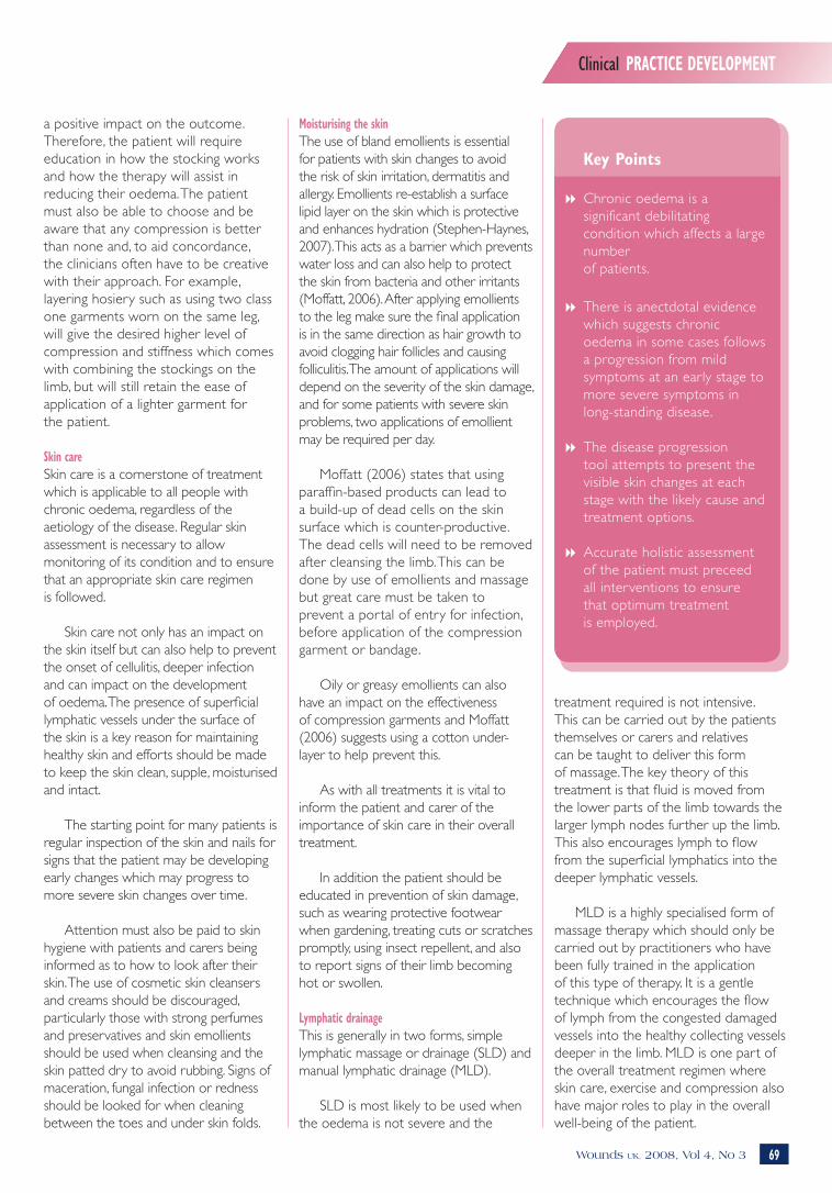
a positive impact on the outcome. Therefore, the patient will require education in how the stocking works and how the therapy will assist in reducing their oedema. The patient must also be able to choose and be aware that any compression is better than none and, to aid concordance, the clinicians often have to be creative with their approach. For example, layering hosiery such as using two class one garments worn on the same leg, will give the desired higher level of compression and stiffness which comes with combining the stockings on the limb, but will still retain the ease of application of a lighter garment for the patient.
Skin careSkin care is a cornerstone of treatment which is applicable to all people with chronic oedema, regardless of the aetiology of the disease. Regular skin assessment is necessary to allow monitoring of its condition and to ensure that an appropriate skin care regimen is followed.
Skin care not only has an impact on the skin itself but can also help to prevent the onset of cellulitis, deeper infection and can impact on the development of oedema. The presence of superficial lymphatic vessels under the surface of the skin is a key reason for maintaining healthy skin and efforts should be made to keep the skin clean, supple, moisturised and intact.
The starting point for many patients is regular inspection of the skin and nails for signs that the patient may be developing early changes which may progress to more severe skin changes over time.
Attention must also be paid to skin hygiene with patients and carers being informed as to how to look after their skin. The use of cosmetic skin cleansers and creams should be discouraged, particularly those with strong perfumes and preservatives and skin emollients should be used when cleansing and the skin patted dry to avoid rubbing. Signs of maceration, fungal infection or redness should be looked for when cleaning between the toes and under skin folds.
Moisturising the skinThe use of bland emollients is essential for patients with skin changes to avoid the risk of skin irritation, dermatitis and allergy. Emollients re-establish a surface lipid layer on the skin which is protective and enhances hydration (Stephen-Haynes, 2007). This acts as a barrier which prevents water loss and can also help to protect the skin from bacteria and other irritants (Moffatt, 2006). After applying emollients to the leg make sure the final application is in the same direction as hair growth to avoid clogging hair follicles and causing folliculitis. The amount of applications will depend on the severity of the skin damage, and for some patients with severe skin problems, two applications of emollient may be required per day.
Moffatt (2006) states that using
paraffin-based products can lead to a build-up of dead cells on the skin surface which is counter-productive. The dead cells will need to be removed after cleansing the limb. This can be done by use of emollients and massage but great care must be taken to prevent a portal of entry for infection, before application of the compression garment or bandage.
Oily or greasy emollients can also have an impact on the effectiveness of compression garments and Moffatt (2006) suggests using a cotton under-layer to help prevent this.
As with all treatments it is vital to inform the patient and carer of the importance of skin care in their overall treatment.
In addition the patient should be educated in prevention of skin damage, such as wearing protective footwear when gardening, treating cuts or scratches promptly, using insect repellent, and also to report signs of their limb becoming hot or swollen.
Lymphatic drainage This is generally in two forms, simple lymphatic massage or drainage (SLD) and manual lymphatic drainage (MLD).
SLD is most likely to be used when the oedema is not severe and the
treatment required is not intensive. This can be carried out by the patients themselves or carers and relatives can be taught to deliver this form of massage. The key theory of this treatment is that fluid is moved from the lower parts of the limb towards the larger lymph nodes further up the limb. This also encourages lymph to flow from the superficial lymphatics into the deeper lymphatic vessels.
MLD is a highly specialised form of massage therapy which should only be carried out by practitioners who have been fully trained in the application of this type of therapy. It is a gentle technique which encourages the flow of lymph from the congested damaged vessels into the healthy collecting vessels deeper in the limb. MLD is one part of the overall treatment regimen where skin care, exercise and compression also have major roles to play in the overall well-being of the patient.
Key Points
8 Chronic oedema is a significant debilitating condition which affects a large number of patients.
8 There is anectdotal evidence which suggests chronic oedema in some cases follows a progression from mild symptoms at an early stage to more severe symptoms in long-standing disease.
8 The disease progression tool attempts to present the visible skin changes at each stage with the likely cause and treatment options.
8 Accurate holistic assessment of the patient must preceed all interventions to ensure that optimum treatment is employed.
OedemaNRC.indd 39 5/9/08 13:02:50

Patients who require MLD should always be under the care of a lymphoedema specialist nurse who has undergone the appropriate training to carry this out.
Exercise The importance of exercise in patients with any level of disease cannot be over stated. Even low level passive exercise can make a small difference to the patient’s overall mobility and assist in moving fl uid from the tissues. Exercise also encourages circulation, in particular the venous return from the lower legs. Muscle movement also propels the lymphatic fl uid. The calf muscle pump is one method by which blood and lymph are forced up the limb. Changes in intra-abdominal and intra-thoracic pressure also help to create upper body pressure changes which encourage fl uid to move up the leg. It is, however, more likely that patients with oedema and related problems will not be as active as younger healthier patients. This lack of mobility will result in an increase in oedema due to dependency and gravity.
Exercise regimens should always be tailored to suit the individual patient and should not be set at a level which could cause harm. During exercise the patient should maintain compression therapy to obtain maximum benefi t.
Psychological care It is important that patients are assessed in relation to their psychological well-being which may be affected by the presence of long-term swollen limbs. The impact of the disease on the patients’ body image can be debilitating. If the patient has advanced disease with hyperkeratosis it is likely that there will be malodour associated with this, which will impact on the patient’s ability to socialise and may even cause nausea and affect their appetite. Constant leaking from their limbs will also lead to an increase in odour, with bandages becoming saturated and heavy. This can affect on the patient’s ability to mobilise, to care for themselves and to play a full part in society. Over time this may result in a loss of self-esteem.
Patients need support during this time and require optimal care to ensure that exudate or lymphorrhea is not problematic, that the limb volume is reduced and that associated symptoms are managed. Giving patients information regarding their care and treatments will allow them to become integral to their own care and this may also improve the outcomes. The Leg Club Model (Lyndsay, 2008) has been particularly successful in helping to reduce the stigma which patients face with regards leg ulceration and swollen limbs.
Pain managementPain in oedema will be an individual experience which has to be assessed on an individual basis. Routine analgesics, non-steroidal analgesics may be required and some patients with nerve-based pain may require anti-neuroleptics in addition to normal oral analgesics.
Treatment stagesPreventive treatmentWhen using the disease progression model those patients who present with very early signs of venous disease such as spider veins, atrophie blanche or very mild swelling will require preventive treatment.
This will involve the use of emollients on the skin and application of British standard hosiery class 1 (14–17mmHg) or European class 1 (18–21mmHg) if oedema is present. These garments are relatively simple to apply and can be used with other preventive measures to reduce the risk of disease progression.
Information on exercise should be given and the importance of exercise in helping to reduce swelling should be made clear to the patient, as well as the importance of nutrition and hydration.
Early and medium intervention Patients who have limbs which have progressed to exhibit signs of disease such as severe hyperkeratosis, varicose eczema with possibly a healed ulcer and more extensive swelling will require treatment which refl ects these needs.
For patients who have early to mid stage disease the full assessment
will inform the treatment choice. For some patients British Standard class 2 hosiery may be suffi cient, however, for patients with more established oedema, the use of European class hosiery will be required.
Early treatment will involve an appropriate skin care regimen, and bandaging may be required to reduce swelling. Following this a European hosiery garment may be necessary to maintain limb shape. Patients with varicose eczema may require steroid treatment which should be given as per local protocol. As with all patients, exercise, regardless of how little, should be encouraged which could mean simple ankle exercises or short walks.
Intensive treatmentIntensive treatment will be required for patients who have advanced disease exhibiting signs such as gross limb distortion, hyperkeratosis and ulceration.
Treatment of this patient group is likely to require assessment by a lymphoedema nurse specialist, manual lymphatic drainage (MLD) by a trained specialist, full leg bandaging to reduce swelling, skin care and exercise when tolerated. European class 2–3 hosiery may be required for patients who have had intensive therapy, in order to maintain limb shape and volume. The higher working pressure of these garments can ensure that the improvements made during intensive therapy can be maintained.
ConclusionThere is evidence from clinical practice that patients with venous disease can be identifi ed at an early stage and preventive treatment can be implemented. This may prevent future problems such as grossly swollen and ulcerated limbs and importantly this time can be used to educate patients in how to reduce their risk of disease progression, by teaching about good skin care, exercise and the wearing of compression garments.
The disease progression tool presented here along with the chart
Clinical PRACTICE DEVELOPMENTClinical PRACTICE DEVELOPMENT
70 Wounds UK, 2008, Vol 4, No 3 71Wounds UK, 2008, Vol 4, No 3
For further information, including an application form please email [email protected],or call 01224 637371.
OedemaNRC.indd 40 5/9/08 13:02:54
Clinical PRACTICE DEVELOPMENTClinical PRACTICE DEVELOPMENT
70 Wounds UK, 2008, Vol 4, No 3 71Wounds UK, 2008, Vol 4, No 3
(Table 1) is a simplifi ed version of the signs and symptoms which may be seen in this patient group and can allow the clinician to identify disease at any stage and implement the required treatment as outlined in this article.
References Bianchi J, Cameron J (2008) Assessment of skin integrity in the elderly (Part:1). Br J Community Nursing 13(3): s26–32
CREST (1998) Guidelines for the Assessment and Management of Leg Ulceration, Recommendations for Practice. Clinical Resource Effi ciency Support Team, Belfast
Fletcher A (1995) Epidemiology of leg ulcers. In: Cullum N, Roe B (eds) Leg Ulcers Nursing Management: A Research-based Guide. Scutari Press, London
Greenlee R, Hoyme H, Witte M, Crowe P, Witte C (1993) Developmental disorders of the lymphatic system. Lymphology 26: 156–68
Hardy D (2006) Managing long-term conditions, non cancer-related lymphoedema. Br J Nursing 15(8): 444–52
Kirsner RS, Pardes JB, Eaglstein WH et al (1993) The clinical spectrum of lipodermato-sclerosis. J Am Acad Dermatol 28: 623–7
Lyndsay E (2008) The Leg Club Model promoting the health of patients’ lower limbs through collaborative working, Wounds UK 4(2): 49–60
Melzack R (1975) The McGill pain questionnaire: major properties and scoring methods. Pain 1: 277–99
Meige H (1898) Dystrophie odemateuse hereditaire. Presse Med 6: 341–3
Milroy WF (1892) An undescribed variety of hereditary oedema. NY Med J 56: 503
Moffatt C (2007) Lymphoedema; the hidden epidemic. Br J Community Nurs 12(4): S3–S4
Mortimer PS (1995) Managing lymphoedema. Clin Exp Dermatol 20: 98–106
Royal College of Nursing (2006) The Nursing Management of Patients with Venous Leg
Ulcers, Recommendations, Clinical Practice Guidelines. RCN publishing, London
Romanelli M, Romanelli P (2007) Dermatological aspects of leg ulcers. In: Morison MJ, Moffatt CJ, Franks PJ (eds) Leg Ulcers: A Problem Based Learning Approach. Mosby Elsevier, London
Ruckley CV, Evans CJ, Allan PL et al (2002) Chronic venous insufficiency: clinical duplex correlations. The Edinburgh Vein Study of venous disorders in the general population. J Vasc Surgery 36: 520–5
Sibbald RG, Cameron J (2001) Dermatological aspects of wound care. In: Krasner DL, Rodeheaver GT, Sibbald RG (eds) Chronic Wound Care: A Clinical Source Book for Healthcare Professionals (3rd edn). HMP Communications, Wayne, PA
Scottish Intercollegiate Guidelines Network (1998) The care of patients with a chronic leg ulcer. A national clinical guideline. SIGN, Edinburgh
Timmons JP (2005) Recognising necrotising fasciitis. Wounds UK 1(2): 40–7
For further information, including an application form please email [email protected],or call 01224 637371.
WUK
OedemaNRC.indd 41 5/9/08 13:02:58