Embed Size (px)
Citation preview

SC I ENCE ADVANCES | R E S EARCH ART I C L E
GENET I CS
1Department of Immunology, St. Jude Children’s Research Hospital, Memphis, TN38105, USA. 2Blood Research Institute, Blood Center of Wisconsin, Milwaukee, WI53226, USA. 3Departments of Structural Biology and Developmental Neuro-biology, St. Jude Children’s Research Hospital, Memphis, TN 38105, USA. 4St. JudeProteomics Facility, St. Jude Children’s Research Hospital, Memphis, TN 38105,USA. 5Integrated Biomedical Sciences Program, University of Tennessee HealthScience Center, Memphis, TN 38163, USA. 6Hartwell Center for Bioinformaticsand Biotechnology, St. Jude Children’s Research Hospital, Memphis, TN 38105, USA.*Present address: Division of Rheumatology, Department of Medicine, and De-partment of Immunology, Mayo Clinic, Rochester, MN 55905, USA.†Corresponding author. Email: [email protected] (H.C.); [email protected] (D.W.); [email protected] (J.P.)
Zeng et al., Sci. Adv. 2018;4 : eaar5701 31 January 2018
Copyright © 2018
The Authors, some
rights reserved;
exclusive licensee
American Association
for the Advancement
of Science. No claim to
originalU.S. Government
Works. Distributed
under a Creative
Commons Attribution
NonCommercial
License 4.0 (CC BY-NC).
Discrete roles and bifurcation of PTEN signaling andmTORC1-mediated anabolic metabolism underlieIL-7–driven B lymphopoiesis
Hu Zeng,1* Mei Yu,2 Haiyan Tan,3,4 Yuxin Li,3,4 Wei Su,1,5 Hao Shi,1 Yogesh Dhungana,1 Cliff Guy,1Geoffrey Neale,6 Caryn Cloer,1 Junmin Peng,3,4† Demin Wang,2† Hongbo Chi1†
Dow
nloaded from
Interleukin-7 (IL-7) drives early B lymphopoiesis, but the underlying molecular circuits remain poorly under-stood, especially how Stat5 (signal transducer and activator of transcription 5)–dependent and Stat5-independent pathways contribute to this process. Combining transcriptome and proteome analyses and mousegenetic models, we show that IL-7 promotes anabolic metabolism and biosynthetic programs in pro-B cells. IL-7–mediated activation of mTORC1 (mechanistic target of rapamycin complex 1) supported cell proliferation andmetabolism in a Stat5-independent, Myc-dependent manner but was largely dispensable for cell survival orRag1 and Rag2 gene expression. mTORC1 was also required for Myc-driven lymphomagenesis. PI3K (phospha-tidylinositol 3-kinase) and mTORC1 had discrete effects on Stat5 signaling and independently controlled B celldevelopment. PI3K was actively suppressed by PTEN (phosphatase and tensin homolog) in pro-B cells to ensure prop-er IL-7R expression, Stat5 activation, heavy chain rearrangement, and cell survival, suggesting the unexpected bi-furcation of the classical PI3K-mTOR signaling. Together, our integrative analyses establish IL-7R–mTORC1–Mycand PTEN-mediated PI3K suppression as discrete signaling axes driving B cell development, with differential effectson IL-7R–Stat5 signaling.
http
on May 23, 2019
://advances.sciencemag.org/
INTRODUCTIONB lymphopoiesis is a highly ordered developmental process character-ized by the sequential rearrangements of immunoglobulin heavy (IgH)and light chain (IgL) loci that accompany differentiation of lymphoidprecursors to pro-B cells, pre-B cells, and immature B cells in the bonemarrow (BM). The stepwise progression of B cell development isdependent upon a network of transcription factors and extrinsic signals(1), especially interleukin-7 (IL-7) (2). IL-7 is produced by BM stromalcells and signals through IL-7 receptor (IL-7R; composed of the IL-7Rachain and a common gc chain) expressed on pro-B and pre-B cells topromote their proliferation, survival, and differentiation (3). Because ofits potent trophic effect on lymphocytes, IL-7 therapy has been devel-oped for clinical application. In particular, infusion of IL-7 induces amarked expansion of B cell precursors in humanpatients (4, 5), suggest-ing a strong trophic effect of IL-7 in adult human B cell precursors. Yet,the lack of full understanding of the IL-7–mediated signaling networkhinders its rational utilization in clinics (6). Despite its importance in Blymphopoiesis, how IL-7 influences pro-B cell development, especiallyabout the mechanisms linking IL-7R activation to transcriptional andtranslational events, has not been examined in a systematic manner.Furthermore, although IL-7 has been linked to lymphocyte metabolism(7–9), the detailed processes and molecular regulators are unclear.
Binding of IL-7 to IL-7R initiates phosphorylation of Janus kinase1 (Jak1) and Jak3, which recruit and activate the transcription factorStat5 (signal transducer and activator of transcription 5). IL-7R–mediated proliferation and survival of pro-B cells are criticallydependent on the Jak1/3-Stat5 signaling axis (10–13). Ectopic expres-sion of constitutively active Stat5 considerably, although incompletely,rescues B cell developmental defects in IL-7Ra–deficient mice (14),suggesting the involvement of other pathways downstream of IL-7R.Published studies indicate that phosphatidylinositol 3-kinase (PI3K)signaling mediates IL-7R signaling in pre-B cells (15), although forpro-B cell development, deletion of p110a and p110d (16), pharmaco-logical inhibition of PI3K, or deficiency of p85a does not exert a strongeffect (17). Thus, the nature and function of Stat5-independent pathwaysin IL-7R signaling, and the extent to which PI3K contributes to earlyB cell development, remain elusive. Furthermore, PI3K has beentraditionally associated with the activation of the mechanistic targetof rapamycin (mTOR) (18, 19). Despite recent genetic studies ofmTORsignaling in B cell development and function (20, 21), the upstream anddownstream regulators of mTOR and the relationship with PI3Ksignaling in B lymphopoiesis are not well understood. Moreover, howmTOR, PI3K, and Stat5 signaling are integrated is largely unknown inlymphocyte development.
To understand the proteome landscape and signaling networksmediated by IL-7 signaling, we performed temporal proteomic profilingand network analysis of IL-7–stimulated pro-B cells using multi-plexed tandem mass tag (TMT) and two-dimensional (2D) liquidchromatography–tandem mass spectrometry (LC/LC-MS/MS) (22, 23).Our proteomic profiling, together with transcriptome analysis, revealedenrichment of anabolic metabolism and activation of mTOR and Mycin IL-7–stimulated pro-B cells. Furthermore, using hCd2-iCre–mediated targeted mutagenesis (24), we genetically defined the rolesand mechanisms of mTOR and its two complexes, mTORC1 andmTORC2, as well as Myc and PI3K signaling, in early B cell develop-ment. Our results showed that mTORC1, but not mTORC2, is critical
1 of 19

SC I ENCE ADVANCES | R E S EARCH ART I C L E
D
for B lymphopoiesis, by linking IL-7R signaling to pro-B cell metabo-lism. These effects are separated fromPI3K-Akt and are independent ofStat5 signaling and cell survival, but mTORC1 activity is required forIL-7–induced Myc protein translation. Further, mTORC1 and Mycform a feed-forward circuitry in lymphomagenesis, and Raptor de-ficiency completely blocks tumor development in a model of Myc-induced B cell lymphoma (25, 26). Unexpectedly, we found that activesuppression of PI3K activity by PTEN (phosphatase and tensin homo-log) is required for proper B cell development. Uncontrolled PI3K dis-rupts pro-B cell development, associated with impaired IL-7R–Stat5signaling, reduced lineage transcription factor expression, diminishedIgH chain rearrangement, and greatly elevated apoptosis. Together,our results uncover two signaling axes, namely, PTEN-mediatedPI3K suppression and IL-7R–mTORC1–Myc, crucial for early B celldevelopment with discrete mechanisms and effects on IL-7R–Stat5signaling and immunoglobulin rearrangement, and point to the un-expected bifurcation of the conventional PI3K-mTOR signaling axis inIL-7–driven B lymphopoiesis.
on May 23, 2019
http://advances.sciencemag.org/
ownloaded from
RESULTSIL-7 activates mTOR, Myc, and anabolic metabolism inpro-B cellsCell metabolism is closely associated with lymphocyte activation andfunction, but its role in immune development remains poorly under-stood (27). To investigate metabolic regulation during B cell develop-ment, we examined mitochondria respiration and glycolysis in freshlyisolated BM B cell subsets (the gating strategy is shown in fig. S1A) bymeasuring oxygen consumption rate (OCR) and extracellular acidifica-tion rate (ECAR), respectively. B cell precursors [Lin−B220+CD43+IgM−,including fraction A, B, C, and C′ cells (28)] had the highest OCR,relative to B220+CD25+ pre-B cells, immature B cells (B220+IgM+),and circulating mature B cells (B220hiIgM+; Fig. 1A). ECAR wasmodest in all cell subsets examined, but B cell precursors still con-tained the highest level (fig. S1B). We next examined additionalmetabolic parameters, including the expression of the nutrient receptortransferrin (CD71), which correlates with cell metabolic rate (29) (fig.S1C), proliferation via 5-ethynyl-2′-deoxyuridine (EdU) incorpora-tion assay (fig. S1D), and glucose uptake, as indicated by staining withthe glucose analog 2-(N-(7-nitrobenz-2-oxa-1,3-diazol-4-yl)amino)-2-deoxyglucose (2-NBDG; fig. S1E). We found that developingB cells gradually increased CD71 expression, proliferation, and glu-cose uptake during their developmental progression from fractionA [Lin−B220+CD43+IgM−BP-1−CD24−, according to the Hardy classi-fication (30)] to fraction B (Lin−B220+CD43+IgM−BP-1−CD24+) andthen fraction C and C′ (Lin−B220+CD43+IgM−BP-1+CD24+) stage(see fig. S1A for gating), whereas pre-B cells down-regulated all theseparameters relative to fraction C/C′ cells (fig. S1, C to E). Accompany-ing the dynamic changes of metabolic activities, the expression of IL-7Ra was actively regulated during B cell development. Specifically,IL-7Ra expression was markedly increased on fraction B and frac-tion C/C′ cells compared to fraction A cells, followed by sequentialdown-regulation in later developmental stages (fig. S1F), suggestingcoordinated regulation of IL-7R expression and metabolic programsduring B cell development.
Further bioinformatics analysis of transcriptome of developing B cellsubsets in the Immunological Genome Project (ImmGen) (31) revealed2499 genes that had significantly altered expression between any twosubsets, that is, differentially expressed (DE) genes [false discovery rate
Zeng et al., Sci. Adv. 2018;4 : eaar5701 31 January 2018
(FDR) < 0.01, with at least 1.5 fold change (FC); data S1A]. We appliedweighted gene correlation network analysis (WGCNA) (32) of theseDEgenes to identify RNA clusters (RCs) of highly correlated genes, whichmay share similar biological functions or mechanisms of regulation(33). We identified nine clusters that contained at least 10 DE genes(Fig. 1B and fig. S1G). The largest cluster, RC7 (865 genes), showed agradual up-regulation from fraction A to fraction C′ cells and thendown-regulation (Fig. 1B), a pattern similar to metabolic changes andIL-7Ra expression described above. Pathway analysis revealed that RC7was enrichedwithmTORC1 signaling,Myc targets,metabolic pathways(including glycolysis, oxidative phosphorylation, and one-carbon me-tabolism), and cell cycle regulators (Fig. 1C, fig. S1H, and data S1B),further supporting the dynamic regulation ofmetabolic activities in ear-ly B cell development.
To comprehensively examine the direct roles and temporal effectsof IL-7 on the proteome landscape and metabolic programs in pro-B cells, we applied multiplexed TMT and LC/LC-MS/MS approaches(22, 23, 34) to quantify the whole proteome of freshly isolated (0 hour)or IL-7–stimulated (1, 4, and 16 hours) pro-B cells (fig. S1I). We quan-tified 7733 unique proteins. The null comparisons of the experimentalreplicates showed random distribution (fig. S1J, left), whereas compar-isons of the two time points (16 hours versus 0 hour) showed signifi-cant differences between freshly isolated and activated pro-B cells butconsistency between replicates (fig. S1J, right). Clustering analysis ofDEproteins (Fig. 1D) further verified the reproducibility of the results.We identified 854 DE proteins that significantly changed betweenany two time points using one-way analysis of variance (ANOVA;10% FDR, z score of at least 2; Fig. 1D and data S2A). We then ap-plied WGCNA for these DE proteins and identified five clusters ofcoexpressed proteins, named as whole protein clusters WPC1 toWPC5. WPC1, the largest cluster, showed late down-regulation at16 hours of IL-7 stimulation, whereas WPC2, the second largestcluster, exhibited late up-regulation at 16 hours. In addition, WPC3to WPC5 showed up-regulation at 4 hours but had distinct patterns ofexpression at 16 hours, namely, continued up-regulation (amplification),unchanged expression (plateau), or marked down-regulation (atten-uation), respectively (Fig. 1E).
Pathway enrichment analysis revealed that WPC1 contained pro-teins involved in cytoskeletal regulation, including actin-binding pro-teins and multiple integrin molecules, suggesting a potential effect ofIL-7 on cytoskeletal reorganization in pro-B cells (Fig. 1F and dataS2B). Negative regulators of the cell cycle were also identified in WPC1,including CDKN2C and CDKN2D. BCL6 (B cell lymphoma 6), atranscriptional regulator suppressed by IL-7 signaling and inducedby pre-BCR (B cell receptor) (35), was also identified in WPC1 (dataS2B).WPC2 andWPC3 contained themajority of proteins up-regulatedby IL-7. In addition to the modestly enriched Jak-Stat and IL-2–Stat5pathways, mTORC1 signaling and cholesterol homeostasis pathwayswere more significantly enriched inWPC2 andWPC3, including someof the key enzymes in lipid biosynthesis such as HMGCS1, PMVK,MVD, SQLE, IDI1, ACSL3, and FASN (data S2B). Enzymes involvedin amino acid metabolism, such as BCAT1, SLC7A5, and ASNS, werealso identified inWPC2 andWPC3 (data S2B). Furthermore, Myc tar-gets and organelle formation pathways were enriched inWPC2 (Fig. 1Fand data S2B). The heat maps of mTORC1 signaling components(Fig. 1G and fig. S1K) and Myc targets (Fig. 1H) in WPC2 and WPC3demonstrated a pronounced up-regulation of these pathways by IL-7 atthe 16-hour time point, indicating a relatively late activation kinetics.WPC4 and WPC5 showed enrichment of the E2F target pathway
2 of 19

SC I ENCE ADVANCES | R E S EARCH ART I C L E
on May 23, 2019
http://advances.sciencemag.org/
Dow
nloaded from
r
A
B
E
I
G H
F
C D
Fig. 1. Regulation of metabolic activity in developing B cells and IL-7–mediated proteome landscape in pro-B cells. (A) OCR of Lin−B220+CD43+IgM− cells, CD25+
pre-B cells (Lin−B220+CD43−IgM−CD25+), immature B cells (B220+IgM+), and circulating mature B cells (B220hiIgM+) measured using a Seahorse XF24 analyzer. (B) RC7from microarray data of common lymphoid progenitors (CLPs) and each developing B cell subset from ImmGen (gene expression relative to CLP). The central rectanglerepresents the first to third quartile, with notch in the plot corresponding to the median expression of all genes in the sample. End of the whiskers represents minimumand maximum level of expression of the genes in the cluster. (C) Functional annotations of RCs by Gene Ontology (GO), Kyoto Encyclopedia of Genes and Genomes(KEGG), and Hallmark databases (FDR < 0.2). (D) Cluster analysis of DE proteins in temporal proteomic profiling of pro-B cells stimulated with 0, 1, 4, and 16 hours of IL-7.(E) Five WPCs during pro-B cell activation were defined by WGCNA. Each line indicates the relative abundance of each protein and is color-coded by the clustermembership determined by correlation analysis between individual protein and the consensus of the WPC (see the color bar). (F) Functional annotations of WPCsby GO, KEGG, and Hallmark databases (FDR < 0.2). (G and H) IL-7–mediated temporal expression of mTORC1 signaling (G) and Myc targets (H). (I) Four interconnectedmodules of organelles and ribosome components were derived from WPC2. The number and names of proteins and the representative functional term for eachmodule are shown. See also fig. S1 and data S1 and S2.
Zeng et al., Sci. Adv. 2018;4 : eaar5701 31 January 2018 3 of 19

SC I ENCE ADVANCES | R E S EARCH ART I C L E
on May 23, 2019
http://advances.sciencemag.org/
Dow
nloaded from
and sialyltransferase activity (36), respectively, indicating dynamiceffects of IL-7 on cell cycle progression and protein glycosylation inpro-B cells. Finally, despite close connection between PI3K and mTORsignaling pathways (19), the PI3K-Akt pathway was not identified inany of the WPCs (data S2B). Thus, in response to IL-7 stimulation,pro-B cells likely undergo changes in their metabolic features associatedwith the activation of mTORC1 and Myc pathways.
We further superimposed proteins within each WPC onto theprotein-protein interaction (PPI) network to identify putative func-tional modules altered during temporal IL-7 stimulation on thebasis of the assumption that coexpressed genes/proteins are morelikely to share common molecular functions (33, 34). We identified39 modules, with sizes ranging from 2 to 35 proteins (data S2C). Thelargest module (WPC1.M5) was composed of molecules involved incell adhesion, migration, and cytoskeleton, including multiple integrinfamily members (data S2C). In WPC2, we found four interconnectedmodules (WPC2.M1 to WPC2.M4) with 86 combined componentsthat were enriched for intracellular organelle or ribosome compo-nents (Fig. 1I), suggesting that IL-7 may coordinate up-regulation oforganelle biogenesis. Collectively, our integrative network analysespredict that IL-7 likely regulates cell adhesion and migration andconcomitantly activates mTORC1 and Myc signaling, biosyntheticmetabolism, and organelle biogenesis in pro-B cells.
mTORC1, but not mTORC2, activity is critical for pro-B topre-B cell transitionWenext investigated the functional importance ofmTOR signaling in Bcell development. We crossed mice bearing conditional loxP-flankedMtor (Mtorfl/fl) allele with hCd2-iCre (Cd2icre) transgenicmice, inwhichan optimized variant of Cre recombinase (iCre) is expressed under ahuman CD2 promoter and locus control region. This transgene drivesiCre expression in lymphocyte lineages (24), leading to efficient recom-bination in all developingB cells including fractionAcells (37). Previousstudies have shown that a hypomorphic Mtor allele with constitutivebut partial loss ofmTOR activity partly blocks pro-B to pre-B transitionand reduces pre-B cells, but Cd19-Cre–driven [which initiatesincomplete target gene deletion at the pro-B/pre-B cell stage (38)] dele-tion ofMtor does not affect B cell development in the BM (39, 40). Incontrast, we observed an accumulation of B220+CD43+IgM− B cell pre-cursors (Fig. 2A) in the absence of mTOR, which was attributed toincreased fraction C/C′ cells with abnormally high expression ofBP-1 (Fig. 2B). In addition, there were few CD25+ pre-B cells (Fig. 2C)and immature and circulating mature B cells in the BM (Fig. 2D) ofCd2icreMtorfl/fl mice. Thus, the mTOR kinase is essential for early B celldevelopment.
mTOR signaling consists of mTORC1 and mTORC2 complexes.To investigate the extent to which mTORC1 and mTORC2 contrib-ute to early B cell development, we generated Cd2icreRptorfl/fl andCd2icreRictorfl/fl mice, in which Raptor and Rictor were deleted in de-veloping B cells. Flow cytometry analysis of BM cells revealed thatCd2icreRptorfl/fl mice had a significant reduction of total B220+ cells(fig. S2A) and an increased number of B220+CD43+IgM− B cell pre-cursors (Fig. 2E). Similar to Cd2icreMtorfl/fl mice, Cd2icreRptorfl/fl micehad normal numbers of fraction A and fraction B cells but signifi-cantly increased numbers of fraction C/C′ cells with unusually highexpression of BP-1 (Fig. 2F). Strikingly, B220+CD25+ pre-B cells(Fig. 2G), immature B cells (Fig. 2H), and circulating mature B cells(Fig. 2H and fig. S2B) were all severely diminished in the absence ofRaptor. Consequently, Cd2icreRptorfl/fl mice were devoid of peripheral
Zeng et al., Sci. Adv. 2018;4 : eaar5701 31 January 2018
B cells (fig. S2C), including follicular (CD21intCD23hi; fig. S2D) andmarginal zone (CD21hiCD23low) B cells in the spleen (fig. S2E). There-fore, Raptor and mTOR kinase are required for fraction C/C′ to pre-B cell transition, and their deficiencies lead to disrupted pro-B celldevelopment, a greatly diminished pre-B cell compartment, and de-velopment of profound lymphopenia of peripheral B cells.
In contrast, BM ofCd2icreRictorfl/fl mice contained a small (P > 0.05)increase of B220+CD43+IgM− B cell precursors (Fig. 2E) and a modestelevation of B220+CD25+ pre-B cells (Fig. 2G). These observations areconsistent with a recent report using Mx1-Cre–mediated systemic de-letion of Rictor (41). Within the pro-B cell compartment, Rictor defi-ciency did not substantially alter the distribution of pro-B cell subsets(Fig. 2F). Despite increased pre-B cells, Cd2icreRictorfl/fl mice had anormal immature B cell compartment (Fig. 2H), indicating that themodest perturbation during earlier B cell development did not affectimmature B cell generation. However, Cd2icreRictorfl/fl mice had signif-icantly reduced circulating mature B cells in the BM (fig. S2B) anddiminished numbers of splenic B cells, including follicular andmarginalzone B cells (fig. S2, C to E). These data indicate that mTORC2 ac-tivity is not essential for early B cell development but contributes toperipheral B cell maturation, consistent with a recent report using pan-hematopoietic and acute deletion systems (21). Combined deletion ofbothRaptor andRictor led to developmental phenotypes thatwere large-ly similar to deletion of Raptor alone (Fig. 2, E toH, and fig. S2, A to E) ormTOR (Fig. 2, A to D). Furthermore, to examine whether B cell devel-opmental defects due to Raptor deficiency were cell-intrinsic, we gener-ated mixed BM chimeras that contained 50% of WT congenic cells andfound that mTORC1-dependent B cell development was cell-intrinsic(fig. S2, F and G). Collectively, mTORC1 activity plays a predominant,intrinsic role in pro-B cell development and in the pro-B to pre-Btransition. In contrast, mTORC2 has a relatively small suppressive effecton early B cell generation but contributes to peripheral B cellmaturation.
mTORC1 mediates IL-7R signaling in a Stat5-independentbut Myc-dependent mannerWe next explored the mechanisms underlying mTORC1-mediatedearly B cell development. Raptor-deficient fraction C/C′ cells, butnot fraction A or fraction B cells, had reduced CD71 expression(Fig. 3A) and EdU incorporation (Fig. 3B). However, Raptor deficiencydid not affect cell apoptosis, as indicated by active caspase-3 staining(Fig. 3C). Similar phenotypes were observed in Cd2icreMtorfl/fl mice(fig. S3, A and B), indicating that mTORC1 impinges upon a specificset of physiological activities.
Because IL-7 is the major growth factor driving early B cell pro-liferation, we tested in vitro IL-7–induced proliferative response ofBM cells from WT and Cd2icreRptorfl/fl mice. WT BM cells prolifer-ated in response to IL-7 stimulation, but Raptor-deficient BM cellsshowed no population expansion (Fig. 3D). To circumvent thepotential complication from the altered composition of developingB cells, we purified lineage-negative hematopoietic progenitor cellsfromWTandCd2icreRptorfl/fl mice and, as controls, those fromRag-1−/−
and Jak3−/− mice and cultured them with OP9 stromal cells in thepresence of IL-7. The normal and defective B cell differentiation fromRag-1−/− and Jak3−/− progenitor cells, respectively, indicated that B celldifferentiation is independent of pre-BCR but is dependent upon IL-7Rsignaling in this experimental system (fig. S3C). Raptor-deficientprogenitor cells, similar to Jak3-deficient cells, failed to differentiate intoB cells (fig. S3C), indicating that IL-7R signaling is severely impaired inRaptor-deficient progenitor cells.
4 of 19

SC I ENCE ADVANCES | R E S EARCH ART I C L E
on May 23, 2019
http://advances.sciencemag.org/
Dow
nloaded from
01234
01234
0.00.51.01.52.02.5
0.0
0.5
1.0
1.5
0.0
0.2
0.4
0.6
0.00.51.01.52.02.5
0.00.51.01.52.02.5
A
C
E
F
G
H
D
B
Fig. 2. mTORC1, but not mTORC2, is required for pro-B cell development and pro-B to pre-B cell transition. (A to D) Representative flow cytometry plots of BM cellsfrom wild-type (WT) and Cd2icreMtorfl/fl mice. (A) Expression of B220 and CD43 on BM lymphocytes, with the percentages of B220+CD43+IgM− cells indicated (after furthergating IgM+ cells out of B220+CD43+ cells; see fig. S1A for gating scheme). Right: Number of designated populations. (B) Expression of BP-1 and CD24 on B220+CD43+IgM−
cells, with the percentages of fraction A (CD24−BP-1−), fraction B (CD24+BP-1−), and fraction C/C′ (CD24+BP-1+) cells indicated. Right: Number of indicated subset cells.(C) Expression of B220 and CD25 on BM lymphocytes, with the percentage of B220+CD25+ pre-B cells indicated. Right: Number of B220+CD25+ pre-B cells. (D) Expres-sion of B220 and IgM on BM lymphocytes, with the percentages of pro-B/pre-B cells (B220+IgM−), immature B cells (B220+IgM+), and circulating mature B cells(B220hiIgM+) indicated. Right: Number of immature and mature B cells. (E to H) Representative flow cytometry plots of BM cells of WT, Cd2icreRptorfl/fl, Cd2icreRictorfl/fl, andCd2icreRptorfl/flRictorfl/fl mice and the number of indicated populations. (E) Expression of B220 and CD43 on BM lymphocytes, with the percentage of B220+CD43+IgM− cellsindicated. (F) Hardy classification of pro-B cell subsets in the B220+CD43+IgM− gate [from (E)], with the percentages of fraction A (CD24−BP-1−), fraction B (CD24+BP-1−), andfraction C/C′ (CD24+BP-1+) cells indicated. (G) Expression of B220 and CD25 on BM lymphocytes, with the percentage of B220+CD25+ pre-B cells indicated. (H) Expression ofB220 and IgM on BM lymphocytes, with the percentages of pro-B/pre-B cells (B220+IgM−), immature B cells (B220+IgM+; statistics are on the right), and circulatingmature B cells (B220hiIgM+) indicated. NS, not significant. *P < 0.05, **P < 0.01, ***P < 0.001, ****P < 0.0001 (one-way ANOVA with Tukey’s test). Results represent at leastfour independent experiments. Data are means ± SEM. Numbers indicate percentage of cells in gates. See also fig. S2.
Zeng et al., Sci. Adv. 2018;4 : eaar5701 31 January 2018 5 of 19
SC I ENCE ADVANCES | R E S EARCH ART I C L E
on May 23, 2019
http://advances.sciencemag.org/
Dow
nloaded from
The failure of Raptor-deficient BM cells to respond to IL-7prompted us to examine the IL-7R signaling machinery. WhereasWT mice substantially up-regulated IL-7Ra expression in fractionB and C/C′ cells relative to fraction A cells, Raptor-deficient fractionB andC/C′ cells showed onlymodest up-regulation of IL-7Ra expression(fig. S3D). However, Rictor-deficient, Raptor/Rictor-deficient, andmTOR-deficient B cell precursor subsets maintained largely normalIL-7Ra expression (fig. S3, D and E). Except for the differential IL-7Ra expression, Cd2icreRptorfl/flRictorfl/fl and Cd2icreMtorfl/fl mice sharealmost identical B cell developmental defects asCd2icreRptorfl/fl mice,as described above, thereby largely excluding the possibility that thereduced IL-7Ra expression in Cd2icreRptorfl/fl mice is functionallyimportant for their B cell developmental defects.
We therefore examined IL-7–induced Stat5 activation and othersignaling events in pro-B cells. Raptor-deficient pro-B cells had largelynormal Stat5 activation in response to IL-7 stimulation (Fig. 3E). Also,IL-7–induced Stat5 nuclear translocation was not altered by mTORC1inhibition (fig. S3F). Further downstream, the IL-7R–Stat5 signalingpathway drives the expression of a number of transcription factors thatorchestrate B cell lineage differentiation; for example, TCF3 (alsoknown as E2A), through activation of Ebf1 and Pax5, promotes B celldevelopment (2, 10, 42–44). The expression of Tcf3, Ebf1, and Pax5 inRaptor-deficient pro-B cells was largely normal, indicative of compara-ble Stat5-dependent function (fig. S3G). In contrast, IL-7 induced po-tent mTORC1 activation, as indicated by phosphorylation of S6 and4EBP1 (eukaryotic translation initiation factor 4E-binding protein 1),which was completely abolished in Raptor-deficient cells (Fig. 3E). Sim-ilarly, Raptor/Rictor double deficiency and mTOR deficiency did notaffect IL-7–induced Stat5 phosphorylation but abolished mTORC1 ac-tivation (fig. S3, H and I).We further tested the role ofmTORC1 in IL-7signaling via pharmacological approaches. IL-7 treatment of in vitrocultured B cells induced rapid S6 kinase (S6K) and S6 phosphorylation,which was blocked by rapamycin. However, rapamycin treatment didnot affect IL-7–mediated Stat5 activation (Fig. 3F). Together, Raptoracts downstream of IL-7R tomediatemTORC1 activation but not Stat5signaling, suggesting a unique program orchestrated by Raptor-mTORC1in B cell development.
To directly examine the role of IL-7–mTORC1 signaling in earlyB cell development in vivo, we injected recombinant IL-7 into micetreated with or without rapamycin, followed by analysis of pro-B cellsubsets after 2 days of IL-7 treatment. Exogenous IL-7 expanded bothfractionC andC′ populations and enhancedCD71 expression (Fig. 3, GandH). Strikingly, treatment of rapamycin nearly completely blocked theexpansion of fraction C′ cells and CD71 up-regulation induced by IL-7(Fig. 3, G and H). Thus, transient inhibition of mTORC1 blocks IL-7–mediated pro-B cell developmental progression.
Recent studies establish an important role of IL-7 in B cell de-velopment in adult humans (45, 46). To test whether IL-7 can ac-tivate human pro-B cells, we sorted BM pro-B cells from healthydonors and stimulated them with IL-7. IL-7 stimulation inducedCD71 up-regulation and cell growth, indicating that IL-7 may alsoenhance cell metabolism in human pro-B cells (Fig. 3I).
The proto-oncogene Myc is essential for B cell development(26, 47, 48). Given the concomitant activation of mTORC1 and Mycin response to IL-7 stimulation (Fig. 1, G and H), we investigated theeffect of Raptor deficiency on Myc expression in developing B cellsubsets.MycmRNAwas low and comparable in fraction A cells fromWTandCd2icreRptorfl/fl mice butwas elevated in fractionB andC/C′ cellsin Raptor-deficient mice compared with WT counterparts (fig. S4A).
Zeng et al., Sci. Adv. 2018;4 : eaar5701 31 January 2018
To accurately measure Myc protein expression on a single-cell level,we crossedWT and Cd2icreRptorfl/fl mice withMycEGFP mice, which ex-press anN-terminal fusion enhanced green fluorescent protein (EGFP)–Myc protein that faithfully marks endogenous Myc expression (49).In WT cells, EGFP-Myc expression progressively increased fromfraction A to fraction C′ cells, with the highest expression in the moremature fraction C′ cells, consistent with a previous study (Fig. 3J andfig. S4B) (48). The increased EGFP-Myc expression was markedly re-duced in Raptor-deficient fraction C and C′ cells (Fig. 3J, left panel, andfig. S4B). IL-7 treatment further induced EGFP-Myc expression inWTfraction B and C/C′ cells but not in fraction A cells (Fig. 3J, right panel,and fig. S4B), in line with the expression of IL-7R in these cell subsetsdescribed above. IL-7–induced EGFP-Myc expression was greatlydiminished in the absence of Raptor in fraction C and C′ cells (Fig. 3Jand fig. S4B). Further, whereas the percentage of EGFP-Myc+ cells infraction B cells from Cd2icreRptorfl/fl mice was not significantly reducedcompared to that ofWT cells, the mean fluorescence intensity (MFI) ofEGFP-Myc in Raptor-deficient cells was significantly lower than that inWT cells (fig. S4C). Thus, although IL-7 is known to promote Myctranscription in developing B cells (50), our results identify a novelmTORC1-dependent regulation of Myc protein expression down-stream of IL-7 signaling. The functional significance of this interactionwas further verified by the drastically impaired B cell development inhCd2-iCre–mediated deletion of Myc in developing B cells (fig. S4, Dto F).Myc deficiency blocked B cell development at an earlier stage thanRaptor deficiency (fig. S4E), which may reflect, in part, the differencebetween complete and partial reduction of Myc expression in thesemodels (Fig. 3J and fig. S4, B and C). These results collectively demon-strate that mTORC1 activity is required for IL-7–induced Myc expres-sion but not for Stat5 activation in pro-B cells.
mTORC1 promotes B cell development throughtranscriptional and translational programsWe next determined mTORC1-dependent transcriptional programs.To circumvent potential secondary effects introduced by chronic defi-ciency of Raptor, we crossed Rptorfl/fl mice with Rosa26-Cre-ERT2 (aCre-ER fusion gene was recombined into the ubiquitously expressedRosa26 locus) mice to generate CreERRptorfl/fl mice, in which Rptorwas acutely deleted after injection of tamoxifen.Using the acute deletionsystem, we performed functional genomics to identify IL-7–mediatedmolecular pathways that are controlled by mTORC1. Pro-B cells fromtamoxifen-treatedCreERRptorfl/fl and control (CreERRptorfl/+)mice werecultured in the presence or absence of IL-7 for 24 hours, and RNA wassubjected to global gene expression profiling. Principal componentsanalysis showed that Raptor-deficient pro-B cells had distinct gene ex-pression profiles compared to control pro-B cells and that IL-7 had amore pronounced effect on the gene expression of control pro-B cellsthan that of Raptor-deficient cells (fig. S5A). Specifically, IL-7 treatmentof control pro-B cells resulted in a total of 1315 up-regulated and 713down-regulated probes (by greater than 0.5 log2 FC). By comparison,IL-7 treatment of Raptor-deficient cells resulted in only 452 increasedand 213 decreased probes (fig. S5B). Gene set enrichment analysis(GSEA) identified cell cycle–related genes as one of the most down-regulated pathways in Raptor-deficient cells with or without IL-7 stim-ulation (fig. S5C), consistentwith the highly reducedEdU incorporation(Fig. 3B) in Raptor-deficient pro-B cells. Furthermore, although IL-7modestly promoted the expression of cell cycle genes in Raptor-deficient pro-B cells, possibly through intact Stat5 signaling, the mag-nitude of activation was much smaller than that in control pro-B cells
6 of 19

SC I ENCE ADVANCES | R E S EARCH ART I C L E
on May 23, 2019
http://advances.sciencemag.org/
Dow
nloaded from
0.0
0.5
1.0
1.5
05101520
0.00.51.01.52.0
0
5
10
15
20
A
D
G
H
I
K
L M
N
J
E F
B C
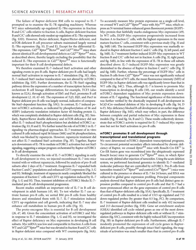
Fig. 3. Raptor-mTORC1 is essential to mediate IL-7 signaling and Myc induction but not Stat5 activation. (A) Expression of CD71 on fraction A, B, and C/C′ cellsfrom WT and Cd2icreRptorfl/fl mice. Numbers indicate the MFI of CD71 from a representative experiment. (B and C) EdU incorporation (at 1 hour after intravenousinjection of EdU) (B) and active caspase-3 staining (C) in fraction A, B, and C/C′ cells from WT and Cd2icreRptorfl/fl mice, with the percentages of EdU+ and activecaspase-3+ cells indicated from a representative experiment plotted in the graphs. (D) Cellularity of BM cells after IL-7 stimulation in vitro. Total BM cells were culturedin the presence of IL-7 (5 ng/ml) for 1 week, and cell numbers were counted on days 0, 4, and 7. (E) Immunoblot analysis of p-Stat5, p-S6, and p-4EBP1 in freshly sortedpro-B cells with or without IL-7 stimulation. (F) Immunoblot analysis of p-Stat5, p-S6K, and p-S6 in the in vitro cultured B cells. Lin− BM cells were cultured with OP9stromal cells with IL-7 for 5 days, and cells were washed and rested overnight before they were restimulated with IL-7 for the indicated time points in the presence orabsence of rapamycin. DMSO, dimethyl sulfoxide. (G) Expression of BP-1 and CD24 on BM Lin−B220+CD43+IgM− cells from mice injected with vehicle control, recom-binant IL-7 (250 mg/kg), rapamycin (15 mg/kg), or IL-7 plus rapamycin, with the percentages of fraction A, B, C, and C′ cells indicated. Right: Number of fraction C and C′cells. (H) Relative CD71 expression on fraction C cells from mice treated in (G). The MFI of CD71 from mice treated with vehicle control was set as 1. (I) Human BM pro-Bcells (CD3−CD19+CD10+CD34+CD127+) were sorted from healthy donors. Cells were stimulated with recombinant human IL-7 (10 ng/ml) for 24 hours. Expression ofCD71 (left) and cell size [measured by forward light scatter (FSC); right] were analyzed by flow cytometry. Numbers in plots indicate the MFI. (J) Frequency of EGFP-Myc+
cells in fraction A, B, C, and C′ cells from WT/MycEGFP and Cd2icreRptorfl/fl/MycEGFP mice cultured for 4 hours in medium (left) or with IL-7 (right). (K) Five RWPCs duringpro-B cell activation were defined by WGCNA. The central rectangle represents the first to third quartile, with notch in the plot corresponding to the median expressionof all proteins in the sample. End of the whiskers represents minimum and maximum level of expression of the proteins in the cluster. (L) Functional annotations ofRWPCs by Hallmark and KEGG databases (FDR < 0.1). (M and N) Rapamycin-dependent IL-7–induced temporal expression of proteins in mTORC1 signaling (M) and Myctargets (N). **P < 0.01, ***P < 0.001 [Mann-Whitney test (J) and one-way ANOVA with Tukey’s test (G and H)]. Results represent three (A to C, E, and F) or two (D, G, and I)independent experiments or are pooled from four (J) or two (G and H) independent experiments. Data are means ± SEM. Numbers indicate percentage of cells in gates.See also figs. S3 to S5 and data S3.
Zeng et al., Sci. Adv. 2018;4 : eaar5701 31 January 2018 7 of 19

SC I ENCE ADVANCES | R E S EARCH ART I C L E
on May 23, 2019
http://advances.sciencemag.org/
Dow
nloaded from
(fig. S5C). In addition to the cell cycle pathway, the cholesterol bio-synthesis pathway was also significantly down-regulated in Raptor-deficient cells (fig. S5D).
Because mTORC1 also controls gene expression at the translationallevel, as in the case of regulatingMyc expression (Fig. 3J and fig. S4, A toC), we profiled the mTORC1-dependent proteome in IL-7–stimulatedpro-B cells. The proteome of freshly isolated (0 hour), IL-7–stimulated(16 hours), or IL-7/rapamycin–treated pro-B cells was analyzed usingmultiplexed TMT and LC/LC-MS/MS proteomic profiling. Varianceanalysis revealed consistency between replicates (fig. S5E). We iden-tified that 455DEproteins (data S3A) significantly changed using one-way ANOVA (10% FDR, z score of at least 2 between IL-7 and IL-7/rapamycin treatment groups). We applied WGCNA for these proteinsand identified five clusters of coexpressed proteins, named as rapamycin-dependent whole protein clusters RWPC1 to RWPC5 (Fig. 3K and dataS3B).RWPC1andRWPC2, the two largest clusters, showedup-regulationupon IL-7 treatment but partial andmore complete suppression withrapamycin, respectively. RWPC4 also showed rapamycin-mediatedsuppression despite the lack of strong effects induced by IL-7, whereasRWPC3 and RWPC5 revealed rapamycin-mediated activation.Pathway enrichment analysis revealed that RWPC1 andRWPC2wereenriched with Myc and mTORC1 signaling targets, as well as differ-ent organelle components and multiple lipid biosynthesis pathways(Fig. 3L). The inhibitory effects of rapamycin on IL-7–inducedmTORC1and Myc activation and lipid biosynthesis were illustrated in the heatmaps (Fig. 3, M and N, and fig. S5, F and G). These data demon-strate that IL-7–mediated multiple metabolic programs may dependupon mTORC1 signaling and provide additional validations of theconclusions from Fig. 1 as well as our extensive biochemical and ge-netic analyses.
Deficiency of mTORC1 protects mice from Myc-inducedB cell lymphomaMyc is one of the most studied proto-oncogenes, whereas aberrant up-regulation of mTORC1 activity is also observed in multiple types oftumors including B cell malignancies (51). To investigate howmTORC1 regulates B cell tumorigenesis and further explore the inter-play between mTORC1 and Myc, we bred Cd2icreRptorfl/fl with Em-Myc transgenic mice, which develop B cell lymphoma induced byoverexpression of Myc via the IgH enhancer (Em) (25). Strikingly,Raptor deficiency completely suppressed Myc-induced lymphomagen-esis and early death (Fig. 4A). Lymphomas arising from Em-Myc miceare at various stages of B cell development (52). In the BM, B220+ cellsfrom most pre-tumor Em-Myc mice showed a continuous expressionof CD43, with an increased frequency of B220+CD43+IgM− B cell pre-cursors, whereas B cell development in Cd2icreRptorfl/flEm-Mycmice waslargely arrested at this precursor stage (Fig. 4B and fig. S6A). Within theB cell precursor compartment, cells from Cd2icreRptorfl/flEm-Myc miceshowed a phenotype similar to that of Cd2icreRptorfl/fl mice, with highexpression of BP-1 and an expanded fraction C/C′ cell popula-tion, whereas B cells from Em-Myc mice were either BP-1+CD24+
or BP-1−CD24+ (fig. S6, B and C). Em-Myc mice had reduced im-mature and circulating mature B cells, which was further exacerbatedin Cd2icreRptorfl/flEm-Myc mice (Fig. 4C and fig. S6D). Myc overex-pression drove increased DNA synthesis (measured by EdU incor-poration; fig. S6E), anabolic metabolism (indicated by enhancedCD71 and CD98 expression), and cell growth (measured by FSCprofile; Fig. 4D and fig. S6F), which were all largely rectified byRaptor deficiency. Consequently, Cd2icreRptorfl/flEm-Myc mice, like
Zeng et al., Sci. Adv. 2018;4 : eaar5701 31 January 2018
Cd2icreRptorfl/fl mice, had highly reduced B cells in circulation(Fig. 4E). Thus, these data demonstrate that mTORC1 signaling inearly B cell progenitors is critical for Myc-induced B cell lymphoma.
Mechanistically, Myc overexpression led to increased phosphoryl-ation of 4EBP1 but reduced phosphorylation of S6 in pro-B cells(Fig. 4F). Because IL-7–mediated Myc up-regulation was dependenton mTORC1 (Fig. 3G), we tested whether Myc overexpression inEm-Mycmicewas also dependent onmTORC1.Myc protein expressionin Cd2icreRptorfl/flEm-Myc pro-B cells was almost as low as that in WTcells, in contrast to the massive Myc expression found in Em-Myc cells(Fig. 4G). Various mTOR inhibitors also reduced Myc expression inBurkitt’s lymphoma cells (fig. S6G). In contrast, Myc mRNA levelwas similar between Em-Myc and Cd2icreRptorfl/flEm-Myc pro-B cells(fig. S6H), suggesting that mTORC1 regulates Myc protein expressionin Em-Mycmice. Furthermore,Myc protein stability was not affected bymTORC1 inhibition (fig. S6I). To directly examineMyc protein synthe-sis, we used stochastic optical reconstruction microscopy (STORM) toimage the nascent Myc protein binding to polyribosomes in IL-7–stimulated pro-B cells (Fig. 4H and fig. S6J). We found a significantreduction of Myc protein binding to polyribosomes in Raptor-deficientpro-B cells (Fig. 4H), indicating that mTORC1 activity is required forefficient Myc protein synthesis.
Translation initiation is a major step for protein synthesis and animportant target for cancer therapies (53). Eukaryotic translationinitiation complex 4F (eIF4F) is a ternary complex composed ofeIF4A, the cap-binding subunit eIF4E, and the scaffolding proteineIF4G. The binding of eIF4E to eIF4G is controlled by 4EBP1, whosephosphorylation and degradation are mediated by mTORC1 (53).How mTORC1 and Myc coordinate eIF4F activity is poorly defined(54). We found that Myc overexpression led to increased phospho-rylation of eIF4E but did not significantly affect eIF4G phosphoryl-ation (Fig. 4G). In contrast, the phosphorylation of both eIF4E andeIF4G was abolished in Cd2icreRptorfl/fl and Cd2icreRptorfl/flEm-Mycpro-B cells (Fig. 4G) or markedly suppressed by mTOR inhibition(fig. S6G), suggesting that mTORC1 activity is required for eIF4Gphosphorylation and Myc overexpression–induced eIF4E phosphoryl-ation. Together, Myc overexpression promotes B cell transformationthrough a Myc-mTORC1 feed-forward circuitry, which can beinterrupted through mTORC1 inhibition (fig. S6K).
Pro-B cell development requires active suppression of PI3KPI3K-Akt is a well-established activator of mTOR in many cell types(19). However, deletion of p110a and p110d (16), or p85a (17), doesnot affect pro-B cell development. Moreover, we observed elevated ex-pression of PTEN, a crucial negative regulator of PI3K, in pro-B cellsand pre-B cells than in other subsets (Fig. 5A), which correlated withincreased IL-7Ra expression (fig. S1F). To test the functional impor-tance of PI3K regulation, we deleted Pten using the hCd2-iCre system.Cd2icrePtenfl/fl mice contained a normal number of B220+CD43+IgM−
cells (Fig. 5B), including normal numbers of fraction A and fractionB cells, but markedly reduced fraction C/C′ cells (Fig. 5C). Further,B220+CD25+ pre-B cell percentage and number were decreased sub-stantially in the BM of Cd2icrePtenfl/fl mice (Fig. 5D). In addition, BMimmature and circulating mature B cells (fig. S7A) were all profoundlylost in the absence of PTEN. These data show that deficiency of PTENselectively blocks B cell development.
Because PTEN has nuclear functions independent of PI3K-Akt(55, 56), we used hCd2-iCre to ectopically express a constitutivelyactive form of the PI3K catalytic subunit p110a (encoded by Pik3ca)
8 of 19

SC I ENCE ADVANCES | R E S EARCH ART I C L E
on May 23, 2019
http://advances.sciencemag.org/
Dow
nloaded from
(57) in developing B cells (referred to as Pik3ca*). B cell developmentwas blocked at the pro-B cell stage, with substantial loss of fractionC/C′ cells in Cd2icrePik3ca* mice (fig. S7B), and few pre-B cellsemerged from Cd2icrePik3ca* mice (fig. S7C). Therefore, uncontrolledPI3K in pro-B cells blocks early B cell development, highlighting theimportance of active suppression of PI3K in pro-B cells.
Zeng et al., Sci. Adv. 2018;4 : eaar5701 31 January 2018
PI3K and mTORC1 independently controlB cell developmentThe above observations were incongruent with the conventional modelin which PI3K-Akt functions upstream to activate mTORC1 (19); forexample, mTORC1 is required for mediating PTEN-deficient pheno-types in other contexts including hematopoietic stem cells (58). To
0 20 40 600
50
100A
B
C
D
H
E
G
F
Fig. 4. Loss of Raptor prevents Myc-induced B cell malignancy. (A) Survival curve of Em-Myc, Cd2icreRptorfl/+Em-Myc, and Cd2icreRptorfl/flEm-Myc mice (P = 0.0005, log-rank test). (B and C) Representative flow cytometry plots of BM cells of WT, Em-Myc, Cd2icreRptorfl/fl, and Cd2icreRptorfl/flEm-Myc mice. (B) Expression of B220 and CD43 onBM lymphocytes, with the percentages of B220+CD43− and B220+CD43+ cells indicated. (C) Expression of B220 and IgM on BM lymphocytes, with the percentages ofpro-B/pre-B cells (B220+IgM−), immature B cells (B220+IgM+), and circulating mature B cells (B220hiIgM+) indicated. (D) Analysis of CD71 and CD98 expression and cellsize (via FSC) of pro-B cells. Numbers indicate the MFI. (E) Frequency of CD19+ B cells in the blood of WT, Em-Myc, Cd2icreRptorfl/fl, and Cd2icreRptorfl/flEm-Mycmice. (F) Immunoblotanalysis of p-S6 and p-4EBP1 in pro-B cells from WT, Em-Myc, Cd2icreRptorfl/fl, and Cd2icreRptorfl/flEm-Mycmice. (G) Immunoblot analysis of Myc expression and phosphorylation ofeIF4G and eIF4E in pro-B cells from WT, Em-Myc, Cd2icreRptorfl/fl, and Cd2icreRptorfl/flEm-Mycmice. (H) 3D rendering of STORM imaging of polyribosomes (red) and Myc (green) inpro-B cells treated with IL-7. Left: Representative images of unoccupied (top) and Myc-occupied polyribosome (bottom). Right: The percentage of Myc-occupied polyribosomesin each imaged cell was enumerated. Data were pooled from three independent experiments, with a total of 35 cells fromWT and 45 cells from Cd2icreRptorfl/fl mice examined.***P < 0.001, ****P < 0.0001 [one-way ANOVA with Tukey’s test (E) and Mann-Whitney test (H)]. Results represent four (B and C), three (D, F, and H), or two (E and G) independentexperiments. Data are means ± SEM. Numbers indicate percentage of cells in gates. See also fig. S6.
9 of 19

SC I ENCE ADVANCES | R E S EARCH ART I C L E
on May 23, 2019
http://advances.sciencemag.org/
Dow
nloaded from
012345
0.00.51.01.52.02.5
012345
0
1
2
3
0.0
0.5
1.0
1.5
2.0
0.0
0.5
1.0
1.5
2.0
A
C
E
F
G
H
D
B
Fig. 5. Deletion of PTEN inhibits B cell development independent of mTORC1. (A) Immunoblot analysis of PTEN in fraction A cells, pro-B cells, pre-B cells, andimmature B cells with or without IL-7 stimulation for 4 hours. (B to D) Left: Representative flow cytometry plots of BM cells of WT and Cd2icrePtenfl/fl mice. Right: Absolutenumber of indicated populations. (B) Expression of B220 and CD43 on BM lymphocytes, with the percentage of B220+CD43+ cells indicated. Right: Number of B220+CD43+IgM−
B cell precursors. (C) Expression of BP-1 and CD24 on B cell precursors, with the percentages of fraction A (CD24−BP-1−), fraction B (CD24+BP-1−), and fraction C/C′ (CD24+BP-1+)cells indicated. Right: Number of each subset. (D) Expression of B220 and CD25 on BM lymphocytes, with the percentage of B220+CD25+ pre-B cells indicated. Right: Number ofB220+CD25+ pre-B cells. (E to H) Representative flow cytometry plots of BM cells of WT, Cd2icrePtenfl/fl, Cd2icreRptorfl/fl, and Cd2icrePtenfl/flRptorfl/fl mice. (E) Expression of B220 andCD43 on BM lymphocytes, with the percentage of B220+CD43+IgM− cells indicated. Right: Number of B220+CD43+IgM− cells. (F) Hardy classification of pro-B cell subsets in theB220+CD43+IgM− gate [from (B)], with the percentages of fraction A (CD24−BP-1−), fraction B (CD24+BP-1−), and fraction C/C′ (CD24+BP-1+) cells indicated. Bottom: Numbers offraction A, B, and C/C′ cells. (G) Expression of B220 and CD25 on BM lymphocytes, with the percentage of B220+CD25+ pre-B cells indicated. Right: Number of B220+CD25+ pre-Bcells. (H) Expression of B220 and IgM on BM lymphocytes, with the percentage of pro-B/pre-B cells (B220+IgM−), immature B cells (B220+IgM+), and mature B cells (B220hiIgM+)indicated. Right: Number of immature B cells. *P < 0.05, **P < 0.01, ****P < 0.0001 (one-way ANOVA with Tukey’s test). Results represent at least four independent experiments.Data are means ± SEM. Numbers indicate percentage of cells in gates. See also fig. S7.
Zeng et al., Sci. Adv. 2018;4 : eaar5701 31 January 2018 10 of 19

SC I ENCE ADVANCES | R E S EARCH ART I C L E
on May 23, 2019
http://advances.sciencemag.org/
Dow
nloaded from
conclusively test whether PI3K modulates B cell development throughmTORC1, we crossed Cd2icrePtenfl/fl mice with Cd2icreRptorfl/fl mice togenerate Cd2icrePtenfl/flRptorfl/fl mice. Deletion of PTEN and Raptor ledto B cell developmental arrest at the pro-B cell stage associated withreduction of fraction C/C′ cells (Fig. 5, E and F) and profound loss ofB220+CD25+ pre-B cells (Fig. 5G) and immature and mature B cells(Fig. 5H). Deletion of Raptor in Cd2icrePik3ca* mice led to a pheno-type similar to that of Cd2icrePtenfl/flRptorfl/fl mice (fig. S7, D to F).Cd2icrePtenfl/flRptorfl/fl mice did not accumulate fraction C/C′ cells as ob-served in Cd2icreRptorfl/fl mice, possibly due to an earlier developmentalblock in the absence of PTEN (Fig. 5C). These results demonstrate thatloss of mTORC1 does not restore B cell development in the absence ofPTEN, and additionally, PI3K overactivation fails to rescue B cell devel-opmental block induced by Raptor deficiency. We therefore concludethat mTORC1 and PTEN-PI3K pathways independently control earlyB cell development.
PTEN promotes pro-B cell survival anddifferentiation through Foxo1-dependent andFoxo1-independent mechanismsFurther mechanistic analyses of Cd2icrePtenfl/fl mice revealed apronounced down-regulation of IL-7Ra expression, particularly in frac-tion B cells (Fig. 6A). It is well established that activation of the PI3K-Aktpathway suppresses Foxo1 activity, which is critical for IL-7Ra expres-sion and B cell development (59). To test whether impaired Foxo1 ac-tivity could account for the B cell developmental defect in Cd2icrePtenfl/fl
mice, we introduced a Rosa26-floxed stop mutant Foxo1 allele, which isrefractory to Akt-induced inhibition and thus constitutively active (de-signated as Foxo1-CA) (60), into Cd2icrePtenfl/fl mice. Examination ofIL-7Ra expression revealed that the reduced expression of IL-7Rawas largely restored in Cd2icrePtenfl/flFoxo1-CAmice (Fig. 6B). How-ever, Cd2icrePtenfl/flFoxo1-CA mice had an even lower percentage ofB220+CD43+IgM− precursor cells (Fig. 6C), with a small increase of frac-tion C/C′ cell proportion (Fig. 6D) and virtually no B220+CD25+ pre-Bcells (Fig. 6E). Thus, Foxo1 likely contributes to IL-7Ra expression butdoes not account for B cell developmental block in Cd2icrePtenfl/fl mice.
To better understand how PTEN deficiency affects pro-B cell devel-opment, we compared gene expression profiles of pro-B cells fromWTand Cd2icrePtenfl/fl mice. Ingenuity pathway analysis (IPA) showed thatmost of the top biological processes (based on P value) affected byPTEN deletion were down-regulated compared to WT cells, such aslymphocyte development and cellularity (fig. S8A). In addition, reducedcell viability was identified in PTEN-deficient pro-B cells, which wasdistinct from the prosurvival role of PI3K in mature B cells (57). Wevalidated this finding by annexin V staining (Fig. 6F). PTEN-deficientB cell precursors also expressed more proapoptotic molecule Bim(Fig. 6G), whereas cell proliferation was largely normal (Fig. 6H). In ad-dition, mRNA expression of two transcription factors critical for B celldevelopment, Myc and Myb (61), was significantly reduced in PTEN-deficient pro-B cells (Fig. 6I). Moreover, immunoblot analysis showedthat PTEN-deficient pro-B cells had impaired IL-7–induced Stat5activation (Fig. 6J), consistent with the prosurvival effect of Stat5signaling (12). As expected, PTEN-deficient pro-B cells had elevatedAkt phosphorylation and mTORC1 activation (fig. S8B). The expres-sion of Stat5-regulated genes Pax5, Ebf1, and Tcf3 was considerably re-duced in PTEN-deficient early and fraction C/C′ cells (Fig. 6K). Inaddition, IL-7–induced population expansion in vitro was reduced inPTEN-deficient BM cells (fig. S8C). Therefore, PTEN function is re-quired for the activation of Stat5, the expression of IL-7Ra and many
Zeng et al., Sci. Adv. 2018;4 : eaar5701 31 January 2018
Stat5-dependent transcription factors critical for B cell development,and the survival of pro-B cells. Deletion of PTEN selectively blocksB cell development at early to fraction C/C′ cell transition by affectingboth Foxo1-dependent and Foxo1-independent pathways.
Raptor and PTEN deficiencies impair IgH rearrangement, butenforced expression of IgH and IgL does not rescue B celldevelopmental defectsB cell development is characterized by tightly controlled rearrange-ments of IgH and IgL chains, with intracellular expression of m chaindetected after successful rearrangement of the IgH locus in pro-B cells.IgH expression depends uponmTORC1 (20). Moreover, we found thatdeficiency of mTOR (Fig. 7A) or PTEN (Fig. 7B), greatly reduced IgHexpression, indicating that heavy chain rearrangement could be im-paired. Semiquantitative RT-PCR analysis showed that the expressionof rearranged proximal VH7183-DJCm and distal VHJ558-DJm tran-scripts was substantially reduced in Raptor- and PTEN-deficient frac-tion B cells (Fig. 7, C and D). However, Raptor-deficient fraction C/C′cells had a largely normal rearrangement, indicative of a temporarydelay (Fig. 7C), whereas PTEN-deficient fraction C/C′ cells remaineddefective for IgH rearrangement (Fig. 7D). Thus, whereas bothmTORC1 and PTEN are required for initiating intracellular heavychain expression, only PTEN is essential for efficient IgH rearrange-ment in fraction C/C′ cells. Mechanistically, Ig rearrangement dependsupon the induction of Rag1 and Rag2 expression. PTEN-deficient Bcells, but not Raptor-deficient B cells, had highly reduced Rag1 andRag2 expression (fig. S9, A and B), which may partly explain the differ-ential defects in IgH rearrangement between these two genetic models.
To test whether the impaired IgH and IgL expression accounts forB cell developmental defects inRaptor-deficient orPik3ca*-expressingpro-B cells, we attempted to introduce rearranged IgH and IgL bycrossing Cd2icreRptorfl/fl or Cd2icrePik3ca* mice with IgHEL mice,which carry a rearranged IgM/IgD transgene specific for the hen egglysozyme (62). The IgHEL transgene was successfully introduced intoCd2icreRptorfl/fl mice (fig. S9C), but B cells failed to develop into circu-lating mature B cells (fig. S9D) in the BM or migrate to periphery(fig. S9E). In contrast, Cd2icrePik3ca*IgHEL mice expressed very littleIgHEL transgene (Fig. 7E), and there were few mature B cells in theBM (Fig. 7F) or the spleen (Fig. 7G). The marked contrast betweenCd2icreRptorfl/flIgHEL and Cd2icrePik3ca*IgHEL mice indicated thatwhereas loss of mTORC1 permits IgH transgene expression, suppres-sion of PI3K is critically required for IgH expression. Furthermore,because the IgHEL transgene is able to rescue B cell development inRag2-deficient mice (63), these results suggest that mechanisms otherthan defective Rag gene expression contribute to B cell developmentaldefects in Cd2icrePtenfl/fl mice. Finally, successful IgHEL expressioncannot rescue B cell development in the absence of Raptor, indicatingthat mTORC1 is likely important for receptor-mediated downstreamevents.
DISCUSSIONDespite the well-recognized roles of IL-7R and Stat5 signaling in B celldevelopment, the importance and mechanism of Stat5-independentpathways are poorly defined, and the global impact of IL-7 on pro-Bcells is obscure. Further, how IL-7R–Stat5 signaling is regulated andthe function of PI3K in pro-B cell development remain unclear. Here,we combined systems biology and mouse genetic models to investigateIL-7–dependent signaling programs in pro-B cells and dissect the
11 of 19

SC I ENCE ADVANCES | R E S EARCH ART I C L E
on May 23, 2019
http://advances.sciencemag.org/
Dow
nloaded from
0.0
0.5
1.0
1.5
0.0
0.5
1.0
1.5
2.0
0.0
0.5
1.0
1.5
2.0
α
0.0
0.5
1.0
1.5
0.0
0.5
1.0
1.5
α
A F
G
H
B
C
D
E
I J K
Fig. 6. PTEN promotes early B cell development through Foxo1-dependent and Foxo1-independent mechanisms. (A) Expression of IL-7Ra on fraction A, B, andC/C′ cells of WT and Cd2icrePtenfl/fl mice. Numbers indicate the MFI. (B to E) Representative flow cytometry plots of BM cells from WT, Cd2icrePtenfl/fl, Cd2icrePtenfl/+Foxo1-CA,and Cd2icrePtenfl/flFoxo1-CA mice. (B) Expression of IL-7Ra in pro-B cells. Numbers indicate the MFI. (C) Expression of B220 and CD43 on BM lymphocytes, with the per-centage of B220+CD43+ and B220+CD43− cells indicated. (D) Hardy classification of pro-B cell subsets in the B220+CD43+IgM− gate (from C), with the percentages offraction A (CD24−BP-1−), fraction B (CD24+BP-1−), and fraction C/C′ (CD24+BP-1+) cells indicated. (E) Expression of B220 and CD25 on BM lymphocytes, with the percentageof B220+CD25+ pre-B cells indicated. (F) Analysis of apoptotic cell death via annexin V staining on fraction A, B, and C/C′ cells. (G) Expression of Bim in fraction A, B, and C/C′ cellsof WT and Cd2icrePtenfl/fl mice. Numbers indicate the MFI. (H) EdU incorporation in fraction A, B, and C/C′ cells of WT and Cd2icrePtenfl/fl mice. (I) Reverse transcription polymerasechain reaction (RT-PCR) analysis of Myc and Myb expression in pro-B cells. (J) Immunoblot analysis of p-Stat5 in pro-B cells of WT and Cd2icrePtenfl/fl mice. (K) RT-PCR analysis ofPax5, Ebf1, and Tcf3 expression in fraction B and C/C′ cells of WT and Cd2icrePtenfl/fl mice. *P < 0.05, **P < 0.01, ***P < 0.001 (Mann-Whitney test for cell frequencies and unpairedStudent’s t test for cell numbers). Data represent four (A and F), three (J), or two (B to E, G to I, and K) independent experiments. Data are means ± SEM. Numbers indicatepercentage of cells in gates. See also fig. S8.
Zeng et al., Sci. Adv. 2018;4 : eaar5701 31 January 2018 12 of 19

SC I ENCE ADVANCES | R E S EARCH ART I C L E
on May 23, 2019
http://advances.sciencemag.org/
Dow
nloaded from
functions of PI3K, mTORC1, and mTORC2 during early B cell devel-opment. Our comprehensive transcriptome and proteome analysesshow that pro-B cell development is characterized by IL-7–mediatedanabolic metabolism, organelle biogenesis, and mTORC1 and Mycactivation. Deficiency of mTORC1 arrested B cell development mainlyat the fraction C/C′ cell stage and disrupted pro-B cell subsets but with-out affecting Stat5 activation, B cell lineage transcription factor expres-sion, or cell survival. In contrast, overactivation of PI3K due to PTENdeficiency blocked early B cell development at the fraction B stage,associated with impaired IL-7R–Stat5 signaling, reduced expressionof B cell lineage transcription factors, and excessive apoptosis. Addition-al analyses of compoundmutantmice in these two pathways allowed usto genetically dissociate mTORC1 and PI3K signaling, a conventionalupstream signal of mTORC1. Notably, deletion of mTORC2 led to amodest increase of pro-B and pre-B cells but a normal immature Bcell compartment. The B cell developmental defects in the absence ofRaptor were largely phenocopied by loss of both mTORC1 andmTORC2, ormTOR itself, suggesting a predominant role ofmTORC1,but not mTORC2, in early B cell development. Therefore, we havegenetically defined two distinct signaling axes, mTORC1-Myc andPTEN-mediated PI3K suppression, crucial for early B cell development.Finally, mTORC1 promotes B cell development by activating Myc,and mTORC1 and Myc form a feed-forward circuitry to drive B celllymphoma. Therefore, mTORC1 is a crucial regulator of both B celldevelopment and malignancy and could serve as a legitimate thera-peutic target.
The function of IL-7 in humanB cell development appears to be age-dependent. Earlier studies on infant patients with severe combined im-
Zeng et al., Sci. Adv. 2018;4 : eaar5701 31 January 2018
munodeficiency, including mutations in IL-7R, common g chain, andJak3, suggested that IL-7 signaling is not essential for human B celldevelopment (64–67). Recent studies have revealed that IL-7 plays in-creasingly important roles for B cell production as ontogeny progresses,and it is essential for adult B lymphogenesis (45, 46, 68). Our datashowed that the IL-7 and metabolic axis is operational in human pro-B cells, highlighting the relevance of our findings to human physiology.
Whereas the Jak1/3-Stat5 pathway is an established mechanism forIL-7–mediated pro-B cell development, the role of the PI3K-Aktpathway remains unclear. A recent study has shown that compoundmutation of the PI3K catalytic subunits p110a and p110d arrests B celldevelopment at the pro-B to pre-B cell transition, but the pro-B cellcompartment remains largely normal (16). However, p85a deficiencyor pharmacological inhibition of PI3K does not affect pro-B cell pro-liferation (17). The PI3K-Akt pathway is the most well-knownupstream activator for both mTORC1 and mTORC2 in many cellularcontexts (19). Whereas p110a and p110d double deficiency leads toincreased VDJH rearrangement due to impaired down-regulationof Rag1 and Rag2 expression (16), Raptor-deficient B cells had re-duced IgH rearrangement in fraction B cells, normal IgH rearrange-ment in fraction C/C′ cells, and normal Rag1 and Rag2 expression.These divergent phenotypes indicate that PI3K likely functions inde-pendently ofmTORC1 during early B cell development. One possibleupstream activator of mTORC1 could be 3-phosphoinositide-dependent protein kinase 1 (PDK1) because it activates mTORC1 inCD8+ T cells independent of PI3K (69), and PDK1 deficiency throughVav1-Cre–mediated deletion leads to some B cell phenotypes similarto mTORC1 deficiency (70).
A
D
F G
E
B C
Fig. 7. Loss of mTORC1 and PI3K overactivation exert differential effects on immunoglobulin gene rearrangement and expression. (A and B) Intracellular (i.c.)expression of Igm chains in pro-B cells from Cd2icreMtorfl/fl (A) or Cd2icrePtenfl/fl mice (B). (C and D) Semiquantitative RT-PCR analysis of the expression of the rearrangedVH7183-DJCm and VHJ558-DJm transcripts in fivefold serial dilutions of complementary DNA prepared from sorted fraction B and C/C′ cells from WT and Cd2icreRptorfl/fl (C)or Cd2icrePtenfl/fl mice (D). (E to G) Representative flow cytometry plots of BM cells (E and F) and splenocytes (G) of WT, IgHEL, and Cd2icrePik3ca*IgHEL mice. (E and F)Expression of B220 and HEL (E) and B220 and IgM (F) on BM lymphocytes. (G) Expression of T cell receptor b (TCRb) and B220 on splenocytes. Results represent three (Aand B) or two (C to G) independent experiments. Numbers indicate percentage of cells in quadrants or gates. See also fig. S9.
13 of 19

SC I ENCE ADVANCES | R E S EARCH ART I C L E
on May 23, 2019
http://advances.sciencemag.org/
Dow
nloaded from
Further underscoring the above point, although we anticipatedan accelerated B cell development upon PI3K overactivation or PTENdeficiency, we were surprised to observe a profound block of B celldevelopment at the fraction B stage, earlier than Raptor deficiency.It was reported earlier that PTEN is required for early B cell devel-opment, but the cellular and molecular mechanisms have been un-clear (71). Mechanistically, PTEN deletion impaired IL-7R–Stat5signaling and caused excessive apoptosis, distinct from the Stat5-independent activity of mTORC1. These observations are consistentwith a prosurvival role of PTEN in pre-B acute lymphoblastic leuke-mia (ALL) (72) but are distinct from the prosurvival role of PI3K inmature B cells (57). The B cell developmental block caused by PTENdeletion is largely independent of Foxo1, a major target of PI3K-Aktand a critical factor for early B cell development (59). The expressionof a constitutively active Foxo1 did not rescue B cell developmentbut actually exacerbated the reduced B cell generation in PTEN-deficient mice. The underlying reason is unclear, but it may reflectthe need of a precise temporal control of Foxo1 activity during earlyB cell development. These results suggest that B cell development re-quires two distinct and independent molecular signals: activation ofmTORC1 to promote metabolic and proliferative activities, and propercontrol of PI3K activity via PTEN function to permit IL-7R-Stat5 sig-naling, cell survival, lineage transcription factor expression, and IgHrearrangement.
Whereas a previous study indicates that rapamycin can inhibit IL-7signaling in ALL cells (73), how mTORC1 is regulated in normal B celldevelopment has been unclear. Using both early and acute deletionsystems, we provide the unequivocal evidence that mTORC1 activ-ity is critically required for pro-B cell development downstream ofIL-7R. Raptor-deficient B cell progenitorswere unable to proliferate anddifferentiate in response to IL-7. Several lines of evidence suggest thatRaptor-deficient pro-B cells have largely intact IL-7R–Stat5 signaling.First, IL-7–induced Stat5 phosphorylation was normal in the ab-sence of Raptor. Second, freshly isolated Raptor-deficient pro-B cellsdid not exhibit increased cell death because one of the majorfunctions of Stat5 is the maintenance of cell survival (12). Third,the expression of Stat5-dependent B cell lineage transcription factorsTcf3, Ebf1, and Pax5 was undisturbed in Raptor-deficient pro-B cells.Although IgH rearrangement was reduced in Raptor-deficient fractionB cells, it was recovered in fraction C/C′ cells. IgHEL transgene couldbe expressed in Raptor-deficient mice but not in PTEN-deficientmice, suggesting that mTORC1 is not absolutely required for IgH ex-pression. Together, we conclude that the IL-7R–mTORC1 signalingpathway orchestrates a unique program essential for B cell develop-ment, distinct from Stat5 signaling, PTEN signaling, or IL-7R expres-sion (fig. S9F).
IL-7 and pre-BCR coordinate early B cell development (3, 74). Theloss of heavy chain expression precludes analysis of pre-BCR signalingin Raptor- or PTEN-deficient mice. However, the fact that IgHEL can besuccessfully expressed in Raptor-deficient mice, yet it fails to rescueB cell development, suggests that mTORC1may also play a role in pre-BCR signaling. The potential function of mTOR in pre-BCR signalingwarrants further investigation.
Our results reveal the intricate interplay betweenmTORC1 andMycin both normal B cell development and lymphomagenesis. Similar toCd2icreRptorfl/fl mice, Cd2icreMycfl/fl mice show developmental arrest atthe pro-B cell stage (albeit with elevated severity), and these observa-tions are recapitulated in Mb1-Cre–mediated deletion systems(20, 47). Further, overexpression ofMyc leads to aggressive fatal B cell
Zeng et al., Sci. Adv. 2018;4 : eaar5701 31 January 2018
lymphoma (25) and can partially rescue B cell development in Jak3-deficient mice, indicating that Myc-mediated signaling events canpartly compensate for defective IL-7–induced Jak3-Stat5 signaling(75). The mechanisms that control Myc expression in B cell physiologyand malignancy have not been well studied. In cell lines, Stat5 has beenshown to promote Myc mRNA transcription (76, 77). Here, we dem-onstrate that mTORC1 is required for IL-7–mediated Myc proteinsynthesis during normal B cell development and Myc overexpressionduring lymphomagenesis. Similar mTOR-dependent Myc translationalregulation is also operative in myeloid cell lines (78). This raises aninteresting model in which IL-7 promotes Myc expression throughtwo independent pathways, namely, Stat5-dependent transcriptionand mTORC1-dependent translation (fig. S9F). Moreover, mTORC1and Myc form a feed-forward circuitry in that Myc reinforces selectivemTORC1 activity during lymphomagenesis. Specifically, Myc overex-pression activates the 4EBP1, but not S6K (79) or S6, branch ofmTORC1 and mTORC1-dependent eIF4E, but not eIF4G. Inhibitionof mTORC1 interrupts this circuitry and blocks lymphoma develop-ment. Previous studies have shown thatmodulation of 4EBP1-mediatedprotein synthesis through ectopic expression of a degradation-resistant4EBP1 mutant allele only has a partial inhibitory effect on tumor pro-gression in Em-Myc tumor model (79). Instead, our data indicate thattargeting mTORC1 at the pro-B stage completely blocks Myc-drivenlymphoma formation. Aberrant activation of the mTORC1 pathwayhas been observed in many types of B cell lymphomas, and mTOR in-hibitors are in early stages of clinical development (51). Our results pro-vide mechanistic basis for targeting mTORC1 for B cell precursormalignancies.
MATERIALS AND METHODSAnimalsC57BL/6, CD45.1+, Rag1−/−, hCd2-iCre (Cd2icre), MycEGFP, Em-Myc,Mtorfl, and IgHEL mice were purchased from the Jackson Laboratory.ROSA26-Cre-ERT2,Rptorfl,Rictorfl, Ptenfl,Mycfl, and Pik3ca*mice havebeen previously described (57, 80–82). BM chimeras were generated bytransferring 8 × 10⁶ T cell–depleted BM cells into sublethally irradiated(5.5 Gy) Rag1−/−mice, followed by reconstitution for at least 2 months.For treatment of tamoxifen, mice were injected intraperitoneally withtamoxifen (1 mg per mouse) in corn oil daily for four consecutive daysand analyzed 7days after the last injection. IL-7 (250mg/kg)was injectedintraperitoneally daily for 2 days. Rapamune (15 mg/kg) was injectedintraperitoneally daily for 3 days. All mice were kept in a specificpathogen–free condition in the Animal Resource Center at the St. JudeChildren’s Research Hospital. All animal protocols were approved bythe Institutional Animal Care and Use Committee of the St. JudeChildren’s Research Hospital.
Human samplesAdult BM cells were purchased from Lonza Inc.
Flow cytometryFor analysis of surfacemarkers, cellswere stained in phosphate-bufferedsaline (PBS) containing 2% (w/v) fetal bovine serum (FBS) with indi-cated antibodies. The following antibodies were used: anti-B220 (RA3-6B2), anti-CD24 (M1/69), anti–BP-1 (6C3), anti-IgM (II/41), anti-IgD[11-26c (11–26)], anti–c-kit (ACK2), anti-CD21 (eBio8D9), anti-CD23(B3B4), anti-CD93 (AA4.1), anti–IL-7Ra (A7R34), and anti-CD71(R17217)—all were from eBioscience. Anti-CD25 (PC61) and Brilliant
14 of 19

SC I ENCE ADVANCES | R E S EARCH ART I C L E
on May 23, 2019
http://advances.sciencemag.org/
Dow
nloaded from
Violet 711–conjugated anti-B220 (RA3-6B2) were purchased fromSony Biotechnology. Anti-CD43 (S7) and anti-Igk (187.1) were fromBD Biosciences. Brilliant Violet 605–conjugated anti-B220 (RA3-6B2),anti-CD98 (RL388), anti-human CD19 (HIB19), anti-human CD10(HI10a), anti-human CD34 (581), anti-human CD127 (A019D5), andanti-human CD71 (CY1G4) were from BioLegend. To exclude lineage-positive cells, cells were stained with a biotin mouse lineage panel(BD Biosciences), including anti-CD3e, anti–Ly-6G/Ly-6C, anti-CD11b, and anti–TER-119. Intracellular staining of Igm was performedusing the Foxp3/Transcription Factor Staining Buffer Set (eBioscience).EdU (Life Technologies), active caspase-3 (BDBiosciences), and annex-in V (BD Biosciences) staining were performed per the manufacturer’sinstructions. Glucose uptake was measured by intravenous injection of2-NBDG (Life Technologies) followed by flow cytometry analysis 30minafter injection. Flow cytometry data were acquired on an LSR II orLSRFortessa flow cytometer (BDBiosciences) and analyzed using FlowJosoftware (Tree Star).
Cell purification and cultureBMcells were extracted from the femur, tibia, and pelvis. Pro-B cell sub-setswere sorted on aReflection or SY3200 cell sorter (i-Cyt). Sorted pro-B cells were cultured in Click’s medium (plus b-mercaptoethanol)supplemented with 10% (v/v) FBS and 1% penicillin-streptomycin. Cellswere stimulatedwith IL-7 at 2 or 25 ng/ml and harvested at the indicatedtime points. For long-term IL-7 culture, total BM cells were cultured inClick’s medium with IL-7 (5 ng/ml). Cell numbers were determinedusing trypan blue exclusion. For the in vitro B cell differentiation assay,BM lineage-negative (B220−CD11b−Gr-1−CD4−CD8−TER119−) cellswere purified by magnetic bead depletion. One hundred thousandcells were cultured with OP9 stromal cells in the presence of IL-7(5 ng/ml). The numbers of B220+ cells were counted on day 9 by flowcytometry.
High-resolution fluorescence imagingSTORMwas performed as previously described (83). Briefly, cells werecultured onCell Tak–coated (5 mg/ml) LabTek II chambered coverslipsin the presence of IL-7 for 2 hours before fixation for 10 min with 4%paraformaldehyde. Following rinsing with PBS, free reactive groupswere quenched by incubation in sodium borohydride for 10 min. Cellswere permeabilized for 3 min in PBS containing 0.1% Triton X-100,followed by blocking for 30 min in PBS buffer containing 2% bovineserum albumin (BSA) and 0.05% Tween 20. Samples were incubatedwith anti-Myc (250 ng/ml; Cell Signaling Technology, D84C12) andanti-RPL26 (25 ng/ml; Sigma, PLA0299), or anti-Stat5 (BioLegend,9C8B50) in PBS containing BSA overnight at 4°C, followed by detec-tion with Alexa Fluor 647–conjugated donkey anti-rabbit and AlexaFluor 568–conjugated donkey anti-goat secondary antibodies (eachat 500 ng/ml; Thermo Fisher Scientific) at room temperature for1 hour. Samples were postfixed in 1% paraformaldehyde before im-age acquisition. Direct activator STORM (dSTORM) imaging was per-formed in tris-buffered saline buffer (pH 7.0) containing 10% glucose,10 mM cysteamine, glucose oxidase, and catalase, as previously de-scribed (83). Images were acquired using a Nikon Ti-E inverted mi-croscope equipped with a high-power Agilent laser launch, DU-897high-speed electron-multiplying charge-coupled device camera, and100× 1.45 numerical aperture (NA) oil objective. Image acquisitionand analysis were facilitated using Nikon Elements software and al-gorithms for 3D molecule fitting and drift correction, as previouslydescribed (83, 84).
Zeng et al., Sci. Adv. 2018;4 : eaar5701 31 January 2018
RNA and immunoblot analysisRT-PCR analysis was performed as described (80) using primers andprobe sets from Applied Biosystems or using the Power SYBR GreenMaster Mix from Life Technologies. Immunoblots were performedand quantified as previously described (80) using the following anti-bodies: anti–p-Stat5 (Tyr694), anti–p-S6K (108D2), anti–p-S6(D57.2.2E), anti–p-4EBP1 (236B4), anti–p-Akt Ser473 (D9E), anti-Raptor (24C12), anti-Myc (D84C12), anti–p-eIF4E (Ser209), and anti–p-eIF4G (Ser1108) (all from Cell Signaling Technology) and actin(Sigma-Aldrich).
Metabolic assaysTotal B cell precursors (Lin−B220+CD43+IgM−), B220+CD25+IgM−
pre-B cells, and B220intIgM+ immature B cells were sorted by flowcytometry and used for Seahorse assays. Specifically, OCR and ECARwere measured in Seahorse XF Assay Media (unbuffered Dulbecco’smodified Eagle’s medium containing 25 mM glucose, 2 mM L-glutamine,and 1 mM sodium pyruvate) under basal condition and in response to0.25 mM oligomycin, 1 mM fluorocarbonyl cyanide phenylhydrazone(FCCP), and 0.5 mM rotenone + 0.5 mM antimycin A with the XF-24Extracellular Flux Analyzer (all from Seahorse Bioscience).
Whole proteome profiling by multiplex TMT-LC/LC-MS/MSPro-B cells for proteome profilingPro-B cells (Lin−B220+CD43+IgM−CD24+BP-1+/−) were sorted fromBM cells of C57BL/6 mice by flow cytometry. Cells were stimulatedwith IL-7 (25 ng/ml) for 1, 4, and 16 hours (Fig. 1) or were pretreatedwith rapamycin (50 nM) for 30 min followed by IL-7 stimulation for16 hours in the presence of rapamycin (Fig. 3, G and H).Protein extraction, digestion, labeling, and poolingCells were washed twice with ice-cold PBS, and cell pellets from the 10samples were lysed in fresh lysis buffer [50 mM Hepes (pH 8.5), 8 Murea, and 0.5% sodium deoxycholate]. The protein concentration oflysate was quantified by the BCA Protein Assay (Thermo Fisher Scien-tific). Proteins (10 mg) from each sample were loaded and run on a 10%SDS–polyacrylamide gel electrophoresis gel until all sampleswere insideof the gel. The gel was stained, and each sample was sliced and furtherchopped into 1-mm3 pieces. After gel detaining, the proteins were re-duced with 5 mM dithiothreitol at 37°C for 30 min and alkylated with10mM iodoacetamide at room temperature in the dark for 30min. Pro-teins were then digested in-gel with trypsin in 50 mMHepes overnightat 37°C, and the protein-to-trypsin ratio (w/w) was 50:1. Peptides foreach sample were extracted, dried, and labeled with 10-plex TMTreagents following the manufacturer’s instruction. Finally, the TMT-labeled samples were equally mixed.Offline basic pH reversed-phase LCThe mixture of the 10 TMT-labeled samples was desalted, dried, andsolubilized in 60 ml of buffer A (10 mM ammonium formate, pH 8)and separated on an XBridge C18 column (particle size, 3.5 mm;4.6 mm × 25 cm; Waters) into 44 fractions with an 88-min gradientfrom 15 to 45% buffer B (95% acetonitrile, 10mMammonium formate,pH 8; flow rate, 0.4 ml/min). Each fraction was dried for whole pro-teome analysis.Acidic pH reversed-phase LC-MS/MSThe analysis was performed on the basis of our optimized platform(85). Forwhole proteome analysis, the dried peptideswere reconstitutedin 5% formic acid and loadedona reversed-phase column (75mm×30cm,1.9-mmC18 resin; Dr. Maisch GmbH) interfaced with a Q Exactive HFmass spectrometer (Thermo Fisher Scientific). Peptides were eluted by
15 of 19

SC I ENCE ADVANCES | R E S EARCH ART I C L E
on May 23, 2019
http://advances.sciencemag.org/
Dow
nloaded from
12 to 36% buffer B gradient for 2.5 hours [buffer A: 0.2% formic acidand 3% DMSO; buffer B: buffer A plus 67% acetonitrile (flow rate of0.25 ml/min)]. The column was heated at 65°C by a butterfly portfolioheater (Phoenix S&T) to reduce backpressure. Mass spectrometer wasoperated in data-dependent mode with a survey scan in Orbitrap[60,000 resolution, 1 × 10⁶ automatic gain control (AGC) target,50-ms maximal ion time] and 20 MS/MS high-resolution scans[60,000 resolution, 1 × 10⁵AGC target, 105-msmaximal ion time, high-energy collisional dissociation (HCD), 35 normalized collision energy,1.0 mass/charge ratio (m/z) isolation window, and 20-s dynamic exclu-sion]. All raw data are available upon request and will be deposited inpublic databases upon manuscript acceptance.Proteomic data analysisThe analysis was performed by our in-house JUMP search engine,which has been used in the data processing of numerous publications(23, 86–89). Briefly, acquired MS/MS raw files were converted intomzXML format and searched by the JUMP algorithm against acomposite target/decoy database to estimate FDR. The target proteindatabase was downloaded from the UniProt mouse database (52,490protein entries), and the decoy protein database was generated byreversing all target protein sequences. Searches were performed with10 ppm (parts per million) mass tolerance for both precursor ionsand product ions, fully tryptic restriction, twomaximal missed cleav-ages, and the assignment of a, b, and y ions. TMT tags on lysine residuesand peptide N termini (+229.162932 Da) and carbamidomethylationof cysteine residues (+57.021 Da) were used for static modifications,and oxidation of methionine residues (+15.99492 Da) was used for dy-namicmodification. The assigned peptides were filtered bymass accuracy,minimal peptide length, matching scores, charge state, and trypticityto reduce protein FDR to below 1%.TMT-based protein quantificationThe analysis was performed by in-house JUMP software suite, as pre-viously reported (34, 88). Briefly, TMT reporter ion intensities of eachpeptide spectrummatch (PSM)were extracted, and the PSMswith verylow intensity were removed. The raw intensities were then corrected onthe basis of isotopic distribution of each labeling reagent and loadingbias. The mean-centered intensities across samples were calculated,and protein relative intensities were derived by averaging relatedPSMs. Finally, protein absolute intensities were determined by multi-plying the relative intensities by the grand mean of three most highlyabundant PSMs.
Differential expression analysis of proteinsDifferential expression events during B cell developmentwere evaluatedusing R, as previously described (34). ANOVAwas used for calculatingthe statistical significance followed by multiple testing correction usingtheBenjamini-Hochberg (BH)method (90, 91) on eight samples of pro-B cells. After applying threshold of corrected P value (<0.1), 2306 pro-teinswere changed significantly during pro-B cell activation.We furtherapplied an FC cutoff of 1.22, which was 2 SDs away from the mean ofwithin replicate FC, that is, null comparison (in at least one pairwisecomparison between time points). This resulted in 854 DE proteins.
WGCNA clustering analysis of proteomic dataCoexpression clustering analysis was performed using the WGCNA Rpackage (32), as previously described (34). Briefly, using the eightsamples of pro-B cells and the 854DEproteins, the algorithmcalculateda Pearson correlation matrix (with direction, that is, for building signedcoexpression network), which was then converted to an adjacency
Zeng et al., Sci. Adv. 2018;4 : eaar5701 31 January 2018
matrix. Coexpression clusters were defined by the hybrid dynamictree-cutting method (92) and merged if necessary. For each coexpres-sion cluster, a consensus trend was calculated on the basis of the firstprincipal component (also known as eigengene), and cluster member-ship was defined as Pearson correlation between individual protein andthe consensus of theWPC. Proteins were assigned to themost correlatedcoexpression cluster (that is, WPC) with a cutoff of r of at least 0.7. In-dividual WPC was annotated using three pathway databases [GO,KEGG, and Hallmark downloaded from Molecular SignaturesDatabase (MSigDB) (93)] by Fisher’s exact test (with BH FDR < 0.2).
PPI network analysisProteins in each WPC were superimposed onto the PPI databasefollowing the procedure that we previously described (34). The PPIdatabase was constructed by merging the STRING PPI database (ver-sion 10) (94) with the affinity purification–MS–based BioPlex PPIdatabase (using the latest version of June 2015) (95). Edges in theSTRING database were filtered with the following requirements: (i)Edges with evidence of physical interactions (for example, throughcoimmunoprecipitation or yeast two-hybrid experiments) and (ii) edgesof high confidence, as determined by the STRING score (with cutoffdetermined by requiring comparable degree distributionswithBioPlex),were considered. The filtered STRING database was then combinedwith BioPlex to construct a composite PPI database. Modules weredefined by a two-step procedure: (i) only retaining PPI edges of bothnodes (that is, the two connected proteins) from the sameWPC, result-ing in a filtered PPI network defined by the DE proteins, and (ii)calculating a topological overlapping matrix (96) for this filteredPPI network andmodularizing such a network into individual modulesby the hybrid dynamic tree-cuttingmethod (92). Individualmodulewasannotated using three pathway databases (GO, KEGG, and Hallmark)by Fisher’s exact test and visualized by Cytoscape (97).
WGCNA clustering analysis of transcriptome data setsfrom ImmGenGene expression microarray data set corresponding to BM CLPs and Bcell subsets, including fractionsA, BC, C′, D, E, and F, were downloadedfromGene Expression Omnibus (GEO; GSE15907) using GEO2R. Theexpression matrix obtained was log2-transformed followed by differen-tial expression analysis using one-wayANOVA.ANOVAP values werecorrected for multiple testing using the BH method implemented in R.In addition, all pairwise comparisons were performed to calculateFCs for each gene. Finally, 2499 genes were called DE at BH-correctedP < 0.01 and absolute value of log2FC or |log2FC| > 1.5. Coexpressionclustering analysis was performed on 2499 DE genes usingWGCNA Rpackage (32), which calculated a Pearson correlationmatrix followed bycalculation of an adjacencymatrix by raising the correlation matrix to apower of 14 using scale-free topology criterion (98). Coexpressionclusters were defined after the implementation of the dynamic tree-cutting method (92) with a minimum height of 0.15 to merge clusters.Each coexpression cluster was annotated using three pathway databases[GO, KEGG, and HALLMARK downloaded from MSigDB (93)] byFisher’s exact test (BH FDR < 0.2).
Gene expression profiling and GSEAB220+CD43+IgM− pro-B cells were sorted fromWT and CreERRptorfl/fl
mice 7 days after tamoxifen treatment. Cells were cultured in the pres-ence or absence of IL-7 (25 ng/ml) for 24 hours. RNA samples wereanalyzed with the Mouse Gene 2.0 ST Signals array. Differentially
16 of 19

SC I ENCE ADVANCES | R E S EARCH ART I C L E
expressed transcripts were identified by ANOVA (Partek GenomicsSuite v6.5), and the BH method was used to estimate the FDR as pre-viously described (80). GSEA was performed as previously described(80). Lists of DE genes at the 0.5 log2 (FC) cutoffs were used for IPAcanonical pathway and upstream signaling analyses. Microarray datahave been deposited into the GEO series database (GSE81844).
Statistical analysisP values were calculated with Student’s t test, Mann-Whitney test, orANOVA (GraphPad Prism), as specified in the figure legends. Tukey’smultiple comparisons test was performed following one-way ANOVAanalysis.
http://adD
ownloaded from
SUPPLEMENTARY MATERIALSSupplementary material for this article is available at http://advances.sciencemag.org/cgi/content/full/4/1/eaar5701/DC1fig. S1. IL-7Ra mediates dynamic metabolic changes during B cell development.fig. S2. mTORC1, but not mTORC2, is required for B cell development.fig. S3. mTORC1 promotes B cell development independent of Stat5.fig. S4. mTORC1 promotes Myc protein expression to support B cell development.fig. S5. mTORC1-mediated transcriptional and translational programs in pro-B cells.fig. S6. An mTORC1-Myc circuitry controls B cell malignancy.fig. S7. Deficiency of PTEN or overactivation of PI3K blocks early B cell development.fig. S8. Impaired differentiation and survival in PTEN-deficient pro-B cells.fig. S9. Expression of Rag1 and Rag2 in Raptor- and PTEN-deficient B cells.data S1. Bioinformatics analysis of gene expression in developing B cell subsets.data S2. Whole proteome analysis of IL-7–stimulated pro-B cells.data S3. Whole proteome analysis of IL-7– and rapamycin-treated pro-B cells.
on May 23, 2019
vances.sciencemag.org/
REFERENCES AND NOTES1. E. V. Rothenberg, Transcriptional control of early T and B cell developmental choices.
Annu. Rev. Immunol. 32, 283–321 (2014).
2. K. Kikuchi, A. Y. Lai, C.-L. Hsu, M. Kondo, IL-7 receptor signaling is necessary for stagetransition in adult B cell development through up-regulation of EBF. J. Exp. Med.201, 1197–1203 (2005).
3. M. R. Clark, M. Mandal, K. Ochiai, H. Singh, Orchestrating B cell lymphopoiesis throughinterplay of IL-7 receptor and pre-B cell receptor signalling. Nat. Rev. Immunol. 14, 69–80(2014).
4. I. Sereti, R. M. Dunham, J. Spritzler, E. Aga, M. A. Proschan, K. Medvik, C. A. Battaglia,A. L. Landay, S. Pahwa, M. A. Fischl, D. M. Asmuth, A. R. Tenorio, J. D. Altman, L. Fox, S. Moir,A. Malaspina, M. Morre, R. Buffet, G. Silvestri, M. M. Lederman; ACTG 5214 Study Team,IL-7 administration drives T cell–cycle entry and expansion in HIV-1 infection. Blood 113,6304–6314 (2009).
5. C. Sportès, R. R. Babb, M. C. Krumlauf, F. T. Hakim, S. M. Steinberg, C. K. Chow, M. R. Brown,T. A. Fleisher, P. Noel, I. Maric, M. Stetler-Stevenson, J. Engel, R. Buffet, M. Morre,R. J. Amato, A. Pecora, C. L. Mackall, R. E. Gress, Phase I study of recombinant humaninterleukin-7 administration in subjects with refractory malignancy. Clin. Cancer Res.16, 727–735 (2010).
6. C. L. Mackall, T. J. Fry, R. E. Gress, Harnessing the biology of IL-7 for therapeuticapplication. Nat. Rev. Immunol. 11, 330–342 (2011).
7. S. R. Jacobs, R. D. Michalek, J. C. Rathmell, IL-7 is essential for homeostatic control of T cellmetabolism in vivo. J. Immunol. 184, 3461–3469 (2010).
8. J. C. Rathmell, E. A. Farkash, W. Gao, C. B. Thompson, IL-7 enhances the survival andmaintains the size of naive T cells. J. Immunol. 167, 6869–6876 (2001).
9. J. A. Wofford, H. L. Wieman, S. R. Jacobs, Y. Zhao, J. C. Rathmell, IL-7 promotes Glut1trafficking and glucose uptake via STAT5-mediated activation of Akt to support T-cellsurvival. Blood 111, 2101–2111 (2008).
10. Z. Yao, Y. Cui, W. T. Watford, J. H. Bream, K. Yamaoka, B. D. Hissong, D. Li, S. K. Durum, Q. Jiang,A. Bhandoola, L. Hennighausen, J. J. O’Shea, Stat5a/b are essential for normal lymphoiddevelopment and differentiation. Proc. Natl. Acad. Sci. U.S.A. 103, 1000–1005 (2006).
11. A. Hoelbl, B. Kovacic, M. A. Kerenyi, O. Simma, W. Warsch, Y. Cui, H. Beug,L. Hennighausen, R. Moriggl, V. Sexl, Clarifying the role of Stat5 in lymphoid developmentand Abelson-induced transformation. Blood 107, 4898–4906 (2006).
12. S. Malin, S. McManus, C. Cobaleda, M. Novatchkova, A. Delogu, P. Bouillet, A. Strasser,M. Busslinger, Role of STAT5 in controlling cell survival and immunoglobulin generecombination during pro-B cell development. Nat. Immunol. 11, 171–179 (2010).
Zeng et al., Sci. Adv. 2018;4 : eaar5701 31 January 2018
13. X. Dai, Y. Chen, L. Di, A. Podd, G. Li, K. D. Bunting, L. Hennighausen, R. Wen, D. Wang, Stat5is essential for early B cell development but not for B cell maturation and function.J. Immunol. 179, 1068–1079 (2007).
14. C. A. Goetz, I. R. Harmon, J. J. O’Neil, M. A. Burchill, M. A. Farrar, STAT5 activation underliesIL7 receptor-dependent B cell development. J. Immunol. 172, 4770–4778 (2004).
15. K. Ochiai, M. Maienschein-Cline, M. Mandal, J. R. Triggs, E. Bertolino, R. Sciammas,A. R. Dinner, M. R. Clark, H. Singh, A self-reinforcing regulatory network triggered bylimiting IL-7 activates pre-BCR signaling and differentiation. Nat. Immunol. 13, 300–307(2012).
16. F. Ramadani, D. J. Bolland, F. Garcon, J. L. Emery, B. Vanhaesebroeck, A. E. Corcoran,K. Okkenhaug, The PI3K isoforms p110a and p110d are essential for pre–B cell receptorsignaling and B cell development. Sci. Signal. 3, ra60 (2010).
17. S. E. Powers, M. Mandal, S. Matsuda, A. V. Miletic, M. H. Cato, A. Tanaka, R. C. Rickert,S. Koyasu, M. R. Clark, Subnuclear cyclin D3 compartments and the coordinatedregulation of proliferation and immunoglobulin variable gene repression. J. Exp. Med.209, 2199–2213 (2012).
18. H. Chi, Regulation and function of mTOR signalling in T cell fate decisions. Nat. Rev.Immunol. 12, 325–338 (2012).
19. R. A. Saxton, D. M. Sabatini, mTOR signaling in growth, metabolism, and disease. Cell 169,361–371 (2017).
20. T. N. Iwata, J. A. Ramírez, M. Tsang, H. Park, D. H. Margineantu, D. M. Hockenbery,B. M. Iritani, Conditional disruption of raptor reveals an essential role for mTORC1 in B celldevelopment, survival, and metabolism. J. Immunol. 197, 2250–2260 (2016).
21. K. Lee, L. Heffington, J. Jellusova, K. T. Nam, A. Raybuck, S. H. Cho, J. W. Thomas,R. C. Rickert, M. Boothby, Requirement for Rictor in homeostasis and function of matureB lymphoid cells. Blood 122, 2369–2379 (2013).
22. M. Niu, J.-H. Cho, K. Kodali, V. Pagala, A. A. High, H. Wang, Z. Wu, Y. Li, W. Bi, H. Zhang,X. Wang, W. Zou, J. Peng, Extensive peptide fractionation and y1 ion-based interferencedetection method for enabling accurate quantification by isobaric labeling and massspectrometry. Anal. Chem. 89, 2956–2963 (2017).
23. X. Wang, Y. Li, Z. Wu, H. Wang, H. Tan, J. Peng, JUMP: A tag-based database search toolfor peptide identification with high sensitivity and accuracy. Mol. Cell. Proteomics 13,3663–3673 (2014).
24. J. de Boer, A. Williams, G. Skavdis, N. Harker, M. Coles, M. Tolaini, T. Norton, K. Williams,K. Roderick, A. J. Potocnik, D. Kioussis, Transgenic mice with hematopoietic and lymphoidspecific expression of Cre. Eur. J. Immunol. 33, 314–325 (2003).
25. J. M. Adams, A. W. Harris, C. A. Pinkert, L. M. Corcoran, W. S. Alexander, S. Cory,R. D. Palmiter, R. L. Brinster, The c-myc oncogene driven by immunoglobulin enhancersinduces lymphoid malignancy in transgenic mice. Nature 318, 533–538 (1985).
26. W. Y. Langdon, A. W. Harris, S. Cory, J. M. Adams, The c-myc oncogene perturbsB lymphocyte development in Em-myc transgenic mice. Cell 47, 11–18 (1986).
27. N. J. MacIver, R. D. Michalek, J. C. Rathmell, Metabolic regulation of T lymphocytes.Annu. Rev. Immunol. 31, 259–283 (2013).
28. T. Nagasawa, Microenvironmental niches in the bone marrow required for B-celldevelopment. Nat. Rev. Immunol. 6, 107–116 (2006).
29. Y. Zheng, S. L. Collins, M. A. Lutz, A. N. Allen, T. P. Kole, P. E. Zarek, J. D. Powell, A role formammalian target of rapamycin in regulating T cell activation versus anergy. J. Immunol.178, 2163–2170 (2007).
30. R. R. Hardy, C. E. Carmack, S. A. Shinton, J. D. Kemp, K. Hayakawa, Resolution andcharacterization of pro-B and pre-pro-B cell stages in normal mouse bone marrow. J. Exp.Med. 173, 1213–1225 (1991).
31. T. S. P. Heng, M. W. Painter; Immunological Genome Project Consortium, TheImmunological Genome Project: Networks of gene expression in immune cells.Nat. Immunol. 9, 1091–1094 (2008).
32. P. Langfelder, S. Horvath, WGCNA: An R package for weighted correlation networkanalysis. BMC Bioinformatics 9, 559 (2008).
33. R. Jansen, D. Greenbaum, M. Gerstein, Relating whole-genome expression data withprotein-protein interactions. Genome Res. 12, 37–46 (2002).
34. H. Tan, K. Yang, Y. Li, T. I. Shaw, Y. Wang, D. B. Blanco, X. Wang, J.-H. Cho, H. Wang, S. Rankin,C. Guy, J. Peng, H. Chi, Integrative proteomics and phosphoproteomics profiling revealsdynamic signaling networks and bioenergetics pathways underlying T cell activation.Immunity 46, 488–503 (2017).
35. C. Duy, J. J. Yu, R. Nahar, S. Swaminathan, S.-M. Kweon, J. M. Polo, E. Valls, L. Klemm,S. Shojaee, L. Cerchietti, W. Schuh, H.-M. Jäck, C. Hurtz, P. Ramezani-Rad, S. Herzog, H. Jumaa,H. P. Koeffler, I. M. de Alborán, A. M. Melnick, B. H. Ye, M. Müschen, BCL6 is critical forthe development of a diverse primary B cell repertoire. J. Exp. Med. 207, 1209–1221 (2010).
36. T. Hennet, D. Chui, J. C. Paulson, J. D. Marth, Immune regulation by the ST6Galsialyltransferase. Proc. Natl. Acad. Sci. U.S.A. 95, 4504–4509 (1998).
37. S. Siegemund, J. Shepherd, C. Xiao, K. Sauer, hCD2-iCre and Vav-iCre mediated generecombination patterns in murine hematopoietic cells. PLOS ONE 10, e0124661 (2015).
38. E. Hobeika, S. Thiemann, B. Storch, H. Jumaa, P. J. Nielsen, R. Pelanda, M. Reth, Testinggene function early in the B cell lineage in mb1-cre mice. Proc. Natl. Acad. Sci. U.S.A.103, 13789–13794 (2006).
17 of 19

SC I ENCE ADVANCES | R E S EARCH ART I C L E
on May 23, 2019
http://advances.sciencemag.org/
Dow
nloaded from
39. S. Zhang, M. Pruitt, D. Tran, W. Du Bois, K. Zhang, R. Patel, S. Hoover, R. M. Simpson,J. Simmons, J. Gary, C. M. Snapper, R. Casellas, B. A. Mock, B cell–specific deficiencies inmTOR limit humoral immune responses. J. Immunol. 191, 1692–1703 (2013).
40. S. Zhang, J. A. Readinger, W. DuBois, M. Janka-Junttila, R. Robinson, M. Pruitt, V. Bliskovsky,J. Z. Wu, K. Sakakibara, J. Patel, C. A. Parent, L. Tessarollo, P. L. Schwartzberg, B. A. Mock,Constitutive reductions in mTOR alter cell size, immune cell development, and antibodyproduction. Blood 117, 1228–1238 (2011).
41. Y. Zhang, T. Hu, C. Hua, J. Gu, L. Zhang, S. Hao, H. Liang, X. Wang, W. Wang, J. Xu, H. Liu, B. Liu,T. Cheng, W. Yuan, Rictor is required for early B cell development in bone marrow.PLOS ONE 9, e103970 (2014).
42. C. A. Goetz, I. R. Harmon, J. J. O’Neil, M. A. Burchill, T. M. Johanns, M. A. Farrar, RestrictedSTAT5 activation dictates appropriate thymic B versus T cell lineage commitment.J. Immunol. 174, 7753–7763 (2005).
43. S. Hirokawa, H. Sato, I. Kato, A. Kudo, EBF-regulating Pax5 transcription is enhanced bySTAT5 in the early stage of B cells. Eur. J. Immunol. 33, 1824–1829 (2003).
44. E. M. Mandel, R. Grosschedl, Transcription control of early B cell differentiation. Curr. Opin.Immunol. 22, 161–167 (2010).
45. T.-A. M. Milford, R. J. Su, O. L. Francis, I. Baez, S. R. Martinez, J. S. Coats, A. J. Weldon,M. N. Calderon, M. C. Nwosu, A. R. Botimer, B. T. Suterwala, X.-B. Zhang, C. L. Morris,D. J. Weldon, S. Dovat, K. J. Payne, TSLP or IL-7 provide an IL-7Ra signal that is critical forhuman B lymphopoiesis. Eur. J. Immunol. 46, 2155–2161 (2016).
46. Y. K. Parrish, I. Baez, T.-A. Milford, A. Benitez, N. Galloway, J. W. Rogerio, E. Sahakian,M. Kagoda, G. Huang, Q.-L. Hao, Y. Sevilla, L. W. Barsky, E. Zielinska, M. A. Price, N. R. Wall,S. Dovat, K. J. Payne, IL-7 dependence in human B lymphopoiesis increases duringprogression of ontogeny from cord blood to bone marrow. J. Immunol. 182, 4255–4266(2009).
47. M. Vallespinós, D. Fernández, L. Rodríguez, J. Alvaro-Blanco, E. Baena, M. Ortiz,D. Dukovska, D. Martínez, A. Rojas, M. R. Campanero, I. Moreno de Alborán, B lymphocytecommitment program is driven by the proto-oncogene c-myc. J. Immunol. 186,6726–6736 (2011).
48. G. J. Sandoval, D. B. Graham, D. Bhattacharya, B. P. Sleckman, R. J. Xavier, W. Swat, Cuttingedge: Cell-autonomous control of IL-7 response revealed in a novel stage of precursorB cells. J. Immunol. 190, 2485–2489 (2013).
49. C.-Y. Huang, A. L. Bredemeyer, L. M. Walker, C. H. Bassing, B. P. Sleckman, Dynamicregulation of c-Myc proto-oncogene expression during lymphocyte developmentrevealed by a GFP-c-Myc knock-in mouse. Eur. J. Immunol. 38, 342–349 (2008).
50. M. A. Morrow, G. Lee, S. Gillis, G. D. Yancopoulos, F. W. Alt, Interleukin-7 induces N-mycand c-myc expression in normal precursor B lymphocytes. Genes Dev. 6, 61–70 (1992).
51. M. Waibel, G. Gregory, J. Shortt, R. W. Johnstone, Rational combination therapiestargeting survival signaling in aggressive B-cell leukemia/lymphoma. Curr. Opin. Hematol.21, 297–308 (2014).
52. R. Wen, Y. Chen, L. Bai, G. Fu, J. Schuman, X. Dai, H. Zeng, C. Yang, R. P. Stephan,J. L. Cleveland, D. Wang, Essential role of phospholipase Cg2 in early B-cell developmentand Myc-mediated lymphomagenesis. Mol. Cell. Biol. 26, 9364–9376 (2006).
53. M. Bhat, N. Robichaud, L. Hulea, N. Sonenberg, J. Pelletier, I. Topisirovic, Targeting thetranslation machinery in cancer. Nat. Rev. Drug Discov. 14, 261–278 (2015).
54. L. So, J. Lee, M. Palafox, S. Mallya, C. G. Woxland, M. Arguello, M. L. Truitt, N. Sonenberg,D. Ruggero, D. A. Fruman, The 4E-BP–eIF4E axis promotes rapamycin-sensitive growthand proliferation in lymphocytes. Sci. Signal. 9, ra57 (2016).
55. M. S. Song, A. Carracedo, L. Salmena, S. J. Song, A. Egia, M. Malumbres, P. P. Pandolfi,Nuclear PTEN regulates the APC-CDH1 tumor-suppressive complex in a phosphatase-independent manner. Cell 144, 187–199 (2011).
56. W. H. Shen, A. S. Balajee, J. Wang, H. Wu, C. Eng, P. P. Pandolfi, Y. Yin, Essential role fornuclear PTEN in maintaining chromosomal integrity. Cell 128, 157–170 (2007).
57. L. Srinivasan, Y. Sasaki, D. P. Calado, B. Zhang, J. H. Paik, R. A. DePinho, J. L. Kutok,J. F. Kearney, K. L. Otipoby, K. Rajewsky, PI3 kinase signals BCR-dependent mature B cellsurvival. Cell 139, 573–586 (2009).
58. D. Kalaitzidis, S. M. Sykes, Z. Wang, N. Punt, Y. Tang, C. Ragu, A. U. Sinha, S. W. Lane,A. L. Souza, C. B. Clish, D. Anastasiou, D. G. Gilliland, D. T. Scadden, D. A. Guertin,S. A. Armstrong, mTOR complex 1 plays critical roles in hematopoiesis and Pten-loss-evokedleukemogenesis. Cell Stem Cell 11, 429–439 (2012).
59. H. S. Dengler, G. V. Baracho, S. A. Omori, S. Bruckner, K. C. Arden, D. H. Castrillon,R. A. DePinho, R. C. Rickert, Distinct functions for the transcription factor Foxo1 at variousstages of B cell differentiation. Nat. Immunol. 9, 1388–1398 (2008).
60. W. Ouyang, W. Liao, C. T. Luo, N. Yin, M. Huse, M. V. Kim, M. Peng, P. Chan, Q. Ma, Y. Mo,D. Meijer, K. Zhao, A. Y. Rudensky, G. Atwal, M. Q. Zhang, M. O. Li, Novel Foxo1-dependenttranscriptional programs control Treg cell function. Nature 491, 554–559 (2012).
61. S. P. Fahl, R. B. Crittenden, D. Allman, T. P. Bender, c-Myb is required for pro-B celldifferentiation. J. Immunol. 183, 5582–5592 (2009).
62. D. Y. Mason, M. Jones, C. C. Goodnow, Development and follicular localization of tolerantB lymphocytes in lysozyme/anti-lysozyme IgM/IgD transgenic mice. Int. Immunol. 4,163–175 (1992).
Zeng et al., Sci. Adv. 2018;4 : eaar5701 31 January 2018
63. H. Park, K. Staehling, M. Tsang, M. W. Appleby, M. E. Brunkow, D. Margineantu,D. M. Hockenbery, T. Habib, H. D. Liggitt, G. Carlson, B. M. Iritani, Disruption of Fnip1reveals a metabolic checkpoint controlling B lymphocyte development. Immunity 36,769–781 (2012).
64. P. Macchi, A. Villa, S. Giliani, M. G. Sacco, A. Frattini, F. Porta, A. G. Ugazio, J. A. Johnston,F. Candotti, J. J. O’Shea, P. Vezzoni, L. D. Notarangelo, Mutations of Jak-3 gene in patientswith autosomal severe combined immune deficiency (SCID). Nature 377, 65–68 (1995).
65. M. Noguchi, H. Yi, H. M. Rosenblatt, A. H. Filipovich, S. Adelstein, W. S. Modi, O. W. McBride,W. J. Leonard, Interleukin-2 receptor g chain mutation results in X-linked severecombined immunodeficiency in humans. Cell 73, 147–157 (1993).
66. A. Puel, S. F. Ziegler, R. H. Buckley, W. J. Leonard, Defective IL7R expression in T−B+NK+
severe combined immunodeficiency. Nat. Genet. 20, 394–397 (1998).67. S. M. Russell, N. Tayebi, H. Nakajima, M. C. Riedy, J. L. Roberts, M. J. Aman, T.-S. Migone,
M. Noguchi, M. L. Markert, R. H. Buckley, J. J. O’Shea, W. J. Leonard, Mutation of Jak3 in apatient with SCID: Essential role of Jak3 in lymphoid development. Science 270, 797–800(1995).
68. S. C. Bendall, K. L. Davis, E.-a.D. Amir, M. D. Tadmor, E. F. Simonds, T. J. Chen,D. K. Shenfeld, G. P. Nolan, D. Pe’er, Single-cell trajectory detection uncovers progressionand regulatory coordination in human B cell development. Cell 157, 714–725 (2014).
69. D. K. Finlay, E. Rosenzweig, L. V. Sinclair, C. Feijoo-Carnero, J. L. Hukelmann, J. Rolf,A. A. Panteleyev, K. Okkenhaug, D. A. Cantrell, PDK1 regulation of mTOR andhypoxia-inducible factor 1 integrate metabolism and migration of CD8+ T cells.J. Exp. Med. 209, 2441–2453 (2012).
70. R. K. C. Venigalla, V. A. McGuire, R. Clarke, J. C. Patterson-Kane, A. Najafov, R. Toth,P. C. McCarthy, F. Simeons, L. Stojanovski, J. S. C. Arthur, PDK1 regulates VDJ recombination,cell-cycle exit and survival during B-cell development. EMBO J. 32, 1008–1022 (2013).
71. A. Alkhatib, M. Werner, E. Hug, S. Herzog, C. Eschbach, H. Faraidun, F. Köhler, T. Wossning,H. Jumaa, FoxO1 induces Ikaros splicing to promote immunoglobulin generecombination. J. Exp. Med. 209, 395–406 (2012).
72. S. Shojaee, L. N. Chan, M. Buchner, V. Cazzaniga, K. N. Cosgun, H. Geng, Y. H. Qiu,M. D. von Minden, T. Ernst, A. Hochhaus, G. Cazzaniga, A. Melnick, S. M. Kornblau,T. G. Graeber, H. Wu, H. Jumaa, M. Müschen, PTEN opposes negative selection and enablesoncogenic transformation of pre-B cells. Nat. Med. 22, 379–387 (2016).
73. V. I. Brown, J. Fang, K. Alcorn, R. Barr, J. M. Kim, R. Wasserman, S. A. Grupp, Rapamycin isactive against B-precursor leukemia in vitro and in vivo, an effect that is modulated byIL-7-mediated signaling. Proc. Natl. Acad. Sci. U.S.A. 100, 15113–15118 (2003).
74. H. E. Fleming, C. J. Paige, Pre-B cell receptor signaling mediates selective response to IL-7 atthe pro-B to pre-B cell transition via an ERK/MAP kinase-dependent pathway. Immunity15, 521–531 (2001).
75. S. R. Dillon, M. S. Schlissel, Partial restoration of B cell development in Jak-3(−/−) miceachieved by co-expression of IgH and E(mu)-myc transgenes. Int. Immunol. 14, 893–904(2002).
76. J. D. Lord, B. C. McIntosh, P. D. Greenberg, B. H. Nelson, The IL-2 receptor promoteslymphocyte proliferation and induction of the c-myc, bcl-2, and bcl-x genes through thetrans-activation domain of Stat5. J. Immunol. 164, 2533–2541 (2000).
77. S. Pinz, S. Unser, A. Rascle, Signal transducer and activator of transcription STAT5 isrecruited to c-Myc super-enhancer. BMC Mol. Biol. 17, 10 (2016).
78. M. Wall, G. Poortinga, K. M. Hannan, R. B. Pearson, R. D. Hannan, G. A. McArthur,Translational control of c-MYC by rapamycin promotes terminal myeloid differentiation.Blood 112, 2305–2317 (2008).
79. M. Pourdehnad, M. L. Truitt, I. N. Siddiqi, G. S. Ducker, K. M. Shokat, D. Ruggero, Myc andmTOR converge on a common node in protein synthesis control that confers syntheticlethality in Myc-driven cancers. Proc. Natl. Acad. Sci. U.S.A. 110, 11988–11993 (2013).
80. H. Zeng, K. Yang, C. Cloer, G. Neale, P. Vogel, H. Chi, mTORC1 couples immune signals andmetabolic programming to establish Treg-cell function. Nature 499, 485–490 (2013).
81. K. Yang, S. Shrestha, H. Zeng, P. W. F. Karmaus, G. Neale, P. Vogel, D. A. Guertin, R. F. Lamb,H. Chi, T cell exit from quiescence and differentiation into Th2 cells depend on raptor-mTORC1-mediated metabolic reprogramming. Immunity 39, 1043–1056 (2013).
82. R. Wang, C. P. Dillon, L. Z. Shi, S. Milasta, R. Carter, D. Finkelstein, L. L. McCormick,P. Fitzgerald, H. Chi, J. Munger, D. R. Green, The transcription factor Myc controls metabolicreprogramming upon T lymphocyte activation. Immunity 35, 871–882 (2011).
83. S. Liedmann, E. R. Hrincius, C. Guy, D. Anhlan, R. Dierkes, R. Carter, G. Wu, P. Staeheli,D. R. Green, T. Wolff, J. A. McCullers, S. Ludwig, C. Ehrhardt, Viral suppressors of theRIG-I-mediated interferon response are pre-packaged in influenza virions. Nat. Commun. 5,5645 (2014).
84. B. Huang, W. Wang, M. Bates, X. Zhuang, Three-dimensional super-resolution imaging bystochastic optical reconstruction microscopy. Science 319, 810–813 (2008).
85. H. Wang, Y. Yang, Y. Li, B. Bai, X. Wang, H. Tan, T. Liu, T. G. Beach, J. Peng, Z. Wu,Systematic optimization of long gradient chromatography mass spectrometry for deepanalysis of brain proteome. J. Proteome Res. 14, 829–838 (2015).
86. J. A. Hanna, M. R. Garcia, J. C. Go, D. Finkelstein, K. Kodali, V. Pagala, X. Wang, J. Peng,M. E. Hatley, PAX7 is a required target for microRNA-206-induced differentiation of fusion-negative rhabdomyosarcoma. Cell Death Dis. 7, e2256 (2016).
18 of 19

SC I ENCE ADVANCES | R E S EARCH ART I C L E
http:/D
ownloaded from
87. Y. Li, X. Wang, J.-H. Cho, T. I. Shaw, Z. Wu, B. Bai, H. Wang, S. Zhou, T. G. Beach, G. Wu,J. Zhang, J. Peng, JUMPg: An integrative proteogenomics pipeline identifyingunannotated proteins in human brain and cancer cells. J. Proteome Res. 15, 2309–2320(2016).
88. J. Mertz, H. Tan, V. Pagala, B. Bai, P.-C. Chen, Y. Li, J. H. Cho, T. Shaw, X. Wang, J. Peng,Sequential elution interactome analysis of the mind bomb 1 ubiquitin ligase reveals anovel role in dendritic spine outgrowth. Mol. Cell. Proteomics 14, 1898–1910 (2015).
89. W. K. Pierce, C. R. Grace, J. Lee, A. Nourse, M. R. Marzahn, E. R. Watson, A. A. High, J. Peng,B. A. Schulman, T. Mittag, Multiple weak linear motifs enhance recruitment andprocessivity in SPOP-mediated substrate ubiquitination. J. Mol. Biol. 428, 1256–1271(2016).
90. Y. Benjamini, Y. Hochberg, Controlling the false discovery rate: A practical and powerfulapproach to multiple testing. J. R. Stat. Soc. Series B Stat. Methodol. 57, 289–300 (1995).
91. J. D. Storey, R. Tibshirani, Statistical significance for genomewide studies. Proc. Natl. Acad.Sci. U.S.A. 100, 9440–9445 (2003).
92. P. Langfelder, B. Zhang, S. Horvath, Defining clusters from a hierarchical cluster tree: TheDynamic Tree Cut package for R. Bioinformatics 24, 719–720 (2008).
93. A. Liberzon, A. Subramanian, R. Pinchback, H. Thorvaldsdóttir, P. Tamayo, J. P. Mesirov,Molecular signatures database (MSigDB) 3.0. Bioinformatics 27, 1739–1740 (2011).
94. D. Szklarczyk, A. Franceschini, S. Wyder, K. Forslund, D. Heller, J. Huerta-Cepas,M. Simonovic, A. Roth, A. Santos, K. P. Tsafou, M. Kuhn, P. Bork, L. J. Jensen, C. von Mering,STRING v10: Protein–protein interaction networks, integrated over the tree of life.Nucleic Acids Res. 43, D447–D452 (2015).
95. E. L. Huttlin, L. Ting, R. J. Bruckner, F. Gebreab, M. P. Gygi, J. Szpyt, S. Tam, G. Zarraga,G. Colby, K. Baltier, R. Dong, V. Guarani, L. P. Vaites, A. Ordureau, R. Rad, B. K. Erickson,M. Wühr, J. Chick, B. Zhai, D. Kolippakkam, J. Mintseris, R. A. Obar, T. Harris,S. Artavanis-Tsakonas, M. E. Sowa, P. De Camilli, J. A. Paulo, J. W. Harper, S. P. Gygi, TheBioPlex network: A systematic exploration of the human interactome. Cell 162, 425–440(2015).
96. E. Ravasz, A. L. Somera, D. A. Mongru, Z. N. Oltvai, A.-L. Barabasi, Hierarchical organizationof modularity in metabolic networks. Science 297, 1551–1555 (2002).
Zeng et al., Sci. Adv. 2018;4 : eaar5701 31 January 2018
97. P. Shannon, A. Markiel, O. Ozier, N. S. Baliga, J. T. Wang, D. Ramage, N. Amin,B. Schwikowski, T. Ideker, Cytoscape: A software environment for integrated models ofbiomolecular interaction networks. Genome Res. 13, 2498–2504 (2003).
98. B. Zhang, S. Horvath, A general framework for weighted gene co-expression networkanalysis. Stat. Appl. Genet. Mol. Biol. 4, Article17 (2005).
Acknowledgments: We acknowledge M. Li for Foxo1-CA mice, the St. Jude Immunology FACScore facility for cell sorting, and N. Chapman and Y. Wang for scientific editing. Funding: Thiswork was supported in part by NIH grants AI105887, AI101407, CA176624, and NS064599 and theAmerican Asthma Foundation (to H.C.) and NIH grants PO1 HL44612 and R01 AI079087 (to D.W.)and R01 AG053987 (to J.P.). Author contributions: H.Z. designed and performed cellular,molecular, and biochemical experiments with all the mutant mice and wrote the manuscript;M.Y. designed and performed in vitro BM culture and Ig rearrangement assay; H.T. performedproteomic assay; Y.L., Y.D., and G.N. performed bioinformatics analysis; W.S. performed RT-PCR, flowcytometry, and immunoblot analysis; H.S. prepared samples for proteomic profiling and flowcytometry;C.G. performed Stat5 nuclear translocation, Myc, and polyribosome imaging studies; C.C. performedsurvival curve analysis; D.W. and J.P. designed experiments and provided direction; and H.C.designed experiments, wrote themanuscript, and provided overall direction. Competing interests:The authors declare that they have no competing interests. Data and materials availability:All data needed to evaluate the conclusions in the paper are present in the paper and/or theSupplementary Materials. Additional data related to this paper may be requested from the authors.
Submitted 22 November 2017Accepted 4 January 2018Published 31 January 201810.1126/sciadv.aar5701
Citation: H. Zeng, M. Yu, H. Tan, Y. Li, W. Su, H. Shi, Y. Dhungana, C. Guy, G. Neale, C. Cloer,J. Peng, D. Wang, H. Chi, Discrete roles and bifurcation of PTEN signaling and mTORC1-mediated anabolic metabolism underlie IL-7–driven B lymphopoiesis. Sci. Adv. 4, eaar5701(2018).
/ad
19 of 19
on May 23, 2019
vances.sciencemag.org/

driven B lymphopoiesis−underlie IL-7Discrete roles and bifurcation of PTEN signaling and mTORC1-mediated anabolic metabolism
Peng, Demin Wang and Hongbo ChiHu Zeng, Mei Yu, Haiyan Tan, Yuxin Li, Wei Su, Hao Shi, Yogesh Dhungana, Cliff Guy, Geoffrey Neale, Caryn Cloer, Junmin
DOI: 10.1126/sciadv.aar5701 (1), eaar5701.4Sci Adv
ARTICLE TOOLS http://advances.sciencemag.org/content/4/1/eaar5701
MATERIALSSUPPLEMENTARY http://advances.sciencemag.org/content/suppl/2018/01/29/4.1.eaar5701.DC1
REFERENCES
http://advances.sciencemag.org/content/4/1/eaar5701#BIBLThis article cites 98 articles, 44 of which you can access for free
PERMISSIONS http://www.sciencemag.org/help/reprints-and-permissions
Terms of ServiceUse of this article is subject to the
registered trademark of AAAS.is aScience Advances Association for the Advancement of Science. No claim to original U.S. Government Works. The title
York Avenue NW, Washington, DC 20005. 2017 © The Authors, some rights reserved; exclusive licensee American (ISSN 2375-2548) is published by the American Association for the Advancement of Science, 1200 NewScience Advances
on May 23, 2019
http://advances.sciencemag.org/
Dow
nloaded from