Embed Size (px)
Citation preview

Pediatric Anesthesia and Critical Care Journal 2015;3(2):129-139 doi:10.14587/paccj.2015.26
Di Pede et al. Children and spinal muscular atrophy
129
129
Key points
Spinal muscular atrophy is a genetic neuromuscular disorder that affect children; it leads to severe hypotonia and swallowing difficulties; caregiver's training in the management of daily home care is crucial to guarantee a safe and early discharge at home of these children.
Discharge of children affected by spinal muscular atrophy type 1 and type 2 1C. Di Pede, 2C. Agosto, 1S. Masiero, 2F. Benini 1Department of Physical and Rehabili tation Medicine, Padua University, Italy 2Pediatric Palliative Care and Pain Service, Department of Women’s and Children’s Health, Padua University, Italy
Corresponding author: 1C. Di Pede, Department of Physical and Rehabilitation Medicine, Padua University, Italy. Email: [email protected]
Abstract
Spinal Muscular Atrophy (SMA) is an inherited
neuromuscular disease that leads to severe global
hypotonia in children.
These patients are often known during critical episodes
in the context of intensive care; they have a lot of
special needs to satisfie at home.
A proper management of swallowing and feeding
difficulties, together with a correct postural alignment
are critical in this kind of children.
Granting a good quality of life should be the most
important goal for these children and their family.
Caregivers' training in the management of daily home
care is crucial to guarantee a safe and early discharge at
home.
Keywords. spinal muscular atrophy, swallowing,
posture, daily home care, management, physiotherapy.
Introduction
Spinal muscular atrophy (SMA) is an inherited
autosomal recessive neuromuscular disease caused by
the deletion of the SMN1 gene (1), responsible for the
production of the protein SMN (Survival Motor
Neuron). The lack of production of this protein leads to
the degeneration of motor neurons in the anterior horn
of the spinal cord (second motor neuron) assigned to the
control of the striated muscle, causing a progressive
denervation, muscle wasting and weakness (2).
The muscle weakness is usually symmetrical and more
proximal than distal; weakness in the legs is greater than
in the arms. The severity of weakness generally corre-
lates with the age of onset; the respiratory muscles are
interested in a heterogeneous way, depending on the
severity of the clinical form; in the most severe forms
also oro-pharingo-laryngeal muscles can be involved,
because of degeneration of bulbar neurons (3,4).
The clinical presentation of the disease is
heterogeneous; according to the international literature,
SMA is classified into 4 forms, as reported in Table 1
(5,6)
Table 1. Clinical presentation

Pediatric Anesthesia and Critical Care Journal 2015;3(1):129-139 doi:10.14587/paccj.2015.26
Di Pede et al. Children and spinal muscular atrophy
130
130
Epidemiology
SMA has a global incidence of 1/6000-1/10000 (7,8)
live births; the most serious forms account for the major
part of the patients in charge to the Veneto Center for
Paediatric Palliative Care in Padua.
SMA1 (non sitters)
It is the most severe form of SMA.
Type 1 patients have also been subclassified into types
1a (neonatal or antenatal onset), 1b (typical Werdnig-
Hoffmann disease with onset after neonatal period), and
1c (later onset, better head control in supported sitting,
mild feeding or respiratory difficulties during the first 6
months of life) (9,10).
SMA 1a occurs at birth or within the first month of life;
from the motor point of view these patients have global
hypotonia, with impossibility to perform even the
slightest antigravitary movements; they present severe
respiratory failure with collapse of the chest wall,
abdominal balance and serious difficulties in feeding;
they may have arthrogryposis, clubfoot and early
multidistrectual muscle-tendon retractions. Life
expectancy is generally less than 3 months (9).
SMA1b occurs between 1 and 3 months of age; we
observe poor antigravitary movements of the limbs;
head control is absent and respiratory failure occurs in
the first months of life; also these children may show
early muscle-tendon retractions and consequent skeletal
deformities. The life expectancy without ventilatory
support is usually 6-12 months (9,10).
SMA 1c is characterized by an onset between three and
six months of life; at least initially, these children have a
partial head control and are able to perform some anti-
gravitary movements of the limbs. The early onset of
muscle-tendon retraction is a common occurrence. Life
expectancy, in the absence of supportive interventions,
is typically less than one year of life, but reaches 24
months and in some cases a stabilization of the clinical
picture can be achieved after few years with proper
ventilatory and nutritional support (9.10).
These children are often known during critical episodes
in the context of intensive care; they have a lot of
rehabilitative needs to satisfie at home; the priority is an
early training of the caregivers with the aim of discharg-
ing them at home as soon as possible to ensure a better
quality of life for the children and their family.
Swallowing and feeding
Swallowing and feeding difficulties are common in non
sitters (11). The general hypotonia affects also the bulb-
ar muscles involved in swallowing, causing weak suc-
tion and an important suction-breathing-swallowing in-
coordination. In addition to the difficulty in feeding by
mouth, also the difficulty in managing saliva can occur;
if saliva is not handled properly, it can be inhaled, espe-
cially during changing posture (11,12).
Poor head control may also be a factor in the develop-
ment of feeding difficulties, precluding neck tuck or
other compensatory postures to enhance the safety of
swallowing (13). This dysfunction can cause aspiration
pneumonias, which are the main cause of death in these
patients (14).
Key symptoms of feeding difficulties include:
- prolonged mealtime;
- fatigue with oral feeding;
- poor weight gain;
- evident choking or coughing during or after
swallowing;
- presence of recurrent pneumonias (15).
Clinical examination of the oropharyngeal apparatus and
observation of the patient during the meal, are critical
(11,12).
Videofluoroscopic swallowing study is indicated when
dysphagia is suspected, in order to identify proper
therapeutic strategies, such as adapted food texture and
positioning (11, 12, 14).
Management of swallowing and feeding difficulties
Children suffering from the most severe forms of SMA1
(1a, 1b) show difficulties during suction and during
swallowing since the first months of life, while children
suffering from milder forms of SMA1 (1c), typically
Pediatric Anesthesia and Critical Care Journal 2015;3(1):129-139 doi:10.14587/paccj.2015.26
Di Pede et al. Children and spinal muscular atrophy
131
131
preserve mechanism of sucking, experiencing the
greatest difficulties when they are tired (11,16). In the
latter category of patients, the weaning may take place
in ways and times comparable to those we find in
healthy children.
It's important to train parents to identify any "red flags":
does the child sweat while eating? Does he/she seem
tired while eating? Does he/she cough? Has he/she
prolonged mealtime? (15).
If the child is breastfed, he/she should be gradually
accustomed to the prone position, as it exposes the child
to a lower risk of inhalation (Figure 1); if the baby is fed
with baby bottles, small size baby bottles should be
used, with a not too wide nipple hole, getting the baby
used to position on the side or with the head turned to
the side (17).
When oral feeding leads to excessive fatigue and to an
increased risk of inhalation, starting enteral feeding is
indicated. (18). Recent clinical studies suggest early
placement of gastrostomy with the aim of ensuring an
adequate caloric and idric intake and facilitate handling
of the child during the acute phase of the disease (15).
In clinical practice, nutritional supplementation via
nasogastric tube is the first instance to start enteral
feeding, with the aim of integrating the oral feeding,
which has become no longer sufficient. It's important to
train parents to early management of nasogastric tube
and enteral feeding pump.
Posture and postural changes
The early training of parents to the proper positioning of
their child is critical and has three main aims: to grant
good breathing (minimizing the risk of inhalation of
saliva / milk), to contain the onset of pain from
musculoskeletal hypomobility and tendon retractions, to
allow the child a social relationship as satisfying as
possible.
The concept is that the more we can protect the body of
the child through proper postural alignment, the more
we can preserve residual motor functions and maintain
effective ventilation (19).
It's essential to place the baby on special decubitus ulcer
prevention devices (pearl millet pillows) (17) and to car-
ry out posture changes at least every 2 hours.
The millet pillows are cotton pillows filled with millet
seeds which have the purpose of supporting parts of the
body that would otherwise be crushed on the support
surface and contain the baby's body (17).
The "table mat" is an important device to use especially
when the baby is still small; it consists of a pad of foam
glued to a board of plywood, on which you place the
baby with millet pillows (17). The purpose is to allow
parents to easily move the baby from one room to an-
other while keeping him/her correctly positioned.
Let's see which are the possible postures for a child with
SMA1, their indications and benefits:
- Supine posture (“relationship posture"): the baby
can be maintained supine until he/she is able to
manage the saliva, so that the care giver can look
the baby in his/her eyes getting a rewarding
relationship;
- Posture on the side (“play posture") (figure 2):
when the baby experiences disconfort or difficuties
in maintaining the supine position or when we want
to allow him/her to manipulate small and light toys
in a situation of reduced gravity, it's possible to
place him/her on the side, so that saliva can easily
come out from the mouth;
- Prone posture (“safety position”) (figure 3): it must
be considered the safest posture for all children
with SMA1, as it allows better ventilation of the
lung bases and is always indicated during
emergency situations; in this position the abundant
secretions present in the pharynx can leak from the
mouth and be sucked smoothly, avoiding that they
invade the airways; the baby can be positioned with
the help of the millet pillows or can be held in the
arms supporting the abdomen and chest with the
forearm, and supporting the head rotated 90° with
the other hand, in order to enable him/her to
observe the surrounding environment.

Pediatric Anesthesia and Critical Care Journal 2015;3(1):129-139 doi:10.14587/paccj.2015.26
Di Pede et al. Children and spinal muscular atrophy
132
132
- It's important to preserve the head-trunk-pelvis axis
during every postural change, considering the head
as the point of “support” and the pelvis as the point
of “hub”.
Physiotherapy
Currently no studies address physical and occupational
therapy as general therapies; reccomendations expressed
in the Consensus Statement for Standar of Care in Spi-
nal Muscular Atrophy (15) are followed in clinical prac-
tice.
In non sitters limited range of motion, head control, pos-
tural control and alignment, and progressive scoliosis
are found.
In these patients it is important that parents are trained
to daily perform global mobilization with the aim of
preventing the onset of muscle-tendon contractures and
consequent musculoskeletal deformities (upper and
lower limbs, spine and rib cage). The baby should be
mobilized several times a day, in particular just after
awakening, before nocturnal sleep and after the bath.
Stretching exercises of the spine are of crucial im-
portance and aim to maintain the spine flexible; one of
the most used maneuvres of spine elongation is the so
called "ferret maneuver" (17), which consists in the
manual traction of the spine (figure 4).
Figure 1
Figure 2
Figure 3
Figure 4

Pediatric Anesthesia and Critical Care Journal 2015;3(1):129-139 doi:10.14587/paccj.2015.26
Di Pede et al. Children and spinal muscular atrophy
133
133
Chest physiotherapy
The main objectives of chest physiotherapy are: to
maintain the rib cage elastic and prevent its deformity,
to encourage secretions drainage and ensure their
elimination (20). Airway clearance is very important in
both acute and chronic management of all patients with
spinal muscular atrophy.
Caregivers of these patients should learn to assist
coughing in all patients with ineffective cough. These
techniques include manually and mechanically assisted
cough. Mechanical insufflation-exsufflation (Cough
Machine) is widely accepted in management of
neuromuscular diseases, combined with manually
assisted cough manouvers (abdominal thrust); secretion
mobilization techniques are also helpful and include
chest physiotherapy and postural drainage. (21,22).
Oral suctioning can assist in secretion management after
assisted coughing (15).
Aquatic therapy
Water is an ideal environment for children in general,
and particularly in children with neuromuscular diseases
(23); thanks to the anti-gravity force facilitating
flotation, aquatic therapy allows children affected by
SMA to perform exercises and movements in conditions
of reduce gravity and in total safety.
As long as children are small it's possible to immerse
them in hot water in the bathtub at home every day,
taking advantage of the myorelaxant action of hor water;
when they grow up, it's possible to set an appropriate
rehabilitation program in aquatic environment (water
temperature > 32-33 ° C), defining the individual needs
and goals of the child.
SMA2 (sitters)
The second from of SMA is defined “intermediate”;
these children have delayed motor milestones; some
learn to achieve independent sitting, whereas others
need help to sit up. The defining characteristic is an
ability to maintain a sitting position unsupported.; inde-
pendent ambulation is never achieved (15).
SMA 2 presents an extreme variability in its clinical
manifestations and is therefore classified into ten
subgroups, according to the classification of Dubowitz
(5), based on the score obtained at the Hammersmith
Functional Motor Scale (25,26).
At the strongest end of this category are those who can
stand with a standing frame or long leg braces but are
not able to walk independently (25).
Swallowing and feeding
Swallowing and feeding difficulties can often occur in
sitters (27); the typical symptons related to these
difficulties are:
- prolonged mealtimes;
- poor growth or weight loss;
- coughs during swallowing.
In these patients the main causes of these difficulties are
due to:
Pre-oral phase:
- limited mouth opening due to reduced
temporomandibular joint range of motion;
- difficulties in getting food to the mouth for self-
feeding resulting from upper limbs weakness;
Oral phase:
- weak bite force;
- increased fatigue of the masticatory muscles;
- craniofacial deformities (including dental
malocclusion, anterior open bite..)
Pharyngeal phase:
- pharyngeal muscles dysfunction;
- poor coordination of the swallow with airway
closure. (27)
Poor head control may also be a factor in the
development of feeding difficulties, precluding neck
tuck or other compensatory postures to enhance the
safety of swallowing. (13) In these children clinical
examination of oropharyngeal structures that influence
feeding efficiency and consideration of the effect of
positioning and head control on feeding and swallowing
are essential.

Pediatric Anesthesia and Critical Care Journal 2015;3(1):129-139 doi:10.14587/paccj.2015.26
Di Pede et al. Children and spinal muscular atrophy
134
134
Videofluoroscopic swallow studies should be carried
out after initial assessment if there are concerns about
swallow safety and to identify proper therapeutic
strategies. (11,12,14)
Management of swallowing and feeding difficulties
Specific treatments should aim at reducing the risk of
aspiration during swallow and optimizing efficiency of
feeding and promote enjoyable mealtimes. (15)
There is currently no supporting evidence that oral
motor treatment programs impact safety or efficiency of
oral feeding. (13)
Let's see some of the compensatory strategies to
implement daily:
- Changing food consistency: a semisolid diet can be
adopted to compensate for poor chewing and reduce
length of mealtimes; thickened liquids may protect
against aspiration of thin fluids. (13)
- Ensuring proper sitting position and use appropriate
orthotic devices, if necessary, in order to enhance
self-feeding ability and increase swallowing
efficacy and efficiency. (11,12)
- Performing passive mobilization of the cervical
spine and temporomandibular joint to prevent
stiffness (13).
- Performing oral tactile stimulation with foods of
different tastes and temperatures to strengthen the
swallowing reflex (11,12).
According to international consensus optimal
management requires proactive nutritional
supplementation as soon as inadequate oral intake is
recognized.
Physiotherapy
Contracture management and exercise are a major focus
of treatment, with implementation of a regular stretching
to preserve flexibility and prevent joint stiffness.
Regular exercise to maintain fitness should be
encouraged and may include swimming, aquatic
therapy, horseback riding, and adaptive sports. (15)
Chest physiotherapy
Most children with SMA2 don't have intrinsic lung dis-
ease that can limit the effectiveness of the mucociliary
system in clearing secretions from the airways; other-
wise respiratory muscle weakness results in limited
cough strength that requires cough-augmentation
therapy.
Manual cough augmentation can be administered by
supporting either hyperinflation or forced expiration
alone or by combining both therapies to improve cough
strength. Manual hyperinflation can be administered by
using a self-inflating resuscitator bag combined with a
1-way valve and a mouthpiece. (30)
Patients with weak inspiratory muscle strength and
adequate expiratory muscle strength may be able to
significantly increase cough flows to clear secretions
with manual hyperinflation therapy alone. Patients with
adequate inspiratory muscle strength and weak
expiratory muscle strength may benefit from an
abdominal thrust maneuver to improve peak cough
flows. (31)
Manual hyperinflation may also be used as a
maintenance therapy to maintain lung inflation
by preventing atelectasis and improving chest wall
compliance. A 2- or 3-times-daily regimen of 8 to 10
hyperinflation maneuvers with a 5-second breath hold at
the end of each hyperinflation maneuver has been
suggested as a maintenance therapy for pulmonary and
chest wall compliance. (30)
Mechanical in-exsufflation (MIE) can be used to
support limited cough function by combining pressure
preset insufflation and exsufflation by means of a
switch-activated reversible flow and an adjustable flow
generator.
The CoughAssist device has been shown to produce a
higher peak cough flow when compared with combined
manual Cough Assist therapies alone. (32)
MIE is administered by using preset in-exsufflation
pressures.
Mean in-exsufflation pressures of 30 cwp with a range
of insufflation of 15 to 40 cm H2O
Pediatric Anesthesia and Critical Care Journal 2015;3(1):129-139 doi:10.14587/paccj.2015.26
Di Pede et al. Children and spinal muscular atrophy
135
135
and exsufflation of 20 to 50 cmH2O have been
suggested for the application of MIE for pediatric
patients.
It's suggested to perform 4-5 cycles of insufflation-
exsufflation followed by spontaneous breathing (or
mechanical ventilation).
The CoughAssist device can also be used to apply
hyperinflation therapy. A twice-per-day treatment
regimen using manual insufflation cycles of 5 to 6
seconds at 50 cmH2= can be applied to prevent
atelectasis and improve chest wall compliance.
It'important a proper training of the caregiver and the
child to guarantee the efficacy of this therapy. (15)
In children with severe thoracic deformities and poor rib
cage compliance, chest pain from stretching of the
musculo-skeletal structures may arise from the use of
the machine. The effectiveness of MIE may be limited
in patients with a weak or enlarged tongue that may
block exsufflation flow. (30)
Skin care in SMA 1-2
Proper care of the skin is essential in this kind of
patients who are at high risk of developing decubitus
lesions because of reduced mobility.
In particular, in children who use oro-nasal or nasal
masks for non invasive ventilation, bedsores are
frequently found right where the interface lays (usually
forehead and nose).
If redness appears, a different mask must be used in the
following days, to prevent contact on the same area; it's
also advisable to apply special barrier creams on the red
area in order to prevent ulceration.
Orthotics
Motor physiotherapy is not sufficient to prevent and
contain the onset of muscle-tendon retractions and
consequent skeletal deformities (15); custom made
orthoses and special adaptive aids must be used during
the day and during the night, if well tolerated, with the
aim to contain skeletal deformities and delay the
surgical option as soon as possible.
In selecting and fabricating an orthosis for patients with
spinal muscular atrophy, it is important that
rehabilitation physician, orthotist, therapist, and family
work together to ensure that the appropriate orthosis is
fabricated and allows wearers to meet their functional
goal. (15).
Custom made orthoses and aids must be early proposed
to the child and his/her family:
- Thoracic-lumbar-sacral orthoses (TLSO; ex.
Cheneau brace) with/without head support with an
abdominal cutout to allow appropriate
diaphragmatic excursion and access to gastrostomy
tubes where present (figure 5). Spinal orthoses may
be used for postural support, but there is insufficient
evidence to support delayed curve progression. (15)
The use of the brace while sitting promotes a better
head control and allows the children to take
advantage of the residual strenght of the upper
limbs to manipulate small and light objects, control
a power wheelchair with special interfaces, use
personal computers and touch screen devices to
communicate, play and at school.
- Ankle-foot orthoses (AFO): to wear during the day
to prevent foot flexors retraction (figure 6).
- Knee- ankle- foot orthoses (KAFO): to wear during
the night rest to prevent knee flexors retraction
(figure 7) or for standing/assisted ambulation with a
walker for patients with sufficient strength.
- Hip-Knee-ankle-foot orthoses (HKAFO): for
standing (figure 8).
- Upper extremity or hand orthoses: to use during the
night rest to correct postural deviations.
- Upper extremity orthotics with mobile arm supports
or slings augment active range of motion and
functional abilities.
- Postural seat units can be used in different
situations (at home, inside the car, in a stroller , in a
wheelchair…) to guarantee a better posture.
A standing frame or a mobile stander should be early
considered to promove the upright position, respecting

Pediatric Anesthesia and Critical Care Journal 2015;3(1):129-139 doi:10.14587/paccj.2015.26
Di Pede et al. Children and spinal muscular atrophy
136
136
basic criteria as body alignment and comfort. Some
SMA1 children who are able to maintain a sufficient
head-trunk control with support and all SMA2 children
can early experience autonomy on a wheelchair.
Evaluations for manual and power wheelchair may be
conducted as early as 18 to 24 months of age. The
following wheelchair types are more frequently
proposed:
- manual ultralight wheelchair (figure 9): for very
young children (1.5 -5 years), to move in indoor
environments;
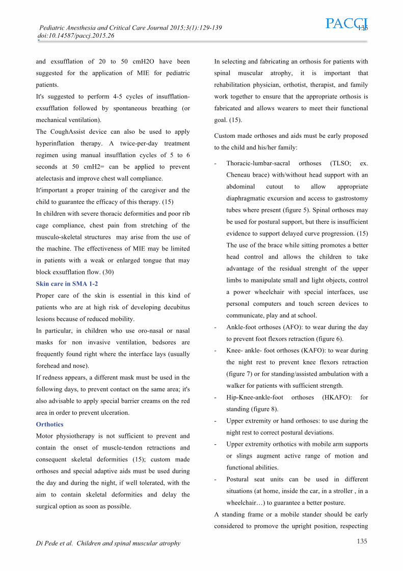
- manual light wheelchair (figure 10): for children
who preserve a reasonable upper limb function , to
move in indoor and outdoor environments;
- power wheelchair (figure 11): for children with
minimal upper limb function; thanks to adapted
interfaces (proper joysticks, table control systems,
chin/foot/breath control systems...) they can easily
drive the wheelchair and experience independence.
Figure 5
Figure 6
Figure 7
Figure 8
Figure 9
Pediatric Anesthesia and Critical Care Journal 2015;3(1):129-139 doi:10.14587/paccj.2015.26
Di Pede et al. Children and spinal muscular atrophy
137
137
Figure 10
Figure 11
The role of play in SMA1-2
Play is one of the best ways to explore the world and the
interpersonal relationships, to develop motor and
cognitive skills; it's clear that play has a critical role also
in SMA children.
Every child has the right to experience the joy of play;
choosing the appropriate toys is essential, complying the
child's motor difficulties and avoiding him/her to
experience the frustration of not being able to use them
because they are too big, too heavy...
Different categories of toys are available:
- Commonly available toys suitable to be used by
children with severe muscle weakness (for
newborns: inflated balloons, rag dolls to slip on the
fingers, gyms with objects hanging low enough to
be reached, bright rattles... for children 1-3 years:
soap bubbles, clay, finger crayons; for preschool
children: highlighters, crayons to connect to the
wheelchair base, stensils; for age school children:
racetracks, make up, videogames...)
- Commonly available and easily adaptable toys:
puzzle with small magnets for easy positioning.
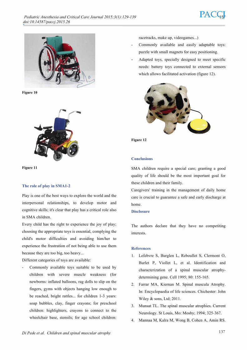
- Adapted toys, specially designed to meet specific
needs: battery toys connected to external sensors
which allows facilitated activation (figure 12).
Figure 12
Conclusions
SMA children require a special care; granting a good
quality of life should be the most important goal for
these children and their family.
Caregivers' training in the management of daily home
care is crucial to guarantee a safe and early discharge at
home.
Disclosure
The authors declare that they have no competiting
interests. References 1. Lefebvre S, Burglen L, Reboullet S, Clermont O,
Burlet P, Viollet L, et al. Identification and
characterization of a spinal muscular atrophy-
determining gene. Cell 1995; 80: 155-165.
2. Farrar MA, Kiernan M. Spinal muscula Atrophy.
In: Encyclopaedia of life sciences. Chichester: John
Wiley & sons, Ltd; 2011.
3. Munsat TL. The spinal muscular atrophies. Current
Neurology. St Louis, Mo: Mosby; 1994; 325-367.
4. Mannaa M, Kalra M, Wong B, Cohen A, Amin RS.

Pediatric Anesthesia and Critical Care Journal 2015;3(1):129-139 doi:10.14587/paccj.2015.26
Di Pede et al. Children and spinal muscular atrophy
138
138
Survival probabilities of patients with childhood
spinal muscle atrophy. Journal of clinical
neuromuscular disease 2009; 10:85-89.
5. Dubowitz V. Chaos in the classification of SMA: a
possible resolution. Neuromuscular disorders 1995;
5:3-5.
6. Merlini L, Granata C, Dubowitz V. Current
Concepts in childhood spinal muscular atrophy.
Wien; New York :Springer-Verlag; Bologna: Aulo
Gaggi Editore, 1989.
7. Pearn J. The gene frequency of acute Werdnig-
Hoffman disease (SMA type 1): a total population
survey in North-East England. J Med Genet 1973;
10: 260-265.
8. Pearn J. Incidence, prevalence and gene frequency
studies of chronic childhood spinal muscular
atrophy. J Med 1978; 15: 409-413.
9. Bertini E, Burghes A, Bushby K, et al. 134th
ENMC International Workshop: Outcome
Measures and Treatment of Spinal Muscular
Atrophy, 11-13 February 2005, Naarden, the
Netherlands. Neuromuscul Disord. 2005; 15:802-
816.
10. Dubowitz V. Very severe spinal muscular atrophy
(SMA type 0): an expanding clinical phenotype.
Eur J Paediatr Neurol. 1999; (3): 49 – 51.
11. Willig TN, Paulus J, Lacau-Saint-Guily J, et al.
Swallowing problems in neuromuscular disorders.
Arch Phys Med Rehab. 1994;75:1175-1181.
12. Tilton AH MM, Khoshoo V. Nutrition and
swallowing in pediatric neuromuscular patients.
Semin Pediatr Neurol. 1998; 5:106-115.
13. Houston KD, Buschang PH, Iannaccone ST, Seale
NS. Craniofacial morphology of spinal muscular
atrophy. Pediatr Res. 1994; 36:265-269.
14. Grunebaum M, Nutman J, Nitzan M. The
pharyngo-laryngeal deficit in the acute form of
infantile spinal muscular atrophy (Werdnig-
Hoffmann disease). Pediatr Radiol. 1981; 11:67-70.
15. Ching H. Wang, Richard S. Finkel, Enrico S.
Bertini, Mary Schroth, Anita Simonds, Brenda
Wong, Annie Aloysius, Leslie Morrison, Marion
Main, Thomas O. Crawford, Anthony Trela and
Participants of the International Conference on
SMA Standard of Care. Consensus Statement for
Standard of Care in Spinal Muscular Atrophy. J
Child Neurol 2007; 22:1027.
16. Granger MW, Buschang PH, Throckmorton GS,
Iannaccone ST. Masticatory muscle function in
patients with spinal muscular atrophy. Am J Orthod
Dentofacial Orthop. 1999; 115:697-702.
17. Mastella C, Ottonello GC: La cura del corpo del
bambino nella vita quotidiana. In: SMA1 Abita con
noi. Print LAb S.r.l – Spazio Aperto S.c.a.r.l.
Buccinasco 2009, pp 71 – 107.
18. Birnkrant DJ, Pope JF, Martin JE, et al. Treatment
of type I spinal muscular atrophy with noninvasive
ventilation and gastrostomy feeding. Pediatr
Neurol. 1998; 18:407-410.
19. Hough JL, Johnston L, Brauer SG, Woodgate PG,
Pham TMT, Schibler A. Effect of body position on
ventilation distribution in preterm infants on
continuous positive airway pressure. Pediatr Crit
Care Med 2012; 13: 446 – 451.
20. Bush A, Fraser J, Jardine E, et al. Respiratory
management of the infant with type 1 spinal
muscular atrophy. Arch Dis Child. 2005;90:709-
711.
21. Chatwin M, Ross E, Hart N, et al. Cough
augmentation with mechanical
insufflation/exsufflation in patients with
neuromuscular weakness. Eur Respir J. 2003;
21:502-508.
22. Finder JD, Birnkrant D, Carl J, et al. Respiratory
care of the patient with Duchenne muscular
dystrophy: ATS consensus statement. Am J Respir
Crit Care Med. 2004; 170:456-465.
23. Plecash AR, Leavitt BR. Aquatherapy for
neurodegenerative disorders. J Hungtington disease
2014; 3:5-11.

Pediatric Anesthesia and Critical Care Journal 2015;3(1):129-139 doi:10.14587/paccj.2015.26
Di Pede et al. Children and spinal muscular atrophy
139
139
24. Pini A, Ghezzo A. Atrofia muscolare spinale tipo I
o grave (Malattia di Werdnig – Hoffmann). In:
Dopo la diagnosi. Il monitoraggio delle malattie
neuromuscolari ad esordio in età evolutiva. Pini A,
Ghezzo A. Alberto Perdisa eds. 2007; pp 102 – 103.
25. Main M, Kairon H, Mercuri E, Muntoni F. The
Hammersmith functional motor scale for children
with spinal muscular atrophy: a scale to test ability
and monitor progress in children with limited
ambulation. Eur J Paediatr Neurol. 2003; 7:155 -
159 .
26. Mercuri E, Messina S, Bertini E, et al. Reliability of
the Hammersmith functional motor scale for spinal
muscular atrophy in a multicentric study.
Neuromuscul Disord. 2006; 16: 93 – 98.
27. Messina S, Pane M, De Rose P, Vasta I, Sorleti D,
Aloysius A, Sciarra F, Mangiola F, Kinali M,
Bertini E, Mercuri E. Feeding problems and
malnutrition in spinal muscular atrophy type II.
Neuromuscular Disorders 2008; 389–393.
28. Jenkins HM, A. Stocki, D. Kriellaars, H.
Pasterkamp. Breath Stacking in Children With
Neuromuscular Disorders. Paediatric Pulmonology
2014; 49:544-553.
29. Tzeng AC, Bach JR. Prevention of pulmonary
morbidity for patients with neuromuscular disease.
Chest 2000;118:1390–1396.
30. Louis J. Boitano. Equipment Options for Cough
Augmentation, Ventilation, and Noninvasive.
Pediatrics 2009;123;S226.
31. Kirby N, Barnerias MS, Siebens AA. An evaluation
of assisted cough in quadriparetic patients. Arch
Phys Med Rehabil. 1966; 47:705–710.
32. Fiona CE Moran, Alicia Spittle, Clare Delany,
Colin F Robertson and John Massie. Effect of home
mechanical in-exsufflation on hospitalisation and
life-style in neuromuscular disease: A pilot study.
Journal of Paediatrics and Child Health 2013;
49:233–237.