Embed Size (px)
Citation preview
Molecular Biology of the CellVol. 9, 3119–3131, November 1998
Direct Involvement of N-Cadherin–mediated Signalingin Muscle DifferentiationPolina Goichberg and Benjamin Geiger*
Department of Molecular Cell Biology, The Weizmann Institute of Science, Rehovot 76100, Israel
Submitted June 22, 1998; Accepted September 8, 1998Monitoring Editor: Mary C. Beckerle
Cell–cell interactions, mediated by members of the cadherin family of Ca21-dependentadhesion molecules, play key roles in morphogenetic processes as well as in the trans-duction of long-range growth and differentiation signals. In muscle differentiation celladhesion is involved in both early stages of myogenic induction and in later stages ofmyoblast interaction and fusion. In this study we have explored the involvement of aspecific cadherin, namely N-cadherin, in myogenic differentiation. For that purpose wehave treated different established lines of cultured myoblasts with beads coated withN-cadherin–specific ligands, including a recombinant N-cadherin extracellular domain,and anti-N-cadherin antibodies. Immunofluorescent labeling for cadherins and cateninsindicated that treatment with the cadherin-reactive beads for several hours enhances theassembly of cell–cell adherens-type junctions. Moreover, immunofluorescence and im-munoblotting analyses indicated that treatment with the beads for 12–24 h inducesmyogenin expression and growth arrest, which are largely independent of cell platingdensity. Upon longer incubation with the beads (2–3 d) a major facilitation in theexpression of several muscle-specific sarcomeric proteins and in cell fusion into myo-tubes was observed. These results suggest that surface clustering or immobilization ofN-cadherin can directly trigger signaling events, which promote the activation of amyogenic differentiation program.
INTRODUCTION
Intercellular adhesion plays key roles in tissue forma-tion and in the transduction of transmembrane signalsaffecting cell growth, motility, and differentiation. Oneof the most prominent and widespread groups ofadhesion molecules involved in such interactions isthe cadherin family, whose members mediate ho-mophilic and Ca21-dependent cell–cell adhesion in awide variety of tissues (for review, see Geiger andAyalon, 1992; Overduin et al., 1995; Shapiro et al., 1995;Takeichi, 1995). Cadherins are transmembrane mole-cules that interact with similar cadherins on neighbor-ing cells via their ectodomains and with the actin-based cytoskeleton via their cytoplasmic regions. Inaddition, it has recently been established that a varietyof signaling molecules are associated with cadherin-containing junctions, including receptor tyrosine ki-
nases and cytoplasmic kinases of src family (Geiger etal, 1990, 1995; Yamada and Geiger, 1997). This colocal-ization suggested that accumulation of these mole-cules in junctional sites may lead to their activationand to adhesion-mediated signaling. Interestingly, thedeterioration of these complexes as a result of cad-herin or vinculin deficiency (Rodriguez Fernandez etal., 1993; Birchmeier, 1995; Volberg et al., 1995) orextensive tyrosine phosphorylation (Volberg et al.,1992; Ayalon and Geiger, 1997) is commonly found inmalignant cells, leading to their anaplastic morphol-ogy and deregulated growth (Tsukita et al., 1993;Birchmeier and Behrens, 1994; Birchmeier, 1995). Re-cent evidence also indicates that junctional proteins,such as b-catenin and plakoglobin, can play a criticalrole in regulating cell fate not just by controlling theassembly of adherens junctions but also by directlyactivating transcription in the nucleus (Barth et al.,1997). Cadherin-mediated adhesion is also indirectlyimplicated in the differentiation of various cell types,including muscle cells (Knudsen, 1990; Knudsen et al.,
* Corresponding author. E-mail address: [email protected].
© 1998 by The American Society for Cell Biology 3119
1990; Zeschingk et al., 1995), chondrocytes (Oberlenderand Tuan, 1994), osteoclasts (Mbalaviele et al., 1995),and neural cells (Doherty and Walsh, 1994).
Myogenesis is a particularly appealing system tostudy the role of cadherin-mediated adhesion in celldifferentiation, because Ca21-dependent cell adhesion,followed by cell fusion, is an intrinsic step in thedifferentiation process. The differentiation of culturedskeletal myoblasts is commonly activated by growthfactor withdrawal and accompanied by transcriptionalactivation of muscle-specific genes, growth arrest, andfusion to form multinucleated myotubes (Olson, 1992,1993). The muscle-specific basic helix–loop–helix tran-scription factors, including MyoD, Myf5, myogenin,and Mrf4, orchestrate the entire expression program ofthe various muscle-specific genes.
Several lines of indirect evidence suggest that cad-herin-mediated interactions are also involved in theregulation of skeletal myogenesis. These include theinhibition of fusion by calcium depletion or by anti-N-cadherin antibodies and HAV-containing inhibitorypeptide (Knudsen et al., 1990; Mege et al., 1992). Inaddition, the somites formed in N-cadherin knockoutmice are small and irregularly shaped (Radice et al.,1997). In addition to the effect of N-cadherin on termi-nal stages of skeletal muscle differentiation, it has beenshown to affect the expression of genes before the cellfusion stage: injection of a dominant negative cadherinRNA suppresses the expression of MyoD in Xenopusembryos and affects the subsequent expression ofmuscle-specific genes (Holt et al., 1994). Avian embry-onic progenitor cells expressing only N-cadherin andnot E-cadherin differentiate into skeletal muscle, andtreatment with anti-N-cadherin antibodies inhibits theaccumulation of myosin in chick embryo cells derivedfrom different stages of avian embryonic development(George-Weinstein et al., 1997). Overexpression of N-cadherin in baby hamster kidney cells stimulates ex-pression of sarcomeric myosin in these cells (Redfieldet al., 1997). However, although these data suggest thatcadherin-mediated interactions are involved in muscledifferentiation, they do not indicate whether they areinvolved merely in the promotion of myoblast–myo-blast adhesion per se or also induce long-range, myo-genic signals that promote muscle gene expression.
In the present study we examined the effect of directlong-range signaling induced by N-cadherin cluster-ing or immobilization on myogenic differentiation. Weshow here that beads conjugated to different N-cad-herin ligands can trigger myogenesis, manifested byaccelerated myoblast adhesion, myogenin expression,formation and assembly of various structural sarco-meric components, and myoblast fusion. Stimulationof myogenin expression by the N-cadherin–reactivebeads occurred irrespective of myoblast density, sug-gesting that activation of this key step in myogenesisis directly induced by N-cadherin signaling.
MATERIALS AND METHODS
Cell CultureAll myoblast lines examined in this study, including C2 mouseskeletal myoblasts and L8 and L84 rat skeletal myoblasts, werekindly provided by Dr. D. Yaffe (The Weizmann Institute of Science)(Yaffe and Saxel, 1976, 1977). The cells were cultured in subconfluentdensities at 37°C in a humidified atmosphere containing 8% CO2 indishes coated with 0.1% gelatin. C2 cells were cultured in Dulbec-co’s modified Eagle’s medium (DMEM) supplemented with 20%heat-inactivated FCS (BioLabs, Israel), glutamine, and antibiotics. L8and L84 cells were cultured in Waymouth’s medium containing15% FCS. Myogenic differentiation of L8 and L84 cells was inducedby changing the growth medium to DMEM containing 2% heat-inactivated horse serum (Biological Industries, Israel) and 4 IU/mlinsulin (Humulin R; Lilly, France). To trigger the differentiation ofC2 myoblasts, cells were either plated at high density or stimulatedby insulin and 10% horse serum in DMEM.
Preparation and Application of Cadherin-reactiveBeadsN-cadherin ectodomain (NEC) was produced as described by Lev-enberg et al. (1998a). Briefly, 108 latex Polybead amino microsperes(mean diameter, 6 mm; Polysciences, Warrington, PA) were washedwith phosphate-buffered saline (PBS; pH 7.4), activated overnightwith 8% glutaraldehyde, washed with PBS, and incubated for 5 hwith 500 mg/ml bovine serum albumin (BSA; Sigma Chemical, St.Louis, MO), purified NEC (Levenberg et al., 1998), or anti-N-cad-herin monoclonal antibodies (clone BE; Volk and Geiger, 1986). Freesites were blocked with 0.5 M ethanolamine for 30 min, followed byincubation with 10 mg/ml BSA for 30 min, and the beads wereresuspended in storage buffer (PBS containing 10 mg/ml BSA, 0.1%sodium azide, and 5% glycerol, pH 7.4). Aliquots containing 5 3 105
beads were added to cell monolayers in 35-mm-diameter culturedishes.
Cytochemical StainingMyoblasts were cultured on 35-mm tissue culture dishes (Falcon,Becton Dickinson, Palo Alto, CA), coated with 0.1% gelatin, washedtwice in PBS, and fixed for 10 min with methanol at room temper-ature. The monolayer was washed twice with PBS and stained for 25min with 10% Giemsa solution (Fluka, Buchs, Switzerland), exten-sively washed with water, and dry mounted for microscopic exam-ination.
Immunochemical Reagents and ProceduresMyoblasts cultured on glass coverslips coated with 0.1% gelatinwere washed with 0.1 M 4-morpholinepropanesulfonic acid buffer(pH 6.0), permeabilized for 2 min by 0.5% Triton X-100 in 0.1 M4-morpholinepropanesulfonic acid buffer, and fixed for 25 min with3% paraformaldehyde in PBS. All of these procedures were carriedout at room temperature. Anti-skeletal a-actin (5C5), anti-skeletala-actinin (EA53), anti-skeletal myosin (MY32), anti-desmin(DEU10), and anti-pan-cadherin (CH19) were purchased fromSigma. Anti-b-catenin (94.5) was a gift from Dr. M. Wheelock (Uni-versity of Toledo, Toledo, OH). Anti-titin (T12) and anti-myomesin(BB78) were obtained from Dr. W. Obermann and Dr. D. Furst(Max-Plank-Institut for Biophysical Chemistry, Gottingen, Germa-ny). Anti-myogenin antibodies were obtained from Dr. BarbaraWinter and Dr. H. Arnold (Technical University, Braunschweig,Germany). Anti-5-bromo-29-deoxyuridine (BrdU) was purchasedfrom Becton Dickinson. For BrdU labeling cells were incubated for45 min with 10 mM BrdU (Sigma) in culture medium, fixed, perme-abilized for 4 min with 0.5% Triton X-100 in 3% paraformaldehyde,and post-fixed for 25 min with 3% paraformaldehyde. For anti-BrdUand 49,6-diamidino-2-phenylinodole (DAPI, Sigma) labeling, the
P. Goichberg and B. Geiger
Molecular Biology of the Cell3120
cells were treated with 2 M HCl in 0.5% Triton X-100 for an addi-tional 15 min. The secondary antibodies were Cy-3-conjugated goatanti-mouse immunoglobulin (Jackson ImmunoResearch Laborato-ries, West Grove, PA). Nuclei were indirectly immunolabeled andcounterstained by 10 min incubation with 2.5 mg/ml DAPI, and thecells were mounted in Elvanol (Mowiol 4-88; Hoechst, Frankfurt,Germany). Immunofluorescence microscopy was carried out withan Axiophot microscope (Zeiss, Oberkochen, Germany) equippedfor multiple fluorescence examination.
Immunoblot AnalysisWhole cells were washed with PBS and extracted with Laemmli sam-ple buffer. Proteins were separated by 10% SDS-PAGE (Laemmli, 1970)and transferred by electroblotting to Hybond-C nitrocellulose mem-branes (Amersham, Buckinghamshire, United Kingdom). Membraneswere blocked for 1 h with a 4% solution of dry milk in PBS and thenincubated overnight at 4°C with the primary antibodies diluted in PBS.After washing in PBS, the membranes were incubated for 45 min atroom temperature with HRP-conjugated goat anti-mouse immuno-globulin G (Amersham), and immunoreactive bands were visualizedusing the Enhanced Chemiluminiscence system (Amersham).
Transmission Electron MicroscopyC2 cells were plated overnight on gelatin-coated 35-mm dishes.After 48 h of treatment with beads the cells were fixed inKranovsky’s fixative (3% paraformaldehyde, 2% glutaraldehyde, 5mM CaCl2, and 0.1 M sucrose in 0.1 M cacodylate buffer, pH 7.4)and post-fixed with 1% osmium tetroxide, 0.5% potassium dichro-mate, and 0.5% potassium hexacyanoferrate in 0.1 M cacodylatebuffer. The cells were stained en bloc with 2% aqueous uranylacetate, followed by ethanol dehydration. The dishes were embed-ded in Epon 812 (Tuosimis, MD). Sections were cut using a diamondknife (Diatome, Biel, Switzerland) and examined using a Philips(Mahwah, NJ) CM-12 transmission electron microscope operating atan accelerating voltage of 100 kV.
RESULTS
Interactions of Cadherin-reactive Beads withCultured MyoblastsTo test the effect of N-cadherin–mediated interactionson myogenic differentiation, established myoblast celllines were treated with 6-mm beads, coated with N-cadherin ligands (NEC or anti-N-cadherin monoclonalantibodies [BE]), as described by Levenberg et al.(1998a). BSA-coated beads were used as controls.
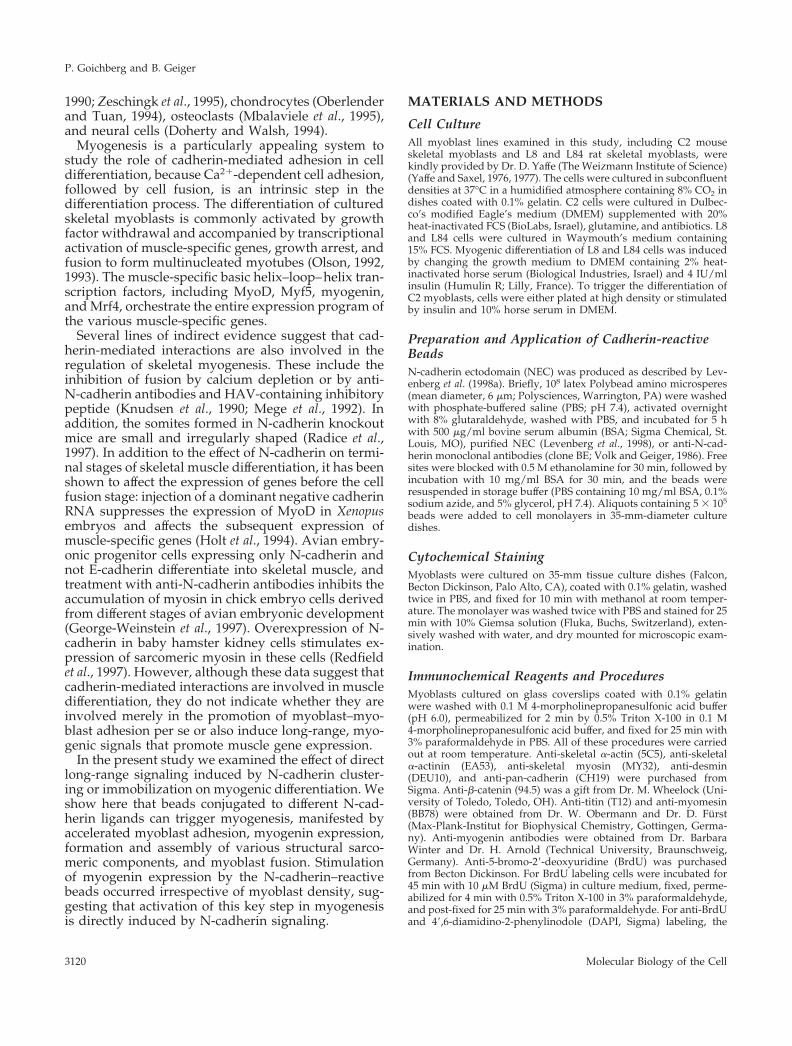
Transmission electron microscopy of C2 myoblastsafter 48 h of incubation with the beads, coated eitherwith NEC (Figure 1B) or with BSA (Figure 1A) indi-cated that both types of beads attach firmly to the cellsurface. BSA-coated beads attached to the plasmamembrane via a continuos close contact area and wereengulfed by the cells after several hours of incubation,
Figure 1. Transmission electron micrographs of C2 myoblaststreated with beads coated with BSA (A) or NEC (B and C). The cellswere incubated with the beads for 48 h in growth medium, fixed,
Figure 1 (cont). and processed for transmission electron micros-copy. Notice that the contact region with the NEC-coated bead ischaracterized by focal, foot-like adhesions (arrows), whereas theattachment to the BSA beads is tight and uniform. Sarcomericfilament bundles were found in the cytoplasm of the N-cadherin–stimulated cells (C) (arrowheads point to Z lines). Bars, 1 mm.
N-Cadherin in Muscle Differentiation
Vol. 9, November 1998 3121
whereas the NEC-coated beads, were attached to thecells through electron-dense “foot-like processes,” re-sembling focal contacts, and were usually not exten-sively engulfed.
Promotion of Myotube Formation by N-Cadherin–mediated StimulationTo test the direct involvement of N-cadherin–medi-ated signaling in skeletal muscle differentiation, we
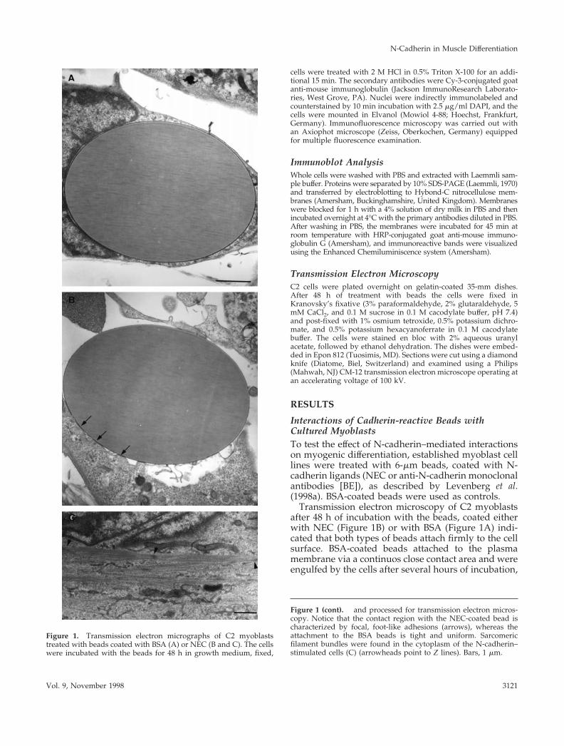
have examined the effects of N-cadherin–reactive andcontrol beads on the rate of myotube formation by thedifferent myogenic cell lines under conditions that donot favor differentiation (i.e., high serum concentra-tion and low plating density). As demonstrated inFigure 2, the binding of cadherin-reactive beads(beads coated with NEC or with anti N-cadherin an-tibodies) to the cells significantly increased the num-ber of myotubes in these myogenic cultures from ;4/
Figure 2. Effect of the N-cadherin stimula-tion on myotube formation in C2, L8, and L84myogenic cell lines. C2, L8, and L84 myo-blasts were treated with NEC and anti-N-cadherin (anti N-cad) BE beads for the indi-cated periods, fixed, and stained withGiemsa. C2 cells were maintained in growthmedium, whereas for L8 and L84 cells theculture medium was replaced with differen-tiation medium simultaneously with the ad-dition of beads. Media were changed everysecond day. Notice that the number of myo-tubes in the field is higher for the cells treatedwith cadherin-reactive beads. Arrows point tosome of the multinucleated myotubes. Bar,100 mm.
P. Goichberg and B. Geiger
Molecular Biology of the Cell3122
mm2 (BSA-coated beads) to 7 or 8/mm2 (BE- andNEC-coated beads, respectively). It is noteworthy thatmyotubes formed after treatment with cadherin-reac-tive beads were usually larger than those formed aftertreatment with control beads. The number of nucleiper individual tube was, however, variable, usually
displaying clusters of 5–20 nuclei, and apparently didnot depend on the type or number of bound beads.
Transmission electron microscopy of C2 cells, after48 h treatment with cadherin-reactive beads, revealedscattered sarcomeric structures in the cytoplasm (Fig-ure 1C), which could be found in essentially all sec-
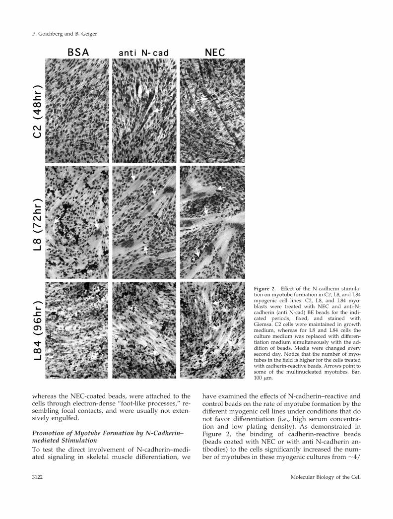
Figure 3. Expression of skeletal myosin in myocytes treated with beads conjugated to NEC, anti-N-cadherin BE antibodies (anti N-cad) orBSA. C2, L8, and L84 myoblasts were treated with different beads for 48 h, permeabilized, fixed, and immunostained with anti-skeletalmyosin antibodies. C2 cells were maintained in growth medium, whereas for L8 and L84 cells growth medium was replaced withdifferentiation medium simultaneously with the addition of beads. The number of cells per field was approximately equal. Notice the increasein myosin expression in the cultures after treatment with the cadherin-reactive beads. The position of individual beads was detected byphase-contrast microscopy, and their location is indicated by arrowheads. Bar, 10 mm.
N-Cadherin in Muscle Differentiation
Vol. 9, November 1998 3123
tions. These sarcomers were similar to those formedlater in the course of differentiation induced bygrowth factors deprivation. Such organized filamentswere not detected at that time point in .30 sectionsderived from C2 cells treated with control beads.
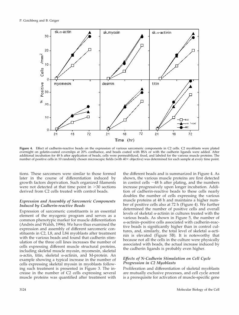
Expression and Assembly of Sarcomeric ComponentsInduced by Cadherin-reactive BeadsExpression of sarcomeric constituents is an essentialelement of the myogenic program and serves as acommon phenotypic marker for muscle differentiation(Andres and Walsh, 1996). We have thus examined theexpression and assembly of different sarcomeric con-stituents in C2, L8, and L84 myoblasts after treatmentwith the various beads and found that cadherin stim-ulation of the three cell lines increases the number ofcells expressing different muscle structural proteins,including skeletal muscle myosin, myomesin, skeletala-actin, titin, skeletal a-actinin, and M-protein. Anexample showing a typical increase in the number ofcells expressing skeletal myosin in myoblasts follow-ing such treatment is presented in Figure 3. The in-crease in the number of C2 cells expressing severalmuscle proteins was quantified after treatment with
the different beads and is summarized in Figure 4. Asshown, the various muscle proteins are first detectedin control cells ;48 h after plating, and the numbersincrease progressively upon longer incubation. Addi-tion of cadherin-reactive beads to these cells nearlydoubles the number of cells expressing the variousmuscle proteins at 48 h and maintains a higher num-ber of positive cells also at 72 h (Figure 4). We furtherdetermined the number of positive cells and overalllevels of skeletal a-actinin in cultures treated with thevarious beads. As shown in Figure 5, the number ofa-actinin–positive cells associated with cadherin-reac-tive beads is significantly higher than in control cul-tures, and, similarly, the total level of skeletal a-acti-nin is elevated (Figure 5B). It is noteworthy thatbecause not all the cells in the culture were physicallyassociated with beads, the actual increase induced bythe cadherin ligands is probably even higher.
Effects of N-Cadherin Stimulation on Cell CycleProgression in C2 MyoblastsProliferation and differentiation of skeletal myoblastsare mutually exclusive processes, and cell cycle arrestis a prerequisite for activation of muscle-specific gene
Figure 4. Effect of cadherin-reactive beads on the expression of various sarcomeric components in C2 cells. C2 myoblasts were platedovernight on gelatin-coated coverslips at 20% confluence, and beads coated with BSA or with the cadherin ligands were added. Afteradditional incubation for 48 h after application of beads, cells were permeabilized, fixed, and labeled for the various muscle proteins. Thenumber of positive cells in 10 randomly chosen microscopic fields (with 403 objective) was determined for each sample at every time point.
P. Goichberg and B. Geiger
Molecular Biology of the Cell3124
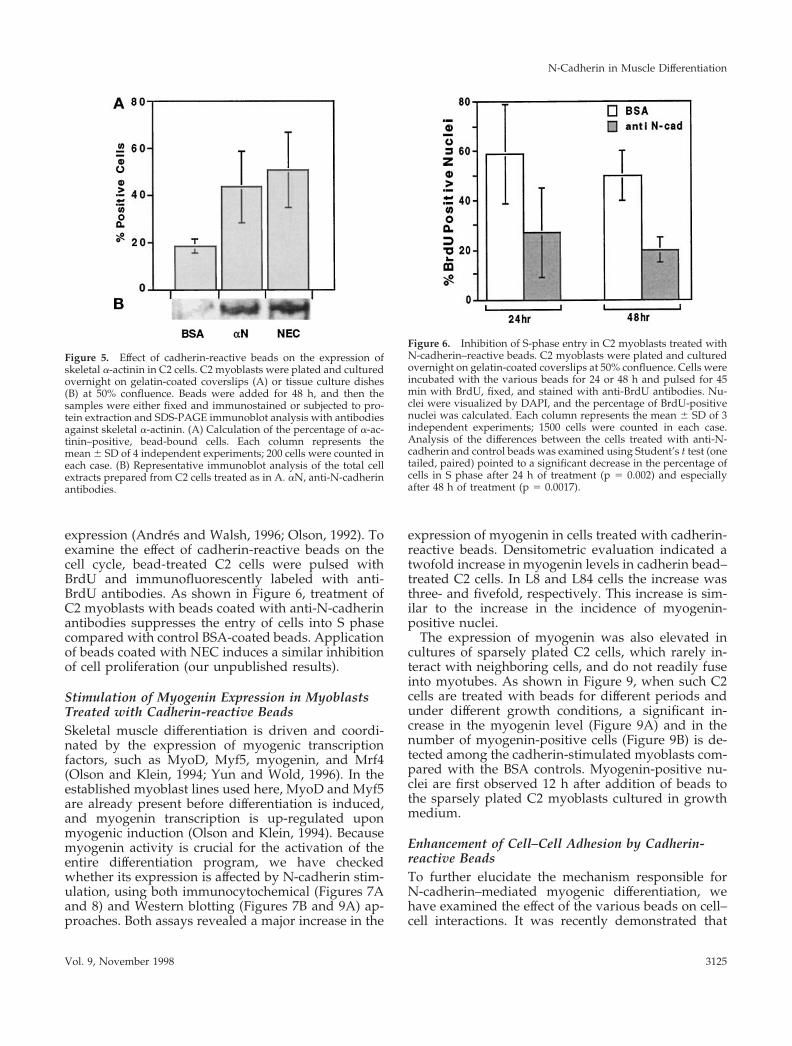
expression (Andres and Walsh, 1996; Olson, 1992). Toexamine the effect of cadherin-reactive beads on thecell cycle, bead-treated C2 cells were pulsed withBrdU and immunofluorescently labeled with anti-BrdU antibodies. As shown in Figure 6, treatment ofC2 myoblasts with beads coated with anti-N-cadherinantibodies suppresses the entry of cells into S phasecompared with control BSA-coated beads. Applicationof beads coated with NEC induces a similar inhibitionof cell proliferation (our unpublished results).
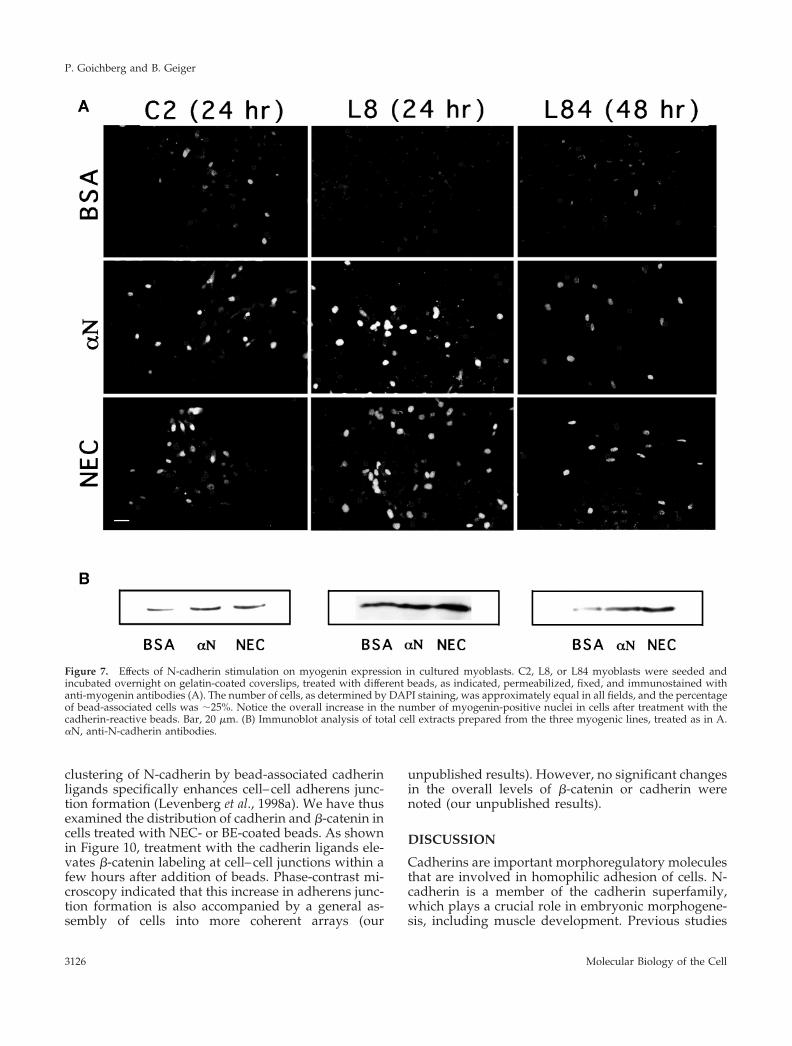
Stimulation of Myogenin Expression in MyoblastsTreated with Cadherin-reactive BeadsSkeletal muscle differentiation is driven and coordi-nated by the expression of myogenic transcriptionfactors, such as MyoD, Myf5, myogenin, and Mrf4(Olson and Klein, 1994; Yun and Wold, 1996). In theestablished myoblast lines used here, MyoD and Myf5are already present before differentiation is induced,and myogenin transcription is up-regulated uponmyogenic induction (Olson and Klein, 1994). Becausemyogenin activity is crucial for the activation of theentire differentiation program, we have checkedwhether its expression is affected by N-cadherin stim-ulation, using both immunocytochemical (Figures 7Aand 8) and Western blotting (Figures 7B and 9A) ap-proaches. Both assays revealed a major increase in the
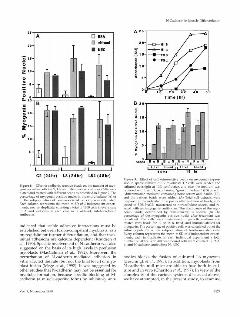
expression of myogenin in cells treated with cadherin-reactive beads. Densitometric evaluation indicated atwofold increase in myogenin levels in cadherin bead–treated C2 cells. In L8 and L84 cells the increase wasthree- and fivefold, respectively. This increase is sim-ilar to the increase in the incidence of myogenin-positive nuclei.
The expression of myogenin was also elevated incultures of sparsely plated C2 cells, which rarely in-teract with neighboring cells, and do not readily fuseinto myotubes. As shown in Figure 9, when such C2cells are treated with beads for different periods andunder different growth conditions, a significant in-crease in the myogenin level (Figure 9A) and in thenumber of myogenin-positive cells (Figure 9B) is de-tected among the cadherin-stimulated myoblasts com-pared with the BSA controls. Myogenin-positive nu-clei are first observed 12 h after addition of beads tothe sparsely plated C2 myoblasts cultured in growthmedium.
Enhancement of Cell–Cell Adhesion by Cadherin-reactive BeadsTo further elucidate the mechanism responsible forN-cadherin–mediated myogenic differentiation, wehave examined the effect of the various beads on cell–cell interactions. It was recently demonstrated that
Figure 5. Effect of cadherin-reactive beads on the expression ofskeletal a-actinin in C2 cells. C2 myoblasts were plated and culturedovernight on gelatin-coated coverslips (A) or tissue culture dishes(B) at 50% confluence. Beads were added for 48 h, and then thesamples were either fixed and immunostained or subjected to pro-tein extraction and SDS-PAGE immunoblot analysis with antibodiesagainst skeletal a-actinin. (A) Calculation of the percentage of a-ac-tinin–positive, bead-bound cells. Each column represents themean 6 SD of 4 independent experiments; 200 cells were counted ineach case. (B) Representative immunoblot analysis of the total cellextracts prepared from C2 cells treated as in A. aN, anti-N-cadherinantibodies.
Figure 6. Inhibition of S-phase entry in C2 myoblasts treated withN-cadherin–reactive beads. C2 myoblasts were plated and culturedovernight on gelatin-coated coverslips at 50% confluence. Cells wereincubated with the various beads for 24 or 48 h and pulsed for 45min with BrdU, fixed, and stained with anti-BrdU antibodies. Nu-clei were visualized by DAPI, and the percentage of BrdU-positivenuclei was calculated. Each column represents the mean 6 SD of 3independent experiments; 1500 cells were counted in each case.Analysis of the differences between the cells treated with anti-N-cadherin and control beads was examined using Student’s t test (onetailed, paired) pointed to a significant decrease in the percentage ofcells in S phase after 24 h of treatment (p 5 0.002) and especiallyafter 48 h of treatment (p 5 0.0017).
N-Cadherin in Muscle Differentiation
Vol. 9, November 1998 3125
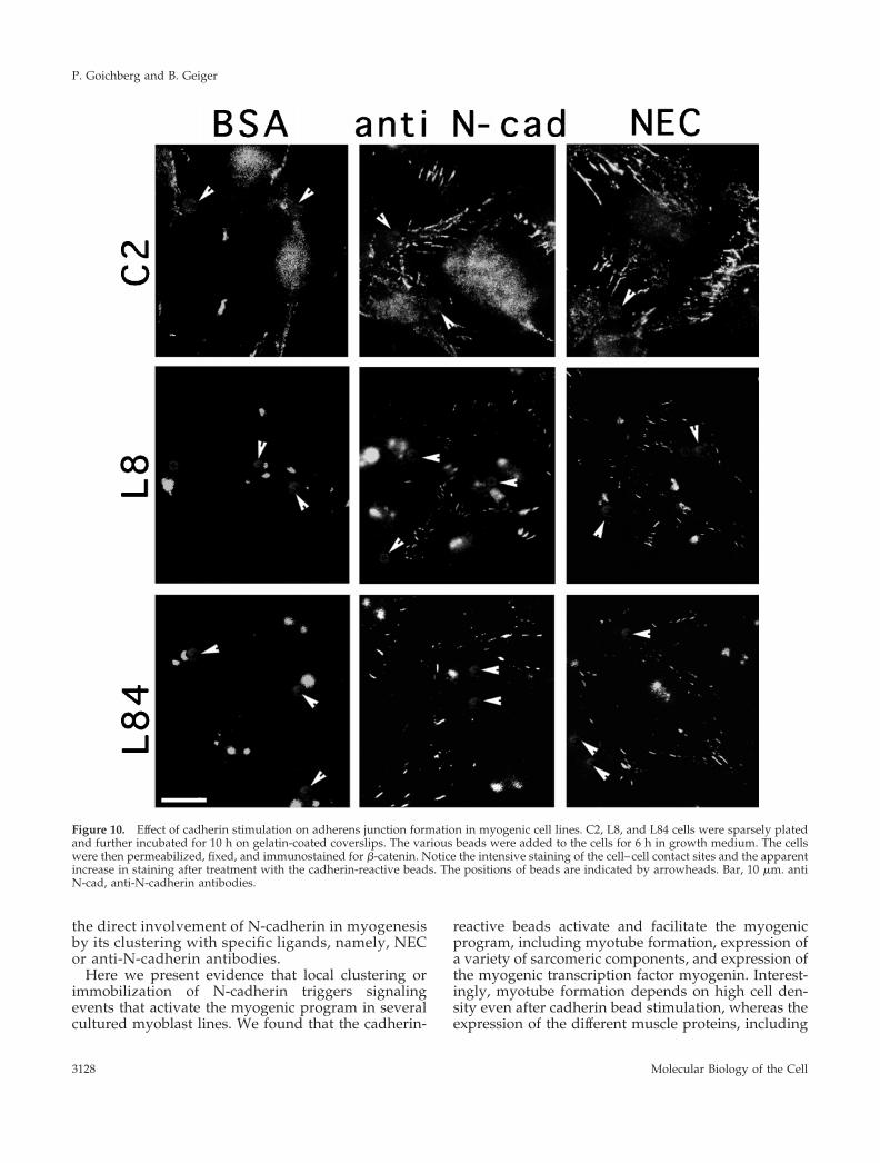
clustering of N-cadherin by bead-associated cadherinligands specifically enhances cell–cell adherens junc-tion formation (Levenberg et al., 1998a). We have thusexamined the distribution of cadherin and b-catenin incells treated with NEC- or BE-coated beads. As shownin Figure 10, treatment with the cadherin ligands ele-vates b-catenin labeling at cell–cell junctions within afew hours after addition of beads. Phase-contrast mi-croscopy indicated that this increase in adherens junc-tion formation is also accompanied by a general as-sembly of cells into more coherent arrays (our
unpublished results). However, no significant changesin the overall levels of b-catenin or cadherin werenoted (our unpublished results).
DISCUSSION
Cadherins are important morphoregulatory moleculesthat are involved in homophilic adhesion of cells. N-cadherin is a member of the cadherin superfamily,which plays a crucial role in embryonic morphogene-sis, including muscle development. Previous studies
Figure 7. Effects of N-cadherin stimulation on myogenin expression in cultured myoblasts. C2, L8, or L84 myoblasts were seeded andincubated overnight on gelatin-coated coverslips, treated with different beads, as indicated, permeabilized, fixed, and immunostained withanti-myogenin antibodies (A). The number of cells, as determined by DAPI staining, was approximately equal in all fields, and the percentageof bead-associated cells was ;25%. Notice the overall increase in the number of myogenin-positive nuclei in cells after treatment with thecadherin-reactive beads. Bar, 20 mm. (B) Immunoblot analysis of total cell extracts prepared from the three myogenic lines, treated as in A.aN, anti-N-cadherin antibodies.
P. Goichberg and B. Geiger
Molecular Biology of the Cell3126
indicated that stable adhesive interactions must beestablished between fusion-competent myoblasts, as aprerequisite for further differentiation, and that theseinitial adhesions are calcium dependent (Knudsen etal., 1990). Specific involvement of N-cadherin was alsosuggested on the basis of its high levels in prefusionmyoblasts (MacCalman et al., 1992). Moreover, theperturbation of N-cadherin–mediated adhesion invitro affected the rate (but not the final level) of myo-blast fusion (Mege et al., 1992). It was suggested byother studies that N-cadherin may not be essential formyotube formation, because specific blocking of M-cadherin (a muscle-specific form) by inhibitory anti-
bodies blocks the fusion of cultured L6 myocytes(Zeschingk et al., 1995). In addition, myoblasts fromN-cadherin–null mice are able to fuse both in cul-ture and in vivo (Charlton et al., 1997). In view of thecomplexity of the various systems discussed above,we have attempted, in the present study, to examine
Figure 8. Effect of cadherin-reactive beads on the number of myo-genin-positive cells in C2, L8, and L84 myoblast cultures. Cells wereplated and treated with different beads as described in Figure 7. Thepercentage of myogenin positive nuclei in the entire culture (A) orin the subpopulation of bead-associated cells (B) was calculated.Each column represents the mean 6 SD of 3 independent experi-ments, each in duplicate, counting a total of 1000 cells in every casein A and 250 cells in each case in B. aN-cad, anti-N-cadherinantibodies.
Figure 9. Effect of cadherin-reactive beads on myogenin expres-sion in sparse cultures of C2 myoblasts. C2 cells were seeded andcultured overnight at 10% confluence, and then the medium wasreplaced with fresh FCS-containing “growth medium” (FS) or with“differentiation medium” containing horse serum and insulin (HI),and the various beads were added. (A) Total cell extracts wereprepared at the indicated time points after addition of beads, sub-jected to SDS-PAGE, transferred to nitrocellulose sheets, and re-acted with anti-myogenin antibodies. The absorbance of the myo-genin bands, determined by densitometry, is shown. (B) Thepercentage of the myogenin positive nuclei after treatment wascalculated. The cells were maintained in growth medium andtreated with beads for 12 or 18 h, fixed, and immunolabeled formyogenin. The percentage of positive cells was calculated out of theentire population or the subpopulation of bead-associated cells.Every column represents the mean 6 SD of 3 independent experi-ments, each in duplicate. In each individual experiment a totalnumber of 900 cells or 200 bead-bound cells were counted. B, BSA;a, anti-N-cadherin antibodies; N, NEC.
N-Cadherin in Muscle Differentiation
Vol. 9, November 1998 3127
the direct involvement of N-cadherin in myogenesisby its clustering with specific ligands, namely, NECor anti-N-cadherin antibodies.
Here we present evidence that local clustering orimmobilization of N-cadherin triggers signalingevents that activate the myogenic program in severalcultured myoblast lines. We found that the cadherin-
reactive beads activate and facilitate the myogenicprogram, including myotube formation, expression ofa variety of sarcomeric components, and expression ofthe myogenic transcription factor myogenin. Interest-ingly, myotube formation depends on high cell den-sity even after cadherin bead stimulation, whereas theexpression of the different muscle proteins, including
Figure 10. Effect of cadherin stimulation on adherens junction formation in myogenic cell lines. C2, L8, and L84 cells were sparsely platedand further incubated for 10 h on gelatin-coated coverslips. The various beads were added to the cells for 6 h in growth medium. The cellswere then permeabilized, fixed, and immunostained for b-catenin. Notice the intensive staining of the cell–cell contact sites and the apparentincrease in staining after treatment with the cadherin-reactive beads. The positions of beads are indicated by arrowheads. Bar, 10 mm. antiN-cad, anti-N-cadherin antibodies.
P. Goichberg and B. Geiger
Molecular Biology of the Cell3128
myogenin, was also detected in sparse cultures, ap-parently independently of cell fusion. This is in linewith the common sequence of myogenic events trig-gered by growth factor withdrawal, which start withmyogenin expression, induction of growth arrest, ex-pression of structural sarcomeric components, and,finally, fusion into myotubes (Andres and Walsh,1996). It is, however, noteworthy that the growth ar-rest induced by N-cadherin–reactive beads is notunique to the myogenic differentiation pathway, andtreatment of a variety of mesenchymal cells with thesame types of beads inhibits proliferation and blocksthe cell cycle at the G1 phase. The mechanism under-lying this growth inhibiting signaling process will bedescribed in detail elsewhere (Levenberg et al., 1998b).Our data are consistent with the notion that growtharrest precedes the expression of the various structuralsarcomeric components by ;24 h.
The crucial events in skeletal muscle differentiationare coordinated by the expression of muscle regula-tory proteins that act in cooperation with the MEF2family of transcription factors to activate muscle-spe-cific gene expression (Yun and Wold, 1996). Theseproteins were also shown to interact with and beregulated by other transcription factors and the cellcycle regulatory system to coordinately activate thedifferentiation program and to inhibit proliferation(Olson, 1992, 1993; Rao et al., 1994; Skapek et al., 1995,1996). The fine balance between proliferation and dif-ferentiation appears to be critical for the induction andprogression of the myogenic program. For instance, incommitted myoblasts MyoD and Myf5 proteins areexpressed, although their activity is apparently inhib-ited by the presence of growth-promoting factors, andthus the progression of differentiation depends ongrowth factor withdrawal, leading to myogenin ex-pression and activation of the myogenic cascade (An-dres and Walsh, 1996).
Numerous studies indicate that in the course ofmyogenic differentiation inhibition of cell proliferationand cell death are coordinately regulated, and theinability to exit the cell cycle leads to apoptotic death(Walsh and Perlman, 1997; Fimia et al., 1998). Cellcycle inhibitors, such as p21 or Rb, are able to preventthis apoptosis most probably by the induction of cellcycle arrest (Wang and Walsh, 1996; Wang et al., 1997;Zacksenhaus et al., 1996). As described above, treat-ment with cadherin-reactive beads inhibits cell cycleprogression in C2 myoblasts. However, no apparentdifferences in the number of apoptotic nuclei (definedby DAPI staining) were observed after application ofthe various beads (our unpublished results). Currentreports demonstrate that the decision to exit the cellcycle and further differentiate or to die is made at thelevel of myogenin-induced cell cycle arrest, i.e., at thestages of myogenesis when cells already express myo-genin. Because cadherin-reactive beads promote myo-
genin expression, it seems to us unlikely that stimula-tion of cadherin-mediated adhesion directly affects theapoptotic process.
Another aspect raised by the present study is thespecificity of the effects on myogenesis to N-cadherin.As indicated above, additional members of the cad-herin family are also expressed in muscle tissues, in-cluding M- and R-cadherins (Zeschingk et al., 1995;Rosenberg et al., 1997) and cadherin-11 (Kimura et al.,1995), and perturbation of some of these can affectmyogenesis (Zeschingk et al., 1995). We have no directevidence or claim that the effect shown here for N-cadherin stimulation is unique to this isoform andcannot be obtained by the clustering or immobiliza-tion of other cadherins. It is noteworthy that thesethree cadherins show considerable overall homologywith N-cadherin along their cytoplasmic domains (82,50, and 54% identity), which are presumably involvedin the transduction of N-cadherin–mediated signals.
The data presented here are in agreement with theview that activation of cadherin-mediated signalingleads to the expression of myogenin, which in turninhibits cell cycle progression, triggers the differenti-ation program, including the expression of sarcomericproteins, and promotes myotube formation. Themechanism underlying this cadherin-induced activa-tion of myogenin expression is, however, not clear. Itwas previously shown that cadherin-reactive beadsspecifically activate tyrosine phosphorylation at adhe-rens junctions and enhance cadherin-mediated cell–cell adhesion in a variety of mesenchymal cells (Lev-enberg et al., 1998). This is consistent with the presentresults, showing that cadherin-induced stimulationleads to a specific and generalized enhancement ofmyoblast–myoblast adhesion (Figure 10). This, in turn,could have two distinct effects that are highly relevantto the progression of myogenic differentiation: 1) thesignals triggered by the beads might be directly in-volved in the stimulation of myogenin expression; and2) the apparent increase in cell adhesion, triggered bythe beads, might further promote the myogenin-in-duced progression of differentiation.
Another possible pathway for cadherin-induced ef-fects might involve the catenin system. b-Catenin,which is an intrinsic component of adherens junctions,is also implicated in Wnt and Wg signaling (Willertand Nusse, 1998) and in malignant transformation(Korinek et al., 1997; Morin et al., 1997; Redfield et al.,1997). In view of the capacity of extrajunctional b-cate-nin to translocate to the nucleus and to be involved ingene transactivation, together with LEF and Tcf tran-scription factors (Cavallo et al., 1997), it might be in-teresting to explore the possibility that some of thegenes whose expression is regulated during myogen-esis are under the control of b-catenin, and thatchanges in b-catenin stability, localization, and/or ac-
N-Cadherin in Muscle Differentiation
Vol. 9, November 1998 3129
tivity might affect the myogenic process. Some of theseaspects are currently under study.
ACKNOWLEDGMENTS
We express our gratitude to Dr. D. Yaffe (The Weizmann Institute)for many illuminating discussions and helpful advice, as well as forproviding the various cell lines used in this study. We are gratefulto Ilana Sabanay for her expert help with the electron microscopicwork and to Drs. B. Winter, H.H. Arnold, W. Obermann, D. Furst,and M. Wheelock for providing antibodies used in this study. Thiswork was supported by the Israel Research Foundation and by theRita Markus Foundation. B.G. holds the Erwin Neter Chair in Celland Tumor Biology.
REFERENCES
Andres, V., and Walsh, K. (1996). Myogenin expression, cell cyclewithdrawal, and phenotypic differentiation are temporally separa-ble events that precede cell fusion upon myogenesis. J. Cell Biol. 132,657–666.
Ayalon, O., and Geiger, B. (1997). Cyclic changes in the organizationof cell adhesions and the associated cytoskeleton, induced by stim-ulation of tyrosine phosphorylation on bovine aortic endothelialcells. J. Cell Sci. 110, 547–556.
Barth, A.I., Nathke, I.S., and Nelson, W.J. (1997). Cadherins, cateninsand APC protein: interplay between cytoskeletal complexes andsignaling pathways. Curr. Opin. Cell Biol. 9, 683–690.
Birchmeier, W. (1995). E-cadherin as a tumor (invasion) suppressorgene. Bioessays 17, 97–99.
Birchmeier, W., and Behrens, J. (1994). Cadherins expression incarcinomas: role in the formation of cell junctions and the preven-tion of invasiveness. Biochim. Biophys. Acta 1198, 11–26.
Cavallo, R., Rubinstein, D., and Peifer, M. (1997). Armadillo anddTCF: a marriage made in the nucleus. Curr. Opin. Genet. Dev. 7,459–466.
Charlton, C.A., Mohler, W.A., Radice, G.L., Hynes, R.O., and Blau,H.M. (1997). Fusion competence of myoblasts rendered geneticallynull for N-cadherin in culture. J. Cell Biol. 138, 331–336.
Doherty, P., and Walsh, F.S. (1994). Signal transduction events un-derlying neurite outgrowth stimulated by cell adhesion molecules.Curr. Opin. Neurobiol. 4, 49–95.
Fimia, G.M., Gottifredi, V., Belli, B., Ricciardi, M.R., Tafuri, A.,Amati, P., and Maione, R. (1998). The activity of differentiationfactors induced apoptosis in polyoma virus large T-expressing myo-blasts. Mol. Biol. Cell 9, 1449–1463.
Geiger, B., and Ayalon, O. (1992). Cadherins. Annu. Rev. Cell Biol.8, 307–332.
Geiger, B., Bershadsky, A., and Yehuda-Levenberg, S. (1995). Mo-lecular interactions in the submembrane plaque of cell-cell andcell-matrix adhesions. Acta Anat. 154, 46–62.
Geiger, B., Ginsberg, D., Salomon, D., and Volberg, T. (1990). Themolecular basis for the assembly and modulation of adherens-typejunctions. Cell Differ. Dev. 32, 343–53.
George-Weinstein, M., Gehart, J., Blitz, J., Simak, E., and Knudsen,K.A. (1997). N-cadherin promotes the commitment and differentia-tion of skeletal muscle precursor cells. Dev. Biol. 185, 14–24.
Holt, C.E., Lemaire, P., and Gurdon, J.B. (1994). Cadherin-mediatedcell interactions are necessary for the activation of MyoD in Xenopusmesoderm. Proc. Natl. Acad. Sci. USA 91, 10844–10848.
Kimura, Y., Matsunami, H., Inoue, T., Shimamura, K., Uchida, N.,Ueno, T., Miyazaki, T., and Takeichi, M. (1995). Cadherin-11 ex-
pressed in association with mesenchymal morphogenesis in thehead, somite, and limb bud of early mouse embryos. Biol. Dev. 169,347–358.
Knudsen, K.A. (1990). Cell adhesion molecules in myogenesis. Curr.Opin. Cell Biol. 2, 902–906.
Knudsen, K.A., Myers, L., and McElwee, S. (1990). A role for theCa21-dependent adhesion molecules, N-cadherin, in myoblast in-teraction during myogenesis. Exp. Cell Res. 188, 175–184.
Korinek, V., Barker, N., Morin, P.J., van Wichen, D., de Weger, R.,Kinzler, K.W., Vogelstein, B., and Clevers, H. (1997). Constitutivetranscriptional activation by a b-catenin-Tcf complexes in APC2/2colon carcinoma. Science 275, 1784–1787.
Laemmli, U.K. (1970). Cleavage of structural proteins during theassembly of bacteriophage T4. Nature 227, 680–685.
Levenberg, S., Katz, B.-Z., Yamada, K., and Geiger, B. (1998a).Long-range and selective autoregulation of cell-cell or cell-matrixadhesions by cadherin or integrin ligands. J. Cell Sci. 111, 347–357.
Levenberg, S., Yarden, A., Kam, Z., and Geiger, B. (1998b). p27 isinvolved in N-cadherin-mediated contact inhibition of cell growthand S-phase entry. Oncogene (in press).
MacCalman, C., Bardeesy, N., Holland, P.C., and Blaschuk, O.W.(1992). Noncoordinated developmental regulation of N-cadherin,N-CAM, integrin, and fibronectin mRNA levels during myoblastterminal differentiation. Dev. Dyn. 195, 127–132.
Mbalaviele, G., Chen, H., Boyce, B.F., Mundy, G.R., and Yoneda, T.(1995). The role of cadherin in the generation of multinucleatedosteoclasts from mononucleated precursors in murine marrow.J. Clin. Invest. 95, 2757–2765.
Mege, R.M., Goudou, D., Diaz, C., Nicolet, M., Garcia, L., Geraud,G., and Rieger, F. (1992). N-cadherin and N-CAM in myoblastfusion: compared localization and effect of blockage by peptides andantibodies. J. Cell Sci. 103, 897–906.
Morin, P.J., Sparks, A., Korinek, V., Barker, N., Clevers, H., Vo-gelstein, B., and Kinzler, K.W. (1997). Activation of b-catenin-Tcfsignaling in colon cancer by mutations in b-catenin or APC. Science275, 11787–11789.
Oberlender, S.A., and Tuan, R.S. (1994). Expression and functionalinvolvement of N-cadherin in embryonic limb chondrogenesis. De-velopment 120, 177–187.
Olson, E.N. (1992). Interplay between proliferation and differentia-tion within the myogenic lineage. Dev. Biol. 154, 261–272.
Olson, E.N. (1993). Signal transduction pathways that regulate skel-etal muscle gene expression. Mol. Endocrinol. 7, 1369–1378.
Olson, E.N., and Klein, W.H. (1994). bHLH factors in muscle devel-opment: dead lines and commitments, what to leave in and what toleave out. Genes Dev. 8, 1–8.
Overduin, M., Harvey, T.S., Bagby, S., Tong, K.I., Yau, P., Takeichi,M., and Ikura, M. (1995). Solution structure of the epithelial cad-herin domain responsible for selective cell adhesion. Science 267,386–389.
Radice, G.L., Rayburn, H., Matsunami, H., Knudsen, K.A., Takeichi,M., and Hynes, R.O. (1997). Developmental defects in mouse em-bryos lacking N-cadherin. Dev. Biol. 181, 64–78.
Rao, S.S., Chu, C., and Stave Kontz, D. (1994). Ectopic expression ofcyclin D1 prevents activation of gene transcription by myogenicbasic helix-loop-helix regulators. Mol. Cell. Biol. 14, 5259–5267.
Redfield, A., Nieman, M.T., and Knudsen, K.A. (1997). Cadherinspromote skeletal muscle differentiation in three-dimensional cul-tures. J. Cell Biol. 138, 1323–1331.
Rodrigues Fernandez, J.L., Geiger, B., Salomon, D., and Ben Ze’ev,A. (1993). Suppression of vinculin expression by antisense transfec-
P. Goichberg and B. Geiger
Molecular Biology of the Cell3130
tion confers changes in cell morphology, motility, and anchorage-dependent growth of 3T3 cells. J. Cell Biol. 122, 1285–1294.
Rosenberg, P., Esni, F., Sjodin, A., Larue, L., Carlsson, L., Gullberg,D., Takeichi, M., Kemler, R., and Semb, H. (1997). A potential role ofR-cadherin in striated muscle formation. Dev. Biol. 187, 55–70.
Shapiro, L., Fannon, A.M., Kwong, P.D., Thompson, A., Lehman,M.S., Grubel, G., Legrand, J.-F., Als-Neilsen, J., Colman, D.R., andHendrickson, W.A. (1995). Structural basis of cell-cell adhesion bycadherins. Nature 374, 327–333.
Skapek, S.X., Rhee, J., Kim, P.S., Novitch, B.G., and Lassar, A.B.(1996). Cyclin-mediated inhibition of muscle gene expression via amechanism that is independent of pRb hyperphosphorylation. Mol.Cell. Biol. 16, 7043–7053.
Skapek, S.X., Rhee, J., Spicer, D.B., and Lassar, A.B. (1995). Inhibitionof myogenic differentiation in proliferating myoblasts by cyclinD1-dependent kinase. Science 267, 1022–1024.
Takeichi, M. (1995). Morphogenetic roles of classic cadherins. Curr.Opin. Cell Biol. 7, 619–627.
Tsukita, S., Itoh, M., Nagafuchi, A., Yonamura, S., and Tsukita, S.(1993). Submembranous junctional plaque proteins include poten-tial tumor suppressor molecules. J. Cell Biol. 123, 1049–1053.
Volberg, T., Geiger, B., Kam, Z., Pankov, R., Simcha, I., Sabanay, H.,Coll, J.L., Adamson, E., and Ben-Ze’ev, A. (1995). Focal adhesionformation by F9 embryonal carcinoma cells after vinculin genedisruption. J. Cell Sci. 108, 2253–2260.
Volberg, T., Zick, Y., Dror, R., Sabanay, I., Gilon, C., Levitzki, A.,and Geiger, B. (1992). The effect of tyrosine-specific protein phos-phorylation on the assembly of adherens-type junctions. EMBO J.11, 1733–1742.
Volk, T., and Geiger, B. (1986). A-CAM: a 135-kD receptor of inter-cellular adherens junctions. II.Antibody-mediated modulation ofjunction formation. J. Cell Biol. 103, 1451–1464.
Wang, J., Guo, K., Wills, K.N., Walsh, K. (1997). Rb functions toinhibit apoptosis during myocyte differentiation. Cancer Res. 57,351–354.
Wang, J., and Walsh, K. (1996). Resistance to apoptosis conferred byCdk inhibitors during myocyte differentiation. Science 273, 359–361.
Walsh, K., and Perlman, H. (1997). Cell cycle exit upon myogenicdifferentiation. Curr. Opin. Genet. Dev. 7, 597–602.
Willert, K., and Nusse, R. (1998). b-catenin: a key modulator of Wntsignaling. Curr. Opin. Genet. Dev. 8, 95–102.
Yaffe, D., and Saxel, O. (1976). A myogenic cell line with alteredserum requirements for differentiation. Differentiation 7, 159–166.
Yaffe, D., and Saxel, O. (1977). Serial passaging and differentiation ofmyogenic cells isolated from dystrophic mouse muscle. Nature 270,725–727.
Yamada, K.M., and Geiger, B. (1997). Molecular interactions in celladhesion complexes. Curr. Opin. Cell. Biol. 9, 76–85.
Yun, K., and Wold, B. (1996). Skeletal muscle determination anddifferentiation: story of a core regulatory network and its context.Curr. Opin. Cell Biol. 8, 877–889.
Zacksenhaus, E., Jiang, Z., Chung, D., Marth J.D., Phillips, R.A., andGallie, B.L. (1996). pRb controls proliferation, differentiation, anddeath of skeletal muscle cells and other lineages during embryogen-esis. Genes Dev. 10, 3051–3064.
Zeschingk, M., Kozian, D., Kuch, C., Schamoll, M., and Starzinski-Powitz, A. (1995). Involvement of M-cadherin in terminal differen-tiation of skeletal muscle cells. J. Cell Sci. 108, 2973–2981.
N-Cadherin in Muscle Differentiation
Vol. 9, November 1998 3131


![Review Article Methods of Cell Propulsion through the Local ...N-Cadherin mediated cell-cell adhesion, in contrast to E-Cadherin, is required for collective cell migration [ ]. Normally](https://img.pdfslide.us/doc/110x75/6129aae2923cac55295adc23/review-article-methods-of-cell-propulsion-through-the-local-n-cadherin-mediated.jpg)