Embed Size (px)
Citation preview
Biomolecular Engineering
16 (1999) 45–55
Direct force measurements of the streptavidin–biotin interaction
Joyce Wong a, Ashutosh Chilkoti b, Vincent T. Moy c,*a Boston Uni6ersity, Department of Biomedical Engineering, 44 Cummington Street, Boston, MA 02215, USA
b Duke Uni6ersity, 136 Hudson Hall, Durham, NC 27708-0281, USAc Department of Physiology and Biophysics, Uni6ersity of Miami School of Medicine, Miami, FL 33136, USA
Abstract
The interaction between streptavidin and its ligand, biotin, were studied by direct force measurements. The complimentaryapproaches of surface force apparatus (SFA) and atomic force microscopy (AFM) were used to elucidate both long-range andshort-range adhesive interactions of the streptavidin–biotin interaction. The high spatial resolution of the SFA provided a detailedprofile of the intersurface forces of apposing surfaces functionalized with streptavidin and biotin. Measurements obtained by theSFA corresponded to long and intermediate-range forces that are important in determining ligand–receptor association. AFMwas used to measure the unbinding force of individual streptavidin–biotin complexes. These measurements revealed theshort-range interactions (i.e. hydrophobic and hydrogen bonding forces) that stabilize the intermolecular bond. © 1999 ElsevierScience B.V. All rights reserved.
Keywords: Streptavidin; Surface force aparatus; Atomic force microscopy
www.elsevier.com/locate/geneanabioeng
1. Introduction
Living systems make use of both strong and weakchemical interactions [1]. Strong, covalent bonds arenaturally suited for static connections; their formationand breakage require enzyme assistance and oftenstored chemical energy. Once created, the individualcovalent bond can withstand the onslaught of thermalagitation. On the other hand, noncovalent weak bonds(i.e. hydrogen bonds and van der Waals interactions)are transient and more suitable for connections thatneed to be formed and broken rapidly. An individualweak bond has a life expectancy typically several ordersof magnitude shorter than the characteristic times asso-ciated with most biological processes. However, severalweak bonds can combine to form stable, highly specific,intermolecular connections. These molecular recogni-tion interactions, achieved by multiple, individuallyweak noncovalent bonds between complementary bind-ing partners, underlie the spatial architectures ofproteins and nucleic acids, the transient associationsformed between enzymes and their substrates, the bind-ing of messenger molecules by their receptors, and therecognition of antigens by antibodies [2].
The dynamical properties of biological systems de-pend on the reversibility and specificity of the ligand–receptor interactions. These interactions have beenstudied by thermodynamic analysis and structural ap-proaches such as NMR and X-ray crystallography thatcharacterize systems near equilibrium. Recently, directforce measurements have enabled researchers to investi-gate properties of systems far from equilibrium and toexplore the energy landscape of ligand–receptor inter-actions. This article is a review of recent developmentsin this field. The majority of direct force measurementstudies of ligand–receptor recognition have focused onthe interaction of biotin with avidin/streptavidin [3–9].The reasons for the popularity of (strept)avidin–biotinas the model ligand–receptor system of choice stemfrom its unique structural and functional features.These features include the high affinity (dissociationconstant of 10−13–10−15 M) and specificity of theinteraction [10], and the 222 point symmetry of the(strept)avidin homotetramer, which facilitates orienta-tion-specific immobilization of streptavidin via the bi-otin-binding sites present on one side of the protein,leaving free biotin-binding sites on the opposite face[11–13]. Streptavidin is also extremely stable, as shownby its ability to retain its tertiary structure in the
* Corresponding author. Tel.: +1-305-2436821.E-mail address: [email protected] (V.T. Moy)
1389-0344/99/$ - see front matter © 1999 Elsevier Science B.V. All rights reserved.PII: S 1 0 5 0 -3862 (99 )00035 -2
J. Wong et al. / Biomolecular Engineering 16 (1999) 45–5546
presence of a high concentration of sodium dodecylsulfate, a potent denaturant at elevated temperatures[10,14]. Furthermore, the reactive, carboxy terminus ofbiotin allows facile attachment of linkers with reactivegroups for the immobilization of the ligand to asubstrate.
As we will expand on in this review, ligand–receptorinteractions involve both long-range and short-rangeforces. Long and intermediate-range forces are impor-tant determinants of ligand–receptor association [15,16]and have been studied with the Surface Force Appara-tus (SFA) [17]. The SFA measurements represent thecooperative, ensemble average of interactions betweenmany molecules immobilized on apposing substrates.Inter-surface force is measured as a function of separa-tion, which is obtained independently by an opticalinterference technique. Short-range forces are involvedin specific recognition and stability of ligand–receptorcomplexes. Direct measurements of short-range bindingforces between ligand–receptor complexes wereachieved using the Atomic Force Microscope (AFM)[4,6,18–20]. In the AFM experiments, the area overwhich ligand and receptor interact was limited to thetip of the AFM probe. This restricted the interaction toa small number of molecules and consequently, therupture force of a single complex was frequentlyrecorded. Another feature of the AFM is its ability toinvestigate the local inhomogeneity of the substrate inthe affinity imaging mode [21]. Together, the SFA and
AFM experiments furnish complementary informationthat have been used to develop a more comprehensivedescription of the interaction forces between ligand–re-ceptor complexes. This review focuses on results ob-tained with the SFA [17,22] and AFM [23,24]. Othertechniques including micropipette aspiration measure-ments [25,26], optical tweezers [27], magnetic torsionmeasurements [28], and shear flow measurements [29]while undeniably useful in force measurements are notcovered in this review.
2. Streptavidin–biotin interaction assayed by thesurface forces apparatus (SFA)
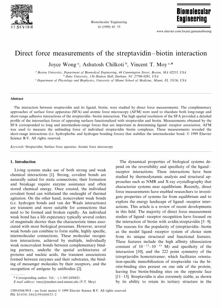
The SFA allows one to directly measure the interac-tion forces between surfaces at the molecular level and,independently and unambiguously, the geometry andabsolute separation distance between two surfaces. Fig.1 shows the central components of the experimentalsetup [30]. The substrates consist of two transparentback-silvered mica sheets of equal thickness glued ontocylindrically curved silica disks. Mica is used because itcan be easily cleaved to be atomically smooth overlarge areas (:a few cm2). The separation distancebetween the two surfaces can be controlled over a rangeof 5 mm with a resolution of 1 A, by a four-stagemechanism of increasing sensitivity. The separation be-tween the surfaces can be measured to 91 A, bymonitoring the movement of the multiple beam inter-ference fringes known as Fringes of Equal ChromaticOrder (FECO) produced when white light passes nor-mally through the two surfaces. When strongly attrac-tive forces are measured, mechanical instabilities canoccur causing the surfaces to jump from one stableposition to another. Instabilities occur whenever thegradient of the force exceeds the spring stiffness.
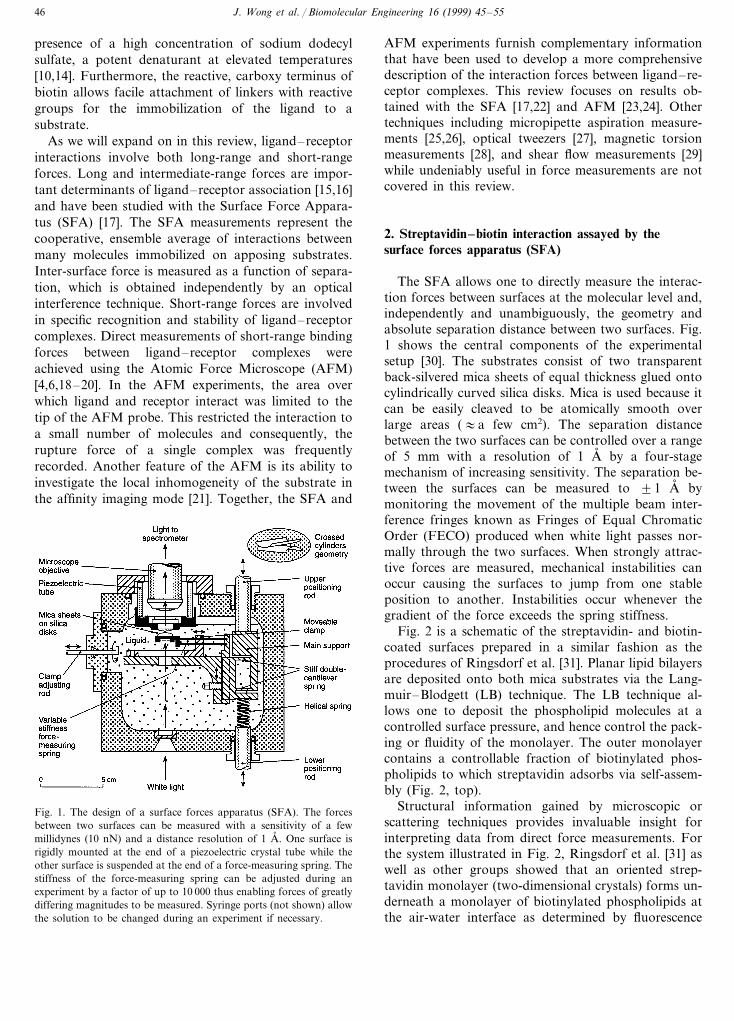
Fig. 2 is a schematic of the streptavidin- and biotin-coated surfaces prepared in a similar fashion as theprocedures of Ringsdorf et al. [31]. Planar lipid bilayersare deposited onto both mica substrates via the Lang-muir–Blodgett (LB) technique. The LB technique al-lows one to deposit the phospholipid molecules at acontrolled surface pressure, and hence control the pack-ing or fluidity of the monolayer. The outer monolayercontains a controllable fraction of biotinylated phos-pholipids to which streptavidin adsorbs via self-assem-bly (Fig. 2, top).
Structural information gained by microscopic orscattering techniques provides invaluable insight forinterpreting data from direct force measurements. Forthe system illustrated in Fig. 2, Ringsdorf et al. [31] aswell as other groups showed that an oriented strep-tavidin monolayer (two-dimensional crystals) forms un-derneath a monolayer of biotinylated phospholipids atthe air-water interface as determined by fluorescence
Fig. 1. The design of a surface forces apparatus (SFA). The forcesbetween two surfaces can be measured with a sensitivity of a fewmillidynes (10 nN) and a distance resolution of 1 A, . One surface isrigidly mounted at the end of a piezoelectric crystal tube while theother surface is suspended at the end of a force-measuring spring. Thestiffness of the force-measuring spring can be adjusted during anexperiment by a factor of up to 10 000 thus enabling forces of greatlydiffering magnitudes to be measured. Syringe ports (not shown) allowthe solution to be changed during an experiment if necessary.
J. Wong et al. / Biomolecular Engineering 16 (1999) 45–55 47
Fig. 2. Schematic representation of a streptavidin-coated surface anda biotin-coated surface. The streptavidin is physisorbed to a biotiny-lated phospholipid bilayer, which is in turn physisorbed to theunderlying mica surface. The tetravalent nature of streptavidin allowsit to be anchored to the bilayer, leaving two empty sites for interac-tion with the apposing biotinylated surface. The silica disks support-ing the mica substrates are in crossed cylinder geometry, and for twocrossed cylinders with 1 cm radii of curvature, the effective contactarea is approximately 100 mm2. The magnitude of this contact areascales with the geometric average radius R; thus, data are alwaysreported as F/R, the force normalized by the average radius ofcurvature.
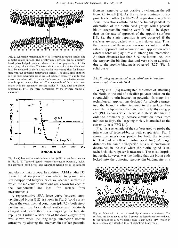
from net negative to net positive by changing the pHfrom 7.2 to 6.0 [17]. As the surfaces continue to ap-proach each other (:10–20 A, separation), repulsivesteric interactions attributed to the time-dependent re-orientation of the biotin head groups which precedebiotin–streptavidin binding were found to be depen-dent on the rate of approach of the apposing surfaces[17], i.e. the steric repulsion is not observed if thesurfaces are approached at a much slower rate. Thus,the time-scale of the interaction is important in that therates of approach and separation and application of anexternal force all play a role in altering the interaction.At short distances less than 5 A, , the biotins lock intothe streptavidin binding sites and very strong adhesiondue to the specific binding is observed [3,22] (Fig. 3,solid curve).
2.1. Probing dynamics of tethered-biotin interactionwith strepta6idin with SFA
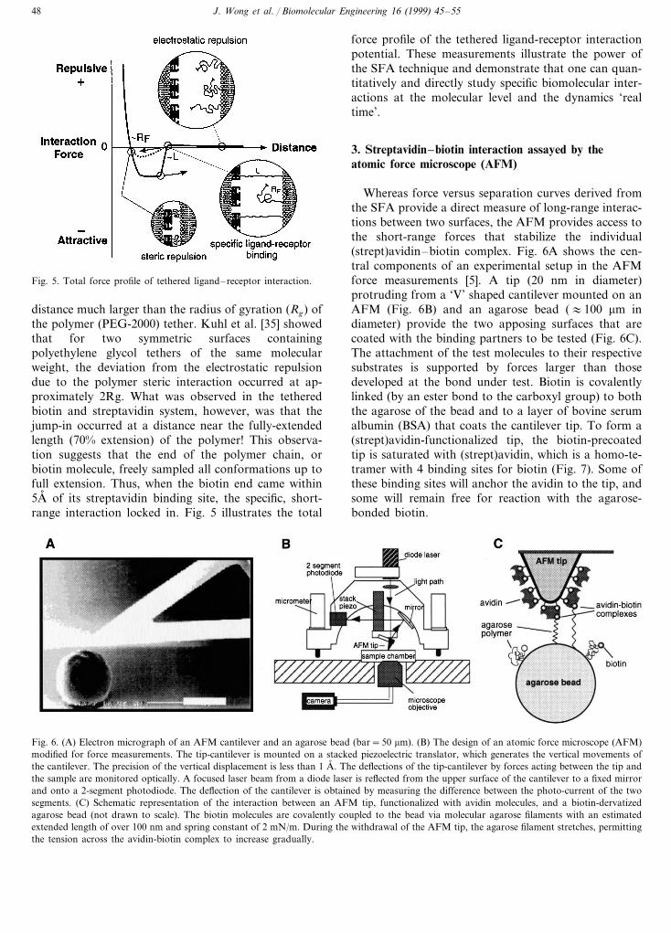
Wong et al. [33] investigated the effect of attachingthe biotin to the end of a flexible polymer tether on thestreptavidin–biotin interaction potential. In many bio-technological applications designed for selective target-ing, the ligand is often tethered to the surface. Forexample, in liposomes decorated with polyethylene gly-col (PEG) chains which serve as a steric stabilizer inorder to dramatically increase circulation times fromminutes to days, the targeting moiety is attached at theextremity of a PEG [34].
Fig. 4 is a schematic of the surfaces used to probe theinteraction of tethered-biotin with streptavidin. Fig. 3shows the interaction profile for both the tethered(circles) and untethered biotin (solid line). At largedistances the same non-specific DLVO interaction asdetermined in the case when the biotin ligand is at-tached via short spacer is measured. The most surpris-ing result, however, was the finding that the biotin endslocked into the opposing streptavidin binding site at aFig. 3. (A) Biotin–streptavidin interaction (solid curve) for schematic
in Fig. 2. (B) Tethered ligand–receptor interaction potential, includ-ing approach (open circles) and separation (closed circles) of surfaces.
Fig. 4. Schematic of the tethered ligand–receptor surfaces. Thesurfaces are the same as in Fig. 2 except the ligands are now tetheredto the surface via a polyethylene glycol chain (2000 MW) which inturn is covalently attached to a phospholipid headgroup.
and electron microscopy. In addition, AFM studies [32]showed that streptavidin can adsorb to planar sub-strate-supported bilayers. Such well-defined surfaces inwhich the molecular dimensions are known for each ofthe components are ideal for surface forcemeasurements.
A representative SFA force curve between strep-tavidin and biotin [3,22] is shown in Fig. 3 (solid curve).Under the experimental conditions (pH 7.2), both strep-tavidin and the biotinylated surface are negativelycharged and hence there is a long-range electrostaticrepulsion. Further verification of the double-layer forcewas shown when the long-range interaction becameattractive by altering the streptavidin surface potential
J. Wong et al. / Biomolecular Engineering 16 (1999) 45–5548
Fig. 5. Total force profile of tethered ligand–receptor interaction.
force profile of the tethered ligand-receptor interactionpotential. These measurements illustrate the power ofthe SFA technique and demonstrate that one can quan-titatively and directly study specific biomolecular inter-actions at the molecular level and the dynamics ‘realtime’.
3. Streptavidin–biotin interaction assayed by theatomic force microscope (AFM)
Whereas force versus separation curves derived fromthe SFA provide a direct measure of long-range interac-tions between two surfaces, the AFM provides access tothe short-range forces that stabilize the individual(strept)avidin–biotin complex. Fig. 6A shows the cen-tral components of an experimental setup in the AFMforce measurements [5]. A tip (20 nm in diameter)protruding from a ‘V’ shaped cantilever mounted on anAFM (Fig. 6B) and an agarose bead (:100 mm indiameter) provide the two apposing surfaces that arecoated with the binding partners to be tested (Fig. 6C).The attachment of the test molecules to their respectivesubstrates is supported by forces larger than thosedeveloped at the bond under test. Biotin is covalentlylinked (by an ester bond to the carboxyl group) to boththe agarose of the bead and to a layer of bovine serumalbumin (BSA) that coats the cantilever tip. To form a(strept)avidin-functionalized tip, the biotin-precoatedtip is saturated with (strept)avidin, which is a homo-te-tramer with 4 binding sites for biotin (Fig. 7). Some ofthese binding sites will anchor the avidin to the tip, andsome will remain free for reaction with the agarose-bonded biotin.
distance much larger than the radius of gyration (Rg) ofthe polymer (PEG-2000) tether. Kuhl et al. [35] showedthat for two symmetric surfaces containingpolyethylene glycol tethers of the same molecularweight, the deviation from the electrostatic repulsiondue to the polymer steric interaction occurred at ap-proximately 2Rg. What was observed in the tetheredbiotin and streptavidin system, however, was that thejump-in occurred at a distance near the fully-extendedlength (70% extension) of the polymer! This observa-tion suggests that the end of the polymer chain, orbiotin molecule, freely sampled all conformations up tofull extension. Thus, when the biotin end came within5A, of its streptavidin binding site, the specific, short-range interaction locked in. Fig. 5 illustrates the total
Fig. 6. (A) Electron micrograph of an AFM cantilever and an agarose bead (bar=50 mm). (B) The design of an atomic force microscope (AFM)modified for force measurements. The tip-cantilever is mounted on a stacked piezoelectric translator, which generates the vertical movements ofthe cantilever. The precision of the vertical displacement is less than 1 A, . The deflections of the tip-cantilever by forces acting between the tip andthe sample are monitored optically. A focused laser beam from a diode laser is reflected from the upper surface of the cantilever to a fixed mirrorand onto a 2-segment photodiode. The deflection of the cantilever is obtained by measuring the difference between the photo-current of the twosegments. (C) Schematic representation of the interaction between an AFM tip, functionalized with avidin molecules, and a biotin-dervatizedagarose bead (not drawn to scale). The biotin molecules are covalently coupled to the bead via molecular agarose filaments with an estimatedextended length of over 100 nm and spring constant of 2 mN/m. During the withdrawal of the AFM tip, the agarose filament stretches, permittingthe tension across the avidin-biotin complex to increase gradually.
J. Wong et al. / Biomolecular Engineering 16 (1999) 45–55 49
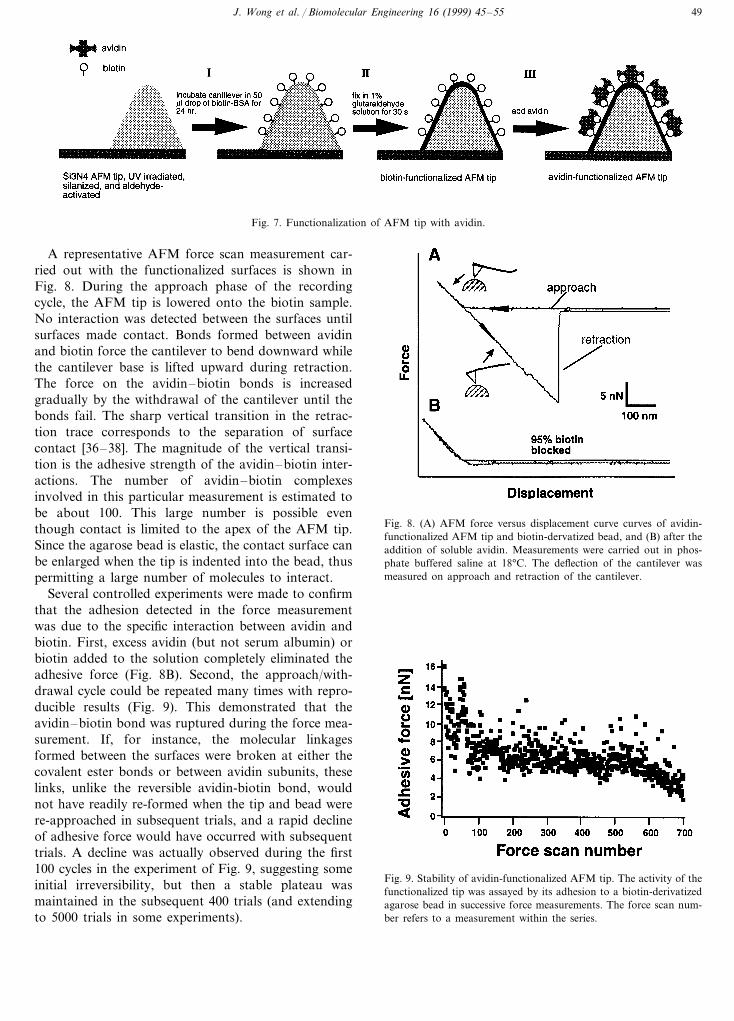
Fig. 7. Functionalization of AFM tip with avidin.
A representative AFM force scan measurement car-ried out with the functionalized surfaces is shown inFig. 8. During the approach phase of the recordingcycle, the AFM tip is lowered onto the biotin sample.No interaction was detected between the surfaces untilsurfaces made contact. Bonds formed between avidinand biotin force the cantilever to bend downward whilethe cantilever base is lifted upward during retraction.The force on the avidin–biotin bonds is increasedgradually by the withdrawal of the cantilever until thebonds fail. The sharp vertical transition in the retrac-tion trace corresponds to the separation of surfacecontact [36–38]. The magnitude of the vertical transi-tion is the adhesive strength of the avidin–biotin inter-actions. The number of avidin–biotin complexesinvolved in this particular measurement is estimated tobe about 100. This large number is possible eventhough contact is limited to the apex of the AFM tip.Since the agarose bead is elastic, the contact surface canbe enlarged when the tip is indented into the bead, thuspermitting a large number of molecules to interact.
Several controlled experiments were made to confirmthat the adhesion detected in the force measurementwas due to the specific interaction between avidin andbiotin. First, excess avidin (but not serum albumin) orbiotin added to the solution completely eliminated theadhesive force (Fig. 8B). Second, the approach/with-drawal cycle could be repeated many times with repro-ducible results (Fig. 9). This demonstrated that theavidin–biotin bond was ruptured during the force mea-surement. If, for instance, the molecular linkagesformed between the surfaces were broken at either thecovalent ester bonds or between avidin subunits, theselinks, unlike the reversible avidin-biotin bond, wouldnot have readily re-formed when the tip and bead werere-approached in subsequent trials, and a rapid declineof adhesive force would have occurred with subsequenttrials. A decline was actually observed during the first100 cycles in the experiment of Fig. 9, suggesting someinitial irreversibility, but then a stable plateau wasmaintained in the subsequent 400 trials (and extendingto 5000 trials in some experiments).
Fig. 8. (A) AFM force versus displacement curve curves of avidin-functionalized AFM tip and biotin-dervatized bead, and (B) after theaddition of soluble avidin. Measurements were carried out in phos-phate buffered saline at 18°C. The deflection of the cantilever wasmeasured on approach and retraction of the cantilever.
Fig. 9. Stability of avidin-functionalized AFM tip. The activity of thefunctionalized tip was assayed by its adhesion to a biotin-derivatizedagarose bead in successive force measurements. The force scan num-ber refers to a measurement within the series.
J. Wong et al. / Biomolecular Engineering 16 (1999) 45–5550
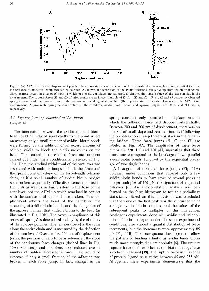
Fig. 10. (A) AFM force versus displacement profile. Under conditions where a small number of avidin–biotin complexes are permitted to form,the breakage of individual complexes can be detected. As shown, the separation of the avidin-functionalized AFM tip from the biotin-function-alized agarose occurs in a series of steps in which one to six complexes are ruptured. f3 denotes the rupture force of the last complex in themeasurement. The rupture forces (f1 and f2) of prior events are an integer multiple of f3; f1=2f3 and f2= f3. k1, k2 and k3 denote the observedspring constants of the system prior to the rupture of the designated bond(s). (B) Representation of elastic elements in the AFM forcemeasurement. Approximate spring constant values of the cantilever, avidin–biotin bond, and agarose polymer are 60, 2, and 200 mN/m,respectively.
3.1. Rupture force of indi6idual a6idin–biotincomplexes
The interaction between the avidin tip and biotinbead could be reduced significantly to the point whereon average only a small number of avidin–biotin bondswere formed by the addition of an excess amount ofsoluble avidin to block the biotin molecules on thebead. The retraction trace of a force measurementcarried out under these conditions is presented in Fig.10A. Here, the gradual withdrawal of the cantilever wasassociated with multiple jumps in both the force andthe spring constant (slope of the force-length relation-ship), as if a small number of avidin–biotin bridgeswere broken sequentially. (The displacement plotted inFig. 10A as well as in Fig. 8 refers to the base of thecantilever, not the AFM tip which remained in contactwith the surface until all bonds are broken. This dis-placement reflects the bend of the cantilever, thestretching of avidin-biotin bonds, and the elongation ofthe agarose filament that anchors biotin to the bead (asillustrated in Fig. 10B). The overall compliance of thisseries of ‘springs’ is determined mainly by the elasticityof the agarose polymer. The tension (force) is the samealong the entire chain and is measured by the deflectionof the cantilever.) Over the first 150 nm of displacement(using the position of zero force as reference), the slopeof the continuous force changes (dashed lines in Fig.10A) was steep and not detectably reduced over anumber of sudden decreases in force. This would beexpected if only a small fraction of the adhesion wasbroken in each force jump. In fact, changes in the
spring constant only occurred at displacements atwhich the adhesion force had dropped substantially.Between 200 and 300 nm of displacement, there was aninterval of small slope and zero tension, as if followingthe preceding force jump there was slack in the remain-ing bridges. Three force jumps (f1, f2 and f3) arelabeled in Fig. 10A. The amplitudes of these forcejumps are 320, 160 and 160 pN, suggesting that thesetransitions correspond to the breakage of two parallelavidin-biotin bonds, followed by the sequential break-age of two single bonds.
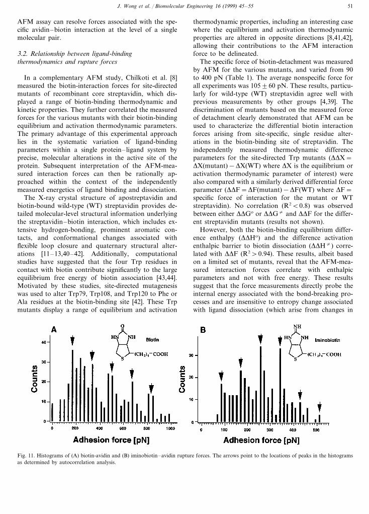
A histogram of measured force jumps (Fig. 11A)obtained under conditions that allowed only a fewavidin-biotin bonds to form revealed several peaks atinteger multiples of 160 pN, the signature of a quantalbehavior [6]. An autocorrelation analysis was per-formed on the force histogram to test this periodicitystatistically. Based on this analysis, it was concludedthat the value of the first peak was the rupture force ofa single avidin–biotin complex, and the values of thesubsequent peaks to multiples of this interaction.Analogous experiments done with avidin and iminobi-otin, a biotin analogue, under the same experimentalconditions, also yielded a quantal distribution of forceincrements, but the increments were approximately 85pN (Fig. 11B). The force quanta thus appear to followthe pattern of binding affinity, as avidin binds biotinmuch more strongly than iminobiotin [6]. The unitaryrupture force of three other avidin-biotin analogs havealso been measured [39]. The rupture force in this seriesof protein–ligand pairs varies between 85 and 255 pN.Altogether, these experiments demonstrate that the
J. Wong et al. / Biomolecular Engineering 16 (1999) 45–55 51
AFM assay can resolve forces associated with the spe-cific avidin–biotin interaction at the level of a singlemolecular pair.
3.2. Relationship between ligand-bindingthermodynamics and rupture forces
In a complementary AFM study, Chilkoti et al. [8]measured the biotin-interaction forces for site-directedmutants of recombinant core streptavidin, which dis-played a range of biotin-binding thermodynamic andkinetic properties. They further correlated the measuredforces for the various mutants with their biotin-bindingequilibrium and activation thermodynamic parameters.The primary advantage of this experimental approachlies in the systematic variation of ligand-bindingparameters within a single protein–ligand system byprecise, molecular alterations in the active site of theprotein. Subsequent interpretation of the AFM-mea-sured interaction forces can then be rationally ap-proached within the context of the independentlymeasured energetics of ligand binding and dissociation.
The X-ray crystal structure of apostreptavidin andbiotin-bound wild-type (WT) streptavidin provides de-tailed molecular-level structural information underlyingthe streptavidin–biotin interaction, which includes ex-tensive hydrogen-bonding, prominent aromatic con-tacts, and conformational changes associated withflexible loop closure and quaternary structural alter-ations [11–13,40–42]. Additionally, computationalstudies have suggested that the four Trp residues incontact with biotin contribute significantly to the largeequilibrium free energy of biotin association [43,44].Motivated by these studies, site-directed mutagenesiswas used to alter Trp79, Trp108, and Trp120 to Phe orAla residues at the biotin-binding site [42]. These Trpmutants display a range of equilibrium and activation
thermodynamic properties, including an interesting casewhere the equilibrium and activation thermodynamicproperties are altered in opposite directions [8,41,42],allowing their contributions to the AFM interactionforce to be delineated.
The specific force of biotin-detachment was measuredby AFM for the various mutants, and varied from 90to 400 pN (Table 1). The average nonspecific force forall experiments was 105960 pN. These results, particu-larly for wild-type (WT) streptavidin agree well withprevious measurements by other groups [4,39]. Thediscrimination of mutants based on the measured forceof detachment clearly demonstrated that AFM can beused to characterize the differential biotin interactionforces arising from site-specific, single residue alter-ations in the biotin-binding site of streptavidin. Theindependently measured thermodynamic differenceparameters for the site-directed Trp mutants (DDX=DX(mutant)−DX(WT) where DX is the equilibrium oractivation thermodynamic parameter of interest) werealso compared with a similarly derived differential forceparameter (DDF=DF(mutant)−DF(WT) where DF=specific force of interaction for the mutant or WTstreptavidin). No correlation (R2B0.8) was observedbetween either DDGo or DDG" and DDF for the differ-ent streptavidin mutants (results not shown).
However, both the biotin-binding equilibrium differ-ence enthalpy (DDHo) and the difference activationenthalpic barrier to biotin dissociation (DDH") corre-lated with DDF (R2\0.94). These results, albeit basedon a limited set of mutants, reveal that the AFM-mea-sured interaction forces correlate with enthalpicparameters and not with free energy. These resultssuggest that the force measurements directly probe theinternal energy associated with the bond-breaking pro-cesses and are insensitive to entropy change associatedwith ligand dissociation (which arise from changes in
Fig. 11. Histograms of (A) biotin-avidin and (B) iminobiotin–avidin rupture forces. The arrows point to the locations of peaks in the histogramsas determined by autocorrelation analysis.
J. Wong et al. / Biomolecular Engineering 16 (1999) 45–5552
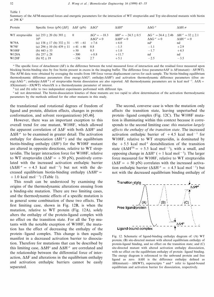
Table 1Summary of the AFM-measured forces and energetic parameters for the interaction of WT streptavidin and Trp site-directed mutants with biotinat 298 Ka
DDF (pN) DDG° DDH°Protein DDG"Specific force (pN) [DF] DDH"
DH°=−24.590.50WT streptavidin DG°=−18.3(a) 253920 (b) 3939 DG"=24.492.4b DH"=3292.110b DDG"=0DDH°=0 DDH"=0DDG°=0
−95 −61 +7.6W79A +6.0(a) 158917 (b) 332931 ndc nd+41 +46 0.8 −1.5(a) 294910 (b) 439911 −1.1W79F +2.9
(b) 443933W108F +50 0.5 +1.0 −1.7 +4.5W120A −300(b) 257928 +8.8 +11.7 nd nd
−136 2.7 +5.1 −2.5(b) 92919 −3.5W120F
a The specific force of detachment (DF) is the difference between the total measured force of interaction and the residual force measured uponblocking biotin-binding sites by free biotin present in excess in the imaging buffer. The difference force parameterDDF is DF(mutant)−DF(WT).The AFM data were obtained by averaging the results from 100 force versus displacement curves for each sample. The biotin-binding equilibriumthermodynamic difference parameters (free energy:DDG°; enthalpy:DDH°) and activation thermodynamic difference parameters ((free en-ergy:DDG"; enthalpy:DDH") of streptavidin–biotin dissociation are also reported. All thermodynamic parameters are in kcal mol−1. DDX=DX(mutant)−DX(WT) whereDX is a thermodynamic parameter of interest.
b (a) and (b) refer to two independent experiments performed with different tips.c nd: not determined. The biotin-dissociation kinetics of these mutants are too rapid to allow determination of the activation thermodynamic
parameters by the methods utilized for the other mutants in this study.
the translational and rotational degrees of freedom ofligand and protein, dilution effects, changes in proteinconformation, and solvent reorganization) [45,46].
However, there was an important exception to thisoverall trend for one mutant, W108F, which allowedthe apparent correlation of DDF with both DDHo andDDH" to be examined in greater detail. The activationenthalpy for dissociation (DH") and the equilibriumbiotin-binding enthalpy (DHo) for the W108F mutantare altered in opposite directions, relative to WT strep-tavidin. The larger interaction force for W108F, relativeto WT streptavidin (DDF= +50 pN), positively corre-lated with the increased activation enthalpic barrier(DDH"= +4.5 kcal mol−1) but not with the de-creased equilibrium biotin-binding enthalpy (DDHo=+1.0 kcal mol−1) (Table 1).
This result can be understood by examining theorigins of the thermodynamic alterations ensuing froma binding-site mutation. There are two limiting cases,and the thermodynamic effects of a specific mutation isin general some combination of these two effects. Thefirst limiting case, shown in Fig. 12B, is when themutation, relative to WT protein (Fig. 12A), solelyalters the enthalpy of the protein-ligand complex withno effect on the transition state. For all the Trp mu-tants with the notable exception of W108F, the muta-tion has the effect of decreasing the enthalpy of theprotein–ligand complex. This change is then equallymanifest in a decreased activation barrier to dissocia-tion. Therefore for mutations that can be described bythis limiting case, DDHo and DDH" are correlated andthe relationship between the differential force of inter-action, DDF and alterations in the equilibrium enthalpyand activation enthalpic barriers cannot be easilyseparated.
The second, converse case is when the mutation onlyaffects the transition state, leaving unperturbed theprotein–ligand complex (Fig. 12C). The W108F muta-tion is illuminating within this context because it corre-sponds to the second limiting case: this mutation largelyaffects the enthalpy of the transition state. The increasedactivation enthalpic barrier of +4.5 kcal mol−1 forW108F, relative to WT streptavidin, is dominated bythe +5.5 kcal mol-1 destabilization of the transitionstate (DDHTS= +5.5 kcal mol−1), with a small, andopposing change in DDHo (+1 kcal mol−1). The largerforce measured for W108F, relative to WT streptavidin(DDF= +50 pN) correlates with the increased activa-tion enthalpic barrier (DDH"= +4.5 kcal mol−1) butnot with the decreased equilibrium binding enthalpy of
Fig. 12. Schematic of ligand-binding enthalpy diagram of: (A) WTprotein; (B) site-directed mutant with altered equilibrium enthalpy ofprotein-ligand binding, and no effect on the transition state; and (C)site-directed mutant with altered activation enthalpy dissociation,with no effect on the equilibrium enthalpy of protein–ligand binding.The energy diagram is referenced to the unbound protein and freeligand as zero. DDH is the difference enthalpy defined asDH(mutant)−DH(WT), superscripts ° and" refer to ligand-boundequilibrium and activation barrier for dissociation, respectively.
J. Wong et al. / Biomolecular Engineering 16 (1999) 45–55 53
W108F (DDHo= +1 kcal mol−1). These results sug-gest that the AFM detachment process for this mutantfollows the enthalpic barrier to dissociation, and thatthe apparent correlation of DDFo with DDHo for all theother mutants arises because changes in the enthalpylevel of the ligand-bound state, i.e. the protein–ligandcomplex, are folded into the magnitude of the activa-tion enthalpy.
4. Discussion
4.1. Energetics of tethered receptor– ligand interaction
The results from the SFA tethered receptor–ligandexperiments demonstrate that the total energy of thesystems is controlled by both the ligand-receptor bondenergy and the length of the tether. The polymer chaindynamics of the tethered receptor may be a generalmeans of controlling the effective receptor–ligand on-rate process independently from the specific ligand–re-ceptor affinity in biological interactions. The questionthen becomes if this is reasonable from an energeticstandpoint. One needs to estimate the energy needed tostretch a polymer chain to near full extension. Theoriesof end-grafted polymer chains have predicted that thefree chain ends are, on average, located at a distance of0.7 Rgfrom the anchoring surface. However, thermalfluctuations allow the polymer to sample all possibleconfigurations ranging from a random-coil to nearlyfully extended state. The typical exploration time, t, fora particle diffusing in an external potential, Eext(D), (inour case, the time it takes for a tethered biotin to findits binding site) is given by Kramers’ equation:
t(D)=t0exp[Eext(D)/kT ] (1)
where k is the Boltzmann constant and T is tempera-ture. The basic attempt rate for a chain to reach nearfull extension, t0, is the Zimm time
t0:hRg
3
kT:10−8s (2)
where h is the viscosity of water. A rough estimate ofEext(D) for D\Rg is to model the polymer chain as aspring with a parabolic potential:
Eext(D)=(D/Rg)2kT
2(3)
For PEG (2000 MW), D/Rg=5, and Eqs. (3) and (2)and Eq. (1) give t in the millisecond range which isconsistent with the observation of a rapid locking-on.
However, a polymer does not strictly follow aparabolic potential. A more refined Monte-Carlo simu-lation of a single polymer chain (manuscript in prepara-tion) shows that a polymer chain of 45 monomer units(2000 MW) is able to reach 70% extension in 1 s, which
is of the time-scale of our measurements. Thus, themeasured tethered interaction potential and its dynam-ics can still be modeled using standard theories ofpolymer and colloidal interactions.
4.2. Separation of surfaces: ligand–receptor bondfailure 6ersus membrane failure
In contrast to the AFM experiments where the strep-tavidin-biotin bond is ruptured, in SFA experimentslipid pull-out was observed. In the SFA experiments,streptavidin and biotin were immobilized onto planarlipid bilayers which leads to competing failure mecha-nisms: [1] ligand-receptor bond failure or [2] membranefailure. When lipids are extracted from a membrane,the energy to pull out a lipid from a bilayer is 16 kT.Assuming that potential changes linearly over the entirelength of the lipid (in our case, 28 A, ), the force neededto extract the lipid from the membrane is 23 pN [3]. Onthe other hand, the streptavidin–biotin bond energy is31 kT, and its effective length is approximately 9 A,giving a force of 140 pN. Thus, for both the PEG-teth-ered biotinylated lipid and the biotinylated lipid, amuch lower force is needed to pull out lipids than tobreak individual streptavidin–biotin bonds. In addi-tion, significant hysteresis in the force curve givingdecreased adhesion on subsequent approach of thesurfaces as well as an increase in the layer thicknessindicates that the lipids are being pulled out of themembrane and remain on the surface coated with strep-tavidin rather than streptavidin–biotin bond breakage.This has been tested for a series of biotin-analogs withaffinities ranging over 10 orders of magnitude [47] inwhich for sufficiently weak biotin-analogs, the recep-tor–ligand bond was the weaker link and the expectedchangeover in the bond failure mechanism wasobserved.
4.3. Theoretical approaches: dynamic strength ofligand–receptor interactions
Close to physiological conditions, the dissociation ofthe avidin–biotin complex is slow, and involves cross-ing an activation energy barrier of more than 20 kcal/mol. As demonstrated by the AFM measurement, thedissociation time can be reduced significantly by theapplication of an external force. Theoretically, the ex-ternal force causes a distortion in the pair potential ofthe streptavidin-biotin complex that reduces the lifetimeof the bond. Bell argued that the activation energy ofbiochemical bond is reduced by f°g, where f° is theapplied force and g is a displacement parameter thatcharacterizes the interaction [48,49]. The lifetime of thecomplex is then: t=toexp[(Eo-f°g)/kT ], where Eo is theintrinsic dissociation energy barrier and to is a phe-nomenological prefactor. More recently, Evans ex-
J. Wong et al. / Biomolecular Engineering 16 (1999) 45–5554
tended the Bell model to consider the effects of viscousdamping and hydrodynamics on the forced dissociationof biochemical bonds [50]. Applying Kramers’ theoryfor reaction kinetics in condensed liquid to the bonddissociation under external force [51], Evans andRitchie showed that the unbinding force of biochemicalbonds progresses through three regimes of loading rate.In the slow-loading regime that characterized manyphysiological processes and the AFM experiments, theunbinding force increased as a weak power of theloading rate. At intermediate loading rates, the strengthof the bond increased as the logarithm of the loadingrate. In the ultrafast regime that approaches the regimeof molecular dynamic (MD) calculations, the loadingforce overwhelmed the bonding potential and only fric-tion remained to retard dissociation.
While the theoretical models have provided a keylink between the AFM unbinding force and loadingrate, many questions regarding the energetics and reac-tion trajectories remain to be elucidated. MD simula-tion has provided some insight into the unbindingpathway of the streptavidin-biotin complex. Grub-muller et al. [52] simulated the forced unbinding ofbiotin from the streptavidin monomer in a watersphere. The dissociation of the complex was induced bythe displacement of a spring potential assigned to thecarboxyl group of biotin. The trajectory of the restoringforce revealed an overall increase of force, followed bya drop to the baseline as separation was achieved. Theforce profile also revealed several jumps that coincidedwith the separation of individual hydrogen bonds be-tween the binding pocket wall and biotin. In addition,the simulation identified water in playing a prominentrole throughout the dissociation process by bridging theinteractions between biotin and the hydrophilic residuesin the binding pocket.
In a separate MD study, Izrailev et al. [53] examinedthe unbinding of biotin from the avidin tetramer in theabsence of water. The ground state of the avidin–biotincomplex is stabilized by hydrogen bonds formed be-tween the head group of biotin and polar residues inthe binding pocket, most notably Asn12 and Tyr33,and to an lesser extent, by van der Waals interactionswith Phe79 and Trp97. With increasing applied force,the system undergoes transitions into 2 well-definedintermediate states in which positions of biotin re-mained relatively constant with increased force applica-tion. The first intermediate state is stabilized byhydrogen bonds involving Thr35 and Ser16, and vdwinteractions involving Trp110 and Trp70. After thebiotin is extracted from the binding pocket, it stillremained associated with the avidin via interactionbetween the biotin head group and the avidin residuesoutside the pocket, including those from the 3–4 loopof an adjacent subunit.
It should be noted that the MD calculations werecarried out with separation velocities that were at leastsix orders of magnitude higher than the fastest velocitythat was tested experimentally. While a direct compari-son of the AFM experiments and MD simulations maynot be appropriate, the MD calculations raise the possi-bility that the unbinding of the streptavidin–biotincomplex may involve transitions between intermediatestates before total separation occur. The dissociation ofa complex that involves series of intermediates mayhave unbinding forces significantly lower than com-plexes that undergo a cooperative dissociation of alltheir intermolecular bonds. Future experiments thatinvestigate the kinetics of the stressed streptavidin–bi-otin bond may provide more direct evidence for theexistence of intermediate states. Finally, expandingthese studies to include complementary experimentalsystems where ligand-binding is dominated by entropicprocesses rather than enthalpy should further clarifythe relationship between solution thermodynamicparameters and the AFM-measured forces.
Acknowledgements
The authors would like to thank J.N. Israelachvili forhelpful discussions. J.Y.W. is grateful for support froma NIH NRSA postdoctoral fellowship (GM17876).V.T.M is supported by the NIH (1 R29 GM55611-01).
References
[1] Creighton TE. Proteins. 2nd ed. New York: WH Freeman andCompany, 1993.
[2] Fersht A. Enzyme structure and mechanism. 2nd ed. New York:WH Freeman and Company, 1985.
[3] Helm C, Knoll W, Israelachvili JN. Proc Natl Acad Sci USA1991;88:8169–73.
[4] Lee GU, Kidwell DA, Colton RJ. Langmuir 1994;10:354–61.[5] Moy VT, Florin E-L, Gaub HE. Colloids Surfaces 1994;93:343–
8.[6] Florin E-L, Moy VT, Gaub HE. Science 1994;264:415–7.[7] Pierce M, Stuart J, Pungor A, Dryden P, Hlady V. Langmuir
1994;10:3217–21.[8] Chilkoti A, Boland T, Ratner BD, Stayton PS. Biophys J
1995;69:2125–30.[9] Allen S, Davies J, Dawkes AC, Davies MC, Edwards JC, Parker
MC, et al. FEBS Lett 1996;390:161–4.[10] Green NM. Adv Prot Chem 1975;29:85–133.[11] Weber PC, Ohlendorf DH, Wendoloski JJ, Salemme FR. Science
1989;243:85–8.[12] Hendrickson WA, Pahler A, Smith JL, Satow Y, Merritt EA,
Phizackerley RP. Proc Natl Acad Sci USA 1989;86:2190–4.[13] Livnah O, Bayer EA, Wilchek M, Sussman JL. Proc Natl Acad
Sci USA 1993;90:5076–80.[14] Sano T, Pandori MW, Smith CL, Cantor CR. In: Uhlen M,
Hornes E, Olsvik Ø, editors. Advances in Biomagnetic Separa-tion. Natick, MA: Eaton, 1994:21–9.
J. Wong et al. / Biomolecular Engineering 16 (1999) 45–55 55
[15] Israelachvili JN. Intermolecular and surface forces. 2nd ed.London: Academic Press, 1992.
[16] Gilson MK, Straatsma TP, McCammon JA, Ripoll DR, Faer-man CH, Axelsen PH, et al. Science 1994;263:1276–8.
[17] Leckband DE, Schmitt FJ, Israelachvili JN, Knoll W. Biochem-istry 1994;33:4611–24.
[18] Hinterdorfer P, Baumgartner W, Gruber HJ, Schilcher K, Schin-dler H. Proc Natl Acad Sci USA 1996;93:3477–81.
[19] Lee GU, Chrisey LA, Colton RJ. Science 1994;266:771–3.[20] Dammer U, Popescu O, Wagner P, Anselmetti D, Guntherodt
H-J, Misevic GN. Science 1995;267:1173–5.[21] Ludwig M, Dettmann W, Gaub HE. Biophys J 1997;72:445–8.[22] Leckband DE, Israelachvili JN, Schmitt FJ, Knoll W. Science
1992;255:1419–21.[23] Binnig G, Rohrer H. Rev Mod Phys 1987;59:615–25.[24] Frommer J. Angew Chem Int Ed Engl 1992;31:1265–82.[25] Evans E, Berk D, Leung A. Biophys J 1991;59:838–48.[26] Evans E, Ritchie K, Merkel R. Biophys J 1995;68:2580–7.[27] Stout AL, Webb WW. Meth Cell Biol 1998;55:99–116.[28] Wang N, Bulter JP, Ingber PE. Science 1993;260:1124–7.[29] Kuo SC, Lauffenburger DA. Biophys J 1993;65:2191–200.[30] Israelachvili JN, McGuiggan PM. J Mater Res 1990;5:2223–31.[31] Darst SA, Ahlers M, Meller P, Kubalek EW, Blankenburg R,
Ribi HO, et al. Biophys J 1991;59:387–96.[32] Weisenhorn AL, Schmitt FJ, Knoll W, Hansma PK. Ultrami-
croscopy 1992;42–44(Part B):1125–32.[33] Wong JY, Kuhl TL, Israelachvili JN, Mullah N, Zalipsky S.
Science 1997;275:820–2.[34] Allen TM, Brandeis E, Hansen CB, Kao GY, Zalipsky S.
Biochim Biophys Acta 1995;1237:99–108.
[35] Kuhl TL, Leckband DE, Lasic DD, Israelachvili JN. Biophys J1994;66:1479–88.
[36] Pethica JB, Oliver WC. Physica Scripta 1987;T19:61–6.[37] Burnham NA, Colton RJ, Pollock HM. J Vac Soc Technol
1991;A9:2548–56.[38] Hoh JH, Cleveland JP, Prater CB, Revel JP, Hansma PK. J Am
Chem Soc 1992;114:4917–8.[39] Moy VT, Florin E-L, Gaub HE. Science 1994;266:257–9.[40] Weber PC, Wendoloski JJ, Pantoliano MW, Salemme FR. J Am
Chem Soc 1992;114:3197–200.[41] Chilkoti A, Stayton PS. J Am Chem Soc 1995;117:10622–8.[42] Chilkoti A, Tan PH, Stayton PS. Proc Natl Acad Sci USA
1995;92:17544–8.[43] Miyamoto S, Kollman PA. Proteins 1993;16:226–45.[44] Miyamoto S, Kollman PA. Proc Natl Acad Sci USA
1993;90:8402–6.[45] Spolar RS, Record Jr MT. Science 1994;263:777–84.[46] Vajda S, Weng Z, Rosenfeld R, DeLisi C. Biochemistry
1994;33:13977–88.[47] Leckband D, Muller W, Schmitt FJ, Ringsdorf H. Biophys J
1995;69:1162–9.[48] Bell GI. Science 1978;200:618–27.[49] Dembo M, Torney DC, Saxman K, Hammer D. Proc R Soc
Lond Ser B 1988;234:55–83.[50] Evans E, Ritchie K. Biophys J 1997;72:1541–55.[51] Hanggi P, Talkner P, Borkovec M. Rev Mod Phys 1990;62:251–
341.[52] Grubmuller H, Heymann B, Tavan P. Science 1996;271:997–9.[53] Izrailev S, Stepaniants S, Balsera M, Oono Y, Schulten K.
Biophys J 1997;72:1568–81.
.