Embed Size (px)
Citation preview

International Journal of Applied Engineering Research ISSN 0973-4562 Volume 12, Number 23 (2017) pp. 13552-13564
© Research India Publications. http://www.ripublication.com
13552
A Biotin–Streptavidin Module for Signal Amplification in
Immunochromatographic Analysis to Control Antibiotic Levels
O.D. Hendrickson, A.E. Urusov, D.A. Kuznetsova, A.V. Zherdev and B.B. Dzantiev*
A.N. Bach Institute of Biochemistry, Federal Research Center «Fundamentals of Biotechnology» of the Russian Academy of Sciences,
Leninsky prospect 33, 119071, Moscow, Russia. * Corresponding author
Orcid: 0000-0003-3799-9383
Abstract
In this study, we proposed a new amplification scheme to
improve the sensitivity of immunochromatographic analysis
(ICA) and maintain high-intensity coloration. It was based on
the indirect introduction of a label into immune complexes
formed via a biotin–streptavidin module. The registered signal
(test zone coloration) was amplified by multiplying the label
binding. During the assay, biotinylated antibodies interacted
with colored conjugates of streptavidin and gold nanoparticles
(GNPs), and the addition of a biotinylated protein promoted
aggregation of the label. In the developed assay, the detection
limit of streptomycin (STR) was 30 ng/mL; the achieved
characteristics exceeded those of standard competitive ICA
using antibodies linked directly to a label. Because of the
universal nature of the proposed amplification scheme, it
could be applied to other compounds, including antibiotics
from different chemical classes. Such a sensitive ICA
amplification scheme could also be considered an efficient
screening tool for antibiotics in medical monitoring and food
safety.
Keywords: immunochromatography, amplification, biotin–
streptavidin module, antibiotics, nanoparticles aggregation
INTRODUCTION
Antibiotics are not only considered efficient therapeutic
preparations but also important risk factors in human health.
Resistance to antibiotics is one of the most serious health
concerns related to the uncontrolled consumption of
antibiotics. In addition, changes in intestinal microflora cause
disbacteriosis, leading to the emergence of allergic diseases,
the inhibition of vitamin synthesis, and the propagation of
pathogenic microbes in organs and tissues [1]. Thus, efficient
analytical tools to screen for antibiotic levels are in high
demand, both for making therapeutic decisions and ensuring
food safety.
Various physicochemical and biochemical approaches are
used for antibiotic detection [2-4]. Among the latter,
immunochemical methods are often used, due to their
simplicity and sensitivity, and that they can be utilized
without complex, high-cost equipment [5-7].
Immunochromatographic analysis most closely meets the
demands of rapid and accurate monitoring [8]. This assay
(also known as lateral flow immunoassay) uses the contact
between a multi-membrane test-strip with immobilized
reactants and a test sample. After this contact, the reagents
move along the test strip, resulting in specific interactions and
the formation of colored zones. Furthermore, ICA can be
applied without the use of additional equipment and reagents.
However, ICA is typically less sensitive than other
immunochemical methods [9]. Thus, new assay formats are
necessary for the ICA of compounds (including antibiotics)
that should be controlled in samples containing extremely low
antibiotics’ concentration.
The immunochromatographic assays used for low molecular
weight compounds traditionally include analyte-specific
antibodies directly linked to a colored marker, gold
nanoparticles [8]. Reducing the concentration of antibody–
GNP conjugate for the purpose of lowering the detection limit
decreases the concentration of colored markers and the
magnitude of the detected signal, thus reducing sensitivity. An
indirect introduction of the marker into detectable specific
immune complexes (so-called indirect labeling) may solve
this problem [10-12].
In this study, we proposed a new type of competitive ICA that
indirectly introduces a label via the streptavidin–biotin
module and the subsequent aggregation of GNPs. The biotin–
streptavidin system is characterized by its unique high affinity
(Kd = 10-15 М) and specificity of interaction [13]. The binding
of streptavidin–biotin modules with any label (fluorophore,
enzyme, or nanoparticle) is often used to introduce markers in
immunoassays [14, 15]. The detected signal was amplified
after multiple interactions (each streptavidin molecule binds
four molecules of biotin). Thus, applying this ICA
amplification scheme may ensure high specificity, rapid
interaction, and resistance to changes in temperature, pH, and
denaturizing agents.
In the proposed assay, conjugates of GNPs with streptavidin,
and biotinylated monoclonal antibodies and bovine serum
albumin (BSA) were used instead of the traditional conjugates
of specific antibodies with GNPs. These structures formed
large complexes in the analytic zone of the test strip:

International Journal of Applied Engineering Research ISSN 0973-4562 Volume 12, Number 23 (2017) pp. 13552-13564
© Research India Publications. http://www.ripublication.com
13553
biotinylated antibodies bound to streptavidin–GNPs
conjugates and BSA–biotin aggregated several markers. Such
multiple interactions led to the appearance of a test line with
high color intensity; thereby allowing for a lower detection
limit.
This study investigated streptomycin (STR) which belongs to
the group of aminoglycosides used to treat a number of
bacterial infections in cattle, such as tuberculosis,
endocarditis, brucellosis, plague, tularemia, and fever [16-18].
However, STR is known to promote the growth of
microorganisms resistant to antibiotics [18, 19], which means
there is an urgent need to control STR. Several studies have
focused on STR detection by immunoassay techniques,
including immunochromatography [20-22]. However, they
were based on traditional ICA formats and did not
demonstrate the high sensitivity required for medical
monitoring. In contrast, the universal character of the
proposed amplification scheme allows for ICA to be applied
to other antibiotics, as well as other low molecular weight
analytes.
EXPERIMENTAL
Materials, reagents, and equipment
The BSA, 3,3’,5,5’-tetramethylbenzidine, chloroauric acid,
Triton X-100, dimethyl sulfoxide (DMSO), cyanuric chloride,
and triethylamine were from Sigma-Aldrich (USA); the
biotin-N-hydroxysuccinimide ester was from ICN
Biomedicals (USA); the STR sulfate was from AppliChem
(Germany); and the recombinant STR was from IMTEK
(Russia). The monoclonal antibodies against STR were
obtained from the All-Russian Center of Molecular
Diagnostics and Therapy (Moscow, Russia), the goat anti-
mouse (GAM) polyclonal antibodies and peroxidase-labeled
anti-mouse immunoglobulins were obtained from the
Gamaleya Institute of Microbiology and Epidemiology
(Moscow, Russia). The purity of all other reagents (salts,
acids, alkalis, and organic solvents) was of analytical grade or
higher. Milli-Q deionized water (Millipore, USA) was used to
prepare the solutions. Costar 9018 transparent microplates
(Corning, USA) were used for the enzyme-linked
immunosorbent assay (ELISA). Optical density was measured
using a microplate photometer Zenyth 3100 (Anthos Labtec
Instruments, Austria).
The preparation of the GNPs
GNPs with an average diameter of 30 nm were synthesized
according to [23]. Briefly, 0.5 mL of a 1% water solution of
HAuCl4 was added to 48.75 mL of water. The mixture was
brought to the boil, and then 0.75 mL of a 1% sodium citrate
solution was added. After boiling for 30 min, the preparation
was cooled. The obtained GNPs were stored at 4∘C.
The characterization of GNPs by transmission electron
microscopy (TEM)
The aliquot of the GNP preparation was dropped onto a
formvar film-coated grid and studied by transmission electron
microscopy (TEM) on a JEM-100C microscope (Jeol, Japan)
at an accelerating voltage of 80 kV. The obtained images were
analyzed using the ImageTool program (UTHSCSA, USA).
The selection of protein concentration for conjugation with
GNPs
For conjugation with GNPs, 100 µL of aqueous solutions of
anti-STR antibodies or streptavidin (at concentrations from 5
to 200 µg/mL) were mixed with 1 mL of the GNPs (OD520 =
1) and incubated for 10 min at room temperature while
stirring. Then, 0.1 mL of a 10% NaCl solution was added and
incubated for 10 min at room temperature while stirring.
Finally, the optical density at 580 nm was measured.
The immobilization of the anti-STR antibodies and
streptavidin on GNPs
The pH of the GNPs solution was adjusted to 8.5–9.0 with
potassium carbonate. Then, anti-STR antibodies or
straptavidin (5 and 20 μg per mL of the GNPs solution,
respectively) were diluted in 10 mM Tris buffer, pH 8.5, were
added. The resulting mixture was incubated for 45 min at
room temperature, followed by the addition of a 10% aqueous
solution of BSA (VGNPs:Vprotein = 40:1) and 15 min of vigorous
stirring. The GNPs were pelleted by centrifugation at 15 000 g
for 15 min at 4°C. The supernatant was removed, and the
residue was dissolved in 10 mM Tris buffer, pH 8.5, with 1%
BSA and 1% sucrose (TBSA) and subjected to centrifugation
in the same environment. The resulting sediment was
dissolved in TBSA containing 0.05% sodium azide and stored
at 4°C.
The synthesis of the STR–BSA conjugate
STR was coupled with BSA in 10:1 molar ratio according to
[24] with modifications. Cyanuric chloride (9 mg) was
dissolved in 1 mL of acetone, cooled to 4oC, and added
dropwise while stirring to a cold solution of STR (36 mg, 25
µmol) in 1 mL of 50 mM carbonate buffer, pH 9.6 (CB),
containing 50 µL of triethylamine. The reaction mixture was
incubated at 4oC for 1 h. The activated STR was diluted by
CB:acetone:triethylamine, 30:20:1, to obtain a 0.05 µmol
concentration of the activated STR. Then, 1 mL of the
resulting solution was added dropwise with vigorous stirring
to a solution of BSA (34 mg, 0.5 µmol) in 4 mL of CB and
incubated for 1 h at room temperature. After that, the reaction
mixture was dialyzed against 50 mM potassium phosphate

International Journal of Applied Engineering Research ISSN 0973-4562 Volume 12, Number 23 (2017) pp. 13552-13564
© Research India Publications. http://www.ripublication.com
13554
buffer, pH 7.4, containing 100 mM NaCl (phosphate-buffered
saline, PBS). The conjugates were stored at -20oC.
The biotinylation of the anti-STR antibodies and BSA
Both monoclonal antibodies to STR and BSA were
biotinylated according to a previously described method [25].
A solution of activated biotin ester in DMSO was added to a
solution of protein in PBS, to achieve a protein:biotin molar
ratio of 1:20 or 1:8 for BSA and antibodies, respectively. The
reaction was carried out for 2 h at room temperature while
stirring. Then, the reaction mixture was dialyzed against PBS
for 16 h at 4°C. After dialysis, the conjugates were separated
by centrifugation for 15 min at 14 000 g in centrifuge tubes
(Corning, USA) with cellulose acetate filters (0.45 μm pore
diameter). The biotinylated antibodies and BSA were stored at
4°C.
The production of the immunochromatographic assays
MdiEasypack membrane kits (Advanced Microdevices, India)
were used to manufacture the immunochromatographic test
kits. They included a plastic support, a working nitrocellulose
membrane CNPC with a 15 µm pore size, a GFB-R4
separation membrane, and an AP045 adsorption membrane.
The reagents were immobilized on the membranes using an
IsoFlow automated dispenser (Imagene Technology, USA) at
a rate of 0.1 µL per mm. After dispensing, the membranes
were dried at room temperature for at least 20 h.
The working and terminal absorbent pads were fixed on a
plastic pad. After the assembly of the membrane components,
the obtained sheets were cut with an Index Cutter-1 (A-Point
Technologies, USA) into test strips of 3.5 mm in width and
stored at 20–22°C in a sealed package containing silica gel.
Both for the standard and amplification ICA schemes, the
analytical zone of the test strips was formed by the STR–BSA
conjugate (1 mg/mL in PBS). To form the control zone, GAM
antibodies were immobilized on the working membrane at a
concentration of 0.25 µg/mL.
The microplate ELISA
The STR–BSA conjugate (100 μL, 1 μg/mL) in PBS was
adsorbed in microplate wells at 4°С overnight. Then, the
microplate was washed four times with PBS containing 0.05%
Triton Х-100 (PBST). After that, 50 μL of STR solution in
PBST was added to the wells (at concentrations from 3 pg/mL
to 2 µg/mL), followed by the addition of 50 μL of specific
antibodies (500 ng/mL in PBST) to each well. The mixtures
were incubated for 1 h at 37°С. After washing the microplate
with PBST, 100 μL of peroxidase-labeled anti-mouse
immunoglobulins (1:3 000 dilution in PBST) was added to the
wells, and the plate was incubated for 1 h at 37°С. The
microplate was washed (three times with PBST and once with
distilled water). To measure the activity of the formed
immune complexes, 100 μL of a 0.4 mM solution of 3,3’,5,5’-
tetramethylbenzidine in 40 mM Na-citrate buffer, рН 4.0,
containing 3 mM of Н2О2 was added to the wells and
incubated for 15 min at room temperature. The reaction was
stopped by the addition of 50 μL of 1 М H2SO4, and the
optical density was recorded at 450 nm.
The standard ICA scheme
For the standard ICA scheme, 50 μL of a STR dilution (at
concentrations from 0.01 ng/mL to 10 µg/mL) and 50 μL of
specific antibodies at concentrations of 400 ng/mL were
mixed, and the test strips were immersed in these solutions.
After 20 min of incubation, the strips were scanned for
detection. For statistical processing, all the measurements
were performed in triplicate.
The ICA amplification scheme
For the ICA amplification scheme, 100 μL of a STR dilution
(at concentrations from 0.01 ng/mL to 10 µg/mL in PBST or
spiked milk samples), 50 μL of biotinylated antibodies at a
concentration of 100 ng/mL, 25 μL of biotinylated BSA at a
concentration of 12.5 ng/mL, and 25 μL of streptavidin–GNPs
conjugate at a dilution corresponding to OD520 = 0.03 were
mixed. The test strips were immersed in these mixtures and
incubated for 20 min. Then, detection was performed as
described above. For statistical processing, all the
measurements were performed in triplicate.
In the preliminary experiments, the concentrations of the
reagents varied from 3–400 ng/mL for biotinylated antibodies
and 0.1–800 ng/mL for biotinylated BSA. The dilutions of
streptavidin–GNPs conjugate varied to obtain OD520 = 0.004–
2.
The registration of the ICA data
The binding in the test zones was recorded with the use of a
CanoScan LiDE 90 scanner (Canon, Japan), with a resolution
of 600 dpi without correcting for contrast or color, followed
by digital processing of the images with TotalLab software
(Nonlinear Dynamics, UK). This program was used to
determine the spot boundaries, add the intensities of all pixels
belonging to a particular unit, and normalize the sums to the
spot surface area, thereby representing color intensity in
relative units.
Processing the ELISA and ICA data
The plots of optical density or color intensity (y) versus the
analyte concentrations in the samples (x) were fitted to a four-
parameter logistic function using Origin 7.5 software
(OriginLab, USA):
International Journal of Applied Engineering Research ISSN 0973-4562 Volume 12, Number 23 (2017) pp. 13552-13564
© Research India Publications. http://www.ripublication.com
13555
p
x
x
AAAy
0
212
1
,
where A1 was the maximum signal value, A2 was the
minimum signal value, p was the slope of the calibration
curve, and x0 was the antigen concentration causing 50%
inhibition of the STR–BSA binding (IC50).
The antigen concentration resulting in 10% inhibition (IC10)
was calculated from this fitting and then regarded as the
instrumental detection limit of the assay [26].
The visual limit of detection in the ICA was interpreted as the
minimal analyte concentration that caused the complete
absence of coloration in the test zone.
The preparation of the spiked milk samples
The milk samples were purchased from the local store. A
standard solution of STR (1 mg/mL) was added to undiluted
and 5- and 10-fold diluted milk samples to obtain a row of
STR dilutions in the range of 0.01 ng/mL – 10 µg/mL. The
obtained spiked samples were thoroughly mixed.
RESULTS AND DISCUSSION
The immunochromatographic detection of low molecular
weight compounds is usually based on competitive
interactions. In the standard scheme, the antigens in the
sample and that immobilized in the working membrane of the
test strip competitively interact with specific antibodies
labeled with colored markers, GNPs. The lower the analyte
content in the sample, the more labeled antibodies bind with
antigens that are immobilized in the analytic zone, increasing
the color intensity of the test line. Reducing the concentration
of specific antibodies is among the different approaches for
decreasing the detection limit of ICA. In such cases, even
small amounts of antigens in the test samples prevent the
binding of immunoglobulins in the test zones. However, the
decrease of antibody–marker conjugates’ concentration also
reduced the color intensity of the test line, which decreased
the signal intensity and, thus, assay reliability.
To solve this problem, we proposed a new format of
competitive ICA based on the indirect introduction of a label
via the streptavidin–biotin module. Signal amplification was
achieved via binding multiplication and the aggregation of
GNPs.
The synthesis and characterization of the immunoreagents
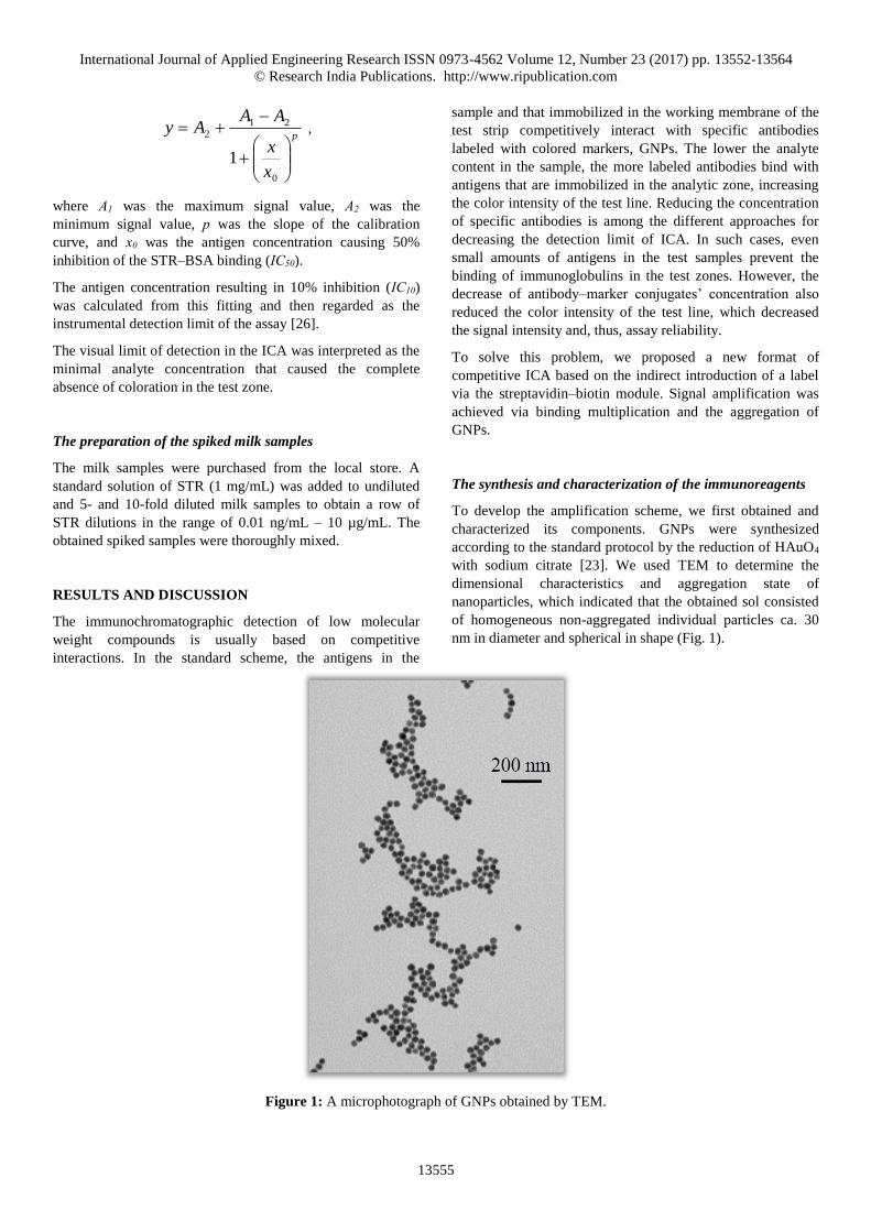
To develop the amplification scheme, we first obtained and
characterized its components. GNPs were synthesized
according to the standard protocol by the reduction of HAuO4
with sodium citrate [23]. We used TEM to determine the
dimensional characteristics and aggregation state of
nanoparticles, which indicated that the obtained sol consisted
of homogeneous non-aggregated individual particles ca. 30
nm in diameter and spherical in shape (Fig. 1).
Figure 1: A microphotograph of GNPs obtained by TEM.

International Journal of Applied Engineering Research ISSN 0973-4562 Volume 12, Number 23 (2017) pp. 13552-13564
© Research India Publications. http://www.ripublication.com
13556
The obtained GNPs were conjugated with anti-STR
monoclonal antibodies and streptavidin, as recommended in
[24]. Preliminary experiments were performed to determine
the optimal loading of the immobilized proteins on GNP
surfaces. The colloidal aggregation test allowed us to study
GNP stabilization according to the concentration of protein
absorbed upon the addition of salt. As a result of this test, we
obtained flocculation curves (the plots of the optical density at
580 nm versus protein concentrations; data not shown).
According to these dependencies, we selected the protein
concentrations that best stabilized the conjugates. Therefore,
for further conjugations, anti-STR antibodies and streptavidin
were taken at 5 and 20 µg per mL of GNPs, respectively. The
functional properties of specific antibodies conjugated to
GNPs were confirmed by binding with STR–BSA
immobilized on the membrane of a test strip.
The competitive ELISA of STR
The immunoreagents were characterized by ELISA. Under the
selected ELISA regime, namely, the STR–BSA conjugate
immobilized at a concentration of 1 μg/mL and specific
antibodies at a concentration of 500 ng/mL, STR was
determined with a detection limit of 20 ng/mL (Fig. 2).
The standard ICA scheme
During the standard ICA, the test strip was immersed in a
solution containing an analyte and conjugates of anti-STR
antibodies with GNPs. STR in the sample reacted with the
active sites of the antibodies, thereby preventing them from
interacting with the STR–BSA adsorbed in the test zone. The
excess conjugate bound anti-species antibodies in the control
zone (Fig. 3A). Therefore, we observed an inverse
dependence of color intensity in the test zone and STR
concentration in the sample.
1 10 100 1000
0,10
0,15
0,20
0,25
0,30
0,35
OD
45
0
[STR], ng/mL
Figure 2: The competitive curve for ELISA of STR.

International Journal of Applied Engineering Research ISSN 0973-4562 Volume 12, Number 23 (2017) pp. 13552-13564
© Research India Publications. http://www.ripublication.com
13557
A
B
Figure 3: The schemes for standard ICA (A) and ICA with amplification (B).
The calibration curve for the standard ICA of STR and the
coloration of their respective test strips is shown in Fig. 4. The
visual limit of STR detection was 3.3 µg/mL and IC10 was 100
ng/mL. Although the sensitivity of the assay satisfied the
maximum permitted levels of STR in food (200 ng/g
according to Commission Regulation [EC] No. 1881/2016),
improving its analytical characteristics seems to be quite
important, taking into account the possible pre-treatment of
the detected probes.

International Journal of Applied Engineering Research ISSN 0973-4562 Volume 12, Number 23 (2017) pp. 13552-13564
© Research India Publications. http://www.ripublication.com
13558
А
0,01 0,1 1 10 100 1000 10000
0
5
10
15
20
25
30
35
40
45
50
55
L
ine i
nte
ncit
y. a
rb
. u
nit
s
[STR], ng/mL
В
10000 3333 1111 370.4 123.5 41.2 13.7 0.01
ng/mL
Figure 4: The calibration curve of STR for standard ICA (A) and the appearance of the test strips (B).
The ICA amplification scheme
In this study, we proposed an ICA amplification scheme to
solve the problem of the contradiction between high assay
sensitivity and low signal intensity in the standard ICA
scheme. We suggested an indirect introduction of the label to
simultaneously increase the amount of marker and reduce the
concentration of specific antibodies. A variation of this
scheme was developed previously using anti-species
antibodies conjugated with GNPs [10, 11]. In the current
study, the indirect introduction of the label was performed by
the streptavidin–biotin module: streptavidin was conjugated
with GNPs, and biotin was conjugated with antibodies.
Furthermore, biotinylated BSA was added to the system to
ensure the binding of the GNPs conjugate with streptavidin,
thus creating an aggregation processes in the analytic zone
and changing the antibody:marker ratio. The scheme of this
format is demonstrated in Fig. 3B.
The conditions for assembling specific complexes in the ICA
amplification scheme were optimized at zero antigen
concentration. The optimization of the streptavidin–GNPs
concentration was carried out by varying its dilution in the
range corresponding to OD520 = 0.002–2. In this case, only
biotinylated antibodies at a concentration of 200 ng/mL were
used. The obtained concentration dependence (Fig. 5)

International Journal of Applied Engineering Research ISSN 0973-4562 Volume 12, Number 23 (2017) pp. 13552-13564
© Research India Publications. http://www.ripublication.com
13559
demonstrated that the maximal signal intensity was observed
at the conjugate concentration corresponding to OD520 = 0.02.
Therefore, a dilution of the streptavidin–GNPs conjugate to
OD520 = 0.03 seemed to be optimal and was used for further
optimization experiments.
1E-4 1E-3 0,01
0
5
10
15
20
25
30
35
40
45
50L
ine i
nte
nsi
ty,
arb
. u
nit
s
OD 520 of streptavidin-GNPs
Figure 5: The dependence of the test zone coloration on the dilution of streptavidin–GNP conjugate.
The concentration of biotinylated antibodies was also
optimized. For this purpose, assays were carried out with
different amounts of antibodies (3–400 ng/mL) and fixed
concentrations of streptavidin–GNPs and BSA–biotin
conjugates (corresponding to OD520 = 0.03 and 60 ng/mL,
respectively). The dependence of the test zone coloration on
the concentration of biotinylated antibodies is presented in
Fig. 6. Because the signals were of the highest intensity at a
concentration of 200 ng/mL, this value was selected.
10 100
0
5
10
15
20
25
30
35
40
45
50
Lin
e i
nte
nsi
ty. a
rb
. u
nit
s
[Biotinylated antibodies], ng/mL
Figure 6: The dependence of the test zone coloration on the concentration of biotinylated antibodies.

International Journal of Applied Engineering Research ISSN 0973-4562 Volume 12, Number 23 (2017) pp. 13552-13564
© Research India Publications. http://www.ripublication.com
13560
The optimal concentration of biotinylated BSA in the reaction
media was found to be ca. 8–30 ng/mL to ensure high
analytical signals. Adding more biotinylated BSA resulted in a
sharp decrease in the test zone coloration (Fig. 7).
0,1 1 10 100
0
5
10
15
20
25
30
35
40
45
50
Lin
e i
nte
nsi
ty, a
rb
. u
nit
s
[Biotinylated BSA], ng/mL
Figure 7: The dependence of the test zone coloration on the concentration of biotinylated BSA.
After preliminary determining the reagents’ concentrations,
we carried out a two-factor optimization of biotinylated BSA
and antibody concentrations. For a fixed amount of a GNPs
conjugate (OD520 = 0.03), the BSA–biotin and antibody–biotin
concentrations were varied in wider ranges (0.1–800 and 25–
400 ng/mL, respectively). Satisfactory color intensity was
achieved when using BSA–biotin and antibody–biotin at
concentrations of 12.5 and 100 ng/mL, respectively. Even
though the latter was lower than what we determined in the
preliminary optimization experiment (200 ng/mL), thus
slightly lowering the intensity of the band color, this
concentration allowed us using half as much reagent.
During final step of the optimization, the streptavidin–GNPs
conjugate was applied in different dilutions (corresponding to
OD520 = 2–0.004) at the aforementioned concentrations of all
other components. The dependence is shown in Fig. 8.
0,01 0,1 1
0
10
20
30
40
50
60
70
Lin
e in
ten
sity
, a
rb.
un
its
OD 520 of streptavidin-GNPs
Figure 8: The dependence of the test zone coloration on the dilution of the streptavidin–GNPs conjugate.

International Journal of Applied Engineering Research ISSN 0973-4562 Volume 12, Number 23 (2017) pp. 13552-13564
© Research India Publications. http://www.ripublication.com
13561
Fig. 8 shows that the dependence of test zone coloration on
the concentration has a bell-shaped shape; the earlier selected
value (OD520 = 0.03) lay in the high-signal range. This low
concentration significantly reduced the consumption of
colloidal gold.
Finally, the optimum combination of test zone coloration and
the assay sensitivity was achieved using biotinylated
antibodies and BSA at concentrations of 100 and 12.5 ng/mL,
respectively, and streptavidin–GNPs conjugate at the dilution
corresponding to OD520 = 0.03.
Fig. 9 demonstrates the appearance of the test strips and the
STR calibration curve. The visual detection limit was 370
ng/mL, and IC10 was 30 ng/mL. The estimated visual
detection limit and IC10 values were 10-fold and 3-fold higher,
respectively, than those obtained as a result of the standard
ICA scheme.
A
0,01 0,1 1 10 100 1000 10000
0
5
10
15
20
25
30
35
40
45
50
Lin
e i
nte
nsi
ty, a
rb
.un
its
[STR], ng/mL
B
10000 3333 1111 370.4 123.5 41.2 13.7 0.01
ng/mL
Figure 9: The calibration curve of STR for ICA in the amplification scheme (A) and the appearance of the test strips (B).
Testing the ICA amplification scheme on food samples
The developed amplification scheme was tested for STR
detection in milk samples. In the first case, the milk samples
were tested directly, without sample preparation or dilution. In
the second case, the milk samples were diluted 5- and 10-fold
by PBST, spiked with various concentrations of STR, and
then used in the ICA amplification scheme. The appearance of

International Journal of Applied Engineering Research ISSN 0973-4562 Volume 12, Number 23 (2017) pp. 13552-13564
© Research India Publications. http://www.ripublication.com
13562
the test strips after the assays is shown in Fig. 10. For the
undiluted samples, the liquid did not reach the absorption
membrane and no bands were formed on the working
membrane. For the diluted samples, the membranes looked
similar to those observed for STR detection in the buffer.
10000
3333
1111
370
124
41,2
13,7
0.0
1
10000
3333
1111
370
124
41.2
13.7
0.0
1
10000
3333
1111
370
123
41.2
13.7
0.0
1
ng/mL
Undiluted milk 5-fold diluted milk 10-fold diluted milk
Figure 10: The appearance of the test strips after the detection of STR in milk samples in the ICA amplification scheme.
Fig. 11 shows the competitive curves of STR in 5- and 10-fold
diluted milk samples. The detection limits for STR detection
did not differ reliably from those obtained as a result of the
assay in PBST (IC10 = 30 ng/mL). The biological matrix of
the probe did not influence the immune interactions, showing
no significant shift in the amplitude of the analytical signals.
Therefore, a pre-treatment of the tested samples, which
requires no organic solvents or complex manipulations, can
precede the ICA amplification scheme. Taking into account
the maximum permitted levels of STR in food (200 ng/g), the
ICA amplification scheme is recommended as a sensitive and
rapid method to screen for STR contamination.
0,01 0,1 1 10 100 1000 10000
0
10
20
30
40
50
60
b
Lin
e i
nte
nsi
ty, a
rb
. u
nit
s
[STR], ng/mL
a
Figure 11: The calibration curves of STR for the ICA amplification scheme in 5-fold (a) and 10-fold (b) diluted milk.

International Journal of Applied Engineering Research ISSN 0973-4562 Volume 12, Number 23 (2017) pp. 13552-13564
© Research India Publications. http://www.ripublication.com
13563
CONCLUSION
The results confirmed the efficiency of the proposed ICA
amplification scheme to reach a lower detection limit. It
showed a 10-fold increase in sensitivity compared to the
standard ICA scheme. Furthermore, the universal nature of the
proposed amplification scheme could be applied to the
immunodetection of antibiotics from other classes (i.e.,
tetracyclines and beta-lactams) with similar effects on signal
enhancement, shifting the calibration curves for lower analyte
concentrations and, thus, providing efficient tools for rapid
screening in food safety and medical monitoring.
ACKNOWLEDGMENTS
The authors are grateful to Prof. P.G. Sveshnikov (All-
Russian Center of Molecular Diagnostics and Therapy,
Moscow, Russia) for providing monoclonal antibodies against
STR, and S.M. Pridvorova (Federal Research Center
«Fundamentals of Biotechnology», Moscow, Russia) for the
TEM studies.
This study was financially supported by the Ministry of
Science and Education of the Russian Federation (grant
agreement No. 14.613.21.0061 on 17.07.2017, unique
identifier RFMEFI61317X0061).
LIST OF SYMBOLS AND ABBREVIATIONS
BSA – bovine serum albumin
CB – carbonate buffer
DMSO – dimethyl sulfoxide
ELISA – enzyme-linked immunosorbent assay
GAM – goat anti-mouse
GNPs – gold nanoparticles
ICA – immunochromatographic analysis
PBS – phosphate-buffered saline
PBST – PBS containing 0.05% Triton X-100
STR – streptomycin
TBSA – Tris buffer containing BSA and sucrose
TEM – transmission electron microscopy
°C
h
IC10
IC50
min
mL
mol
μg
µg/mL
µL
ng/g
ng/mL
nm
pg/mL
REFERENCES
[1] Mikkelsen, K.H., Allin, K.H., and Knop, F.K., 2016,
“Effect of antibiotics on gut microbiota, glucose
metabolism and body weight regulation: a review of
the literature”, Diabetes Obes. Metab., 18(5): pp.
444-453.
[2] Chen, T., Cheng, G., Ahmed, S., Wang, Y., Wang,
X., Hao, H., and Yuan, Z., 2017, “New
methodologies in screening of antibiotic residues in
animal-derived foods: biosensors”, Talanta, 175, pp.
435-442.
[3] Greno, M., Castro-Puyana, M., Garcia, M. A.,
Marina, M. L., 2017, “Analysis of antibiotics by CE
and CEC and their use as chiral selectors: an update”.
Electrophoresis. DOI: 10.1002/elps.201700306.
[4] Gaudin, V., 2017, “Advances in biosensor
development for the screening of antibiotic residues
in food products of animal origin - a comprehensive
review”. Biosens. Bioelectron., 90, pp. 363-377.
[5] Raeisossadati, M.J., Danesh, N.M., Borna, F.,
Gholamzad, M., Ramezani, M., Abnous, K., and
Taghdisi, S.M., 2016, “Lateral flow based
immunobiosensors for detection of food
contaminants”. Biosens Bioelectron., 86, pp. 235-
246.
[6] Hao, N., and Wang, K., 2016, “Recent development
of electrochemiluminescence sensors for food
analysis”. Anal. Bioanal. Chem., 408(25), pp. 7035-
7048.
[7] Dmitrienko, S.G., Kochuk, E.V., Apyari, V.V.,
Tolmacheva, V.V., and Zolotov, Y.A., 2014, “Recent
advances in sample preparation techniques and
methods of sulfonamides detection – a review”,
Anal. Chim. Acta, 850, pp. 6-25.
[8] Dzantiev, B.B., Byzova, N.A., Urusov, A.E., and
Zherdev, A.V., 2014, “Immunochromatographic
methods in food analysis”, TRAC – Trends in Anal.
Chem.,. 55, pp. 81-93.

International Journal of Applied Engineering Research ISSN 0973-4562 Volume 12, Number 23 (2017) pp. 13552-13564
© Research India Publications. http://www.ripublication.com
13564
[9] Babington, R., Matas, S., Marco, M. P., and Galve,
R., 2012, “Current bioanalytical methods for
detection of penicillins”. Anal. Bioanal. Chem.,
403(6), pp. 1549-1566.
[10] Urusov, A.E., Petrakova, A.V., Zherdev, A.V., and
Dzantiev, B.B., 2016, "Multistage in one touch"
design with a universal labelling conjugate for high-
sensitive lateral flow immunoassays”. Biosens.
Bioelectron., 86, pp. 575-579.
[11] Urusov, A.E., Zherdev, A.V., and Dzantiev, B.B.,
2014, “Use of gold nanoparticle-labeled secondary
antibodies to improve the sensitivity of an
immunochromatographic assay for aflatoxin B1”,
Microchim. Acta, 181(15), pp. 1939-1946.
[12] Taranova N.A., Urusov A.E., Sadykhov E.G.,
Zherdev A.V., and Dzantiev B.B., 2017,
“Bifunctional gold nanoparticles as agglomeration
enhancing tool for high sensitive lateral flow test: a
case study with procalcitonin”, Microchim. Acta,
184(10), pp. 4189-4195.
[13] Green, N.M., 1975, “Avidin”, Adv. Protein Chem.,
29, pp. 85-133.
[14] Wu, J., Chen, Y., Yang, M., Wang, Y., Zhang, C.,
Sun, J., Xie, M., and Jiang, X., 2017, “Streptavidin-
biotin-peroxidase nanocomplex-amplified
microfluidics immunoassays for simultaneous
detection of inflammatory biomarkers”. Anal. Chim.
Acta, 982, pp. 138-147.
[15] You, Y., Lim, S., Hahn, J., Choi, Y.J., and
Gunasekaran, S., 2018, “Bifunctional linker-based
immunosensing for rapid and visible detection of
bacteria in real matrices”. Biosens. Bioelectron., 100,
pp. 389-395.
[16] Boisset, S., Caspar, Y., Sutera, V., and Maurin, M.,
2014, “New therapeutic approaches for treatment of
tularaemia: a review”. Front. Cell Infect. Microbiol,
4, p. 40.
[17] Riva, M.A., 2014, “From milk to rifampicin and back
again: history of failures and successes in the
treatment for tuberculosis”. J. Antibiot. (Tokyo),
67(9), pp. 661-665.
[18] Chandrika, N.T., and Garneau-Tsodikova, S., 2016,
“A review of patents (2011-2015) towards combating
resistance to and toxicity of aminoglycosides”,
MedСhemСomm., 7(1), pp. 50-68.
[19] Takahashi, Y., and Igarashi, M., “Destination of
aminoglycoside antibiotics in the 'post-antibiotic
era'”. J. Antibiot. (Tokyo), 2017. DOI:
10.1038/JA.2017.117.
[20] Song, E., Yu, M., Wang, Y., Hu, W., Cheng, D.,
Swihart, M.T., and Song, Y., 2015, “Multi-color
quantum dot-based fluorescence immunoassay array
for simultaneous visual detection of multiple
antibiotic residues in milk”, Biosens. Bioelectron.,
72, pp. 320-325.
[21] Taranova, N.A., Berlina, A.N., Zherdev, A.V.,
Dzantiev, B.B., 2015, “'Traffic light'
immunochromatographic test based on multicolor
quantum dots for the simultaneous detection of
several antibiotics in milk”, Biosens. Bioelectron.,
63, pp. 255-261.
[22] Byzova, N.A., Zvereva, E.A., Zherdev, A.V.,
Eremin, S.A., Sveshnikov, P.G., and Dzantiev, B.B.,
2011, “Pretreatment-free immunochromatographic
assay for the detection of streptomycin and its
application to the control of milk and dairy
products”. Anal. Chim. Acta, 701(2), pp. 209-217.
[23] Byzova, N.A., Zvereva, E.A., Zherdev, A.V.,
Eremin, S.A., and Dzantiev, B.B., 2010, “Rapid
pretreatment-free immunochromatographic assay of
chloramphenicol in milk”. Talanta, 81(3), pp. 843-
848.
[24] Abuknesha, R.A., and Luk, C., 2005, “Enzyme
immunoassays for the analysis of streptomycin in
milk, serum and water: development and assessment
of a polyclonal antiserum and assay procedures using
novel streptomycin derivatives”, Analyst, 130(6), pp.
964-970.
[25] Bayer, E.A., and Wilchek, M., 1990, “Protein
biotinylation”. Methods Enzymol., 184, pp. 138-160.
[26] Sittampalam, G.S., Smith, W.C., Miyakawa, T.W.,
Smith, D.R., and Mcmorris, C., 1996, “Application
of experimental design techniques to optimize a
competitive ELISA”. J. Immunol. Methods, 190(2),
pp. 151-161.


![[XLS] · Web viewSTR 20015 STR 30105 STR 30115 STR 30123 STR 30125 STR 30130 STR 40090 ORİ STR 40115 STR 41090 ORİ STR 44115 STR 45111 STR 50020 STR 50103A STR 50112 STR 50113A](https://img.pdfslide.us/doc/110x75/5ad04b0c7f8b9a1d328e1e93/xls-viewstr-20015-str-30105-str-30115-str-30123-str-30125-str-30130-str-40090.jpg)