Embed Size (px)
Citation preview
Digestive SystemANS 215
Physiology and Anatomy ofDomesticated Animals
I. Digestive TractA. Animals are classified according to the diet in their natural state as:
1. Carnivores2. Herbivores3. Omnivores
B. Because of the diversity of diet, various parts of the digestive system developed indifferent ways.
Comparisons of gastrointestinal tracts of, A) the dog, B) the horse, C) and cattle.1. Stomach; 2. small intestine; 3. cecum;
4. ascending colon (dog), large colon (horse), coiled colon (cattle); 5. descending colon.
C. Mouth1. Most cranial part of the digestive system
a. also referred to as oral cavityb. site of reduction of food particle sizec. teeth and tongue are structures that assist in digestiond. salivary enzymes are also added to digesta
1
2. Teetha. mechanically reduce the size of ingested food
i. increases surface areab. teeth are also used for cutting food (incisors) and defense (canines)c. Types of teeth:
i. identified by location and use- incisors
. most forward
. used for cutting
. also called nippers- canines
. also called fangs, eyeteeth, tusks
. used for tearing
. located posterior to incisors- premolars
. used for grinding
. located posterior to canines- molars
. located caudal to premolars
. used for grinding
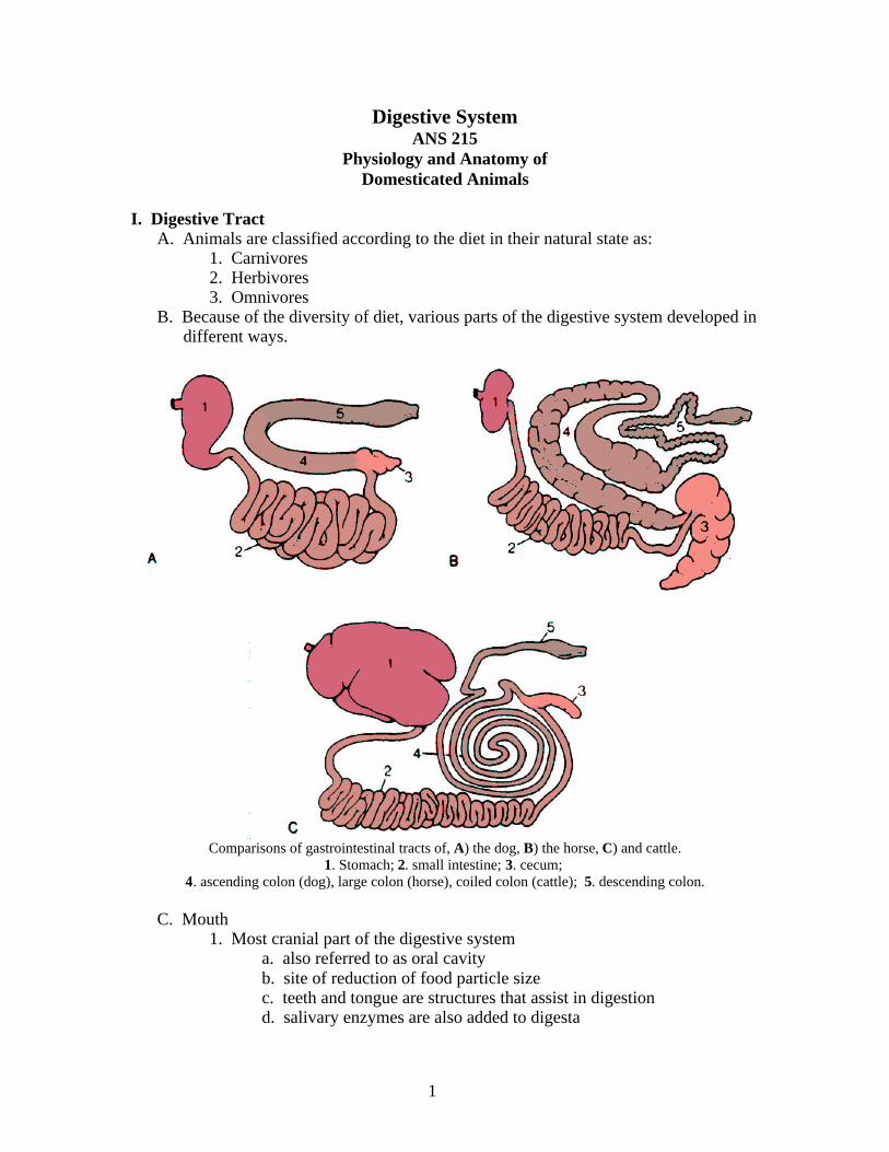
Dental Formulas and Eruption Times for Permanent TeethTeeth Horse Cow Sheep Pig Dog
Permanent Formula3 1 3 or 4 3 0 0 3 3 0 0 3 3 3 1 4 3 3 1 4 22( I-C-P-M-) 2( I-C-P-M-) 2( I-C-P-M-) 2( I-C-P-M-) 2( I-C-P-M-)
3 1 3 3 4 0 3 3 4 0 3 3 3 1 4 3 3 1 4 3Permanent Eruption
Incisors11 2½ yr 1½ - 2 yr 1 - 1½ yr 1 yr 3 - 5 mo12 3½ yr 2 - 2½ yr 1½ - 2 yr 16 - 20 mo 3 - 5 mo13 4½ yr 3 yr 2½ - 3 yr 8 - 10 mo 4 - 5 mo14 3½ - 4 yr 3½ - 4 yr
CaninesC 4 - 5 yr 9 - 10 mo 4 - 6 mo
PremolarsP1 5 - 6 mo 2 - 2½ yr 1½ - 2 yr 12 - 15 mo 4 - 5 moP2 2½ yr 1½ - 2½ yr 1½ - 2 yr 12 - 15 mo 5 - 6 moP3 3 yr 2½ - 3 yr 1½ - 2 yr 12 - 15 mo 5 - 6 moP4 4 yr 12 - 15 mo 5 - 6 mo
MolarsM1 9 - 12 mo 5 - 6 mo 43 - 5 mo 4 - 6 mo 5 - 6 moM2 2 yr 1½ yr 9 - 12 mo 8 - 12 mo 6 - 7 moM3 3½ - 4 yr 2 - 2½ yr 1½ - 2 yr 18 - 20 mo 6 - 7 mo
I = incisors, C = canines, P = premolars, M = molars, wk = week, mo = month, yr = year
d. Dental formulai. indicates the number of incisors (I), canines (C), premolars
2
(P), and molars (M) on one side of the mouthii. dental formula of the cow is I0/4C0/0P3/3M3/3
- numerator represents upper jaw, denominator is lower jaw
- cow has a firm dental pad instead of incisors in upper jaw
iii. formula represents teeth on one side of the mouth
e. Exposed surface of teethi. Several terms are used to describe the exposed surface of a
tooth.- table (grinding surface) – makes contact with the ‘
surface of opposite jaw- lingual surface – side of tooth next to tongue- labial surface – outer surface next to lips- buccal surface – outer surface next to cheeks- contact surface – next to a neighboring tooth ot the same arcade (row)
- upper arcades of cheek teeth (molars and premolars) are slightly wider apart than the lower arcades of cheek teeth
- upper cheek teeth have a wider table surface than the lower cheek teeth
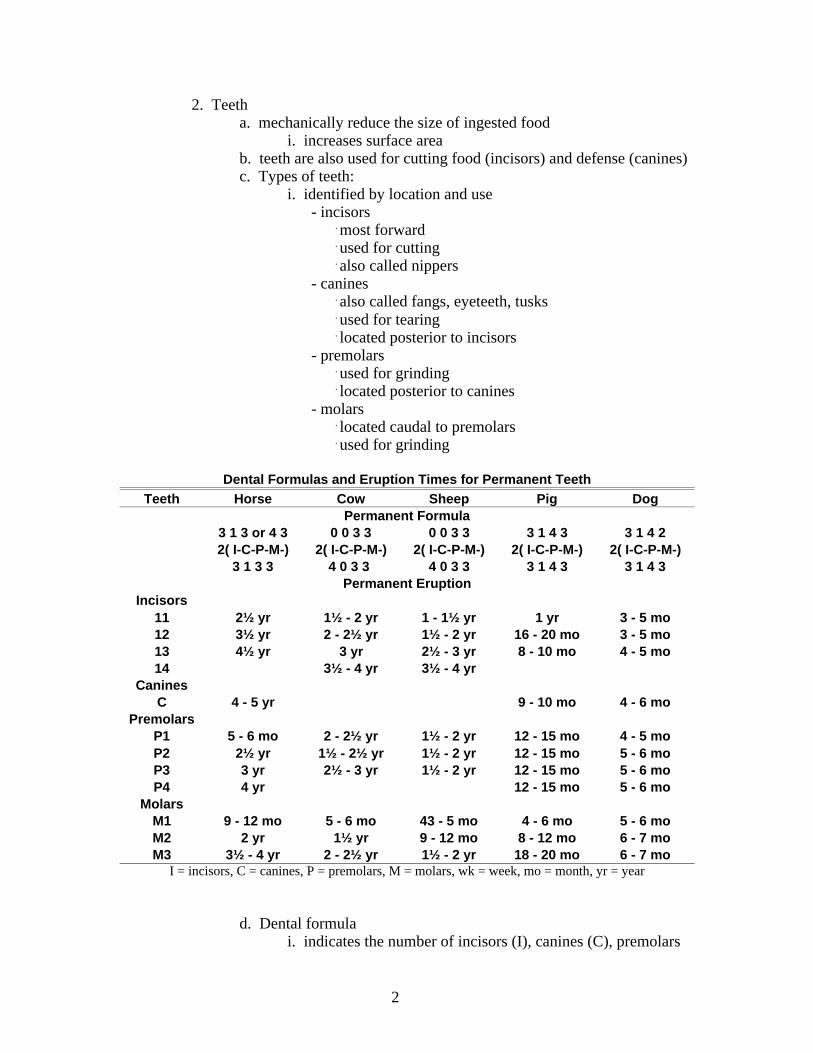
UJ = upper jaw; LJ = lower jaw; RM = right molar; LM = left molar; RLM = right lower molar; LLM = left lower molar
Schematic transverse section of the upper and lower jaws of the horse at the level of the fourth molarsshowing the position of the tables of the teeth during rest and mastication. 1. Position of the teeth duringrest. The outside edge of the lower row is in apposition with the inside edge of the upper. 2. Jaws fully
crossed, masticating from left to right (lower jaw movement). The tables of both upper and lower molarsnow rest on each other. 3. Position halfway through mastication. The outer half of the lower teeth wearsagainst the inner half of the upper. Note the potential for developing “points” on the cheek side of the
uppers and on the tongue side of the lowers.
ii. uneven wear of tooth surface
3
- can develop, particularly in horses- points can develop on teeth that injure the buccal or lingual membranes causing pain
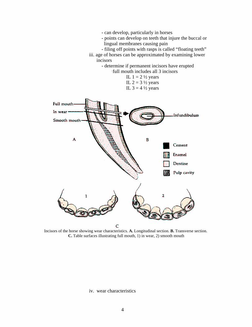
- filing off points with rasps is called “floating teeth”iii. age of horses can be approximated by examining lower
incisors- determine if permanent incisors have erupted
. full mouth includes all 3 incisorsIL 1 = 2 ½ years IL 2 = 3 ½ yearsIL 3 = 4 ½ years
Incisors of the horse showing wear characteristics. A. Longitudinal section. B. Transverse section. C. Table surfaces illustrating full mouth, 1) in wear, 2) smooth mouth
iv. wear characteristics
4
- mouth is in wear when two complete enamel rings are present on the table surface of each incisor
- approximate ages for each pair of incisors to be in wearare 6, 7, and 8 years for I1, I2, and I3
- judgment is made regarding the loss of the inner enamel ring and appearance of the pulp cavity (dental star)
. 11, 12, and 13 years for I1, I2, and I3- a horse has a smooth mouth when these occur in all three pairs of incisors
- rough approximation of horse ages for. full mouth 5 years. in wear 10 years. smooth mouth 15 years
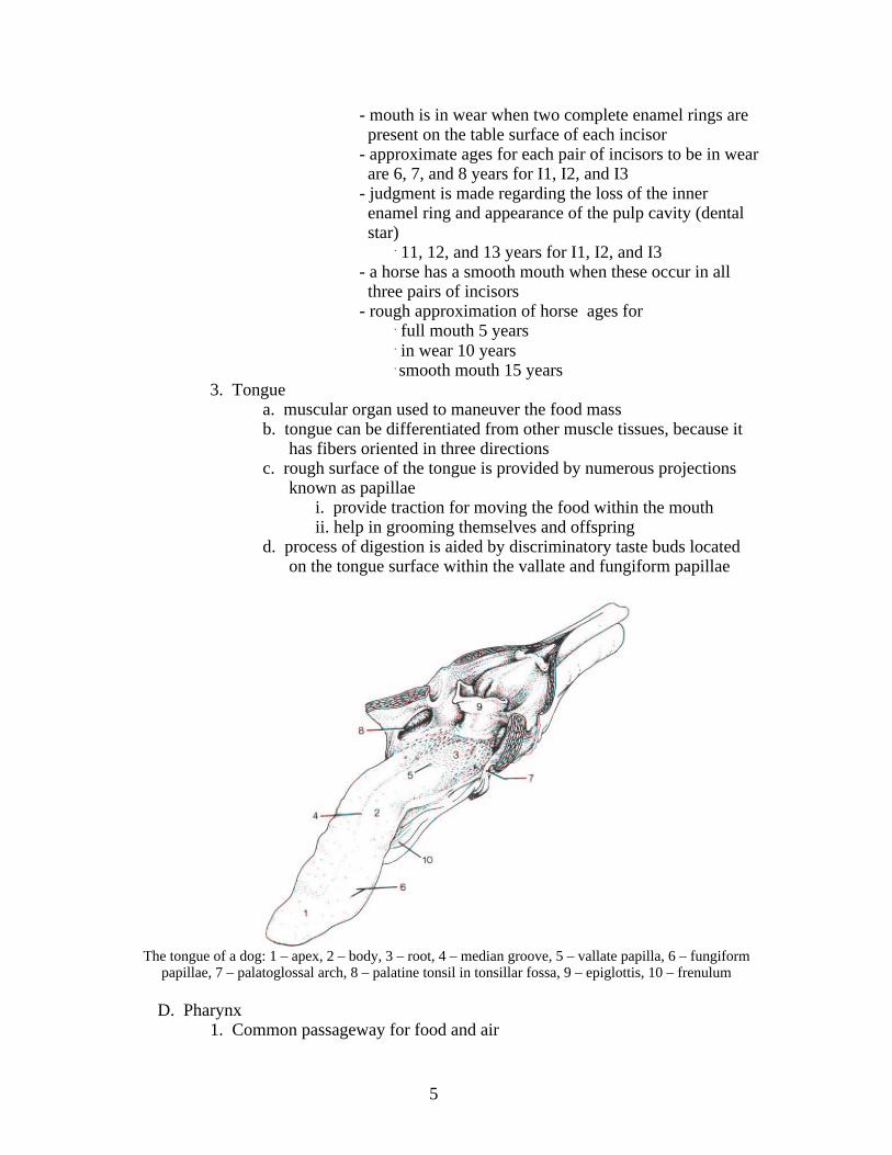
3. Tonguea. muscular organ used to maneuver the food massb. tongue can be differentiated from other muscle tissues, because it
has fibers oriented in three directionsc. rough surface of the tongue is provided by numerous projections
known as papillaei. provide traction for moving the food within the mouthii. help in grooming themselves and offspring
d. process of digestion is aided by discriminatory taste buds located on the tongue surface within the vallate and fungiform papillae
The tongue of a dog: 1 – apex, 2 – body, 3 – root, 4 – median groove, 5 – vallate papilla, 6 – fungiformpapillae, 7 – palatoglossal arch, 8 – palatine tonsil in tonsillar fossa, 9 – epiglottis, 10 – frenulum
D. Pharynx1. Common passageway for food and air
5
2. Opens into mouth, nasal cavities, eustachian tubes, larynx and esophagus3. Passage of food through the pharynx and into the larynx and nasal cavities
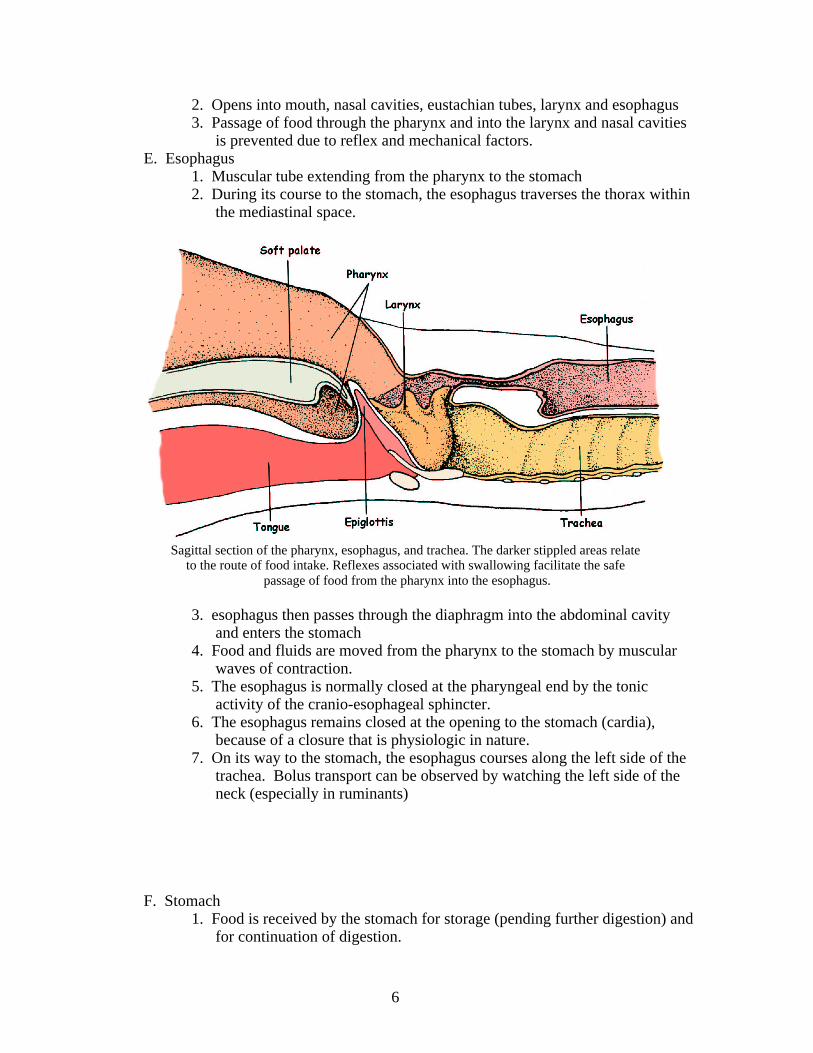
is prevented due to reflex and mechanical factors.E. Esophagus
1. Muscular tube extending from the pharynx to the stomach2. During its course to the stomach, the esophagus traverses the thorax within
the mediastinal space.
Sagittal section of the pharynx, esophagus, and trachea. The darker stippled areas relate to the route of food intake. Reflexes associated with swallowing facilitate the safe
passage of food from the pharynx into the esophagus.
3. esophagus then passes through the diaphragm into the abdominal cavity and enters the stomach
4. Food and fluids are moved from the pharynx to the stomach by muscular waves of contraction.
5. The esophagus is normally closed at the pharyngeal end by the tonic activity of the cranio-esophageal sphincter.
6. The esophagus remains closed at the opening to the stomach (cardia), because of a closure that is physiologic in nature.
7. On its way to the stomach, the esophagus courses along the left side of the trachea. Bolus transport can be observed by watching the left side of the neck (especially in ruminants)
F. Stomach1. Food is received by the stomach for storage (pending further digestion) and
for continuation of digestion.
6
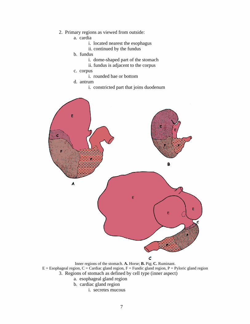
2. Primary regions as viewed from outside:a. cardia
i. located nearest the esophagusii. continued by the fundus
b. fundusi. dome-shaped part of the stomachii. fundus is adjacent to the corpus
c. corpusi. rounded bae or bottom
d. antrumi. constricted part that joins duodenum
Inner regions of the stomach. A. Horse; B. Pig; C. Ruminant.E = Esophageal region, C = Cardiac gland region, F = Fundic gland region, P = Pyloric gland region
3. Regions of stomach as defined by cell type (inner aspect)a. esophageal gland regionb. cardiac gland region
i. secretes mucous
7
c. fundic gland regioni. entire space between cardiac and pyloric gland regionsii. These glands are sometimes called gastric glands.iii. secrete hydrochloric acid and pdpsinogen
d. pyloric gland regioni. secrete mucous and the hormone gastrin
4. Ruminant stomachsa. foregut fermentationb. includes the rumen, reticulum (omasum) and abomasumsc. true stomach is the abomasum
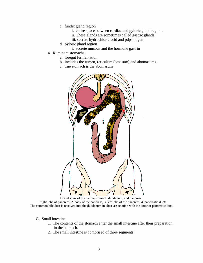
Dorsal view of the canine stomach, duodenum, and pancreas. 1. right lobe of pancreas, 2. body of the pancreas, 3. left lobe of the pancreas, 4. pancreatic ducts
The common bile duct is received into the duodenum in close association with the anterior pancreatic duct.
G. Small intestine1. The contents of the stomach enter the small intestine after their preparation
in the stomach. 2. The small intestine is comprised of three segments:
8
a. duodenumi. closely connected to the pancreasii. receives pancreatic secretionsiii. also receives bile
b. jejunumc. ilium
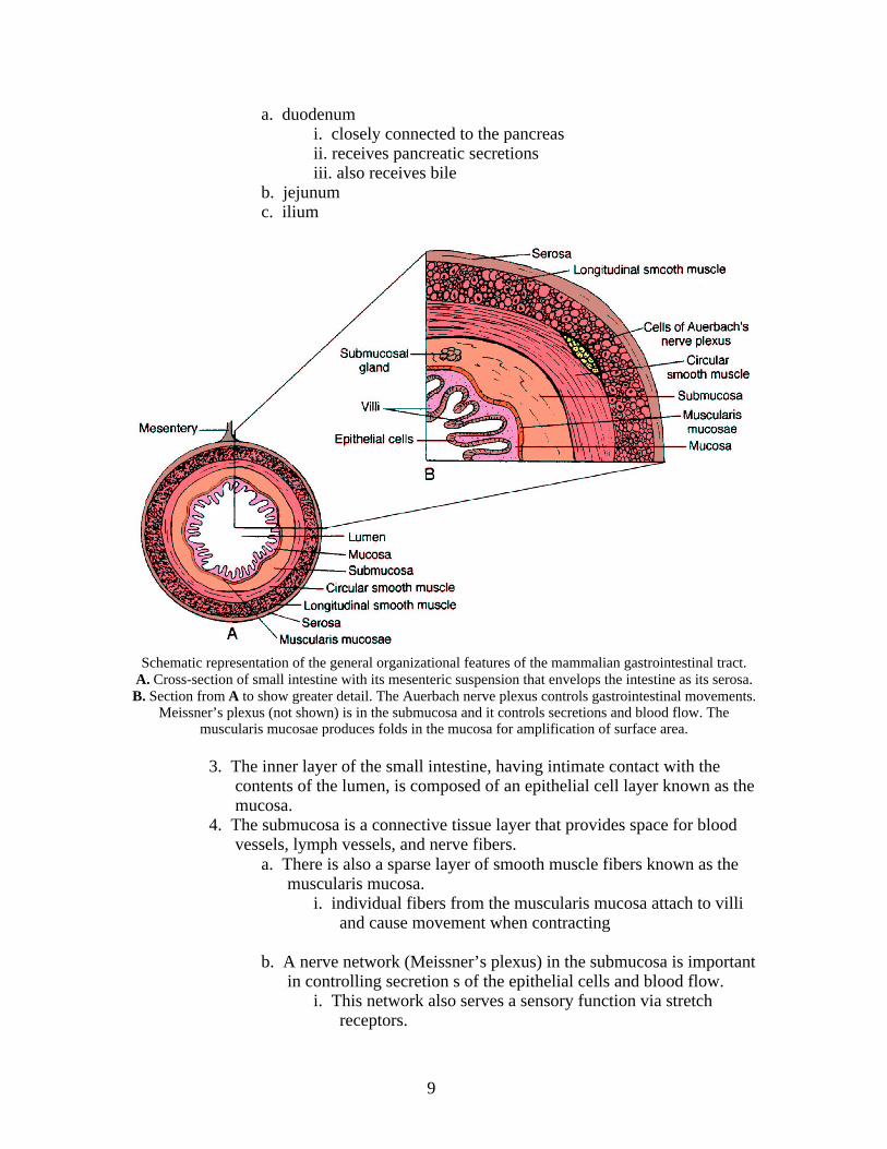
Schematic representation of the general organizational features of the mammalian gastrointestinal tract.A. Cross-section of small intestine with its mesenteric suspension that envelops the intestine as its serosa.B. Section from A to show greater detail. The Auerbach nerve plexus controls gastrointestinal movements.
Meissner’s plexus (not shown) is in the submucosa and it controls secretions and blood flow. Themuscularis mucosae produces folds in the mucosa for amplification of surface area.
3. The inner layer of the small intestine, having intimate contact with the contents of the lumen, is composed of an epithelial cell layer known as themucosa.
4. The submucosa is a connective tissue layer that provides space for blood vessels, lymph vessels, and nerve fibers.
a. There is also a sparse layer of smooth muscle fibers known as the muscularis mucosa.
i. individual fibers from the muscularis mucosa attach to villi and cause movement when contracting
b. A nerve network (Meissner’s plexus) in the submucosa is importantin controlling secretion s of the epithelial cells and blood flow.
i. This network also serves a sensory function via stretch receptors.
9
ii. Another nerve plexus (Auerbach’s plexus), between the inner circular and outer longitudinal muscle layers is important in controlling gastrointestinal movement.
- These two nerve plexus are referred to as the enteric nervous system, which extends from the esophagus to the anus.
c. The outer layer of the intestine is the serosa. It covers the intestine and is continuous with the mesentery, which suspends the intestinewithin the abdominal cavity.
i. the mesentery is in turn continuous with the peritoneumd. The surface are of the intestine is increased by long length and
folding of the tissue within the intestine.i. folds or placations are covered with villi and the individual
cells that cover the villi have their own micriovilliii. microvilli provide for the greatest amplification of surface
area and constitute the brush borderiii. this amplification increases the total surface area of the
small intestine about 600 times that of a smooth cylinder ofthe same size
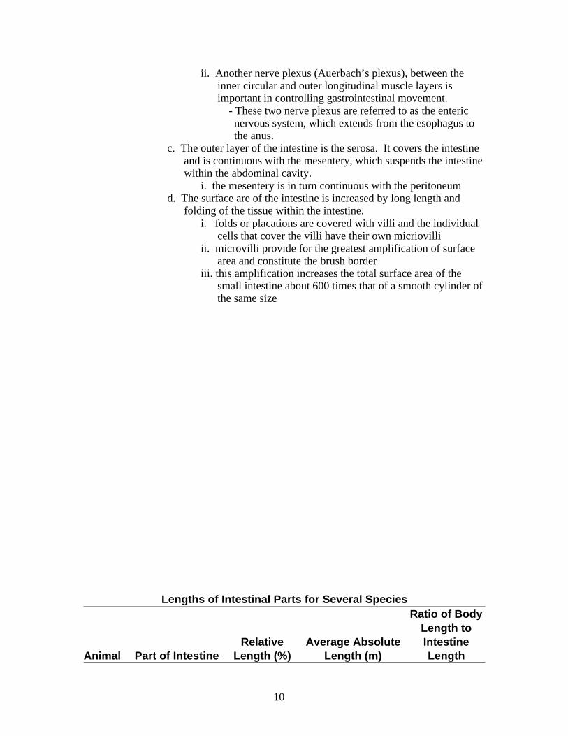
Lengths of Intestinal Parts for Several Species
Animal Part of IntestineRelative
Length (%)Average Absolute
Length (m)
Ratio of BodyLength toIntestineLength
10
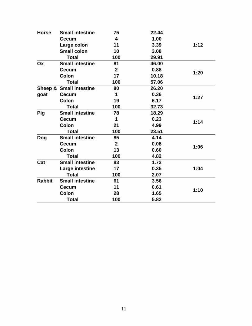
Horse Small intestine 75 22.44Cecum 4 1.00Large colon 11 3.39Small colon 10 3.08
Total 100 29.91
1:12
Ox Small intestine 81 46.00Cecum 2 0.88Colon 17 10.18
Total 100 57.06
1:20
Sheep &goat
Small intestine 80 26.20Cecum 1 0.36Colon 19 6.17
Total 100 32.73
1:27
Pig Small intestine 78 18.29Cecum 1 0.23Colon 21 4.99
Total 100 23.51
1:14
Dog Small intestine 85 4.14Cecum 2 0.08Colon 13 0.60
Total 100 4.82
1:06
Cat Small intestine 83 1.72Large intestine 17 0.35
Total 100 2.071:04
Rabbit Small intestine 61 3.56Cecum 11 0.61Colon 28 1.65
Total 100 5.82
1:10
11
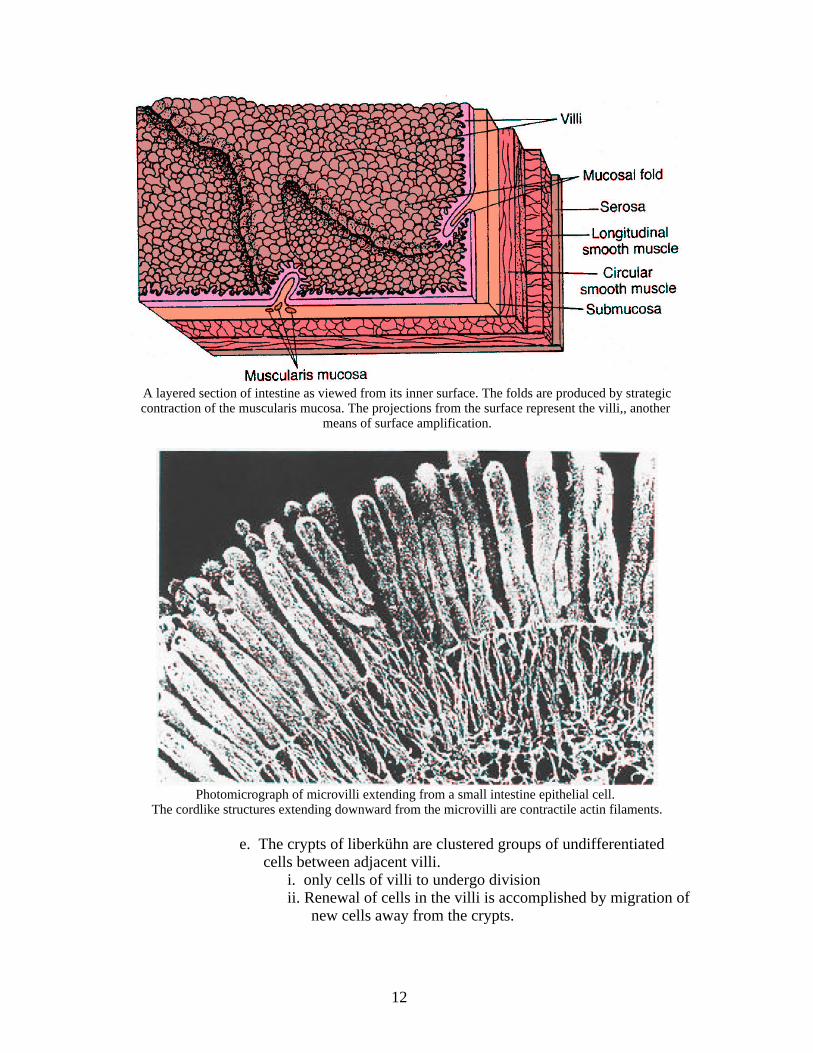
A layered section of intestine as viewed from its inner surface. The folds are produced by strategiccontraction of the muscularis mucosa. The projections from the surface represent the villi,, another
means of surface amplification.
Photomicrograph of microvilli extending from a small intestine epithelial cell. The cordlike structures extending downward from the microvilli are contractile actin filaments.
e. The crypts of liberkühn are clustered groups of undifferentiated cells between adjacent villi.
i. only cells of villi to undergo divisionii. Renewal of cells in the villi is accomplished by migration of
new cells away from the crypts.
12
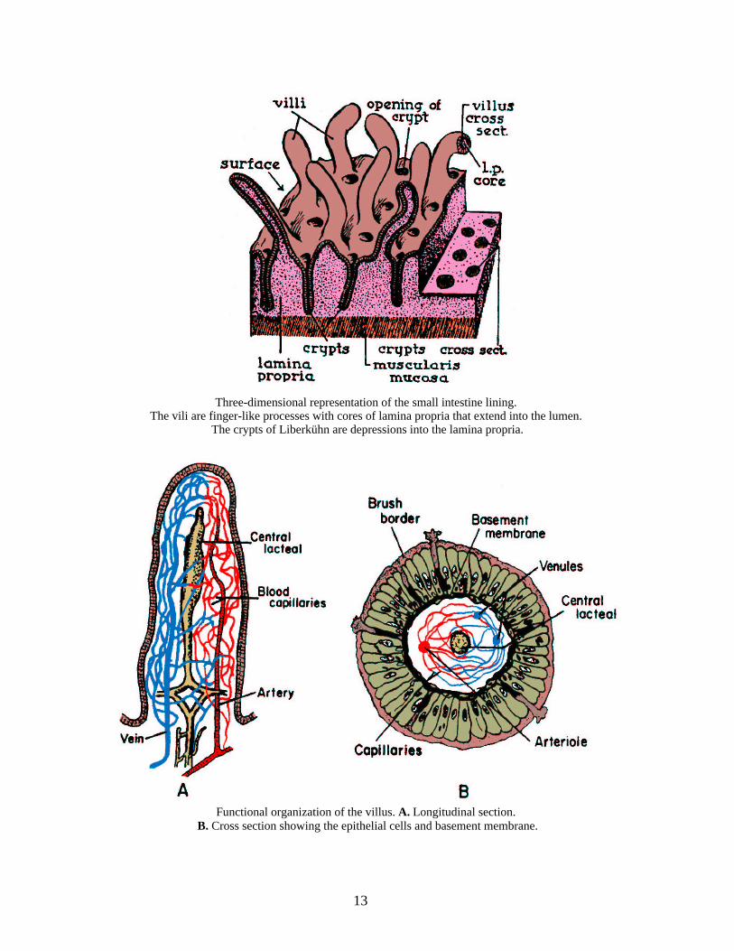
Three-dimensional representation of the small intestine lining. The vili are finger-like processes with cores of lamina propria that extend into the lumen.
The crypts of Liberkühn are depressions into the lamina propria.
Functional organization of the villus. A. Longitudinal section. B. Cross section showing the epithelial cells and basement membrane.
13
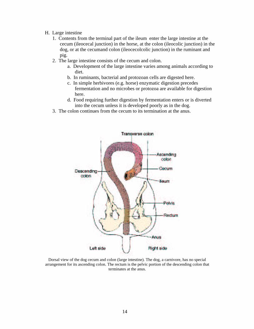
H. Large intestine1. Contents from the terminal part of the ileum enter the large intestine at the
cecum (ileocecal junction) in the horse, at the colon (ileocolic junction) in the dog, or at the cecumand colon (ileocecolcolic junction) in the ruminant and pig.
2. The large intestine consists of the cecum and colon.a. Development of the large intestine varies among animals according to
diet.b. In ruminants, bacterial and protozoan cells are digested here.c. In simple herbivores (e.g. horse) enzymatic digestion precedes
fermentation and no microbes or protozoa are available for digestion here.
d. Food requiring further digestion by fermentation enters or is diverted into the cecum unless it is developed poorly as in the dog.
3. The colon continues from the cecum to its termination at the anus.
Dorsal view of the dog cecum and colon (large intestine). The dog, a carnivore, has no specialarrangement for its ascending colon. The rectum is the pelvic portion of the descending colon that
terminates at the anus.
14

Gastrointestinal tract of the cow showing the colic spiral (asna spiralis).
Schematic representation of the cecum and colon of the horse
15
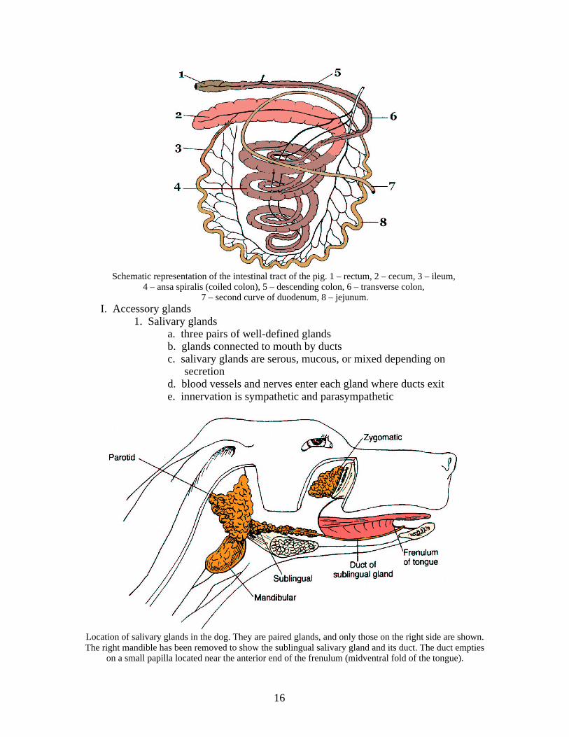
Schematic representation of the intestinal tract of the pig. 1 – rectum, 2 – cecum, 3 – ileum, 4 – ansa spiralis (coiled colon), 5 – descending colon, 6 – transverse colon,
7 – second curve of duodenum, 8 – jejunum.I. Accessory glands
1. Salivary glandsa. three pairs of well-defined glandsb. glands connected to mouth by ductsc. salivary glands are serous, mucous, or mixed depending on
secretiond. blood vessels and nerves enter each gland where ducts exite. innervation is sympathetic and parasympathetic
Location of salivary glands in the dog. They are paired glands, and only those on the right side are shown.The right mandible has been removed to show the sublingual salivary gland and its duct. The duct empties
on a small papilla located near the anterior end of the frenulum (midventral fold of the tongue).
16
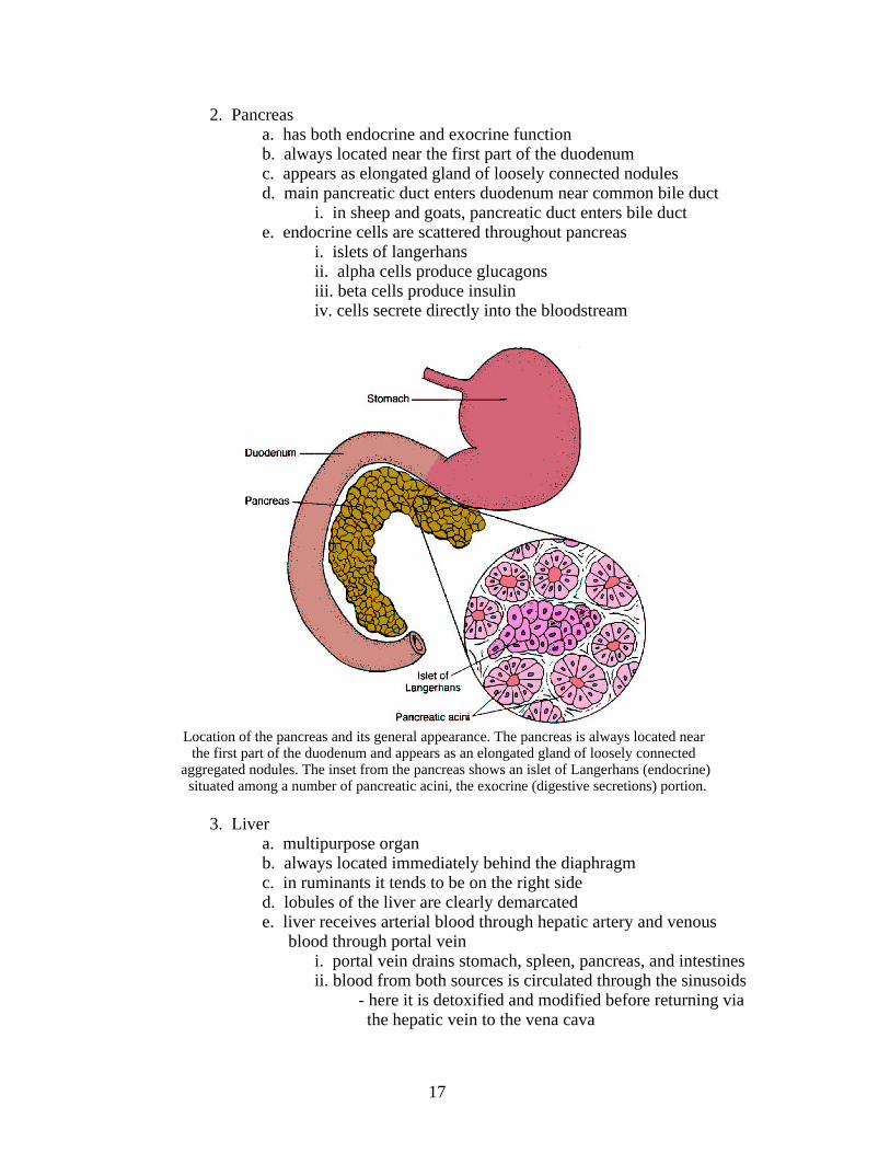
2. Pancreasa. has both endocrine and exocrine functionb. always located near the first part of the duodenumc. appears as elongated gland of loosely connected nodulesd. main pancreatic duct enters duodenum near common bile duct
i. in sheep and goats, pancreatic duct enters bile ducte. endocrine cells are scattered throughout pancreas
i. islets of langerhansii. alpha cells produce glucagonsiii. beta cells produce insuliniv. cells secrete directly into the bloodstream
Location of the pancreas and its general appearance. The pancreas is always located near the first part of the duodenum and appears as an elongated gland of loosely connected
aggregated nodules. The inset from the pancreas shows an islet of Langerhans (endocrine) situated among a number of pancreatic acini, the exocrine (digestive secretions) portion.
3. Livera. multipurpose organb. always located immediately behind the diaphragmc. in ruminants it tends to be on the right sided. lobules of the liver are clearly demarcatede. liver receives arterial blood through hepatic artery and venous
blood through portal veini. portal vein drains stomach, spleen, pancreas, and intestinesii. blood from both sources is circulated through the sinusoids
- here it is detoxified and modified before returning via the hepatic vein to the vena cava
17
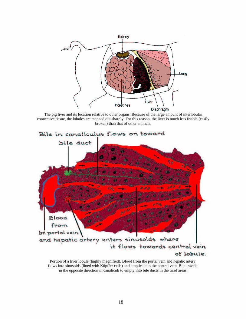
The pig liver and its location relative to other organs. Because of the large amount of interlobularconnective tissue, the lobules are mapped out sharply. For this reason, the liver is much less friable (easily
broken) than that of other animals.
Portion of a liver lobule (highly magnified). Blood from the portal vein and hepatic artery flows into sinusoids (lined with Küpffer cells) and empties into the central vein. Bile travels
in the opposite direction in canaliculi to empty into bile ducts in the triad areas.
18
II. Physical and Mechanical FactorsA. Prehension
1. Seizing and conveying food into the moutha. mouth, tongue, and lips
B. Mastication1. mechanical breakdown of food in the mouth2. A bolus of food is formed by the mastication process.3. bolus is mixed with saliva, which provides adhesiveness
C. Deglutination1. act of swallowing2. conveys the food mass from the mouth to the stomach3. some degree of consciousness is required, because activity is voluntary
a. inhaling food and vomitus is possible when unconsciousD. Smooth muscle activity
1. Once food reaches the stomach, its movement is controlled by the activity of the smooth muscles of the stomach and intestine.
2. muscle activity is spontaneous and controlled by autonomic nervous system
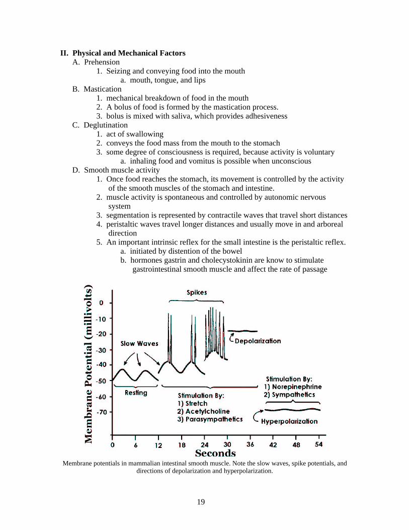
3. segmentation is represented by contractile waves that travel short distances4. peristaltic waves travel longer distances and usually move in and arboreal
direction5. An important intrinsic reflex for the small intestine is the peristaltic reflex.
a. initiated by distention of the bowelb. hormones gastrin and cholecystokinin are know to stimulate
gastrointestinal smooth muscle and affect the rate of passage
Membrane potentials in mammalian intestinal smooth muscle. Note the slow waves, spike potentials, anddirections of depolarization and hyperpolarization.
19
E. Physical functions of the stomach and intestine1. mixing food with secretions2. control of emptying of its contents
a. Delay of gastric emptyingi. Neural mechanism (enterogastric reflex)
- osmoreceptors detect hypertonicity- excessive protein or carbohydrate is also effective in delaying gastric emptying
- other receptors respond to high hydrogen ion concentrations
- cholecystokinin slows gastric emptying to aid in fat digestion
- gastric inhibitory polypeptide (GIP) is secreted by the jejunal mucosa in response to high lipid and
carbohydrate content of the diet
III. Composition of FoodstuffsA. Six basic foodstuffs are classified as:
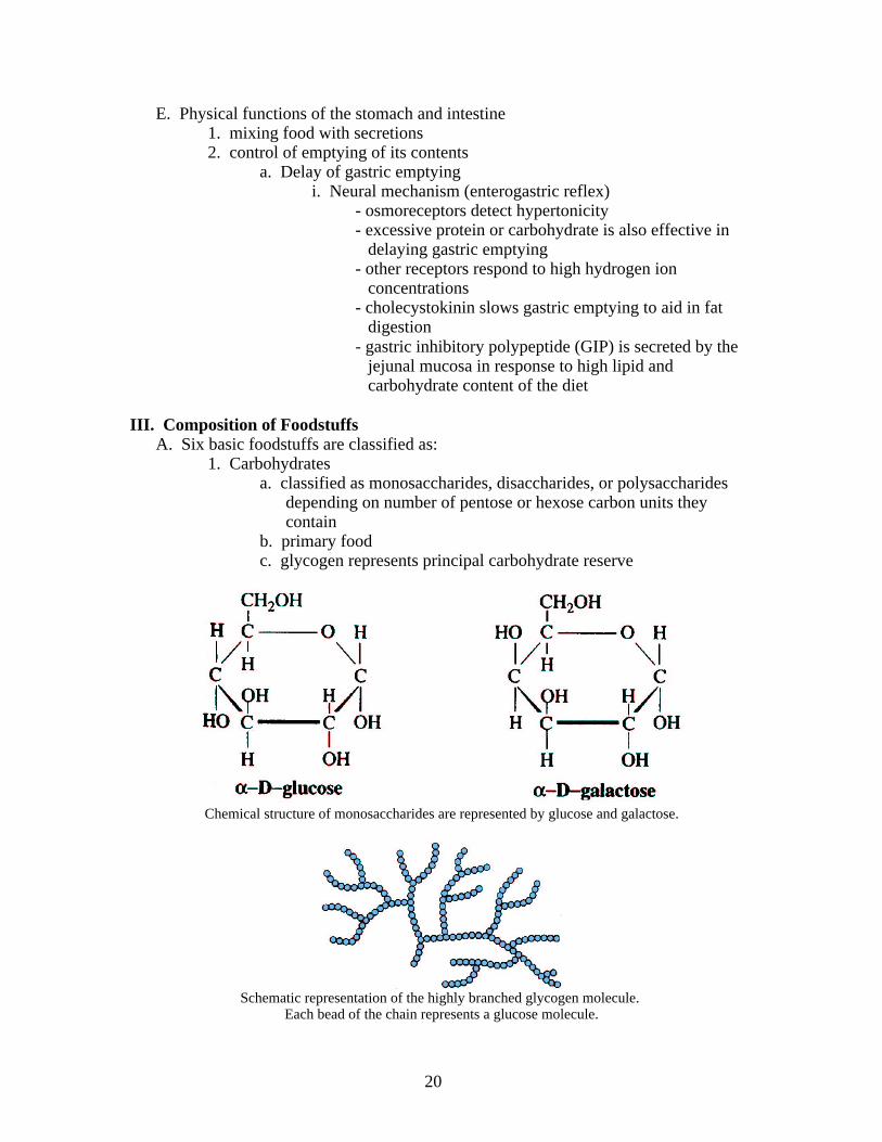
1. Carbohydratesa. classified as monosaccharides, disaccharides, or polysaccharides
depending on number of pentose or hexose carbon units they contain
b. primary foodc. glycogen represents principal carbohydrate reserve
Chemical structure of monosaccharides are represented by glucose and galactose.
Schematic representation of the highly branched glycogen molecule. Each bead of the chain represents a glucose molecule.
20
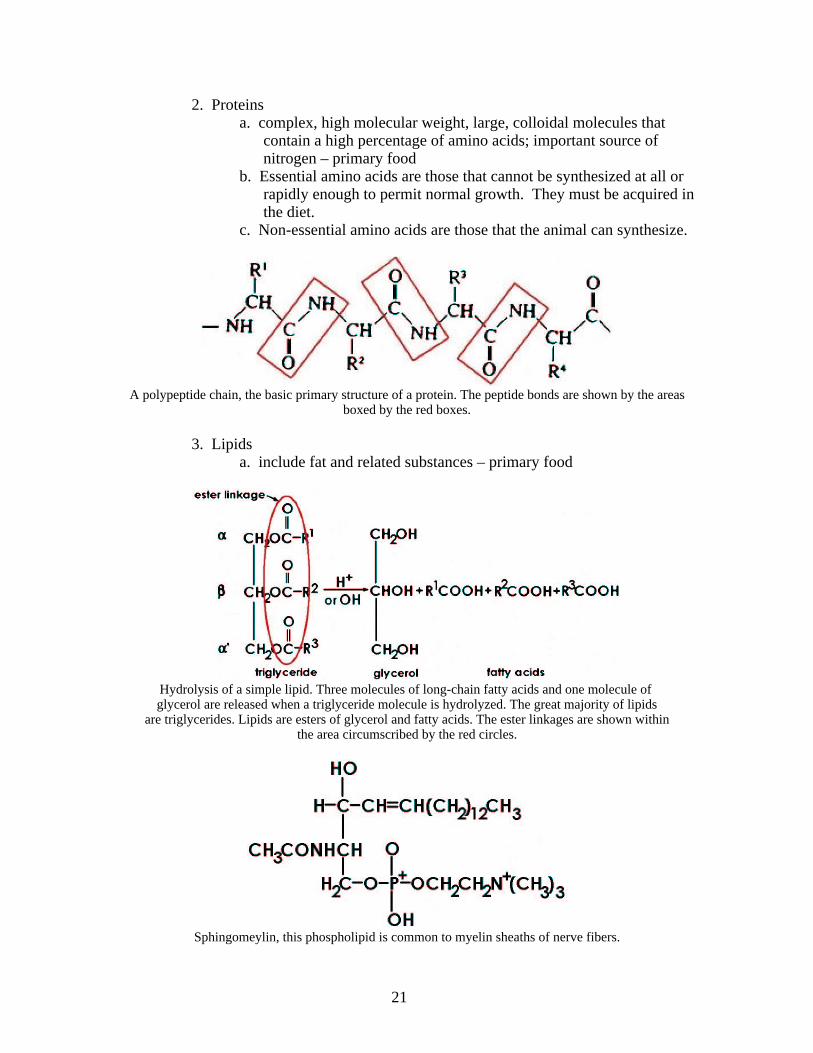
2. Proteinsa. complex, high molecular weight, large, colloidal molecules that
contain a high percentage of amino acids; important source of nitrogen – primary food
b. Essential amino acids are those that cannot be synthesized at all or rapidly enough to permit normal growth. They must be acquired inthe diet.
c. Non-essential amino acids are those that the animal can synthesize.
A polypeptide chain, the basic primary structure of a protein. The peptide bonds are shown by the areasboxed by the red boxes.
3. Lipidsa. include fat and related substances – primary food
Hydrolysis of a simple lipid. Three molecules of long-chain fatty acids and one molecule of glycerol are released when a triglyceride molecule is hydrolyzed. The great majority of lipids
are triglycerides. Lipids are esters of glycerol and fatty acids. The ester linkages are shown within the area circumscribed by the red circles.
Sphingomeylin, this phospholipid is common to myelin sheaths of nerve fibers.
21

4. Watera. Water is considered an accessory food, but it is essential to live.
5. Inorganic saltsa. minerals are required for skeletal development and many chemical
reactions that occur during metabolismb. minerals only required in trace amounts are called trace mineralsc. combined amount of mineral in the diet is determined by ashing
(burning) of the food sourced. considered an accessory food
6. Vitaminsa. group of chemically unrelated organic compoundsb. generally function as metabolic catalysts usually in the form of
coenzymesc. considered an accessory food
IV. Digestive Secretions and Their FunctionsA. Saliva – facilitates mastication and deglutition
1. volume varies considerably among species2. cow can produce 25 – 50 gallons per day3. can perform cooling function4. contains amylase for starch digestion
B. Gastric secretions1. mucous – protective function2. pepsinogen – converted to pepsin by HCl3. gastrin – stimulates secretion of HCl4. hydrochloric acid5. rennin – in young ruminants – coagulates milk protein
C. Pancreatic secretions1. HCO3
- neutralizes HCl content of stomach contents entering the duodenum2. enzymes included all those needed for digestion of fat, protein, and
carbohydrates a. amylase
b. trypsinogenc. chymotrypsinogend. elastasee. carboxypeptidasef. pancreatic lipase
D. Biliary secretion1. Bile is a greenish-yellow solution of bile salts, bilirubin, cholesterol,
lecithin, and electrolytes.2. Bile salts are produced continuously by the liver, but the amount required
for digestion far exceeds production, therefore they are recirculated from the intestine to the hepatic cells (portal circulation)
3. Bile salts are synthesized from cholesterol4. The only domestic animal without a gallbladder is the horse.5. Fat in the intestine is emulsified by bile salts and lecithin.
V. Breakdown and Absorption of Carbohydrates, Proteins, and FatsA. Most digestion of carbohydrates, proteins, and fats occurs in the small intestine
22
(except in ruminants).1. Carbohydrate
a. amylase breaks down starch to maltoseb. maltose is further degraded by maltase to monosaccharidesc. fructose, glucose, and sucrose are all absorbed at brush border
2. Proteina. gastric and pancreatic proteases hydrolyze proteins into smaller
unitsb. oligopeptidases at the brush border break down oligopeptides to
individual amino acids for absorptionc. amino acids are actively transported into the blood
3. Fata. dietary triglycerides are emulsified by bile saltsb. triglycerides broken down into glycerol and fatty acids by lipase
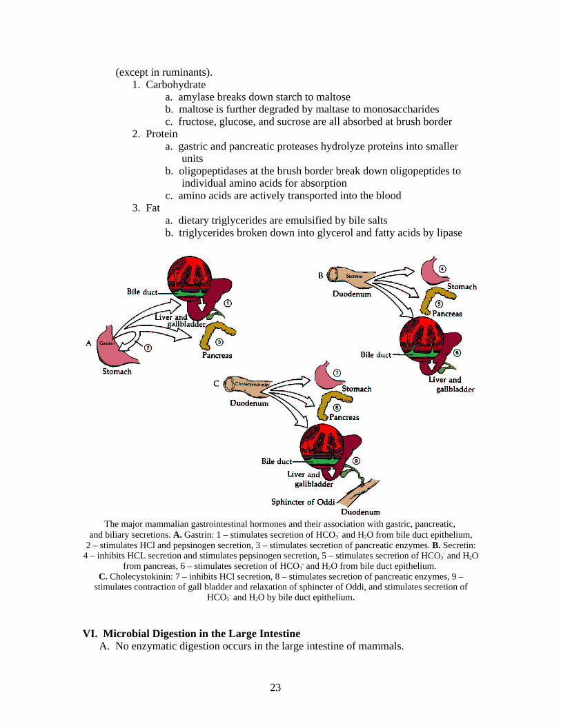
The major mammalian gastrointestinal hormones and their association with gastric, pancreatic, and biliary secretions. A. Gastrin: 1 – stimulates secretion of HCO3
- and H2O from bile duct epithelium, 2 – stimulates HCl and pepsinogen secretion, 3 – stimulates secretion of pancreatic enzymes. B. Secretin: 4 – inhibits HCL secretion and stimulates pepsinogen secretion, 5 – stimulates secretion of HCO3
- and H2Ofrom pancreas, 6 – stimulates secretion of HCO3
- and H2O from bile duct epithelium. C. Cholecystokinin: 7 – inhibits HCl secretion, 8 – stimulates secretion of pancreatic enzymes, 9 –
stimulates contraction of gall bladder and relaxation of sphincter of Oddi, and stimulates secretion ofHCO3
- and H2O by bile duct epithelium.
VI. Microbial Digestion in the Large IntestineA. No enzymatic digestion occurs in the large intestine of mammals.
23
B. Digestion that occurs results from microbial action which is significant for non-ruminant herbivores and omnivores
C. End products of digestion are volatile fatty acids (VFA’s)1. acetate2. proprionate3. butyrate
D. Horses obtain as much as 75% of their energy requirement from large intestinal absorption of VFA’s.
E. Large intestine fermentation salvages otherwise lost calories as VFA’s and decreases the effective osmotic pressure of the large intestine contents, so that water can be reabsorbed.
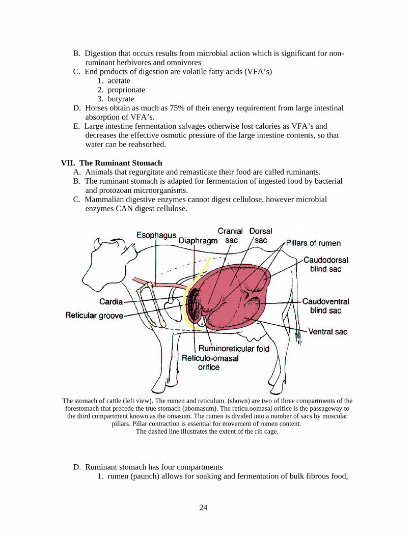
VII. The Ruminant StomachA. Animals that regurgitate and remasticate their food are called ruminants.B. The ruminant stomach is adapted for fermentation of ingested food by bacterial
and protozoan microorganisms. C. Mammalian digestive enzymes cannot digest cellulose, however microbial
enzymes CAN digest cellulose.
The stomach of cattle (left view). The rumen and reticulum (shown) are two of three compartments of theforestomach that precede the true stomach (abomasum). The reticu.oomasal orifice is the passageway tothe third compartment known as the omasum. The rumen is divided into a number of sacs by muscular
pillars. Pillar contraction is essential for movement of rumen content. The dashed line illustrates the extent of the rib cage.
D. Ruminant stomach has four compartments1. rumen (paunch) allows for soaking and fermentation of bulk fibrous food,
24
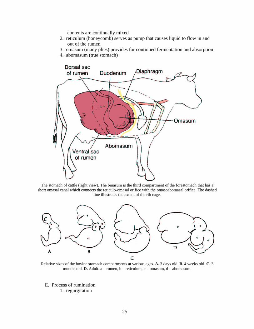
contents are continually mixed2. reticulum (honeycomb) serves as pump that causes liquid to flow in and
out of the rumen3. omasum (many plies) provides for continued fermentation and absorption4. abomasum (true stomach)
The stomach of cattle (right view). The omasum is the third compartment of the forestomach that has ashort omasal canal which connects the reticulo-omasal orifice with the omasoabomasal orifice. The dashed
line illustrates the extent of the rib cage.
Relative sizes of the bovine stomach compartments at various ages. A. 3 days old. B. 4 weeks old. C. 3months old. D. Adult. a – rumen, b – reticulum, c – omasum, d – abomasum.
E. Process of rumination1. regurgitation
25
2. remastication3. reinsalivation4. redeglutition
F. Byproducts of fermentation1. methane (30 – 40% of rumen gas)2. carbon dioxide (60 – 70% of rumen gas)
a. gases mostly eliminated by eructation3. volatile fatty acids4. protozoa5. bacteria
VIII. Avian DigestionA. Birds have no teeth, therefore the mechanical breakdown of their ingested food is
accomplished by the beak and gizzard.B. Salivary glands are present in birds and well developed in species which consume
dry food.C. The esophagus is divided into precrop and postrcrop segments.D. The crop is a dilation of the esophagus and has a food storage function.E. The proventriculus is located between the postcrop, esophagus, and gizzard.F. The gastric secretions HCl and pepsinogen as well as mucus are secreted by the
proventriculusG. The small intestine has a well-defined duodenum with the pancreas located
between its loops, but there is no distinction between6the jejunum and ileum.H. The ceca, which are paired structures, are located at the junction of the small and
large intestine. Here microbial digestion of cellulose occurs.I. Digestive tract ends with the cloaca, the site that is common to the digestive,
urinary, and reproductive tracts in birds.
26
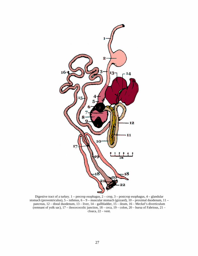
Digestive tract of a turkey. 1 – precrop esophagus, 2 – crop, 3 – postcrop esophagus, 4 – glandularstomach (proventriculus), 5 – isthmus, 6 – 9 – muscular stomach (gizzard), 10 – proximal duodenum, 11 –
pancreas, 12 – distal duodenum, 13 – liver, 14 – gallbladder, 15 – ileum, 16 – Meckel’s diverticulum(remnant of yolk sac), 17 – ileocecocolic junction, 18 – ceca, 19 – colon, 20 – bursa of Fabrious, 21 –
cloaca, 22 – vent.
27