Embed Size (px)
Citation preview

DIGESTIVE SYSTEMBy GERALDINE SABATE RIDAD, RN
LEAH MAY P. MADJUS, RN, MN
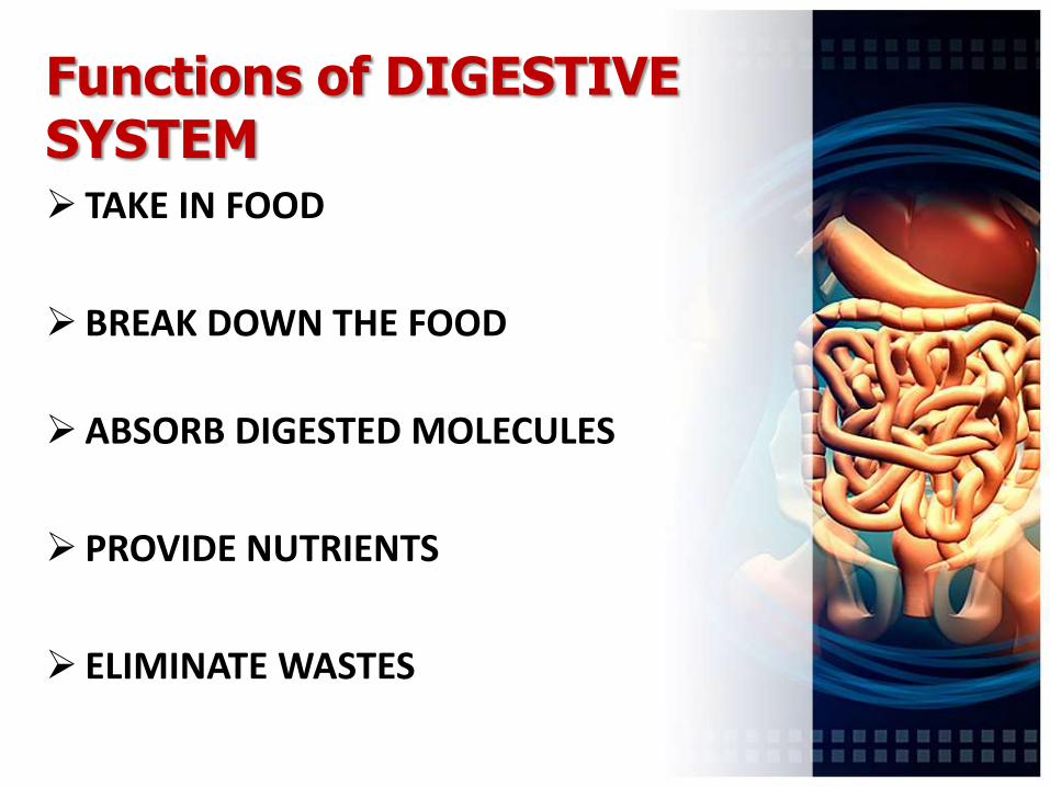
Functions of DIGESTIVE SYSTEM TAKE IN FOOD
BREAK DOWN THE FOOD
ABSORB DIGESTED MOLECULES
PROVIDE NUTRIENTS
ELIMINATE WASTES
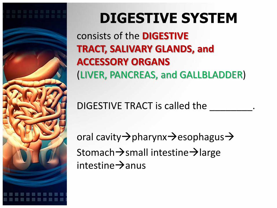
DIGESTIVE SYSTEMconsists of the DIGESTIVE TRACT, SALIVARY GLANDS, and ACCESSORY ORGANS (LIVER, PANCREAS, and GALLBLADDER)
DIGESTIVE TRACT is called the ________.
oral cavitypharynxesophagus
Stomachsmall intestinelargeintestineanus


4 Layers Of Digestive Tract1. MUCOSA – consists of mucous epithelium,
lamina propria and muscularis mucosa
2. SUBMUCOSA- thick layer of connective tissue containing NERVES, BLOOD VESSELS, and SMALL GLANDS.
3. MUSCULARIS- consists of an inner layer of CIRCULAR SMOOTH MUSCLE and an outer layer of LONGITUDINAL SMOOTH MUSCLE
4. SEROSA or ADVENTITIA>SEROSA- peritoneum and underlying connective tissue>ADVENTITIA- not covered by peritoneum

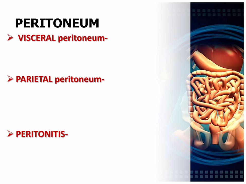
PERITONEUM VISCERAL peritoneum-
PARIETAL peritoneum-
PERITONITIS-

MESENTERIES- hold many of the organs in place within the abdominal cavity.
RETROPERITONEAL ORGANS- other abdominal organs lie against the abdominal wall, have no mesenteries.


ORAL CAVITY
MOUTH, is the FIRST PART of the digestive tract, bounded by lips and cheeks and contains the teeth and tongue.
MASTICATION- chewing; begins the process of MECHANICAL DIGESTION
TONGUE- moves food in mouth, holds food in place during mastication, facilitates SWALLOWING , major sensory organ for TASTE and SPEECH

TEETH
There are 32 teeth in normal adult mouth located in the mandible or maxillary.
Teeth of adults are PERMANENT or SECONDARY TEETH replacement of the 20 DECIDUOUS/ PRIMARY TEETH
Facilitates in MECHANICAL DIGESTION of food


PALATE ROOF of oral cavity
2 parts:
1.HARD PALATE
2.SOFT PALATE
>>separates the oral cavity from the nasal cavity and prevents food from passing into it during chewing and swallowing!!!

TONSILS
TONSILS- are located in the lateral posterior wall of the oral cavity, in the nasopharynxand posterior surface of tongue


SALIVARY GLANDS
Produce SALIVA which is a mixture of SEROUS and MUCUS FLUIDS
Saliva helps keep the oral cavity MOIST and contains enzymes that begin the process of CHEMICAL DIGESTION
1. PAROTID- largest; just anterior to the ear;
(***mumps)
2. SUBMANDIBULAR- produce more serous than mucous secretions
3. SUBLINGUAL- smallest; produce primarily mucus secretions

SECRETIONS of ORAL CAVITY
1. SALIVARY AMYLASE- digestive enzyme in saliva, DIGESTS STARCH (to glucose or isomaltose or maltose)
2. MUCIN – PROTEOGLYCAN that gives a lubricating quality to saliva
Regulated by AUTONOMIC NERVOUS SYSTEM
P
S

FLUID OR ENZYME
FUNCTION
SALIVA Moistens and lubricates food
SALIVARY AMYLASE
Starch digestion (conversion to maltose and isomaltose)
LIPASE Begins LIPID DIGESTION
LYSOZYME Weak antibacterial action

MASTICATION
Breaks LARGE food particles into many small ones; since digestive enzymes act on molecules only at the surface of the food particles, mastication increases EFFICIENCY OF DIGESTION
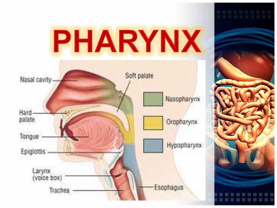

PHARYNX
THROAT, which connects MOUTH with ESOPHAGUS
3 PARTS
1.NASOPHARYNX
2.OROHARYNX
3.LARYNGOPHARYNX
PHARYNGEAL LIPASE- facilitates lipid digestion
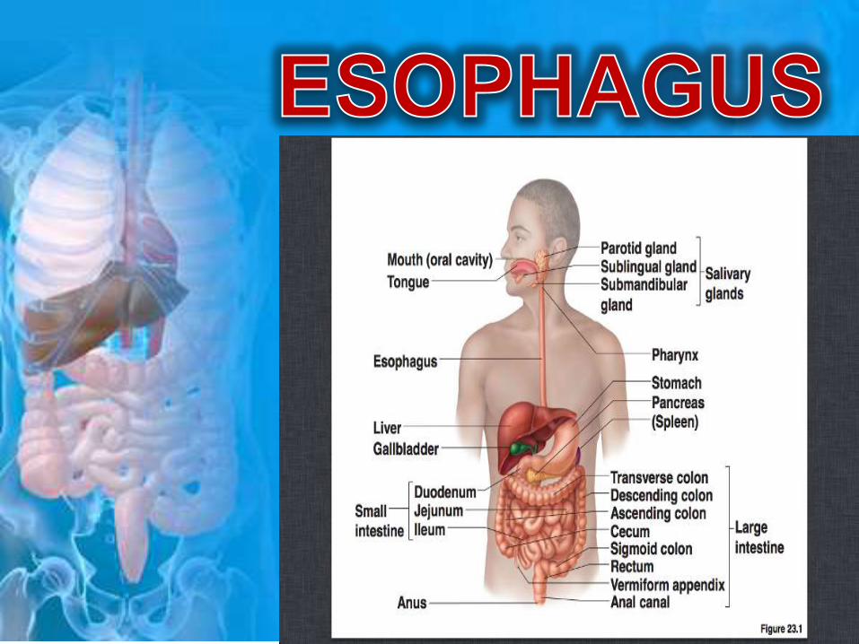

ESOPHAGUS
lined with STRATIFIED SQUAMOUS EPITHELIUM, that extends from pharynx to the stomach
Passes through the DIAPHRAGM
UPPER ESOPHAGEAL SPHINCTER
LOWER ESOPHAGEAL SPHINCTER-CARDIAC SPHINCTER; regulate the movement of food into and out of esophagus.
peristalsis
DEGLUTITION swallowing
3 separate phases:
1. VOLUNTARY PHASE- BOLUS is formed, pushed by tongue against the hard palate into the oropharynx
2. PHARYNGEAL PHASE- begins with the elevation of soft palate, which closes the passage between the nasopharynxand oropharynx
- EPIGLOTTIS is tipped posteriorly covering the LARYNX
3. ESOPHAGEAL PHASE- moving food from pharynx to stomach
>>peristaltic contractions associated w/ swallowing cause relaxation of lower esophageal sphincter as peristaltic waves approach the stomach.


STOMACH Located in the LEFTSUPERIOR part of
the abdomen( )


RUGAE- large folds that allow the STOMACH to stretch when stomach is filled

lined with SIMPLE COLUMNAR EPITHELIUM
gastric pits are openings for GASTRIC GLANDS
5 types of EPITHELIAL CELLS:
1. SURFACE MUCOUS CELLS- produce MUCUS which coats and protects the stomach lining and gastric pits
2. MUCOUS NECK CELLS

3. PARIETAL CELLS-produce HCL acid and intrinsic factor
4. ENDOCRINE CELLS-produce regulatory hormones
5.CHIEF CELLS-produce pepsinogen
SECRETIONS of the STOMACH Functions primarily as a storage and mixing
chamber for ingested food
_________ CHYME
HYDROCHLORIC ACID
INTRINSIC FACTOR
PEPSINOGEN
GASTRIN

REGULATION of STOMACH SECRETIONS CEPHALIC PHASE
GASTRIC
INTESTINAL PHASE
-when CHYME (W/ ph 2.0) enters the DUODENUM SECRETIN and CHOLECYSTOKININ is released
HEARTBURN-
>>painful or burning sensation in the chest usually associated w/ an increase in gastric secretion and /or backflush of acidic chyme into the esophagus


SMALL INTESTINEAbout 6m long; the site at which
greatest amount of digestion and absorption occur.
Duodenum
Jejunum
Ileum
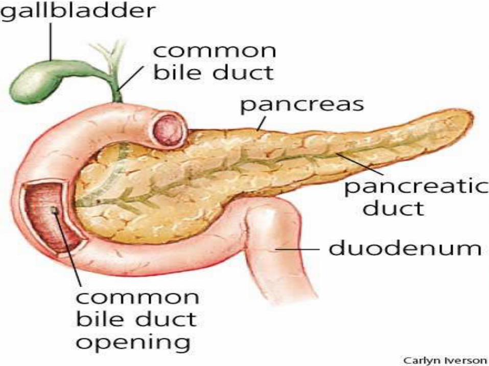
DUODENUM-
common bile duct+pancreatic duct


VILLI- covered by SIMPLE COLUMNAR EPITHELIUM
>> contains blood capillary network and LACTEAL
MAJOR CELL TYPES:
1. ABSORPTIVE CELLS
2. GOBLET CELLS
3. GRANULAR CELLS(PANETH’S CELLS)
4. ENDOCRINE CELLS

PEYER’S PATCHES- found in ILEUM
VIT B12 ABSORPTION???
ILEOCECAL JUNCTION
ILEOCECAL SPHINCTER and VALVE
SECRETIONS of SMALL INTESTINE Peptidases
Sucrases
Maltase
Lipase
Cholecystikinin
Secretin
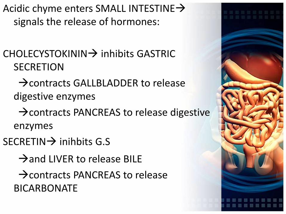
Acidic chyme enters SMALL INTESTINEsignals the release of hormones:
CHOLECYSTOKININ inhibits GASTRIC SECRETION
contracts GALLBLADDER to release digestive enzymes
contracts PANCREAS to release digestive enzymes
SECRETIN inihbits G.S
and LIVER to release BILE
contracts PANCREAS to release BICARBONATE

MOVEMENTS of SMALL INTESTINE PERISTALTIC CONTRACTIONS- proceed
along the length of the intestine for variable distances and cause chyme to move along the small intestine
SEGMENTAL CONTRACTIONS- propagated for only short distances and function to mix intestinal contents

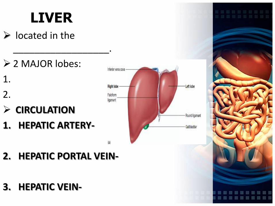
LIVER located in the
__________________.
2 MAJOR lobes:
1.
2.
CIRCULATION
1. HEPATIC ARTERY-
2. HEPATIC PORTAL VEIN-
3. HEPATIC VEIN-

LIVER1. The hepatic ducts from the
liver lobes combine to formthe common hepatic duct.
2. The common hepatic ductcombines with the cysticduct from the gallbladder toform the common bile duct.
3. The common bile duct andthe
pancreatic duct combine to formthe hepatopancreaticampulla.
4. The hepatopancreatic ampullaempties into the duodenumat the major duodenalpapilla.
5. Pancreatic secretions alsoenter the duodenum throughthe
hepatopancreatic ampulla. The
accessory pancreatic duct alsoempties into the duodenum.


FUNCTIONS OF THE LIVER
DIGESTION
EXCRETION Excretion of CHOLESTEROL, FATS, and BILIRUBIN
NUTRIENTSTORAGE
Store glucose (as GLYCOGEN in liver), fats, VITAMINS (ADEK, B12), and MINERALS (copper and IRON)
NUTRIENT CONVERSION
1.GLYCOGENOLYSIS
2.
DETOXIFICATION 1.AMMONIA 2.DRUG-FIRST PASS
SYNTHESIS of NEW
MOLECULES
Blood proteins: ALBUMIN, FIBRINOGEN, GLOBULINS, and CLOTTING FACTORS
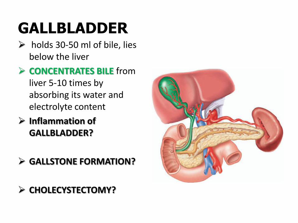
GALLBLADDER holds 30-50 ml of bile, lies
below the liver
CONCENTRATES BILE from liver 5-10 times by absorbing its water and electrolyte content
Inflammation of GALLBLADDER?
GALLSTONE FORMATION?
CHOLECYSTECTOMY?


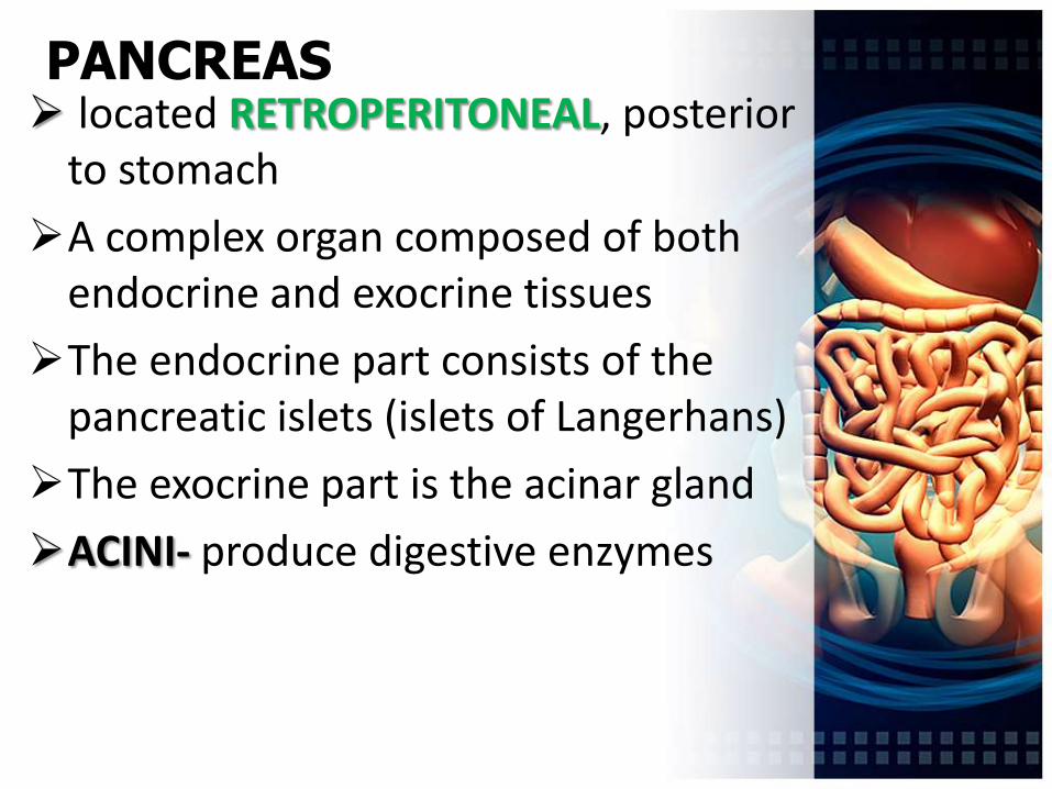
PANCREAS located RETROPERITONEAL, posterior
to stomach
A complex organ composed of both endocrine and exocrine tissues
The endocrine part consists of the pancreatic islets (islets of Langerhans)
The exocrine part is the acinar gland
ACINI- produce digestive enzymes


FUNCTIONS of PANCREAS PROTEOLYTIC ENZYMES:
1.Chymotrypsin
2.Trypsin
3.Carboxypeptidase
4.Peptidase
PANCREATIC AMYLASE – continues the polysaccharide digestion that was initiated in the oral cavity
PANCREATIC LIPASES – break down lipids

FUNCTIONS of PANCREAS• CCKCHOLECYSTOKININ inhibits
GASTRIC SECRETION/ contracts GALLBLADDER to release digestive enzymes
• SECRETIN inihbits G.S/ contracts GALLBLADDER to release BICARBONATE and LIVER to release BILE
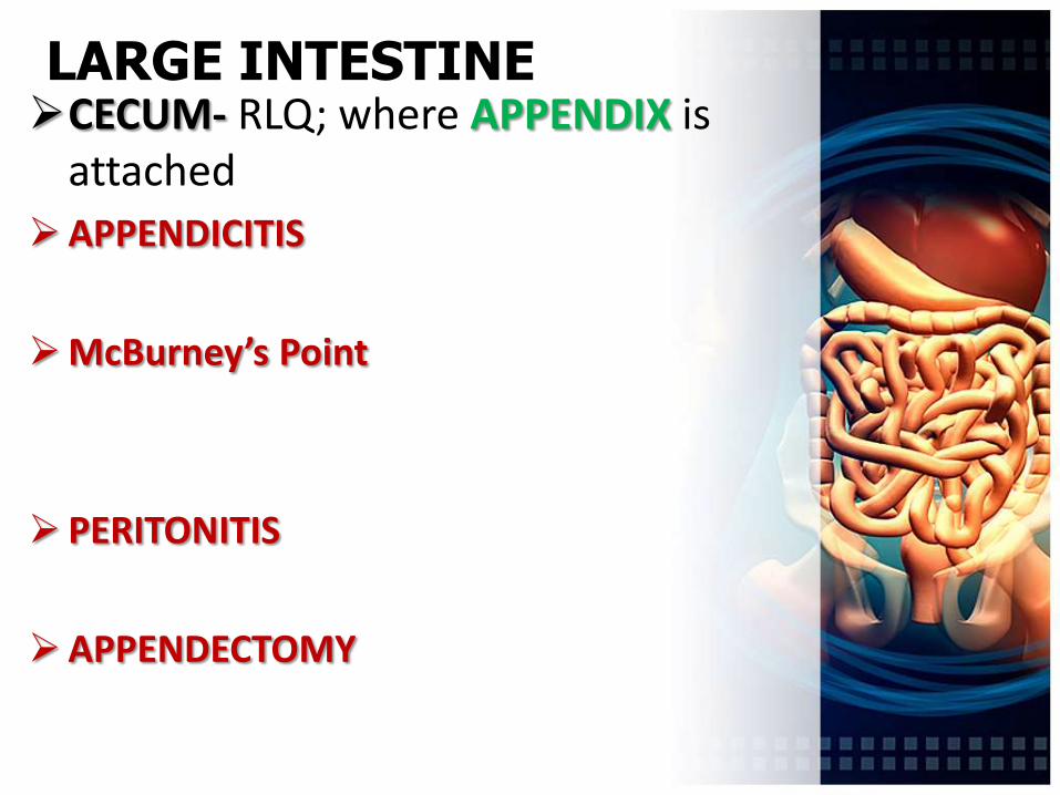
LARGE INTESTINECECUM- RLQ; where APPENDIX is
attached
APPENDICITIS
McBurney’s Point
PERITONITIS
APPENDECTOMY

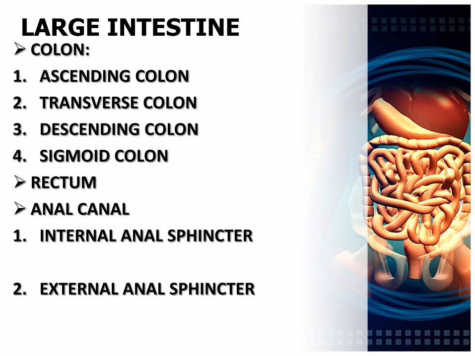
LARGE INTESTINE COLON:
1. ASCENDING COLON
2. TRANSVERSE COLON
3. DESCENDING COLON
4. SIGMOID COLON
RECTUM
ANAL CANAL
1. INTERNAL ANAL SPHINCTER
2. EXTERNAL ANAL SPHINCTER


FUNCTIONS of LARGE INTESTINE chyme is converted into FECES
absorbs WATER and SALTS
DEFECATION
some bacteria in the colon synthesize VITAMIN K, which is passively absorb in the intestine.
MASS MOVEMENTS- propel the colon contents a considerable distance toward the anus

HEMORRHOIDS
CONSTIPATION
DIARRHEA