Embed Size (px)
Citation preview
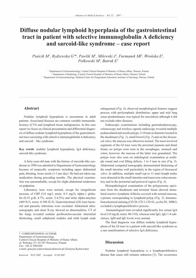
296 Piaścik M, et al. · Advances in Medical Sciences · Vol. 52 · 2007 ·
Abstract
Nodular lymphoid hyperplasia is uncommon in adult patients. Associated diseases are common variable immunode-ficiency (CVI) and lymphoid tissue malignancies. In this case report we focus on clinical presentation and differential diagno-sis of diffuse nodular lymphoid hyperplasia of the gastrointesti-nal tract coexisting with selective immunoglobulin A deficiency and sarcoid – like syndrome.
Key words: nodular lymphoid hyperplasia, IgA deficiency, sarcoid-like syndrome.
A forty-year-old man with the history of sarcoide-like syn-drome in 1998 was admitted to Department of Gastroenterology because of unspecific symptoms including upper abdominal pain, bloating, loose stools (1-3 per day). He had not taken any medication during preceding months. The physical examina-tion was unremarkable, except for slight abdominal tenderness on palpation.
Laboratory tests were normal, except for insignificant increase of CRP (5.8 mg/l, norm: 0-5 mg/l), alpha-1 globu-lin (0.35 g/dl, 4.7%, norm: 2-4.5%) and urine alpha-amylase (469 IU/l, norm: 0-380 IU/l). Gastrointestinal (GI) tract bacte-rial and parasitic infections were excluded. Abdominal ultra-sonography and chest X-ray showed no abnormalities. CT of the lungs revealed nodular peribronchovascular interstitial thickening, small subpleural nodules and mild lymph node
enlargement (Fig. 6), observed morphological features suggest process with perilymphatic distribution, upper and mid lung zones predominance was typical for sarcoidosis although it did not exclude other diseases.
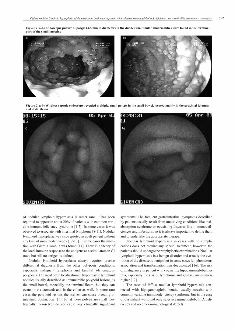
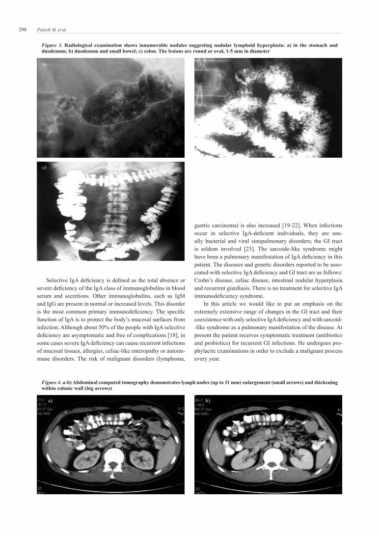
Endoscopic examinations including gastroduodenoscopy, colonoscopy and wireless capsule endoscopy revealed multiple pedunculated and sessile polyps, 2-10 mm in diameter located in the duodenum (Fig. 1), small bowel (Fig. 2) and on the ileocae-cal valve; the mucosa was otherwise normal. The most involved segments of the GI tract were the proximal jejunum and distal ileum; no polyps were seen in the oesophagus, stomach and colon, however, the mucosa of the latter was granulated. The polyps were also seen on radiological examination as multi-ple round and oval filling defects, 1 to 5 mm in size (Fig. 3). Abdominal computed tomography demonstrated thickening of the small intestine wall particularly in the region of ileocaecal valve. In addition, multiple small (up to 11 mm) lymph nodes were detected in the small intestine and transverse colon mesen-tery and in the periaortal and pericaval region (Fig. 4).
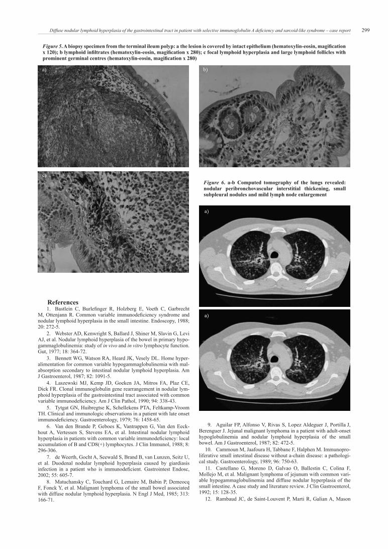
Histopathological examination of the polypectomy speci-men from the duodenum and terminal ileum showed stimu-lated reactive lymphatic follicles covered with normal mucosa, a picture corresponding to lymphoid polyp (Fig. 5). Immuno-histochemical staining (CD 20, CD 3, CD 43, cyclin D1, MIB1) excluded a lymphoproliferative process.
Immunological tests revealed significantly low serum IgA level (10 mg/dl, norm: 80-310), whereas total IgG, IgG 1-4 sub-classes, IgM and IgE levels were normal.
The final diagnosis was diffuse nodular lymphoid hyper-plasia of the GI tract in a patient with sarcoid-like syndrome as a rare manifestation of selective IgA deficiency.
Discussion
Nodular lymphoid hyperplasia is a lymphoproliferative disease that cause still remains unknown [1]. The occurrence
Diffuse nodular lymphoid hyperplasia of the gastrointestinal tract in patient with selective immunoglobulin A deficiency
and sarcoid-like syndrome – case report
Piaścik M 1, Rydzewska G1*, Pawlik M 1, Milewski J 1, Furmanek MI2, Wrońska E3, Polkowski M3, Butruk E3
1 Department of Gastroenterology, Central Clinical Hospital of Ministry of Home Affairs, Warsaw, Poland2 Department of Radiology, Central Clinical Hospital of Ministry of Home Affairs, Warsaw, Poland
3 Department of Gastroenterology, Medical Centre for Postgraduate Education, Institute of Oncology, Warsaw, Poland
* CORREsPOnDInG AUTHOR: Department of Gastroenterology Central Clinical Hospital of Ministry of Home Affairsul. Wołoska 137, 02-507 Warszawa, PolandFax: +48 22 5081044e-mail: [email protected] (Grażyna Rydzewska)
Received 30.07.2007 Accepted 01.08.2007
297Diffuse nodular lymphoid hyperplasia of the gastrointestinal tract in patient with selective immunoglobulin A deficiency and sarcoid-like syndrome – case report
of nodular lymphoid hyperplasia is rather rare. It has been reported to appear in about 20% of patients with common vari-able immunodeficiency syndrome [1-7]. In some cases it was observed to associate with intestinal lymphoma [8-11]. Nodular lymphoid hyperplasia was also reported in adult patient without any kind of immunodeficiency [12-13]. In some cases the infec-tion with Giardia lamblia was found [14]. There is a theory of the local immune response to the antigens as a stimulators in GI tract, but still no antigen is defined.
Nodular lymphoid hyperplasia always requires precise differential diagnosis from the other polyposis conditions, especially malignant lymphoma and familial adenomatous polyposis. The most often localisation of hyperplastic lymphoid nodules usually described as innumerable polypoid lesions, is the small bowel, especially the terminal ileum, but they can occur in the stomach and in the colon as well. In some rare cases the polypoid lesions themselves can cause bleeding or intestinal obstruction [15], but if these polyps are small they typically themselves do not cause any clinically significant
symptoms. The frequent gastrointestinal symptoms described by patients usually result from underlying conditions like mal-absorption syndrome or coexisting diseases like immunodefi-ciences and infections, so it is always important to define them and to undertake the appropriate therapy.
Nodular lymphoid hyperplasia in cases with no compli-cations does not require any special treatment, however, the patients should undergo the prophylactic examinations. nodular lymphoid hyperplasia is a benign disorder and usually the evo-lution of the disease is benign but in some cases lymphomatous association and transformation was documented [16]. The risk of malignancy in patient with coexisting hipogammaglobuline-mia, especially the risk of lymphoma and gastric carcinoma is higher [17].
The cases of diffuse nodular lymphoid hyperplasia con-nected with hipogammaglobulinemia, usually coexist with common variable immunodeficiency syndrome, but in the case of our patient we found only selective immunoglobulin A defi-ciency and no other immunological defects.
Figure 1. a-b) Endoscopic picture of polyps (3-5 mm in diameter) in the duodenum. Similar abnormalities were found in the terminal part of the small intestine
Figure 2. a-b) Wireless capsule endoscopy revealed multiple, small polyps in the small bowel, located mainly in the proximal jejunum and distal ileum
298 Piaścik M, et al.
selective IgA deficiency is defined as the total absence or severe deficiency of the IgA class of immunoglobulins in blood serum and secretions. Other immunoglobulins, such as IgM and IgG are present in normal or increased levels. This disorder is the most common primary immunodeficiency. The specific function of IgA is to protect the body’s mucosal surfaces from infection. Although about 50% of the people with IgA selective deficiency are asymptomatic and free of complications [18], in some cases severe IgA deficiency can cause recurrent infections of mucosal tissues, allergies, celiac-like enteropathy or autoim-mune disorders. The risk of malignant disorders (lymphoma,
gastric carcinoma) is also increased [19-22]. When infections occur in selective IgA-deficient individuals, they are usu-ally bacterial and viral sinopulmonary disorders; the GI tract is seldom involved [23]. The sarcoide-like syndrome might have been a pulmonary manifestation of IgA deficiency in this patient. The diseases and genetic disorders reported to be asso-ciated with selective IgA deficiency and GI tract are as follows: Crohn’s disease, celiac disease, intestinal nodular hyperplasia and recurrent giardiasis. There is no treatment for selective IgA immunodeficiency syndrome.
In this article we would like to put an emphasis on the extremely extensive range of changes in the GI tract and their coexistence with only selective IgA deficiency and with sarcoid- -like syndrome as a pulmonary manifestation of the disease. At present the patient receives symptomatic treatment (antibiotics and probiotics) for recurrent GI infections. He undergoes pro-phylactic examinations in order to exclude a malignant process every year.
Figure 3. Radiological examination shows innumerable nodules suggesting nodular lymphoid hyperplasia: a) in the stomach and duodenum; b) duodenum and small bowel; c) colon. The lesions are round or oval, 1-5 mm in diameter
Figure 4. a-b) Abdominal computed tomography demonstrates lymph nodes (up to 11 mm) enlargement (small arrows) and thickening within colonic wall (big arrows)
299Diffuse nodular lymphoid hyperplasia of the gastrointestinal tract in patient with selective immunoglobulin A deficiency and sarcoid-like syndrome – case report
References 1. Bastlein C, Burlefinger R, Holzberg E, Voeth C, Garbrecht M, Ottenjann R. Common variable immunodeficiency syndrome and nodular lymphoid hyperplasia in the small intestine. Endoscopy, 1988; 20: 272-5. 2. Webster AD, Kenwright s, Ballard J, shiner M, slavin G, Levi AJ, et al. nodular lymphoid hyperplasia of the bowel in primary hypo-gammaglobulinemia: study of in vivo and in vitro lymphocyte function. Gut, 1977; 18: 364-72. 3. Bennett WG, Watson RA, Heard JK, Vesely DL. Home hyper-alimentation for common variable hypogammaglobulinemia with mal-absorption secondary to intestinal nodular lymphoid hyperplasia. Am J Gastroenterol, 1987; 82: 1091-5. 4. Laszewski MJ, Kemp JD, Goeken JA, Mitros FA, Plaz CE, Dick FR. Clonal immunoglobulin gene rearrangement in nodular lym-phoid hyperplasia of the gastrointestinal tract associated with common variable immunodeficiency. Am J Clin Pathol, 1990; 94: 338-43. 5. Tytgat Gn, Huibregtse K, schellekens PTA, Feltkamp-Vroom TH. Clinical and immunologic observations in a patient with late onset immunodeficiency. Gastroenterology, 1979; 76: 1458-65. 6. Van den Brande P, Geboes K, Vantrappen G, Van den Eeck-hout A, Vertessen s, stevens EA, et al. Intestinal nodular lymphoid hyperplasia in patients with common variable immunodeficiency: local accumulation of B and CD8(+) lymphocytes. J Clin Immunol, 1988; 8: 296-306. 7. de Weerth, Gocht A, seewald s, Brand B, van Lunzen, seitz U, et al. Duodenal nodular lymphoid hyperplasia caused by giardiasis infection in a patient who is immunodeficient. Gastrointest Endosc, 2002; 55: 605-7. 8. Matuchansky C, Touchard G, Lemaire M, Babin P, Demeocq F, Fonck Y, et al. Malignant lymphoma of the small bowel associated with diffuse nodular lymphoid hyperplasia. n Engl J Med, 1985; 313: 166-71.
9. Aguilar FP, Alfonso V, Rivas s, Lopez Aldeguer J, Portilla J, Berenguer J. Jejunal malignant lymphoma in a patient with adult-onset hypoglobulinemia and nodular lymphoid hyperplasia of the small bowel. Am J Gastroenterol, 1987; 82: 472-5. 10. Cammoun M, Jaafoura H, Tabbane F, Halphen M. Immunopro-liferative small intestinal disease without a-chain disease: a pathologi-cal study. Gastroenterology, 1989; 96: 750-63. 11. Castellano G, Moreno D, Galvao O, Ballestin C, Colina F, Mollejo M, et al. Malignant lymphoma of jejunum with common vari-able hypogammaglobulinemia and diffuse nodular hyperplasia of the small intestine. A case study and literature review. J Clin Gastroenterol, 1992; 15: 128-35. 12. Rambaud JC, de saint-Louvent P, Marti R, Galian A, Mason
Figure 6. a-b Computed tomography of the lungs revealed: nodular peribronchovascular interstitial thickening, small subpleural nodules and mild lymph node enlargement
Figure 5. A biopsy specimen from the terminal ileum polyp: a the lesion is covered by intact epithelium (hematoxylin-eosin, magification x 120); b lymphoid infiltrates (hematoxylin-eosin, magification x 280); c focal lymphoid hyperplasia and large lymphoid follicles with prominent germinal centres (hematoxylin-eosin, magification x 280)

300 Piaścik M, et al.
DY, Wassef M, et al. Diffuse follicular lymphoid hyperplasia of the small intestine without primary immunoglobulin deficiency. Am J Med, 1982; 73: 125-32. 13. Tomita s, Kojima M, Imura J, Ueda Y, Koitabasi A, suzuki Y, et al. Diffuse nodular lymphoid hyperplasia of the large bowel without hypogammaglobulinemia or malabsorption syndrome: a case report and literature review. Int J surg Pathol, 2002; 10: 297-302. 14. Ward H, Jalan Kn, Maitra TK, Agarwal sK, Mahalanabis D. small intestinal nodular lymphoid hyperplasia in patient with giardiasis and normal serum immunoglobulins. Gut, 1983; 24: 120-6. 15. Chandra s. Benign nodular lymphoid hyperplasia of colon: a report of two cases. Indian J Gastroenterol, 2003; 22: 145-6. 16. Matuchansky C, Touchard G, Lemaire M, Babin P, Deme-ocq F, Fonk Y, et al. Malignant lymphoma of small bowelassociated with diffuse nodular lymphoid hyperplasia. n Engl J Med, 1985; 313: 1661-71. 17. Lai Ping so A, Mayer L. Gastrointestinal manifestations of
primary immunodeficiency disorders. semin Gastrointest Dis, 1997; 8: 22-32. 18. Jakóbisiak M. Izolowany niedobór IgA. In: Immunologia. Warszawa: PWn; 1995, pp. 537-9. 19. Amman AJ, Hong R. selective IgA deficiency. Presentation of 30 cases and a review of the literature. Medicine, 1971; 50: 223-36. 20. Fasth A. Primary immunodeficiency disorders in sweden: cases among children, 1974-1979. J Clin Immunol, 1982; 2: 86-92. 21. Hanson LA, Bjorkander J, Oxelius V. selective IgA deficiency. In: Primary and secondary immunodeficiency disorders. Chandra RK (editor). new York: Churchil Livingstone; 1983, pp. 62-84. 22. Luzi G, Businco L, Aiuti F. Primary immunodeficiency syn-dromes in Italy: a report of the national register in children and adults. J Clin Immunol, 1983; 3: 316-20. 23. spickett GP, Misbah sA, Chapel HM. Primary antibody defi-ciency in adults. Lancet, 1991; 337: 281-4.