Embed Size (px)
Citation preview
Akhter et al. Diagnostic Pathology 2014, 9:93http://www.diagnosticpathology.org/content/9/1/93
CASE REPORT Open Access
Polypoid nodular histiocytic hyperplasiaassociated with endometrioid adenocarcinoma ofthe endometrium: report of a caseShabnam Akhter, W Dwayne Lawrence and M Ruhul Quddus*
Abstract
A 45 year old woman underwent Laparoscopy-assisted total hysterectomy with staging procedure following adiagnosis of endometrial endometrioid adenocarcinoma on her endometrial biopsy. The hysterectomy specimenshowed a FIGO I stage 1a, endometrioid carcinoma. A separate polypoid lesion in the endometrium, distinct from thecarcinoma, was also identified. Microscopically the polypoid lesion was “nodular histiocytic hyperplasia”. The H&E,immunohistochemical staining findings and the differential diagnoses are discussed in this report. Althoughdescription of similar lesions is available in the literature, the current lesion is unique as it is identified in ahysterectomy specimen in its entirety and its association with an endometrial endometrioid carcinoma.Virtual Slides: The virtual slide(s) for this article can be found here: http://www.diagnosticpathology.diagnomx.eu/vs/1060511915121922
Keywords: Endometrium, Nodular histiocytic hyperplasia, Endometrioid carcinoma
IntroductionNodular histiocytic hyperplasia, initially thought to be ofmesothelial origin because of their sites of occurrences,was reported in hernia sac [1]. Subsequently similar le-sions have been reported from various other body sites,e.g., the pericardial sacs and cardiac valves [2], periton-eum [3] and pleura [4]. Their presence in endometrialbiopsy may mimic neoplasia. A recent series publishedseven cases of nodular histiocytic hyperplasia [5]. Thecases, so far reported in the literature, are associatedwith benign lesions only. Since the previously reportedcases were encountered in endometrial biopsies the le-sions were fragmented. We report here a case of a nodu-lar histiocytic hyperplasia associated with FIGO I/IIIendometrial endometrioid adenocarcinoma. We also de-scribe the lesion in its entirety; as noted above, all previ-ous descriptions of this entity were based on theirpresence in biopsy specimens.
* Correspondence: [email protected] of Pathology and Laboratory Medicine, Women & InfantsHospital, Alpert Medical School of Brown University, 101 Dudley Street,Providence, Rhode Island 02905, USA
© 2014 Akhter et al.; licensee BioMed CentralCommons Attribution License (http://creativecreproduction in any medium, provided the orDedication waiver (http://creativecommons.orunless otherwise stated.
Case presentationA 45 year-old woman with a history of uterine fibroidspresented with vaginal bleeding. She was Gravida 1 andPara 0, with a history of termination of pregnancy. Apelvic ultrasound showed a 9.1 x 8.0 x 4.0 cm partiallydistorted uterus with multiple hypoechoeic nodularareas, the largest being 4 x 3 cm and a thickened,1.8 cm, endometrial stripe.
Pathologic findingsAn endometrial biopsy revealed a FIGO I endometrialendometrioid type adenocarcinoma. After weighing-inall the options presented to her, the patient opted for aLaparoscopy-assisted vaginal hysterectomy with bilateralsalpingo-oophorectomy and staging procedure.Gross examination of the specimen revealed a 172
gms, 9.6 x 6.5 x 5.6 cm distorted uterus with multiplesubserosal and intramural fibroids. Upon opening theuterus, a 4.5 x 4.2 cm shaggy polypoid lesion was identi-fied in the endometrial cavity involving both uterinewalls. In addition a 0.7 x 0.5 cm polypoid lesion(Figure 1a) was found on the anterior wall of the endo-myometrium with smooth surface. The fibroids werenoted in the intramural and subserosal locations.
Ltd. This is an Open Access article distributed under the terms of the Creativeommons.org/licenses/by/4.0), which permits unrestricted use, distribution, andiginal work is properly credited. The Creative Commons Public Domaing/publicdomain/zero/1.0/) applies to the data made available in this article,
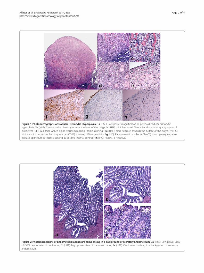
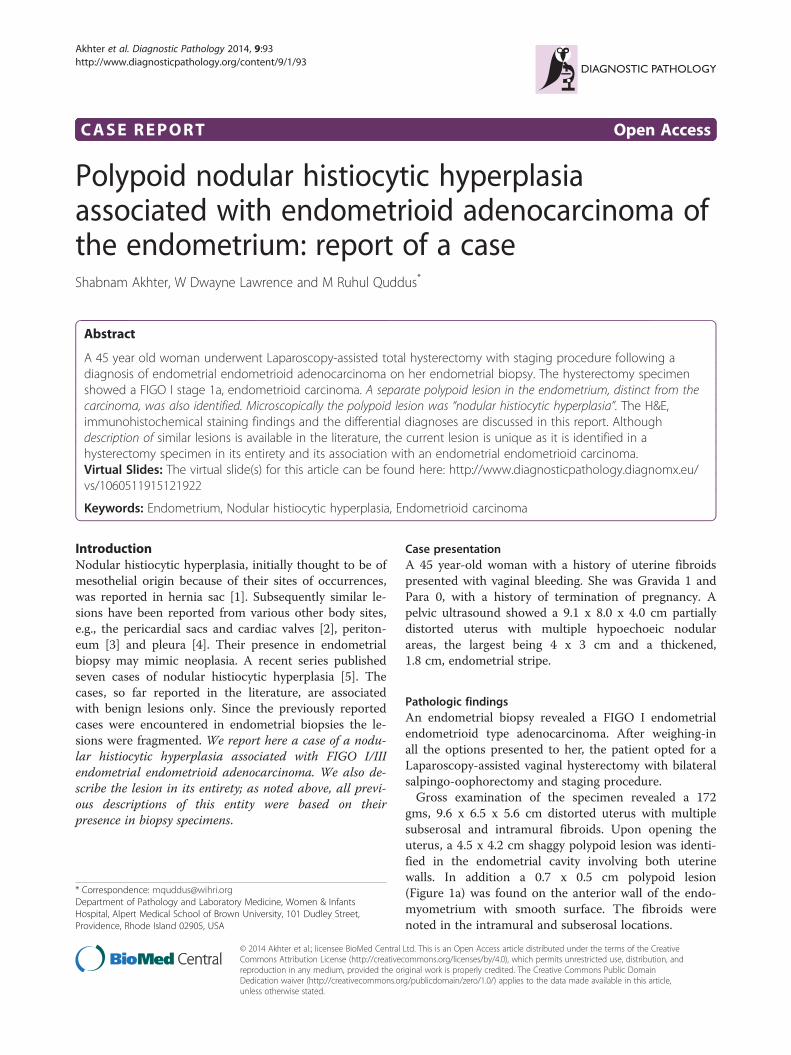
Figure 1 Photomicrographs of Nodular Histiocytic Hyperplasia. 1a (H&E): Low power magnification of polypoid nodular histiocytichyperplasia; 1b (H&E): Closely packed histiocytes near the base of the polyp; 1c (H&E): pink hyalinized fibrous bands separating aggregates ofhistiocytes; 1d (H&E): thick-walled blood vessel mimicking “onion-skinning”; 1e (H&E): more sclerosis towards the surface of the polyp; 1f (IHC):histiocytic immunohistochemistry marker (CD68) showing diffuse positivity; 1g (IHC): Pancytokeratin marker (AE1/AE3) is completely negative(surface epithelium is reactive serving as positive internal control); 1h (IHC): HMB45 is negative.
Figure 2 Photomicrographs of Endometrioid adenocarcinoma arising in a background of secretory Endometrium. 2a (H&E): Low power viewof FIGO I endometrioid carcinoma; 2b (H&E): high power view of the same tumor; 2c (H&E): Carcinoma is arising in a background of secretoryendometrium.
Akhter et al. Diagnostic Pathology 2014, 9:93 Page 2 of 4http://www.diagnosticpathology.org/content/9/1/93
Akhter et al. Diagnostic Pathology 2014, 9:93 Page 3 of 4http://www.diagnosticpathology.org/content/9/1/93
Microscopically, a smooth surfaced polypoid lesionwas found to consist mostly of aggregates of histiocytesthat were more closely packed near the endomyometrialjunction, i.e., the base of the lesion (Figure 1b). Pink hya-linized fibrous bands are noted throughout the polypoidlesion (Figure 1c). Many blood vessels with thickenedhyalinized walls are present throughout the lesion, morecommonly towards the surface (Figure 1d). Towards thesurface, the histiocytes were relatively sparse and sepa-rated by sclerosis (Figure 1e). The polyp was lined bya thin, attenuated, layer of endometrial epithelium(Figure 1e). The histiocytes resembled perivascular epi-thelioid cells. And presence of thick walled blood vesselswithin the lesion raised the question of a perivascularepithelioid cell tumor (PEComa). Immunohistochemicalstains reveal that the lesional cells are strongly and dif-fusely reactive to a histiocytic marker, CD68, (Figure 1f )and non-reactive to HMB45 (Figure 1h), pancytokeratin(AE1/AE3), (1 g) Epithelial membrane antigen (EMA),Desmin, Smooth Muscle Actin (SMA), CD10, Vimentin.FIGO grade I stage 1a endometrioid adenocarcinoma
was noted in the endometrium as well (Figure 2a andFigure 2b). The tumor involves areas of adenomyosis;however, true myometrial invasion was not present. Thecarcinoma was arising in background of functional endo-metrium (Figure 2c). The lower uterine segment andcervix were free of tumor. No lymph-vascular space in-vasion was identified. The bilateral ovaries and fallopiantubes were unremarkable. The regional lymph node dis-section was all negative for metastatic carcinoma.
Discussion and conclusionsNodular histiocytic hyperplasia is a rare lesion often inci-dentally encountered in endometrial biopsies. Majority ofthese lesions are reported in the literature as single casereport. The largest series, so far has been reported in theliterature, is based on 7 cases [5]. The lesion, when en-countered in endometrial biopsies and present in smallfragments, may mimic a neoplasm. So far all the re-ported cases are, however, associated with benign dis-eases. The current report is based on a case of nodularhistiocytic hyperplasia associated with a FIGO I/IIIendometrial endometrioid adenocarcinoma. The lesionitself is identified grossly in its entirety as a distinct en-tity presenting as an endometrial polyp.The differential diagnoses of this entity include Lang-
erhans cell histiocytosis, xanthogranulomatous endomet-ritis, malakoplakia, signet-ring cell changes of theendometrial stromal cells, etc. Available reports in theliterature have elaborated how to distinguish nodularhistiocytic hyperplasia from their mimics. The currentcase showed some resemblance with perivascular epithe-lioid cell tumor (PEComa) or an epithelioid smoothmuscle tumor with many small hyalinized blood vessels.
Microscopic and multifocal PEComa of the female geni-tal tract have been reported [6,7]. One interesting find-ing noted in the current case is the presence ofhyalinized fibrous strands throughout the lesion; a fea-ture often seen in endometrial stromal tumors. The cyto-logic appearance of the tumor cells seen in endometrialstromal tumor is, however, completely different fromwhat was seen in nodular histiocytic hyperplasia. Thecells in endometrial stromal tumor are small, mostlyround and darkly stained, especially in low grade stro-mal sarcoma.Special stains that were performed to rule out the
mimics show the lesional cells of nodular histiocytichyperplasia are strongly and diffusely reactive to histio-cytic marker, CD68. All other markers including, epithe-lial markers (AE1/AE3), smooth muscle markers, andendometrial stromal cell marker (CD10) were non-reactive. HMB45, a marker for PEComa, was alsonegative.The site of nodular histiocytic hyperplasia was clearly
located on the surface of endometrium with smooth lin-ing and protruding into the endometrial cavity.It is speculated that the nodular histiocytic aggregates
may result from previous endometrial biopsy. Thecurrent case did have a recent history of endometrial bi-opsy one month prior to her hysterectomy. The previousbiopsy in this case revealed endometrial carcinoma withsquamous differentiation and no evidence of histiocyticaggregate. It is also of note that the appearance of thefoamy histiocytes often seen in endometrial biopsies isalso different from the histiocytes present in nodularhyperplasia as they lack the foamy cytoplasm.Mazur and Kurman [8] proposed that these histiocytes
apparently reside in the endometrial cavity and reportedtheir presence in association with hydrometra and be-nign bleeding patterns. The authors postulated that itmay represent a response to what they have proposed as“intracavitary debris.” The current case was associatedwith an endometrioid adenocarcinoma so presence ofsome “intracavitary debris” is not unlikely. As noted be-fore the polypoid lesion was seen projecting into theendometrial cavity.A possibly related, but morphologically dissimilar, his-
tiocytic endometrial lesion has been reported by Iezzoniand Mills in their study of non-neoplastic endometrialsignet-ring cells [9]. No signet-ring cells are identified inthis case.Kim et al. proposed that the endometrial stromal cells
showing progestational changes with atrophic endomet-rial glands trapped in the middle may produce histologicsimilarities that may vaguely resemble histiocytic aggre-gate [10]. Unlike nodular histiocytic aggregate, decidua-lized stromal cells do not form a discrete nodule. Kimet al. also speculated that the nodules may not originate
Akhter et al. Diagnostic Pathology 2014, 9:93 Page 4 of 4http://www.diagnosticpathology.org/content/9/1/93
in the endometrium because no vasculature was seen intheir cases [10]. The current case documents many bloodvessels in the nodule and thus contradicts that specula-tion of Kim et al.In conclusion, nodular histiocytic hyperplasia may not
always be associated with benign/inflammatory lesionsas previously reported in the literature. The current casedocuments its association with endometrioid carcinomaof the endometrium.The patient is currently followed routinely and is dis-
ease free 80 months after surgery.
ConsentThe patient has given consent for the use of the imagesand case presentation for educational and scientific pur-poses provided the unique patient identification is notrevealed.
Competing interestThe authors declare no competing financial interest. All the authors haveactively participated in the diagnosis and manuscript writing.
Authors’ contributionsSA is the Stuart Lauchlan International Visiting Fellow in Gynecologic andBreast Pathology and participated in writing up the case report and MRQ isthe attending Pathologist on the case. WDL offered his expert opinion infinalizing the case. All authors read and approved the manuscript.
Received: 19 February 2014 Accepted: 9 April 2014Published: 12 May 2014
References1. Rosai J, Dehner LP: Nodular mesothelial hyperplasia in hernia sacs. A
benign reactive condition simulating a neoplastic process. Cancer 1975,35:165–175.
2. Luthringer DJ, Virmani R, Weiss SW, Rosai J: A distinctive cardiovascularlesion resembling histiocytoid (epithelioid) hemangioma. Evidencesuggesting mesothelial participation. Am J Surg Pathol 1990, 14:993–1000.
3. Clement PB: Reactive tumor-like lesions of the peritoneum. Am J ClinPathol 1995, 103:673–76.
4. Ordonez NG, Ro JY, Ayala AG: Lesions described as nodular mesothelialhyperplasia are primarily composed of histiocytes. Am J Sung Pathol 1998,22:285–92.
5. Prakash V, Domfeh AB, Fadare O: Nodular histiocytic aggregates in theendometrium: a report of 7 cases. Int J Gynecol Pathol 2013, 33:52–57.
6. Chia-Lang F, Yun-Ho C, Wei-Yu C: Microscopic endometrial perivascularepithelioid cell nodules: a case report with the earliest presentation of auterine perivascular epithelioid cell tumor. Diagn Pathol 2012, 7:117.
7. Wang Y, Gao L, Zheng W-Q: Multifocal PEComa (PEComatosis) of thefemale genital tract and pelvis: a case report and review of theliterature. Diagn Pathol 2012, 7:23.
8. Mazur MT, Kurman RJ: Artifacts and contaminants. In Diagnosis ofEndometrial Biopsies and Curettings: A Practical Approach. Edited by MazurMT, Kurman RJ. New York: Springer; 1995:22.
9. Iezzoni JC, Mills SE: Nonneoplastic endometrial signet-ring cells.Vacuolated decidual cells and stromal histiocytes mimickingadenocarcinoma. Am J Clin Pathol 2001, 15:249–55.
10. Kim K-R, Lee YH, Ro JY: Nodular histiocytic hyperplasia of theendometrium. Int J of Gynecol Pathol 2002, 21:141–146.
doi:10.1186/1746-1596-9-93Cite this article as: Akhter et al.: Polypoid nodular histiocytic hyperplasiaassociated with endometrioid adenocarcinoma of the endometrium:report of a case. Diagnostic Pathology 2014 9:93.
Submit your next manuscript to BioMed Centraland take full advantage of:
• Convenient online submission
• Thorough peer review
• No space constraints or color figure charges
• Immediate publication on acceptance
• Inclusion in PubMed, CAS, Scopus and Google Scholar
• Research which is freely available for redistribution
Submit your manuscript at www.biomedcentral.com/submit