Embed Size (px)
Citation preview
Leading Edge
Review
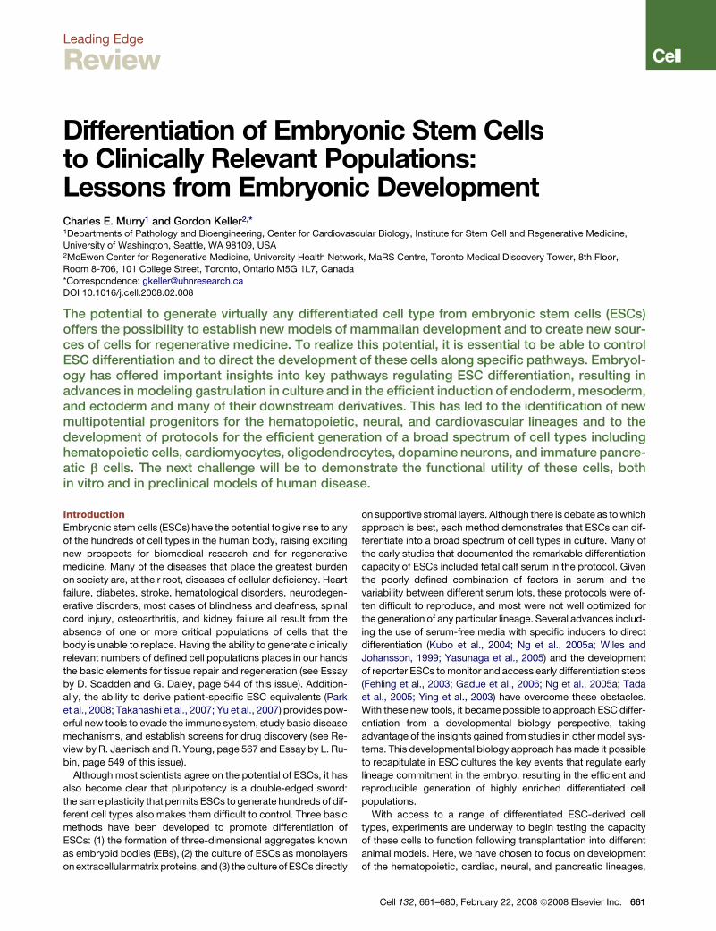
Differentiation of Embryonic Stem Cellsto Clinically Relevant Populations:Lessons from Embryonic DevelopmentCharles E. Murry1 and Gordon Keller2,*1Departments of Pathology and Bioengineering, Center for Cardiovascular Biology, Institute for Stem Cell and Regenerative Medicine,
University of Washington, Seattle, WA 98109, USA2McEwen Center for Regenerative Medicine, University Health Network, MaRS Centre, Toronto Medical Discovery Tower, 8th Floor,
Room 8-706, 101 College Street, Toronto, Ontario M5G 1L7, Canada
*Correspondence: [email protected]
DOI 10.1016/j.cell.2008.02.008
The potential to generate virtually any differentiated cell type from embryonic stem cells (ESCs)offers the possibility to establish new models of mammalian development and to create new sour-ces of cells for regenerative medicine. To realize this potential, it is essential to be able to controlESC differentiation and to direct the development of these cells along specific pathways. Embryol-ogy has offered important insights into key pathways regulating ESC differentiation, resulting inadvances in modeling gastrulation in culture and in the efficient induction of endoderm, mesoderm,and ectoderm and many of their downstream derivatives. This has led to the identification of newmultipotential progenitors for the hematopoietic, neural, and cardiovascular lineages and to thedevelopment of protocols for the efficient generation of a broad spectrum of cell types includinghematopoietic cells, cardiomyocytes, oligodendrocytes, dopamine neurons, and immature pancre-atic b cells. The next challenge will be to demonstrate the functional utility of these cells, bothin vitro and in preclinical models of human disease.
IntroductionEmbryonic stem cells (ESCs) have the potential to give rise to any
of the hundreds of cell types in the human body, raising exciting
new prospects for biomedical research and for regenerative
medicine. Many of the diseases that place the greatest burden
on society are, at their root, diseases of cellular deficiency. Heart
failure, diabetes, stroke, hematological disorders, neurodegen-
erative disorders, most cases of blindness and deafness, spinal
cord injury, osteoarthritis, and kidney failure all result from the
absence of one or more critical populations of cells that the
body is unable to replace. Having the ability to generate clinically
relevant numbers of defined cell populations places in our hands
the basic elements for tissue repair and regeneration (see Essay
by D. Scadden and G. Daley, page 544 of this issue). Addition-
ally, the ability to derive patient-specific ESC equivalents (Park
et al., 2008; Takahashi et al., 2007; Yu et al., 2007) provides pow-
erful new tools to evade the immune system, study basic disease
mechanisms, and establish screens for drug discovery (see Re-
view by R. Jaenisch and R. Young, page 567 and Essay by L. Ru-
bin, page 549 of this issue).
Although most scientists agree on the potential of ESCs, it has
also become clear that pluripotency is a double-edged sword:
the same plasticity that permits ESCs to generate hundreds of dif-
ferent cell types also makes them difficult to control. Three basic
methods have been developed to promote differentiation of
ESCs: (1) the formation of three-dimensional aggregates known
as embryoid bodies (EBs), (2) the culture of ESCs as monolayers
on extracellular matrix proteins,and (3) the culture of ESCs directly
on supportive stromal layers. Although there is debate as to which
approach is best, each method demonstrates that ESCs can dif-
ferentiate into a broad spectrum of cell types in culture. Many of
the early studies that documented the remarkable differentiation
capacity of ESCs included fetal calf serum in the protocol. Given
the poorly defined combination of factors in serum and the
variability between different serum lots, these protocols were of-
ten difficult to reproduce, and most were not well optimized for
the generation of any particular lineage. Several advances includ-
ing the use of serum-free media with specific inducers to direct
differentiation (Kubo et al., 2004; Ng et al., 2005a; Wiles and
Johansson, 1999; Yasunaga et al., 2005) and the development
of reporter ESCs to monitor and access early differentiation steps
(Fehling et al., 2003; Gadue et al., 2006; Ng et al., 2005a; Tada
et al., 2005; Ying et al., 2003) have overcome these obstacles.
With these new tools, it became possible to approach ESC differ-
entiation from a developmental biology perspective, taking
advantage of the insights gained from studies in other model sys-
tems. This developmental biology approach has made it possible
to recapitulate in ESC cultures the key events that regulate early
lineage commitment in the embryo, resulting in the efficient and
reproducible generation of highly enriched differentiated cell
populations.
With access to a range of differentiated ESC-derived cell
types, experiments are underway to begin testing the capacity
of these cells to function following transplantation into different
animal models. Here, we have chosen to focus on development
of the hematopoietic, cardiac, neural, and pancreatic lineages,
Cell 132, 661–680, February 22, 2008 ª2008 Elsevier Inc. 661
as access to such cell populations may provide new therapies for
some of society’s most devastating diseases.
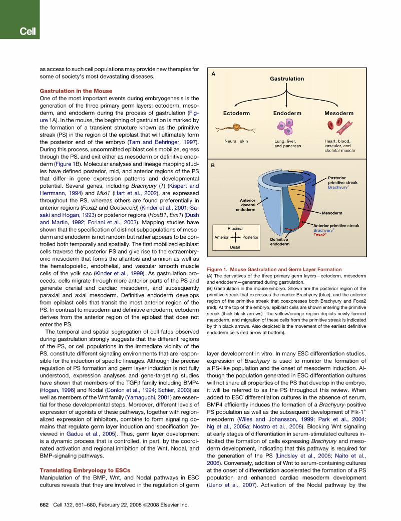
Gastrulation in the MouseOne of the most important events during embryogenesis is the
generation of the three primary germ layers: ectoderm, meso-
derm, and endoderm during the process of gastrulation (Fig-
ure 1A). In the mouse, the beginning of gastrulation is marked by
the formation of a transient structure known as the primitive
streak (PS) in the region of the epiblast that will ultimately form
the posterior end of the embryo (Tam and Behringer, 1997).
During this process, uncommitted epiblast cells mobilize, egress
through the PS, and exit either as mesoderm or definitive endo-
derm (Figure 1B). Molecular analyses and lineage mapping stud-
ies have defined posterior, mid, and anterior regions of the PS
that differ in gene expression patterns and developmental
potential. Several genes, including Brachyury (T) (Kispert and
Herrmann, 1994) and Mixl1 (Hart et al., 2002), are expressed
throughout the PS, whereas others are found preferentially in
anterior regions (Foxa2 and Goosecoid) (Kinder et al., 2001; Sa-
saki and Hogan, 1993) or posterior regions (HoxB1, Evx1) (Dush
and Martin, 1992; Forlani et al., 2003). Mapping studies have
shown that the specification of distinct subpopulations of meso-
derm and endoderm is not random but rather appears to be con-
trolled both temporally and spatially. The first mobilized epiblast
cells traverse the posterior PS and give rise to the extraembry-
onic mesoderm that forms the allantois and amnion as well as
the hematopoietic, endothelial, and vascular smooth muscle
cells of the yolk sac (Kinder et al., 1999). As gastrulation pro-
ceeds, cells migrate through more anterior parts of the PS and
generate cranial and cardiac mesoderm, and subsequently
paraxial and axial mesoderm. Definitive endoderm develops
from epiblast cells that transit the most anterior region of the
PS. In contrast to mesoderm and definitive endoderm, ectoderm
derives from the anterior region of the epiblast that does not
enter the PS.
The temporal and spatial segregation of cell fates observed
during gastrulation strongly suggests that the different regions
of the PS, or cell populations in the immediate vicinity of the
PS, constitute different signaling environments that are respon-
sible for the induction of specific lineages. Although the precise
regulation of PS formation and germ layer induction is not fully
understood, expression analyses and gene-targeting studies
have shown that members of the TGFb family including BMP4
(Hogan, 1996) and Nodal (Conlon et al., 1994; Schier, 2003) as
well as members of the Wnt family (Yamaguchi, 2001) are essen-
tial for these developmental steps. Moreover, different levels of
expression of agonists of these pathways, together with region-
alized expression of inhibitors, combine to form signaling do-
mains that regulate germ layer induction and specification (re-
viewed in Gadue et al., 2005). Thus, germ layer development
is a dynamic process that is controlled, in part, by the coordi-
nated activation and regional inhibition of the Wnt, Nodal, and
BMP-signaling pathways.
Translating Embryology to ESCsManipulation of the BMP, Wnt, and Nodal pathways in ESC
cultures reveals that they are involved in the regulation of germ
662 Cell 132, 661–680, February 22, 2008 ª2008 Elsevier Inc.
layer development in vitro. In many ESC differentiation studies,
expression of Brachyury is used to monitor the formation of
a PS-like population and the onset of mesoderm induction. Al-
though the population generated in ESC differentiation cultures
will not share all properties of the PS that develop in the embryo,
it will be referred to as the PS throughout this review. When
added to ESC differentiation cultures in the absence of serum,
BMP4 efficiently induces the formation of a Brachyury-positive
PS population as well as the subsequent development of Flk-1+
mesoderm (Wiles and Johansson, 1999; Park et al., 2004;
Ng et al., 2005a; Nostro et al., 2008). Blocking Wnt signaling
at early stages of differentiation in serum-stimulated cultures in-
hibited the formation of cells expressing Brachyury and meso-
derm development, indicating that this pathway is required for
the generation of the PS (Lindsley et al., 2006; Naito et al.,
2006). Conversely, addition of Wnt to serum-containing cultures
at the onset of differentiation accelerated the formation of a PS
population and enhanced cardiac mesoderm development
(Ueno et al., 2007). Activation of the Nodal pathway by the
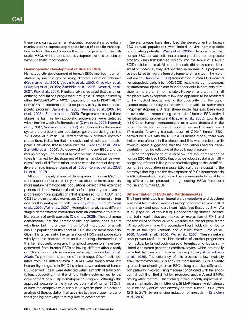
Figure 1. Mouse Gastrulation and Germ Layer Formation
(A) The derivatives of the three primary germ layers—ectoderm, mesoderm
and endoderm—generated during gastrulation.
(B) Gastrulation in the mouse embryo. Shown are the posterior region of the
primitive streak that expresses the marker Brachyury (blue), and the anterior
region of the primitive streak that coexpresses both Brachyury and Foxa2
(red). At the top of the embryo, epiblast cells are shown entering the primitive
streak (thick black arrows). The yellow/orange region depicts newly formed
mesoderm, and migration of these cells from the primitive streak is indicated
by thin black arrows. Also depicted is the movement of the earliest definitive
endoderm cells (red arrow at bottom).
addition of activin A (activin) induced a PS population and the
subsequent formation of endoderm or mesoderm, depending
on the strength of the signal (Kubo et al., 2004). Collectively,
these observations indicate that these signaling pathways play
some role in the early stages of ESC differentiation. However,
the precise stage at which they function and their interactions
with each other were not established in these studies.
While PS formation and germ layer induction are often consid-
ered part of gastrulation, these stages likely represent distinct
developmental steps that are regulated by different signaling
pathways. To investigate regulation of these early stages, Gadue
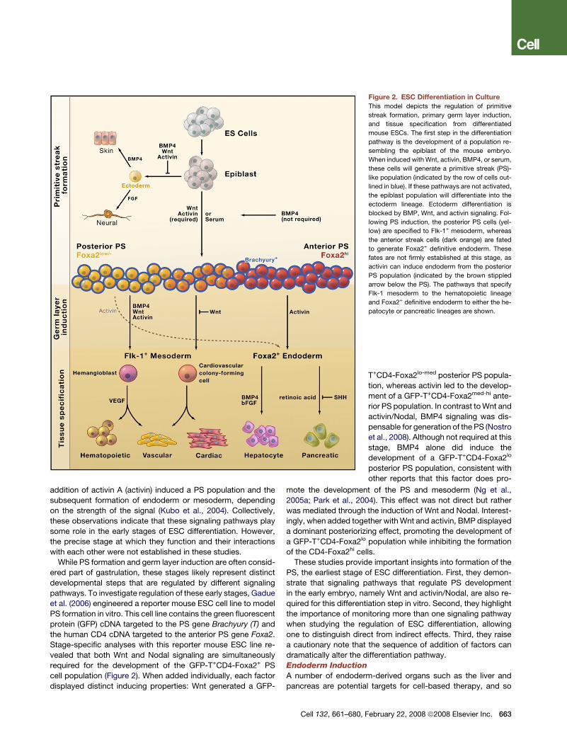
et al. (2006) engineered a reporter mouse ESC cell line to model
PS formation in vitro. This cell line contains the green fluorescent
protein (GFP) cDNA targeted to the PS gene Brachyury (T) and
the human CD4 cDNA targeted to the anterior PS gene Foxa2.
Stage-specific analyses with this reporter mouse ESC line re-
vealed that both Wnt and Nodal signaling are simultaneously
required for the development of the GFP-T+CD4-Foxa2+ PS
cell population (Figure 2). When added individually, each factor
displayed distinct inducing properties: Wnt generated a GFP-
Cell 132, 661–680, F
T+CD4-Foxa2lo-med posterior PS popula-
tion, whereas activin led to the develop-
ment of a GFP-T+CD4-Foxa2med-hi ante-
rior PS population. In contrast to Wnt and
activin/Nodal, BMP4 signaling was dis-
pensable for generation of the PS (Nostro
et al., 2008). Although not required at this
stage, BMP4 alone did induce the
development of a GFP-T+CD4-Foxa2lo
posterior PS population, consistent with
other reports that this factor does pro-
mote the development of the PS and mesoderm (Ng et al.,
2005a; Park et al., 2004). This effect was not direct but rather
was mediated through the induction of Wnt and Nodal. Interest-
ingly, when added together with Wnt and activin, BMP displayed
a dominant posteriorizing effect, promoting the development of
a GFP-T+CD4-Foxa2lo population while inhibiting the formation
of the CD4-Foxa2hi cells.
These studies provide important insights into formation of the
PS, the earliest stage of ESC differentiation. First, they demon-
strate that signaling pathways that regulate PS development
in the early embryo, namely Wnt and activin/Nodal, are also re-
quired for this differentiation step in vitro. Second, they highlight
the importance of monitoring more than one signaling pathway
when studying the regulation of ESC differentiation, allowing
one to distinguish direct from indirect effects. Third, they raise
a cautionary note that the sequence of addition of factors can
dramatically alter the differentiation pathway.
Endoderm Induction
A number of endoderm-derived organs such as the liver and
pancreas are potential targets for cell-based therapy, and so
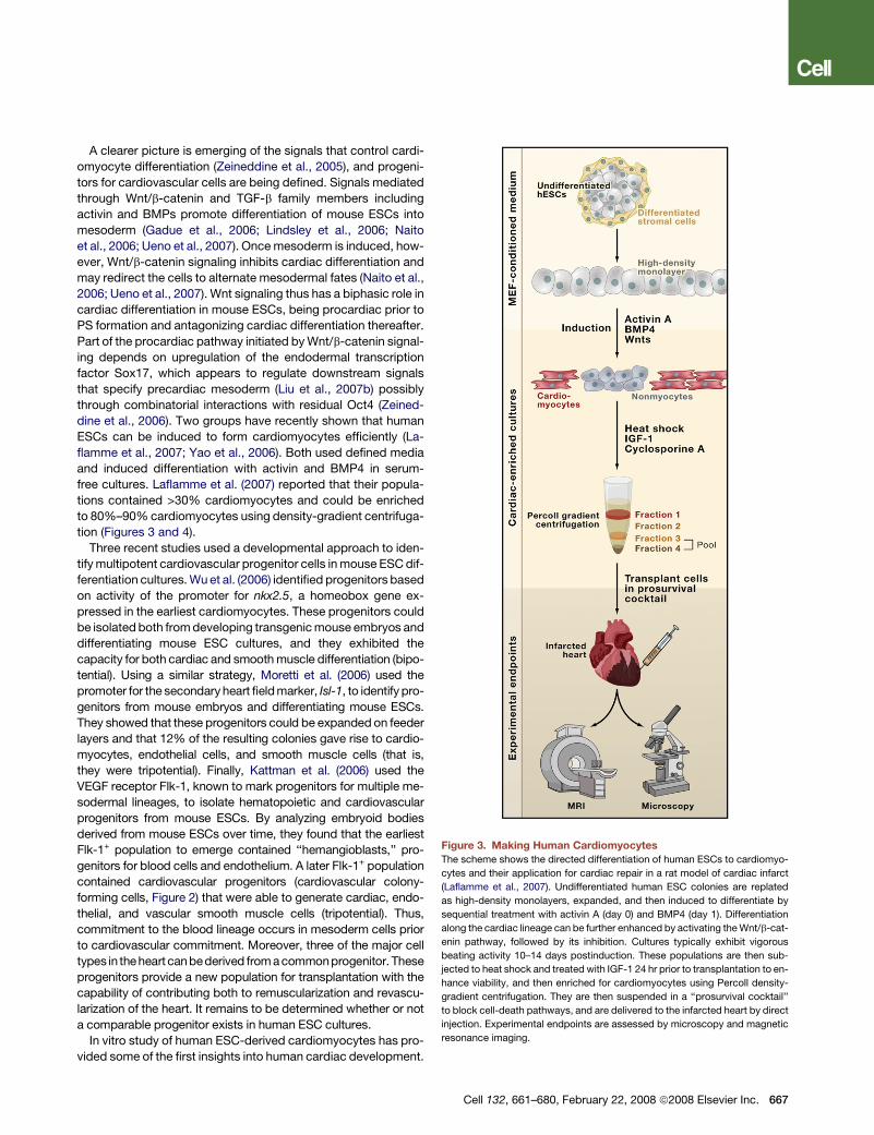
Figure 2. ESC Differentiation in Culture
This model depicts the regulation of primitive
streak formation, primary germ layer induction,
and tissue specification from differentiated
mouse ESCs. The first step in the differentiation
pathway is the development of a population re-
sembling the epiblast of the mouse embryo.
When induced with Wnt, activin, BMP4, or serum,
these cells will generate a primitive streak (PS)-
like population (indicated by the row of cells out-
lined in blue). If these pathways are not activated,
the epiblast population will differentiate into the
ectoderm lineage. Ectoderm differentiation is
blocked by BMP, Wnt, and activin signaling. Fol-
lowing PS induction, the posterior PS cells (yel-
low) are specified to Flk-1+ mesoderm, whereas
the anterior streak cells (dark orange) are fated
to generate Foxa2+ definitive endoderm. These
fates are not firmly established at this stage, as
activin can induce endoderm from the posterior
PS population (indicated by the brown stippled
arrow below the PS). The pathways that specify
Flk-1 mesoderm to the hematopoietic lineage
and Foxa2+ definitive endoderm to either the he-
patocyte or pancreatic lineages are shown.
ebruary 22, 2008 ª2008 Elsevier Inc. 663
there is great interest in understanding the pathways that regu-
late the induction and specification of this germ layer. High levels
of activin/Nodal signaling will efficiently induce definitive endo-
derm in mouse ESC cultures (Kubo et al., 2004; Yasunaga
et al., 2005). When analyzed at the PS stage, one step prior to
induction of definitive endoderm, activin-induced populations
identified either by the coexpression of Brachyury and Foxa2
(GFP-T+CD4-Foxa2hi) or expression of the anterior marker
Goosecoid (tagged with GFP, Gsc-GFP) were found to contain
both mesoderm and endoderm (Gouon-Evans et al., 2006;
Tada et al., 2005). Clonal analysis revealed that individual cells
within the Gsc-GFP population had the potential to generate
both endoderm and mesoderm derivatives, suggesting that
they may represent mesendoderm progenitors. Thus, the first
step in the generation of definitive endoderm may be the
formation of mesendoderm. Future studies should determine if
all endoderm and anterior mesoderm is derived from such
progenitors.
Progression of the anterior PS population (GFP-T+CD4-
Foxa2hi) to definitive endoderm (GFP-TnegCD4-Foxa2hi) de-
pends on sustained activin signaling (Gadue et al., 2006),
consistent with increased Nodal signaling required for definitive
endoderm formation in the early embryo (Schier, 2003). Interest-
ingly, when exposed to high levels of activin, the GFP-T+CD4-
Foxa2lo posterior PS population was also able to generate endo-
derm, indicating that germ-layer fates are not yet fixed at the PS
stage in mouse ESC differentiation cultures. Once induced, en-
doderm forms an epithelial sheet that undergoes specification
to distinct regions known as foregut, midgut, and hindgut (Wells
and Melton, 1999). This specification is controlled in part by fac-
tors secreted by surrounding mesoderm-derived tissues. By
translating findings from the embryo to ESC cultures, FGF and
BMP4 together were shown to induce a hepatic fate in activin-in-
duced endoderm (Gouon-Evans et al., 2006), whereas retinoic
acid combined with inhibition of sonic hedgehog (SHH) resulted
in specification to a pancreatic fate (D’Amour et al., 2006).
Mesoderm Induction
The hematopoietic, vascular, cardiac, and skeletal muscle line-
ages develop from subpopulations of mesoderm induced in a
defined temporal pattern. Understanding the mechanisms that
regulate mesoderm induction is a prerequisite for generating
each of these cell types. The early stages of mesoderm induction
from the PS can be monitored by the upregulation of Flk-1 and
PDGFR, receptors that are broadly expressed on subpopula-
tions of this lineage (Ema et al., 2006; Kataoka et al., 1997). Al-
though most studies have not distinguished PS formation from
mesoderm induction, several have provided insights into the sig-
naling pathways that regulate this step. Park et al. (2004) showed
that BMP signaling is required to induce Flk-1+ hematopoietic
mesoderm from populations that express Brachyury, indicating
that this pathway functions at the level of mesoderm induction.
In a more recent study, Nostro et al. (2008) demonstrated that
the generation of hematopoietic mesoderm from a Brachyury-
GFP tagged PS population is dependent on a combination of
Wnt, activin/Nodal, and BMP signaling (Figure 2), and that upre-
gulation of Flk-1 correlated with commitment to a mesoderm fate
(this population could no longer undergo respecification to endo-
derm following activin treatment).
664 Cell 132, 661–680, February 22, 2008 ª2008 Elsevier Inc.
Although the emergence of cardiac mesoderm from the PS
has not been investigated in as much detail as hematopoietic
mesoderm, findings from several studies suggest that transient
inhibition of Wnt/b-catenin signaling at this stage is essential
for the generation of this population (Naito et al., 2006; Ueno
et al., 2007). Wnt/b-catenin is thus required for mesoderm induc-
tion, whereas its inhibition is subsequently required for specifica-
tion of precardiac mesoderm. Recent studies using various con-
centrations of BMP4 or a combination of BMP4 and activin
indicate that subpopulations of mesoderm can be generated in
mouse ESC cultures by manipulating different signaling path-
ways at appropriate developmental stages (Era et al., 2007).
Ectoderm Induction
The neural lineages and skin are derived from ectoderm, which is
induced from epiblast cells in the anterior region of the embryo
that do not traverse the PS. The induction of ectoderm in ESC
cultures is often referred to as the ‘‘default’’ pathway, as neuro-
ectoderm readily develops in cultures that contain no serum
or other PS inducers. Neuroectoderm induction is inhibited by
BMP, Wnt, and activin/Nodal signaling, consistent with the ob-
servation that these pathways are not active in the region of ec-
toderm induction in the early embryo (Aubert et al., 2002; Kubo
et al., 2004; Ying et al., 2003). Ying et al. (2003) studied develop-
ment of the neuroectoderm lineage from mouse ESCs using a re-
porter cell line with GFP targeted to the neuroectoderm-specific
gene Sox1. They clearly showed that induction of neuroecto-
derm is dependent on FGF signals endogenously produced by
the differentiating ESCs. Thus, ‘‘default’’ differentiation to neuro-
ectoderm still proceeds through signaling pathways involved in
embryogenesis, similar to the formation of mesoderm and endo-
derm. Neuroectoderm generated from ESCs can be specified to
neuronal subtypes, using factor combinations known to regulate
these steps in the early embryo. In addition to the neural
lineages, ESC-derived ectoderm can also generate epidermal
lineage cells. As observed in vivo, the BMP-signaling pathway
does play a role in ectoderm specification in ESC cultures as
BMP signaling blocked neural differentiation and promoted epi-
dermal development (Kawasaki et al., 2000).
PS formation and Germ-Layer Induction in Human ESCsMouse and human ESCs may represent different stages of devel-
opment and clearly display different requirements for growth and
maintenance in the undifferentiated state (Reubinoff et al., 2000;
Thomson et al., 1998). However, the signaling pathways that reg-
ulate human ESC differentiation are similar to those that regulate
these processes in other organisms and in mouse ESC cultures.
As with mouse ESCs, the most successful human ESC differen-
tiation strategies are those that recapitulate normal develop-
ment. For example, activin signaling leads to the efficient induc-
tion of definitive endoderm in human ESC cultures (D’Amour
et al., 2005). Molecular analysis showed that the induced popu-
lations progressed through a PS stage to definitive endoderm in
a time frame similar to that observed in the mouse cultures. Me-
soderm induction from human ESCs also shows similarities to
mouse. Studies with wild-type human ESCs and a human ESC
reporter line expressing GFP from the MIXL1 gene (GFP-
MIXL1) demonstrated that induction with BMP4 leads to a rapid
increase in BRACHYURY and MIXL1 expression and the
subsequent formation of KDR+ (Flk-1+) and PDGFR+ mesoderm
(Davis et al., 2008; Kennedy et al., 2007; Ng et al., 2005b; Pick
et al., 2007). PS induction by BMP appears to require active
FGF and TGF-b/Nodal/activin signaling, as inhibition of these
pathways blocked the BMP4 response (Zhang et al., 2008).
Blocking Wnt signaling early in serum-induced cultures reduced
hematopoietic development, suggesting that this pathway acts
early in either PS formation or mesoderm induction (Woll et al.,
2007). Stimulating human ESCs with activin in addition to
BMP4 led to the induction of cardiac mesoderm, suggesting
that different subpopulations of mesoderm can be induced by
manipulating different signaling pathways (Laflamme et al.,
2007). These findings document the success of translating differ-
entiation strategies from mouse ESC to the human ESC system.
ESCs and the Hematopoietic LineageAs many of the early ESC differentiation studies focused on
blood cell development, hematopoiesis is one of the best-stud-
ied programs in ESC cultures (reviewed in Keller, 2005; Olsen
et al., 2006). The goal of many investigators working in this
area has been to induce the formation of transplantable hemato-
poietic stem cells (HSCs). With the relative ease of generating
blood cell lineages from ESCs, it was assumed that differentia-
tion to HSCs would be straightforward. However, despite exten-
sive efforts, the development of HSCs from ESCs that have not
been genetically modified remains a challenge, which may re-
flect the complexities of embryonic hematopoietic development
where different hematopoietic programs are generated at differ-
ent times from different embryonic sites.
Hematopoiesis in the early mouse embryo is initiated indepen-
dently at two distinct sites: the yolk sac and the para-aortic
splanchnopleura (P-Sp), an intraembryonic region in the caudal
portion of the embryo that later contains the developing aorta,
gonads, and mesonephros (AGM) (reviewed in Cumano and
Godin, 2007; Dzierzak, 2005; see Review by S.H. Orkin and
L. I. Zon, page 631 of this issue). Of significance for ESC differen-
tiation studies is the finding that these sites display different he-
matopoietic potential. The yolk sac generates primitive erythro-
cytes as well as a subset of other hematopoietic populations
including macrophages and progenitors of the definitive ery-
throid, megakaryocyte, and mast cell lineages. When analyzed
prior to the onset of circulation, the yolk sac displays little, if
any, lymphoid or HSC potential. In contrast, the P-Sp-derived
hematopoietic population generates HSCs as well as multipo-
tential progenitors that give rise to myeloid, lymphoid, and defin-
itive erythroid lineages in vitro. Hematopoietic development in
the human embryo follows a similar pattern (reviewed in Tavian
and Peault, 2005). Given these differences, it will be necessary
to recapitulate these developmental stages in vitro and to identify
the equivalent of P-Sp-derived hematopoiesis in order to isolate
HSCs from ESCs.
Recapitulating Hematopoiesis in Mouse ESC Cultures
Mouse ESCs undergo rapid and synchronous differentiation to
the hematopoietic lineages when cultured with serum or under
serum-free conditions supplemented with inducers (reviewed
in Keller, 2005). Gene expression and progenitor cell analysis
revealed that the differentiation program in these cultures closely
parallels that in the early embryo, progressing through a PS
stage, to Flk-1+ mesoderm, and subsequently to a yolk sac-
like hematopoietic program. Detailed analysis of these early
stages led to the identification of the hemangioblast, a progenitor
that displays hematopoietic and vascular potential and one that
defines the onset of hematopoiesis (Choi et al., 1998). A compa-
rable progenitor was identified in the posterior PS region of the
early mouse embryo and may represent the yolk-sac hemangio-
blast (Huber et al., 2004). After the hemangioblast appears, prim-
itive erythroid progenitors develop in ESC cultures, establishing
the primitive erythropoiesis phase of hematopoiesis. In addition
to primitive erythrocytes, other progenitors including those of the
macrophage, definitive erythroid, megakaryocyte, and mast cell
lineages develop in the differentiation cultures with a kinetic pat-
tern similar to that observed in the yolk sac. HSCs and progeni-
tors of the lymphoid lineage are not generated during this early
stage of hematopoiesis. These patterns of lineage development
suggest that the first hematopoietic population to develop from
ESCs represents the equivalent of yolk-sac hematopoiesis.
The striking similarities in yolk-sac hematopoietic development
observed between the ESC cultures and the early embryo sug-
gests that lineage commitment in this in vitro system recapitu-
lates that found in vivo.
The yolk-sac stage of hematopoiesis is well characterized
in mouse ESC cultures, but little is known about specification
and development of P-Sp hematopoiesis. Lymphoid potential
is one characteristic defining the P-Sp and mouse ESCs do gen-
erate these lineages under appropriate conditions. When cocul-
tured with OP9 stromal cells, mouse ESCs will give rise to cells of
the B lymphoid lineage (Cho et al., 1999; Nakano et al., 1994). If
cocultured with OP9 stromal cells engineered to express the
Notch ligand Delta-like 1, mouse ESCs will differentiate along
the T cell rather than the B cell lineage (Schmitt et al., 2004).
These observations suggest that a population equivalent to
the P-Sp region is generated in these cultures. However, given
that the ESCs were differentiated in complex cultures with stro-
mal cells in serum-based media, isolation of the lymphoid pro-
genitors and identification of the signaling pathways involved
remains a challenge.
The differentiation of mouse ESCs to HSCs that are capable of
long-term engraftment in recipient animals remains one of the
greatest challenges in the field of ESC biology. Although the de-
velopment of transplantable cells from ESCs in serum-stimulated
cultures was reported (Burt et al., 2004), the routine isolation of
HSCs using this approach is not widespread, suggesting that
success may depend on a specific batch of serum. As factors
regulating the specification of HSCs have yet to be defined, pro-
tocols for the efficient generation of these cells from ESCs do not
exist. To promote the development, survival, and expansion of
mouse ESC-derived HSCs, Daley and colleagues (Kyba et al.,
2002; Wang et al., 2005b) forced the expression of HoxB4 and
the caudal-related homeobox-containing factor Cdx4 in mouse
ESC-derived hematopoietic populations prior to transplantation.
Cdx4 promotes hematopoiesis when expressed in mouse ESC
cultures (Davidson et al., 2003), and HoxB4 induces extensive
self-renewal and expansion of bone marrow-derived HSCs
when expressed in this population (Sauvageau et al., 1995).
The ESC-derived cells expressing these two genes yielded multi-
lineage repopulation of recipient animals, demonstrating that
Cell 132, 661–680, February 22, 2008 ª2008 Elsevier Inc. 665
these cells can acquire hematopoietic repopulating potential if
manipulated to express appropriate levels of specific transcrip-
tion factors. The next step on the road to generating clinically
useful HSCs will be to induce development of this population
without genetic modification.
Hematopoietic Development of Human ESCsHematopoietic development of human ESCs has been demon-
strated by multiple groups using different induction schemes
(Kaufman et al., 2001; Vodyanik et al., 2005; Chadwick et al.,
2003; Ng et al., 2005b; Zambidis et al., 2005; Kennedy et al.,
2007; Pick et al., 2007). Kinetic analysis revealed that the differ-
entiating populations progressed through a PS stage defined by
either BRACHYURY or MIXL1 expression, then to KDR+ (Flk-1+)
or PDGFR+ mesoderm and subsequently to a yolk-sac hemato-
poietic program (Davis et al., 2008; Kennedy et al., 2007; Ng
et al., 2005b; Zambidis et al., 2005). Progression through these
stages is fast, as hematopoietic progenitors were detected
within the first week of differentiation (Davis et al., 2008; Kennedy
et al., 2007; Vodyanik et al., 2006). As observed in the mouse
system, the predominant population generated during the first
7–10 days of human ESC differentiation is primitive erythroid
progenitors, indicating that the equivalent of yolk-sac hemato-
poiesis develops first in these cultures (Kennedy et al., 2007;
Zambidis et al., 2005). As observed with mouse ESCs and the
mouse embryo, the onset of hematopoiesis in human ESC cul-
tures is marked by development of the hemangioblast between
days 2 and 4 of differentiation, prior to establishment of the prim-
itive erythroid lineage (Davis et al., 2008; Kennedy et al., 2007;
Lu et al., 2007).
Although the early stages of development in human ESC cul-
tures appear to represent the yolk-sac phase of hematopoiesis,
more mature hematopoietic populations develop after extended
periods of time. Analysis of cell surface phenotypes revealed
progression from populations that expressed KDR, CD31, and
CD34 to those that also expressed CD45, a marker found on fetal
and adult hematopoietic cells (Kennedy et al., 2007; Vodyanik
et al., 2005; Woll et al., 2007). Hemoglobin analysis at different
stages demonstrated maturation from an embryonic to a fetal-
like pattern of erythropoiesis (Qiu et al., 2008). These changes
demonstrate that the hematopoietic population does mature
with time, but it is unclear if they reflect maturation of a yolk
sac-like population or the onset of P-Sp-derived hematopoiesis.
Given this uncertainty, the generation of HSCs and progenitors
with lymphoid potential remains the defining characteristic of
this hematopoietic program. T lymphoid progenitors have been
generated from human ESCs following differentiation directly
on OP9 stromal cells in serum-containing media (Galic et al.,
2006). To promote maturation of the lineage, CD34+ cells iso-
lated from the differentiation cultures were transplanted into
human thymic grafts in SCID-hu mice. Low numbers of human
ESC-derived T cells were detected within a month of transplan-
tation, suggesting that the differentiation scheme led to the
development of a P-Sp hematopoietic program. Although this
approach documents the lymphoid potential of human ESCs in
culture, the complexities of the culture system preclude detailed
analysis of the population that gave rise to these progenitors or of
the signaling pathways that regulate its development.
666 Cell 132, 661–680, February 22, 2008 ª2008 Elsevier Inc.
Several groups have described the development of human
ESC-derived populations with limited in vivo hematopoietic
repopulating potential. Wang et al. (2005a) demonstrated that
human ESC-derived cells mature and produce hematopoietic
progeny when transplanted directly into the femur of a NOD/
SCID recipient animal. Although the cells did show some differ-
entiation potential, they did not display normal HSC properties
as they failed to migrate from the femur to other sites in the recip-
ient animal. Tian et al. (2006) transplanted human ESC-derived
hematopoietic cells into NOD/SCID recipients by intravenous
or intrafemoral injection and found donor cells in both sets of re-
cipients more than 3 months later. However, engraftment in all
recipients was exceptionally low and appeared to be restricted
to the myeloid lineage, raising the possibility that the trans-
planted population may be reflective of the yolk sac rather than
P-Sp hematopoiesis. A fetal sheep model has also been used
to evaluate the repopulating potential of human ESC-derived
hematopoietic progenitors (Narayan et al., 2006). Low levels
(<1.0%) of human hematopoietic cells were detected in the
bone marrow and peripheral blood of recipient animals 5 and
17 months following transplantation of CD34+ human ESC-
derived cells. As with the NOD/SCID mouse model, there was
limited engraftment in the sheep, and this was predominantly
myeloid, again suggesting that the population used for trans-
plantation may be reflective of the yolk-sac program.
These transplantation studies show that the identification of
human ESC-derived HSCs that provide robust sustained multili-
neage engraftment is likely to be as challenging as the identifica-
tion of this population in mouse ESC cultures. Elucidating the
pathways that regulate the development of P-Sp hematopoiesis
in ESC differentiation cultures will be a prerequisite for establish-
ing differentiation protocols for generating HSCs from both
mouse and human ESCs.
Differentiation of ESCs into CardiomyocytesThe heart originates from lateral plate mesoderm and develops
in at least two distinct waves of myogenesis from regions called
the primary and secondary heart fields (see Essay by S.M. Wu
et al., page 537 of this issue). Lineage-tracing studies indicate
that both heart fields are marked by expression of Flk-1 and
the transcription factor Nkx2.5, whereas the transcription factor
Isl1 selectively marks the secondary heart field, giving rise to
much of the right ventricle and outflow tracts (Ema et al.,
2006; Moretti et al., 2006; Wu et al., 2006). These markers
have proven useful in the identification of cardiac progenitors
from ESCs. Embryoid body-based differentiation of ESCs stim-
ulated with serum generates cardiomyocytes, which are readily
detected by their spontaneous beating activity (Doetschman
et al., 1985). The efficiency of this process is low, typically
1%–3% from mouse ESCs and <1% from human ESCs. An early
approach for directing human ESCs along a cardiac differentia-
tion pathway involved using medium conditioned with the endo-
dermal cell line, End-2 (which produces activin A and BMPs,
among other factors). This technique was recently improved us-
ing a small molecule inhibitor of p38 MAP kinase, which almost
doubled the yield of cardiomyocytes from human ESCs (from
12% to 25%) by enhancing induction of mesoderm (Graichen
et al., 2007).
o
t
t
a
m
e
e
m
2
c
P
P
i
f
t
t
d
E
fl
a
f
t
t
t
t
f
o
p
b
d
c
t
p
g
T
l
m
t
V
s
p
d
F
g
c
f
t
c
t
t
p
c
l
a
v
A clearer picture is emerging of the signals that control cardi-
myocyte differentiation (Zeineddine et al., 2005), and progeni-
ors for cardiovascular cells are being defined. Signals mediated
hrough Wnt/b-catenin and TGF-b family members including
ctivin and BMPs promote differentiation of mouse ESCs into
esoderm (Gadue et al., 2006; Lindsley et al., 2006; Naito
t al., 2006; Ueno et al., 2007). Once mesoderm is induced, how-
ver, Wnt/b-catenin signaling inhibits cardiac differentiation and
ay redirect the cells to alternate mesodermal fates (Naito et al.,
006; Ueno et al., 2007). Wnt signaling thus has a biphasic role in
ardiac differentiation in mouse ESCs, being procardiac prior to
S formation and antagonizing cardiac differentiation thereafter.
art of the procardiac pathway initiated by Wnt/b-catenin signal-
ng depends on upregulation of the endodermal transcription
actor Sox17, which appears to regulate downstream signals
hat specify precardiac mesoderm (Liu et al., 2007b) possibly
hrough combinatorial interactions with residual Oct4 (Zeined-
ine et al., 2006). Two groups have recently shown that human
SCs can be induced to form cardiomyocytes efficiently (La-
amme et al., 2007; Yao et al., 2006). Both used defined media
nd induced differentiation with activin and BMP4 in serum-
ree cultures. Laflamme et al. (2007) reported that their popula-
ions contained >30% cardiomyocytes and could be enriched
o 80%–90% cardiomyocytes using density-gradient centrifuga-
ion (Figures 3 and 4).
Three recent studies used a developmental approach to iden-
ify multipotent cardiovascular progenitor cells in mouse ESC dif-
erentiation cultures. Wu et al. (2006) identified progenitors based
n activity of the promoter for nkx2.5, a homeobox gene ex-
ressed in the earliest cardiomyocytes. These progenitors could
e isolated both from developing transgenic mouse embryos and
ifferentiating mouse ESC cultures, and they exhibited the
apacity for both cardiac and smooth muscle differentiation (bipo-
ential). Using a similar strategy, Moretti et al. (2006) used the
romoter for the secondary heart field marker, Isl-1, to identify pro-
enitors from mouse embryos and differentiating mouse ESCs.
hey showed that these progenitors could be expanded on feeder
ayers and that 12% of the resulting colonies gave rise to cardio-
yocytes, endothelial cells, and smooth muscle cells (that is,
hey were tripotential). Finally, Kattman et al. (2006) used the
EGF receptor Flk-1, known to mark progenitors for multiple me-
odermal lineages, to isolate hematopoietic and cardiovascular
rogenitors from mouse ESCs. By analyzing embryoid bodies
erived from mouse ESCs over time, they found that the earliest
lk-1+ population to emerge contained ‘‘hemangioblasts,’’ pro-
enitors for blood cells and endothelium. A later Flk-1+ population
ontained cardiovascular progenitors (cardiovascular colony-
orming cells, Figure 2) that were able to generate cardiac, endo-
helial, and vascular smooth muscle cells (tripotential). Thus,
ommitment to the blood lineage occurs in mesoderm cells prior
o cardiovascular commitment. Moreover, three of the major cell
ypes in the heart can bederived from a commonprogenitor. These
rogenitors provide a new population for transplantation with the
apability of contributing both to remuscularization and revascu-
arization of the heart. It remains to be determined whether or not
comparable progenitor exists in human ESC cultures.
In vitro study of human ESC-derived cardiomyocytes has pro-
ided some of the first insights into human cardiac development.
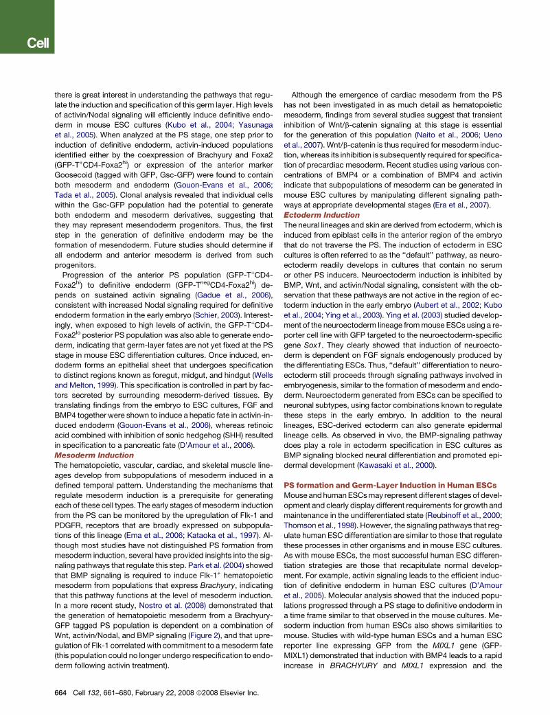
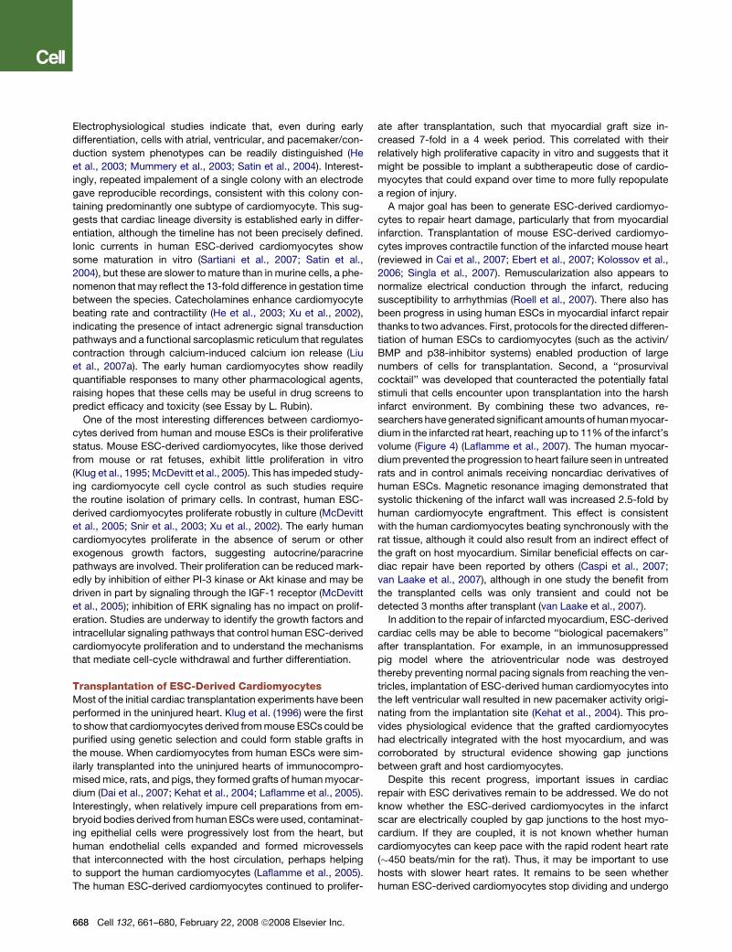
Figure 3. Making Human Cardiomyocytes
The scheme shows the directed differentiation of human ESCs to cardiomyo-
cytes and their application for cardiac repair in a rat model of cardiac infarct
(Laflamme et al., 2007). Undifferentiated human ESC colonies are replated
as high-density monolayers, expanded, and then induced to differentiate by
sequential treatment with activin A (day 0) and BMP4 (day 1). Differentiation
along the cardiac lineage can be further enhanced by activating the Wnt/b-cat-
enin pathway, followed by its inhibition. Cultures typically exhibit vigorous
beating activity 10–14 days postinduction. These populations are then sub-
jected to heat shock and treated with IGF-1 24 hr prior to transplantation to en-
hance viability, and then enriched for cardiomyocytes using Percoll density-
gradient centrifugation. They are then suspended in a ‘‘prosurvival cocktail’’
to block cell-death pathways, and are delivered to the infarcted heart by direct
injection. Experimental endpoints are assessed by microscopy and magnetic
resonance imaging.
Cell 132, 661–680, February 22, 2008 ª2008 Elsevier Inc. 667
Electrophysiological studies indicate that, even during early
differentiation, cells with atrial, ventricular, and pacemaker/con-
duction system phenotypes can be readily distinguished (He
et al., 2003; Mummery et al., 2003; Satin et al., 2004). Interest-
ingly, repeated impalement of a single colony with an electrode
gave reproducible recordings, consistent with this colony con-
taining predominantly one subtype of cardiomyocyte. This sug-
gests that cardiac lineage diversity is established early in differ-
entiation, although the timeline has not been precisely defined.
Ionic currents in human ESC-derived cardiomyocytes show
some maturation in vitro (Sartiani et al., 2007; Satin et al.,
2004), but these are slower to mature than in murine cells, a phe-
nomenon that may reflect the 13-fold difference in gestation time
between the species. Catecholamines enhance cardiomyocyte
beating rate and contractility (He et al., 2003; Xu et al., 2002),
indicating the presence of intact adrenergic signal transduction
pathways and a functional sarcoplasmic reticulum that regulates
contraction through calcium-induced calcium ion release (Liu
et al., 2007a). The early human cardiomyocytes show readily
quantifiable responses to many other pharmacological agents,
raising hopes that these cells may be useful in drug screens to
predict efficacy and toxicity (see Essay by L. Rubin).
One of the most interesting differences between cardiomyo-
cytes derived from human and mouse ESCs is their proliferative
status. Mouse ESC-derived cardiomyocytes, like those derived
from mouse or rat fetuses, exhibit little proliferation in vitro
(Klug et al., 1995; McDevitt et al., 2005). This has impeded study-
ing cardiomyocyte cell cycle control as such studies require
the routine isolation of primary cells. In contrast, human ESC-
derived cardiomyocytes proliferate robustly in culture (McDevitt
et al., 2005; Snir et al., 2003; Xu et al., 2002). The early human
cardiomyocytes proliferate in the absence of serum or other
exogenous growth factors, suggesting autocrine/paracrine
pathways are involved. Their proliferation can be reduced mark-
edly by inhibition of either PI-3 kinase or Akt kinase and may be
driven in part by signaling through the IGF-1 receptor (McDevitt
et al., 2005); inhibition of ERK signaling has no impact on prolif-
eration. Studies are underway to identify the growth factors and
intracellular signaling pathways that control human ESC-derived
cardiomyocyte proliferation and to understand the mechanisms
that mediate cell-cycle withdrawal and further differentiation.
Transplantation of ESC-Derived CardiomyocytesMost of the initial cardiac transplantation experiments have been
performed in the uninjured heart. Klug et al. (1996) were the first
to show that cardiomyocytes derived from mouse ESCs could be
purified using genetic selection and could form stable grafts in
the mouse. When cardiomyocytes from human ESCs were sim-
ilarly transplanted into the uninjured hearts of immunocompro-
mised mice, rats, and pigs, they formed grafts of human myocar-
dium (Dai et al., 2007; Kehat et al., 2004; Laflamme et al., 2005).
Interestingly, when relatively impure cell preparations from em-
bryoid bodies derived from human ESCs were used, contaminat-
ing epithelial cells were progressively lost from the heart, but
human endothelial cells expanded and formed microvessels
that interconnected with the host circulation, perhaps helping
to support the human cardiomyocytes (Laflamme et al., 2005).
The human ESC-derived cardiomyocytes continued to prolifer-
668 Cell 132, 661–680, February 22, 2008 ª2008 Elsevier Inc.
ate after transplantation, such that myocardial graft size in-
creased 7-fold in a 4 week period. This correlated with their
relatively high proliferative capacity in vitro and suggests that it
might be possible to implant a subtherapeutic dose of cardio-
myocytes that could expand over time to more fully repopulate
a region of injury.
A major goal has been to generate ESC-derived cardiomyo-
cytes to repair heart damage, particularly that from myocardial
infarction. Transplantation of mouse ESC-derived cardiomyo-
cytes improves contractile function of the infarcted mouse heart
(reviewed in Cai et al., 2007; Ebert et al., 2007; Kolossov et al.,
2006; Singla et al., 2007). Remuscularization also appears to
normalize electrical conduction through the infarct, reducing
susceptibility to arrhythmias (Roell et al., 2007). There also has
been progress in using human ESCs in myocardial infarct repair
thanks to two advances. First, protocols for the directed differen-
tiation of human ESCs to cardiomyocytes (such as the activin/
BMP and p38-inhibitor systems) enabled production of large
numbers of cells for transplantation. Second, a ‘‘prosurvival
cocktail’’ was developed that counteracted the potentially fatal
stimuli that cells encounter upon transplantation into the harsh
infarct environment. By combining these two advances, re-
searchers have generated significant amounts of human myocar-
dium in the infarcted rat heart, reaching up to 11% of the infarct’s
volume (Figure 4) (Laflamme et al., 2007). The human myocar-
dium prevented the progression to heart failure seen in untreated
rats and in control animals receiving noncardiac derivatives of
human ESCs. Magnetic resonance imaging demonstrated that
systolic thickening of the infarct wall was increased 2.5-fold by
human cardiomyocyte engraftment. This effect is consistent
with the human cardiomyocytes beating synchronously with the
rat tissue, although it could also result from an indirect effect of
the graft on host myocardium. Similar beneficial effects on car-
diac repair have been reported by others (Caspi et al., 2007;
van Laake et al., 2007), although in one study the benefit from
the transplanted cells was only transient and could not be
detected 3 months after transplant (van Laake et al., 2007).
In addition to the repair of infarcted myocardium, ESC-derived
cardiac cells may be able to become ‘‘biological pacemakers’’
after transplantation. For example, in an immunosuppressed
pig model where the atrioventricular node was destroyed
thereby preventing normal pacing signals from reaching the ven-
tricles, implantation of ESC-derived human cardiomyocytes into
the left ventricular wall resulted in new pacemaker activity origi-
nating from the implantation site (Kehat et al., 2004). This pro-
vides physiological evidence that the grafted cardiomyocytes
had electrically integrated with the host myocardium, and was
corroborated by structural evidence showing gap junctions
between graft and host cardiomyocytes.
Despite this recent progress, important issues in cardiac
repair with ESC derivatives remain to be addressed. We do not
know whether the ESC-derived cardiomyocytes in the infarct
scar are electrically coupled by gap junctions to the host myo-
cardium. If they are coupled, it is not known whether human
cardiomyocytes can keep pace with the rapid rodent heart rate
(�450 beats/min for the rat). Thus, it may be important to use
hosts with slower heart rates. It remains to be seen whether
human ESC-derived cardiomyocytes stop dividing and undergo
hypertrophic growth to match their host counterparts. Finally,
which stage of ischemic heart disease would benefit most from
human ESC-derived cardiomyocytes: hearts with acute infarcts
or those with old scars and established heart failure? It will be
essential to answer these questions to move the field forward.
Differentiation of ESCs into Neural PhenotypesEarly methods to direct the differentiation of ESCs to neural fates
used treatment with retinoic acid (Bain et al., 1995), sequential
culture in serum and serum-free media (Okabe et al., 1996), or
coculture with specific stromal cell lines such as PA6 (Kawasaki
et al., 2000). It is well established that trilineage neural progeni-
tors—capable of giving rise to neurons, astrocytes, and oligoden-
drocytes—can be generated from ESCs (reviewed in Joannides
et al., 2007). Neural progenitors are commonly derived from differ-
entiatingESCculturesbygrowing themunderconditionsoptimized
for adult neural progenitors, including growth as three-dimensional
spheroids (neurospheres) in the presence of EGF and FGF2.
Although ESC-derived neural progenitors resemble adult and
fetal neural progenitors in their trilineage capacity, microarray
and DNA methylation assays indicate that there are many differ-
ences between these two progenitor populations (Shin et al.,
2007). These differences are important to keep in mind as the
two cell populations can be expected to behave differently in
many settings. Although generating cells with a neural pheno-
type from ESCs is relatively straightforward, it should be noted
that there are many neuronal and glial subtypes with different
physiological functions. Obtaining a purified subtype for basic
research or for tissue repair is considerably more challenging.
Many signaling pathways known to regulate neural cell fate in
the embryo have been exploited to control neural differentiation
from ESCs, including Notch (reviewed in Androutsellis-Theotokis
et al., 2006; Hitoshi et al., 2002; Lowell et al., 2006), sonic hedge-
hog (Maye et al., 2004), Wnts (Davidson et al., 2007; Lamba et al.,
2006), the FGF family (Rao and Zandstra, 2005), and members of
the TGF-b superfamily (Smith et al., 2008). The Notch pathway
has emerged as a particularly important axis for controlling neu-
ral differentiation. Hitoshi et al. (2002) showed that neural pro-
genitors could form in the absence of Notch signaling, but that
these cells did not self-renew and hence were quickly lost to dif-
ferentiation. Other investigators demonstrated that activation of
Notch in mouse ESC derivatives after withdrawal of leukemia
inhibitory factor (LIF) promoted exclusively neural differentiation,
whereas inhibition of Notch blocked formation of neural progen-
itors. The ability of Notch ligands to promote neural progenitor
formation required FGF receptor-mediated signaling (Lowell
et al., 2006). Taken together, these data implicate Notch signal-
ing as a key player in establishment of neural progenitor cells,
principally through effects on cell survival and promoting expan-
sion of the progenitors by blocking their differentiation.
Joannides et al. (2007) have developed a protocol for neural
induction of human ESCs that uses chemically defined media
at each step. Supplements include common amino acids and
taurine; trace metals; vitamins; and the growth factors insulin,
EGF, and FGF2. After optimizing techniques for passaging to
generate small clumps of human ESCs, cells were induced to
form neural progenitors and were expanded in defined media.
Some cultures approached 90% nestin-negative/Pax6-positive
cells that were trilineage-competent, and these cells could un-
dergo 5- log expansion with a stable karyotype. Thus, defined
media facilitate mechanistic studies and should help to promote
translation to the clinic.
Wichterle et al. (2002) were the first to derive a protocol for the
directed differentiation of ESCs to a specific neural type, using
induction with retinoic acid and a sonic hedgehog analog to
induce transplantable murine spinal motor neurons (Wichterle
et al., 2002). Following this pioneering work, multiple investiga-
tors developed techniques to induce differentiation of ESCs
into specific neuronal populations, including progenitors for ret-
inal photoreceptors, cerebellar granule neurons, and cerebral-
type neurons that use glutamate, GABA, and dopamine as their
major neurotransmitters. Different lines of human ESCs appear
to preferentially make one neuron type over another.
Differentiation to Dopamine NeuronsDopamine neurons are of particular interest because of their cen-
tral role in Parkinson’s disease. Many studies now show that
mouse and human ESCs can form dopamine neurons, and they
appear to arise through the neural progenitor stage described
above. These neurons express tyrosine hydroxylase (required
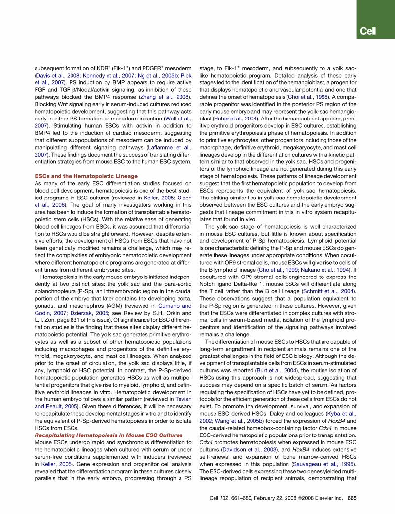
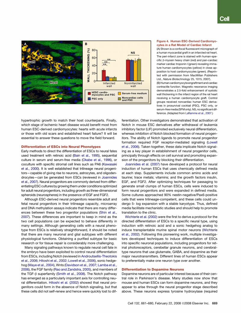
Figure 4. Human ESC-Derived Cardiomyo-
cytes in a Rat Model of Cardiac Infarct
(A) Shown is a confocal fluorescent micrograph of
a human myocardial graft in an infarcted rat heart.
The peri-infarct zone is stained with human-spe-
cific b-myosin heavy chain (red) and pan-cardiac
marker cardiac troponin I (green) revealing imma-
ture human cardiomyocytes (yellow) in close ap-
position to host cardiomyocytes (green). (Reprin-
ted with permission from MacMillan Publishers
Ltd., Nature Biotechnology 25, 1015, 2007).
(B)Humancardiomyocyteengraftment andcardiac
contractile function. Magnetic resonance imaging
demonstrates a 2.5-fold enhancement of systolic
wall thickening in the infarct region of the rat heart
receiving a human cardiomyocyte graft. Control
groups received noncardiac human ESC deriva-
tives in prosurvival cocktail (PSC), PSC only, or
serum-freemedia (SFMonly). NS,nosignificantdif-
ference. (Adapted from Laflamme et al., 2007.)
Cell 132, 661–680, February 22, 2008 ª2008 Elsevier Inc. 669
for dopamine synthesis), release dopamine upon depolarization,
and format least rudimentarysynapses in vitrowith transmitter re-
uptake abilities (reviewed in Kim et al., 2007). The combined use
of FGF8 and SHH effectively induces dopamine neurons from
ESC-derived neural progenitors generated from either mouse
ESCs (Lee et al., 2000) or human ESCs (Yan et al., 2005). Although
recombinant factors are now routinely used, most protcols do
include undefined reagents at one or more stages of dopamine
neuron production, due to coculture with stromal cell lines or
the use of conditioned media. One of the best-defined protocols
for human ESC differentiation into dopamine neurons was vali-
dated in three human ESC lines and two monkey ESC lines (Per-
rier et al., 2004). Neural progenitors were induced in this study
using stromal cell coculture, followed by SHH and FGF8 to specify
a neuronal fate. Addition of ascorbate, BDNF, glial-derived neuro-
trophic factor, dibutyryl cyclic-AMP, and TGF-b3 yielded cultures
that were 30%–50% neurons expressing b-III tubulin. Of these
neurons, 65%–80% expressed tyrosine hydroxylase,and the ma-
jority fired action potentials that could be blocked by tetrodotoxin,
a Na+ channel blocker. The remainder of the culture comprised
nestin-positive progenitors with low frequencies of other differen-
tiated neural cell types. It would be interesting to determine
whether combining the above-mentioned protocol for producing
neural progenitors (Joannides et al., 2007) with this regimen for
inducing midbrain neurogenesis would yield dopamine neurons
from human ESCs using completely defined factors.
Differentiation to OligodendrocytesAstrocytes and oligodendrocytes are the two neuroglial types in
the central nervous system. Diseases of the central nervous sys-
tem typically involve proliferation of astrocytes and loss of oligo-
dendrocytes and the protective myelin sheath they produce.
Thus, derivation of oligodendrocytes from ESCs has been an
important goal for cell replacement therapy. The most common
protocols involve an initial differentiation step to neural progeni-
tors, followed by expansion, further differentiation, and selec-
tion. Oligodendrocytes were first efficiently derived from mouse
ESCs (Brustle et al., 1999), where medium containing FGF2 and
EGF was used to expand progenitors, followed by a switch to
FGF2 and PDGF to yield bipotential glial progenitors. These glial
progenitors were transplanted into the spinal cords of rats with
a genetic deficiency in myelin production, yielding myelinated fi-
bers in the majority of animals. Transplantation of these glial pro-
genitors into the brains of developing rats (at embryonic day 17)
resulted in widespread myelin-producing cells of mouse origin.
Oligodendrocytes were first generated from human ESCs
by Zhang et al. (2001b), who used a similar strategy involving
FGF treatment followed by growth as neurospheres. They re-
ported occasional cells expressing the oligodendrocyte marker
O4, with many more cells expressing neuronal or astrocytic
markers. No human oligodendrocytes were detected after trans-
plantation into the brains of newborn mice, although human
neurons and some astrocytes were found to have engrafted.
The first detailed protocol for directed differentiation of oligo-
dendrocytes from human ESCs involved generation of neuro-
spheres, followed by several rounds of expansion and selection
in various media containing, among other things, the multicom-
ponent additive B27, thyroid hormone, retinoic acid, FGF2,
670 Cell 132, 661–680, February 22, 2008 ª2008 Elsevier Inc.
EGF, and insulin (Nistor et al., 2005). After �42 days of culture,
the desired cells were found in yellow spheroids, which upon dif-
ferentiation as low-density monolayers formed 85%–95% oligo-
dendrocytes (based on expression of the markers GalC, RIP, and
O4). The remaining cells were astrocytes or neurons. The authors
observed significant amounts of cell death and noted that
cells seemed to be selected based on preferential adherence.
Importantly, the derived oligodendrocyte progenitors were able
to myelinate host axons when transplanted into the shiverer
mouse, which has a genetic defect preventing myelination.
Kang et al. (2007) recently reported a simplified protocol for iso-
lation of oligodendrocyte progenitors from human ESCs, using a
multistep procedure that yielded �80% oligodendrocytes that
were capable of myelinating fetal neural explants in vitro. These
experiments show that human oligodendrocytes can be gener-
ated in large numbers and used to restore myelination under
some circumstances in mice.
Transplantation for Parkinson’s DiseaseParkinson’s disease is characterized by loss of a critical popula-
tion of dopamine-producing midbrain neurons with cell bodies
in the substantia nigra. These neurons project to the striatum
and are essential for motor processing. Parkinson’s patients suf-
fer from multiple motor problems including a resting tremor, diffi-
culty in walking, and loss of facial expression. The disease is typ-
ically progressive due to ongoing loss of neurons. Parkinson’s
disease was one of the first diseases of solid tissue to be treated
with cellular therapy, in this case with cells derived from human
fetal midbrains transplanted into the striatum of patients (Goya
et al., 2007). Early clinical trials with fetal tissue appeared promis-
ing (Lindvall and Hagell, 2001), but the results of randomized, con-
trolled studies were less robust with modest improvements seen
only in younger patients (Freed et al., 2001). Of note, some pa-
tients receiving these transplants developed dyskinesias, move-
ment disorders associated with excessive dopamine levels in
the brain. Dyskinesias may have resulted from overdosing with
graft cells, although this is controversial (Hagell et al., 2002).
Thesemixed results may stem, in part, fromthe variability inherent
in using human fetal tissue as a source of therapeutic cells. One
advantage afforded by human ESCs is their ability to reproducibly
generate well-characterized cell populations for transplantation.
In a common animal model for Parkinson’s disease, 6-hydrox-
ydopamine, a toxin for dopamine neurons, is injected into the
midbrain to produce a unilateral lesion. Treating the animal
with an agent that stimulates motion, such as amphetamines
or opioids, results in a characteristic unilateral turning behavior
that can be readily quantified. Using 6-hydroxydopamine in a
rat model, Rodriguez-Gomez et al. (2007) demonstrated long-
term benefit of dopamine neural grafts derived from mouse
ESCs. They showed that mouse ESC neural derivatives survived
for 32 weeks in the brains of immunosuppressed Parkinsonian
rats, and that the grafts produced dopamine and induced persis-
tent improvements in rotational behavior.
We are aware of one study that attempted a head-to-head
comparison of fetal-derived midbrain cells to mouse ESC-de-
rived cells (Yurek and Fletcher-Turner, 2004). These investigators
induced mouse ESCs toward a neural phenotype, achieving
�10% dopamine neurons, which were transplanted into the
striatum of immunosuppressed rats previously injured with 6-
hydroxydopamine. Another group of rats received transplants
of intact rat fetal ventral mesencephalic tissue, apparently with-
out immunosuppression. Both cell populations yielded viable
grafts in the striatum with comparable numbers of cells express-
ing tyrosine hydroxylase. However, the rats receiving mouse
ESC derivatives did not show an improvement in their rotational
score, whereas rats receiving fetal rat midbrain tissue did. The
lack of benefit with mouse ESC derivatives may reflect compar-
ison of a xenograft under immunosuppressive conditions with
an allograft without immunosuppression.
In one of the most detailed studies involving human ESC deriv-
atives, Ben-Hur et al. (2004), transplanted human ESC-derived
neurospheres into the chemically lesioned brains of immunosup-
pressed rats. The human cells persisted for 12 weeks and formed
nestin+ progenitor cells, astrocytes, and tyrosine hydroxylase-
positive neurons (these neurons comprising �0.2% of the total
human cells in the graft). Despite the relatively small population
of dopamine neurons, the grafts reduced turning behavior by
25%–50% after opiate or amphetamine treatment. Interestingly,
there was a linear correlation between the number of dopamine
human neurons and the reduction in turning behavior, and no
benefit was seen in lesioned rats that did not receive a surviving
graft. The percentage of dopamine neurons was similar to that
observed with in vitro differentiation of human ESCs, suggesting
that differentiation signals are absent from the brain and need to
be provided before transplantation.
A drawback to extensive predifferentiation in vitro was high-
lighted by Park et al. (2005), who directed differentiation of hu-
man ESCs to a population comprising >95% neurons or nestin+
progenitors. Although 40% of these cells expressed tyrosine
hydroxylase and released dopamine upon depolarization in vitro,
the cells did not survive after transplantation into the lesioned
brains of immunosuppressed rats and had no influence on turn-
ing behavior. Thus, more differentiated dopamine neurons may
be more likely to die after transplantation.
Stem Cells and Spinal-Cord InjuryTrauma to the spinal cord kills neurons, myelinating cells, blood
vessels, and other resident cells (reviewed in Coutts and Keirst-
ead, 2008; Kim et al., 2007). The main determinants of morbidity
are the extent of tissue disruption (mild contusion versus full tran-
section) and the point at which the spinal cord sustains injury (cra-
nial versus caudal). Hemorrhage and inflammation are thought to
inhibit axon regeneration by promoting glial scarring and through
direct chemical signals that block axon growth. Stem cells could
have beneficial effects by being directly incorporated into tissue
or by promoting repair by endogenous cells. Possible benefits
include new neurons to bridge the gap, enhanced remyelination,
and controlling host responses like cell survival, axon sprouting,
inflammation, angiogenesis, and gliosis.
Neural progenitors derived from mouse ESCs promote hin-
dlimb motor recovery when implanted in a mouse (Kimura
et al., 2005) or rat spinal cord injury model (McDonald et al.,
1999). Tracking with bromodeoxyuridine suggested that the cells
differentiated into oligodendrocytes, astrocytes, and neurons.
Liu et al. (2000) reported that these cells promoted remyelination
of a chemically demyelinated spinal cord in immunosuppressed
rats. Human ESC-derived oligodendrocyte progenitor cells
populate a�1 cm region when engrafted in a rat model of spinal-
cord contusion (Keirstead et al., 2005). The human cells
promoted remyelination of host axons and enhanced motor
function when administered 7 days postinjury. However, when
the human ESC-derived oligodendrocyte progenitor cells were
transplanted into a 10-week-old rat spinal injury model with glial
scarring, remyelination was inhibited and there was no improve-
ment in motor function. This suggests that, like fibrosis in the
heart, glial scarring in the spinal cord is a barrier to regeneration.
Despite these setbacks, spinal cord injury is likely to be the
first clinical setting in which human ESC derivatives will be
tested, with a multicenter clinical trial expected to commence
in 2008–2009.
Differentiation of ESCs to Pancreatic CellsThe potential to generate functional pancreatic b cells from ESCs
differentiated in culture has raised the exciting possibility of
a new source of insulin-producing cells for transplantation to
treat type I diabetes. Given the therapeutic potential of ESC-de-
rived b cells, significant efforts have focused on isolating such
cells in both mouse and human ESC cultures. Initial attempts
to generate the pancreatic lineage used mouse ESCs (reviewed
in Spence and Wells, 2007), but the most successful differentia-
tion along this pathway has been recently achieved with human
ESCs (D’Amour et al., 2006). The key to generating pancreatic
lineage cells from human ESCs relies on recapitulating the criti-
cal signals that regulate endocrine pancreas development in the
embryo.
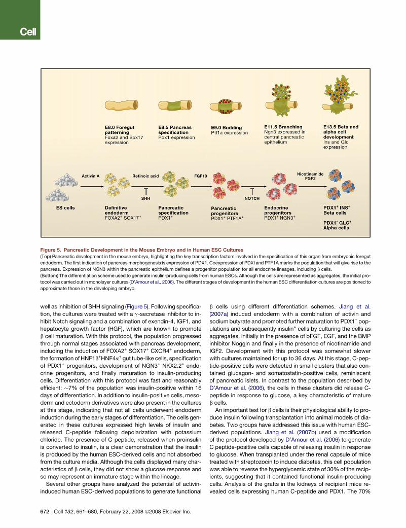
The pancreas develops from foregut endoderm, and the earli-
est stages of induction are controlled in part by retinoic acid (RA)
and the inhibition of SHH signaling (reviewed in Collombat et al.,
2006; Murtaugh, 2007; Spence and Wells, 2007). The first indica-
tion of pancreas morphogenesis is the upregulation of Pdx1,
a gene encoding a transcription factor that is essential for devel-
opment of this tissue (Figure 5). Although indicative of pancreas
specification, expression of Pdx1 is not restricted to pancreatic
tissues as it is also found in the region of the foregut that will
give rise to the pyloric region of the stomach and the proximal
duodenum. Coexpression of the transcription factor encoded
by the Ptf1a/P48 gene together with Pdx1 marks the population
that will give rise to the pancreas. Recent evidence suggests that
expansion of the pancreatic progenitor population is supported
by the surrounding mesenchyme through FGF10 secretion.
FGF10 enhances Notch signaling, which represses expression
of the transcription factor Ngn3 and promotes expansion of pan-
creatic progenitors. Expression of Ngn3 within the pancreatic
epithelium defines the development of a progenitor population
for all endocrine lineages, including the b cells. With further mat-
uration, cohorts of factors function to establish the different en-
docrine lineages. b cell development is dependent, in part, on the
combined activity of Nkx2.2, Nkx6.1, Pax4, Pax6, and MafA.
Through the sequential activation of different signaling path-
ways, D’Amour et al. (2006) demonstrated that it is possible
to recapitulate many of these developmental stages in human
ESC cultures. In this study, endoderm induced by activin signal-
ing in monolayer cultures was specified to a pancreatic fate
through a combination of FGF and retinoic acid signaling as
Cell 132, 661–680, February 22, 2008 ª2008 Elsevier Inc. 671
well as inhibition of SHH signaling (Figure 5). Following specifica-
tion, the cultures were treated with a g-secretase inhibitor to in-
hibit Notch signaling and a combination of exendin-4, IGF1, and
hepatocyte growth factor (HGF), which are known to promote
b cell maturation. With this protocol, the population progressed
through normal stages associated with pancreas development,
including the induction of FOXA2+ SOX17+ CXCR4+ endoderm,
the formation of HNF1b+HNF4a+ gut tube-like cells, specification
of PDX1+ progenitors, development of NGN3+ NKX2.2+ endo-
crine progenitors, and finally maturation to insulin-producing
cells. Differentiation with this protocol was fast and reasonably
efficient: �7% of the population was insulin-positive within 16
days of differentiation. In addition to insulin-positive cells, meso-
derm and ectoderm derivatives were also present in the cultures
at this stage, indicating that not all cells underwent endoderm
induction during the early stages of differentiation. The cells gen-
erated in these cultures expressed high levels of insulin and
released C-peptide following depolarization with potassium
chloride. The presence of C-peptide, released when proinsulin
is converted to insulin, is a clear demonstration that the insulin
is produced by the human ESC-derived cells and not absorbed
from the culture media. Although the cells displayed many char-
acteristics of b cells, they did not show a glucose response and
so may represent an immature stage within the lineage.
Several other groups have analyzed the potential of activin-
induced human ESC-derived populations to generate functional
672 Cell 132, 661–680, February 22, 2008 ª2008 Elsevier Inc.
b cells using different differentiation schemes. Jiang et al.
(2007a) induced endoderm with a combination of activin and
sodium butyrate and promoted further maturation to PDX1+ pop-
ulations and subsequently insulin+ cells by culturing the cells as
aggregates, initially in the presence of bFGF, EGF, and the BMP
inhibitor Noggin and finally in the presence of nicotinamide and
IGF2. Development with this protocol was somewhat slower
with cultures maintained for up to 36 days. At this stage, C-pep-
tide-positive cells were detected in small clusters that also con-
tained glucagon- and somatostatin-positive cells, reminiscent
of pancreatic islets. In contrast to the population described by
D’Amour et al. (2006), the cells in these clusters did release C-
peptide in response to glucose, a key characteristic of mature
b cells.
An important test for b cells is their physiological ability to pro-
duce insulin following transplantation into animal models of dia-
betes. Two groups have addressed this issue with human ESC-
derived populations. Jiang et al. (2007b) used a modification
of the protocol developed by D’Amour et al. (2006) to generate
C peptide-positive cells capable of releasing insulin in response
to glucose. When transplanted under the renal capsule of mice
treated with streptozocin to induce diabetes, this cell population
was able to reverse the hyperglycemic state of 30% of the recip-
ients, suggesting that it contained functional insulin-producing
cells. Analysis of the grafts in the kidneys of recipient mice re-
vealed cells expressing human C-peptide and PDX1. The 70%
Figure 5. Pancreatic Development in the Mouse Embryo and in Human ESC Cultures
(Top) Pancreatic development in the mouse embryo, highlighting the key transcription factors involved in the specification of this organ from embryonic foregut
endoderm. The first indication of pancreas morphogenesis is expression of PDX1. Coexpression of PDXI and PTF1A marks the population that will give rise to the
pancreas. Expression of NGN3 within the pancreatic epithelium defines a progenitor population for all endocrine lineages, including b cells.
(Bottom) The differentiation scheme used to generate insulin-producing cells from human ESCs. Although the cells are represented as aggregates, the initial pro-
tocol was carried out in monolayer cultures (D’Amour et al., 2006). The different stages of development in the human ESC differentiation cultures are positioned to
approximate those in the developing embryo.
of recipients that still showed hyperglycemia had far fewer C
peptide-positive cells in the graft than did the mice that achieved
normal glucose levels. No teratomas were detected up to
3 months after transplantation, indicating that the graft did not
contain significant numbers of undifferentiated human ESCs.
In the second study, Shim et al. (2007) used a combination of
activin and retinoic acid to induce differentiation of human ESCs
to the PDX1+ progenitor stage. These immature cells were trans-
planted to the kidney capsule of streptozocin-treated recipients
to determine if this environment would promote survival and
maturation of the transplanted population. Insulin-positive cells,
not present in the population prior to transplantation, were
detected 28 days following transplantation, indicating that fur-
ther differentiation had occurred in vivo. Some of these insulin-
positive cells also expressed glucagon, suggesting that they still
represent an immature stage of development. These grafts
appeared to rescue the streptozocin-induced hyperglycemia
as the animals showed a significant reduction in blood glucose
levels within 4 days of transplantation. This is somewhat surpris-
ing, given the immature status of the population used for trans-
plantation and the presumptive requirement that the grafts be
vascularized for proper endocrine function. Despite the imma-
ture stage of development, the transplanted cells appeared to
be responsible for the reversal of hyperglycemia as the mice re-
verted back to a hyperglycemic state following removal of the
kidney with the graft. Given the reversal of hyperglycemia by
immature cells in streptozocin-treated animals, it will be impor-
tant to use additional preclinical models of diabetes to verify
the therapeutic potential of insulin-producing cells generated
by this approach.
Taken together, these studies highlight remarkable progress
over the last two years in the development of b cells from human
ESCs. Although the populations generated may not represent
mature functional cells, differentiation in most studies was effi-
cient and relied on protocols that incorporated aspects of pan-
creatic development in the embryo. A major hurdle is the estab-
lishment of culture conditions that will promote efficient
maturation of the population to end stage functional b cells.
The transplantation studies of Brolen et al. (2005) suggest that
the fetal pancreas may provide a source of regulators that
promote these maturation steps. These investigators cotrans-
planted differentiated human ESC populations expressing
PDX1 together with fetal mouse pancreas under the kidney cap-
sule of recipient mice. The human ESC-derived cells matured to
generate insulin-positive cells when transplanted with mouse
pancreatic tissue, but not when transplanted with fetal liver or
telencephalon cells, suggesting that factors within the pancre-
atic environment induced the human cells to mature. Future
studies will be required to determine if the fetal pancreas can
influence human ESC differentiation in vitro.
Challenges for Clinical ApplicationsSafety
Pluripotent cells such as ESCs or iPS cells present a safety
concern because of their potential to form tumors. When these
cells are transplanted in the undifferentiated state they form
teratomas, tumors derived from all three germ layers. In our ex-
perience, normal or injured adult tissues lack the cues required to
induce ESCs to form appropriate cell types (Nussbaum et al.,
2007). Currently, the only way to ensure that teratomas do not
form is to differentiate the ESCs in advance, enrich for the
desired cell type, and screen for the presence of undifferentiated
cells. When such procedures were rigorously followed, tera-
tomas were not observed in over 200 animals transplanted
with human ESC-derived cardiomyocytes (Laflamme et al.,
2007). But more differentiated tumor-like growths can occur.
For example, transplanting a population of human ESC neural
derivatives enriched for dopamine neurons into Parkinsonian
rats resulted in a primitive population of nestin+ neuroepithelial
cells that continued to proliferate in the striatum 70 days after
transplantation (Roy et al., 2006). This raises a cautionary flag
and suggests that even committed progenitors can proliferate
excessively after transplantation, a problem that may be solved
by improving purification methods.
Other types of safety concerns exist not only with ESC-based
approaches but also with adult cell grafts. For example, cell
grafts into the heart may provoke arrhythmias (due, for example,
to aberrant pacemaker activity or conduction properties) or, sim-
ilarly, induce seizure activity in the central nervous system. Also,
it is possible to give patients too many normal cells. This has
been suggested as an explanation for Parkinson’s patients re-
ceiving fetal neural tissue, who subsequently developed dyski-
nesias (Hagell et al., 2002). It will be critical to choose the initial
patient population correctly to minimize risk, likely starting with
the sickest patients, such as patients awaiting heart transplants
or those with longstanding complete spinal-cord transaction
injuries. Unfortunately, although beginning with the sickest pa-
tients minimizes risks, it also reduces the chance of success. It
undoubtedly will be easier to prevent physiological deterioration
in the early phases of illness rather than to try and reverse severe
and chronic damage.
Purification
Despite considerable progress in directing the differentiation of
ESCs, purification of desired differentiated cell types is required
for most applications. Microdissection of cellular aggregates
with a visible phenotype (e.g., beating cardiomyocytes or fluo-
rescent reporter gene expression) is perhaps the simplest ap-
proach, but the purity of the resulting cultures is only moderate,
and the system cannot be scaled up for the clinic. Density-gradi-
ent centrifugation can be used for purifying human ESC-derived
cardiomyocytes (Laflamme et al., 2005, 2007; Xu et al., 2002),
but in our experience, offers only �5-10-fold enrichment. Thus,
the final purity is highly dependent on the purity of the input
population.
Surface molecules on cells are commonly used to select a
desired population or to remove unwanted cell populations.
Fluorescence-activated cell sorting (FACS) has been used to
select many ESC derivatives, including hematopoietic cells, me-
sodermal progenitors, and endothelial cells (Gadue et al., 2006;
Kattman et al., 2006; Kouskoff et al., 2005; Wang et al., 2004).
FACS offers many advantages, including the ability to analyze
multiple surface markers simultaneously to refine selection of
the desired subpopulation. The biggest drawback to FACS is
its relatively low throughput, necessitating expansion of some
cell types after selection. A related technique, magnetically acti-
vated cell sorting (MACS), permits higher throughput selection of
Cell 132, 661–680, February 22, 2008 ª2008 Elsevier Inc. 673
ESC derivatives based on surface markers (Vodyanik et al.,
2006) but is less amenable to the use of multiple markers than
is FACS.
Finally, genetic selection of cells based on expression of a
selectable marker driven by a lineage-restricted promoter offers
excellent purity and scalability. This approach was first devel-
oped to select cardiomyocytes at > 99% purity by expressing
an antibiotic resistance gene via a cardiac-restricted promoter
(Klug et al., 1996). Genetic selection has been expanded to
include fluorescent reporter proteins and has been used with
excellent results to purify endothelial cells and insulin-producing
islet cells from ESCs. Importantly, genetic selection of clinically
relevant numbers of highly purified cells has been achieved in
bioreactors (Zandstra et al., 2003). The principal drawback to ge-
netic selection is the necessity (at present) of inserting a selection
cassette into the host genome, which may increase the risk of
tumorigenesis.
Controlling Graft Size
Death of transplanted cells remains a major limitation for repair of
the heart (Murtuza et al., 2004; Zhang et al., 2001a), for treatment
of Parkinson’s disease (Cicchetti et al., 2002; Emgard et al., 2003;
Marchionini et al., 2004; Schierle et al., 1999), in islet cell trans-
plantation for diabetes (Contreras et al., 2002; Nakano et al.,
2004), and for myogenic precursor transplantation for muscular
dystrophy (Guerette et al., 1997; Skuk et al., 2003). Current evi-
dence suggests that cell-death pathways are initiated by multiple
stresses associated with transplantation, including ischemia,
loss of matrix attachments, and inflammation. In studies of human
ESC-derived cardiomyocytes in the infarcted heart, a prosurvival
cocktail that targeted multiple points in the death signaling net-
work succeeded in generating physiologically significant graft
sizes, whereas individual interventions were without benefit (La-
flamme et al., 2007). This suggests that there are multiple path-
ways through which transplanted cells can die, and that blocking
individual paths simply leads to death through alternate routes. It
will be of interest to determine whether the prosurvival cocktail
developed for cardiomyocytes can benefit other cell types.
Proliferation of grafted cells offers the prospect of exponential
growth. Delivery of growth factors to the cells may regulate their
proliferation, but it has the potential to induce undesired effects
on the host such as stimulation of fibrosis or glial scarring. One
route to control proliferation involves genetic modification of
the graft cells to render them responsive to a signaling pathway
controlled by a small molecule, to which the host cells are unre-
sponsive. One such approach has been to fuse the cytoplasmic
domains of growth-factor receptors to drug-binding domains,
typically variants of FK506-binding proteins (Belshaw et al.,
1996). Administering bivalent ligands for the modified binding
proteins induces dimerization of the receptor domains, repro-
ducing signaling induced by a native ligand selectively in the
graft cells. This approach has been used to promote hematopoi-
esis after bone-marrow transplantation (Weinreich et al., 2006)
and was recently used selectively to control graft cell prolifera-
tion in the heart (Stevens et al., 2007), yielding a pharmacologi-
cally tunable system for infarct repair.
Scarring
In the heart, scar tissue associated with the infarct typically forms
a barrier that interferes with integration of grafted cardiomyo-
674 Cell 132, 661–680, February 22, 2008 ª2008 Elsevier Inc.
cytes into host myocardium. As a consequence, grafts may
remain electrically insulated from the host tissue and beat asyn-
chronously, or they may have delayed activation that leads to
arrhythmias. In the central nervous system, proliferation of astro-
cytes following injury leads to ‘‘glial scarring’’ (Keirstead et al.,
2005). In this context, cytoplasmic projections of astrocytes
insulate axonal projections and prevent them from finding their
distal tracks or forming new synapses with transplanted neural
progenitors. The simplest approach to these problems would
be to intervene relatively early, before the scar tissue forms.
Unfortunately, this option is not always clinically feasible. The
pathogenesis of scarring in the setting of stem cell transplanta-
tion has not been well studied. Additional research is needed
to rationally identify targets to address this important problem.
Finding the Right Model for Preclinical Testing
Preclinical research with human ESC derivatives requires xeno-
transplantation, raising the question of which species are best
suited as hosts. An ideal model would (1) support human xeno-
grafts, (2) recapitulate key aspects of the disease, (3) have phys-
iological properties that make it predictive of the human re-
sponse to therapy, and (4) be cost-effective enough to permit
high-throughput screening necessary at this early stage of devel-
opment. Clearly, no single model meets all of these criteria.
A large animal with drug-induced immunosuppression, such as
a dog, pig, sheep or nonhuman primate, probably provides the
best physiological mimic of the human. These models are
extraordinarily expensive, however, and studies requiring multi-
ple iterations are difficult to do in large animals. On the other end
of the spectrum, the mouse is inexpensive, and there are many
murine models for human diseases, including genetically immu-
nodeficient strains. Murine physiology differs significantly from
human physiology; for example, the mouse heart rate is 600
beats/min and that for human is 70 beats/min. This makes elec-
trophysiological results from mouse less predictive of human
responses. Furthermore, scaling up a process 2500-fold for the
human can raise new problems in cell production or delivery
that were not anticipated from murine studies. One clear draw-
back to all animal studies is that the immune response to xeno-
geneic transplantation does not predict the response to alloge-
neic transplantation. From an immune standpoint, we probably
will learn more from animal-to-animal allotransplantation studies
than we will from xenotransplantation. Most investigators sup-
port the development of therapeutic approaches in small ani-
mals where higher throughput permits more rapid progress.
However, key concepts should be tested in large animals,
including nonhuman primates, where real-world issues of scale,
delivery, and assessment of efficacy can be developed before
attempting studies in patients.
Immune rejection
The immune system presents a formidable barrier to allogeneic
cell transplantation. ESCs express few class I major histocom-
patibility antigens and have virtually no expression of class II
molecules (Drukker et al., 2002; Nussbaum et al., 2007; Swijnen-
burg et al., 2005), which initially raised hopes that ESC deriva-
tives might not induce an immune response when transplanted.
However, once transplanted, ESC derivatives show increased
expression of histocompatibility antigens, with further increases
in response to cytokine stimulation. Although cell transplants
may not have the full antigenicity of solid organs (due, for exam-
ple, to the absence of professional antigen-presenting cells),
there is now clear evidence that differentiated progeny of ESCs
will be rejected in an allogeneic setting (Nussbaum et al., 2007).
There are several strategies to address this problem (reviewed
in Cabrera et al., 2006; Drukker, 2004). The first approach is im-
munosuppression, similar to that used for solid organ transplan-
tation, and this is likely where clinical trials will begin. Banking of
ESCs can provide cells with a repertoire of tissue types, but large
numbers of cell lines (likely thousands) would be needed to cover
a genetically diverse population such as that in the United States
(Taylor et al., 2005). The number of required cells can be reduced
exponentially by using human ESCs that are homozygous at the
HLA histocompatibility loci. Others have suggested develop-
ment of a ‘‘universal donor cell,’’ for example by using blood
type O cells and suppressing expression of HLA molecules (Cab-
rera et al., 2006; Drukker, 2004). HLA suppression, although con-
ceptually appealing, makes ESC derivatives targets for killing by
NK cells of the immune system. Another notion is engineering
ESC progeny to secrete locally immunosuppressive molecules
such as a soluble interleukin-1 receptor or agents that block
coreceptors for antigen stimulation. A final approach is to take
a lesson from hematopoietic cell transplantation, where animals
with hematopoietic chimerism tolerate solid organ transplants
from the marrow donors (Menendez et al., 2005; Priddle et al.,
2006). If hematopoietic cells capable of inducing tolerance could
be derived from human ESCs, they would enable cell transplants
from the same human ESCs to survive in the host without broad
spectrum immunosuppression.
Three groups recently reported the derivation of induced plu-
ripotent stem (iPS) cells from human somatic cells. These studies
demonstrated reprogramming of either the differentiated prog-
eny of stem cells (Park et al., 2008), fetal and neonatal fibroblasts
(Yu et al., 2007), or adult fibroblasts (Takahashi et al., 2007). The
groups used a cocktail of four retrovirally encoded transcription
factors—either Oct4, Sox2, c-myc and Klf-4 or Oct4, Sox2,
Nanog, and Lin28—to reprogram fibroblasts to cells that closely
resemble human ESCs. These iPS cells had gene-expression
profiles and DNA-methylation patterns closely resembling
human ESCs, grew vigorously while expressing telomerase,
maintained a normal karyotype, and formed teratomas after
transplantation into immunocompromised mice. Importantly,
Takahashi et al. (2007) showed that the human iPS cells could
be directed down the cardiomyocyte lineage using activin/
BMP4 and toward neurons using PA6 stromal cell coculture.
Most recently, Nakagawa et al. (2008) demonstrated that iPS
cells could be derived without c-myc, removing a potential onco-
genic factor from the reprogramming cocktail.
If these results hold up as anticipated there are important impli-
cations for the field. The ability to generate pluripotent cells from
readily available fibroblasts should obviate ethical concerns sur-
rounding human ESCs as no human embryos are harmed in the
process. Furthermore, iPS cells offer the potential to generate
patient-specific cells that would be recognized as ‘‘self’’ by the
immune system, thus preventing rejection of cell transplants. In
addition, iPS cells could be generated from patients with specific
diseases having genetic components and allowed to differentiate
into relevant populations (such as dopamine neurons from Par-
kinson’s patients) enabling study of disease pathogenesis and
development of new treatment strategies. There are, however,
two drawbacks to the clinical use of iPS cells. The first is the cur-
rent need to use integrating retroviruses to deliver the reprogram-
ming factors; this may be solved with transient gene transfer or
through delivery of the protein factors in cell-permeant forms.
The second is that iPS cells are not an ‘‘off-the-shelf’’ product
and would likely only be produced after the patient becomes ill,
precluding their use in the acute phase of the disease. Quality
control is also likely to be difficult and expensive, because a sep-
arate batch of iPS cells would have to be made for each patient.
Summary and ConclusionsInsights gained from developmental biology have facilitated our
understanding of the control of ESC growth and lineage-specific
differentiation. These insights enable the reproducible genera-
tion of highly enriched cell populations from a number of different
lineages. The demonstration that somatic cells can be reprog-
rammed to an ESC-like cell suggests it will be possible to gener-
ate such populations from patients in the near future. With these
advances, we can now to begin testing the function of these cell
types through the transplantation of highly enriched, well-char-
acterized populations into different preclinical models of dis-
ease. Using protocols with defined reagents ensures that differ-
ent groups will be able to reproducibly isolate comparable cell
populations for such studies. Access to lineage-specific progen-
itors for transplantation will allow comparison to more mature
populations to determine which stage integrates best into the
adult tissue and which ultimately provides the most benefit.
The availability of highly enriched cell populations from different
lineages also provides an opportunity for cell biologists to inter-
act with tissue engineers to generate culture systems that will
more accurately mimic important three-dimensional aspects of
organogenesis. Such engineered tissues may be more effective
following transplantation and may also support more efficient
maturation of the different cell types in culture. With these tools
at hand, the therapeutic potential of ESCs is now ready to be
tested.
ACKNOWLEDGMENTS
We thank members of the Murry and Keller labs for helpful discussions, Tom
Reh and Phil Horner for reviewing the neurobiology sections, and Steve Katt-
man and Cristina Nostro for assistance with the figures.
REFERENCES
Androutsellis-Theotokis, A., Leker, R.R., Soldner, F., Hoeppner, D.J., Ravin,
R., Poser, S.W., Rueger, M.A., Bae, S.K., Kittappa, R., and McKay, R.D.
(2006). Notch signalling regulates stem cell numbers in vitro and in vivo. Nature
442, 823–826.
Aubert, J., Dunstan, H., Chambers, I., and Smith, A. (2002). Functional gene
screening in embryonic stem cells implicates Wnt antagonism in neural differ-
entiation. Nat. Biotechnol. 20, 1240–1245.
Bain, G., Kitchens, D., Yao, M., Huettner, J.E., and Gottlieb, D.I. (1995). Embry-
onic stem cells express neuronal properties in vitro. Dev. Biol. 168, 342–357.
Belshaw, P.J., Ho, S.N., Crabtree, G.R., and Schreiber, S.L. (1996). Controlling
protein association and subcellular localization with a synthetic ligand that
induces heterodimerization of proteins. Proc. Natl. Acad. Sci. USA 93, 4604–
4607.
Cell 132, 661–680, February 22, 2008 ª2008 Elsevier Inc. 675
Ben-Hur, T., Idelson, M., Khaner, H., Pera, M., Reinhartz, E., Itzik, A., and Reu-
binoff, B.E. (2004). Transplantation of human embryonic stem cell-derived
neural progenitors improves behavioral deficit in Parkinsonian rats. Stem Cells
22, 1246–1255.
Brolen, G.K., Heins, N., Edsbagge, J., and Semb, H. (2005). Signals from the
embryonic mouse pancreas induce differentiation of human embryonic stem
cells into insulin-producing beta-cell-like cells. Diabetes 54, 2867–2874.
Brustle, O., Jones, K.N., Learish, R.D., Karram, K., Choudhary, K., Wiestler,
O.D., Duncan, I.D., and McKay, R.D. (1999). Embryonic stem cell-derived glial
precursors: a source of myelinating transplants. Science 285, 754–756.
Burt, R.K., Verda, L., Kim, D.A., Oyama, Y., Luo, K., and Link, C. (2004). Embry-
onic stem cells as an alternate marrow donor source: engraftment without
graft-versus-host disease. J. Exp. Med. 199, 895–904.
Cabrera, C.M., Cobo, F., Nieto, A., and Concha, A. (2006). Strategies for pre-
venting immunologic rejection of transplanted human embryonic stem cells.
Cytotherapy 8, 517–518.
Cai, J., Yi, F.F., Yang, X.C., Lin, G.S., Jiang, H., Wang, T., and Xia, Z. (2007).
Transplantation of embryonic stem cell-derived cardiomyocytes improves car-
diac function in infarcted rat hearts. Cytotherapy 9, 283–291.
Caspi, O., Huber, I., Kehat, I., Habib, M., Arbel, G., Gepstein, A., Yankelson, L.,
Aronson, D., Beyar, R., and Gepstein, L. (2007). Transplantation of human em-
bryonic stem cell-derived cardiomyocytes improves myocardial performance
in infarcted rat hearts. J. Am. Coll. Cardiol. 50, 1884–1893.
Chadwick, K., Wang, L., Li, L., Menendez, P., Murdoch, B., Rouleau, A., and
Bhatia, M. (2003). Cytokines and BMP-4 promote hematopoietic differentia-
tion of human embryonic stem cells. Blood 102, 906–915.
Cho, S.K., Webber, T.D., Carlyle, J.R., Nakano, T., Lewis, S.M., and Zuniga-
Pflucker, J.C. (1999). Functional characterization of B lymphocytes generated
in vitro from embryonic stem cells. Proc. Natl. Acad. Sci. USA 96, 9797–9802.
Choi, K., Kennedy, M., Kazarov, A., Papadimitriou, J.C., and Keller, G. (1998).
A common precursor for hematopoietic and endothelial cells. Development
125, 725–732.
Cicchetti, F., Costantini, L., Belizaire, R., Burton, W., Isacson, O., and Fodor,
W. (2002). Combined inhibition of apoptosis and complement improves neural
graft survival of embryonic rat and porcine mesencephalon in the rat brain.
Exp. Neurol. 177, 376–384.
Collombat, P., Hecksher-Sorensen, J., Serup, P., and Mansouri, A. (2006).
Specifying pancreatic endocrine cell fates. Mech. Dev. 123, 501–512.
Conlon, F.L., Lyons, K.M., Takaesu, N., Barth, K.S., Kispert, A., Herrmann, B.,
and Robertson, E.J. (1994). A primary requirement for nodal in the formation
and maintenance of the primitive streak in the mouse. Development 120,
1919–1928.
Contreras, J.L., Bilbao, G., Smyth, C.A., Eckhoff, D.E., Jiang, X.L., Jenkins, S.,
Thomas, F.T., Curiel, D.T., and Thomas, J.M. (2002). Cytoprotection of pancre-
atic islets before and early after transplantation using gene therapy. Kidney Int.
61, 79–84.
Coutts, M., and Keirstead, H.S. (2008). Stem cells for the treatment of spinal
cord injury. Exp. Neurol. 209, 368–377.
Cumano, A., and Godin, I. (2007). Ontogeny of the hematopoietic system.
Annu. Rev. Immunol. 25, 745–785.
D’Amour, K.A., Agulnick, A.D., Eliazer, S., Kelly, O.G., Kroon, E., and Baetge,
E.E. (2005). Efficient differentiation of human embryonic stem cells to definitive
endoderm. Nat. Biotechnol. 23, 1534–1541.
D’Amour, K.A., Bang, A.G., Eliazer, S., Kelly, O.G., Agulnick, A.D., Smart, N.G.,
Moorman, M.A., Kroon, E., Carpenter, M.K., and Baetge, E.E. (2006). Produc-
tion of pancreatic hormone-expressing endocrine cells from human embryonic
stem cells. Nat. Biotechnol. 24, 1392–1401.
Dai, W., Field, L.J., Rubart, M., Reuter, S., Hale, S.L., Zweigerdt, R., Graichen,
R.E., Kay, G.L., Jyrala, A.J., Colman, A., et al. (2007). Survival and maturation
of human embryonic stem cell-derived cardiomyocytes in rat hearts. J. Mol.
Cell. Cardiol. 43, 504–516.
Davidson, A.J., Ernst, P., Wang, Y., Dekens, M.P., Kingsley, P.D., Palis, J.,
Korsmeyer, S.J., Daley, G.Q., and Zon, L.I. (2003). cdx4 mutants fail to specify
676 Cell 132, 661–680, February 22, 2008 ª2008 Elsevier Inc.
blood progenitors and can be rescued by multiple hox genes. Nature 425, 300–
306.
Davidson, K.C., Jamshidi, P., Daly, R., Hearn, M.T., Pera, M.F., and Dottori, M.
(2007). Wnt3a regulates survival, expansion, and maintenance of neural
progenitors derived from human embryonic stem cells. Mol. Cell. Neurosci.
36, 408–415.
Davis, R.P., Ng, E.S., Costa, M., Mossman, A.K., Sourris, K., Elefanty, A.G.,
and Stanley, E.G. (2008). Targeting a GFP reporter gene to the MIXL1 locus
of human embryonic stem cells identifies human primitive streak-like cells
and enables isolation of primitive hematopoietic precursors. Blood 111,
1876–1884.
Doetschman, T.C., Eistetter, H., Katz, M., Schmidt, W., and Kemler, R. (1985).
The in vitro development of blastocyst-derived embryonic stem cell lines: for-
mation of visceral yolk sac, blood islands and myocardium. J. Embryol. Exp.
Morphol. 87, 27–45.
Drukker, M. (2004). Immunogenicity of human embryonic stem cells: can we
achieve tolerance? Springer Semin. Immunopathol. 26, 201–213.
Drukker, M., Katz, G., Urbach, A., Schuldiner, M., Markel, G., Itskovitz-Eldor,
J., Reubinoff, B., Mandelboim, O., and Benvenisty, N. (2002). Characterization
of the expression of MHC proteins in human embryonic stem cells. Proc. Natl.
Acad. Sci. USA 99, 9864–9869.
Dush, M.K., and Martin, G.R. (1992). Analysis of mouse Evx genes: Evx-1 dis-
plays graded expression in the primitive streak. Dev. Biol. 151, 273–287.
Dzierzak, E. (2005). The emergence of definitive hematopoietic stem cells in
the mammal. Curr. Opin. Hematol. 12, 197–202.
Ebert, S.N., Taylor, D.G., Nguyen, H.L., Kodack, D.P., Beyers, R.J., Xu, Y.,
Yang, Z., and French, B.A. (2007). Noninvasive tracking of cardiac embryonic
stem cells in vivo using magnetic resonance imaging techniques. Stem Cells
25, 2936–2944.
Ema, M., Takahashi, S., and Rossant, J. (2006). Deletion of the selection
cassette, but not cis-acting elements, in targeted Flk1-lacZ allele reveals
Flk1 expression in multipotent mesodermal progenitors. Blood 107, 111–117.
Emgard, M., Hallin, U., Karlsson, J., Bahr, B.A., Brundin, P., and Blomgren, K.
(2003). Both apoptosis and necrosis occur early after intracerebral grafting of
ventral mesencephalic tissue: a role for protease activation. J. Neurochem. 86,
1223–1232.
Era, T., Izumi, N., Hayashi, M., Tada, S., Nishikawa, S., and Nishikawa, S.I.
(2007). Multiple mesoderm subsets give rise to endothelial cells whereas
hematopoietic cells are differentiated only from a restricted subset in ES cell
differentiation culture. Stem Cells, in press.
Fehling, H.J., Lacaud, G., Kubo, A., Kennedy, M., Robertson, S., Keller, G., and
Kouskoff, V. (2003). Tracking mesoderm induction and its specification to the
hemangioblast during embryonic stem cell differentiation. Development 130,
4217–4227.
Forlani, S., Lawson, K.A., and Deschamps, J. (2003). Acquisition of Hox codes
during gastrulation and axial elongation in the mouse embryo. Development
130, 3807–3819.
Freed, C.R., Greene, P.E., Breeze, R.E., Tsai, W.Y., DuMouchel, W., Kao, R.,
Dillon, S., Winfield, H., Culver, S., Trojanowski, J.Q., et al. (2001). Transplanta-
tion of embryonic dopamine neurons for severe Parkinson’s disease. N. Engl.
J. Med. 344, 710–719.
Gadue, P., Huber, T.L., Nostro, M.C., Kattman, S., and Keller, G.M. (2005).
Germ layer induction from embryonic stem cells. Exp. Hematol. 33, 955–964.
Gadue, P., Huber, T.L., Paddison, P.J., and Keller, G.M. (2006). Wnt and TGF-
beta signaling are required for the induction of an in vitro model of primitive
streak formation using embryonic stem cells. Proc. Natl. Acad. Sci. USA
103, 16806–16811.
Galic, Z., Kitchen, S.G., Kacena, A., Subramanian, A., Burke, B., Cortado, R.,
and Zack, J.A. (2006). T lineage differentiation from human embryonic stem
cells. Proc. Natl. Acad. Sci. USA 103, 11742–11747.
Gouon-Evans, V., Boussemart, L., Gadue, P., Nierhoff, D., Koehler, C.I., Kubo,
A., Shafritz, D.A., and Keller, G. (2006). BMP-4 is required for hepatic
specification of mouse embryonic stem cell-derived definitive endoderm. Nat.
Biotechnol. 24, 1402–1411.
Goya, R.L., Kuan, W.L., and Barker, R.A. (2007). The future of cell therapies in
the treatment of Parkinson’s disease. Expert Opin. Biol. Ther. 7, 1487–1498.
Graichen, R., Xu, X., Braam, S.R., Balakrishnan, T., Norfiza, S., Sieh, S., Soo,
S.Y., Tham, S.C., Mummery, C., Colman, A., et al. (2007). Enhanced cardio-
myogenesis of human embryonic stem cells by a small molecular inhibitor of
p38 MAPK. Differentiation 10.1111/j.1432-0436.2007.00236.x.
Guerette, B., Skuk, D., Celestin, F., Huard, C., Tardif, F., Asselin, I., Roy, B.,
Goulet, M., Roy, R., Entman, M., et al. (1997). Prevention by anti-LFA-1 of
acute myoblast death following transplantation. J. Immunol. 159, 2522–2531.
Hagell, P., Piccini, P., Bjorklund, A., Brundin, P., Rehncrona, S., Widner, H.,
Crabb, L., Pavese, N., Oertel, W.H., Quinn, N., et al. (2002). Dyskinesias follow-
ing neural transplantation in Parkinson’s disease. Nat. Neurosci. 5, 627–628.
Hart, A.H., Hartley, L., Sourris, K., Stadler, E.S., Li, R., Stanley, E.G., Tam, P.P.,
Elefanty, A.G., and Robb, L. (2002). Mixl1 is required for axial mesendoderm
morphogenesis and patterning in the murine embryo. Development 129,
3597–3608.
He, J.Q., Ma, Y., Lee, Y., Thomson, J.A., and Kamp, T.J. (2003). Human embry-
onic stem cells develop into multiple types of cardiac myocytes: action poten-
tial characterization. Circ. Res. 93, 32–39.
Hitoshi, S., Alexson, T., Tropepe, V., Donoviel, D., Elia, A.J., Nye, J.S., Conlon,
R.A., Mak, T.W., Bernstein, A., and van der Kooy, D. (2002). Notch pathway
molecules are essential for the maintenance, but not the generation, of mam-
malian neural stem cells. Genes Dev. 16, 846–858.
Hogan, B.L. (1996). Bone morphogenetic proteins in development. Curr. Opin.
Genet. Dev. 6, 432–438.
Huber, T.L., Kouskoff, V., Fehling, H.J., Palis, J., and Keller, G. (2004).
Haemangioblast commitment is initiated in the primitive streak of the mouse
embryo. Nature 432, 625–630.
Jiang, J., Au, M., Lu, K., Eshpeter, A., Korbutt, G., Fisk, G., and Majumdar, A.S.
(2007a). Generation of insulin-producing islet-like clusters from human embry-
onic stem cells. Stem Cells 5, 1940–1953.
Jiang, W., Shi, Y., Zhao, D., Chen, S., Yong, J., Zhang, J., Qing, T., Sun, X.,
Zhang, P., Ding, M., et al. (2007b). In vitro derivation of functional insulin-pro-
ducing cells from human embryonic stem cells. Cell Res. 17, 333–344.
Joannides, A.J., Fiore-Heriche, C., Battersby, A.A., Athauda-Arachchi, P.,
Bouhon, I.A., Williams, L., Westmore, K., Kemp, P.J., Compston, A., Allen,
N.D., et al. (2007). A scaleable and defined system for generating neural
stem cells from human embryonic stem cells. Stem Cells 25, 731–737.
Kang, S.M., Cho, M.S., Seo, H., Yoon, C.J., Oh, S.K., Choi, Y.M., and Kim,
D.W. (2007). Efficient induction of oligodendrocytes from human embryonic
stem cells. Stem Cells 25, 419–424.
Kataoka, H., Takakura, N., Nishikawa, S., Tsuchida, K., Kodama, H., Kunisada,
T., Risau, W., Kita, T., and Nishikawa, S.I. (1997). Expressions of PDGF recep-
tor alpha, c-Kit and Flk1 genes clustering in mouse chromosome 5 define dis-
tinct subsets of nascent mesodermal cells. Dev. Growth Differ. 39, 729–740.
Kattman, S.J., Huber, T.L., and Keller, G.M. (2006). Multipotent flk-1+ cardio-
vascular progenitor cells give rise to the cardiomyocyte, endothelial, and vas-
cular smooth muscle lineages. Dev. Cell 11, 723–732.
Kaufman, D.S., Hanson, E.T., Lewis, R.L., Auerbach, R., and Thomson, J.A.
(2001). Hematopoietic colony-forming cells derived from human embryonic
stem cells. Proc. Natl. Acad. Sci. USA 98, 10716–10721.
Kawasaki, H., Mizuseki, K., Nishikawa, S., Kaneko, S., Kuwana, Y., Nakanishi,
S., Nishikawa, S.I., and Sasai, Y. (2000). Induction of midbrain dopaminergic
neurons from ES cells by stromal cell-derived inducing activity. Neuron 28,
31–40.
Kehat, I., Khimovich, L., Caspi, O., Gepstein, A., Shofti, R., Arbel, G., Huber, I.,
Satin, J., Itskovitz-Eldor, J., and Gepstein, L. (2004). Electromechanical inte-
gration of cardiomyocytes derived from human embryonic stem cells. Nat.
Biotechnol. 22, 1282–1289.
Keirstead, H.S., Nistor, G., Bernal, G., Totoiu, M., Cloutier, F., Sharp, K., and
Steward, O. (2005). Human embryonic stem cell-derived oligodendrocyte
progenitor cell transplants remyelinate and restore locomotion after spinal
cord injury. J. Neurosci. 25, 4694–4705.
Keller, G. (2005). Embryonic stem cell differentiation: emergence of a new era
in biology and medicine. Genes Dev. 19, 1129–1155.
Kennedy, M., D’Souza, S.L., Lynch-Kattman, M., Schwantz, S., and Keller, G.
(2007). Development of the hemangioblast defines the onset of hematopoiesis
in human ES cell differentiation cultures. Blood 109, 2679–2687.
Kim, D.S., Kim, J.Y., Kang, M., Cho, M.S., and Kim, D.W. (2007). Stem cell-
based cell therapy for spinal cord injury. Cell Transplant. 16, 117–123.
Kimura, H., Yoshikawa, M., Matsuda, R., Toriumi, H., Nishimura, F., Hirabaya-
shi, H., Nakase, H., Kawaguchi, S., Ishizaka, S., and Sakaki, T. (2005). Trans-
plantation of embryonic stem cell-derived neural stem cells for spinal cord
injury in adult mice. Neurol. Res. 27, 812–819.
Kinder, S.J., Tsang, T.E., Quinlan, G.A., Hadjantonakis, A.K., Nagy, A., and
Tam, P.P. (1999). The orderly allocation of mesodermal cells to the extraem-
bryonic structures and the anteroposterior axis during gastrulation of the
mouse embryo. Development 126, 4691–4701.
Kinder, S.J., Tsang, T.E., Wakamiya, M., Sasaki, H., Behringer, R.R., Nagy, A.,
and Tam, P.P. (2001). The organizer of the mouse gastrula is composed of a
dynamic population of progenitor cells for the axial mesoderm. Development
128, 3623–3634.
Kispert, A., and Herrmann, B.G. (1994). Immunohistochemical analysis of the
Brachyury protein in wild-type and mutant mouse embryos. Dev. Biol. 161,
179–193.
Klug, M.G., Soonpaa, M.H., and Field, L.J. (1995). DNA synthesis and multinu-
cleation in embryonic stem cell-derived cardiomyocytes. Am. J. Physiol. 269,
1913–1921.
Klug, M.G., Soonpaa, M.H., Koh, G.Y., and Field, L.J. (1996). Genetically
selected cardiomyocytes from differentiating embyronic stem cells form stable
intracardiac grafts. J. Clin. Invest. 98, 216–224.
Kolossov, E., Bostani, T., Roell, W., Breitbach, M., Pillekamp, F., Nygren, J.M.,
Sasse, P., Rubenchik, O., Fries, J.W., Wenzel, D., et al. (2006). Engraftment of
engineered ES cell-derived cardiomyocytes but not BM cells restores contrac-
tile function to the infarcted myocardium. J. Exp. Med. 203, 2315–2327.
Kouskoff, V., Lacaud, G., Schwantz, S., Fehling, H.J., and Keller, G. (2005).
Sequential development of hematopoietic and cardiac mesoderm during em-
bryonic stem cell differentiation. Proc. Natl. Acad. Sci. USA 102, 13170–13175.
Kubo, A., Shinozaki, K., Shannon, J.M., Kouskoff, V., Kennedy, M., Woo, S.,
Fehling, H.J., and Keller, G. (2004). Development of definitive endoderm
from embryonic stem cells in culture. Development 131, 1651–1662.
Kyba, M., Perlingeiro, R.C., and Daley, G.Q. (2002). HoxB4 confers definitive
lymphoid-myeloid engraftment potential on embryonic stem cell and yolk
sac hematopoietic progenitors. Cell 109, 29–37.
Laflamme, M.A., Gold, J., Xu, C., Hassanipour, M., Rosler, E., Police, S., Musk-
heli, V., and Murry, C.E. (2005). Formation of human myocardium in the rat
heart from human embryonic stem cells. Am. J. Pathol. 167, 663–671.
Laflamme, M.A., Chen, K.Y., Naumova, A.V., Muskheli, V., Fugate, J.A.,
Dupras, S.K., Reinecke, H., Xu, C., Hassanipour, M., Police, S., et al. (2007).
Cardiomyocytes derived from human embryonic stem cells in pro-survival fac-
tors enhance function of infarcted rat hearts. Nat. Biotechnol. 25, 1015–1024.
Lamba, D.A., Karl, M.O., Ware, C.B., and Reh, T.A. (2006). Efficient generation
of retinal progenitor cells from human embryonic stem cells. Proc. Natl. Acad.
Sci. USA 103, 12769–12774.
Lee, S.H., Lumelsky, N., Studer, L., Auerbach, J.M., and McKay, R.D. (2000).
Efficient generation of midbrain and hindbrain neurons from mouse embryonic
stem cells. Nat. Biotechnol. 18, 675–679.
Lindsley, R.C., Gill, J.G., Kyba, M., Murphy, T.L., and Murphy, K.M. (2006).
Canonical Wnt signaling is required for development of embryonic stem cell-
derived mesoderm. Development 133, 3787–3796.
Lindvall, O., and Hagell, P. (2001). Cell therapy and transplantation in Parkin-
son’s disease. Clin. Chem. Lab. Med. 39, 356–361.
Cell 132, 661–680, February 22, 2008 ª2008 Elsevier Inc. 677
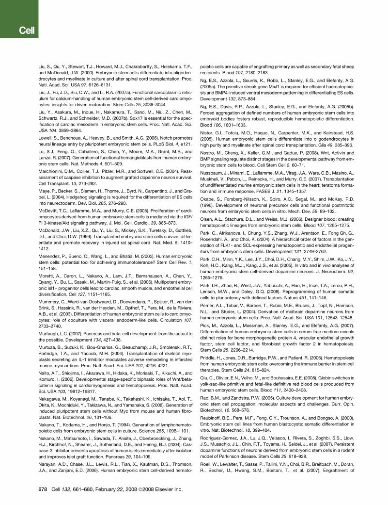
Liu, S., Qu, Y., Stewart, T.J., Howard, M.J., Chakrabortty, S., Holekamp, T.F.,
and McDonald, J.W. (2000). Embryonic stem cells differentiate into oligoden-
drocytes and myelinate in culture and after spinal cord transplantation. Proc.
Natl. Acad. Sci. USA 97, 6126–6131.
Liu, J., Fu, J.D., Siu, C.W., and Li, R.A. (2007a). Functional sarcoplasmic retic-
ulum for calcium-handling of human embryonic stem cell-derived cardiomyo-
cytes: insights for driven maturation. Stem Cells 25, 3038–3044.
Liu, Y., Asakura, M., Inoue, H., Nakamura, T., Sano, M., Niu, Z., Chen, M.,
Schwartz, R.J., and Schneider, M.D. (2007b). Sox17 is essential for the spec-
ification of cardiac mesoderm in embryonic stem cells. Proc. Natl. Acad. Sci.
USA 104, 3859–3864.
Lowell, S., Benchoua, A., Heavey, B., and Smith, A.G. (2006). Notch promotes
neural lineage entry by pluripotent embryonic stem cells. PLoS Biol. 4, e121.
Lu, S.J., Feng, Q., Caballero, S., Chen, Y., Moore, M.A., Grant, M.B., and
Lanza, R. (2007). Generation of functional hemangioblasts from human embry-
onic stem cells. Nat. Methods 4, 501–509.
Marchionini, D.M., Collier, T.J., Pitzer, M.R., and Sortwell, C.E. (2004). Reas-
sessment of caspase inhibition to augment grafted dopamine neuron survival.
Cell Transplant. 13, 273–282.
Maye, P., Becker, S., Siemen, H., Thorne, J., Byrd, N., Carpentino, J., and Gra-
bel, L. (2004). Hedgehog signaling is required for the differentiation of ES cells
into neurectoderm. Dev. Biol. 265, 276–290.
McDevitt, T.C., Laflamme, M.A., and Murry, C.E. (2005). Proliferation of cardi-
omyocytes derived from human embryonic stem cells is mediated via the IGF/
PI 3-kinase/Akt signaling pathway. J. Mol. Cell. Cardiol. 39, 865–873.
McDonald, J.W., Liu, X.Z., Qu, Y., Liu, S., Mickey, S.K., Turetsky, D., Gottlieb,
D.I., and Choi, D.W. (1999). Transplanted embryonic stem cells survive, differ-
entiate and promote recovery in injured rat spinal cord. Nat. Med. 5, 1410–
1412.
Menendez, P., Bueno, C., Wang, L., and Bhatia, M. (2005). Human embryonic
stem cells: potential tool for achieving immunotolerance? Stem Cell Rev. 1,
151–158.
Moretti, A., Caron, L., Nakano, A., Lam, J.T., Bernshausen, A., Chen, Y.,
Qyang, Y., Bu, L., Sasaki, M., Martin-Puig, S., et al. (2006). Multipotent embry-
onic isl1+ progenitor cells lead to cardiac, smooth muscle, and endothelial cell
diversification. Cell 127, 1151–1165.
Mummery, C., Ward-van Oostwaard, D., Doevendans, P., Spijker, R., van den
Brink, S., Hassink, R., van der Heyden, M., Opthof, T., Pera, M., de la Riviere,
A.B., et al. (2003). Differentiation of human embryonic stem cells to cardiomyo-
cytes: role of coculture with visceral endoderm-like cells. Circulation 107,
2733–2740.
Murtaugh, L.C. (2007). Pancreas and beta-cell development: from the actual to
the possible. Development 134, 427–438.
Murtuza, B., Suzuki, K., Bou-Gharios, G., Beauchamp, J.R., Smolenski, R.T.,
Partridge, T.A., and Yacoub, M.H. (2004). Transplantation of skeletal myo-
blasts secreting an IL-1 inhibitor modulates adverse remodeling in infarcted
murine myocardium. Proc. Natl. Acad. Sci. USA 101, 4216–4221.
Naito, A.T., Shiojima, I., Akazawa, H., Hidaka, K., Morisaki, T., Kikuchi, A., and
Komuro, I. (2006). Developmental stage-specific biphasic roles of Wnt/beta-
catenin signaling in cardiomyogenesis and hematopoiesis. Proc. Natl. Acad.
Sci. USA 103, 19812–19817.
Nakagawa, M., Koyanagi, M., Tanabe, K., Takahashi, K., Ichisaka, T., Aoi, T.,
Okita, K., Mochiduki, Y., Takizawa, N., and Yamanaka, S. (2008). Generation of
induced pluripotent stem cells without Myc from mouse and human fibro-
blasts. Nat. Biotechnol. 26, 101–106.
Nakano, T., Kodama, H., and Honjo, T. (1994). Generation of lymphohemato-
poietic cells from embryonic stem cells in culture. Science 265, 1098–1101.
Nakano, M., Matsumoto, I., Sawada, T., Ansite, J., Oberbroeckling, J., Zhang,
H.J., Kirchhof, N., Shearer, J., Sutherland, D.E., and Hering, B.J. (2004). Cas-
pase-3 inhibitor prevents apoptosis of human islets immediately after isolation
and improves islet graft function. Pancreas 29, 104–109.
Narayan, A.D., Chase, J.L., Lewis, R.L., Tian, X., Kaufman, D.S., Thomson,
J.A., and Zanjani, E.D. (2006). Human embryonic stem cell-derived hemato-
678 Cell 132, 661–680, February 22, 2008 ª2008 Elsevier Inc.
poietic cells are capable of engrafting primary as well as secondary fetal sheep
recipients. Blood 107, 2180–2183.
Ng, E.S., Azzola, L., Sourris, K., Robb, L., Stanley, E.G., and Elefanty, A.G.
(2005a). The primitive streak gene Mixl1 is required for efficient haematopoie-
sis and BMP4-induced ventral mesoderm patterning in differentiating ES cells.
Development 132, 873–884.
Ng, E.S., Davis, R.P., Azzola, L., Stanley, E.G., and Elefanty, A.G. (2005b).
Forced aggregation of defined numbers of human embryonic stem cells into
embryoid bodies fosters robust, reproducible hematopoietic differentiation.
Blood 106, 1601–1603.
Nistor, G.I., Totoiu, M.O., Haque, N., Carpenter, M.K., and Keirstead, H.S.
(2005). Human embryonic stem cells differentiate into oligodendrocytes in
high purity and myelinate after spinal cord transplantation. Glia 49, 385–396.
Nostro, M., Cheng, X., Keller, G.M., and Gadue, P. (2008). Wnt, Activin and
BMP signaling regulate distinct stages in the developmental pathway from em-
bryonic stem cells to blood. Cell Stem Cell 2, 60–71.
Nussbaum, J., Minami, E., Laflamme, M.A., Virag, J.A., Ware, C.B., Masino, A.,
Muskheli, V., Pabon, L., Reinecke, H., and Murry, C.E. (2007). Transplantation
of undifferentiated murine embryonic stem cells in the heart: teratoma forma-
tion and immune response. FASEB J. 21, 1345–1357.
Okabe, S., Forsberg-Nilsson, K., Spiro, A.C., Segal, M., and McKay, R.D.
(1996). Development of neuronal precursor cells and functional postmitotic
neurons from embryonic stem cells in vitro. Mech. Dev. 59, 89–102.
Olsen, A.L., Stachura, D.L., and Weiss, M.J. (2006). Designer blood: creating
hematopoietic lineages from embryonic stem cells. Blood 107, 1265–1275.
Park, C., Afrikanova, I., Chung, Y.S., Zhang, W.J., Arentson, E., Fong Gh, G.,
Rosendahl, A., and Choi, K. (2004). A hierarchical order of factors in the gen-
eration of FLK1- and SCL-expressing hematopoietic and endothelial progen-
itors from embryonic stem cells. Development 131, 2749–2762.
Park, C.H., Minn, Y.K., Lee, J.Y., Choi, D.H., Chang, M.Y., Shim, J.W., Ko, J.Y.,
Koh, H.C., Kang, M.J., Kang, J.S., et al. (2005). In vitro and in vivo analyses of
human embryonic stem cell-derived dopamine neurons. J. Neurochem. 92,
1265–1276.
Park, I.H., Zhao, R., West, J.A., Yabuuchi, A., Huo, H., Ince, T.A., Lerou, P.H.,
Lensch, M.W., and Daley, G.Q. (2008). Reprogramming of human somatic
cells to pluripotency with defined factors. Nature 451, 141–146.
Perrier, A.L., Tabar, V., Barberi, T., Rubio, M.E., Bruses, J., Topf, N., Harrison,
N.L., and Studer, L. (2004). Derivation of midbrain dopamine neurons from
human embryonic stem cells. Proc. Natl. Acad. Sci. USA 101, 12543–12548.
Pick, M., Azzola, L., Mossman, A., Stanley, E.G., and Elefanty, A.G. (2007).
Differentiation of human embryonic stem cells in serum-free medium reveals
distinct roles for bone morphogenetic protein 4, vascular endothelial growth
factor, stem cell factor, and fibroblast growth factor 2 in hematopoiesis.
Stem Cells 25, 2206–2214.
Priddle, H., Jones, D.R., Burridge, P.W., and Patient, R. (2006). Hematopoiesis
from human embryonic stem cells: overcoming the immune barrier in stem cell
therapies. Stem Cells 24, 815–824.
Qiu, C., Olivier, E.N., Velho, M., and Bouhassira, E.E. (2008). Globin switches in
yolk-sac-like primitive and fetal-like definitive red blood cells produced from
human embryonic stem cells. Blood 111, 2400–2408.
Rao, B.M., and Zandstra, P.W. (2005). Culture development for human embry-
onic stem cell propagation: molecular aspects and challenges. Curr. Opin.
Biotechnol. 16, 568–576.
Reubinoff, B.E., Pera, M.F., Fong, C.Y., Trounson, A., and Bongso, A. (2000).
Embryonic stem cell lines from human blastocysts: somatic differentiation in
vitro. Nat. Biotechnol. 18, 399–404.
Rodriguez-Gomez, J.A., Lu, J.Q., Velasco, I., Rivera, S., Zoghbi, S.S., Liow,
J.S., Musachio, J.L., Chin, F.T., Toyama, H., Seidel, J., et al. (2007). Persistent
dopamine functions of neurons derived from embryonic stem cells in a rodent
model of Parkinson disease. Stem Cells 25, 918–928.
Roell, W., Lewalter, T., Sasse, P., Tallini, Y.N., Choi, B.R., Breitbach, M., Doran,
R., Becher, U., Hwang, S.M., Bostani, T., et al. (2007). Engraftment of
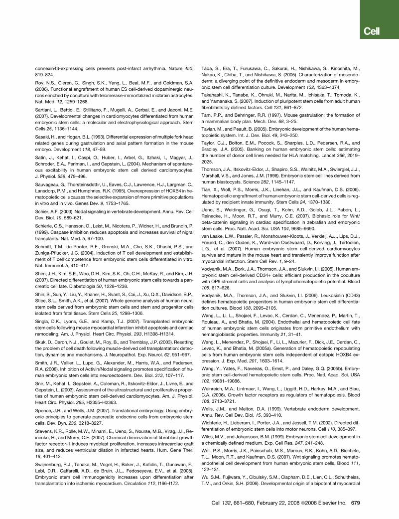
connexin43-expressing cells prevents post-infarct arrhythmia. Nature 450,
819–824.
Roy, N.S., Cleren, C., Singh, S.K., Yang, L., Beal, M.F., and Goldman, S.A.
(2006). Functional engraftment of human ES cell-derived dopaminergic neu-
rons enriched by coculture with telomerase-immortalized midbrain astrocytes.
Nat. Med. 12, 1259–1268.
Sartiani, L., Bettiol, E., Stillitano, F., Mugelli, A., Cerbai, E., and Jaconi, M.E.
(2007). Developmental changes in cardiomyocytes differentiated from human
embryonic stem cells: a molecular and electrophysiological approach. Stem
Cells 25, 1136–1144.
Sasaki, H., and Hogan, B.L. (1993). Differential expression of multiple fork head
related genes during gastrulation and axial pattern formation in the mouse
embryo. Development 118, 47–59.
Satin, J., Kehat, I., Caspi, O., Huber, I., Arbel, G., Itzhaki, I., Magyar, J.,
Schroder, E.A., Perlman, I., and Gepstein, L. (2004). Mechanism of spontane-
ous excitability in human embryonic stem cell derived cardiomyocytes.
J. Physiol. 559, 479–496.
Sauvageau, G., Thorsteinsdottir, U., Eaves, C.J., Lawrence, H.J., Largman, C.,
Lansdorp, P.M., and Humphries, R.K. (1995). Overexpression of HOXB4 in he-
matopoietic cells causes the selective expansion of more primitive populations
in vitro and in vivo. Genes Dev. 9, 1753–1765.
Schier, A.F. (2003). Nodal signaling in vertebrate development. Annu. Rev. Cell
Dev. Biol. 19, 589–621.
Schierle, G.S., Hansson, O., Leist, M., Nicotera, P., Widner, H., and Brundin, P.
(1999). Caspase inhibition reduces apoptosis and increases survival of nigral
transplants. Nat. Med. 5, 97–100.
Schmitt, T.M., de Pooter, R.F., Gronski, M.A., Cho, S.K., Ohashi, P.S., and
Zuniga-Pflucker, J.C. (2004). Induction of T cell development and establish-
ment of T cell competence from embryonic stem cells differentiated in vitro.
Nat. Immunol. 5, 410–417.
Shim, J.H., Kim, S.E., Woo, D.H., Kim, S.K., Oh, C.H., McKay, R., and Kim, J.H.
(2007). Directed differentiation of human embryonic stem cells towards a pan-
creatic cell fate. Diabetologia 50, 1228–1238.
Shin, S., Sun, Y., Liu, Y., Khaner, H., Svant, S., Cai, J., Xu, Q.X., Davidson, B.P.,
Stice, S.L., Smith, A.K., et al. (2007). Whole genome analysis of human neural
stem cells derived from embryonic stem cells and stem and progenitor cells
isolated from fetal tissue. Stem Cells 25, 1298–1306.
Singla, D.K., Lyons, G.E., and Kamp, T.J. (2007). Transplanted embryonic
stem cells following mouse myocardial infarction inhibit apoptosis and cardiac
remodeling. Am. J. Physiol. Heart Circ. Physiol. 293, H1308–H1314.
Skuk, D., Caron, N.J., Goulet, M., Roy, B., and Tremblay, J.P. (2003). Resetting
the problem of cell death following muscle-derived cell transplantation: detec-
tion, dynamics and mechanisms. J. Neuropathol. Exp. Neurol. 62, 951–967.
Smith, J.R., Vallier, L., Lupo, G., Alexander, M., Harris, W.A., and Pedersen,
R.A. (2008). Inhibition of Activin/Nodal signaling promotes specification of hu-
man embryonic stem cells into neuroectoderm. Dev. Biol. 313, 107–117.
Snir, M., Kehat, I., Gepstein, A., Coleman, R., Itskovitz-Eldor, J., Livne, E., and
Gepstein, L. (2003). Assessment of the ultrastructural and proliferative proper-
ties of human embryonic stem cell-derived cardiomyocytes. Am. J. Physiol.
Heart Circ. Physiol. 285, H2355–H2363.
Spence, J.R., and Wells, J.M. (2007). Translational embryology: Using embry-
onic principles to generate pancreatic endocrine cells from embryonic stem
cells. Dev. Dyn. 236, 3218–3227.
Stevens, K.R., Rolle, M.W., Minami, E., Ueno, S., Nourse, M.B., Virag, J.I., Re-
inecke, H., and Murry, C.E. (2007). Chemical dimerization of fibroblast growth
factor receptor-1 induces myoblast proliferation, increases intracardiac graft
size, and reduces ventricular dilation in infarcted hearts. Hum. Gene Ther.
18, 401–412.
Swijnenburg, R.J., Tanaka, M., Vogel, H., Baker, J., Kofidis, T., Gunawan, F.,
Lebl, D.R., Caffarelli, A.D., de Bruin, J.L., Fedoseyeva, E.V., et al. (2005).
Embryonic stem cell immunogenicity increases upon differentiation after
transplantation into ischemic myocardium. Circulation 112, I166–I172.
Tada, S., Era, T., Furusawa, C., Sakurai, H., Nishikawa, S., Kinoshita, M.,
Nakao, K., Chiba, T., and Nishikawa, S. (2005). Characterization of mesendo-
derm: a diverging point of the definitive endoderm and mesoderm in embry-
onic stem cell differentiation culture. Development 132, 4363–4374.
Takahashi, K., Tanabe, K., Ohnuki, M., Narita, M., Ichisaka, T., Tomoda, K.,
and Yamanaka, S. (2007). Induction of pluripotent stem cells from adult human
fibroblasts by defined factors. Cell 131, 861–872.
Tam, P.P., and Behringer, R.R. (1997). Mouse gastrulation: the formation of
a mammalian body plan. Mech. Dev. 68, 3–25.
Tavian, M., and Peault, B. (2005). Embryonic development of the human hema-
topoietic system. Int. J. Dev. Biol. 49, 243–250.
Taylor, C.J., Bolton, E.M., Pocock, S., Sharples, L.D., Pedersen, R.A., and
Bradley, J.A. (2005). Banking on human embryonic stem cells: estimating
the number of donor cell lines needed for HLA matching. Lancet 366, 2019–
2025.
Thomson, J.A., Itskovitz-Eldor, J., Shapiro, S.S., Walnitz, M.A., Swiergiel, J.J.,
Marshall, V.S., and Jones, J.M. (1998). Embryonic stem cell lines derived from
human blastocysts. Science 282, 1145–1147.
Tian, X., Woll, P.S., Morris, J.K., Linehan, J.L., and Kaufman, D.S. (2006).
Hematopoietic engraftment of human embryonic stem cell-derived cells is reg-
ulated by recipient innate immunity. Stem Cells 24, 1370–1380.
Ueno, S., Weidinger, G., Osugi, T., Kohn, A.D., Golob, J.L., Pabon, L.,
Reinecke, H., Moon, R.T., and Murry, C.E. (2007). Biphasic role for Wnt/
beta-catenin signaling in cardiac specification in zebrafish and embryonic
stem cells. Proc. Natl. Acad. Sci. USA 104, 9685–9690.
van Laake, L.W., Passier, R., Monshouwer-Kloots, J., Verkleij, A.J., Lips, D.J.,
Freund, C., den Ouden, K., Ward-van Oostwaard, D., Korving, J., Tertoolen,
L.G., et al. (2007). Human embryonic stem cell-derived cardiomyocytes
survive and mature in the mouse heart and transiently improve function after
myocardial infarction. Stem Cell Rev. 1, 9–24.
Vodyanik, M.A., Bork, J.A., Thomson, J.A., and Slukvin, I.I. (2005). Human em-
bryonic stem cell-derived CD34+ cells: efficient production in the coculture
with OP9 stromal cells and analysis of lymphohematopoietic potential. Blood
105, 617–626.
Vodyanik, M.A., Thomson, J.A., and Slukvin, I.I. (2006). Leukosialin (CD43)
defines hematopoietic progenitors in human embryonic stem cell differentia-
tion cultures. Blood 108, 2095–2105.
Wang, L., Li, L., Shojaei, F., Levac, K., Cerdan, C., Menendez, P., Martin, T.,
Rouleau, A., and Bhatia, M. (2004). Endothelial and hematopoietic cell fate
of human embryonic stem cells originates from primitive endothelium with
hemangioblastic properties. Immunity 21, 31–41.
Wang, L., Menendez, P., Shojaei, F., Li, L., Mazurier, F., Dick, J.E., Cerdan, C.,
Levac, K., and Bhatia, M. (2005a). Generation of hematopoietic repopulating
cells from human embryonic stem cells independent of ectopic HOXB4 ex-
pression. J. Exp. Med. 201, 1603–1614.
Wang, Y., Yates, F., Naveiras, O., Ernst, P., and Daley, G.Q. (2005b). Embry-
onic stem cell-derived hematopoietic stem cells. Proc. Natl. Acad. Sci. USA
102, 19081–19086.
Weinreich, M.A., Lintmaer, I., Wang, L., Liggitt, H.D., Harkey, M.A., and Blau,
C.A. (2006). Growth factor receptors as regulators of hematopoiesis. Blood
108, 3713–3721.
Wells, J.M., and Melton, D.A. (1999). Vertebrate endoderm development.
Annu. Rev. Cell Dev. Biol. 15, 393–410.
Wichterle, H., Lieberam, I., Porter, J.A., and Jessell, T.M. (2002). Directed dif-
ferentiation of embryonic stem cells into motor neurons. Cell 110, 385–397.
Wiles, M.V., and Johansson, B.M. (1999). Embryonic stem cell development in
a chemically defined medium. Exp. Cell Res. 247, 241–248.
Woll, P.S., Morris, J.K., Painschab, M.S., Marcus, R.K., Kohn, A.D., Biechele,
T.L., Moon, R.T., and Kaufman, D.S. (2007). Wnt signaling promotes hemato-
endothelial cell development from human embryonic stem cells. Blood 111,
122–131.
Wu, S.M., Fujiwara, Y., Cibulsky, S.M., Clapham, D.E., Lien, C.L., Schultheiss,
T.M., and Orkin, S.H. (2006). Developmental origin of a bipotential myocardial
Cell 132, 661–680, February 22, 2008 ª2008 Elsevier Inc. 679
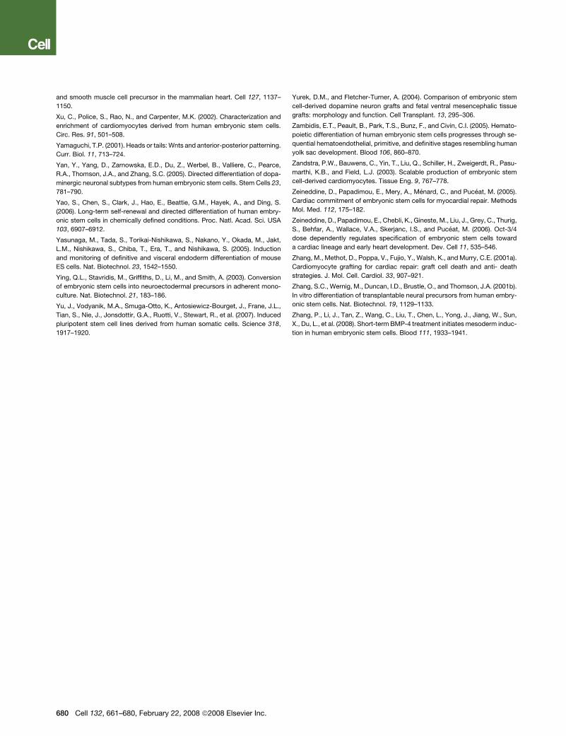
and smooth muscle cell precursor in the mammalian heart. Cell 127, 1137–
1150.
Xu, C., Police, S., Rao, N., and Carpenter, M.K. (2002). Characterization and
enrichment of cardiomyocytes derived from human embryonic stem cells.
Circ. Res. 91, 501–508.
Yamaguchi, T.P. (2001). Heads or tails: Wnts and anterior-posterior patterning.
Curr. Biol. 11, 713–724.
Yan, Y., Yang, D., Zarnowska, E.D., Du, Z., Werbel, B., Valliere, C., Pearce,
R.A., Thomson, J.A., and Zhang, S.C. (2005). Directed differentiation of dopa-
minergic neuronal subtypes from human embryonic stem cells. Stem Cells 23,
781–790.
Yao, S., Chen, S., Clark, J., Hao, E., Beattie, G.M., Hayek, A., and Ding, S.
(2006). Long-term self-renewal and directed differentiation of human embry-
onic stem cells in chemically defined conditions. Proc. Natl. Acad. Sci. USA
103, 6907–6912.
Yasunaga, M., Tada, S., Torikai-Nishikawa, S., Nakano, Y., Okada, M., Jakt,
L.M., Nishikawa, S., Chiba, T., Era, T., and Nishikawa, S. (2005). Induction
and monitoring of definitive and visceral endoderm differentiation of mouse
ES cells. Nat. Biotechnol. 23, 1542–1550.
Ying, Q.L., Stavridis, M., Griffiths, D., Li, M., and Smith, A. (2003). Conversion
of embryonic stem cells into neuroectodermal precursors in adherent mono-
culture. Nat. Biotechnol. 21, 183–186.
Yu, J., Vodyanik, M.A., Smuga-Otto, K., Antosiewicz-Bourget, J., Frane, J.L.,
Tian, S., Nie, J., Jonsdottir, G.A., Ruotti, V., Stewart, R., et al. (2007). Induced
pluripotent stem cell lines derived from human somatic cells. Science 318,
1917–1920.
680 Cell 132, 661–680, February 22, 2008 ª2008 Elsevier Inc.
Yurek, D.M., and Fletcher-Turner, A. (2004). Comparison of embryonic stem
cell-derived dopamine neuron grafts and fetal ventral mesencephalic tissue
grafts: morphology and function. Cell Transplant. 13, 295–306.
Zambidis, E.T., Peault, B., Park, T.S., Bunz, F., and Civin, C.I. (2005). Hemato-
poietic differentiation of human embryonic stem cells progresses through se-
quential hematoendothelial, primitive, and definitive stages resembling human
yolk sac development. Blood 106, 860–870.
Zandstra, P.W., Bauwens, C., Yin, T., Liu, Q., Schiller, H., Zweigerdt, R., Pasu-
marthi, K.B., and Field, L.J. (2003). Scalable production of embryonic stem
cell-derived cardiomyocytes. Tissue Eng. 9, 767–778.
Zeineddine, D., Papadimou, E., Mery, A., Menard, C., and Puceat, M. (2005).
Cardiac commitment of embryonic stem cells for myocardial repair. Methods
Mol. Med. 112, 175–182.
Zeineddine, D., Papadimou, E., Chebli, K., Gineste, M., Liu, J., Grey, C., Thurig,
S., Behfar, A., Wallace, V.A., Skerjanc, I.S., and Puceat, M. (2006). Oct-3/4
dose dependently regulates specification of embryonic stem cells toward
a cardiac lineage and early heart development. Dev. Cell 11, 535–546.
Zhang, M., Methot, D., Poppa, V., Fujio, Y., Walsh, K., and Murry, C.E. (2001a).
Cardiomyocyte grafting for cardiac repair: graft cell death and anti- death
strategies. J. Mol. Cell. Cardiol. 33, 907–921.
Zhang, S.C., Wernig, M., Duncan, I.D., Brustle, O., and Thomson, J.A. (2001b).
In vitro differentiation of transplantable neural precursors from human embry-
onic stem cells. Nat. Biotechnol. 19, 1129–1133.
Zhang, P., Li, J., Tan, Z., Wang, C., Liu, T., Chen, L., Yong, J., Jiang, W., Sun,
X., Du, L., et al. (2008). Short-term BMP-4 treatment initiates mesoderm induc-
tion in human embryonic stem cells. Blood 111, 1933–1941.