Embed Size (px)
Citation preview
Feature Article
Reconstructing the Differentiation Niche ofEmbryonic Stem Cells Using Biomaterials
Laura E. Dickinson,a Sravanti Kusuma,a Sharon Gerecht*
The biochemical cues and topographical architecture of the extracellular environment exten-sively influence ES cell fate. The microenvironment surrounding the developing embryopresents these instructive cues in a complex and interactive manner in order to guide cellfate decisions. Current stem cell research aims to reconstruct this multifaceted embryonicniche to recapitulate development invitro. This review focuses on 2D and3D differentiation niches created fromnatural and synthetic biomaterials toguide the differentiation of ES cellstoward specific lineages. Biomaterialsengineered to present specific physicalconstraints are also reviewed for theirrole in differentiation.
Introduction
The extracellular environment presents a myriad of
biochemical cues and specific topographical architecture
in a spatially and temporally distinct manner to influence
cellular behavior, including the self-renewal and differ-
entiation of embryonic stem (ES) cells. While an important
function of the extracellular matrix (ECM) is to provide the
structural framework to support cellular functions, this
scaffold of proteins, proteoglycans, and glycosaminogly-
cans also provides cell adhesion sites and important
signaling cues. ECM components and structure influence
stem cell fate through integrin-mediated activation and
downstream signaling events.
L. E. Dickinson, S. Kusuma, S. GerechtDepartment of Chemical and Biomolecular Engineering, JohnsHopkins Physical Sciences-Oncology Center and Institute forNanoBioTechnology, 3400 North Charles Street, Baltimore, MD21210, USAFax: þ1 410 516 5510; E-mail: [email protected]. KusumaBiomedical Engineering, Johns Hopkins University, Baltimore, MD21218, USA
a L. E. Dickinson and S. Kusuma contributed equally to this work.
Macromol. Biosci. 2010, 10, 000–000
� 2010 WILEY-VCH Verlag GmbH & Co. KGaA, Weinheim wileyonlin
Early View Publication; these are NOT the
ES cells have attracted attention as a promising source of
cells for tissue engineering, as they can self-renew almost
indefinitely in culture and have the capacity to differentiate
into all cell types of the body (i.e., are pluripotent). ES cells
are isolated from the inner cell mass of the developing
blastocyst. In vitro, they can be maintained as undiffer-
entiated cells via culture on mouse embryonic fibroblasts
(MEFs) or via culture in feeder-free conditions using a
defined medium composition to supply essential cytokines
and nutrients to promote the undifferentiated pheno-
type.[1] Control over their differentiation has been exten-
sively studied via manipulation of the parameters of the
stem cell microenvironment, referred to as the stem cell
niche. Thus, both soluble and insoluble cues of the niche
which modulate the differentiation have been thoroughly
investigated to determine the parameters amenable to
inducing a particular cell fate.
The extracellular microenvironment surrounding the
developing embryo presents a number of spatially and
temporally instructive biochemical cues within a complex
and interactive milieu that guide and govern the sequential
development and cell fate decisions of embryogenesis.[2]
Current stem cell research aspires to deconstruct this
multifaceted embryonic niche, determine the influential
elements involved in lineage differentiation, and recreate
elibrary.com DOI: 10.1002/mabi.201000245 1
final page numbers, use DOI for citation !! R
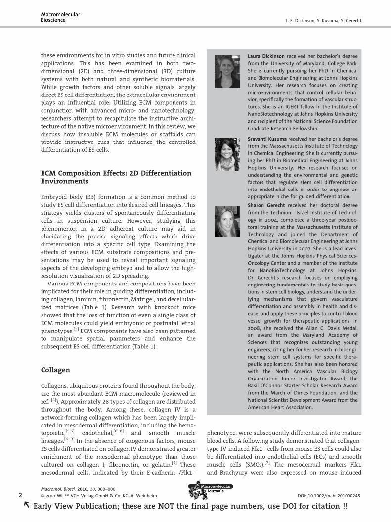
Laura Dickinson received her bachelor’s degreefrom the University of Maryland, College Park.She is currently pursuing her PhD in Chemicaland Biomolecular Engineering at Johns HopkinsUniversity. Her research focuses on creatingmicroenvironments that control cellular beha-vior, specifically the formation of vascular struc-tures. She is an IGERT fellow in the Institute ofNanoBiotechnology at Johns Hopkins Universityand recipient of the National Science FoundationGraduate Research Fellowship.
Sravanti Kusuma received her bachelor’s degreefrom the Massachusetts Institute of Technology
2
REa
L. E. Dickinson, S. Kusuma, S. Gerecht
these environments for in vitro studies and future clinical
applications. This has been examined in both two-
dimensional (2D) and three-dimensional (3D) culture
systems with both natural and synthetic biomaterials.
While growth factors and other soluble signals largely
direct ES cell differentiation, the extracellular environment
plays an influential role. Utilizing ECM components in
conjunction with advanced micro- and nanotechnology,
researchers attempt to recapitulate the instructive archi-
tecture of the native microenvironment. In this review, we
discuss how insoluble ECM molecules or scaffolds can
provide instructive cues that influence the controlled
differentiation of ES cells.
in Chemical Engineering. She is currently pursu-ing her PhD in Biomedical Engineering at JohnsHopkins University. Her research focuses onunderstanding the environmental and geneticfactors that regulate stem cell differentiationinto endothelial cells in order to engineer anappropriate niche for guided differentiation.Sharon Gerecht received her doctoral degreefrom the Technion - Israel Institute of Technol-ogy in 2004, completed a three-year postdoc-toral training at the Massachusetts Institute ofTechnology and joined the Department ofChemical and Biomolecular Engineering at JohnsHopkins University in 2007. She is a lead inves-tigator at the Johns Hopkins Physical Sciences-Oncology Center and a member of the Institutefor NanoBioTechnology at Johns Hopkins.Dr. Gerecht’s research focuses on employingengineering fundamentals to study basic ques-tions in stem cell biology, understand the under-lying mechanisms that govern vasculaturedifferentiation and assembly in health and dis-ease, and apply these principles to control bloodvessel growth for therapeutic applications. In2008, she received the Allan C. Davis Medal,an award from the Maryland Academy ofSciences that recognizes outstanding youngengineers, citing her for her research in bioengi-ECM Composition Effects: 2D DifferentiationEnvironments
Embryoid body (EB) formation is a common method to
study ES cell differentiation into desired cell lineages. This
strategy yields clusters of spontaneously differentiating
cells in suspension culture. However, studying this
phenomenon in a 2D adherent culture may aid in
elucidating the precise signaling effects which drive
differentiation into a specific cell type. Examining the
effects of various ECM substrate compositions and pre-
sentations may be used to reveal important signaling
aspects of the developing embryo and to allow the high-
resolution visualization of 2D spreading.
Various ECM components and compositions have been
implicated for their role in guiding differentiation, includ-
ing collagen, laminin, fibronectin, Matrigel, and decellular-
ized matrices (Table 1). Research with knockout mice
showed that the loss of function of even a single class of
ECM molecules could yield embryonic or postnatal lethal
phenotypes.[3] ECM components have also been patterned
to manipulate spatial parameters and enhance the
subsequent ES cell differentiation (Table 1).
neering stem cell systems for specific thera-peutic applications. She has also been honoredwith the North America Vascular BiologyOrganization Junior Investigator Award, theBasil O’Connor Starter Scholar Research Awardfrom the March of Dimes Foundation, and theNational Scientist Development Award from theAmerican Heart Association.
Collagen
Collagens, ubiquitous proteins found throughout the body,
are the most abundant ECM macromolecule (reviewed in
ref. [4]). Approximately 28 types of collagen are distributed
throughout the body. Among these, collagen IV is a
network-forming collagen which has been largely impli-
cated in mesodermal differentiation, including the hema-
topoietic,[5,6] endothelial,[6–8] and smooth muscle
lineages.[6–9] In the absence of exogenous factors, mouse
ES cells differentiated on collagen IV demonstrated greater
enrichment of the mesodermal phenotype than those
cultured on collagen I, fibronectin, or gelatin.[5] These
mesodermal cells, indicated by their E-cadherin�/Flk1þ
Macromol. Biosci. 2010, 10, 000–000
� 2010 WILEY-VCH Verlag GmbH & Co. KGaA, Weinheim
rly View Publication; these are NOT the fina
phenotype, were subsequently differentiated into mature
blood cells. A following study demonstrated that collagen-
type-IV-induced Flk1þ cells from mouse ES cells could also
be differentiated into endothelial cells (ECs) and smooth
muscle cells (SMCs).[7] The mesodermal markers Flk1
and Brachyury were also expressed on mouse induced
DOI: 10.1002/mabi.201000245
l page numbers, use DOI for citation !!
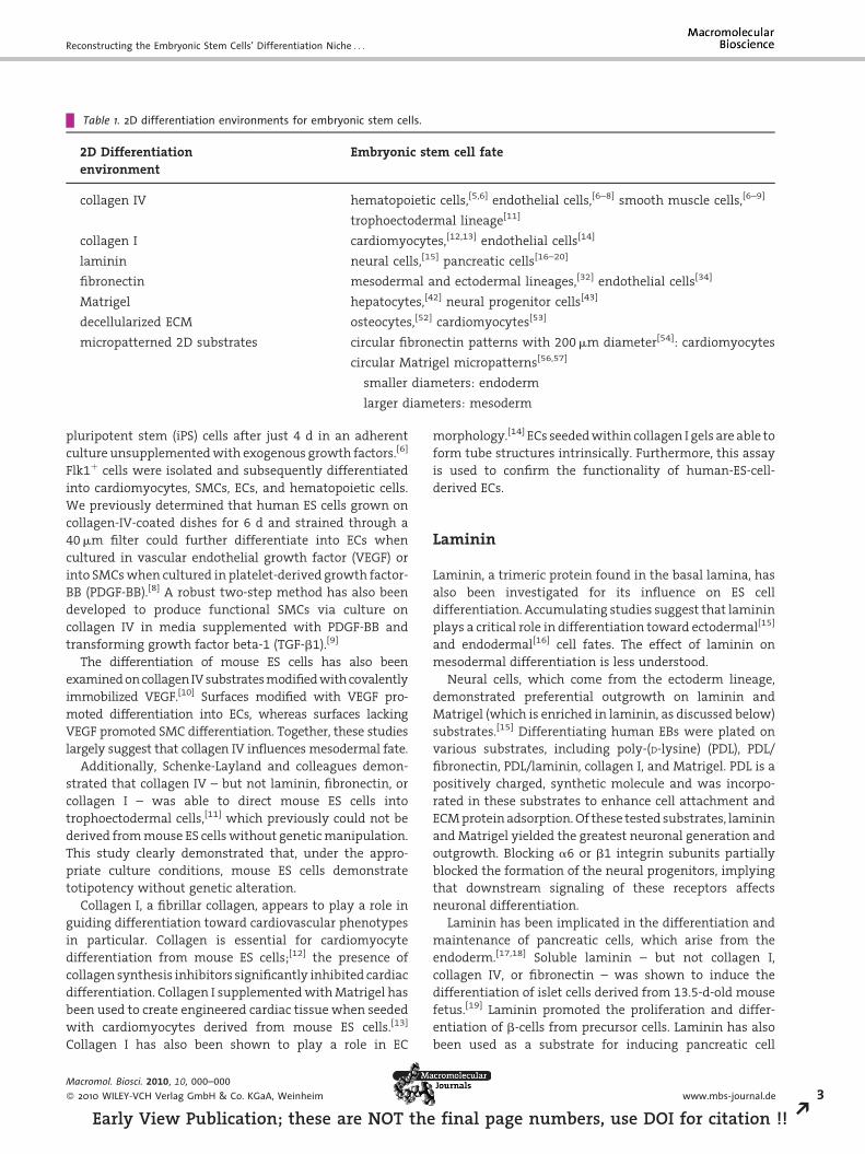
Table 1. 2D differentiation environments for embryonic stem cells.
2D Differentiation
environment
Embryonic stem cell fate
collagen IV hematopoietic cells,[5,6] endothelial cells,[6–8] smooth muscle cells,[6–9]
trophoectodermal lineage[11]
collagen I cardiomyocytes,[12,13] endothelial cells[14]
laminin neural cells,[15] pancreatic cells[16–20]
fibronectin mesodermal and ectodermal lineages,[32] endothelial cells[34]
Matrigel hepatocytes,[42] neural progenitor cells[43]
decellularized ECM osteocytes,[52] cardiomyocytes[53]
micropatterned 2D substrates circular fibronectin patterns with 200 mm diameter[54]: cardiomyocytes
circular Matrigel micropatterns[56,57]
smaller diameters: endoderm
larger diameters: mesoderm
Reconstructing the Embryonic Stem Cells’ Differentiation Niche . . .
pluripotent stem (iPS) cells after just 4 d in an adherent
culture unsupplemented with exogenous growth factors.[6]
Flk1þ cells were isolated and subsequently differentiated
into cardiomyocytes, SMCs, ECs, and hematopoietic cells.
We previously determined that human ES cells grown on
collagen-IV-coated dishes for 6 d and strained through a
40 mm filter could further differentiate into ECs when
cultured in vascular endothelial growth factor (VEGF) or
into SMCs when cultured in platelet-derived growth factor-
BB (PDGF-BB).[8] A robust two-step method has also been
developed to produce functional SMCs via culture on
collagen IV in media supplemented with PDGF-BB and
transforming growth factor beta-1 (TGF-b1).[9]
The differentiation of mouse ES cells has also been
examined on collagen IV substrates modified with covalently
immobilized VEGF.[10] Surfaces modified with VEGF pro-
moted differentiation into ECs, whereas surfaces lacking
VEGF promoted SMC differentiation. Together, these studies
largely suggest that collagen IV influences mesodermal fate.
Additionally, Schenke-Layland and colleagues demon-
strated that collagen IV – but not laminin, fibronectin, or
collagen I – was able to direct mouse ES cells into
trophoectodermal cells,[11] which previously could not be
derived from mouse ES cells without genetic manipulation.
This study clearly demonstrated that, under the appro-
priate culture conditions, mouse ES cells demonstrate
totipotency without genetic alteration.
Collagen I, a fibrillar collagen, appears to play a role in
guiding differentiation toward cardiovascular phenotypes
in particular. Collagen is essential for cardiomyocyte
differentiation from mouse ES cells;[12] the presence of
collagen synthesis inhibitors significantly inhibited cardiac
differentiation. Collagen I supplemented with Matrigel has
been used to create engineered cardiac tissue when seeded
with cardiomyocytes derived from mouse ES cells.[13]
Collagen I has also been shown to play a role in EC
Macromol. Biosci. 2010, 10, 000–000
� 2010 WILEY-VCH Verlag GmbH & Co. KGaA, Weinheim
Early View Publication; these are NOT the
morphology.[14] ECs seeded within collagen I gels are able to
form tube structures intrinsically. Furthermore, this assay
is used to confirm the functionality of human-ES-cell-
derived ECs.
Laminin
Laminin, a trimeric protein found in the basal lamina, has
also been investigated for its influence on ES cell
differentiation. Accumulating studies suggest that laminin
plays a critical role in differentiation toward ectodermal[15]
and endodermal[16] cell fates. The effect of laminin on
mesodermal differentiation is less understood.
Neural cells, which come from the ectoderm lineage,
demonstrated preferential outgrowth on laminin and
Matrigel (which is enriched in laminin, as discussed below)
substrates.[15] Differentiating human EBs were plated on
various substrates, including poly-(D-lysine) (PDL), PDL/
fibronectin, PDL/laminin, collagen I, and Matrigel. PDL is a
positively charged, synthetic molecule and was incorpo-
rated in these substrates to enhance cell attachment and
ECM protein adsorption. Of these tested substrates, laminin
and Matrigel yielded the greatest neuronal generation and
outgrowth. Blocking a6 or b1 integrin subunits partially
blocked the formation of the neural progenitors, implying
that downstream signaling of these receptors affects
neuronal differentiation.
Laminin has been implicated in the differentiation and
maintenance of pancreatic cells, which arise from the
endoderm.[17,18] Soluble laminin – but not collagen I,
collagen IV, or fibronectin – was shown to induce the
differentiation of islet cells derived from 13.5-d-old mouse
fetus.[19] Laminin promoted the proliferation and differ-
entiation of b-cells from precursor cells. Laminin has also
been used as a substrate for inducing pancreatic cell
www.mbs-journal.de 3
final page numbers, use DOI for citation !! R
4
REa
L. E. Dickinson, S. Kusuma, S. Gerecht
differentiation from mouse ES cells in vitro.[20] After EB
formation, early progenitors of the pancreatic lineage were
induced via culture on laminin substrates and in the
presence of nicotinamide and insulin.[20]
Mouse cells genetically altered to lack laminin g1 chain
could not form a basement membrane.[21] In EBs generated
from these knockout cells, ectodermal differentiation was
greatly reduced and mesodermal differentiation was
preferred and accelerated. Cardiomyocytes could still be
differentiated from mouse ES cells deficient in laminin g1
chain.[22] However, they were not able to support electrical
signal propagation, implying that laminin may still be
relevant for the functionality of cardiac cells.
Fibronectin
Fibronectin is an ECM molecule expressed during the early
stages of development that is critical for proper develop-
ment of the mesoderm and neural tube.[23] It is often used as
a cell adhesive layer for biomaterials due to the presence of
the peptide sequence arginine-serine-aspartic acid (RGD),
which is widely involved in integrin-mediated cell adhe-
sion[24–26] and is commonly tethered to biomaterials to
improve cell adhesion.[27,28] Additionally, fibronectin has
been implicated in enhancing the differentiation of stem
cells to specific lineages.[29–31] Binding a5b1 integrin to
specific fibronectin domains conferred differentiation into
mesodermal and ectodermal lineages.[32] Blocking mouse
ES cell interactions with a fibronectin substrate by the
addition of anti-integrin b1 antibody maintained self-
renewal.[33] Fibronectin has also been used to mature hES
cells – and iPS-cell-derived CD34þCD31þ cells – into ECs.[34]
Matrigel
Matrigel is isolated from the natural basement membrane
of mouse sarcoma cells and therefore contains a myriad of
ECM components and growth factors[35,36] conducive to cell
attachment, proliferation, and/or differentiation to all
three lineages;[37–43] its efficacy is most likely due to it
having many factors in optimal native concentrations.
Though collagen IV and heparan sulfate proteoglycans are
also present, laminin is its primary component.[36] Using
Matrigel provides an environment that presents many of
the appropriate extracellular biochemical and biophysical
cues necessary for stem cell differentiation.[39,44] Whereas
many fabricated hydrogels only present one major ECM
component, Matrigel presents many; because it was derived
from basement membrane, it instructs ES cell differentiation
similarly to native basement membranes.[45]
Compared to collagen I and laminin, Matrigel was shown
to be more effective at inducing hepatocyte differentiation
Macromol. Biosci. 2010, 10, 000–000
� 2010 WILEY-VCH Verlag GmbH & Co. KGaA, Weinheim
rly View Publication; these are NOT the fina
from multiple human ES cell lines in the presence of activin
A and hepatocyte growth factor.[42] Matrigel has also been
used to support the growth of neural progenitors.[43] This
study allowed mouse ES cells to differentiate on a mouse
stromal cell layer for 8 d and then transferred them to
matrices made from various ECM components. Of the tested
matrices, which included Matrigel, gelatin, collagen IV, and
laminin, Matrigel proved most effective in supporting the
survival and differentiation of neural progenitor cells.
Decellularized ECM
Of note, Matrigel is isolated from murine species, present-
ing translational disadvantages. Decellularized matrices
provide a bioinstructive template and can be isolated
from various cell types for applications in tissue regenera-
tion.[46–50] Extracted ECM has the potential to provide an
inductive environment that guides undifferentiated cells
to cell lineages supported in vivo.[47,48,51] ECM deposited by
osteoblastic cells enhanced the osteogenic differentiation
of mesenchymal stem cells (MSCs),[48] while a decellular-
ized matrix of adipose tissue produced an inductive
microenvironment for adipogenesis without exogenous
growth factors.[47,51] The ability to extract intact ECM with
conserved 3D structure and biochemical factors could allow
the differentiation of ES cells to any lineage type.
Decellularized ECM has been studied as a potential
substrate for ES cell differentiation.[52] Mouse ES cells were
seeded onto decellularized matrices derived from osteo-
genic and non-osteogenic cells. Differentiation on either of
these matrices was compared with differentiation on a
collagen I matrix. Mineralized tissue formation and
osteogenic gene expression revealed that the osteogenic
cell-derived ECM was most favorable for osteogenic
differentiation. Similarly, ECM derived from cardiac fibro-
blasts, which consists of collagen types I and III, laminin,
fibronectin, proteoglycans, and glycoproteins, enhanced
mouse-ES-cell-derived cardiomyocyte maturation and
functionality better than Matrigel.[53]
Micropatterned 2D Substrates
Micropatterns allow the manipulation of spatial para-
meters involved in lineage commitment, and to this end, 2D
patterned ECM substrates have been extensively examined
for ES cell differentiation. Microcontact printing (mCP) is one
method to achieve micropatterns. This technique uses a
prefabricated silicon template to mold an elastomeric
polymer, polydimethylsiloxane (PDMS), which is used to
‘‘stamp’’ molecules of interest to a substrate in the desired
array. Fibronectin micropatterns have been shown to
influence ES cell fate. When mouse ES cells were cultured
DOI: 10.1002/mabi.201000245
l page numbers, use DOI for citation !!
Reconstructing the Embryonic Stem Cells’ Differentiation Niche . . .
on varying sizes of fibronectin circular micropatterns, they
demonstrated variable differentiation to the cardiomyo-
cyte lineage; the 200 mm diameters circles exhibited the
greatest potential.[54] Additionally, MSCs cultured on
smaller fibronectin islands acquired an adipocyte fate, and
larger islands promoted more osteogenic cell lineages.[55]
Clearly, the surface presentation of biomolecular cues is not
the sole factor in cell differentiation, since fibronectin-coated
substrates yielded varying cell lineages. Cell type, spatial
dimensions, and topography are also crucial parameters in
developing culture systems for controlled cell differentia-
tion. Human ES cells cultured on circular micropatterns of
Matrigel maintained their pluripotency or differentiated to
endoderm on smaller geometries but differentiated to
mesoderm on larger sized islands.[56,57]
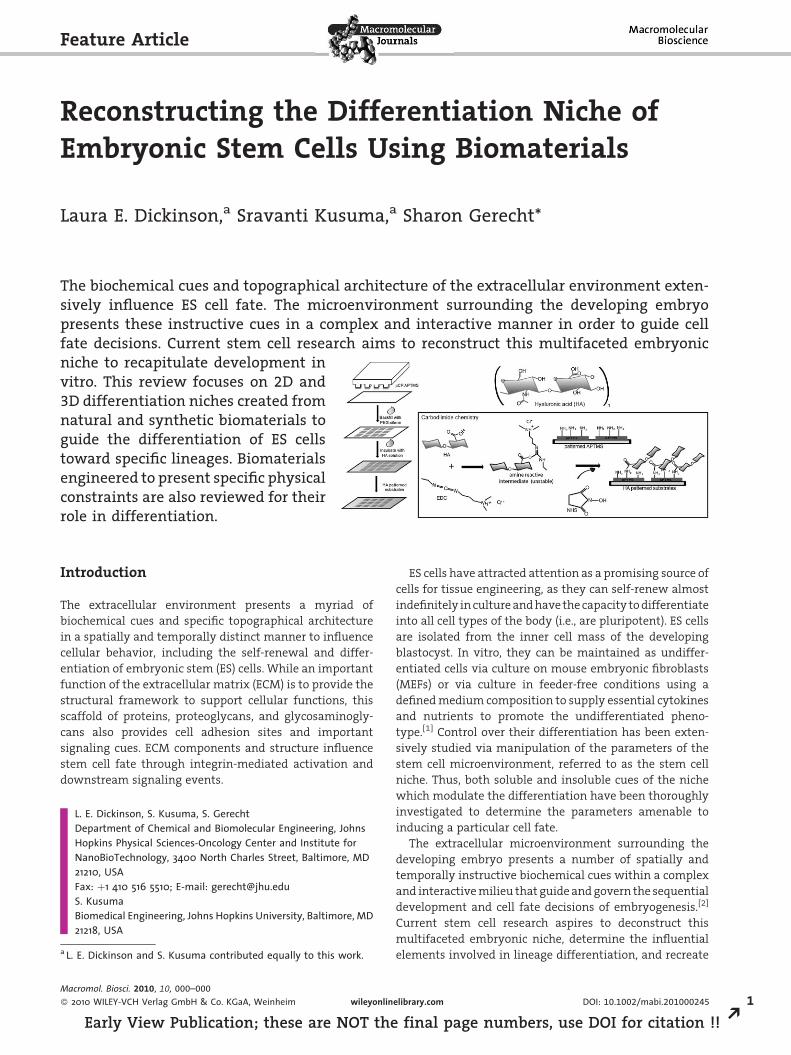
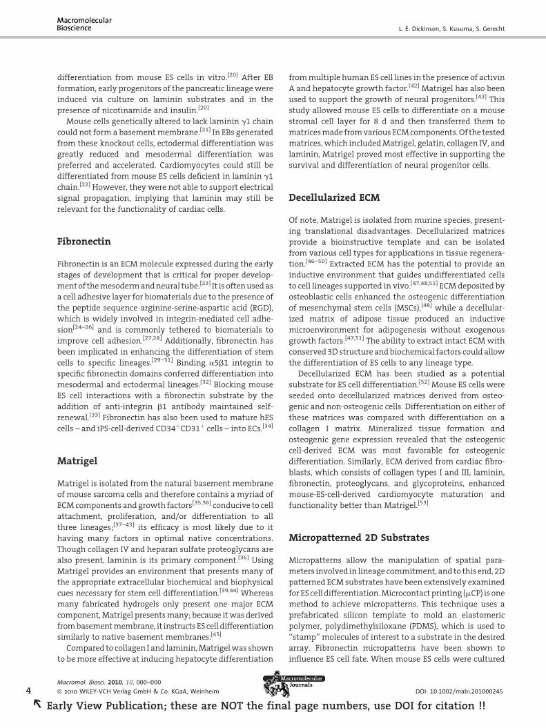
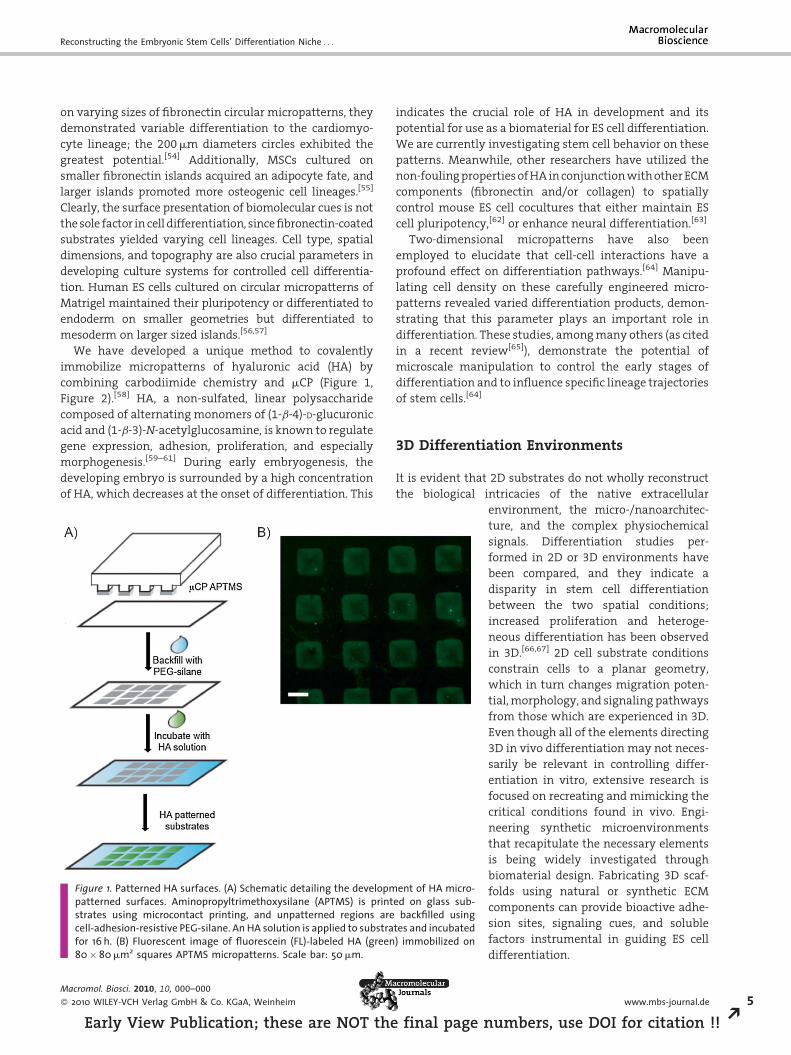
We have developed a unique method to covalently
immobilize micropatterns of hyaluronic acid (HA) by
combining carbodiimide chemistry and mCP (Figure 1,
Figure 2).[58] HA, a non-sulfated, linear polysaccharide
composed of alternating monomers of (1-b-4)-D-glucuronic
acid and (1-b-3)-N-acetylglucosamine, is known to regulate
gene expression, adhesion, proliferation, and especially
morphogenesis.[59–61] During early embryogenesis, the
developing embryo is surrounded by a high concentration
of HA, which decreases at the onset of differentiation. This
Figure 1. Patterned HA surfaces. (A) Schematic detailing the developmpatterned surfaces. Aminopropyltrimethoxysilane (APTMS) is printstrates using microcontact printing, and unpatterned regions arecell-adhesion-resistive PEG-silane. An HA solution is applied to substrafor 16 h. (B) Fluorescent image of fluorescein (FL)-labeled HA (green80�80 mm2 squares APTMS micropatterns. Scale bar: 50 mm.
Macromol. Biosci. 2010, 10, 000–000
� 2010 WILEY-VCH Verlag GmbH & Co. KGaA, Weinheim
Early View Publication; these are NOT the
indicates the crucial role of HA in development and its
potential for use as a biomaterial for ES cell differentiation.
We are currently investigating stem cell behavior on these
patterns. Meanwhile, other researchers have utilized the
non-fouling properties of HA in conjunction with other ECM
components (fibronectin and/or collagen) to spatially
control mouse ES cell cocultures that either maintain ES
cell pluripotency,[62] or enhance neural differentiation.[63]
Two-dimensional micropatterns have also been
employed to elucidate that cell-cell interactions have a
profound effect on differentiation pathways.[64] Manipu-
lating cell density on these carefully engineered micro-
patterns revealed varied differentiation products, demon-
strating that this parameter plays an important role in
differentiation. These studies, among many others (as cited
in a recent review[65]), demonstrate the potential of
microscale manipulation to control the early stages of
differentiation and to influence specific lineage trajectories
of stem cells.[64]
3D Differentiation Environments
It is evident that 2D substrates do not wholly reconstruct
the biological intricacies of the native extracellular
ent of HA micro-ed on glass sub-
backfilled usingtes and incubated) immobilized on
final page n
environment, the micro-/nanoarchitec-
ture, and the complex physiochemical
signals. Differentiation studies per-
formed in 2D or 3D environments have
been compared, and they indicate a
disparity in stem cell differentiation
between the two spatial conditions;
increased proliferation and heteroge-
neous differentiation has been observed
in 3D.[66,67] 2D cell substrate conditions
constrain cells to a planar geometry,
which in turn changes migration poten-
tial, morphology, and signaling pathways
from those which are experienced in 3D.
Even though all of the elements directing
3D in vivo differentiation may not neces-
sarily be relevant in controlling differ-
entiation in vitro, extensive research is
focused on recreating and mimicking the
critical conditions found in vivo. Engi-
neering synthetic microenvironments
that recapitulate the necessary elements
is being widely investigated through
biomaterial design. Fabricating 3D scaf-
folds using natural or synthetic ECM
components can provide bioactive adhe-
sion sites, signaling cues, and soluble
factors instrumental in guiding ES cell
differentiation.
www.mbs-journal.de 5
umbers, use DOI for citation !! R
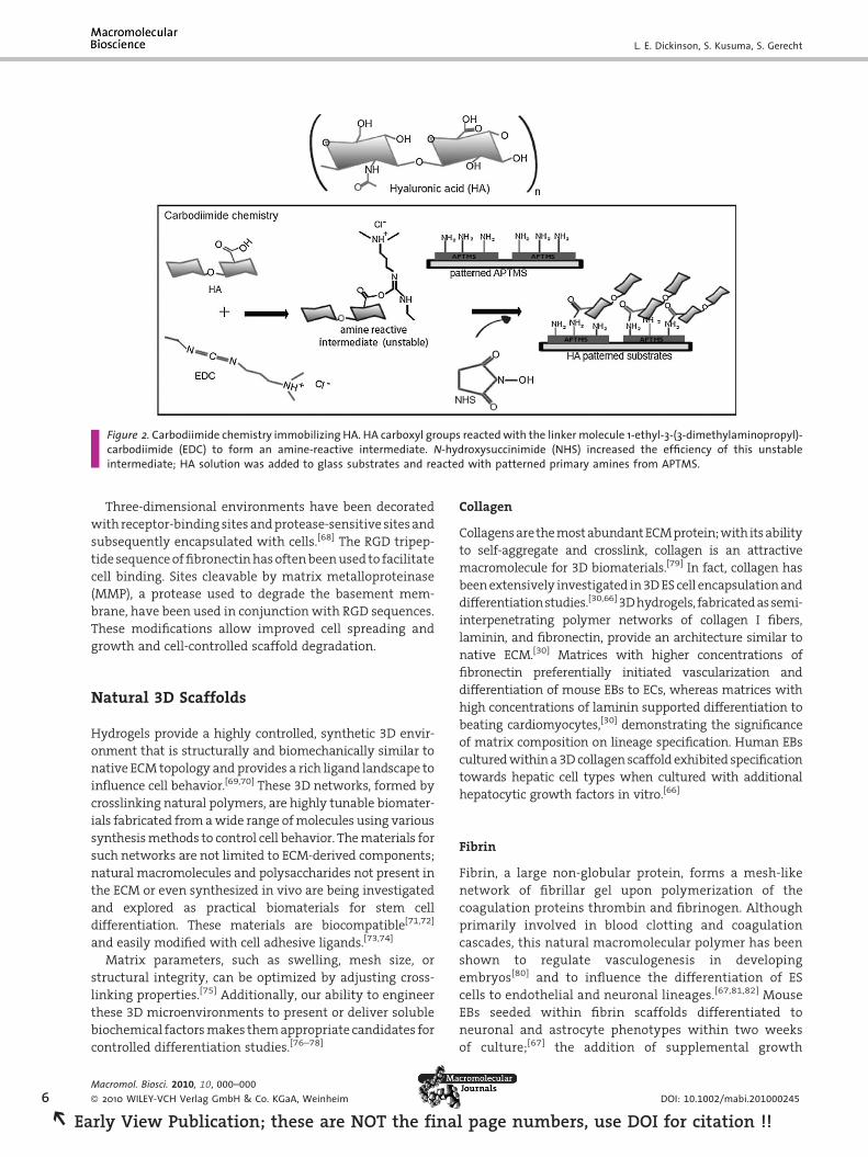
Figure 2. Carbodiimide chemistry immobilizing HA. HA carboxyl groups reacted with the linker molecule 1-ethyl-3-(3-dimethylaminopropyl)-carbodiimide (EDC) to form an amine-reactive intermediate. N-hydroxysuccinimide (NHS) increased the efficiency of this unstableintermediate; HA solution was added to glass substrates and reacted with patterned primary amines from APTMS.
6
REa
L. E. Dickinson, S. Kusuma, S. Gerecht
Three-dimensional environments have been decorated
with receptor-binding sites and protease-sensitive sites and
subsequently encapsulated with cells.[68] The RGD tripep-
tide sequence of fibronectin has often been used to facilitate
cell binding. Sites cleavable by matrix metalloproteinase
(MMP), a protease used to degrade the basement mem-
brane, have been used in conjunction with RGD sequences.
These modifications allow improved cell spreading and
growth and cell-controlled scaffold degradation.
Natural 3D Scaffolds
Hydrogels provide a highly controlled, synthetic 3D envir-
onment that is structurally and biomechanically similar to
native ECM topology and provides a rich ligand landscape to
influence cell behavior.[69,70] These 3D networks, formed by
crosslinking natural polymers, are highly tunable biomater-
ials fabricated from a wide range of molecules using various
synthesis methods to control cell behavior. The materials for
such networks are not limited to ECM-derived components;
natural macromolecules and polysaccharides not present in
the ECM or even synthesized in vivo are being investigated
and explored as practical biomaterials for stem cell
differentiation. These materials are biocompatible[71,72]
and easily modified with cell adhesive ligands.[73,74]
Matrix parameters, such as swelling, mesh size, or
structural integrity, can be optimized by adjusting cross-
linking properties.[75] Additionally, our ability to engineer
these 3D microenvironments to present or deliver soluble
biochemical factors makes them appropriate candidates for
controlled differentiation studies.[76–78]
Macromol. Biosci. 2010, 10, 000–000
� 2010 WILEY-VCH Verlag GmbH & Co. KGaA, Weinheim
rly View Publication; these are NOT the fina
Collagen
Collagens arethe most abundant ECM protein; with its ability
to self-aggregate and crosslink, collagen is an attractive
macromolecule for 3D biomaterials.[79] In fact, collagen has
been extensively investigated in 3D ES cell encapsulation and
differentiation studies.[30,66] 3D hydrogels, fabricated as semi-
interpenetrating polymer networks of collagen I fibers,
laminin, and fibronectin, provide an architecture similar to
native ECM.[30] Matrices with higher concentrations of
fibronectin preferentially initiated vascularization and
differentiation of mouse EBs to ECs, whereas matrices with
high concentrations of laminin supported differentiation to
beating cardiomyocytes,[30] demonstrating the significance
of matrix composition on lineage specification. Human EBs
cultured within a 3D collagen scaffold exhibited specification
towards hepatic cell types when cultured with additional
hepatocytic growth factors in vitro.[66]
Fibrin
Fibrin, a large non-globular protein, forms a mesh-like
network of fibrillar gel upon polymerization of the
coagulation proteins thrombin and fibrinogen. Although
primarily involved in blood clotting and coagulation
cascades, this natural macromolecular polymer has been
shown to regulate vasculogenesis in developing
embryos[80] and to influence the differentiation of ES
cells to endothelial and neuronal lineages.[67,81,82] Mouse
EBs seeded within fibrin scaffolds differentiated to
neuronal and astrocyte phenotypes within two weeks
of culture;[67] the addition of supplemental growth
DOI: 10.1002/mabi.201000245
l page numbers, use DOI for citation !!
Reconstructing the Embryonic Stem Cells’ Differentiation Niche . . .
factors enhanced their differentiation into neurons
and oligodendrytes.[81] We have recently shown that
patterned fibronectin surfaces, optimized in terms of
width and substrate concentration and inverted onto
a fibrin gel to create a 3D environment, guide the
elongation and maturation of endothelial progenitor cells.
The 3D environment also promoted tube formation, as
indicated by the presence of vacuoles and Weibel-Palade
bodies.[82b]
HA
Previously, we examined HA hydrogels capable of maintain-
ing the pluripotency of human ES cells; upon modification,
these hydrogels could induce differentiation.[83] Using
photopolymerization, methacrylated HA was covalently
crosslinked, and human ES cells were encapsulated within
the hydrogel. Human ES cells encapsulated in HA hydrogels
exhibited a preserved self-renewal state, full differential
potential, and normal genetic integrity. Adding the proan-
giogenic growth factor VEGF to differentiation medium
initiated human ES cell differentiation to vascular lineages
within the HA hydrogel. Cell sprouting and elongation were
observed within 48 h, and the presence of endothelial and
smooth muscle markers confirmed their vascular cell fate.[83]
By manipulating fabrication methods, Khademhosseini et al.
fabricated micropatterned HA hydrogels for encapsulation
that supported cell viability and controlled ES cell coloniza-
tion.[84] Photocrosslinked HA hydrogels with spatially
controlled matrix properties allowed the spreading and
proliferation of MSCs,[85] and when enhanced with TGF-b3,
permitted chondrogenesis of adult MSCs.[86] When HA
hydrogels were crosslinked with MMP-cleavable peptides
and coupled with RGD adhesive proteins, encapsulated
MSCs demonstrated spatially controlled cell spreading.[87]
Cell-secreted MMPs degraded ECM proteins, and crosslinking
hydrogels with MMP-cleavable crosslinks allowed cell-
dictated hydrogel degradation and remodeling, enabling
further spreading, interaction, and invasion in the
biomaterial.[88]
Dextran
Dextran is a branched polysaccharide with a molecular
structure similar to that of the ECM macromolecule, HA.
However, dextran is not distributed in the mammalian ECM.
Its chemical structure is easily modified for cell adhesive-
ness.[72,89,90] Pendant functional groups, such as –OH, make
dextran amenable to chemical modifications for greater
flexibility in the formulation of dextran-based hydrogels.
Modifying dextran-based hydrogels with cell adhesive
ligands, such as laminin- or fibronectin-derived RGD
peptides, increases their potential for use in tissue engineer-
Macromol. Biosci. 2010, 10, 000–000
� 2010 WILEY-VCH Verlag GmbH & Co. KGaA, Weinheim
Early View Publication; these are NOT the
ing applications by enhancing cell adhesion and survival.[89]
Ferreira et al. designed a photopolymerized bioactive
dextran-based hydrogel to enhance human ES cell differ-
entiation. These dextran hydrogels, modified with such
regulatory factors as the insoluble cell-adhesive peptide
sequence RGD and the soluble angiogenic growth factor
VEGF, initiated EB formation and vascular differentiation.
Encapsulated EBs formed well-organized vascular networks,
revealed upregulated vascular markers and, upon release
from the hydrogel, proliferated towards the vascular line-
age.[74] The microencapsulation of growth factors within the
dextran hydrogel allowed distinct spatial, temporal, and
biochemical signaling control over encapsulated cells.
Alginate
Alginate is another example of a naturally derived polymer
well-suited for biomaterial scaffolds and the in vitro
differentiation of ES cells. A hydrophilic polysaccharide
isolated from seaweed,[91] alginate resembles native ECM in
macromolecular structure and supports cell adhesion.[73,92]
Biocompatibility properties have made alginate an appro-
priate biomaterial to investigate cell encapsulation[73,92] and
the lineage differentiation of ES cells. Alginate hydrogels for
cell encapsulation are formed when divalent cations, such as
strontium and calcium, interact and ionically crosslink
polymer chains.[91] Alginate-based hydrogels have been
shown to support ES cell proliferation and viability,[93] as
well as osteogenic, hepatic, and chondrogenic differentia-
tion.[94–97] Much emphasis is placed on the influence of
biochemical cues on stem cell differentiation; however,
physical constraints, such as those imposed by pore diameter
size, are also influential in lineage commitment. Encapsula-
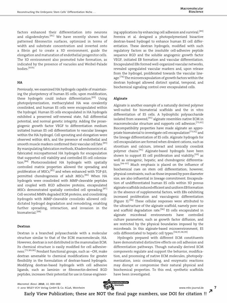
tion of undifferentiated human ES cells within 3D porous
alginate scaffolds induced efficient and uniformEB formation
in the absence of supplemental factors, with EBs exhibiting
increased proliferation and vasculogenic differentiation
(Figure 3).[96] These cellular responses were attributed to
the ultrastructure of the alginate scaffold, namely pore size
and scaffold degradation rate.[96] ES cells encapsulated in
alginate microbead environments have controlled
culture parameters, such as growth factor diffusion, and
are restricted by the physical boundaries imposed by the
microbeads. In this alginate-based microenvironment, ES
cells differentiated to hepatic cell types.[94,95,98,99]
Hydrogels prepared with different ECM constituents
have demonstrated distinctive effects on cell adhesion and
differentiation pathways. Though naturally derived ECM
components regulate and support the behavior, modifica-
tion, and processing of native ECM molecules, photopoly-
merization, ionic crosslinking, and enzymatic reactions
may disrupt or compromise their natural physical and
biochemical properties. To this end, synthetic scaffolds
have been investigated.
www.mbs-journal.de 7
final page numbers, use DOI for citation !! R
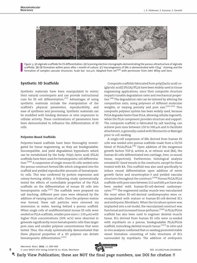
Figure 3. 3D alginate scaffolds for ES differentiation. (A) Scanning electron micrographs demonstrating the porous ultrastructure of alginatescaffolds. (B) EB formation within pores after 1 month of culture. (C) Vasculogenesis of EBs is demonstrated with CD34þ staining and theformation of complex vascular structures. Scale bar: 100 mm. Adapted from ref.[96] with permission from John Wiley and Sons.
8
REa
L. E. Dickinson, S. Kusuma, S. Gerecht
Synthetic 3D Scaffolds
Synthetic materials have been manipulated to mimic
their natural counterparts and can provide instructional
cues for ES cell differentiation.[77] Advantages of using
synthetic materials include the manipulation of the
scaffold’s physical parameters, reproducibility, and
ease of synthesis and processing. Synthetic materials can
be modified with binding domains or sites responsive to
cellular activity. These combinations of parameters have
been demonstrated to influence the differentiation of ES
cells.
Polyester-Based Scaffolds
Polyester-based scaffolds have been thoroughly investi-
gated for tissue engineering, as they are biodegradable,
biocompatible, and yield degradation byproducts which
can be metabolized by the body. Poly(L-lactic acid) (PLLA)
scaffolds have been used for hematopoietic cell differentia-
tion.[100] A suspension of single mouse ES cells seeded onto
the porous construct formed EBs which integrated into the
scaffold and yielded reproducible amounts of hematopoie-
tic cells. This was confirmed by protein expression and
colony-forming ability. A following study systematically
tested the effects of controllable properties of the PLLA
scaffolds on the differentiation of mouse ES cells into
hematopoietic cells.[101] The scaffolds were prepared via
salt leaching; different pore sizes were obtained by the
addition of varying sizes of salts. Once the polymer matrix
was formed, these salt particles were removed via
immersion in water, leaving behind a porous scaffold.
When single cells of undifferentiated mouse ES cells were
seeded on PLLA scaffolds, smaller pore sizes (<150 mm) with
higher PLLA concentrations (20% w/v) were observed to
generate significantly more hematopoietic cells than larger
pore sizes and smaller polymer concentrations that were
tested. Thus, this study systematically demonstrated that
these physical properties of a 3D polymer can dictate
hematopoietic differentiation.[101]
Macromol. Biosci. 2010, 10, 000–000
� 2010 WILEY-VCH Verlag GmbH & Co. KGaA, Weinheim
rly View Publication; these are NOT the fina
Composite scaffolds fabricated from poly[(lactic acid)-co-
(glycolic acid)] (PLGA)/PLLA have been widely used in tissue
engineering applications, since their composite structure
imparts tunable degradation rates and mechanical proper-
ties.[39] The degradation rate can be tailored by altering the
composition ratio, using polymers of different molecular
weights, or varying porosity and pore size.[102,103] This
composite polymer system has been widely used, because
PLGA degrades faster than PLLA, allowing cellular ingrowth,
while the PLLA component provides structure and support.
The composite scaffold is fabricated by salt leaching, can
achieve pore sizes between 150 to 500 mm and to facilitate
attachment, is generally coated with fibronectin or Matrigel
prior to cell seeding.
A single-cell suspension of EBs derived from human ES
cells was seeded onto porous scaffolds made from a 50/50
blend of PLGA/PLLA.[39] Upon addition of the exogenous
growth factors TGF-b, activin A, or retinoic acid (RA), the
human ES cells differentiated into cartilage, liver, or neural
tissue, respectively. Furthermore, histological analysis
revealed EC-lined vessels in the constructs, except for those
treated with RA. This scaffold was also used specifically to
induce neural differentiation upon addition of nerve
growth factor and neurotrophin-3 and yielded vascular
structures throughout the construct.[104] Porous PLGA/PLLA
scaffolds with pore sizes between 212 and 600 mm have also
been seeded with human-ES-cell-derived cardiomyo-
cytes.[105] The engineered cardiac muscle was vascularized
the most when ES-cell-derived cardiomyocytes were co-
encapsulated with mature or human-ES-cell-derived ECs
and embryonic fibroblasts. When the triculture system was
implanted into a rat model, the vascularized construct was
functional and increased tissue perfusion.[106] A PLLA/PLGA
scaffold has also been used to engineer skeletal muscle
tissue. ECs derived from human ES cells were co-seeded
with myoblasts on a porous, biodegradable PLLA/PLGA
scaffold, mimicking skeletal muscle tissue.[107] In vitro and
in vivo analyses confirmed that co-seeding promoted stable
vessel formation consisting of tube structures of ECs
surrounded by myoblasts. The addition of embryonic
DOI: 10.1002/mabi.201000245
l page numbers, use DOI for citation !!
Reconstructing the Embryonic Stem Cells’ Differentiation Niche . . .
fibroblasts increased vascularization dramatically, as
validated by increases in EC number, lumenal area, and
VEGF expression.
Another study demonstrated that porous poly(glycerol-
co-sebacate)-acrylate (PGSA) scaffolds support the growth
and differentiation of encapsulated human ES cells.[108]
In vivo experiments indicated that porous PGSA, and
not non-porous PGSA, promoted tissue ingrowth and
vascularization.
Hepatic differentiation has been studied on fibrous
scaffolds[109] and spheroid foams[110] made from polyesters.
Non-aligned, fibrous scaffolds have been created from a 50/
50 blend of PLLA and poly(glycolic acid) (PGA) and have been
employed in the hepatic differentiation of mouse ES
cells.[111] EB-derived cells were mixed with Matrigel, seeded
onto a scaffold, and allowed to differentiate for 20 d. The
cells showed aggregate growth into the pores of the
scaffold, as well as along the fibers of the scaffold, and
demonstrated differentiation into hepatocyte-like cells.[109]
Porous spheroid foams made from polyurethane have also
been seeded with mouse ES cells, which differentiated into
mature hepatocytes, as evidenced by marker expression
and cell morphology and functionality (ammonia removal)
in the presence of specific growth factors.[110]
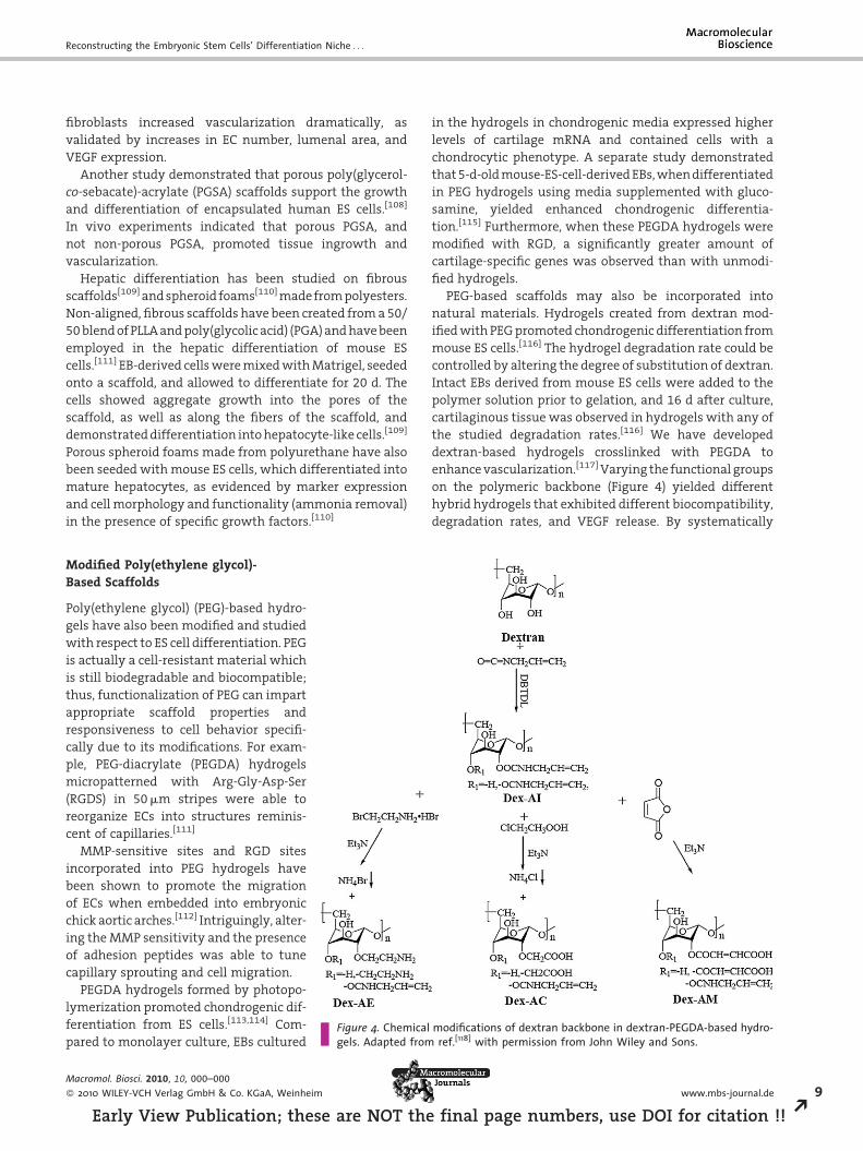
Figure 4. Chemical modifications of dextran backbone in dextran-PEGDA-based hydro-gels. Adapted from ref.[118] with permission from John Wiley and Sons.
Modified Poly(ethylene glycol)-Based Scaffolds
Poly(ethylene glycol) (PEG)-based hydro-
gels have also been modified and studied
with respect to ES cell differentiation. PEG
is actually a cell-resistant material which
is still biodegradable and biocompatible;
thus, functionalization of PEG can impart
appropriate scaffold properties and
responsiveness to cell behavior specifi-
cally due to its modifications. For exam-
ple, PEG-diacrylate (PEGDA) hydrogels
micropatterned with Arg-Gly-Asp-Ser
(RGDS) in 50 mm stripes were able to
reorganize ECs into structures reminis-
cent of capillaries.[111]
MMP-sensitive sites and RGD sites
incorporated into PEG hydrogels have
been shown to promote the migration
of ECs when embedded into embryonic
chick aortic arches.[112] Intriguingly, alter-
ing the MMP sensitivity and the presence
of adhesion peptides was able to tune
capillary sprouting and cell migration.
PEGDA hydrogels formed by photopo-
lymerization promoted chondrogenic dif-
ferentiation from ES cells.[113,114] Com-
pared to monolayer culture, EBs cultured
Macromol. Biosci. 2010, 10, 000–000
� 2010 WILEY-VCH Verlag GmbH & Co. KGaA, Weinheim
Early View Publication; these are NOT the
in the hydrogels in chondrogenic media expressed higher
levels of cartilage mRNA and contained cells with a
chondrocytic phenotype. A separate study demonstrated
that 5-d-old mouse-ES-cell-derived EBs, when differentiated
in PEG hydrogels using media supplemented with gluco-
samine, yielded enhanced chondrogenic differentia-
tion.[115] Furthermore, when these PEGDA hydrogels were
modified with RGD, a significantly greater amount of
cartilage-specific genes was observed than with unmodi-
fied hydrogels.
PEG-based scaffolds may also be incorporated into
natural materials. Hydrogels created from dextran mod-
ified with PEG promoted chondrogenic differentiation from
mouse ES cells.[116] The hydrogel degradation rate could be
controlled by altering the degree of substitution of dextran.
Intact EBs derived from mouse ES cells were added to the
polymer solution prior to gelation, and 16 d after culture,
cartilaginous tissue was observed in hydrogels with any of
the studied degradation rates.[116] We have developed
dextran-based hydrogels crosslinked with PEGDA to
enhance vascularization.[117] Varying the functional groups
on the polymeric backbone (Figure 4) yielded different
hybrid hydrogels that exhibited different biocompatibility,
degradation rates, and VEGF release. By systematically
www.mbs-journal.de 9
final page numbers, use DOI for citation !! R
10
REa
L. E. Dickinson, S. Kusuma, S. Gerecht
examining various functional groups and their concentra-
tions, we determined that incorporating amine groups
favorably affected mechanical properties and in vivo
vascularization.
Physical Constraints
Other methods to control ES cell differentiation using
engineered biomaterials have focused on physical con-
straints, such as nanotopographical cues, EB size, and
mechanical stiffness.
Nanotopographical Cues
The ECM presents an abundance of macromolecules with
feature sizes at the nanometer scale. For example, collagens,
the most abundant ECM macromolecule, exhibit dimen-
sions among the nanoscale regime, with single collagen
monomers approximately 300 nm in width and 1.5 nm in
diameter[118,119] and self-assembled fibrillar structures
extending several micrometers in length and hundreds of
nanometers in diameter.[120] Cells interact with these
nanometer-sized molecular structures[121,122] via contact
guidance, the widely known principle that cellular behavior
and function is influenced by physical topography.[123,124]
This is thought to occur through the reorganization of
membrane-bound integrins, initiating the alteration
of the cytoskeleton and modulating the formation
of associated focal adhesion proteins that ultimately
activate specific signaling cascades, thereby inducing such
cellular events as adhesion, proliferation, migration, and
differentiation.[125–129]
It is well known that cells receive instructive cues from
their topographical environment;[123,130] with the progress
of micro- and nanotechnology, many studies have inves-
tigated specific cellular behavior in response to engineered
micro- and nanoscale substrates[123,131–133] and have
demonstrated that topographical geometry and dimension
clearly correlate with cellular response. 2D patterned cues
have been fabricated in the nanoscale regime, and have
demonstrated a profound effect on cellular response,
including adhesion,[134,135] motility,[136] and stem cell
function.[137] Although there have been recent advances
in nanopatterning techniques and complexities,[138,139]
these designs have yet to be utilized to study stem cell
differentiation. Culturing human ES cells on fibronectin-
coated PDMS substrates with nanogratings enhanced such
morphological changes to ES cells as elongation and
alignment, demonstrating distinct cytoskeletal reorganiza-
tion via contact guidance while also reducing ES cell
proliferation.[127]
Nanofibrous 2D films made of PLLA demonstrated
enhanced osteogenic differentiation of mouse and human
Macromol. Biosci. 2010, 10, 000–000
� 2010 WILEY-VCH Verlag GmbH & Co. KGaA, Weinheim
rly View Publication; these are NOT the fina
ES cells in the presence of pro-osteogenic growth factors, as
compared to flat substrates.[140,141] In the absence of growth
factors, osteogenic differentiation was still observed on
nanofiber films, although to a lesser extent than when
supplemented with growth factors. ES cell lineage differ-
entiation is strongly affected by the cells’ physical
interactions with their environmental topography, and
coupling these biophysical cues with supplemental signal-
ing molecules, such as growth factors or adsorbed proteins,
may have a more pronounced effect on ES cell fate decisions
and produce more homogeneous populations of differen-
tiated cell types. Human ES cells cultured on gelatin-coated
polyurethane nanogratings differentiated to neuronal
lineages after only 5 d in a culture lacking additional
differentiation-inducing agents.[142] Clearly, the presence
(or absence) of different soluble and immobilized biochem-
ical factors, cooperatively with nanotopographical features,
guide lineage specification. These studies demonstrated
that the determination of stem cell lineage is affected by the
dimension, geometry, and composition of nanosized
features.[143]
Electrospinning is another technique that achieves the
complex nanoscale features that essentially mimic native
ECM topography. This technology produces fibrous net-
works from either synthetic or natural polymers. An
electrical field is used to extrude a charged polymer solution
from its source to a grounded collection material, and in
doing so, deposits nanosized fibers.[144] Manipulation of
operating parameters allows distinct control over specific
properties, such as nanofiber diameter and alignment.
These environments recreate extracellular topographies
and structures in vitro, maintain the ability to present
functionalized bioactive molecules, and offer more sites for
cell adhesions and interactions due to a high surface-area-
to-volume ratio.[145,146] Because of these properties, elec-
trospun materials have already been investigated to
determine their effects on ES cell differentiation.
Poly(e-caprolactone) (PCL), a biodegradable polyester
with a slow degradation rate and the ability to maintain
its architectural integrity,[143,147] or PLLA are commonly
utilized polymers for electrospun matrices.[51,143] Uniaxi-
ally aligned PCL nanofibrous scaffolds support the neuronal
differentiation of mouse ES cells with directed neurite
outgrowth, while randomly organized nanofibrous scaf-
folds support mouse ES cell adipogenesis.[145,148] Cells are
capable of sensing their surrounding environment. An
oriented template, such as aligned nanofibers, instructs
cell differentiation through contact guidance. However,
culturing multipotent adult MSCs on nanofibrous PCL
constructs yields either chondrogenic or osteogenic
lineages, demonstrating cell-dependent sensitivity to the
nanoscale scaffolds.[51,143]
Other methods to create a topographically com-
plex environment include cell electrospinning,[149,150]
DOI: 10.1002/mabi.201000245
l page numbers, use DOI for citation !!
Reconstructing the Embryonic Stem Cells’ Differentiation Niche . . .
bio-electrosprays,[151,152] and biojetting.[153,154] These tech-
niques can be used to create oriented scaffolds in a
controlled manner, allow cell seeding to occur simulta-
neously, and thus hold great potential for future studies in
ES cell differentiation.
EB Size
In vitro differentiation of ES cells occurs through the
spontaneous formation of EBs.[155] Differentiation based on
EB formation mimics in vivo embryonic development and is
being used as a template to investigate the effects of spatial
organization on the differentiation to specific cell types. One
critical factor of this differentiation strategy is EB size, which
has been demonstrated to play a role in specific lineage
commitment.[156] A known number of human ES cells
aggregated by centrifugation form uniformly sized EBs and
exhibit reproducible hematopoietic differentiation. Efficient
blood formation required initial EBs formed by morethan 500
human ES cells, with optimal erythropoiesis occurring with
initial aggregations of 1 000 hES cells.[156] Control over the
formation of EBs can lead not only to a more homogeneous
cell population, but also to efficient differentiation.[75]
Microfabrication technologies are now being harnessed
to focus specifically on spatially controlling EB size to
determine its correlation with cell fate.[157,158] Methods
such as photolithography are utilized to fabricate polymeric
microwells that restrict the spatial microenvironments of
ES cells and control the uniformity of EB size.[157–163]
Microwells for this purpose are generally fabricated from
PDMS, a biocompatible elastomeric polymer amenable to
surface functionalization. In fact, different surface functio-
nalizations prove to have varying effects on EB develop-
ment. PEG microwells coated with fibronectin and
subsequently seeded with MEFs, the feeder layer on which
human ES cells maintain their self-renewal properties,
preserved their viability and undifferentiated state,[162]
whereas coating with a cell-repellant and inert material,
such as photocrosslinked PEG, actually initiated the
formation of homogeneously sized EBs.[159] Coating the
bottom of PDMS microwells with a manufactured ECM,
such as Matrigel, and then surrounding the walls with a cell
resistive molecule, provided an ES cell culture microenvir-
onment which was also conducive to uniform EB formation
and possibly regulated differentiation to cardiomyo-
cytes.[160] With the ability to form homogeneous EBs in
vitro, it was recently determined that larger aggregates
enhanced cardiogenesis;[164] also, smaller EBs from more
spatially restrictive microwells were found more likely to
differentiate to endothelial lineages.[163] With such a size-
dependent response of EBs to lineage differentiation,
microwell substrates provide an easy and controllable
method of forming uniform EBs for high-throughput
applications and ES cell differentiation studies.
Macromol. Biosci. 2010, 10, 000–000
� 2010 WILEY-VCH Verlag GmbH & Co. KGaA, Weinheim
Early View Publication; these are NOT the
Stiffness
The mechanical properties of a scaffold regulate cell fate.
Synthetic biomaterials are capable of being tailored with
controllable mechanical properties, thus impacting differ-
entiation. Synthetic biomaterials are advantageous for
cardiac tissue repair, as their mechanical properties can be
tailored to withstand repetitive stress and the activity of the
infarct without obstructing electrical conduction.[165] A
PEG-ylated fibrinogen hydrogel is among the materials
developed for cardiac tissue engineering.[166] Cardiomyo-
cytes derived from human ES cells were encapsulated in a
photopolymerizable PEG-ylated fibrinogen hydrogel,
which induced cell maturation and functionality in two
weeks. The hydrogel was fabricated to achieve precise
control of its mechanical properties and degradation
and was designed to photopolymerize in situ, which would
allow the even dispersal of cells and scaffold when used
in vivo.
Another study examined the mechanical stiffness of
microporous polyurethane tubes on the differentiation of
ES cells into ECs.[167] The material properties were tailored
to achieve compliance similar to that of a human artery.
Under static conditions, the seeded cells expressed smooth
muscle actin (SMA) after 4 d. However, when incubated in
the presence of a pulsatile flow, the cells oriented in the
direction of the flow and expressed endothelial markers.
Additionally, cells of the deeper layers of the tube were
SMAþ, indicating that these conditions were permissive for
segregated differentiation into vascular lineage cell types.
Future Perspectives
As clearly demonstrated, not just a single parameter of the
surrounding milieu impacts and guides ES cell differentia-
tion; rather, differentiation results from the coordinated
actions of various critical parameters, such as scaffold
composition, ligand presentation, ultrastructural topogra-
phy, physical properties, and chemical cues. Indeed, both 2D
and 3D environments with micro- and/or nanoscale
dimensions, along with immobilized or exogenous factors,
profoundly affect the ES cell fate decision, all of which have
been developed in an effort to reconstruct the native
environment as closely as possible. Future studies to
elucidate the effects on the ECM of a wide range of
molecules could incorporate developed combinatorial
arrays for the high-throughput screening of cell fate.[168]
Emerging technologies in micro-/nanofabrication, drug
release, and materials science are enabling the develop-
ment of instructive environments. Recent achievements
emphasize that the fabrication of micropatterned and
gradient-based ECM hydrogels with innate nanoscale
topography can spatially control cell attachment and
www.mbs-journal.de 11
final page numbers, use DOI for citation !! R
12
REa
L. E. Dickinson, S. Kusuma, S. Gerecht
orientation and still maintain the ability to be functiona-
lized with exogenous adhesion molecules.[85,169–171] With
the advent of tunable hydrogels, researchers are gradually
succeeding in synthetically recreating the native micro-
environment.
One important factor to consider is that tissue develop-
ment occurs in a transient environment with bidirectional
signaling between cells and their ECM.[172] However, many
of the scaffolds developed so far may not be conducive to
incorporating this dynamic behavior to truly mimic the
extracellular environment.[173] Cell behavior is constantly
governed by instructive biochemical and biophysical cues
from the ECM; likewise, cells are continuously remodeling
the ECM. Furthermore, the ECM is variable across different
cell types,[174] and synthetic environments should be
individually constructed with distinct cell lineages in mind.
Acknowledgements: We would like to acknowledge funding fromthe AHA Scientist Development Grant and a March of Dimes-O’Conner Starter Scholar award (for S. G.). L. E. D. is an IGERT traineeand a National Science Foundation Graduate Fellow.
Received: June 16, 2010; Revised: July 30, 2010; Published online:DOI: 10.1002/mabi.201000245
Keywords: biocompatibility; biological application of polymers;biomaterials; nanotechnology
[1] J. A. Thomson, J. Itskovitz-Eldor, S. S. Shapiro, M. A. Waknitz,J. J. Swiergiel, V. S. Marshall, J. M. Jones, Science 1998, 282,1145.
[2] R. Timpl, Curr. Opin. Cell Biol. 1996, 8, 618.[3] T. Rozario, D. W. DeSimone, Dev. Biol. 2010, 341, 126.[4] M. K. Gordon, R. A. Hahn, Cell Tissue Res. 2010, 339, 247.[5] S. I. Nishikawa, S. Nishikawa, M. Hirashima, N. Matsuyoshi,
H. Kodama, Development 1998, 125, 1747.[6] K. Schenke-Layland, K. E. Rhodes, E. Angelis, Y. Butylkova, S.
Heydarkhan-Hagvall, C. Gekas, R. Zhang, J. I. Goldhaber, H. K.Mikkola, K. Plath, W. R. Maclellan, Stem Cells 2008, 26, 1537.
[7] J. Yamashita, H. Itoh, M. Hirashima, M. Ogawa, S. Nishikawa,T. Yurugi, M. Naito, K. Nakao, S.-I. Nishikawa, Nature 2000,408, 92.
[8] S. Gerecht-Nir, A. Ziskind, S. Cohen, J. Itskovitz-Eldor, Lab.Investig. 2003, 83, 1811.
[9] E. Vo, D. Hanjaya-Putra, Y. Zha, S. Kusuma, S. Gerecht, StemCell Rev. Rep. 2010, in press.
[10] C. K. Chiang, M. F. Chowdhury, R. K. Iyer, W. L. Stanford, M.Radisic, Acta Biomater. 2010, in press.
[11] K. Schenke-Layland, E. Angelis, K. E. Rhodes, S. Heydarkhan-Hagvall, H. K. Mikkola, W. R. MacLellan, Stem Cells 2007,25, 1529.
[12] H. Sato, M. Takahashi, H. Ise, A. Yamada, S-i. Hirose, Y-i.Tagawa, H. Morimoto, A. Izawa, U. Ikeda, Biochem. Biophys.Res. Commun. 2006, 342, 107.
[13] X.-M. Guo, Y.-S. Zhao, H.-X. Chang, C.-Y. Wang, L.-L. E. X.-A.Zhang, C.-M. Duan, L.-Z. Dong, H. Jiang, J. Li, Y. Song, X. Yang,Circulation 2006, 113, 2229.
[14] M. C. Whelan, D. R. Senger, J. Biol. Chem. 2003, 278, 327.
Macromol. Biosci. 2010, 10, 000–000
� 2010 WILEY-VCH Verlag GmbH & Co. KGaA, Weinheim
rly View Publication; these are NOT the fina
[15] W. Ma, T. Tavakoli, E. Derby, Y. Serebryakova, M. S. Rao, M. P.Mattson, BMC Dev. Biol. 2008, 8.
[16] J. C. Y. Wong, S. Y. Gao, J. G. Lees, M. B. Best, R. Wang, B. E.Tuch, Cell Adhes. Migration 2010, 4, 39.
[17] L. Labriola, W. R. Montor, K. Krogh, F. H. Lojudice, T. Genzini,A. C. Goldberg, F. G. Eliaschewitz, M. C. Sogayar, Mol. Cell.Endocrinol. 2007, 263, 120.
[18] G. Parnaud, E. Hammar, D. G. Rouiller, M. Armanet, P. A.Halban, D. Bosco, Diabetes 2006, 55, 1413.
[19] F. X. Jiang, D. S. Cram, H. J. DeAizpurua, L. C. Harrison,Diabetes 1999, 48, 722.
[20] I. S. Schroeder, A. Rolletschek, P. Blyszczuk, G. Kania, A. M.Wobus, Nat. Protoc. 2006, 1, 495.
[21] H. Fujiwara, Y. Hayashi, N. Sanzen, R. Kobayashi, C. N.Weber, T. Emoto, S. Futaki, H. Niwa, P. Murray, D. Edgar,K. Sekiguchi, J. Biol. Chem. 2007, 282, 29701.
[22] D. Malan, M. Reppel, R. Dobrowolski, W. Roell, N. Smyth, J.Hescheler, M. Paulsson, W. Bloch, B. K. Fleischmann, StemCells 2009, 27, 88.
[23] E. L. George, E. N. Georges-Labouesse, R. S. Patel-King,H. Rayburn, R. O. Hynes, Development 1993, 119, 1079.
[24] E. Ruoslahti, M. D. Pierschbacher, Science 1987, 238, 491.[25] G. Maheshwari, G. Brown, D. A. Lauffenburger, A. Wells,
L. G. Griffith, J. Cell Sci. 2000, 113, 1677.[26] S. I. Aota, M. Nomizu, K. M. Yamada, J. Biol. Chem. 1994, 269,
24756.[27] J. L. Myles, B. T. Burgess, R. B. Dickinson, J. Biomater. Sci.,
Polym. Ed. 2000, 11, 69.[28] J. A. Burdick, K. S. Anseth, Biomaterials 2002, 23, 4315.[29] E. S. Wijelath, S. Rahman, J. Murray, Y. Patel, G. Savidge,
M. Sobel, J. Vasc. Surg. 2004, 39, 655.[30] S. Battista, D. Guarnieri, C. Borselli, S. Zeppetelli, A.
Borzacchiello, L. Mayol, D. Gerbasio, D. R. Keene, L. Ambrosio,P. A. Netti, Biomaterials 2005, 26, 6194.
[31] M. M. Martino, M. Mochizuki, D. A. Rothenfluh, S. A. Rempel,J. A. Hubbell, T. H. Barker, Biomaterials 2009, 30, 1089.
[32] M. D. Singh, M. Kreiner, C. S. McKimmie, S. Holt, C. F. van derWalle, G. J. Graham, Biochem. Biophys. Res. Commun. 2009,390, 716.
[33] Y. Hayashi, M. K. Furue, T. Okamoto, K. Ohnuma, Y. Myoishi,Y. Fukuhara, T. Abe, J. D. Sato, R.-I. Hata, M. Asashima, StemCells 2007, 25, 3005.
[34] K.-D. Choi, J. Yu, K. Smuga-Otto, G. Salvagiotto, W. Rehrauer,M. Vodyanik, J. Thomson, I. Slukvin, Stem Cells 2009, 27, 559.
[35] S. Vukicevic, H. K. Kleinman, F. P. Luyten, A. B. Roberts, N. S.Roche, A. H. Reddi, Exp. Cell Res. 1992, 202, 1.
[36] H. K. Kleinman, M. L. McGarvey, L. A. Liotta, P. G. Robey,K. Tryggvason, G. R. Martin, Biochemistry 1982, 21, 6188.
[37] M. Ruhnke, H. Ungefroren, G. Zehle, M. Bader, B. Kremer,F. Fandrich, Stem Cells 2003, 21, 428.
[38] C. Xu, M. S. Inokuma, J. Denham, K. Golds, P. Kundu,J. D. Gold, M. K. Carpenter, Nat. Biotechnol. 2001, 19, 971.
[39] S. Levenberg, N. F. Huang, E. Lavik, A. B. Rogers, J. Itskovitz-Eldor, R. Langer, Proc. Natl. Acad. Sci. USA 2003, 100, 12741.
[40] S. S. Chen, W. Fitzgerald, J. Zimmerberg, H. K. Kleinman,L. Margolis, Stem Cells 2007, 25, 553.
[41] N. T. Kohen, L. E. Little, K. E. Healy, Biointerphases 2009, 4, 69.[42] T. Ishii, K. Fukumitsu, K. Yasuchika, K. Adachi, E. Kawase, H.
Suemori, N. Nakatsuji, I. Ikai, S. Uemoto, Am. J. Physiol. -Gastrointest. Liver Physiol. 2008, 295, G313.
[43] M. Uemura, M. M. Refaat, M. Shinoyama, H. Hayashi,N. Hashimoto, J. Takahashi, J. Neurosci. Res. 2010, 88, 542.
[44] T. Liu, S. Zhang, X. Chen, G. Li, Y. Wang, Tissue Eng. - Part A2010, 16, 1115.
DOI: 10.1002/mabi.201000245
l page numbers, use DOI for citation !!
Reconstructing the Embryonic Stem Cells’ Differentiation Niche . . .
[45] D. Philp, S. S. Chen, W. Fitzgerald, J. Orenstein, L. Margolis, H.K. Kleinman, Stem Cells 2005, 23, 288.
[46] L. Flynn, G. D. Prestwich, J. L. Semple, K. A. Woodhouse,Biomaterials 2007, 28, 3834.
[47] L. E. Flynn, Biomaterials 2010, 31, 4715.[48] N. Datta, H. L. Holtorf, V. I. Sikavitsas, J. A. Jansen, A. G.
Mikos, Biomaterials 2005, 26, 971.[49] K. Schenke-Layland, I. Riemann, F. Opitz, K. Konig, K. J.
Halbhuber, U. A. Stock, Matrix Biol. 2004, 23, 113.[50] H. C. Ott, T. S. Matthiesen, S. K. Goh, L. D. Black, S. M. Kren, T. I.
Netoff, D. A. Taylor, Nat. Med. 2008, 14, 213.[51] R. A. Thibault, L. Scott Baggett, A. G. Mikos, F. K. Kasper,
Tissue Eng. - Part A 2010, 16, 431.[52] N. D. Evans, E. Gentleman, X. Chen, C. J. Roberts, J. M. Polak,
M. M. Stevens, Biomaterials 2010, 31, 3244.[53] H. Baharvand, M. Azarnia, K. Parivar, S. K. Ashtiani, J. Mol.
Cell. Cardiol. 2005, 38, 495.[54] D. Sasaki, T. Shimizu, S. Masuda, J. Kobayashi, K. Itoga,
Y. Tsuda, J. K. Yamashita, M. Yamato, T. Okano, Biomaterials2009, 30, 4384.
[55] R. McBeath, D. M. Pirone, C. M. Nelson, K. Bhadriraju, C. S.Chen, Dev. Cell 2004, 6, 483.
[56] R. Peerani, B. M. Rao, C. Bauwens, T. Yin, G. A. Wood, A. Nagy,E. Kumacheva, P. W. Zandstra, EMBO J. 2007, 26, 4744.
[57] L. H. Lee, R. Peerani, M. Ungrin, C. Joshi, E. Kumacheva,P. Zandstra, Stem Cell Res. 2009, 2, 155.
[58] L. E. Dickinson, C. C. Ho, G. M. Wang, K. J. Stebe, S. Gerecht,Biomaterials 2010, 31, 5472.
[59] B. P. Toole, Sem. Cell Dev. Biol. 2001, 12, 79.[60] B. P. Toole, Nat. Rev. Cancer 2004, 4, 528.[61] A. P. Spicer, J. Y. L. Tien, Birth Defects Res., Part C - Embryo
Today: Rev. 2004, 72, 89.[62] A. Khademhosseini, K. Y. Suh, J. M. Yang, G. Eng, J. Yeh, S.
Levenberg, R. Langer, Biomaterials 2004, 25, 3583.[63] S. Takahashi, H. Yamazoe, F. Sassa, H. Suzuki, J. Fukuda,
J. Biosci. Bioeng. 2009, 108, 544.[64] J. Tang, R. Peng, J. Ding, Biomaterials 2010, 31, 2470.[65] A. P. Quist, S. Oscarsson, Exp. Opin. Drug Discovery 2010, 5,
569.[66] H. Baharvand, S. M. Hashemi, S. K. Ashtiani, A. Farrokhi, Int.
J. Dev. Biol. 2006, 50, 645.[67] S. M. Willerth, K. J. Arendas, D. I. Gottlieb, S. E. Sakiyama-
Elbert, Biomaterials 2006, 27, 5990.[68] M. P. Lutolf, G. P. Raeber, A. H. Zisch, N. Tirelli, J. A. Hubbell,
Adv. Mater. 2003, 15, 888.[69] K. Y. Lee, D. J. Mooney, Chem. Rev. 2001, 101, 1869.[70] S. Yang, K. F. Leong, Z. Du, C. K. Chua, Tissue Eng. 2001, 7, 679.[71] C. J. De Groot, M. J. A. Van Luyn, W. N. E. Van Dijk-Wolthuis, J.
A. Cadee, J. A. Plantinga, W. D. Otter, W. E. Hennink, Bio-materials 2001, 22, 1197.
[72] S. Moreira, R. M. Gil Da Costa, L. Guardao, F. Gartner, M.Vilanova, M. Gama, J. Bioact. Compat. Polym. 2010, 25, 141.
[73] J. A. Rowley, G. Madlambayan, D. J. Mooney, Biomaterials1999, 20, 45.
[74] L. S. Ferreira, S. Gerecht, J. Fuller, H. F. Shieh, G. Vunjak-Novakovic, R. Langer, Biomaterials 2007, 28, 2706.
[75] O. Z. Fisher, A. Khademhosseini, R. Langer, N. A. Peppas, Acc.Chem. Res. 2010, 43, 419.
[76] R. A. Marklein, J. A. Burdick, Adv. Mater. 2010, 22, 175.[77] M. P. Lutolf, J. A. Hubbell, Nat. Biotechnol. 2005, 23, 47.[78] B. V. Slaughter, S. S. Khurshid, O. Z. Fisher, A. Khademhos-
seini, N. A. Peppas, Adv. Mater. 2009, 21, 3307.[79] M. Van der Rest, R. Garrone, FASEB J. 1991, 5, 2814.
Macromol. Biosci. 2010, 10, 000–000
� 2010 WILEY-VCH Verlag GmbH & Co. KGaA, Weinheim
Early View Publication; these are NOT the
[80] S. E. Francis, K. L. Goh, K. Hodivala-Dilke, B. L. Bader, M. Stark,D. Davidson, R. O. Hynes, Arterioscler. Thromb. Vasc. Biol.2002, 22, 927.
[81] S. M. Willerth, T. E. Faxel, D. I. Gottlieb, S. E. Sakiyama-Elbert,Stem Cells 2007, 25, 2235.
[82] [82a] H. Liu, S. F. Collins, L. J. Suggs, Biomaterials 2006, 27,6004; [82b] L. E. Dickinson, M. E. Moura, S. Gerecht, SoftMatter 2010, 6, 5109.
[83] S. Gerecht, J. A. Burdick, L. S. Ferreira, S. A. Townsend,R. Langer, G. Vunjak-Novakovic, Proc. Natl. Acad. Sci. USA2007, 104, 11298.
[84] A. Khademhosseini, G. Eng, J. Yeh, J. Fukuda, J. Blumling Iii,R. Langer, J. A. Burdick, J. Biomed. Mater. Res. - Part A 2006,79, 522.
[85] R. A. Marklein, J. A. Burdick, Soft Matter 2009, 6, 136.[86] J. A. Burdick, C. Chung, Tissue Eng. - Part A 2009, 15, 243.[87] S. Khetan, J. S. Katz, J. A. Burdick, Soft Matter 2009, 5, 1601.[88] M. P. Lutolf, J. L. Lauer-Fields, H. G. Schmoekel, A. T. Metters,
F. E. Weber, G. B. Fields, J. A. Hubbell, Proc. Natl. Acad. Sci. USA2003, 100, 5413.
[89] S. P. Massia, J. Stark, J. Biomed. Mater. Res. 2001, 56, 390.[90] S. G. Levesque, M. S. Shoichet, Biomaterials 2006, 27, 5277.[91] O. Smidsrod, G. Skjak-Braek, Trends Biotechnol. 1990, 8, 71.[92] A. D. Augst, H. J. Kong, D. J. Mooney, Macromol. Biosci. 2006,
6, 623.[93] B. J. Willenberg, T. Hamazaki, F. W. Meng, N. Terada,
C. Batich, J. Biomed. Mater. Res. - Part A 2006, 79, 440.[94] T. Maguire, E. Novik, R. Schloss, M. Yarmush, Biotechnol.
Bioeng. 2006, 93, 581.[95] T. Maguire, A. E. Davidovich, E. J. Wallenstein, E. Novik,
N. Sharma, H. Pedersen, I. P. Androulakis, R. Schloss,M. Yarmush, Biotechnol. Bioeng. 2007, 98, 631.
[96] S. Gerecht-Nir, S. Cohen, A. Ziskind, J. Itskovitz-Eldor, Bio-technol. Bioeng. 2004, 88, 313.
[97] A. Steinert, M. Weber, A. Dimmler, C. Julius, N. Schutze,U. Noth, H. Cramer, J. Eulert, U. Zimmermann, C. Hendrich,J. Orthop. Res. 2003, 21, 1090.
[98] S. Fang, Y.-D. Qiu, L. Mao, X-l. Shi, D.-C. Yu, Y.-T. Ding, ActaPharmacol. Sin. 2007, 28, 1924.
[99] N. Wang, G. Adams, L. Buttery, F. H. Falcone, S. Stolnik,J. Biotechnol. 2009, 144, 304.
[100] H. Liu, K. Roy, Tissue Eng. 2005, 11, 319.[101] S. Taqvi, K. Roy, Biomaterials 2006, 27, 6024.[102] L. Wu, J. Ding, Biomaterials 2004, 25, 5821.[103] W. Linbo, D. Jiandong, J. Biomed. Mater. Res. Part A 2005,
75A, 767.[104] S. Levenberg, J. A. Burdick, T. Kraehenbuehl, R. Langer, Tissue
Eng. 2005, 11, 506.[105] O. Caspi, A. Lesman, Y. Basevitch, A. Gepstein, G. Arbel, I. H.
M. Habib, L. Gepstein, S. Levenberg, Circ. Res. 2007, 100, 263.[106] A. Lesman, M. Habib, O. Caspi, A. Gepstein, G. Arbel, S.
Levenberg, L. Gepstein, Tissue Eng. - Part A, 16, 115.[107] S. Levenberg, J. Rouwkema, M. Macdonald, E. S. Garfein, D. S.
Kohane, D. C. Darland, R. Marini, C. A. van Blitterswijk, R. C.Mulligan, P. A. D’Amore, R. Langer, Nat. Biotechnol. 2005, 23,879.
[108] S. Gerecht, S. A. Townsend, H. Pressler, H. Zhu, C. L. E. Nijst, J.P. Bruggeman, J. W. Nichol, R. Langer, Biomaterials 2007, 28,4826.
[109] T. Liu, S. Zhang, X. Chen, G. Li, Y. Wang, Tissue Eng. - Part A,16, 1115.
[110] K. Matsumoto, H. Mizumoto, K. Nakazawa, H. Ijima,K. Funatsu, T. Kajiwara, J. Biosci. Bioeng. 2008, 105, 350.
www.mbs-journal.de 13
final page numbers, use DOI for citation !! R
14
REa
L. E. Dickinson, S. Kusuma, S. Gerecht
[111] J. J. Moon, M. S. Hahn, I. Kim, B. A. Nsiah, J. L. West, TissueEng. - Part A 2009, 15, 579.
[112] J. S. Miller, C. J. Shen, W. R. Legant, J. D. Baranski, B. L. Blakely,C. S. Chen, Biomaterials 2010, 31, 3736.
[113] N. S. Hwang, S. K. Myoung, S. Sampattavanich, H. B. Jin,Z. Zhang, J. Elisseeff, Stem Cells 2006, 24, 284.
[114] N. S. Hwang, S. Varghese, P. Theprungsirikul, A. Canver,J. Elisseeff, Biomaterials 2006, 27, 6015.
[115] N. S. Hwang, S. Varghese, Z. Zhang, J. Elisseeff, Tissue Eng.2006, 12, 2695.
[116] J. M. Jukes, L. J. Van Der Aa, C. Hiemstra, T. Van Veen, P. J.Dijkstra, Z. Zhong, J. Feijen, C. A. Van Blitterswijk, J. De Boer,Tissue Eng. - Part A 2010, 16, 565.
[117] G. Sun, Y. I. Shen, C. C. Ho, S. Kusuma, S. Gerecht, J. Biomed.Mater. Res., Part A 2010, 93, 1080.
[118] Y. L. Sun, Z. P. Luo, A. Fertala, K. N. An, Biochem. Biophys. Res.Commun. 2002, 295, 382.
[119] V. K. Yadavalli, D. V. Svintradze, R. M. Pidaparti, Int. J. Biol.Macromol. 2010, 46, 458.
[120] L. Bozec, G. Van Der Heijden, M. Horton, Biophys. J. 2007,92, 70.
[121] R. G. Flemming, C. J. Murphy, G. A. Abrams, S. L. Goodman,P. F. Nealey, Biomaterials 1999, 20, 573.
[122] A. S. G. Curtis, B. Casey, J. O. Gallagher, D. Pasqui, M. A. Wood,C. D. W. Wilkinson, Biophys. Chem. 2001, 94, 275.
[123] A. I. Teixeira, G. A. Abrams, P. J. Bertics, C. J. Murphy, P. F.Nealey, J. Cell Sci. 2003, 116, 1881.
[124] P. Weiss, J. Exp. Zool. 1945, 100, 353.[125] N. Sniadecki, R. Desai, S. Ruiz, C. Chen, Ann. Biomed. Eng.
2006, 34, 59.[126] S. K. Mitra, D. A. Hanson, D. D. Schlaepfer, Nat. Rev. Mol. Cell.
Biol. 2005, 6, 56.[127] S. Gerecht, C. J. Bettinger, Z. Zhang, J. T. Borenstein,
G. Vunjak-Novakovic, R. Langer, Biomaterials 2007, 28, 4068.[128] C. S. Chen, J. L. Alonso, E. Ostuni, G. M. Whitesides, D. E.
Ingber, Biochem. Biophys. Res. Commun. 2003, 307, 355.[129] E. K. F. Yim, K. W. Leong, Nanomed.: Nanotechnol. Biol. Med.
2005, 1, 10.[130] A. Curtis, C. Wilkinson, Biomaterials 1997, 18, 1573.[131] C. J. Bettinger, Z. Zhang, S. Gerecht, J. T. Borenstein, R. Langer,
Adv. Mater. 2008, 20, 99.[132] C. S. Chen, M. Mrksich, S. Huang, G. M. Whitesides, D. E.
Ingber, Science 1997, 276, 1425.[133] E. Martınez, E. Engel, J. A. Planell, J. Samitier, Ann. Anat.
2009, 191, 126.[134] P. Zorlutuna, Z. Rong, P. Vadgama, V. Hasirci, Acta Biomater.
2009, 5, 2451.[135] J. Huang, S. V. Grater, F. Corbellini, S. Rinck, E. Bock, R. Kemkemer,
H. Kessler, J. Ding, J. P. Spatz, Nano Lett. 2009, 9, 1111.[136] E. K. F. Yim, R. M. Reano, S. W. Pang, A. F. Yee, C. S. Chen, K. W.
Leong, Biomaterials 2005, 26, 5405.[137] J. M. Curran, R. Chen, R. Stokes, E. Irvine, D. Graham,
E. Gubbins, D. Delaney, N. Amro, R. Sanedrin, H. Jamil,J. A. Hunt, J. Mater. Sci.: Mater. Med. 2010, 21, 1021.
[138] P. Liu, J. Ding, Langmuir 2010, 26, 492.[139] P. Liu, J. Sun, J. Huang, R. Peng, J. Tang, J. Ding, Nanoscale
2010, 2, 122.[140] L. A. Smith, X. Liu, J. Hu, P. Wang, P. X. Ma, Tissue Eng. Part A
2009, 15, 1855.[141] L. A. Smith, X. Liu, J. Hu, P. X. Ma, Biomaterials 2009, 30, 2516.[142] M. R. Lee, K. W. Kwon, H. Jung, H. N. Kim, K. Y. Suh, K. Kim,
K.-S. Kim, Biomaterials 2010, 31, 4360.[143] J. K. Wise, A. L. Yarin, C. M. Megaridis, M. Cho, Tissue Eng. -
Part A 2009, 15, 913.
Macromol. Biosci. 2010, 10, 000–000
� 2010 WILEY-VCH Verlag GmbH & Co. KGaA, Weinheim
rly View Publication; these are NOT the fina
[144] D. H. Reneker, I. Chun, Nanotechnology 1996, 7, 216.[145] J. Xie, S. M. Willerth, X. Li, M. R. Macewan, A. Rader, S. E.
Sakiyama-Elbert, Y. Xia, Biomaterials 2009, 30, 354.[146] M. M. Stevens, J. H. George, Science 2005, 310, 1135.[147] D. Ratner, J. Mol. Recogn. 1996, 9, 617.[148] X. Kang, Y. Xie, H. M. Powell, L. James Lee, M. A. Belury, J. J.
Lannutti, D. A. Kniss, Biomaterials 2007, 28, 450.[149] S. N. Jayasinghe, S. Irvine, J. R. McEwan, Nanomedicine 2007,
2, 555.[150] A. Townsend-Nicholson, S. N. Jayasinghe, Biomacromole-
cules 2006, 7, 3364.[151] A. Abeyewickreme, A. Kwok, J. R. McEwan, S. N. Jayasinghe,
Integr. Biol. 2009, 1, 260.[152] K. Bartolovic, N. Mongkoldhumrongkul, S. N. Waddington,
S. N. Jayasinghe, S. J. Howe, Analyst 2010, 135, 157.[153] N. Mongkoldhumrongkul, J. M. Flanagan, S. N. Jayasinghe,
Biomed. Mater. 2009, 4.[154] S. Arumuganathar, N. Suter, P. Walzel, S. N. Jayasinghe,
Biotechnol. J. 2009, 4, 64.[155] J. Itskovitz-Eldor, M. Schuldiner, D. Karsenti, A. Eden,
O. Yanuka, M. Amit, H. Soreq, N. Benvenisty, Mol. Med.2000, 6, 88.
[156] E. S. Ng, R. P. Davis, L. Azzola, E. G. Stanley, A. G. Elefanty,Blood 2005, 106, 1601.
[157] J. Park, C. H. Cho, N. Parashurama, Y. Li, F. Berthiaume, M.Toner, A. W. Tilles, M. L. Yarmush, Lab Chip 2007, 7, 1018.
[158] A. M. Bratt-Leal, R. L. Carpenedo, T. C. McDevitt, Biotechnol.Progr. 2009, 25, 43.
[159] J. M. Karp, J. Yeh, G. Eng, J. Fukuda, J. Blumling Iii, K. Y. Suh,J. Cheng, A. Mahdavi, J. T. Borenstein, R. Langer, A.Khademhosseini, Lab Chip 2007, 7, 786.
[160] J. C. Mohr, J. Zhang, S. M. Azarin, A. G. Soerens, J. J. de Pablo,J. A. Thomson, G. E. Lyons, S. P. Palecek, T. J. Kamp, Bioma-terials 2010, 31, 1885.
[161] C. L. Bauwens, R. Peerani, S. Niebruegge, K. A. Woodhouse,E. Kumacheva, M. Husain, P. W. Zandstra, Stem Cells 2008,26, 2300.
[162] A. Khademhosseini, L. Ferreira, J. Blumling Iii, J. Yeh, J. M.Karp, J. Fukuda, R. Langer, Biomaterials 2006, 27, 5968.
[163] Y. S. Hwang, B. G. Chung, D. Ortmann, N. Hattori, H. C.Moeller, A. Khademhosseini, Proc. Natl. Acad. Sci. USA 2009,106, 16978.
[164] Y. Y. Choi, B. G. Chung, D. H. Lee, A. Khademhosseini, J. H.Kim, S. H. Lee, Biomaterials 2010, 31, 4296.
[165] K. L. Christman, R. J. Lee, J. Am. Colloid Cardiol. 2006, 48, 907.[166] K. Shapira-Schweitzer, M. Habib, L. Gepstein, D. Seliktar,
J. Mol. Cell. Cardiol. 2009, 46, 213.[167] H. Huang, Y. Nakayama, K. Qin, K. Yamamoto, J. Ando,
J. Yamashita, H. Itoh, K. Kanda, H. Yaku, Y. Okamoto,Y. Nemoto, J. Artif. Organs 2005, 8, 110.
[168] C. J. Flaim, D. Teng, S. Chien, S. N. Bhatia, Stem Cells Dev.2008, 17, 29.
[169] C. A. Goubko, S. Majumdar, A. Basak, X. Cao, Biomed. Micro-dev. 2010, 1.
[170] S. Trkov, G. Eng, R. Di Liddo, P. P. Parnigotto, G. Vunjak-Novakovic, J. Tissue Eng. Regen. Med. 2010, 4, 205.
[171] H. G. Sundararaghavan, R. B. Metter, J. A. Burdick, Macromol.Biosci. 2010, 10, 265.
[172] N. Gjorevski, C. M. Nelson, Cytokine Growth Factor Rev. 2009,20, 459.
[173] G. C. Reilly, A. J. Engler, J. Biomech. 2010, 43, 55.[174] B. N. Brown, C. A. Barnes, R. T. Kasick, R. Michel, T. W. Gilbert,
D. Beer-Stolz, D. G. Castner, B. D. Ratner, S. F. Badylak,Biomaterials 2010, 31, 428.
DOI: 10.1002/mabi.201000245
l page numbers, use DOI for citation !!