Embed Size (px)
Citation preview
The Journal of Neuroscience, May 1995, 15(5): 3840-3851
Differential Spatiotemporal Expression of K+ Channel Polypeptides in Rat Hippocampal Neurons Developing in sifu and in vitro
Mirjana Maletic-Savatic, lm2 Nicholas J. Lenn,’ and James S. Trimmer2
Departments of ‘Neurology, and 2Biochemistry and Cell Biology, State University of New York, Stony Brook, New York 11794
Hippocampal neurons are highly plastic in their excitable properties, both during development and in the adult brain. As voltage-sensitive K+ channels are major determinants of membrane excitability, one mechanism for generating plasticity is through regulation of K+ channel activity. To gain insights into the regulation of K+ channels in the hip- pocampus, we have analyzed the spatiotemporal expres- sion patterns of five K+ channel polypeptides in rat hip- pocampal neurons developing in situ and in vitro. Delayed rectifier-type channels (Kv1.5, Kv2.1, and Kv2.2) are ex- pressed on all neuronal somata and proximal dendrites, while A-type channels (Kv1.4 and Kv4.2) are present dis- tally on distinct subpopulations of neurons. The develop- ment of these patterns in situ is monotonic; that is, while the time and spatial development varies among the chan- nels, each K+ channel subtype initially appears in its adult pattern, suggesting that the mechanisms underlying spa- tial patterning operate through development. lmmunoblots confirm the differential temporal expression of K+ channels in the developing hippocampus, and demonstrate devel- opmentally regulated changes in the microheterogeneity of some K+ channel polypeptide species. Temporal expres- sion patterns of all five K+ channels observed in situ are retained in vitro, while certain aspects of cellular and sub- cellular localization are altered for some of the K+ channel polypeptides studied. Similarities in K+ channel polypep- tide expression in situ and in vitro indicate that the same regulatory mechanisms are controlling spatiotemporal pat- terning in both situations. However, differences between levels of expression for all subtypes studied except Kv2.1 indicate additional mechanisms operating in situ but ab- sent in vitro that are important in determining polypeptide abundance.
[Key words: ion channels, gene expression, molecular weight, primary neuronal culture, development]
Received Aug. 22, 1994; revised Dec. 5, 1994; accepted Dec. 21, 1994.
We thank Sean Trowbridge for technical assistance with immunostaining in siru, and Kimberley Lopez and Erik Runko for technical assistance with the cultures. We thank Drs. Morgan Sheng and Lily Jan for the generous gifts of the Kv 1.4 and Kv4.2 antibodies, and for critical reading of the manuscript, and Drs. Paul Hwang and Solomon Snyder for the Kv2.2 antibodies. We also thank Dr. Gary Banker and Dr. Michael Caplan for advice during the initiation of this project, and Drs. Paul Adams and Joel Levine for critical reading of the manuscript. This research was supported in part by grants to J.S.T. from the National Institute of Health (NS29876) and from the Council for Tobacco Re- search. This work was done during the tenure of an Established Investigator- ship from the American Heart Association (J.S.T.).
Correspondence should be addressed to Dr. James S. Trimmer at the above address.
Copyright 0 1995 Society for Neuroscience 0270-6474/95/153840-12$05.00/O
Voltage-sensitive K+ channels are integral plasma membrane proteins of major physiological and pathological importance (Hille, 1992). During development, K+ currents are the first volt- age-gated currents to appear in neurons (Spitzer, 1991), and more than a dozen physiologically distinct K+ currents are ob- served during development of the rat brain, with many neurons exhibiting more than one physiologically defined K’ channel type (Halliwell, 1990). Expression of K+ currents varies sub- stantially among specific classes of neurons isolated from either different regions of the brain, or from a given region at different stages of development (Ribera and Spitzer, 1992). The time of expression, cellular abundance and subcellular distribution of specific K+ channel polypeptides are important factors that can influence the physiological properties of neuronal K+ currents.
Hippocampal neurons are highly plastic in their excitable properties, both during development and in the adult brain. One mechanism for generating this plasticity is the regulation of the activity of voltage-sensitive K+ channels (Ribera and Spitzer, 1992). Here we detail and contrast the spatiotemporal develop- mental pattern of five K+ channel polypeptides in rat hippocam- pal neurons differentiating in situ and in vitro, in order to gain insights into extrinsic factors important in the regulation of K+ channels during development. The in vitro system has several advantages over in situ studies including the presence of only a limited number of neurons, the wide spacing between the indi- vidual cells, the absence of afferent axons from remote sites, and the absence of the wide variety of hormonal and humoral influences which are present in situ. The inability to control such factors in situ, either at baseline or after experimental interven- tions, often confounds the interpretation of results. Primary neu- ronal cultures also have obvious advantages over other in vitro systems (e.g., pheochromocytoma cells, transfected fibroblasts, etc) in expressing the precise complement of transmitters, growth factors, receptors and other neural-specific proteins es- sential to the establishment and maintenance of the authentic neuronal phenotype. We have focused these studies on a well- characterized primary culture system for hippocampal neurons (Goslin and Banker, 1991) which provides low density, long- lasting, and almost pure neuronal culture. Stages of differentia- tion (Bartlett and Banker, 1984a,b), development of polarity (Dotti et al., l988), morphology (Benson et al., 1994), neuro- transmitter content (Mattson, 1988), and electrophysiological properties (Segal and Barker, 1984) of neurons in this system have been well characterized, and correspondence to the same parameters in situ have been impressive (Fletcher and Banker, 1989; Kleiman et al., 1990; Pietrini et al., 1992).
Here we report the distinctive features of K+ channel poly-
The Journal of Neuroscience, May 1995, 15(5) 3841
peptide spatiotemporal expression patterns in hippocampal neu- rons developing both in situ, and in these in vitro cultures. Dis- tinct patterns of temporal expression, and spatial distribution at both the cellular and subcellular levels are observed for each of the K+ channel subtypes. Generally, temporal expression pat- terns observed in situ are retained in vitro, while certain aspects of cellular and subcellular localization are altered. This indicates that complex regulatory mechanisms present in situ but not in vitro govern the cellular expression and subcellular targeting of K+ channel polypeptides.
Materials and Methods Materials Timed pregnant rats were from Taconic Farms (Germantown, NY). Horse serum was obtained from JRH Bioscience (Lenexa, KS). Fluo- rescein, rhodamine, and horseradish peroxidase (HRP)-conjugated goat anti-rabbit second antibodies were from Organon Teknika (West Ches- ter, PA), and biotinylated goat second antibody and avidin and biotin coniugated peroxidase from Vector Labs (Burlingame, CA). Enhanced chemiuminkscence (ECL) reagent was from Amersham (Arlington Heights, IL). Prestained molecular weight standards are from Sigma (St. Louis, MO). All other reagents were from Sigma or Boehringer Mann- heim (Indianapolis, IN).
Animals
Fetal (E19) and newborn Sprague-Dawley albino rats were perfused/ sacrificed under anesthesia (Equithesin, 1 .O ml/100 gm body weight, for adults) or deep hypothermia (young animals). Brains were removed, and processed for biochemical or morphological analyses.
Preparation of hippocampal sections
Fetal brains were immersed in 2% paraformaldehyde in 0.1 M PBS (pH 7.4) for 4 hr. Postnatal animals were perfused through the aorta with the same solution for ages postnatal day (P) 1 to P12 and 4% parafor- maldehyde in the same buffer for adult, delivered at 140 torr pressure for 5 min (Lenn and Beebe, 1977). Thereafter, brains were removed from the skull and immersed in the same fixative solution used for perfusion. Fixation was continued for 4 hr at 4°C. Brains were then transferred to 10% sucrose in 0.1 M PBS at 4°C until thev sank. followed by 20% and 30% sucrose until they sank. They were dissected into coronal blocks, which were quickly frozen on powdered dry ice and mounted on chilled chucks covered with OCT. They were stored frozen in the cryostat, sectioned at 30 pm, and stored in PBS at 4°C until used within a week.
We have analyzed the spatiotemporal pattern of K+ channel polypep- tide expression in anterior, middle, and posterior hippocampal sections within the same brain for all five K+ channel subtypes. No differences were observed between the staining pattern among these regions for the same antibody.
Primary cell cultures
Preparation of primary cultures was done according to the method of Banker and Cowan (1977).
Asrrocyte cultures. Cerebral hemispheres from neonatal rat pups (Pl) were dissected in HEPES buffered. calcium and magnesium-free bal- anced salt solution (HBSS), pH 7.35. Tissue was incubated in HBSS with 0.25% trypsin and 1% DNase, 15 min at 37°C. Cell suspension was diluted 1: I with glial plating medium (minimal essential medium (MEM) with 10% horse serum, 0.6% glucose, 100 U/ml penicillin and 100 kg/ml streptomycin). Cells were then collected by centrifugation, resuspended in a glial plating medium and plated at 10h per 10 cm tissue culture dish, coated with 0.1 mg/mL poly-smd-lysine. Cultures were fed twice a week. When confluent, some dishes were used for neuronal cultures within 2 weeks, and some were replated at the original density, for use 15 d later. Subculture was done only once. One to several days before using the glial cultures, their medium was changed to 16 ml of neuronal maintenance medium (below).
Neuronal cultures. El 9 hippocampi dissected in HBSS were digested with 0.25% trypsin, 15 min at 37°C and dispersed by trituration with a constricted Pasteur pipette 15-20 times to produce a homogeneous sus- pension. Cells were plated at 400,000 per 10 cm tissue culture dish,
containing 12 1 mg/ml poly-L-lysine coated coverslips in MEM with 10% horse serum. After 4 hr for the cells to adhere to the substrate, coverslips were transferred into the dishes containing the confluent layer of astrocytes. Neurons did not contact the glia, due to the presence of paraffin wax pedestals. Cultures were maintained in serum-free MEM with N2 supplements, 0.1% ovalbumin and 0.1 mM sodium pyruvate (Goslin and Banker, 1991). After 3 d 5 pM cytosine arabinoside was added to inhibit the proliferation of non-neuronal cells. Cultures were kept at 36°C in a humidified atmosphere of 95% air, 5% CO,. One-third of the culture medium was changed once a week.
Brain membrane preparations
A crude membrane fraction from freshly dissected adult whole brain and hippocampi of rats of different ages [E19, Pl, P5, P7, P9, PI 2, and P92 (adult)] was prepared essentially as described (Trimmer, 1993). Samples were homogenized in 0.3 M sucrose in 10 mM sodium phos- phate, pH 7.4, containing 1 mM EDTA and a protease inhibitor cocktail (I kg/ml leupeptin, 2 pg/ml aprotinin, 2 pg/ml antipain, 10 pglml benzamidine, and 0.5 mM PMSF). The homogenate was centrifuged at 3000 X g for 10 min to remove nuclei. The pellet was then resuspended in homogenization buffer and respun. Combined supernatants were cen- trifuged at 45,000 X g for 90 min. The pellet, which represents crude membrane fraction, was resuspended in homogenization buffer and stored at ~80°C. Protein content was determined using the bicinchon- inic acid method (BCA; Pierce, Rockford, IL) with bovine serum al- bumin as a standard.
Preparation oj’ antibodies
In this study we have used affinity-purified rabbit polyclonal antibodies against Kv1.4, Kv1.5, Kv2.1, Kv2.2, and Kv4.2, a mouse monoclonal antibody against Kv1.5 and site-specific polyclonal antisera raised against two different portions of Kv2.1 (Trimmer, 1991). Antibodies against Kv1.4 and Kv4.2 were provided from Drs. M. Sheng and L. Jan, University of California, San Francisco School of Medicine (Sheng et al., 1992). Polyclonal anti-Kv.l.4 antibody recognizes N-terminal portion of Kvl.4, while two polyclonal anti-Kv4.2 antibodies, Kv4.2N and Kv4.2C, recognize N- and C-terminal portions of Kv4.2, respec- tively. Polyclonal antibodies against C-terminal and N-terminal portions of Kv2.2 were obtained from Drs. I? Hwang and S. Snyder, Johns Hop- kins School of Medicine (Hwang et al., 1992). All other antibodies and antisera were prepared in our laboratory: polyclonal anti-Kv2.1 anti- bodies KC and oGEX-drkl (Trimmer. 199 1). oolvclonal anti-Kv 1.5 an- tibody pGEX-K’41C (Takimdto et al., ‘1993), and monoclonal anti-Kvl.5 antibody K4/209 (J. S. Trimmer, unpublished data). Each batch of an- tiserum and antibody, whether produced here or elsewhere, was tested for specificity and cross-reactivity by immunostaining of K+ channel- transfected COS- 1 cells and immunoblot analysis of transfected COS- 1 cell lysates and brain membrane preparations (Shi et al., 1994; Maletic- Savatic et al., unpublished observations). Control experiments in which antibodies were preincubated with appropriate peptides or fusion pro- teins were also performed to demonstrate the specificity of antibody staining.
SDS/polyacrylamide gels and immunoblot analysis
For immunoblots, 30-100 pg of hippocampal crude membrane sample was added to reducing SDS sample buffer (Maizel, 1971), boiled for 5 min and size-fractionated on 7.5% polyacrylamide-SDS gels. Each lane of the individual blots had the same amount of membrane protein, al- though the amount differed for each K’ channel polypeptide (Kv2.1, 50 ig; Kv2.2, 100 kg; Kv1.5, 100 kg; Kv1.4, 36 pgj. Our SDS gel system utilizes an SDS mixture from Sigma (#L5750) that has been previously shown to promote electrophore‘iic separation’of highly relat- ed polypeptides (e.g., 01 and B tubulin, Best et al., 1981), or different phosphorylation states of the same protein (e.g., particulate guanylate cyclase, Ward et al., 1985), that are not possible with pure SDS. Proteins are electrophoretically transferred to nitrocellulose membranes as de- scribed (Trimmer, 1993). Nonspecific protein-binding sites were blocked by incubation in Blotto [5% (w/v) nonfat dry milk, 0.15 M NaCI, 20 mM Tris-HCl pH 7.5 with 0.01% Antifoam A (Johnson et al., 1984) for 30 min at room temperature. Membranes were incubated with antisera (against Kv2. I ) or affinity-purified antibodies (against Kvl.4, Kvl.5, Kv2.2, and Kv4.2) diluted in Blotto, for 2 hr at room temperature. Appropriate dilution for each antibody/antiserum was determined by titration. Dilutions have been anti-Kv1.4, 1: 1000; rabbit anti-Kvl.5,
3842 Maletic-Savatic et al. * Neuronal Expression of K+ Channels in situ and in vitro
1:25; anti-Kv2. I, 1: 1000; and anti-Kv2.2, 1: IO. After several washes in Blotto, membranes were incubated with HRP-conjugated anti-rabbit IgG secondary antibody (I:2000 in Blotto) for 1 hr at room temperature, followed by several washes in PBS. The blots were then incubated 1 min in the substrate for enhanced chemiluminescence (ECL) and au- toradiographed on Fuji RX x-ray film. Determination of the apparent M, was based on relative mobilities of prestained molecular weight stan- dards (Sigma).
Irnmunostaining
Immunostaining of hippocampal sections. Sections were placed in mesh-bottom baskets, approximately 10 sections/basket, and incubated 2 hr at room temperature with the primary antibody, diluted in PBS containing I% normal goat serum and 0.3% Triton X-100 (PBSGT). The optimal concentration of each antibody/antisera was determined by titration. Dilutions were as follows: polyclonal anti-Kvl.4 and anti- Kv4.2C affinity-purified antibodies, 1: 1000; mouse monoclonal anti- Kv I .5 antibody tissue culture supernatant, 1:2; polyclonal anti-p- GEXdrkl and anti-KC antisera, 1:500; polyclonal anti-Kv2.1 and anti- Kv2.2 affinity-purified antibodies, 1:75. After washing three times in PBS, sections were incubated with biotinylated goat anti-rabbit second- ary antibody (Vector Labs) in PBSGT for I hr at room temperature, followed by washes in PBS. Tissue was then incubated 1 h at room temperature with avidin and biotin conjugated peroxidase (ABC; Vec- tastain, Vector Labs.), 9 ~1 of each reagent diluted in 1 ml of PBS. After washes in PBS, sections were preincubated in 0.07% diaminob- enzidine (DAB) in Tris-HCl buffer pH 7.5, containing 0.015% H,O, for approximately 5 min. Sections were then rinsed in PBS, mounted onto subbed slides, dried overnight, dehydrated, cleared, and coverslipped. An adult section was processed along with the sections from younger animals within each basket, for comparison of the staining intensity. A negative control for the primary antibody (preimmune se;um from the same rabbit and/or PBSGT without urimarv antibody) was run with
A _
each batch, although these were consistently negative. lmmunostuining of cultured hippocampal neurons. Coverslips were
removed from culture dishes with forceps and processed in 35 mm plastic petri dishes. Coverslips were washed in PBS, fixed in 3% para- formaldehyde in PBS for 20 min, washed in PBS again, permeabilized with 0.3% Triton X-100 in PBS for 5 min, rinsed again, and stored at 4°C in PBS with 0.05% sodium azide. Nonspecific protein-binding sites were blocked by incubation in Blotto (see above) containing 0.1% Tri- ton X-100 (Blotto-T) for 30 min at room temperature. Incubation with primary antibody appropriately diluted (as above) in Blotto-T was for 2 hr at room temperature. After several washes in Blotto-T, coverslips were incubated with the appropriate secondary, rhodamine-conjugated antibody (I :500 dilution in Blotto-T) for 1 hr and washed again in PBS containing 0.1% Triton X-100. The samples were mounted in 10% (vol/ vol) glycerol in PBS with 0.1% p-phenylenediamine to retard photo- bleaching. Primary antibody was omitted for controls. Cultured neurons immunostained with anti-Kv4.2 antibody were counted in the following way: 20-25 neurons at four corner fields of six coverslips, each bearing different cultures, were counted under the Zeiss epifluorescence micro- scope at 40X magnification. They were scored for immunofluorescent staining and the percentage of Kv4.2.positive cells was calculated.
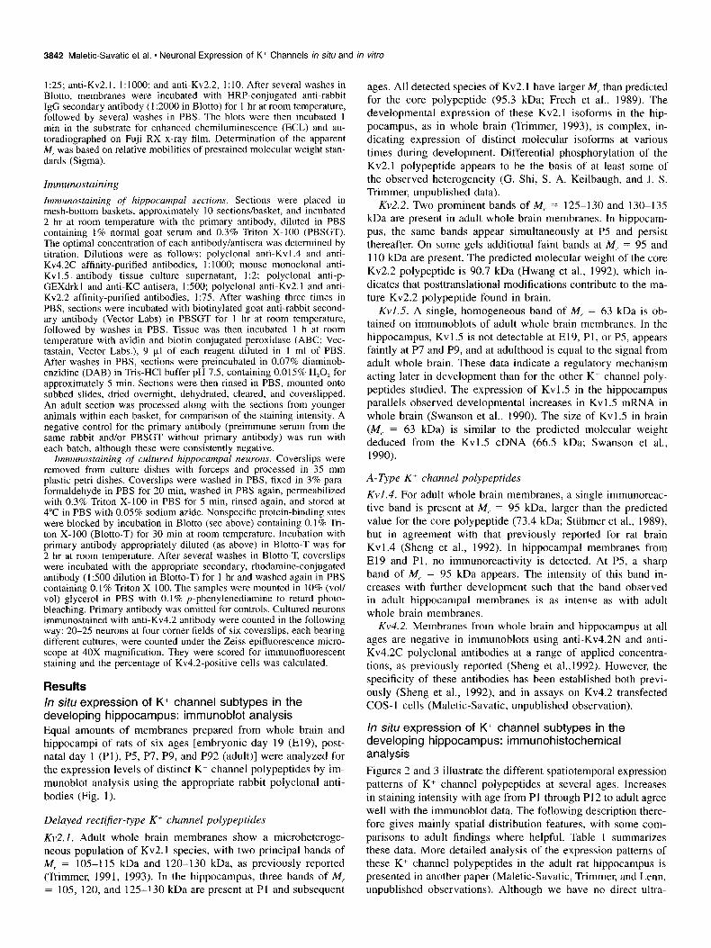
Results In situ expression of K+ channel subtypes in the developing hippocampus: immunoblot analysis Equal amounts of membranes prepared from whole brain and hippocampi of rats of six ages [embryonic day 19 (E19), post- natal day I (Pl), P5, P7, P9, and P92 (adult)] were analyzed for the expression levels of distinct K+ channel polypeptides by im- munoblot analysis using the appropriate rabbit polyclonal anti- bodies (Fig. I).
Delayed recti$er-type K’ channel polypeptides
Kv2.1. Adult whole brain membranes show a microheteroge- neous population of Kv2.I species, with two principal bands of M, = 105-l 15 kDa and 120-I 30 kDa, as previously reported (Trimmer, 1991, 1993). In the hippocampus, three bands of M, = 105, 120, and 125-l 30 kDa are present at PI and subsequent
ages. All detected species of Kv2.1 have larger M,. than predicted for the core polypeptide (95.3 kDa; Frech et al., 1989). The developmental expression of these Kv2.1 isoforms in the hip- pocampus, as in whole brain (Trimmer, 1993), is complex, in- dicating expression of distinct molecular isoforms at various times during development. Differential phosphorylation of the Kv2.1 polypeptide appears to be the basis of at least some of the observed heterogeneity (G. Shi, S. A. Keilbaugh, and J. S. Trimmer, unpublished data).
Kv2.2. Two prominent bands of IV,- = 125-130 and 130-135 kDa are present in adult whole brain membranes. In hippocam- pus, the same bands appear simultaneously at P5 and persist thereafter. On some gels additional faint bands at IV, = 95 and 1 10 kDa are present. The predicted molecular weight of the core Kv2.2 polypeptide is 90.7 kDa (Hwang et al., 1992), which in- dicates that posttranslational modifications contribute to the ma- ture Kv2.2 polypeptide found in brain.
Kv1.5. A single, homogeneous band of M, = 63 kDa is ob- tained on immunoblots of adult whole brain membranes. In the hippocampus, Kvl.5 is not detectable at El9, PI, or P5, appears faintly at P7 and P9, and at adulthood is equal to the signal from adult whole brain. These data indicate a regulatory mechanism acting later in development than for the other K+ channel poly- peptides studied. The expression of Kvl.5 in the hippocampus parallels observed developmental increases in Kvl.5 mRNA in whole brain (Swanson et al., 1990). The size of Kv 1.5 in brain (M,- = 63 kDa) is similar to the predicted molecular weight deduced from the Kv1.5 cDNA (66.5 kDa; Swanson et al., 1990).
A-Type K+ channel polypeptides
Kvl.4. For adult whole brain membranes, a single immunoreac- tive band is present at M, = 95 kDa, larger than the predicted value for the core polypeptide (73.4 kDa; Sttihmer et al., 1989), but in agreement with that previously reported for rat brain Kv1.4 (Sheng et al., 1992). In hippocampal membranes from El9 and Pl, no immunoreactivity is detected. At P5, a sharp band of M,- = 95 kDa appears. The intensity of this band in- creases with further development such that the band observed in adult hippocampal membranes is as intense as with adult whole brain membranes.
Kv4.2. Membranes from whole brain and hippocampus at all ages are negative in immunoblots using anti-Kv4.2N and anti- Kv4.2C polyclonal antibodies at a range of applied concentra- tions, as previously reported (Sheng et a1.,1992). However, the specificity of these antibodies has been established both previ- ously (Sheng et al., 1992), and in assays on Kv4.2 transfected COS-1 cells (Maletic-Savatic, unpublished observation).
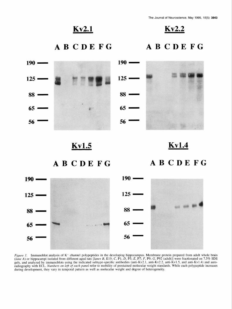
In situ expression of K-’ channel subtypes in the developing hippocampus: immunohistochemical analysis Figures 2 and 3 illustrate the different spatiotemporal expression patterns of K+ channel polypeptides at several ages. Increases in staining intensity with age from PI through PI2 to adult agree well with the immunoblot data. The following description there- fore gives mainly spatial distribution features, with some com- parisons to adult findings where helpful. Table I summarizes these data. More detailed analysis of the expression patterns of these K’ channel polypeptides in the adult rat hippocampus is presented in another paper (Maletic-Savatic, Trimmer, and Lenn, unpublished observations). Although we have no direct ultra-
The Journal of Neuroscience, May 1995, 75(5) 3843
88 -
65 -
56 -
No-
125 -
88 -
65 -
56 -
Kv2.1
ABCDEFG
Kv1.5
ABCD E FG
88 -
65 -
56 -
Kv2.2
ABCDEFG
Kv1.4
ABCDEFG
125 -
88 -
65 -
56 -
Figure I. Immunoblot analysis of K+ channel polypeptides in the developing hippocampus. Membrane protein prepared from adult whole brain (lane A) or hippocampi isolated from different aged rats [lanes B, E19; C, Pl; D, P5; E, P7; F, P9; G, P92 (adult)] were fractionated on 7.5% SDS gels, and analyzed by immunoblots using the indicated subtype-specific antibodies (anti-Kv2.1, anti-Kv2.2, anti-Kvl.5, and anti-Kvl.4) and auto- radiography with ECL. Numbers on left of each panel refer to mobility of prestained molecular weight standards. While each polypeptide increases during development, they vary in temporal pattern as well as molecular weight and degree of heterogeneity.
3844 Maletic-Savatic et al. - Neuronal Expression of K+ Channels in situ and in vitro
PI
P9
A
Figure 2. Immunohistochemical analyses of K+ channel polypeptides in the developing hippocampus. Low power photomicrographs of normal rat hippocampus immunostained for the three delayed-rectifier K’ channel polypeptides, visualized with peroxidase reaction. The pyramidal cell layer of CA1 is marked (large solid urrow at upper left corner) on each photograph, for orientation. Kv2.1 is expressed first at Pl, as light staining of cell bodies and proximal dendrites. At P9, plasma membrane patches are present and the immunostaining is like in adult, although less in intensity. Kv2.2 is absent at Pl. It is first observed at P9 as homogeneous stain in the soma and proximal dendrites. Interneurons are stained more strongly than other cells only in CA3. At adulthood, Kv2.2 is expressed in all neurons, but more in interneurons of all hippocampal layers. Kv1.5 appears at Pl in all pyramidal cell bodies in CA1 and CA3, but not in DG. At P9 and adult the intensity of Kvl.5 immunostaining increases and is more uniform among all hippocampal cells. so, stratum oriens (contains neuropil); pcl, pyramidal cell layer (contains cell bodies of pyramidal cells and some mterneurons); sr, stratum radiatum (contains proximal dendrites of pyramidal cells and interneurons); ml, molecular layer of dentate gyms (contains granule cell dendrites and projections from entorhinal cortex and ipsi- and contralateral dentate gyrus); gel, granule cell layer (contains cell bodies of granule cells). CAI , CA3, and dentate gyms (DG) correspond to the appropriate fields described by Lorente de No (I 934). Note that the micrographs representing the expression of K+ channel polypeptides at PI are overdeveloped in order to make the stain visible after reproduction. Scale bar, 100 pm.
The Journal of Neuroscience, May 1995, 75(5) 3845
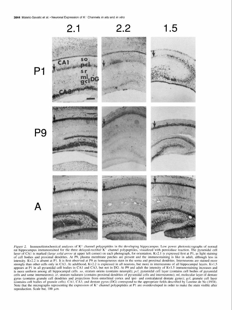
Figure 3. Immunohistochemical anal- yses of K+ channel polypeptides in the developing hippocampus. Low power photomicrographs of normal rat hip- pocampus immunostained for the two A-type K+ channel polypeptides, visu- alized with peroxidase reaction. The pyramidal cell layer of CAI is marked (large solid arrow at upper left corner) on each photograph, for orientation. Kvl.4 is first expressed at P5 as light staining of CA1 neuropil, the mossy fi- bers in CA3 and DC, and the mid-mo- lecular layer of DG. At P9, although less intense, the expression of Kv I .4 is the same as in the adult hippocampus. Kv4.2 is first detected at P5 as mod- erate staining of a few cells, with no definite neuropil staining in any area. At P9 and adult, the intensity of stain- ing increased in both plasma mem- branes and neuropil (SO, SR and the outer molecular layer of DG, arrow at the center), as well as in interneurons (arrow at lower left, compare to Table 1). so, stratum oriens (contains neuro- pil); pcl, pyramidal cell layer (contains cell bodies of pyramidal cells and some interneurons); sr, stratum radiatum (contains proximal dendrites of pyra- midal cells and interneurons); ml, mo- lecular layer of dentate gyrus (contains granule cell dendrites and projections from entorhinal cortex and ipsi- and contralateral dentate gyrus); gel, gran- ule cell layer (contains cell bodies of granule cells). CAI, CA3 and dentate gyrus (DG) correspond to the appro- priate fields described by Lorente de No (1934). Scale bar, 100 urn.
14 .
P5
P9
42 .
3846 Maletic-Savatic et al. * Neuronal Expression of K* Channels in situ and in vitro
Table 1. Immunostaining of K+ channel polypeptides in adult rat hippocampus in situ
K+ channel polypeptide Kv 2.1 Kv 2.2 Kv 1.5 Kv 1.4 Kv 4.2
Neuronal somas and proximal dendrites CA1 , CA2, CA3 + + + -
DG + + + -
Interneurons accentuated CAI, CA2, CA3 - ++ - - - DG - ++ - - -
Only interneuron somas and proximal dendrites
CAI, CA2, CA3 - - + DG - - - - +
Axonal processes in DG and CA3 - - - + - neuropil accentuated - - - + - Distal dendritic processes
accentuated in neuropil CAl, CA2, CA3 - - - - + DG - - - - +
CAI, CA2, CA?, DC = divisions of the hippocampus; see text for further details.
structural or biochemical data to support specific assignments of subcellular localization, for brevity we will hereafter describe immunostaining present at or near the periphery of neuronal cell bodies and processes, as opposed to that which appears to be intracellular or deep within the soma, as “associated with the plasma membrane.”
Deluyed rectifier-type K+ channel polypeptides Kv2.1. Kv2.1 in adult brain is expressed in all neurons, in a characteristic pattern of plasma membrane patches. Develop- mentally, Kv2.1 is absent at E19. At PI, there is light staining of cell bodies and proximal dendrites in CA], CA3 and dentate gyrus (DG). At P5, staining is more intense, with plasma mem- brane accentuations on a minority of cell bodies, especially in CA3 (not shown). The neuropil of the inner half of SO stains homogeneously. In DG all granule cells stain moderately and molecular layer (ML) lightly. At P9 Kv2.1 has increased in in- tensity, with membrane accentuations on all cell bodies and proximal dendrites, which in CA1 extend across stratum radia- turn (SR), but are smaller and sparse compared to the adult. Neuropil staining has become moderate, but still less than the adult.
Kv2.2. Like Kv2. I, Kv2.2 is expressed in all neurons in the adult hippocampus. Unlike Kv2.1, Kv2.2 is expressed more in interneurons than in pyramidal cells. It is first observed at P9 as a faint, homogeneous stain in the soma and proximal dendrites of CA], CA3, and DG. Interneurons stain more strongly than other cells only in CA3 at this age. At P12 (not shown), staining has increased further, and now interneurons are stained more strongly than pyramidal cells in CA1 as well as CA3. Kv2.2 expression at P12 differs from adult only in lower intensity and fewer plasma membrane accentuations.
Kv1.5. There is homogeneous Kvl.5 expression in all neu- ronal cell bodies and proximal dendrites throughout the adult hippocampus, accentuated on the plasma membranes in a uni- form distribution. We first detect Kvl.5 at PI as light, occasion-
ally moderate, staining of all pyramidal cell bodies in CA1 and CA3. This suggests a slightly greater sensitivity of immunostain- ing than present in the immunoblot analysis (Fig. I), which shows no detectable staining at this age, probably due to the expression of the polypeptide in a limited number of small neu- rons. Kv 1.5 is not observed in DG at this age. At P9, the inten- sity of Kvl.5 immunostaining on the cell bodies has slightly increased and is more uniform among cells, still with no neuropil or dendritic staining. At P12 (not shown), the adult pattern is present, except the staining of dendrites in CAI-3 is less exten- sive.
A-Type K’ channel polypeptides Kv1.4. The distribution of Kv1.4 in the adult hippocampus is distinctive. It is detected in the ML of DG, with a strong staining in the middle third, and in mossy fibers in CA3. All neuronal cell bodies and proximal dendrites are unstained. Kv 1.4 is first observed developmentally at P5 as very light staining of SO and stratum lacunosum moleculare (SLM) in CAI, the mossy fibers in CA3 and DG, and the medial entorhinal projection in the middle third of the ML. For comparison, staining in substantia nigra and globus pallidus (data not shown) is already quite strong. At P9, although less intense, the expression of Kv I .4 has the same distribution as in the adult hippocampus. At this age, outer half of SO is more homogeneous with all neuronal cell bodies and proximal dendrites unstained, the neuropil of SO is lightly stained while the mossy fibers and endings in CA3 are strongly stained, and there is a distinct pattern in DG with strong staining of medial entorhinal cortex terminals.
Kv4.2. Kv4.2 in adult hippocampus is expressed on distal den- drites and neuropil of CA]-3. It is first detected at P5, as mod- erate staining of a few cells in CA]-3, with no definite neuropil staining in any area. At P9, the plasma membrane staining of positive cells is increased, and now neuropil in the SO, inner SR, SLM of CA1 only, and ML are lightly and homogeneously stained. At PI2 (not shown), light staining of the hilar neuropil is added. Between P12 and adult, there is a general increase in the intensity of staining, with a relative increase in the outer half of SR.
In vitro expression of K+ channel subtypes in the developing hippocampus: immunohistochemical analysis
To determine the correspondence between our results obtained with hippocampal tissue in situ and hippocampal neurons dif- ferentiated in vitro, we immunostained fixed and permeabilized cultured cells with K+ channel polypeptide-specific antibodies. All cultures utilized neurons from the hippocampi of El9 rats, analyzed at 2, 4, 6, 8, 10, 14, 18, 20, 22, and 26 d in vitro (DIV). No staining was seen with any K+ channel antibody to cultured astrocytes in similar experiments. Figures 4 and 5 il- lustrate the patterns of K+ channel polypeptide expression at 10 and 22 DIV, and Table 2 summarizes the data for seven time points.
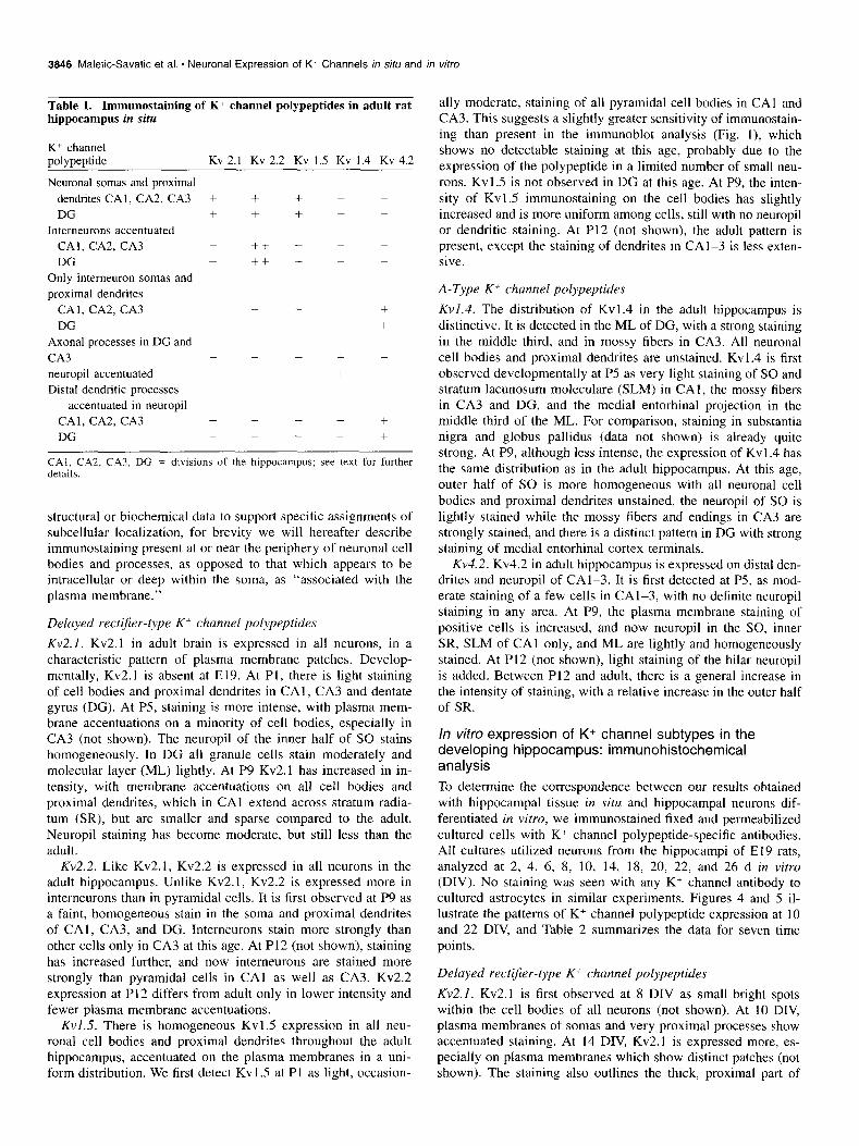
Delayed recti$er-type K+ channel polypeptides Kv2.1. Kv2.1 is first observed at 8 DIV as small bright spots within the cell bodies of all neurons (not shown). At 10 DIV, plasma membranes of somas and very proximal processes show accentuated staining. At 14 DIV, Kv2.1 is expressed more, es- pecially on plasma membranes which show distinct patches (not shown). The staining also outlines the thick, proximal part of
22 DIV
22 DIV
The Journal of Neuroscience, May 1995, 75(5) 3847
Figure 4. Spatiotemporal expression of three delayed rectifier K+ channel polypeptides in cultured hippocampal neurons at 10 and 22 DIV. Neurons on coverslips were visualized with indirect immunofluorescence. First row, 10 DIV, optimal exposure times (Kodak, 400 ISO). Second IOW, 22 DIV, immunofluorescence recorded with the same exposure time for each polypeptide (8 set), the optimal exposure time for Kv2.1. Third ro’ow, 22 DIV; phase contrast micrograph showing morphology of a pyramidal neuron immunostained for the expression of Kv2.2 polypeptide and photomicrographs of the neurons stained with anti-Kv2.2 and anti-Kvl.5 antibodies. Immunofluorescence recorded with the optimal exposure times (15 set). Note intense immunoreactivity of Kv2.1 compared to the lower immunoreactivity of the other K+ channel polypeptides. Detail in text and Table 2. Scale bar, 30 km.
3848 Maietic-Savatic et al. * Neuronal Expression of K+ Channels in situ and in vitro
IO D
42 .
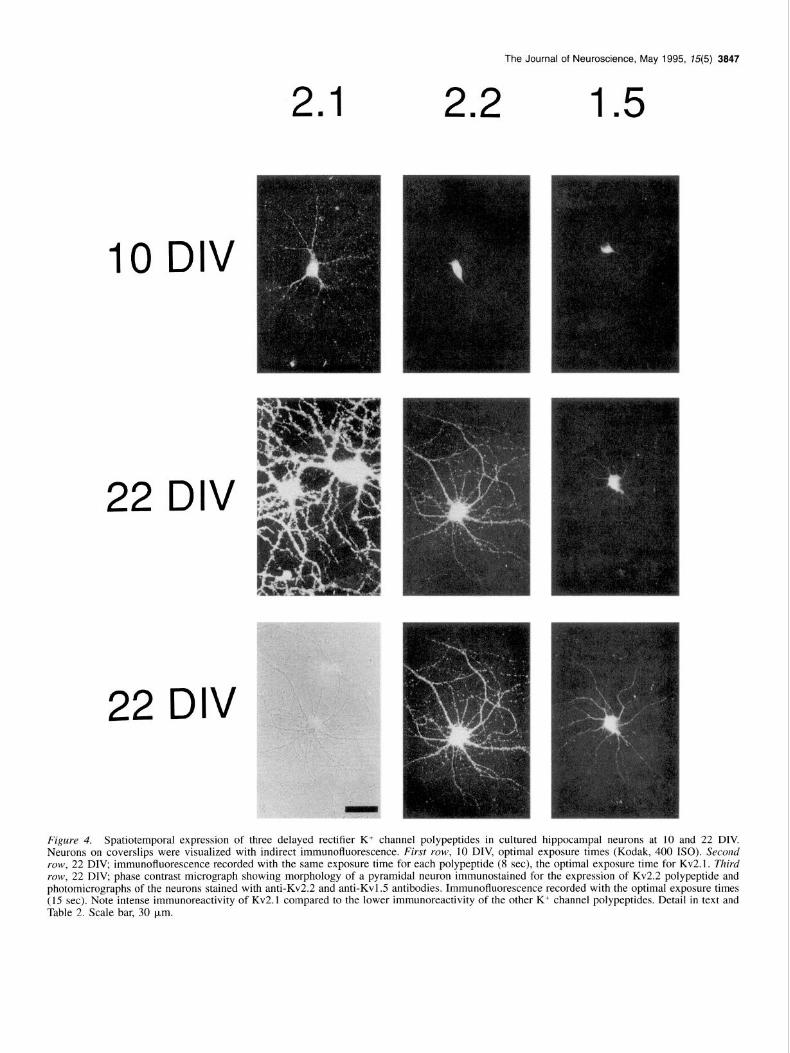
Figure 5. Spatiotemporal expression of two A-type K+ channel polypeptides in cultured hippocampal neurons at 10 and 22 DIV. First row, 10 DIV, phase contrast micrograph showing morphology of a 10 DIV old pyramidal neuron and a neuron unstained with anti-Kv4.2 (arrow; Kodak, 4001SO). Second YOW, immunofluorescence recorded with the same exposure time for both polypeptides (8 set), the optimal exposure time for Kv2.1. Third row, 22 DIV, immunofluorescence recorded with the optimal exposure time for each polypeptides (15 set). Note intense immuno- reactivity of Kv2.1 (Fig. 4) compared to the lower immunoreactivity of the other K+ channel polypeptides. Detail in text and Table 2. Scale bar, 30 pm.
processes. Most processes at 14 DIV are dendrites based on their Kv2.2. Kv2.2 is first observed at 8 DIV as faint intracellular morphology and confirmed by double immunostaining for Kv2.1 staining (not shown) which increases at 10 DIV. A marked and MAP2, a dendritic marker (not shown). At 18 DIV, very change in the pattern of Kv2.2 expression from 10 DIV is seen bright membrane patches of Kv2.1 outline the full length of at 14 DIV, when all cell bodies and processes are strongly almost all dendrites (not shown). The number, size and intensity stained, but some more stained than the rest (not shown). The of the patches increase further at 20, 22, and 26 DIV. These latter are large, with a small number of smoothly curved den- features are very similar to those in situ. drites, and are darker in phase microscopy, corresponding to the
The Journal of Neuroscience, May 1995, 15(5) 3849
Table 2. Summary of normal expression of K+ channel polypeptides in rat hippocampal neurons in vitro
Kv2.1 Kv2.2 Kv1.5 Kv1.4 Kv4.2
6 DIV C 8 DIV C C C C
IO DIV CS C C C cs 14 DIV CSP CSP C C CSP 18 DIV CSP CSP CSP cs CSP 22 DIV CSP CSP CSP CSP CSP 26 DIV CSP CSP CSP CSP CSP
-, No detectable strain; C, intracellular compartments; S, soma plasma mem- brane: P, processes.
established morphology of GABA interneurons in these cultures (Benson et al., 1994). Plasma membrane immunostaining is uni- form, with a few bright accentuations on processes. This pattern and intensity of staining persist at 18, 20, and 22 DIV. Both pyramidal neurons and interneurons express Kv2.2 (Fig. 4, py- ramidal cell at 22 DIV). The pattern, with all neuron cell bodies and proximal processes stained, is the same as found in situ, although in situ the difference in intensity between interneurons and pyramidal cells is greater.
Kv1.5. Kvl.5 exhibits the weakest staining of the K+ channel subtypes studied in vitro, although clearly above controls and with specific changes in localization with time. First detected in somas at 8 DIV (not shown), it is unchanged at 10 and 14 DIV. At 18 and 22 DIV, Kv1.5 is present throughout the somatoden- dritic compartment, with some processes brighter than others. At each time point, the observed immunostaining is diffuse and intracellular. Plasma membrane staining, if present, is unpatter- ned. The gradual increase in staining in all neurons is seen in vitro and in situ, but the intensity is much less at all DIV.
A-Type K’ channel polypeptides
Kvl 4. Kv1.4 is first detected at 10 DIV as faint spots arranged in a crescent inside the cell bodies. At 14 DIV, Kvl.4 is uni- formly expressed throughout the soma and its staining intensity is increased (not shown). The intensity of Kv 1.4 immunostaining increases further at 18, 20, and 22 DIV, but it does not reach the intensity observed in situ. At 18 DIV, immunostaining is accentuated on soma plasma membranes (not shown) and at 22 DIV outlines some processes, although lightly compared to Kv2.1 immunostaining. This is consistent with its limited ex- pression in situ in the classes of neurons which are present in the cultures.
Kv4.2. The special characteristic of Kv4.2 expression pattern is the presence of both stained and unstained neurons at all time points. At 6 DIV, occasional Kv4.2 immunoreactive spots appear intracellularly in some neurons (not shown). At 8 DIV, diffuse immunoreactivity is present inside the cells (not shown). The intensity of intracellular Kv4.2 immunostaining increases at 10 DIV, and in addition staining is accentuated on the plasma mem- branes of the positive somas. At 14 DIV, Kv4.2 is expressed more, particularly on plasma membranes (not shown). The pat- tern and intensity are unchanged at 18, 20, and 22 DIV. There is a similarity between these observations and the in situ pattern in that only some neurons express Kv4.2, although the propor- tion of positive cells appears higher in vitro (88.7% k 4.4% (mean & SD; n = 6 samples of 20-25 cells/sample).
Discussion
The principal findings of this study are (1) the striking differ- ences among the spatiotemporal expression patterns of five K+ channel subtypes in the developing hippocampus, (2) the simi- larities in the temporal expression of each K+ channel polypep- tide in situ compared to in vitro, and (3) the differences in the level of expression and subcellular distribution between some, but not all, K+ channel polypeptides in vitro versus in situ. Our data demonstrate differential regional, cellular, and subcellular distribution and differential temporal expression of K+ channel subtypes in the developing hippocampus, indicating their differ- ent functions and different mechanisms of targeting within neu- rons. Furthermore, we show the distinct spatiotemporal expres- sion patterns of K+ channel subtypes in hippocampal neurons differentiating in vitro, and identify aspects of expression that may be regulated by extrinsic factors in situ but not in vitro.
Spatial distribution of K’ channel polypeptides in situ. The spatial distribution of examined K’ channel subtypes reveals distinct differences in hippocampal cells, including differential regional, cellular, and subcellular distribution between channels associated with delayed rectifier and A-type currents. The de- velopment of these patterns is monotonic; that is, while the time and spatial development varies among the different channels, each K+ channel subtype appears in place, followed only by developmental increases in abundance. These findings indicate that the mechanisms controlling cellular distribution and sub- cellular targeting of channels operate through development, as the same patterns are seen at all ages at which the channels are expressed.
The three delayed rectifier-type K+ channel polypeptides (Kv2.1, Kv2.2, Kvl.5) are present in virtually all neurons, with the greatest concentration on the somatodendritic plasma mem- brane. However, the relative intensity of expression among neu- rons of the hippocampus at various ages are distinctive. A-type K’ channel polypeptides (Kv 1.4, Kv4.2) differ in that they are expressed in distinct subpopulations of neurons, Kvl.4 on the distal axons of the entorhinal and granule cell projections and Kv4.2 on the distal dendritic processes of pyramidal cells and somatodendritic region of a small population of interneurons, as resolved by confocal microscopy on adult brain sections (Mal- etic-Savatic et al., unpublished observations). These patterns hold true throughout development, suggesting that the different mechanisms of targeting within the neurons operate continuous- ly.
Temporal expression patterns of K’ channel polypeptides in situ. By immunoblots, Kv2.1 is expressed first, at P 1, followed by Kvl.4 and Kv2.2 at P5, and Kv1.5 which is faintly present only at P7 and P9. Immunohistochemistry shows the same tem- poral pattern of expression, with Kv2.1 expressed earliest, at Pl . Kv1.4, Kvl.5, and Kv4.2 are first detected at P5, with different patterns of increase after P5. Kv2.2 is detected last, at P9, even in the interneurons which are later strongly stained, with gradual increase in all neurons continuing past P12. However, by im- munoblots, Kv2.2 appears at P5 as a moderately intense band (Fig. 1). This is probably due to the concentrated sample of brain membranes (100 pg per lane). Unlike spatial distribution, tem- poral expression patterns are not related to the A-type/delayed- rectifier type of current.
The microheterogeneity of Kv2.1 and Kv2.2 observed on im- munoblots suggests extensive posttranslational modifications of these neuronal Shab family polypeptides. In addition, the exis-
3850 Maletic-Savatic et al. . Neuronal Expression of K’ Channels in situ and in vitro
tence of three separate bands of Kv2.1 in the hippocampus in- dicate that the heterogeneity of Kv2.1 species observed in whole brain preparations is present in these hippocampal samples. The remaining K+ channel subtypes examined (Kv1.4 and Kv1.5, Shaker family) migrate as single bands on immunoblots, indi- cating that posttranslational modifications, if present, are rela- tively homogeneous compared to the Shah family channel poly- peptides.
The sequential appearance of K+ channel subtypes during hip- pocampal development indicates diverse regulatory mechanisms, either intrinsic or extrinsic, the nature of which are not yet known. However, epigenetic factors related to synaptogenesis and synaptic activity seem likely to have major effects on the expression of K+ channel subtypes. The general increase in polypeptide quantity, observed for all K+ channel polypeptides, but particularly for Kv1.5 after P9 in both immunoblots and immunohistochemical preparations, may be partly due to the postnatal increase in synaptogenesis. This increase in the amount of protein probably reflects both an increase in the number of molecules per unit of plasma membrane (i.e., increase of poly- peptide density) and a relative increase in the plasma membrane because of more highly branched processes, both changes known to be influenced by synaptogenesis in some systems (Shatz, 1990).
Correlation c.fspatiotemporal expression of K’ channel poly- peptides in situ and in vitro. Spatiotemporal expression of all five K+ channel polypeptides in cultured neurons correlates with their expression in situ. Of the five K+ channel subtypes ex- amined, Kv2.1 immunostaining is the strongest both in situ and in vitro. It is present on the cell bodies and dendrites of all neurons as distinct plasma membrane patches in both cases, giv- ing a remarkable appearance compared to the other K+ channel polypeptides tested. However, Kv2. I immunoreactive patches are present throughout the whole length of dendrites in cultured neurons, indicating that factors that influence intracellular tar- geting may differ in situ and in vitro. Temporally, it is detected at 8 DIV in the cell body of all neurons. The number, size and intensity of Kv2.1 patches increase gradually thereafter, as they do in situ.
The expression of Kv2.2 in vitro correlates with the in situ pattern, with minor exceptions. Staining is present in all neurons and is found associated with the plasma membrane. However, in vitro, processes are more prominently stained than observed in situ. In addition, the notable difference between the expres- sion levels in pyramidal cells and interneurons observed in situ is not evident in vitro. Relatively uniform staining of all neurons in vitro may result from a lesser expression in cultured inter- neurons compared to those in situ, possibly related to in vitro culture conditions, which lack the extrinsic inputs to the hip- pocampus.
Staining for Kvl.5 is observed in vitro at considerably lower levels then Kv2.1, even after 26 DIV, with the majority of stain- ing associated with the cell body. This is in sharp contrast to Kvl.5 immunoreactivity in situ, where intense immunoreactivity comparable to Kv2.1 is observed, and processes are clearly stained. In addition, Kvl.5 is detected at 18 DIV, clearly later than in situ where it appears at P5. Its augmented expression in situ may be controlled by the extrinsic connections of the hip- pocampus or other factors not present in vitro, such as specific growth factors, neurotransmitters and neuropeptides. Identifica- tion of such factors through manipulation of these in vitro cul-
tures will yield important insights into the mechanisms of K+ channel expression.
We also observed differences between the in situ and in vitro expression of A-type K+ channel polypeptides. In the cultures, Kv 1.4 is present at low levels, and staining is not restricted to distal axons, as cell bodies are also stained. This finding is not surprising, since the cultures contain neither the neurons which give rise to the perforant path nor mossy fiber axons to which Kvl.4 is predominantly localized to distal axons in situ (Sheng et al., 1992, Maletic-Savatic et al., unpublished observations). Kv1.4 in the hippocampus in situ is also expressed in the neu- ropil of areas containing other afferents, but, in these areas it appears later and at low concentrations. This may correspond to its presence at low concentrations in vitro in the somata and some processes of all neurons, which are now more evident in the isolated cells in vitro. Kv1.4 is expressed relatively late (18 DIV) and does not increase noticeably up to 26 DIV.
Kv4.2 in situ is localized on the most distal dendrites of py- ramidal cells in CAl-3 as observed by confocal microscopy (Maletic-Savatic et al., unpublished observations), and on the somata and dendrites of a small population of interneurons. In vitro, its presence on the somata and proximal dendrites of a subpopulation of neurons is retained (Fig. 5). Kv4.2 immuno- reactive neurons are, however, relatively more numerous in vitro (88.7%) than in situ, and have the morphology of both pyramidal cells and GABAergic interneurons (Benson et al., 1994). In ad- dition, there are no neurons in vitro that express Kv4.2 exclu- sively on small distal dendrites, which are the principal site for this K+ channel polypeptide in situ (Maletic-Savatic et al., un- published observations). During development these dendritic processes differentiate later, influenced by the formation of syn- apses. It is possible that these specific dendritic processes do not form in the cultures, perhaps due to the absence of particular afferent projections. It is interesting to note that Kv2. I and Kv4.2 exhibit distinct spatial segregation on dendritic processes in situ, with Kv2.1 restricted to proximal portions of dendrites, while Kv4.2 is found only distal. This precise segregation in- dicates that distinct functional subdomains of the dendritic mem- brane are established by the differential sorting and/or retention of specific channel polypeptides. It should be noted that this spatial segregation does not arise for either channel in vitro.
Correlation of K’ currents and K’ channel polypeptides in hippocampus. Electrophysiological properties of K+ currents in cultured hippocampal pyramidal cells have been studied in detail (Segal et al., 1984; Ficker and Heinemann, 1992). Storm (1990) lists seven main types of K+ currents recorded from these neu- rons. Each of these currents has distinctive kinetics, voltage de- pendence and pharmacology. They are implicated in a number of electrical features of neurons, but in none of this work has a definite correlation of any K+ current and a channel polypeptide been achieved.
During neuronal development delayed rectifier currents are the first outward currents to appear, followed by A-type currents (reviewed in Ribera and Spitzer, 1992). Our results show that Kv2.1, which yields a delayed rectifier current when expressed in Xenopus oocytes, is the first to appear both in situ and in vitro, followed by Kv4.2, which is an A-type channel. These may represent the major K+ channels expressed in cultured hip- pocampal neurons, perhaps contributing to observed delayed rectifier and A-type currents in these cells. However, the pres- ence of delayed rectifier K+ current has been registered electro- physiologically earlier compared to our data (Bader et al, 1985;
The Journal of Neuroscience, May 1995, 75(5) 3851
Harris et al., 1988). It is possible that the level of expression of channel polypeptides required to give positive immunostaining is higher than the level required for electrophysiological record- ings. In addition, formation of heterotetramers of a-subunits (Sheng et al., 1993; Wang et al., 1993) as well as the presence of P-subunits (Rettig et al., 1994) can significantly influence the characteristics of neuronal K’ currents. Thus, a definitive cor- relation between the expression of a given channel polypeptide and a recorded current is difficult without more detailed infor- mation such as the precise subunit composition of neuronal K+ channel complexes.
In conclusion, we have characterized the expression of K+ channel subtypes during development of hippocampal neurons. Observed differences in the overall level of expression and sub- cellular distribution of some, but not all K+ channels indicate the influence of factors present in situ but not in vitro. Identifi- cation of these factors, such as electrical activity, neurotrans- mitter and neuropeptide release, by manipulation of neuronal cultures will lead to new insights in the regulation of hippocam- pal activity during development and in the adult brain.
References
Bader CR, Bertrand D, Dupin E (1985) Voltage-dependent potassium currents in developing neurons from quail mesencephalic neural crest. J Physiol (Lond) 366:129-151
Banker GA, Cowan WM (1977) Rat hippocampal neurons in dispersed cell culture. Brain Res 126:397-425.
Bartlett WP Banker GA (1984a) An electron microscopic study of the development of axons and dendrites by hippocampal neurons in cul- ture. I. Cells which develop without intercellular contacts. J Neurosci 4:1944-1953.
Bartlett WP, Banker GA (1984b) An electron microscopic study of the development of axons and dendrites by hippocampal neurons in cul- ture. Il. Synaptic relationships. J Neurosci 4: 1954-1965.
Benson DL, Watkins FH, Steward 0, Banker G (1994) Characterization of GABAergic neurons in hippocampal cell cultures. J Neurocytol 231279-295.
Best D, Warr PJ, Gull K (1981) Influence of the composition of com- mercial sodium dodecyl sulfate preparations on the separation of al- pha- and beta-tubulin during polyacrylamide gel electrophoresis. Anal Biochem 114:281-284.
Dotti CG, Sullivan CA, Banker GA (1988) The establishment of po- larity by hippocampal neurons in culture. J Neurosci 8:1454-1468.
Ficker E, Heinemann U (1989) K+ currents in developing hippocampal cells. Pfluegers Arch 414:S125.
Fletcher TL, Banker GA (1989) The establishment of polarity by hip- pocampal neurons: the relationship between the stage of a cell’s de- velopment in situ and its subsequent development in culture. Dev Biol 136:446+54
Frech GC, Van Dongen AMJ, Schuster G, Brown AM, Joho RH (1989) A novel potassium channel with delayed rectifier properties isolated from rat brain by expression cloning. Nature 340:642-645.
Goslin K, Banker G (1991) Rat hippocampal neurons in low-density culture. In: Culturing nerve cells (Goslin K, Banker G, eds), pp 25l- 281. Cambridge, MA: MIT Press.
Halliwell JV (1990) K+ channels in the central nervous system. In: Potassium channels. Structure, function and therapeutic potential (Cook NS, ed), pp 348-381. New York: Halsted.
Harris GL, Henderson LP, Spitzer NC (1988) Changes in densities and kinetics of delayed rectifier potassium channels during neuronal dif- ferentiation. Neuron 1:739-750.
Hille B (1992) Ionic channels of excitable membranes. Sunderland, MA: Sinauer.
Hwang PM, Glatt CE, Bredt DS, Yellen G, Snyder SH (1992) A novel
K+ channel with unique localizations in mammalian brain: molecular cloning and characterization. Neuron 8:473-481.
Hwang PM. Fotuki M. Bredt DS, Cunningham AM. Snvder SH (1993) CoLtrasting immunohistochemical lo&zation in rat brain of two novel K+ channels of the Shab subfamily. J Neurosci 13: 1569-1576.
Johnson DAG, Sportsman JR, Elder JH (1984) Improved technique utilizing nonfat dry milk for analysis of proteins and nucleic acids transferred to nitrocellulose. Gene-Anal Tech 1:3-S.
Kleiman R. Banker G, Steward 0 (1990) Differential subcellular lo- , calization of particular mRNAs in hippocampal neurons in culture. Neuron 5:821-830
Lenn NJ, Beebe B (1979) A simple apparatus for controlled pressure perfusion fixation. Microsc Acta 79: 139-l 44.
Lorente de No R (1934) Studies on the structure of the cerebral cortex. II. Continuation of the study of the ammonic system. J Psycho1 Neu- rol 46: 113-177
Maize1 JV (1971) Polyacrylamide gel electrophoresis of viral proteins. Methods Virol 5: 179-246
Mattson MP (1988) Neurotransmitters in the regulation of neuronal cytoarchitecture. Brain Res Rev 13: 179-212. -
Pietrini G. Matteoli M. Banker G. Caolan MJ (1992) lsoforms of the Na,K-ATPase are present in both axons and dendrites of hippocampal neurons in culture. Proc Nat1 Acad Sci USA 89:8414-8418
Rettig J, Heinemann SH, Wunder F, Lorra C, Parcej DN, Dolly JO, Pongs 0 (1994) Inactivation properties of voltage-gated K+ channels altered by presence of B-subunit. Nature 369:289-294.
Ribera AB, Spitzer NC (1992) Developmental regulation of potassium channels and the impact on neuronal differentiation. In: Ion channels (Narahashi T, ed), pp 1-38. New York: Plenum.
Segal M, Barker JL (1984) Rat hippocampal neurons in culture: po- tassium conductances. J Neurophysiol 5 1: 1409-1433.
Segal M, Rogawski MA, Barker JL (1984) A transient potassium con- ductance regulates the excitability of cultured hippocampal and spinal neurons. J Neurosci 4:604-609.
Shatz CJ (1990) Impulse activity and the patterning of connections during CNS development. Neuron 5:745-756
Sheng M, Tsaur ML, Jan YN, Jan LY (1992) Subcellular segregation of two A-type K’ channel proteins in rat central neurons. Neuron 9:271-284.
Sheng M, Liao YJ, Jan YN, Jan LY (1993) Presynaptic A-current based on heteromultimeric K+ channels detected in viva. Nature 365:72- 75.
Shi G, Kleinklaus A, Marrion N, Trimmer JS (1994) Properties of Kv2.1 K’ channels expressed in transfected mammalian ceils. J Biol Chem 269:23204-23211.
Spitzer NC (1991) A developmental handshake: neuronal control of ionic currents and their control of neuronal differentiation. J Neuro- biol 7:659-673.
Storm JF (1990) Potassium currents in hippocampal pyramidal cells. Prog Brain Res 83:161-187.
Stuhmer W, Ruppersberg JP Schroter KH, Sakmann B, Stocker M, Giese Kp, Perschke A, Baumann A, Pongs 0 (1989) Molecular basis of functional diversity of voltage gated potassium channels in mam- malian brain. EMBO J 8:3235-3244.
Swanson R, Marshall J, Smith JS, Williams JB, Boyle MB, Folander K, Luneau CJ, Antanavage J, Oliva C, Buhrow SA, Bennett C, Stein RB, Kaczmarek LK (1990) Cloning and expression of cDNA and genomic clones encoding three delayed rectifier potassium channels in rat brain. Neuron 4:929-939.
Takimoto K, Fomina AE Gealy R, Trimmer JS, Levitan ES (1993) Dexamethasone rapidly induces Kv 1.5 K+ channel gene transcription and expression in clonal pituitary cells. Neuron I I :359-369.
Trimmer JS (1991) Immunological identification and characterization of a delayed rectifier K+ channel in rat brain. Proc Nat1 Acad Sci USA 88: iO764-10768.
Trimmer JS (1993) Expression of Kv2.1 delayed rectifier K+ channel isoforms in developing rat brain. FEBS Lett 324:205-210.
Wang H, Kunkel DD, Martin TM, Schwartzkroin PA, Tempel BL (1993) Heteromultimeric K+ channels in terminal and iuxtaoaranodal regions of neurons. Nature 365:75-79.
_ _
Ward GE, Garbers DL, Vacquier VD (1985) Effects of extracellular egg factors on sperm guanylate cyclase. Science 227:768-770.


![Spatiotemporal dynamics of continuum neural fields...partial differential equation (PDE) models of diffusively coupled excitable systems [13, 14], neural field models can exhibit](https://img.pdfslide.us/doc/110x75/5f700c514eff5425e92b0db3/spatiotemporal-dynamics-of-continuum-neural-fields-partial-differential-equation.jpg)