Embed Size (px)
Citation preview
TARGETING ORGANIC ANION TRANSPORTING POLYPEPTIDES IN CANCER TO
IMPROVE DIAGNOSTICS AND THERAPY
By
©2012 Amanda Lynne Hays
Submitted to the graduate degree program in Pharmacology, Toxicology and
Therapeutics and the Graduate Faculty of the University of Kansas in partial fulfillment of the
requirements for the degree of Doctor of Philosophy.
__ Bruno Hagenbuch, Ph.D., Chair
__ Udayan Apte, Ph.D.
__ Paige C. Geiger, Ph.D.
__ Gregory A. Reed, Ph.D.
__ Bao-Ting Zhu, Ph.D.
Date Defended: August 13th, 2012
ii
The Dissertation Committee for Amanda Lynne Hays
certifies that this is the approved version of the following dissertation:
TARGETING ORGANIC ANION TRANSPORTING POLYPEPTIDES IN CANCER TO
IMPROVE DIAGNOSTICS AND THERAPY
__ Bruno Hagenbuch, Ph.D., Chair
Date approved: August 13th, 2012
iii
Abstract
Organic Anion Transporting Polypeptides (OATPs) are multispecific transport proteins
that mediate the uptake of numerous endogenous and exogenous compounds into cells.
Recently, OATPs have been shown to have altered expression in cancer tissue compared to
their normal expression profiles. It has been proposed that OATPs can be targeted to improve
cancer therapeutics. Therefore, I tested the hypothesis that expression of OATPs in cancer
combined with their ability to transport cytotoxic anticancer drugs makes them potential targets
for improving cancer diagnosis and therapy.
The hypothesis was tested via the following specific aims: 1) to identify and characterize
OATP expression in cancer, 2) to identify novel anticancer drug substrates of OATPs, and 3) to
identify novel cytotoxic compounds from plant extracts that are substrates of OATPs and can be
developed into anticancer drugs that target OATP-expressing cancers.
In the first specific aim, OATPs expressed in pancreatic cancer were identified by
immunohistochemical staining of pancreatic cancer tissue specimens. Completion of this
specific aim identified four major OATPs expressed in pancreatic adenocarcinomas. Additionally,
OATP1B3 expression was observed to be highest in low stage adenocarcinoma and absent in
metastatic tissue. These results demonstrate that OATP1B3 may serve as a diagnostic marker
and/or therapeutic target in early stage adenocarcinomas.
In specific aim two, novel anticancer drug substrates of OATP1B3 were identified by
screening the NCI/DTP oncology drug set containing all of the FDA approved chemotherapy
drugs. In this study, I determined the effect of the anticancer drugs on transport and cell viability
of OATP1B3-expressing cells. Finally, I demonstrated that the anticancer drugs etoposide,
oxaliplatin and plicamycin are substrates of OATP1B3. These results suggest that the
mentioned cytotoxic anticancer drugs could potentially be used to treat OATP1B3-expressing
cancers.
iv
In the last specific aim, Kansas plant extracts were screened using bioassay guided
fractionation and cell viability assays to isolate novel cytotoxic compounds that are substrates of
OATP1B3. Given that these novel plant compounds are cytotoxic and are also transported by
OATP1B3 suggests that they can be used for lead optimization studies to develop new
anticancer drug entities.
This dissertation demonstrates that OATP1B3 is a potential target for mechanisms of
OATP-mediated anticancer therapy. Ultimately, this knowledge can be used to utilize OATP1B3
expression in cancer as a diagnostic marker, as well as a target for cytotoxic anticancer drug
therapies.

v
Dedication
I would like to dedicate the work herein to my parents, Naser and Iris Obaidat.

vi
Acknowledgments
This work was supported by NIH Grants P20-RR021940, R01-GM077336 and T32-ES07079. I
would like to give my sincere appreciation to the taxpayers of the United States of America
whose tax revenues made my dissertation research possible.
First and foremost, I would like to thank my mentor, Dr. Bruno Hagenbuch. I would have never
thought of choosing this career path had I not been given the opportunity to work with you
during my summer internship. Your love for science and positive attitude was contagious and
made me desire that too. Thank you for all your time, your encouragement, your guidance and
your advice in the lab and in life. Most of all, thank you for your patience and understanding!
You’ve been an excellent role model and father figure over the years and I have truly enjoyed
being a part of your lab.
Committee members: Dr. Apte, Dr. Sittampalam, Dr. Zhu, Dr. Geiger, and Dr. Reed, thank you
for serving on my dissertation committee and for all the instruction and helpful insight into my
project.
I would also like to thank all the Hagenbuch lab members who I had the privilege to work with
throughout the years: Chunshan, Yi, Brett, Megan, Shuichi, Konstanze, Wen, Jessica and
Yuchen. I enjoyed working with you all. Thank you for your friendship and camaraderie.
I would like to give a special thanks to the Pharm/Tox department members. Thank you all for
making me feel like part of a family. Especially, my “lab sisters” Kelli, Steph, Colleen and Megan,
thanks for always being there to listen!

vii
I want to especially thank my wonderful parents, Naser and Iris Obaidat. I can’t thank you
enough for always believing in me. Your unconditional encouragement and support helped me
to get where I am today and for that I am forever grateful. All your countless efforts and
sacrifices are what really made this possible. I love you both so much!
I also want to thank my sisters Sarah and Hanna, and my brother Ramsey. Thank you for
always looking up to me and always cheering me up when I needed it.
I want to thank Jim and Carolyn Hays, for their ongoing support and love. I’m so blessed to have
you in my life.
I have been blessed with a very large close-knit family: The Obaidats, The Quiñones’ and The
Hays’. With a large family like this comes a great amount of support! Thank you to all my aunts,
uncles and cousins for always being there for me and always cheering me on. You have all
been tremendous role models of hard work and success.
Last but not least, to my husband Cody: You are my best friend and my soulmate. I could not
have done this without you. I know anything is possible as long as you are by my side. Thank
you for your 100% support, your companionship and your hard work ethic. You will always be
my hero. I couldn’t imagine life without you and Marley, I love you guys!
viii
Table of Contents in Brief
Acceptance page …………………………………………………………….……………….………… ii
Abstract …………………………………………………………………….……………………………. iii
Dedication …………………………..…………………………….………………….. …….. ………… v
Acknowledgements …………………………..…………………………….……………………………vi
Table of Contents in Brief ………………………………………………….………………………… viii
Table of Contents Expanded ……………………………………………….…………………………. ix
List of Tables ……………………………………………………………….………………………….. xiii
List of Figures …………………………………………………………….…………………………… xiv
List of Abbreviations …………………………………………………….……………………………. xvi
List of Appendices …………………………………………………………………………………… xviii
ix
Table of Contents Expanded
Chapter 1
Background and Significance
1.1 Overview of Transport and Drug Disposition………………………………………………….1
1.2 Introduction to Membrane Transporters……………………………………………………….2
1.3 The Organic Anion Transporting Polypeptide (OATP) Superfamily………………………..3
1.3.1 Nomenclature and Structure…………………………………………………………...3
1.3.2 Distribution of OATPs in Normal Human Tissue…………………………………….9
1.3.3 Expression Profile of OATPs in Cancer Tissue…………………………………….11
1.3.4 Substrate Specificity………………………………………………………………......17
1.3.5 Regulation………………………………………………………………………………21
1.3.6 Polymorphisms and Drug Disposition……………………………………………….22
1.3.7 OATPs and Cancer Development……………………………………………………23
1.3.8 Mechanisms of OATP-mediated Cancer Therapy…………………………………25
1.4 Specific Aims of this Dissertation……………………………………………………………..30
1.4.1 Specific Aim 1: Identify and characterize cancers that express OATPs…………30
1.4.2 Specific Aim 2: Identify and functionally characterize anticancer drug uptake
mediated by OATPs…………………………………………………………………...31
1.4.3 Specific Aim 3: Identify novel cytotoxic compounds from plant extracts that can
be developed into anticancer drugs that target OATP-expressing cancers……..31
x
Chapter 2
Experimental Materials and Methods
2.1 Materials…………………………………………………………………………………………32
2.2 Cell culture………………………………………………………………………………………33
2.3 Quantigene Multiplex Assays………………………………………………………………....33
2.4 Affinity Purification of OATP1B3 Antibody………………………………………………......34
2.5 Immunofluorescence Staining on Fresh Frozen Tissue …………………………………...35
2.6 Immunohistochemistry Staining on Paraffin-embedded Tissue Microarray…………......35
2.7 Pathological Evaluation………………………………………………………………………..37
2.8 Transport Assays……………………………………………………………………………….37
2.9 Plant Compound Extraction and Isolation……………………………………………………38
2.10 Cell Viability Assays……………………………………………………………………………39
2.11 Calculations and Statistical Analysis…………………………………………………………39
Chapter 3
Organic Anion Transporting Polypeptides Expressed in Pancreatic Cancer may Serve as
Potential Diagnostic Markers and Therapeutic Targets for Early Stage Adenocarcinomas
3.1 Abstract………………………………………………………………………………………….40
3.2 Introduction……………………………………………………………………………………...41
3.3 Results…………………………………………………………………………………………..42
3.3.1 Transcripts of OATP1B3, OATP2A1, OATP3A1 and OATP4A1 are expressed in
normal human pancreas and in pancreatic adenocarcinoma tissue……………..42
3.3.2 OATPs expression is higher in pancreatic adenocarcinomas than in normal
pancreas at the protein level………………………………………………………….47
xi
3.3.3 OATP1B3 is expressed in chronic pancreatitis and earlier stages of
adenocarcinoma…………………………………………………………………….....54
3.4 Discussion……………………………………………………………………………………….58
Chapter 4
The Anticancer Drugs Etoposide, Oxaliplatin and Plicamycin are Substrates of OATP1B3
4.1 Abstract………………………………………………………………………………………….62
4.2 Introduction……………………………………………………………………………………...63
4.3 Results…………………………………………………………………………………………..65
4.3.1 Effect of the NCI/DTP oncology drug set on OATP1B3-mediated uptake………65
4.3.2 Effect of anticancer drugs on the cell viability of OATP1B3-expressing cells…..78
4.3.3 OATP1B3 transports etoposide, oxaliplatin and plicamycin………………………82
4.4 Discussion……………………………………………………………………………………….85
Chapter 5
Identification of Cytotoxic Kansas Plant Compounds that are Substrates of Organic Anion
Transporting Polypeptide 1B3 (OATP1B3)
5.1 Abstract………………………………………………………………………………………….88
5.2 Introduction……………………………………………………………………………………...89
5.3 Results…………………………………………………………………………………………..90
5.3.1 The green tea compound epigallocatechin gallate is toxic for OATP-expressing
CHO cells……………………………………………………………………………….90
5.3.2 Identification of Kansas plants with effects on the cell viability of OATP1B3-
expressing CHO cells…………………………………………………………………91
xii
5.3.3 Sub-fractions of Rhus aromatica and Rhus glabra contain potent cytotoxic
substrates of OATP1B3……………………………………………………………….91
5.4 Discussion……………………………………………………………………………………..102
Chapter 6
Summary and Discussion of Dissertation
6.1 Significance……………………………………………………………………………………103
6.2 Specific Aim 1………………………………………………………………………………....104
6.3 Specific Aim 2…………………………………………………………………………………106
6.4 Specific Aim 3…………………………………………………………………………………107
6.5 Future Directions……………………………………………………………………………...108
Reference List...........................................................................................................................113
xiii
List of Tables
Table 1-1: Tissue Expression of OATPs in Normal and Cancer Tissues…………………….18
Table 1-2: Selected Substrates of OATPs……………………………………………………….20
Table 3-1: Pathological Staging and Characteristics of Pancreas Tissue Samples………...44
Table 4-1: Kinetics of OATP1B3-mediated transport in the absence and presence of
anticancer drug inhibitors……………………………………………………………..77
Table 4-2: Effect of Anticancer Drugs on the Cell Viability of OATP1B3-expressing CHO
cells……………………………………………………………………………………...81
Table 5-1: Effect of Sub-fractions from Kansas Plant Extracts on the Cell Viability of Wild-
type and OATP1B3-expressing CHO cells………………………………………..101
xiv
List of Figures
Figure 1-1: Phylogenetic Tree of Human OATPs Along with their Rodent Homologues……..5
Figure 1-2: Structure of OATPs……………………………………………………………………..7
Figure 1-3: Mechanisms of OATP-mediated Anticancer Therapy……………………………..27
Figure 3-1: Messenger RNA Levels of OATPs in Pancreas Tissue Samples………………..45
Figure 3-2: Expression of OATP2A1, OATP3A1 and OATP4A1 in Normal Pancreas and
Pancreatic Adenocarcinoma………………………………………………………….48
Figure 3-3: Expression of OATP1B3 in Normal Pancreas and Pancreatic
Adenocarcinoma……………………………………………………………………….50
Figure 3-4: Co-localization of OATP1B3 and Carbonic Anhydrase II in Pancreatic
Adenocarcinoma……………………………………………………………………….52
Figure 3-5: Expression of OATP1B3 in Different Types and Stages of Pancreatic
Adenocarcinoma……………………………………………………………………….56
Figure 4-1: Inhibition of OATP1B3-mediated Estradiol-17β-Glucuronide Uptake by NCI/DTP
Oncology Drug Set…………………………………………………………………….67
Figure 4-2: Inhibition of OATP1B3-mediated Estrone-3-Sulfate Uptake by NCI/DTP
Oncology Drug Set…………………………………………………………………….69
Figure 4-3: Concentration-dependent effect of actinomycin D, celecoxib, rapamycin and
vinblastine on OATP1B3-mediated estradiol-17β-glucuronide uptake…………..71
Figure 4-4: Effect of actinomycin D, rapaymcin, teniposide and vinblastine on OATP1B3-
mediated uptake of estradiol-17β-glucuronide……………………………………..73
Figure 4-5: Effect of actinomycin D, rapaymcin, teniposide and vinblastine on OATP1B3-
mediated uptake of estrone-3-sulfate………………………………………………..75
Figure 4-6: Effect of Anticancer Drugs on Cell Viability of OATP1B3-expressing CHO
cells……………………………………………………………………………………...79
xv
Figure 4-7: Uptake of Etoposide and Oxaliplatin by OATP1B3………………………………...83
Figure 5-1: Cell viability of wild-type and OATP-expressing CHO cells in the absence and
presence of EGCG…………………………………………………………………….93
Figure 5-2: Initial cell viability screen of wild-type and OATP1B3-expressing CHO cells in the
presence of selected plant extracts of the V series………………………………..95
Figure 5-3: Cell viability of wild-type and OATP1B3-expressing CHO cells in the presence of
the butanol extracts of Rhus aromatica (V345) and Rhus glabra (V585)………..97
Figure 5-4: Cell viability of wild-type and OATP1B3-expressing CHO cells in the presence of
sub-fractions of Rhus aromatica and Rhus glabra…………………………………99
xvi
List of Abbreviations
ABC: ATP-binding cassette
ADME: Absorption, distribution, metabolism and excretion
ATP: Adenosine triphosphate
BCRP: Breast cancer resistance protein (ABCG2)
CCK8: Cholecystokinin-octapeptide
CHO: Chinese hamster ovary
DHEAS: Dehydroepiandosterone sulfate
DMSO: Dimethyl sulfoxide
ECL: Extracellular loop
EGCG: Epigallocatechin gallate
HEPES: 4-(2-hydroxyethyl)-1-peperazineethanesulfonic acid
HCC: Hepatocellular carcinoma
HTS: High throughput screening
IC50: Half maximal inhibitory concentration
Km: Michaelis-Menten constant
MDR1: Multidrug resistance protein (P-glycoprotein)
mRNA: Messenger RNA
MRP: Multidrug resistance associated protein
OATP/Oatp: Organic anion transporting polypeptide
PBS: Phosphate buffered saline
P-gp: P-glycoprotein (MDR1)
PXR: Pregnane X receptor
RT-PCR: Real time polymerase chain reaction
SD: Standard deviation
xvii
SNP: Single nucleotide polymorphism
SLC: Solute carrier
SLCO: Solute carrier family of the OATPs
TM: Transmembrane domain
Vmax: Maximal rate of transport

xviii
List of Appendices
Appendix I: Citations of published papers
Appendix II: List of license agreements for copyrighted materials
1
Chapter 1
Background and Significance
1.1 Overview of Transport and Drug Disposition
All human cells are enclosed by plasma membranes known as the phospholipid bilayer.
The phospholipid bilayer is comprised of phospholipids, cholesterol and other molecules that
make up an amphipathic structure consisting of a hydrophobic core and hydrophilic surfaces
that interact with the extracellular and intracellular environments. The hydrophobic component
of biological membranes acts as a barrier for certain molecules. Whereas small non-polar
molecules are able to freely diffuse through the membrane, large polar molecules require
membrane transporters to move them into cells. Given that most drugs are large polar
compounds, they require membrane transporters for their delivery into cells where they can
exert their therapeutic mechanisms.
The field of pharmacology is comprised of mainly two modules, pharmacodynamics and
pharmacokinetics. Pharmacodynamics describes the effect of drugs on the body while
pharmacokinetics describes the effect of the body on drugs. Pharmacokinetics includes four
main processes: absorption, distribution, metabolism and excretion, often referred to by the
acronym ADME. Membrane transporters play a significant role in all four of these processes.
They are required for the absorption of orally applied drugs into enterocytes, for the distribution
of drugs into different organs for subsequent metabolism, and for the excretion of drugs from the
body. Therefore, drug transporters are crucial for the effectiveness and therapeutic outcomes of
drugs. In contrast, transporters can also be responsible for the ineffectiveness of certain
chemical entities because they are eliminated from the circulation and excreted too fast. Thus,
for drug development, transporters need to be considered for both, efficient delivery and
2
undesired fast elimination. Given the multispecificity of many drug transporters it is also
important to consider the potential for drug-drug interactions at the transporter level. For
example inhibition of transport-mediated drug uptake and inhibition of drug excretion from the
body can lead to undesirable side effects and toxicities in certain organs.
1.2 Introduction to Membrane Transporters
Multispecific drug transporters can be divided into two main classes, solute carriers
(SLC) and ATP-binding cassette (ABC) transporters. SLC transporters are mainly uptake
transporters and mediate transport into cells, whereas ABC transporters are considered efflux
transporters and pump substrates out of cells. Within the SLC superfamily there are the organic
anion transporters (OATs), the organic cation transporters (OCTs), the organic anion
transporting polypeptides (OATPs) and the nucleoside transporters (ENTs, CNTs) that have
been shown to transport anticancer drugs beside their specific endogenous substrates. Several
of the ABC transporters are well characterized with respect to their involvement in multidrug
resistance and cancer. Multidrug resistance protein (MDR, also known as P-gp), multidrug
resistance-associated protein (MRPs) and breast cancer resistance protein (BCRP) are
examples of proteins that belong to the ABC class of transporters. This dissertation focuses on
organic anion transporting polypeptides and their potential role in cancer therapy. A major part
of the introduction to this dissertation is taken from the recent review published in the 52nd
edition of Annual Reviews in Pharmacology and Toxicology (Obaidat et al., 2012).
3
1.3 The Organic Anion Transporting Polypeptide (OATP) Superfamily
OATPs are multispecific transport proteins which mean that they can transport a wide
range of structurally diverse compounds. They are expressed in a wide range of tissues in the
body and are responsible for the Na+-independent uptake of large amphipathic organic anions
into cells. Generally, OATP substrates are anions with molecular weights greater than 350
Daltons. However, OATP substrates are not limited to anions; they transport cationic and
neutral compounds as well.
1.3.1 Nomenclature and Structure
OATPs belong to the superfamily of solute carrier transporters and are classified within
the solute carrier of the OATPs (SLCO) gene family (Hagenbuch and Meier, 2004). There are
11 known human OATPs that are divided into six families, on the basis of a 40% amino acid
sequence identity. Families are further divided into subfamilies, on the basis of a 60% amino
acid sequence identity. The OATP1 family remains the best characterized among the OATP
families. This family includes OATP1A2, OATP1B1, OATP1B3, and OATP1C1.The OATP2
family contains two members, OATP2A1 and OATP2B1, both of which have a narrow substrate
specificity compared with other OATPs. OATP3A1, OATP5A1, and OATP6A1 are the only
members in the OATP3, OATP5, and OATP6 families, respectively. The OATP4 family is
composed of both OATP4A1 and OATP4C1. Orthologs of human OATPs are present in other
species (referred to as Oatps). Owing to species divergence and gene duplication events, more
than one rodent ortholog can correspond to a single human OATP or vice versa. The
phylogenetic tree of all known human OATPs along with their rodent homologs is shown in
Figure 1-1.
4
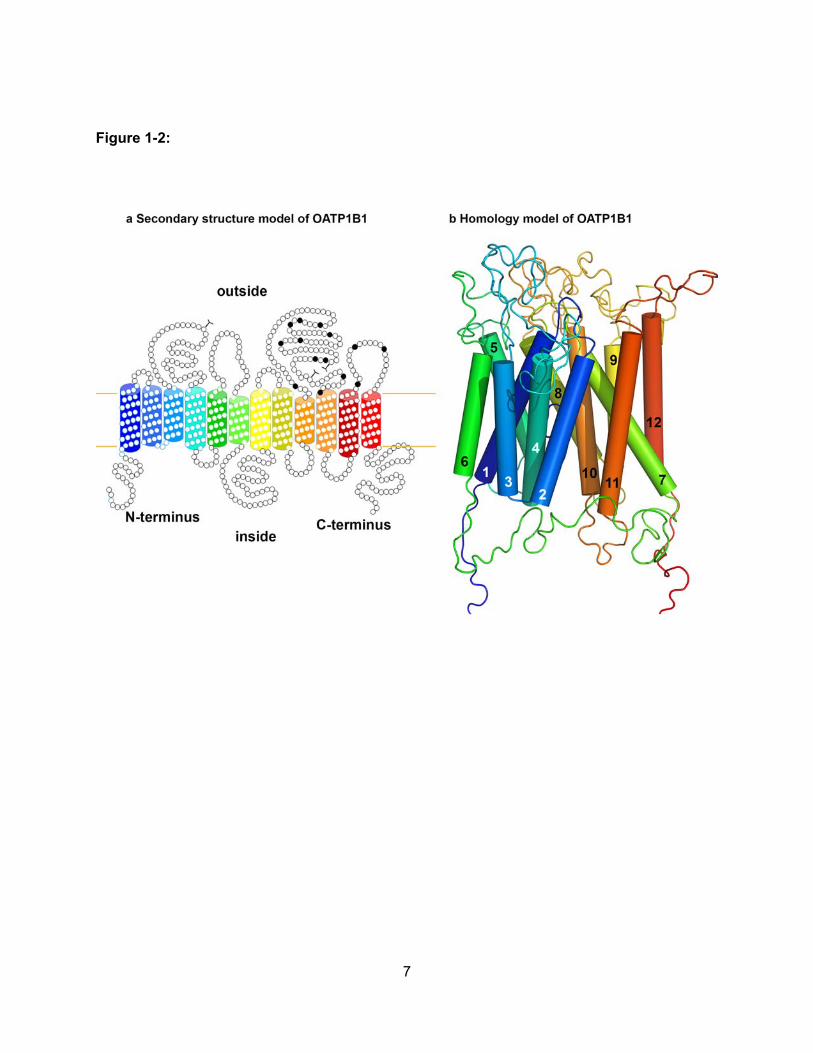
OATPs are predicted to have 12 transmembrane domains with intracellular amino and
carboxy termini (Figure 1-2). Several structural features are important for membrane localization
and transport function. For instance, many OATPs contain a PDZ consensus sequence that is
thought to be important for membrane anchoring (Wang et al., 2005). The fifth extracellular loop
is rich with conserved cysteines, which are thought to form sulfhydryl bonds that may be
important for surface expression (Hanggi et al., 2006). Furthermore, recent structure-function
studies have identified amino acids within transmembrane domain 10 of OATP1B1 and
OATP1B3 that are important in substrate recognition and translocation across the membrane
(Gui and Hagenbuch, 2008; Gui and Hagenbuch, 2009; Miyagawa et al., 2009).
5
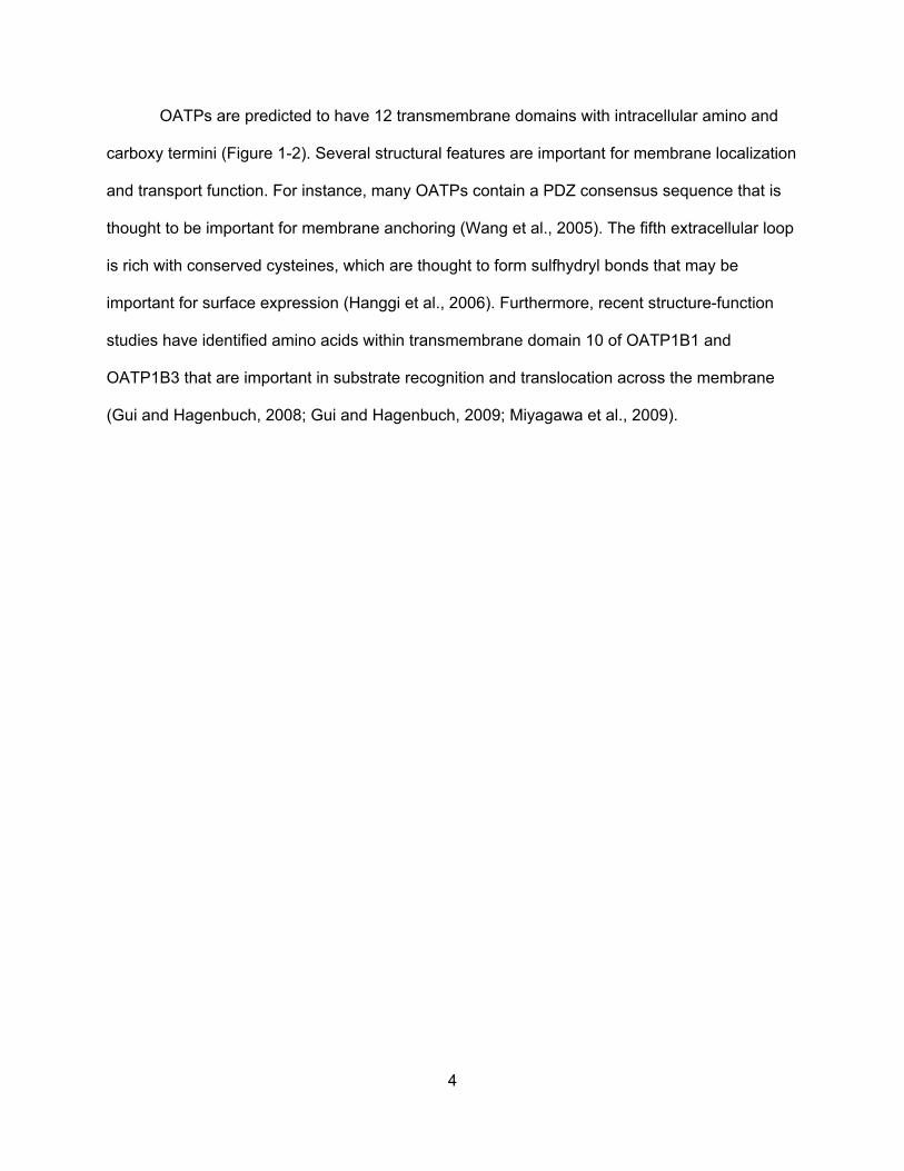
Figure 1-1:
6
Figure 1-1:
Phylogenetic tree of the eleven human OATPs along with their rodent homologs. OATPs
with amino acid sequence identities ≥ 40% belong to the same OATP family (e.g. OATP1,
OATP2, OATP3, OATP4, OATP5, OATP6). OATPs with amino acid sequence identities ≥ 60%
are grouped into subfamilies and represented with a letter after the family number (e.g.
OATP1A, OATP1B, OATP1C, etc.) Individual OATPs are sequentially numbered according to
the chronology of their identification. The Oatp (rodent) and OATP (human) symbols represent
protein, whereas Slco (rodent) and SLCO (human) represent gene symbols. Reproduced with
kind permission from Springer Science+Business Media: (Hagenbuch and Meier, 2004).
7
Figure 1-2:
8
Figure 1-2:
Structure of OATPs. Predicted 12 transmembrane domain (a) secondary structure and (b)
homology model of OATP1B1. (a) Putative transmembrane domains were predicted based on
amino acid hydrophobicity. The highly conserved cysteine residues in the loop between
transmembrane domains 9 and 10 are indicated by black circles. At the C-terminal end, several
OATPs have a PDZ consensus sequence. (b) The homology model for OATP1B1 was
constructed on the basis of the crystal structure of the bacterial multidrug transporter EmrD.
9
1.3.2 Distribution of OATPs in Normal Human Tissue
Some OATPs are expressed in multiple tissues, whereas the expression of other OATPs
is restricted to a single tissue. In the OATP1 family, expression of OATP1A2 mRNA is highest in
the brain, followed by expression in the kidney, liver, lung, testis, and placenta, according to
Northern blot analysis (Kullak-Ublick et al., 1995; Steckelbroeck et al., 2004). OATP1A2
expression was confirmed at the protein level in the blood-brain barrier (Gao et al., 2000; Lee et
al., 2005); at the apical membrane of distal nephrons (Lee et al., 2005); at the apical membrane
of enterocytes, where it is thought to be critical in the absorption of numerous xenobiotics
(Glaeser et al., 2007); and at the apical membrane of cholangiocytes, which make up the bile
duct epithelium (Lee et al., 2005). OATP1B1 and OATP1B3 are examples of tissue-specific
OATPs. Expression of OATP1B1 was shown in adult and fetal liver by Northern blot analysis.
This expression was confirmed at the protein level and localized to the basolateral membrane of
hepatocytes throughout the liver lobule. Similarly, OATP1B3 is expressed exclusively in the liver
at the basolateral membrane of hepatocytes; however, expression was much stronger in the
pericentral region compared with the periportal region (Abe et al., 1999; Hsiang et al., 1999;
Konig et al., 2000b; Konig et al., 2000a; Abe et al., 2001). A recent study based on real-time
polymerase chain reaction (RT-PCR) analysis described OATP1B3 mRNA expression in the
retina; however, protein expression has not been confirmed (Okabe et al., 2008). OATP1C1
mRNA expression is highest in the brain and testis, and, because of its high affinity for T4 and
reverse T3 (in the nanomolar range), OATP1C1 is thought to be a crucial thyroid hormone
transporter (Pizzagalli et al., 2002; Hagenbuch, 2007; Heuer and Visser, 2009). In addition,
OATP1C1 protein was localized to the basolateral membrane of the choroid plexus epithelium
(Roberts et al., 2008), to the basolateral membrane of the pigmented ciliary body epithelium
(Gao et al., 2005), and in leydig cells of the testis (Pizzagalli et al., 2002).
10
OATP2A1, which is also known as the prostaglandin transporter, is thought to be
ubiquitously expressed. OATP2A1 mRNA was detected in brain, colon, heart, liver, kidney,
ovary, lung, pancreas, prostate, skeletal muscle, spleen, and small intestine (Schuster, 2002).
Thus far, expression of OATP2A1 at the protein level has been demonstrated in neurons in the
frontal gyrus of the brain (Choi et al., 2008), in the pyloric glands of the antrum and parietal cells
in the gastrointestinal tract (Mandery et al., 2010), and in the luminal and glandular epithelium of
the endometrium (Kang et al., 2005). OATP2B1, the second member of the OATP2 family, is
expressed in several different tissues in the body, with the highest transcript levels in the liver
(Tamai et al., 2000; Kullak-Ublick et al., 2001). OATP2B1 protein expression was confirmed at
the basolateral membrane of hepatocytes (Kullak-Ublick et al., 2001), at the apical membrane of
enterocytes (Kobayashi et al., 2003), at the endothelium of the blood-brain barrier (Bronger et
al., 2005), at the endothelial cells of the heart (Grube et al., 2006), at the myoepithelium of
mammary ducts (Pizzagalli et al., 2003), and in the placenta (St-Pierre et al., 2002).
OATP3A1 is the most highly conserved OATP among all species and has two different
splice variants in humans (Huber et al., 2007). In general, OATP3A1 is considered to be widely
expressed and was shown to be expressed at the mRNA level in testis, brain, heart, lung,
spleen, peripheral blood leukocytes, and thyroid (Adachi et al., 2003; Huber et al., 2007). At the
protein level, the two splice variants were localized to different cell types or cellular membranes
in various tissues. OATP3A1_v1 was localized to the germ cells of the testes, to the neuroglial
cells of the frontal cortex, and to the basolateral membrane of the choroid plexus. In contrast,
expression of OATP3A1_v2 was shown in Sertoli cells in the testes, at the apical membrane of
the choroid plexus, and in cell bodies of the frontal cortex neurons (Huber et al., 2007).
Recently, OATP3A1 protein expression was demonstrated in epithelial cells of the lactiferous

11
ducts in normal breast tissue (Kindla et al., 2011a). OATP4A1 is another ubiquitously expressed
OATP, with highest mRNA levels in the heart and placenta, followed by levels in the lung, liver,
skeletal muscle, kidney, and pancreas (Tamai et al., 2000; Fujiwara et al., 2001). Thus far,
OATP4A1 protein expression has been confirmed only at the apical membrane of
syncytiotrophoblasts in the placenta (Sato et al., 2003). The other OATP4 family member,
OATP4C1, is expressed only in the kidney, as shown by Northern blot analysis (Mikkaichi et al.,
2004), and thus is considered a kidney-specific OATP. Little is known about OATP5A1 and
OATP6A1. OATP5A1 protein was recently reported to be expressed at the plasma membrane
of the epithelial cells that line the lactiferous ducts in normal breast tissue (Kindla et al., 2011a),
whereas OATP6A1 mRNA expression has been detected in the testes, with low levels in the
spleen, brain, fetal brain, and placenta (Suzuki et al., 2003; Lee et al., 2004).
1.3.3 Expression Profile of OATPs in Cancer Tissue
Aside from their normal expression profiles, OATPs have altered expression in cancer
tissues. OATP1A2 expression has been identified in gliomas, colon polyps and tumors, and
cancers of the breast and bone. A study of human gliomas by RT-PCR showed OATP1A2
expression in different histological subtypes. Through the use of immunofluorescence
microscopy, OATP1A2 was localized in the luminal membrane of the blood-brain barrier
endothelium and in the blood-tumor barrier but not in the glioma cells (Bronger et al., 2005).
Through the use of RT-PCR, OATP1A2 expression was detected in healthy colon tissue;
however, expression was decreased in polyps and in colon cancer tissue (Ballestero et al.,
2006). In breast cancer cell lines, RT-PCR analysis showed that expression of OATP1A2 was
highest in T47-D and ZR-75-1 cells and low in MCF-7, MDA-MB-231, and MDA-MB-468 cells.
OATP1A2 expression was confirmed in tissue obtained from patients with breast cancer and
was localized to the cell membrane and cytoplasm of breast carcinoma cells. However,
12
OATP1A2 expression was not observed in non-neoplastic epithelium, stroma, and adipose
tissue surrounding the carcinoma (Miki et al., 2006). These results were confirmed through the
use of RT-PCR (Meyer zu Schwabedissen et al., 2008; Banerjee et al., 2012). OATP1A2
transcript levels were significantly higher in malignant breast tissue than they were in adjacent
nonmalignant breast tissue, and transcripts of OATP1A2 were highest in stage I and stage IIA
breast cancers (Meyer zu Schwabedissen et al., 2008). Immunofluorescence analysis confirmed
OATP1A2 protein expression and demonstrated that it was restricted to the malignant cells of
the breast tissue samples (Meyer zu Schwabedissen et al., 2008). In contrast, Wlcek et al.
(Wlcek et al., 2008) were unable to detect significant mRNA levels of OATP1A2 in the four
breast cancer cell lines MCF-7, MDA-MB-231, ZR-75-1, and MCF-10A, or in breast cancer
tissue. These discrepancies might arise from differences found in cell lines cultured in different
laboratories (Hayeshi et al., 2008); therefore, such results must be interpreted cautiously if the
exact experimental conditions are not known. OATP1A2 transcripts were detected both in bone
metastases from primary kidney cancer and in the malignant osteosarcoma cell lines HOS and
MG-63 (Liedauer et al., 2009).
In general, the expression of OATP1B1 and OATP1B3, both specific to the liver, tends to
be reduced in hepatocellular carcinomas (HCCs). OATP1B1 and OATP1B3 mRNA was
undetectable or reduced in the Hep3B and HepG2 cell lines (Libra et al., 2006; Monks et al.,
2007), confirming the previously reported reduced expression in HepG2 and PLC cell lines as
well as in HCC tissue samples at the protein level (Cui et al., 2003; Zollner et al., 2005).
Vavricka et al. (Vavricka et al., 2004) also reported reduced expression of OATP1B3 in 60% of
HCC tissues compared with normal surrounding tissue. However, OATP1B1 levels were not
significantly different in HCC samples compared with normal liver samples in their study. The
expression of OATP1B1 and OATP1B3 in different benign liver tumors was investigated by
Vander Borght and colleagues (Vander Borght et al., 2005). They showed reduced expression
13
of both OATPs in hepatocellular adenomas and a strong diffuse expression of both OATPs in
focal nodular hyperplasia. Recently, Tsuboyama et al. (Tsuboyama et al., 2010) supported the
general trend of reduced OATP1B1 and OATP1B3 expression in HCC tissue samples. In
conclusion, the downregulation of OATP1B1 and OATP1B3 in HCC resembles the
downregulation observed in primary cultured hepatocytes (Jigorel et al., 2005) and could be the
result of dedifferentiation of the HCC cells.
Expression of the normally liver-specific OATP1B1 and OATP1B3 has also been
identified in cancers of many different tissues (Abe et al., 2001). Overall, OATP1B3 is
upregulated in a wide range of cancer types. Northern blot analysis showed that OATP1B3 is
expressed in different gastrointestinal cancer cell lines and cancers, including the gastric cancer
cell line KatoIII; the colon cancer cell lines DLD-1, MIP-101, Clone A, and CX-1; the pancreatic
cancer cell lines MIA-Paca2, BXPC-1, PK-8, PK-9, and PK-45P; and the gallbladder cancer cell
lines HuCCT-1, OcuchLM1, and TFK-1. Weak expression levels were also seen in the lung
cancer cell line A549 and the glioblastoma cell line A172. Moreover, immunohistochemical
staining of OATP1B3 was detected in gastric cancer tissue, pancreatic cancer tissue, colon
cancer tissue, and a colon cancer metastatic to a lymph node (Abe et al., 2001). A recent study
showed OATP1B1 expression in the breast cancer cell line MDA-MB-231 via western blot
(Banerjee et al., 2012). OATP1B1 was increased in colon polyps and in colon cancer tissue at
the mRNA level as compared with normal colon tissue (Ballestero et al., 2006). However, the
same study could not demonstrate significant differences in OATP1B3 mRNA expression
between healthy and colon cancer tissue (Ballestero et al., 2006). OATP1B3 expression was
markedly increased in colorectal adenocarcinoma tissues with obvious staining in the cytoplasm
as opposed to membranous expression in normal liver (Lee et al., 2008). Analysis of OATP1B3
expression across different colorectal tumor stages showed that it was highest in earlier-stage
and lower-grade tumors, suggesting that OATP1B3 expression might be indicative of clinical
14
outcome (Lockhart et al., 2008). In non-small-cell lung cancer, OATP1B3 mRNA expression
was significantly increased as compared with the nonmalignant surrounding tissue (Monks et
al., 2007). OATP1B3 transcript levels were observed in prostate cancer tissue through the use
of RT-PCR (Wright et al., 2011). Additionally, OATP1B3 expression was confirmed at the
protein level in prostate tumor tissue but not confirmed in normal prostate or benign prostatic
hyperplasia (Hamada et al., 2008). Muto et al. (Muto et al., 2007) detected OATP1B3 by using
immunohistochemistry in cells of invasive ductal breast carcinoma and suggested that
OATP1B3 expression could be used as a prognostic factor in breast cancer.
Thus far, the only report to show expression of OATP1C1 in cancer demonstrated
OATP1C1 mRNA in several samples from osteosarcomas, in a sample from a kidney cancer
metastasis, and in several specimens from aneurysmal bone cysts, which had the highest levels
(Liedauer et al., 2009).
OATP2A1 mRNA was detected at high levels in bone metastases from kidney cancer
(Liedauer et al., 2009); in breast cancer; and in the breast cancer cell lines MCF-7, MDA-MB-
231, and ZR-75-1 (Wlcek et al., 2008). OATP2A1 mRNA expression was generally higher in
malignant breast tissue compared with adjacent nonmalignant breast tissue, but the difference
did not reach statistical significance (Wlcek et al., 2008). Holla and colleagues (Holla et al.,
2008) showed a trend for decreased OATP2A1 expression in colorectal tumor specimens as
well as in stomach, ovary, lung, and kidney tumors. They were not able to detect OATP2A1 in
several colorectal cancer cell lines, including LS-174T, HCT-116, HT-29, SW-620, SW-480,
HCT-15, and HCA-7, although the colorectal cancer cell line LoVo showed high expression
levels of OATP2A1 mRNA and protein (Holla et al., 2008). OATP2A1 was also detected at the
protein level in hepatocellular carcinoma, in cholangiocellular carcinoma, and in liver
metastases from colon tumors (Wlcek et al., 2011).
15
OATP2B1 mRNA expression was identified in the colon adenocarcinoma cell line CX-1
(Tamai et al., 2000) and was higher in bone cysts than in osteosarcoma tissues (Liedauer et al.,
2009). With respect to breast cancer, OATP2B1 expression was shown in both normal and
breast tumor specimens, and expression increased with increased tumor grade (Al Sarakbi et
al., 2006). This has also been studied by Wlcek et al. (Wlcek et al., 2008), who detected higher
expression levels of OATP2B1 in nonmalignant specimens than in malignant breast tumors.
OATP2B1 expression was also identified in human gliomas, where it was localized to
endothelial cells at the blood-brain barrier and blood-tumor barrier (Bronger et al., 2005).
RT-PCR analysis showed that OATP3A1 expression was significantly higher in
aneurismal bone cysts than in osteosarcomas. OATP3A1 transcripts were found (a) in the
nonmalignant human osteoblast-like cells and bone marrow stromal cells derived from normal
bone marrow and (b) in the osteosarcoma cell lines HOS and MG-63 (Liedauer et al., 2009).
OATP3A1 expression was identified in a variety of additional cancer cell lines, including breast
carcinoma (GI-101), lung carcinoma (LX-1 and GI-117), colon adenocarcinoma (CX-1 and GI-
112), ovarian carcinoma (GI-102), and pancreatic adenocarcinoma (GI-103) (Tamai et al.,
2000). Through RT-PCR, two independent groups showed OATP3A1 expression in the breast
cancer cell line T-47D (Pizzagalli et al., 2003; Nozawa et al., 2004). OATP3A1 expression was
also detected in the breast cancer cell line MCF-7 (Nozawa et al., 2005b), and recently it was
detected in the membrane and cytoplasm of malignant breast tumor specimens (Kindla et al.,
2011a).
OATP4A1 has an expression pattern similar to that of OATP3A1 in various breast
carcinoma, lung carcinoma, colon adenocarcinoma, ovarian carcinoma, and pancreatic
carcinoma cell lines (Tamai et al., 2000), as well as in the breast cancer cell lines T-47D and
MCF-7 (Pizzagalli et al., 2003; Nozawa et al., 2004). OATP4A1 expression is higher in bone
16
cysts than in osteosarcoma tissues, with significantly higher expression in the malignant
osteosarcoma cell lines HOS and MG63 as compared with the nonmalignant human osteoblast-
like cells and bone marrow stromal cells (Liedauer et al., 2009). The expression of OATP4A1
was detected in normal and tumorous breast tissue (Wlcek et al., 2008). In the same study,
expression of OATP4C1 and the poorly characterized OATP5A1 was also revealed in normal
and cancerous breast tissue (Wlcek et al., 2008). Recently, the expression of OATP5A1 was
confirmed at the membrane and in the cytoplasm of malignant breast tumor specimens (Kindla
et al., 2011a). Expression of the presumably gonad-specific OATP6A1 was shown in lung
cancer cell lines, lung cancer, bladder cancer, and esophageal cancer tissues (Lee et al., 2004).
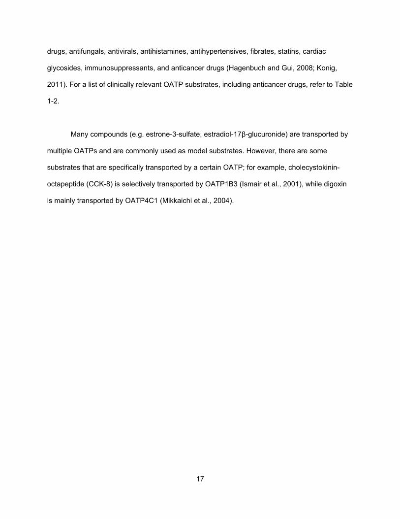
The tissue expression profile of all 11 human OATPs is summarized in Table 1-1.
A few studies have suggested that OATP expression in cancer could be predictive of
patient survival or the success of hormone therapy (Muto et al., 2007; Hamada et al., 2008;
Lockhart et al., 2008). However, because of the many inconsistent reports, it is too early to
propose that the presence or absence of a certain OATP in a given cancer is predictive.
Additional, larger population-based studies are required to confirm the predictive value of
OATPs in different cancer types.
1.3.4 Substrate Specificity
OATPs transport a wide range of structurally unrelated compounds, including numerous
endo- and xenobiotics (Hagenbuch and Gui, 2008; Konig, 2011). Their substrates are generally
organic anions, but OATPs can also transport cations and neutral compounds (van Montfoort et
al., 2003). Among their endogenous substrates are bile acids, conjugated steroid hormones,
thyroid hormones, and cyclic and linear peptides. OATPs are important for drug disposition
because their exogenous substrates include antibiotics, antidiabetic drugs, anti-inflammatory
17
drugs, antifungals, antivirals, antihistamines, antihypertensives, fibrates, statins, cardiac
glycosides, immunosuppressants, and anticancer drugs (Hagenbuch and Gui, 2008; Konig,
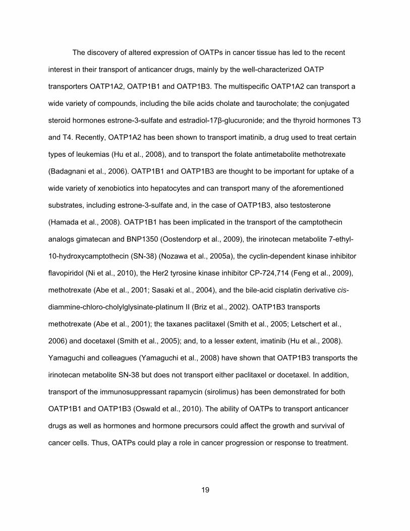
2011). For a list of clinically relevant OATP substrates, including anticancer drugs, refer to Table
1-2.
Many compounds (e.g. estrone-3-sulfate, estradiol-17β-glucuronide) are transported by
multiple OATPs and are commonly used as model substrates. However, there are some
substrates that are specifically transported by a certain OATP; for example, cholecystokinin-
octapeptide (CCK-8) is selectively transported by OATP1B3 (Ismair et al., 2001), while digoxin
is mainly transported by OATP4C1 (Mikkaichi et al., 2004).
18
Table 1-1: Tissue Expression of OATPs in Normal and Cancer Tissues
OATP Normal tissue expression Cancer tissue expression
OATP1A2 Blood brain barrier
Cholangiocytes
Kidney
Enterocytes
Expressed in bone cancer tissues and cell lines
Reduced in colon polyps and cancer
Increased in breast carcinoma cells and
malignant breast tissue
OATP1B1 Liver Reduced in Hepatocellular carcinoma
OATP1B3 Liver
Reduced in Hepatocellular carcinoma
Expressed in: Colorectal adenocarcinoma tissues
Non-small cell lung tumors
Prostate cancer tissue
Invasive ductal carcinoma breast cells
Cell lines of stomach, colon, pancreatic, and gall
bladder cancers
OATP1C1 Brain
Testes
Ciliary body
Expressed in bone cancers
OATP2A1 Ubiquitous
Increased in malignant breast tissue and liver cancers
Reduced in tumors of bowel, stomach, ovary, lung and kidney
OATP2B1 Liver
Blood-brain barrier
Enterocytes
Placenta
Heart
Increased in bone cysts
Altered in breast cancers
OATP3A1 Ubiquitous Expressed in bone cancer and cancer cell lines of
multiple tissues
OATP4A1 Ubiquitous Expressed in bone cancer and cancer cell lines of
multiple tissues
OATP4C1 Kidney
OATP5A1 Lactiferous ducts in breast Expressed in malignant breast tumors
OATP6A1 Testes Expressed in tumors of the lung, bladder and esophagus
19
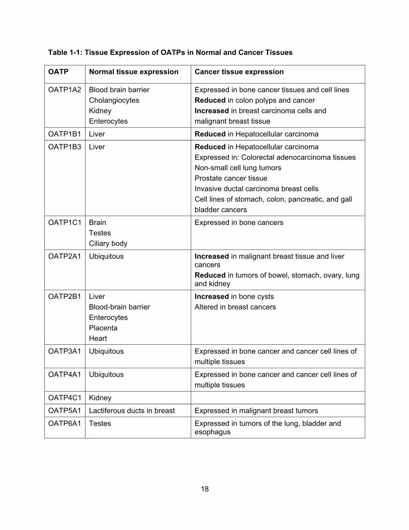
The discovery of altered expression of OATPs in cancer tissue has led to the recent
interest in their transport of anticancer drugs, mainly by the well-characterized OATP
transporters OATP1A2, OATP1B1 and OATP1B3. The multispecific OATP1A2 can transport a
wide variety of compounds, including the bile acids cholate and taurocholate; the conjugated
steroid hormones estrone-3-sulfate and estradiol-17β-glucuronide; and the thyroid hormones T3
and T4. Recently, OATP1A2 has been shown to transport imatinib, a drug used to treat certain
types of leukemias (Hu et al., 2008), and to transport the folate antimetabolite methotrexate
(Badagnani et al., 2006). OATP1B1 and OATP1B3 are thought to be important for uptake of a
wide variety of xenobiotics into hepatocytes and can transport many of the aforementioned
substrates, including estrone-3-sulfate and, in the case of OATP1B3, also testosterone
(Hamada et al., 2008). OATP1B1 has been implicated in the transport of the camptothecin
analogs gimatecan and BNP1350 (Oostendorp et al., 2009), the irinotecan metabolite 7-ethyl-
10-hydroxycamptothecin (SN-38) (Nozawa et al., 2005a), the cyclin-dependent kinase inhibitor
flavopiridol (Ni et al., 2010), the Her2 tyrosine kinase inhibitor CP-724,714 (Feng et al., 2009),
methotrexate (Abe et al., 2001; Sasaki et al., 2004), and the bile-acid cisplatin derivative cis-
diammine-chloro-cholylglysinate-platinum II (Briz et al., 2002). OATP1B3 transports
methotrexate (Abe et al., 2001); the taxanes paclitaxel (Smith et al., 2005; Letschert et al.,
2006) and docetaxel (Smith et al., 2005); and, to a lesser extent, imatinib (Hu et al., 2008).
Yamaguchi and colleagues (Yamaguchi et al., 2008) have shown that OATP1B3 transports the
irinotecan metabolite SN-38 but does not transport either paclitaxel or docetaxel. In addition,
transport of the immunosuppressant rapamycin (sirolimus) has been demonstrated for both
OATP1B1 and OATP1B3 (Oswald et al., 2010). The ability of OATPs to transport anticancer
drugs as well as hormones and hormone precursors could affect the growth and survival of
cancer cells. Thus, OATPs could play a role in cancer progression or response to treatment.
20
Table 1-2: Selected Substrates of OATPs. Anticancer drugs are highlighted in red.
OATP1A2 OATP1B1 OATP1B3 OATP1C1
Hormones and conjugates Estradiol-17β-glucuronide Estrone-3-sulfate DHEA-S Reverse triiodothyronine (rT3) Thyroxine (T4) Triiodothyronine (T3) Prostglandins Prostaglandin E2 Bile acids Cholate Taurocholate Glycocholate Taurochenodeoxycholate Tauroursodeoxycholate Others DPDPE Drugs Acebutolol Rosuvastatin Atenolol Pitavistatin Sotolol Fexofenadine Labetalol Deltorphin II Nadolol Ciprofloxacin Talinolol Gatifloxacin Saquinavir Imatinib Darunavir Levofloxacin Ouabain Methotrexate
Hormones and conjugates Estradiol-17β-glucuronide Estrone-3-sulfate Thyroxine (T4) Triiodothyronine (T3) DHEA-S Prostglandins Prostaglandin E2 Bile acids Cholate Taurocholate Tauroursodeoxycholate Drugs Atorvastatin Olmesartan Lopinavir Phalloidin Darunavir Caspofungin SN-38 Pitavastatin ValsartanCefazolin Enalapril Pravastatin EzetimibeCerivastatin Bosentan Rifampicin SaquinavirRosuvastatin Fluvastatin Temocapril Gimatecan Troglitazone Methotrexate Demethylphalloin
Hormones and conjugates Estradiol-17β-glucuronide Estrone-3-sulfate DHEA-S Drugs Atresentan Dimethylphalloin Bosentan Paclitaxel Cefadroxil Docetaxel Cefazolin Methotrexate Cephalexin Imatinib Digoxin Olmesartan Enalapril Phalloidin Lopinavir Telmisartan SN-38 Fexofenadine Fluvastatin Gimatecan Pitavastatin Telmisartan Rifampicin Rosuvastatin Valsartan
Hormones and conjugates Estradiol-17β-glucuronide Estrone-3-sulfate Thyroxine (T4) Triiodothyronine (T3) Reverse triiodothyronine (rT3) Thyroxine sulfate (T4S) Others BSP
OATP2A1 OATP2B1 OATP3A1 OATP4A1
Prostaglandins Prostaglandin E1 Prostaglandin E2 Prostaglandin F2α Prostaglandin H2 Prostaglandin D2
8-iso-prostaglandin F2α
Thromboxane B2 Drugs Latanoprost
Hormones and conjugates Estrone-3-sulfate DHEA-S Thyroxine (T4) Prostglandins Prostaglandin E2 Drugs Atorvastatin Bosentan Ezetimibe Fluvastatin Glibenclamide Pitavastatin
Hormones and conjugates Thyroxine (T4) Estrone-3-sulfate Prostglandins Prostaglandin E1 Prostaglandin E2 Prostaglandin F2α Drugs Deltorphin BQ-123 Benzylpenicillin Others Vasopressin
Hormones and conjugates Estradiol-17β-glucuronide Estrone-3-sulfate Thyroxine (T4) Triiodothyronine (T3) Reverse triiodothyronine (rT3) Bile acids Prostglandins Drugs
21
1.3.5 Regulation
A recent review provides a detailed summary of the current knowledge about OATP
regulation (Svoboda et al., 2011a). However, there have not been extensive studies on how
OATP expression is regulated within cancer cells. OATP1A2 expression has been associated
with expression of the nuclear receptor PXR (pregnane X receptor) in breast carcinoma tissue
and its cell lines (Miki et al., 2006). In addition, a PXR response element was identified in the
OATP1A2 promoter (Meyer zu Schwabedissen et al., 2008), suggesting that PXR could play a
role in the upregulation of OATP1A2 seen in breast cancers. In 2004, it was suggested that
OATP1B3 expression in HCC was downregulated via transcriptional repression by hepatocyte
nuclear factor 3β (HNF3β) (Vavricka et al., 2004). A more recent study showed that DNA
methylation-dependent gene silencing is involved in the regulation of OATP1B3 expression in
several cancer cell lines (Ichihara et al., 2010). Posttranslational regulation also could be altered
in cancer cells. Although OATPs are normally expressed on cell membranes, strong
cytoplasmic staining is seen for both OATP1A2 in breast cancer andOATP1B3 in colon cancer.
This could result from aberrant posttranslational regulation such as altered phosphorylation,
which regulates cell surface expression of human OATP2B1 (Kock et al., 2010), rat OATP1a1,
and rat OATP1a4 (Guo and Klaassen, 2001; Choi et al., 2011). Owing to the limited knowledge
in this area, further research is required to better understand the mechanism of OATP up- or
downregulation in cancer and to determine what role, if any, it may play in cancer progression or
treatment.
22
1.3.6 Polymorphisms and Drug Disposition
Several studies have documented single-nucleotide polymorphisms (SNPs) of OATPs
that are associated with reduced or absent protein function. Several of these SNPs have been
linked to altered disposition of chemotherapeutic drugs and consequently increased adverse
effects, confirming the importance of OATPs in the disposition of drugs. For example,
methotrexate, a substrate of OATP1A2, has been implicated in serious adverse effects seen in
patients. To identify potential contributors to varying methotrexate pharmacokinetics, common
OATP1A2 polymorphisms were investigated (Badagnani et al., 2006). Among the 12 OATP1A2
polymorphisms studied, four showed altered transport of methotrexate in vitro. The common
I13T OATP1A2 SNP showed enhanced methotrexate uptake, whereas R168C, E172D and
N278DEL variants showed significantly decreased methotrexate uptake (Badagnani et al.,
2006). In addition, common polymorphisms of OATP1B1 may alter disposition of the irinotecan
metabolite SN-38 and potentially contribute to the variable adverse gastrointestinal effects seen
with this drug (Nozawa et al., 2005a). Uptake of SN-38 was determined in cell lines expressing
three common genetic polymorphisms of OATP1B1. Compared with the wild-type OATP1B1*1a,
OATP1B1*15, which has an allelic frequency of 10.3% to 15.0%, had significantly reduced SN-
38 uptake, suggesting that patients with this polymorphism may have altered SN-38
pharmacokinetics (Nozawa et al., 2005a). This prediction was confirmed in vivo: Patients with
the SLCO1B1*15 polymorphism showed higher systemic exposure and lower clearance of SN-
38 (Xiang et al., 2006; Takane et al., 2009). A similar study looked at the effect of three
nonsynonymous SNPs of OATP1B3 on the pharmacokinetics of paclitaxel (Smith et al., 2007).
However, none of the three genetic variations resulted in significantly different paclitaxel
clearance or altered pharmacokinetic parameters.
23
Recently, van de Steeg et al. (van de Steeg et al., 2011) generated the knockout mouse
model Slco1a/1b−/−, which lacks all OATP1A and OATP1B family members, to study the role of
OATPs in the disposition of drugs such as paclitaxel and methotrexate. The Slco1a/1b−/− mice
had higher plasma concentrations of both drugs, suggesting that proteins in the OATP1A or
OATP1B family are involved in their distribution. Introducing polymorphic human OATPs into
these null mice could provide a powerful tool to study their effect on drug disposition (van de
Steeg et al., 2011).
1.3.7 OATPs and Cancer Development
As briefly discussed above, hormones and their conjugates are substrates of many
OATPs, and therefore expression of OATPs in cancer might contribute to the proliferation of
androgen- and estrogen-dependent tumors. Several studies have investigated whether OATP
expression could affect the growth of hormone-dependent cancers such as breast and prostate
cancers.
In 2004, Nozawa et al. showed that uptake of estrone-3-sulfate into T47-D breast cancer
cells led to increased cell proliferation and was mediated by a Na+-independent transport
system (Nozawa et al., 2004). The authors detected OATP3A1 and OATP4A1 in these cells and
suggested that these OATPs might be involved in estrone-3-sulfate uptake into these cells. In a
follow-up study, estrone-3-sulfate uptake was measured into the breast cancer cell line MCF-7,
and inhibition studies suggested that an OATP could be involved (Nozawa et al., 2005b). On the
basis of these findings, inhibition of the estrone-3-sulfate uptake system was proposed as a
potential treatment for estrogen-responsive breast cancers (Nozawa et al., 2005b). Additionally,
recent studies suggested OATP1B3 as one of the transporters involved in the uptake of
estrone-3-sulfate into the breast cancer cell line MCF-7 (Maeda et al., 2010). Meyer zu
24
Schwabedissen et al. (Meyer zu Schwabedissen et al., 2008; Banerjee et al., 2012) showed that
expression of OATP1A2 in T47-D cells was regulated via the nuclear receptor PXR, and
treatment of these cells with the PXR activator rifampicin resulted in both increased expression
of OATP1A2 and increased proliferation of the cells. Furthermore, when these estrogen-
dependent cells were treated with the potent PXR antagonist A-792611, both decreased
OATP1A2 expression and decreased proliferation were observed. This led the authors to
propose targeting the regulation of OATP1A2 as a potential treatment for breast cancer (Meyer
zu Schwabedissen et al., 2008).
OATP1B3 protein expression in various breast carcinomas has been correlated with
various pathological parameters including tumor size, recurrence, and prognosis (Muto et al.,
2007). OATP1B3 expression was shown in 50% of the breast cancer specimens analyzed.
Surprisingly, expression of OATP1B3 was inversely correlated with tumor size and significantly
associated with decreased recurrence and good prognoses. However, this correlation was seen
only in postmenopausal women. OATP1B3 has thus been hypothesized to be implicated in a
hormone-dependent growth mechanism in breast cancer (Muto et al., 2007).
Prostate cancer recurrence and progression have also been correlated with the
expression of OATPs. An OATP1B3-related survival advantage for certain patients was seen in
androgen dependent prostate cancer (Hamada et al., 2008). Although testosterone is a
substrate of wild-type OATP1B3, a variant containing two SNPs, 334G and 699A, does not
transport testosterone. Patients with the 334GG/699AA haplotype had an improved overall
survival over 10 years (Hamada et al., 2008). A separate study demonstrated that patients with
the 334T allele of OATP1B3, which does transport testosterone, had an increased prostate
cancer mortality rate (Wright et al., 2011). This study also showed that patients with the
rs12422149 polymorphism in the SLCO2B1 gene, which had previously been shown to affect
25
OATP2B1-mediated substrate uptake, had an increased risk of prostate-cancer-specific
mortality (Wright et al., 2011). Immunofluorescence micrographs of OATP1B3 staining on
different prostate cancer tissue sections demonstrated that OATP1B3 expression was primarily
observed in the stroma of prostate cancer tumors and not in normal prostate tissue (Pressler et
al., 2011).
OATP1B3 may also be involved in the response to anticancer chemotherapy.
Overexpression of wild-type OATP1B3 in colon cancer cell lines treated with camptothecin and
oxaliplatin conferred an antiapoptotic effect and reduced the transcriptional activity of p53,
whereas a nonfunctional OATP1B3 did not have these effects (Lee et al., 2008).
Taken together, these studies demonstrate that OATP expression in cancer can be
associated with a survival advantage or disadvantage for the tumor. However, additional
research is required to elucidate the underlying molecular mechanisms and to identify the
transported substrates that are potentially involved before such findings will lead to improved
tumor diagnostics and treatment.
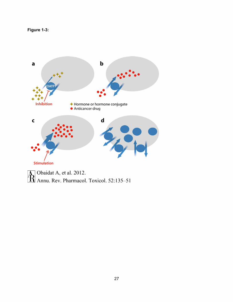
1.3.8 Mechanisms of OATP-mediated Cancer Therapy
OATPs are expressed throughout the body and are generally responsible for the Na+-
independent uptake of a wide range of amphipathic compounds. Expression levels of OATPs
are altered in many different types of cancers, and in several cancers, these altered expression
levels have been correlated with cancer stage. OATPs are capable of transporting multiple
compounds that affect cancer cell growth and survival, including hormones, hormone
precursors, and anticancer drugs.
26
Furthermore, uptake mediated by OATPs can be either inhibited or allosterically
stimulated by small molecules. In addition, OATP polymorphisms have been associated with
altered pharmacokinetics of anticancer drugs, altered transport of hormones, and cancer
outcomes. These findings suggest that OATPs could be valuable targets for anticancer therapy
in four ways: (a) OATP-mediated uptake of hormones, hormone conjugate, or unidentified
growth promoting chemicals could be prevented with OATP-selective inhibitors (Figure 1-3a);
(b) novel anticancer drugs could be developed as OATP substrates to increase their uptake into
OATP-expressing cancer cells (Figure 1-3b); (c) uptake of anticancer drugs could be enhanced
by allosteric stimulators (Figure 1-3c); and (d ) expression of OATPs in the plasma membrane
could be modulated to increase or decrease uptake of various substrates into cancer cells
(Figure 1-3d). Further research is required to elucidate the role OATPs play in cancer
development and anticancer drug transport and to determine how these uptake transporters can
be rationally targeted in cancer treatment.
27
Figure 1-3:
28
Figure 1-3: Mechanisms of OATP-mediated Anticancer Therapy. (a) Because OATPs can
transport hormones, hormone conjugates and additional chemicals (yellow-brown diamonds),
that are beneficial for cancer growth, inhibition of the uptake of these “procancer” compounds
into cancer cells could have antiproliferative effects. (b) OATPs can transport known anticancer
drugs (red circles). Therefore, investigators could attempt anticancer therapy by designing novel
chemotherapeutics that are OATP substrates. (c) Because some OATPs can be allosterically
stimulated with small molecules, uptake of anticancer drugs (red circles) could be enhanced in
the presence of such stimulators. (d) The membrane expression of OATPs in cancer cells could
be regulated to increase or decrease transport into these cells.
29
1.4 Specific Aims of this Dissertation
Despite the discovery of numerous novel anticancer chemotherapeutic drugs, the
problem of multidrug resistance remains a recurrent challenge in cancer treatment (Jemal et al.,
2009). One of the mechanisms that contribute to multidrug resistance is the overexpression of
efflux pumps, such as MDR1 or MRP1, which decrease the intracellular concentrations of these
drugs in cancer cells (Gottesman et al., 2002). Therapeutic approaches to eliminate this
problem currently focus on inhibiting the function of these efflux transporters. Unfortunately the
results of these strategies have so far not been very promising (Daenen et al., 2004; Pusztai et
al., 2005; Friedenberg et al., 2006; Saeki et al., 2007). Thus, there is an urgent need for an
alternative approach to improve drug delivery and overcome multidrug resistance in cancer
treatment. Under normal physiological conditions Organic Anion Transporting Polypeptides
(OATPs) are expressed in certain tissues where they are responsible for the transport of
numerous endogenous compounds and xenobiotics. Recently, OATPs have been found to have
altered expression profiles in cancer tissues. The function of OATPs in cancer cells has not yet
been investigated in detail but because several OATPs have been shown to transport
anticancer drugs, OATPs have been proposed to be potential targets to improve cancer therapy
(Obaidat et al., 2012). By better understanding the expression and function of OATPs in cancer
cells we may be able to improve cancer diagnostics and therapies.
The studies presented in this dissertation were designed to address the lack of
understanding of how OATPs can be used as cancer biomarkers or in cancer drug delivery.
Therefore, the long term goal of the studies presented in this dissertation is to improve cancer
diagnosis and therapy. The objective of this dissertation was to characterize the expression of
OATPs in cancer and to determine their contribution to the transport of cytotoxic
chemotherapeutic drugs into cancer cells. The central hypothesis of this dissertation was that
30
expression of OATPs in cancer combined with their ability to transport cytotoxic
anticancer drugs makes them potential targets for improving cancer diagnosis and
therapy. I formulated this hypothesis based on our preliminary studies that showed that
OATP1B3 can mediate the uptake of several anticancer drugs including etoposide and
paclitaxel and that small molecules can stimulate OATP1B3 mediated uptake of model
substrates. By testing this central hypothesis I have discovered novel information about OATPs
in cancer and expect that this information will lead to a better understanding of the roles OATPs
play in cancer and to an improved diagnosis and drug delivery via OATPs.
The central hypothesis was tested via the following specific aims:
1.4.1: Specific Aim 1: Identify and characterize cancer cells that express OATPs.
Our working hypothesis was that certain OATPs are expressed in human cancers. To
test this hypothesis, we measured mRNA and protein expression of OATPs in several different
cancer tissues on a tissue microarray. Expression of OATPs was further characterized in
pancreatic cancer tissues of different types and stages. Completion of this specific aim identified
four major OATPs upregulated in pancreatic cancer. Furthermore these studies demonstrated
that expression of OATP1B3 was highest in earlier stage pancreatic adenocarcinomas, and
provide a basis for utilizing OATP1B3 as a diagnostic marker and early therapeutic target.
31
1.4.2: Specific Aim 2: Identify and functionally characterize anticancer drug
uptake mediated by OATPs.
Our working hypothesis was that OATPs can transport a variety of anticancer drugs.
Anticancer drugs that interact with OATPs were identified by screening the NCI/DTP oncology
drug set for 1) inhibition of OATP-mediated uptake of two model substrates and 2) for their
effect on cell viability of OATP-expressing cells. Uptake of selected anticancer drugs was
measured. Completion of this specific aim identified etoposide, oxaliplatin, and plicamycin as
three novel anticancer drug substrates of OATP1B3.
1.4.3: Specific Aim 3: Identify cytotoxic compounds from plant extracts that can
be used to develop anticancer drugs that target OATP1B3-expressing cancers.
Our working hypothesis was that Kansas plants are a source for cytotoxic OATP1B3
substrates that can be developed into novel anticancer drugs. A bioassay guided isolation
approach coupled with cell viability assays identified the most potent sub-fractions of two
cytotoxic Kansas plant extracts that were shown to kill cells expressing OATP1B3.
32
Chapter 2
Experimental Materials and Methods
2.1: Materials
[3H]Estrone-3-sulfate and [3H]estradiol-17β-glucuronide were purchased from
PerkinElmer Life and Analytical Sciences (Waltham, MA). [14C]Etoposide and [14C]Oxaliplatin
were purchased from American Radiolabeled Chemicals (St. Louis, MO). The approved
oncology drug set II library of 89 drugs for the treatment of cancer was obtained from the
NCI/DTP Open Chemical Repository (http://dtp.cancer.gov), compounds were dissolved in
DMSO at 10mM concentrations in 96-well plates. Anticancer drugs for confirmatory experiments
were also obtained from NCI/DTP Open Chemical Repository or purchased from Sigma (St.
Louis,MO) or Toronto Research Chemicals (North Tork, Ontario).
The fresh frozen human tissues used in these studies were obtained under IRB approval
of the University of Kansas Cancer Center and provided by the Biospecimen Shared Resource
of the University of Kansas Cancer Center (Kansas City, KS). Quantigene Plex 2.0 System
Reagents were purchased from Panomics Inc. (Fremont, CA). Fetal bovine serum was obtained
from Hyclone (Logan, UT). Anti-OATP1B3, OATP2A1, OATP3A1 and OATP4A1 antibodies
used in the immunoflourescence analyses were purchased from Santa Cruz Biotechnology Inc.
(Santa Cruz, CA). The anti-OATP1B3 antibody used for immunohistochemical staining analyses
on the tissue microarray was raised against the last 14 amino acids at the C-terminal end of
OATP1B3 and was obtained from Sigma (St. Louis, MO). The pancreas tissue microarray was
purchased from US Biomax (Rockville, MD). Dulbecco's modified Eagle's medium (low glucose)
33
was purchased from Caisson Laboratories (North Logan, UT). All other chemicals were
purchased from Sigma-Aldrich or Toronto Research Chemicals.
2.2: Cell culture
Chinese Hamster Ovary (CHO) cells stably expressing OATP1B3 and wild-type CHO
cells were described previously (Gui et al., 2008; Roth et al., 2011b) and were grown at 37 °C in
a humidified 5% CO2 atmosphere in Dulbecco’s Modified Eagle Medium, containing 1 g/l D-
glucose, 2 mM L-glutamine, 25 mM Hepes buffer and 110 mg/l sodium pyruvate, supplemented
with 10% FBS (Hyclone, Logan, UT), 50 µg/ml L-proline, 100 U/ml penicillin and 100 µg/ml
streptomycin (non-select medium), and for the OATP1B3 expressing cells 500 μg/ml G-418
(select medium). Cells were passaged twice a week and used up to passage 60.
2.3: Quantigene Multiplex Assays
Frozen pancreas tissue samples were homogenized with a glass-teflon tissue
homogenizer in a hypotonic buffer (1mM NaCl, 5mM Tris-HCl pH 7.5) containing protease
inhibitors (Complete Protease Inhibitor Cocktail, Roche, Indianapolis, IN) and mRNA expression
of OATPs was determined using the QuantiGene Plex 2.0 Reagent System (Panomics Inc.,
Fremont, CA). Bead-based oligonucleotide probe sets specific for all eleven human OATP
genes were developed by Panomics Inc. Assays were performed according to the
manufacturer’s protocol (Panomics Inc). Briefly, tissue homogenates were lysed with lysis buffer
containing proteinase K. Samples were incubated with capture bead probe set oligonucleotides,
34
capture extenders, label extenders and blocking probes in a 96 well hybridization plate. The
hybridization plate was sealed and incubated at 54ºC while shaking at 300 rpm for 18 hours.
The hybridization plate was centrifuged at 240 x g for 1 minute and samples were transferred to
a 96 well pre-wet filter plate. Samples were washed three times with the provided wash buffer
and filtered at 0.5 psi. The samples were incubated with pre-amplification mixture, amplification
buffer and label probe working reagent at 50ºC while shaking at 600 rpm for 1 hour. Samples
were washed three times with wash buffer. Streptavidin phycoerythrin (SAPE) was added to
each sample and incubated for 30 minutes at room temperature. The filter plate was vacuumed
and washed three times with wash buffer. SAPE was added to resuspend samples and samples
were analyzed using a Bio-Plex System Array reader with Luminex 100xMAP technology, and
data were acquired using Bio-Plex Data Manager software version 5.0 (Bio-Rad Laboratories,
Hercules, CA) All data were standardized to the internal control ribosomal protein L13A.
2.4: Affinity Purification of OATP1B3 Antibody
The anti-OATP1B3 antibody obtained from Sigma (St. Louis, MO) was affinity purified
with an amino-link plus immobilization kit according to the manufacturer’s protocol (Thermo
Scientific, Rockford, IL). Briefly, 2 mg of peptide was dissolved in 3 ml of citrate-carbonate buffer
(0.1 M sodium citrate, 0.05 M sodium carbonate, pH 10.0) and coupled to an equilibrated
amino-link plus column (4 % beaded agarose slurry, 0.02 % sodium azide) by rocking end-over-
end for 4 hours at room temperature. The column was centrifuged at 1000 x g for 1 minute to
remove non-bound peptide. The column was washed twice with PBS and then a final
concentration of 50 mM sodium cyanoborohydride solution was added to the column for 4 hours
at room temperature. The remaining active sites were blocked by washing the column with
quenching buffer for 30 minutes at room temperature (1 M Tris-HCl, 0.05 % sodium azide, pH-
35
7.4). Quenching buffer was removed by centrifugation at 1000 x g for 1 minute and the column
was washed four times with wash solution (1 M NaCl, 0.05 % sodium azide). For affinity
purification, anti-OATP1B3 antibody was added to the column and bound by rocking for 1 hour
at room temperature. The column was washed four times with PBS and the antibody was eluted
by centrifugation at 1000 x g for 1 minute with elution buffer (0.1 M Glycine-HCl, pH=3.0) and
the sample was neutralized with neutralization buffer (1 M Tris-HCl, pH=9.0).
2.5: Immunofluorescence Staining on Fresh Frozen Tissue
Frozen pancreas tissue samples were cut into 6 μm sections with a cryostat onto
positively charged slides. The slides were then fixed and permeabilized using a 2%
paraformaldehyde and 1% Triton X-100 solution (pH=7.4) for 10 minutes, and blocked with 5%
donkey serum in PBS for 1 hour. Sections were incubated overnight at 4 ºC with polyclonal
antibodies for OATPs diluted 1:100 in 1% donkey serum in PBS. After washing three times in
PBS, slides were incubated in the dark with an anti-goat AlexaFluor 594 antibody (Invitrogen,
Carlsbad, CA) diluted 1:1000 in 0.1 % PBS-Tween for 1 hour at room temperature and after
three washes in PBS, sections were mounted in Prolong Gold containing DAPI (Invitrogen,
Carlsbad, CA). For negative controls, the sections were incubated with secondary antibody only.
2.6: Immunohistochemistry Staining on Paraffin-embedded Tissue Microarray
A tissue array slide containing 42 cases of ductal adenocarcinomas (stages 1-3), 3
adenosquamous carcinomas, 1 islet cell carcinoma, 6 metastatic carcinomas, 10 islet cell
tumors, 2 hyperplasias, 10 inflammations, 20 normal tissues adjacent to cancer and 10 normal
36
tissues from autopsies was used for these studies. Following deparrafinization in two changes
of xylene for 5 minutes, sections were rehydrated in two changes of 100 % ethanol for 5
minutes, and two changes of 95 % ethanol for 5 minutes, and deionized water for 5 minutes.
Sections were quenched with 3 % hydrogen peroxide in deionized water for 10 minutes and
washed in three changes of deionized water for 2 minutes. Antigen retrieval was conducted by
boiling the slides in citrate buffer (pH=7.4) for 5 minutes and sub-boiled for 10 minutes, followed
by cooling the sections to room temperature for 30 minutes. Sections were washed in three
changes of deionized water for 2 minutes followed by 0.1 % PBS-Tween for 1 minute. Sections
were blocked with 5 % normal donkey serum (Sigma) in 0.1 % PBS-Tween for 1 hour at room
temperature. Sections were incubated overnight at 4ºC with affinity purified polyclonal anti-
OATP1B3 antibody at a 1:30 dilution in 0.1 % PBS-Tween. After washing, slides were incubated
with biotin-conjugated secondary antibody (Jackson Immuno Research Labs, West Grove, PA)
in 0.1 % PBS-Tween for 30 minutes at room temperature, and the signal was detected using an
ABC Elite kit (Vector Laboratories, Burlingame, CA) for 30 minutes at room temperature. After
washing in three changes of 0.1 % PBS-Tween for 2 minutes, the tissues were stained with
DAB chromagen for 5 minutes, counterstained with hematoxylin for 30 seconds, washed in tap
water for 2 minutes and deionized water for 2 minutes. Sections were dipped in Scott’s Blue dip
12 times followed by incubating in two changes of 95 % ethanol for 5 minutes, two changes of
100 % ethanol for 5 minutes and two changes of xylene for 5 minutes. Sections were
coverslipped and mounted with cytoseal (Richard-Allen scientific, Kalamazoo, MI). For negative
controls, the sections were incubated with secondary antibody only.
37
2.7: Pathological evaluation
OATP1B3 expression was evaluated in all sections of the pancreatic cancer tissue
microarray and scored in three independent evaluations within 14 days of staining. OATP1B3
expression was assessed according to the staining intensity for each specimen and graded as
negative or positive staining. The percentage of staining intensity was calculated by comparing
the stained specimens to total number of specimens for each category.
2.8: Transport Assays
Transport assays were performed as follows: 40,000 cells per well were seeded on 24
well plates or 8,000 cells per well were seeded on 96 well plates for 48 to 72 hours. Once cells
reached confluency, non-specific gene expression was induced by replacing medium with non-
select medium containing 5 mM sodium butyrate. Transport assays were performed 24 hours
after induction as described previously (Gui and Hagenbuch, 2008; Roth et al., 2011b). Briefly,
cells were washed three times with pre-warmed (37 °C) uptake buffer (116.4 mM NaCl, 5.3 mM
KCl, 1 mM NaH2PO4, 0.8 mM MgSO4, 5.5 mM D-glucose and 20 mM Hepes, pH adjusted to 7.4
with Trizma base) and uptake was started by adding uptake buffer containing the indicated
concentration of substrate. After the indicated time period at 37 °C, uptake was stopped by
removing the uptake solution and washing the cells four times with ice-cold uptake buffer. To
measure uptake of radiolabeled compounds, the cell membranes were lysed with 1% Triton X-
100 (dissolved in PBS) and the cell-associated radioactivity was quantified using liquid
scintillation spectroscopy (Research Products International Corp., Mt. Prospect, IL). Plicamycin
in cell lysates from uptake studies was quantified by liquid chromatography-tandem mass
38
spectrometry. Briefly, 10 µL aliquots of cell lysates lysed with 75:50 acetonitrile:water and 0.1 %
formic acid were injected into a Phenomenex Luna C18 column (50 x 2.1 mm, 3.5 µ) maintained
at 40° C. Analytes were eluted with 20% methanol/80% water for 1 min, followed by a 2 min
linear gradient to 100% methanol, with all solvents containing 0.1% formic acid (v/v), at a flow
rate of 0.3 mL/min. Retention times were 4.7 min for both plicamycin and the internal standard
docetaxel. The entire eluate was introduced into the electrospray probe of the mass
spectrometer and analyzed in positive ion mode. Plicamycin was detected and quantified using
the transition from 1107.8 > 427.2, and the internal standard docetaxel was measured using the
808.1 > 527.1 transition. The assay was validated using spiked HeLa cell lysates, and was
linear for plicamycin concentrations between 0.3 and 3000 ng/mL. The rate of uptake in each
well was normalized to its total protein concentration using a BCA protein assay kit (Thermo
Fisher Scientific, Waltham, MA). Net OATP-mediated uptake was determined by subtracting the
uptake in the OATP-expressing cells minus the uptake in the wild-type control cells.
2.9: Plant Compound Extraction and Isolation
Plant collection was performed by Kelly Kindscher from the Kansas Biological Survey at
the University of Kansas, Lawrence, Kansas. Plant compound extraction and isolation was
performed under the supervision of Dr. Barbara N. Timmermann, Department of Medicinal
Chemistry, The University of Kansas, Lawrence, Kansas.
39
2.10: Cell Viability Assays
Cell viability assays were performed by the following procedure. Eight thousand cells per
well were seeded onto 384-well plates and 24 hrs later were induced in non-select medium
containing 5mM sodium butyrate. After another 24 hours the cells were treated with anticancer
drugs or plant extracts and incubated for 48 hours. Then cell viability was measured by adding
Cell Titer Glo® reagent to the cells for 10 minutes and luminescence was measured using a Bio-
Tek Synergy HT microplate reader with an integration time of 1 second per well (Winooski, VT).
2.11: Calculations and Statistical Analysis
All calculations were performed using Prism 5 (GraphPad Software Inc., San Diego,
CA). To determine statistical significance between groups, student’s t-test was performed and P
< 0.05 was considered significant. IC50 values were calculated using the one-site fit LogIC50
equation and kinetic constants were calculated using the Michaelis-Menten equation for non-
linear regression.
40
Chapter 3
Organic Anion Transporting Polypeptides Expressed in Pancreatic Cancer may
Serve as Potential Diagnostic Markers and Therapeutic Targets for Early Stage
Adenocarcinomas
3.1: Abstract
Purpose: Organic Anion Transporting Polypeptides (OATPs) are expressed in various epithelial
tissues in the body. Because they can be expressed in cancers and because they can transport
anticancer drugs, OATPs could be potential targets for cancer therapy. Therefore we examined
their expression in human pancreatic ductal adenocarcinomas.
Methods: Expression of all eleven human OATPs was measured at the mRNA level and
OATPs with highest expression were characterized at the protein level.
Results: Transcripts of SLCO1B3, SLCO2A1, SLCO3A1 and SLCO4A1 were detected in all the
tested pancreatic tissues. OATP1B3, OATP2A1, OATP3A1 and OATP4A1 protein expression
was confirmed in these tissues and expression of all four transporters increased in pancreatic
adenocarcinoma compared to normal pancreas. OATP1B3 expression was highest in
pancreatic hyperplasia and stage one adenocarcinomas compared to stage two and three
adenocarcinomas.
Conclusion: OATP1B3, OATP2A1, OATP3A1 and OATP4A1 are up-regulated in pancreatic
adenocarcinoma and could potentially be used to target anticancer drugs to pancreatic cancer.
Additionally, because expression of OATP1B3 is highest in pancreatitis and stage one
adenocarcinoma, which leads to pancreatic cancer, OATP1B3 is a potential marker to diagnose
patients with early stage pancreatic adenocarcinomas.
41
3.2: Introduction
Pancreatic cancer remains one of the most difficult cancers to treat clinically. Commonly,
patients remain asymptomatic until diagnosed with a late stage of pancreatic adenocarcinoma
and often do not respond to surgery or conventional chemotherapy. For this reason, there is an
urgent need for more effective diagnostics and therapeutics for this disease (Jemal et al., 2009;
Hidalgo, 2010; Zafar and El-Rayes, 2012).
Relapse and incomplete recovery from this disease can be a result of multidrug
resistance (MDR) (Szakacs et al., 2006). A very well characterized mechanism of multidrug
resistance in cancer is the over-expression of efflux transporters, members of the ATP-binding
cassette (ABC) transporter superfamily. Fueled by ATP, these efflux transporters pump
anticancer drugs out of cancer cells leading to drug ineffectiveness and ultimately resistance
(Gottesman et al., 2002). One way to overcome MDR would be to target these efflux
transporters by developing selective inhibitors, also called reversal agents (Daenen et al., 2004;
Pusztai et al., 2005; Friedenberg et al., 2006; Saeki et al., 2007). Unfortunately these attempts
have so far not been successful and recent attention has shifted to a different class of potential
therapeutic targets, namely uptake transporters.
Organic Anion Transporting Polypeptides (OATPs) are a class of uptake transporters
belonging to the solute carrier family of the OATPs (SLCO) gene superfamily (Hagenbuch and
Meier, 2004). OATPs mediate the Na+-independent uptake of a wide range of structurally
diverse endogenous and exogenous compounds including bile acids, hormone conjugates,
peptides, toxins, as well as a multitude of therapeutic drugs (Hagenbuch and Gui, 2008).
OATPs are expressed in the epithelia of many different tissues in the body and have altered
expression profiles in various cancers. For example, the liver-specific OATP1B3, under normal
42
conditions, is exclusively expressed at the basolateral membrane of human hepatocytes (Konig
et al., 2000a; Abe et al., 2001). However, several studies showed OATP1B3 expression in
cancers of the colon (Abe et al., 2001; Cui et al., 2003; Lee et al., 2008), prostate (Hamada et
al., 2008), breast (Muto et al., 2007), ovary (Svoboda et al., 2011b), lung (Monks et al., 2007)
and bone (Liedauer et al., 2009).
The expression of OATPs in many different cancers combined with their ability to
transport cancer drugs, suggests that OATPs could potentially serve as therapeutic targets for
cancer drug delivery into cancer cells (Obaidat et al., 2012). Recently, Kounnis and colleagues
studied the expression of three OATPs in pancreatic cancer (Kounnis et al., 2011). We wanted
to extend this study and evaluated the expression of all eleven human OATPs in pancreatic
cancer and expanded on the understanding of OATP1B3 expression at different stages of
pancreatic adenocarcinomas.
3.3: Results
3.3.1: Transcripts of OATP1B3, OATP2A1, OATP3A1 and OATP4A1 are expressed in
normal human pancreas and in pancreatic adenocarcinoma tissue
To determine which OATPs are expressed in normal and cancerous human pancreatic
tissue, we examined the expression of all eleven human OATPs at the messenger RNA level in
seven normal pancreas tissues and four ductal adenocarcinomas or metastasis (Table 3-1)
using the Quantigene Plex 2.0 reagent system. Among the eleven OATPs, we identified the
expression of only four OATPs in the tested samples: OATP1B3, OATP2A1, OATP3A1 and
43
OATP4A1. The expression of the remaining OATPs, OATP1A2, OATP1B1, OATP1C1,
OATP2B1, OATP4C1, OATP5A1 and OATP6A1 were below the detection limit for all the tested
tissue samples (data not shown). Transcripts of OATP4A1 were highest in all the pancreas
samples followed by OATP2A1, OATP3A1 and OATP1B3 (Figure 3-1). Transcript levels of
OATP2A1 decreased in pancreatic adenocarcinomas compared to normal pancreatic tissues,
however the difference was not statistically significant (p < 0.05). Similarly, the transcript levels
of OATP1B3, OATP3A1 and OATP4A1 were not statistically significant between normal
pancreas and pancreatic adenocarcinoma tissues. These results suggest that OATP1B3,
OATP2A1, OATP3A1 and OATP4A1 are the main OATPs expressed in human pancreas.
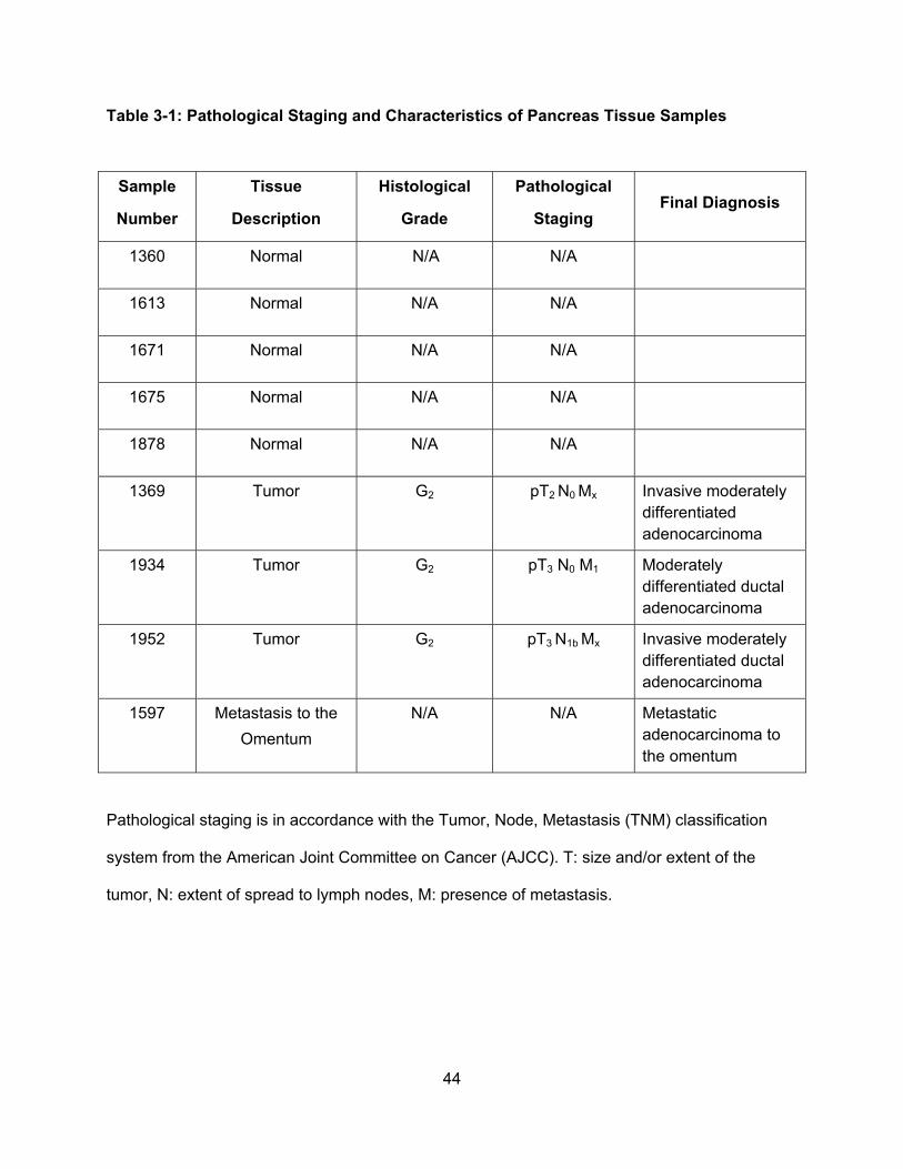
44
Table 3-1: Pathological Staging and Characteristics of Pancreas Tissue Samples
Sample
Number
Tissue
Description
Histological
Grade
Pathological
Staging Final Diagnosis
1360 Normal N/A N/A
1613 Normal N/A N/A
1671 Normal N/A N/A
1675 Normal N/A N/A
1878 Normal N/A N/A
1369 Tumor G2 pT2 N0 Mx Invasive moderately differentiated adenocarcinoma
1934 Tumor G2 pT3 N0 M1 Moderately differentiated ductal adenocarcinoma
1952 Tumor G2 pT3 N1b Mx Invasive moderately differentiated ductal adenocarcinoma
1597 Metastasis to the
Omentum
N/A N/A Metastatic adenocarcinoma to the omentum
Pathological staging is in accordance with the Tumor, Node, Metastasis (TNM) classification
system from the American Joint Committee on Cancer (AJCC). T: size and/or extent of the
tumor, N: extent of spread to lymph nodes, M: presence of metastasis.
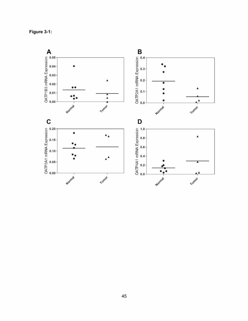
45
Figure 3-1:
46
Figure 3-1: Messenger RNA expression of OATPs in normal pancreas and pancreatic
tumor samples. Messenger RNA expression of A) OATP1B3, B) OATP2A1, C) OATP3A1 and
D) OATP4A1 in normal pancreas and pancreas cancer tissue samples were measured by
Quantigene Plex 2.0 Reagent System. Data were normalized to the housekeeping gene
ribosomal protein L13A. P < 0.05 by unpaired t-test.
47
3.3.2: OATP expression is higher in pancreatic adenocarcinomas than in normal
pancreas at the protein level
To determine changes in OATP protein expression in pancreatic adenocarcinoma and
normal tissues we used immunofluorescence staining. Our data show expression of OATP2A1,
OATP3A1 and OATP4A1 in normal pancreas and adenocarcinoma tissues (Figure 3-2). In
contrast to the transcript data, OATP2A1, OATP3A1 and OATP4A1 protein expression was
higher in pancreatic adenocarcinoma than in normal pancreas tissue as evidenced by more
intense staining observed throughout the adenocarcinoma tumors. Thus OATP2A1, OATP3A1
and OATP4A1 protein expression is up-regulated in pancreatic adenocarcinomas.
Furthermore, we investigated the expression of OATP1B3 in normal pancreas and
pancreatic adenocarcinoma. Immunofluorescence staining revealed low or absent OATP1B3
expression in normal pancreas, which was increased in pancreatic adenocarcinoma tumor
specimens (Figure 3-3). OATP1B3 was expressed in the entire tumor specimen in two out of
three cases and selectively in the epithelial cells that line the pancreatic ducts in one out of
three cases. Ductal expression of OATP1B3 is especially visible in the pancreatic tumor sample
1952 (Figure 3-3, white arrowheads). We used normal human liver tissue as a positive control
for these studies which has constitutive expression of OATP1B3. To determine the location of
OATP1B3 expression in pancreatic tumors, we used co-immunofluorescence with OATP1B3
and carbonic anhydrase II (CAII), a marker for ductal epithelium. Our results indicate that
OATP1B3 was localized, but not restricted, to the ductal epithelium of pancreatic
adenocarcinoma (Figure 3-4). Overall, protein expression of OATP1B3, OATP2A1, OATP3A1
and OATP4A1 was low in normal human pancreas but increased in pancreatic adenocarcinoma
tissue.
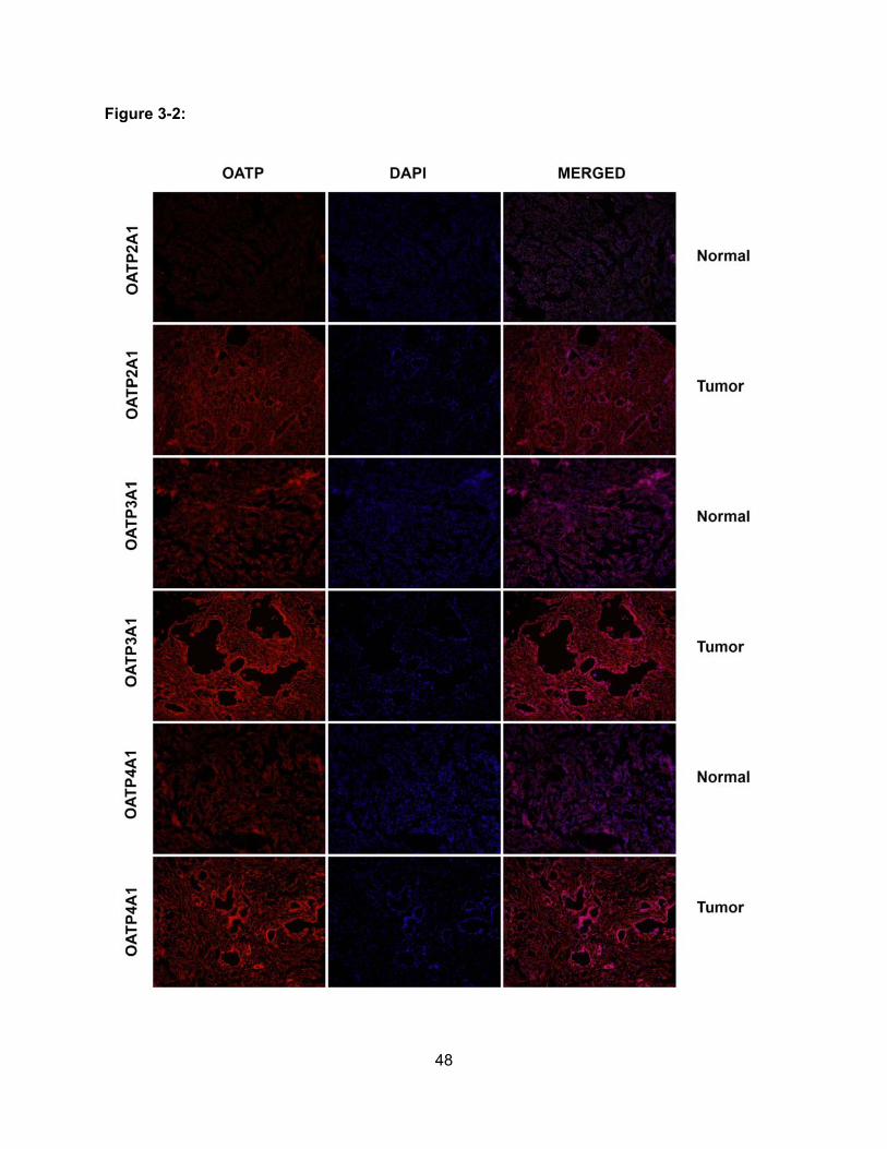
48
Figure 3-2:
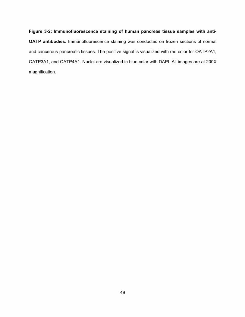
49
Figure 3-2: Immunofluorescence staining of human pancreas tissue samples with anti-
OATP antibodies. Immunofluorescence staining was conducted on frozen sections of normal
and cancerous pancreatic tissues. The positive signal is visualized with red color for OATP2A1,
OATP3A1, and OATP4A1. Nuclei are visualized in blue color with DAPI. All images are at 200X
magnification.
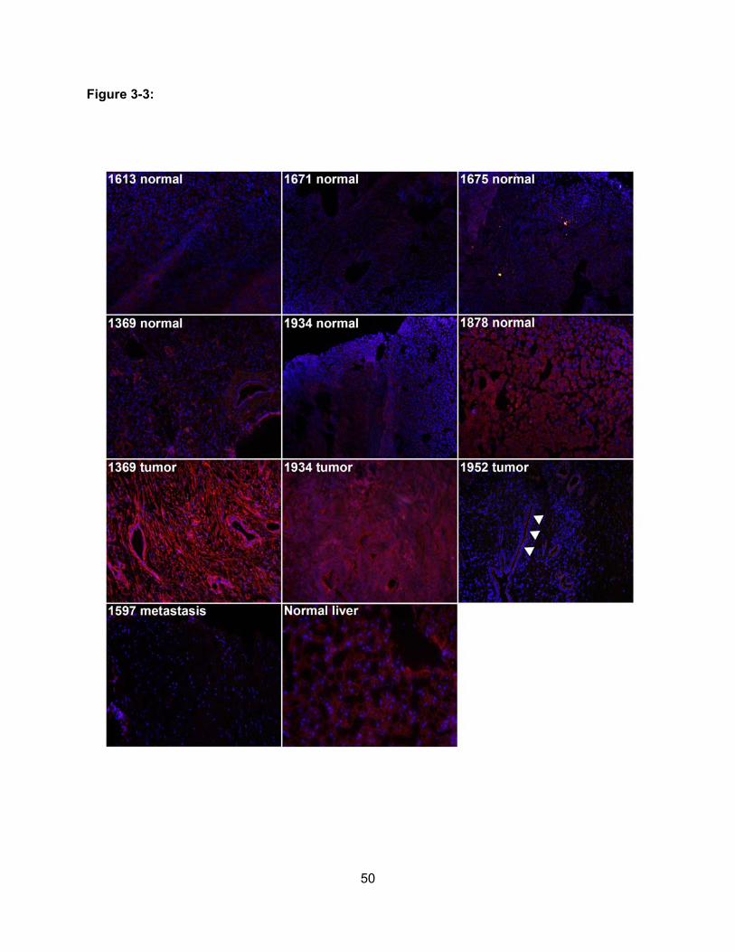
50
Figure 3-3:
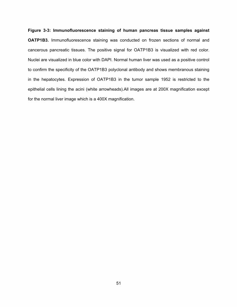
51
Figure 3-3: Immunofluorescence staining of human pancreas tissue samples against
OATP1B3. Immunofluorescence staining was conducted on frozen sections of normal and
cancerous pancreatic tissues. The positive signal for OATP1B3 is visualized with red color.
Nuclei are visualized in blue color with DAPI. Normal human liver was used as a positive control
to confirm the specificity of the OATP1B3 polyclonal antibody and shows membranous staining
in the hepatocytes. Expression of OATP1B3 in the tumor sample 1952 is restricted to the
epithelial cells lining the acini (white arrowheads).All images are at 200X magnification except
for the normal liver image which is a 400X magnification.
52
Figure 3-4:
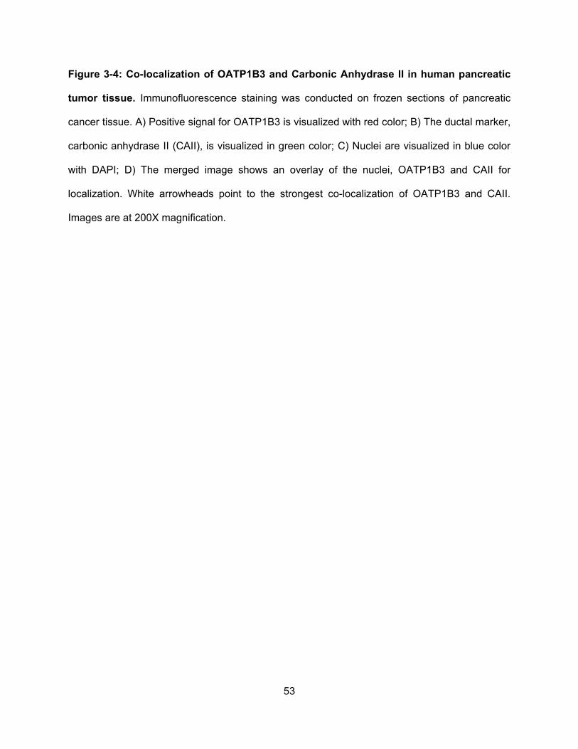
53
Figure 3-4: Co-localization of OATP1B3 and Carbonic Anhydrase II in human pancreatic
tumor tissue. Immunofluorescence staining was conducted on frozen sections of pancreatic
cancer tissue. A) Positive signal for OATP1B3 is visualized with red color; B) The ductal marker,
carbonic anhydrase II (CAII), is visualized in green color; C) Nuclei are visualized in blue color
with DAPI; D) The merged image shows an overlay of the nuclei, OATP1B3 and CAII for
localization. White arrowheads point to the strongest co-localization of OATP1B3 and CAII.
Images are at 200X magnification.
54
3.3.3: OATP1B3 is expressed in chronic pancreatitis and earlier stages of
adenocarcinoma
We further assessed the frequency and extent of OATP1B3 expression at different
stages and types of pancreatic cancer. The rationale to further evaluate the expression of
OATP1B3 was that unlike other OATPs, under normal physiological conditions, OATP1B3 is
selectively expressed in human hepatocytes. However, recent studies revealed OATP1B3
expression in a wide range of different cancers. Thus, given the limited OATP1B3 expression in
normal pancreatic tissue, OATP1B3 could be a good marker for diagnostic purposes and a
practical target for anticancer drug delivery into tumor cells. Therefore, we examined OATP1B3
expression on a pancreas cancer tissue microarray using immunohistochemical staining. The
results confirm previous observations, indicating low expression of OATP1B3 in normal
pancreas (10% of cases; n=10) (Figure 3-5A) and cancer adjacent normal pancreas tissue
(20% of cases; n=20) (Figure 3-5B). Interestingly, OATP1B3 expression was high in hyperplasia,
pancreatic inflammation and chronic pancreatitis specimens (42% of cases; n=12) (Figure 3-5C).
Similarly OATP1B3 was expressed in 44% of stage one pancreatic adenocarcinoma cases
(n=9) (Figure 3-5D), decreased in stage two (28% of cases; n=18) (Figure 3-5E) and stage
three adenocarcinoma (13% of cases; n=15) (Figure 3-5F) and was completely absent in
metastatic tissues from primary pancreatic adenocarcinoma (Figure 3-5G). Aside from
pancreatic adenocarcinoma, we also evaluated OATP1B3 expression in two other types of
pancreatic cancer, islet cell tumors and pancreatic adenosquamous carcinomas. OATP1B3
expression was absent in islet cell tumors (n=11) (Figure 3-5G), and, it was highly expressed in
adenosquamous carcinoma (67% of cases, n=3) (Figure 3-5H). Examples of OATP1B3
expression in chronically inflamed pancreas (Figure 3-5I), in stage one (Figure 3-5J) and in
stage three pancreatic adenocarcinoma (Figure 3-5K) are shown. These results reveal that

55
OATP1B3 is highly expressed in pancreatic hyperplasia and early stage adenocarcinomas with
decreasing expression in higher stage adenocarcinomas.
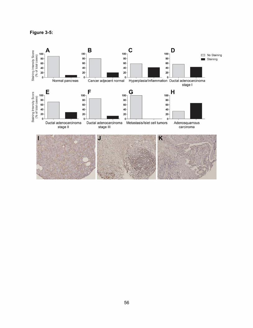
56
Figure 3-5:
57
Figure 3-5: Immunohistochemical staining of OATP1B3 on a pancreas tissue microarray.
Tissue arrays containing paraffin-embedded sections were stained for OATP1B3 using
immunohistochemistry. Staining intensity from the tissue microarray was scored as negative or
positive staining. Percentage was calculated by the number of stained specimens compared to
total number of specimens in A) normal pancreatic tissue, B) normal pancreas adjacent to
cancer tissue, C) hyperplasia and/or inflammation, D) ductal adenocarcinoma stage one, E)
ductal adenocarcinoma stage two, F) ductal adenocarcinoma stage three, G) metastatic tumor
from pancreas and/or islet cell tumors, and H) pancreatic adenosquamous carcinoma.
Representative photomicrographs of I) chronic pancreatitis, J) ductal adenocarcinoma stage
one, and K) ductal adenocarcinoma stage three.
58
3.4: Discussion
Pancreatic cancer remains a difficult cancer to treat clinically with multidrug resistance
as one of the leading causes of relapse in these patients (Jemal et al., 2009). The present study
addressed the question of which major OATPs are expressed in pancreatic adenocarcinoma
and to what extent OATP1B3 would be expressed in the different types and stages of
pancreatic cancer. The results suggest that certain OATPs can potentially be used as a target to
treat early stage pancreatic adenocarcinomas with cytotoxic chemotherapeutic drugs that are
substrates of these OATPs. In addition, the results indicate that OATP1B3 may serve as a
potential biomarker for the development of early stage pancreatic adenocarcinoma.
Our first set of results demonstrates that the four major OATPs expressed in pancreatic
adenocarcinoma include OATP1B3, OATP2A1, OATP3A1 and OATP4A1. Although expression
of the four OATPs at the messenger RNA level was not statistically significant between normal
pancreas and pancreatic adenocarcinoma, we found that OATP1B3, OATP2A1, OATP3A1 and
OATP4A1 expression at the protein level was increased in adenocarcinoma compared to
normal tissue as evidenced by immunofluorescence staining. Although mRNA expression levels
can be useful for preliminary assessment of OATP expression in tissues, it remains important to
assess the expression of OATPs at their functional protein level.
OATP2A1 mainly functions in the transport of prostaglandins and is expressed
ubiquitously throughout the body. Initial studies demonstrated that at the mRNA level OATP2A1
is expressed in many organs including normal pancreas (Schuster, 2002). Follow-up studies
showed OATP2A1 to be up-regulated in numerous different cancers as well as in malignant
breast cancer tissue compared with its adjacent nonmalignant breast tissue (Wlcek et al., 2008).
Both OATP3A1 and OATP4A1 are also ubiquitously expressed in a variety of different tissues in
59
the body. Several reports indicate that both OATPs also share similar expression patterns in
cancer and are expressed in the breast cancer cell lines MCF-7 and T-47D (Pizzagalli et al.,
2003; Nozawa et al., 2004), in malignant breast tumor tissue (Kindla et al., 2011b), in malignant
osteosarcoma cell lines (Liedauer et al., 2009) and in a variety of gastrointestinal cancer cell
lines as well as in the pancreatic cancer cell line GI-103 (Tamai et al., 2000). Due to the ability
of many OATPs to transport hormones and their conjugates, it has been hypothesized that the
expression of OATPs in cancer tissue could potentially contribute to the proliferation of
hormone-dependent cancers (Nozawa et al., 2004; Hamada et al., 2008). Thus, one potential
therapeutic approach would be to develop OATP inhibitors to improve overall survival for
patients with OATP-expressing cancers (Nozawa et al., 2005b; Obaidat et al., 2012). Another
potential approach for OATP-mediated cancer therapy would be the targeting of OATP-
expressing cancers with cytotoxic cancer drugs that are substrates of these OATPs (Obaidat et
al., 2012). However, so far there are no reports available yet that demonstrate transport of
cancer drugs by OATP2A1, OATP3A1 or OATP4A1.
OATP1B3 did get increased attention recently, owing to its expression in a wide variety
of cancers while its expression under normal physiological conditions is exclusive to the liver.
Combined with this expression pattern, its ability to transport several cancer drugs, namely
methotrexate (Abe et al., 2001; Sasaki et al., 2004), paclitaxel, docetaxel (Smith et al., 2005;
Letschert et al., 2006) and imatinib (Hu et al., 2008) makes OATP1B3 a more appropriate target
for the delivery of anticancer drugs that mainly affect rapidly dividing cells. In 2001, Abe et al.
were the first to demonstrate OATP1B3 expression in several pancreatic cancer cell lines by
northern blot analysis. They also confirmed OATP1B3 protein expression in a single case of
pancreatic cancer using immunohistochemical staining (Abe et al., 2001). Recently Kounnis et
al. evaluated the expression of the three well characterized OATP1A2, OATP1B1 and
OATP1B3 in two pancreatic cancer cell lines and in several biopsies of pancreatic cancer tissue
60
(Kounnis et al., 2011). They concluded that all three OATPs are expressed in pancreatic cancer,
but some discrepancies remain due to the use of antibodies with overlapping specificities or
antibodies that have not been validated. In the present study, we confirmed their finding that
OATP1B3 is expressed in pancreatic cancer tissue using a different OATP1B3 specific antibody.
Furthermore, we were among the first to show that expression of OATP1B3 differs in different
tumor types and stages of pancreatic cancer. Interestingly, our results showed the highest
OATP1B3 expression in tissues that have pathological risk factors for the development of
pancreatic cancer including tissues with mild and chronic hyperplasia and inflammation. In
addition, OATP1B3 was expressed at highest levels in stage one followed by stage two
pancreatic adenocarcinoma with little to no staining in stage three pancreatic adenocarcinoma
or metastatic tissue. These results are in accordance with findings reported for the expression of
OATP1B3 in colorectal cancer stages where OATP1B3 expression was highest in lower stage
colorectal tumors (Lockhart et al., 2008). Another study demonstrated an increased trend of
OATP1B3 expression in precancerous colon polyps (Ballestero et al., 2006). In yet another
study, OATP1B3 expression was highest in stage one and two breast carcinoma compared to
stage three breast carcinoma (Muto et al., 2007). Collectively, these data suggest that
OATP1B3 may be used as a potential diagnostic marker in early stage cancer detection.
In future studies it would be interesting to investigate the regulation of OATP1B3
expression during the development of pancreatic ductal adenocarcinoma. Such studies should
examine which signaling pathways regulate OATP1B3 expression during acinar to ductal
metaplasia (ADM), the initial process of pancreatic adenocarcinoma development, where
pancreatic acinar cells with chronic inflammation undergo morphogenesis into abnormal cells
with ductal phenotypes (Reichert and Rustgi, 2011) and might identify additional therapeutic
targets.
61
In conclusion, OATPs may serve as potential diagnostic markers and therapeutic targets
to improve pancreatic cancer therapy. In particular targeting OATP1B3 which has been shown
to transport several anticancer drugs could be as useful as targeting efflux transporters in
cancer therapy. Furthermore, our findings are the basis for further investigation of OATP1B3
regulation in pancreatic cancer and for further evaluation of OATP1B3 as a diagnostic marker
for early cancer detection.
62
Chapter 4
The Anticancer Drugs Etoposide, Oxaliplatin and Plicamycin are Substrates of
OATP1B3
4.1: Abstract
Organic anion transporting polypeptide 1B3 (OATP1B3) is a liver-specific transport
protein that has a very diverse substrate specificity. Because of its ability to transport a
multitude of structurally diverse xenobiotics, OATP1B3 plays a significant role in drug
disposition and is considered a site for drug-drug interactions. Additionally, based on recent
evidence that it is expressed in different cancer tissues OATP1B3 was suggested to be a
potential therapeutic target for cancer drug delivery. In this study, we examined the interactions
of 89 FDA approved oncology drugs on the function of OATP1B3 and compared cell viability of
wild-type with OATP1B3-expressing Chinese Hamster Ovary (CHO) cells in the presence of
these drugs. Uptake of two model substrates estradiol-17β-glucuronide and estrone-3-sulfate
was inhibited by several of the anticancer drugs. However, the results of the inhibition kinetics
were not predictive for known substrates. Therefore, the more direct readout of increased
cytotoxicity for OATP1B3-expressing cells as compared to wild-type cells was used and
bleomycin, etoposide, oxaliplatin, plicamycin and thioguanine were identified as the most potent
cytotoxic compounds. Furthermore, by measuring uptake into OATP1B3-expressing cells
etoposide, oxaliplatin and plicamycin could be demonstrated to be substrates of OATP1B3.
These results suggest that OATP1B3-mediated transport may be involved in the disposition and
hepatotoxicity of some of these anticancer drugs.
63
4.2: Introduction
Cancer is one of the leading causes of death and based on projections, cancer burden
will continue to increase (Boyle and Levin, 2008). Many patients with non-localized or
metastatic cancer often times do not respond well to conventional chemotherapy. Several of
these cases may be attributed to multidrug resistance whereby an increased efflux of
chemotherapy drugs from the cancer cells via efflux transporters leads to ineffective therapeutic
responses (Szakacs et al., 2006). Research aimed at inhibiting these efflux transporters has
shown little promise in overcoming this clinical problem. Consequently, different therapeutic
approaches are sought to improve anticancer drug uptake into cancer cells. Members of the
Organic Anion Transporting Polypeptide (OATP) superfamily of uptake transporters have been
shown to be up-regulated in numerous different cancer types and are considered as potential
therapeutic targets for anticancer drug delivery into cancer cells.
OATPs are multispecific transport proteins that mediate the Na+-independent uptake of
large amphipathic organic anions into cells. They form a superfamily with eleven known human
OATPs that are expressed in a wide range of epithelial tissues in the body (Hagenbuch and
Meier, 2003). A majority of OATP family members are ubiquitously expressed. However, certain
OATPs have restricted expression to a specific tissue type. OATP expression and
polymorphisms have been associated with altered bioavailability of drugs, stressing the
importance of these transporters in drug disposition and ultimately therapy effectiveness.
Additionally, studies have shown single nucleotide polymorphisms (SNPs) of OATPs that have
been associated with altered drug disposition of several anticancer drugs. Subsequent studies
demonstrated the ability of OATPs to transport anticancer drugs. OATP1B3 has been implicated
in the transport of rapamycin, SN-38, imatinib, methotrexate, paclitaxel and docetaxel (Abe et al.,
64
2001; Smith et al., 2005; Letschert et al., 2006; Hu et al., 2008; Yamaguchi et al., 2008; Oswald
et al., 2010).
Because of the up-regulated expression of OATPs in cancer cells and their ability to
transport anticancer drugs, there has been an increased interest in these transporters as
potential targets for anticancer drug therapy. Therefore, a potential mechanism of OATP-
mediated anticancer therapy could be through treating OATP-expressing cancers with known
anticancer drug substrates. Evidence that OATP-mediated transport can be stimulated via small
molecules presents another potential mechanism for increasing drug delivery into cancer cells
(Gui et al., 2008; Roth et al., 2011a).
OATP1B3 is an example of a tissue-specific OATP whereby its expression is restricted
to the liver, specifically at the basolateral membrane of hepatocytes (Abe et al., 1999; Hsiang et
al., 1999; Konig et al., 2000b). OATP1B3 is thought to play a role in the clearance of drugs from
blood into hepatocytes for further metabolism and excretion. Although under normal
physiological conditions OATP1B3 is expressed exclusively in the liver, its expression has been
shown to be down-regulated in hepatocellular carcinomas (HCC), possibly owing to the
dedifferentiation of HCC cells (Cui et al., 2003; Vavricka et al., 2004; Zollner et al., 2005).
Despite the decreased expression in HCC cells, OATP1B3 has been shown to be up-regulated
in a wide variety of different cancers including gastro-intestinal cancers (Abe et al., 2001),
colorectal cancer (Lee et al., 2008), prostate cancer (Hamada et al., 2008), breast cancer (Muto
et al., 2007) and non-small cell lung cancer (Monks et al., 2007). As a result, OATP1B3 is
considered the most attractive candidate for drug targeting into cancer cells. In this study we
aimed at identifying novel anticancer drugs substrates of OATP1B3 by screening 89 compounds
of the NCI/DTP approved oncology drug set.
65
4.3: Results
4.3.1 Effect of the NCI/DTP oncology drug set on OATP1B3-mediated uptake
To determine the effects of the oncology drug set on OATP1B3-mediated transport, we
measured uptake of 1 µM estradiol-17β-glucuronide into OATP1B3-expressing or wild-type
CHO cells in the absence or presence of 100 µM anticancer drug under initial linear rate
conditions. Rifampicin, a known inhibitor of OATP-mediated transport, served as a positive
control in these experiments. Anticancer drugs with greater than 50% inhibition of OATP1B3-
mediated estradiol-17β-glucuronide uptake included rapamycin, teniposide, actinomycin D,
vinorelbine, megestrol, tretinoin, docetaxel, celecoxib, mitoxantrone, gefitinib, dasatinib, imatinib,
plicamycin, vinblastine, vincristine and ixabepilone (Figure 4-1). Of interest, OATP1B3-mediated
uptake of estradiol-17β-glucuronide was unaffected by many anticancer drugs but strongly
stimulated by irinotecan and vorinostat. Based on previous studies that demonstrated substrate-
dependent effects of compounds on OATP-mediated transport (Gui et al., 2008; Roth et al.,
2011a; Roth et al., 2011b), we determined the effects of the oncology drug set on the uptake of
an additional model substrate of OATP1B3, estrone-3-sulfate (0.1 µM). A majority of the
anticancer drugs inhibited estrone-3-sulfate uptake whereas only letrozole and sorafenib
stimulated estrone-3-sulfate uptake (Figure 4-2). We confirmed concentration-dependent
inhibition of four anticancer drugs from the original screen (Figure 4-3) and determined IC50
values for actinomycin D, celecoxib, rapamycin and vinblastine to be 26.0 ± 0.2, 32.4 ± 0.3, 2.4
± 0.2 and 28.1 ± 0.2 µM, respectively. To determine the mechanism(s) of substrate-dependent
effects of these anticancer drug inhibitors on OATP1B3-mediated uptake we preformed
inhibition kinetics of the two model substrates, estradiol-17β-glucuronide (Figure 4-4) and
estrone-3-sulfate (Figure 4-5). Inhibition of OATP1B3-mediated estradiol-17β-glucuronide
uptake was non-competitive for rapamycin (Figure 4-4B), teniposide (Figure 4-4C) and
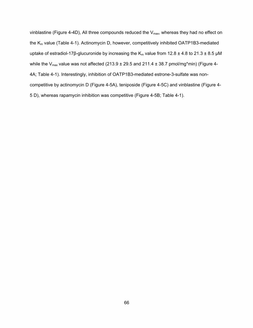
66
vinblastine (Figure 4-4D), All three compounds reduced the Vmax, whereas they had no effect on
the Km value (Table 4-1). Actinomycin D, however, competitively inhibited OATP1B3-mediated
uptake of estradiol-17β-glucuronide by increasing the Km value from 12.8 ± 4.8 to 21.3 ± 8.5 µM
while the Vmax value was not affected (213.9 ± 29.5 and 211.4 ± 38.7 pmol/mg*min) (Figure 4-
4A; Table 4-1). Interestingly, inhibition of OATP1B3-mediated estrone-3-sulfate was non-
competitive by actinomycin D (Figure 4-5A), teniposide (Figure 4-5C) and vinblastine (Figure 4-
5 D), whereas rapamycin inhibition was competitive (Figure 4-5B; Table 4-1).
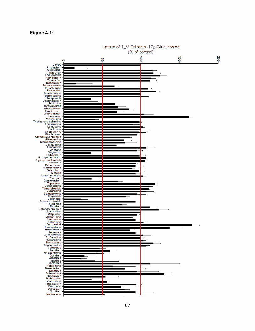
67
Figure 4-1:
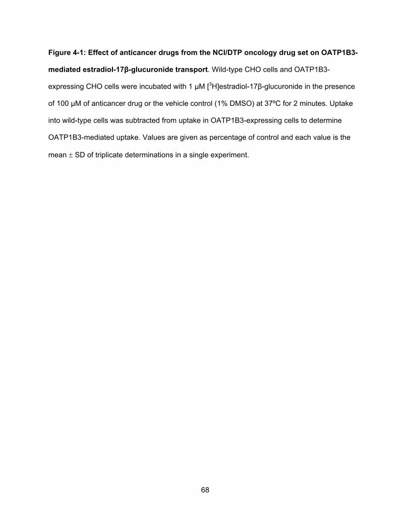
68
Figure 4-1: Effect of anticancer drugs from the NCI/DTP oncology drug set on OATP1B3-
mediated estradiol-17β-glucuronide transport. Wild-type CHO cells and OATP1B3-
expressing CHO cells were incubated with 1 μM [3H]estradiol-17β-glucuronide in the presence
of 100 μM of anticancer drug or the vehicle control (1% DMSO) at 37ºC for 2 minutes. Uptake
into wild-type cells was subtracted from uptake in OATP1B3-expressing cells to determine
OATP1B3-mediated uptake. Values are given as percentage of control and each value is the
mean SD of triplicate determinations in a single experiment.
69
Figure 4-2:
70
Figure 4-2: Effect of anticancer drugs from the NCI/DTP oncology drug set on OATP1B3-
mediated estrone-3-sulfate transport. Wild-type CHO cells and OATP1B3-expressing CHO
cells were incubated with 0.1 μM [3H]estrone-3-sulfate in the presence of 100 μM of anticancer
drug or the vehicle control (1% DMSO) at 37ºC for 2 minutes. Uptake into wild-type cells was
subtracted from uptake in OATP1B3-expressing cells to determine OATP1B3-mediated uptake.
Values are given as percentage of control and each value is the mean SD of triplicate
determinations in a single experiment.
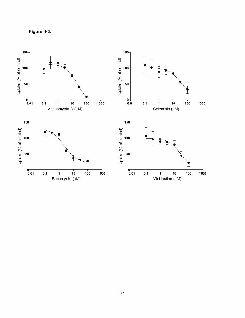
71
Figure 4-3:
0.01 0.1 1 10 100 10000
50
100
150
Actinomycin D (M)
Upt
ake
(% o
f co
ntro
l)
0.01 0.1 1 10 100 10000
50
100
150
Celecoxib (M)
Upt
ake
(% o
f co
ntro
l)
0.01 0.1 1 10 100 10000
50
100
150
Rapamycin (M)
Upt
ake
(% o
f co
ntro
l)
0.01 0.1 1 10 100 10000
50
100
150
Vinblastine (M)
Upt
ake
(% o
f co
ntro
l)
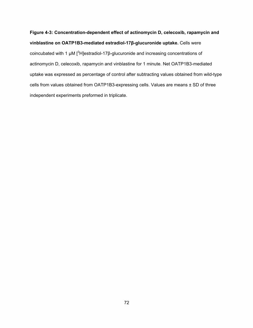
72
Figure 4-3: Concentration-dependent effect of actinomycin D, celecoxib, rapamycin and
vinblastine on OATP1B3-mediated estradiol-17β-glucuronide uptake. Cells were
coincubated with 1 µM [3H]estradiol-17β-glucuronide and increasing concentrations of
actinomycin D, celecoxib, rapamycin and vinblastine for 1 minute. Net OATP1B3-mediated
uptake was expressed as percentage of control after subtracting values obtained from wild-type
cells from values obtained from OATP1B3-expressing cells. Values are means ± SD of three
independent experiments preformed in triplicate.
73
Figure 4-4:
-0.2 -0.1 0.1 0.2 0.3
-0.01
0.01
0.02
0.03
0.04
15MActinomycin D
DMSO
1/estradiol-17-glucuronide
1/
v
(p
mol
/mg*
min
)
-0.2 -0.1 0.1 0.2 0.3
-0.01
0.01
0.02
0.03
0.04DMSO
30M Vinblastine
1/estradiol-17-glucuronide
1
/v
(p
mol
/mg*
min
)
A
-0.2 0.2 0.4 0.6
-0.05
0.05
0.10
0.15
0.20DMSO
5M Rapamycin
1/estradiol-17-glucuronide
1
/v
(pm
ol/m
g*m
in)
B
-0.1 0.1 0.2 0.3
-0.01
0.01
0.02
0.03
0.04DMSO
30M Teniposide
1/estradiol-17-glucuronide
1/
v
(pm
ol/m
g*m
in)
D C
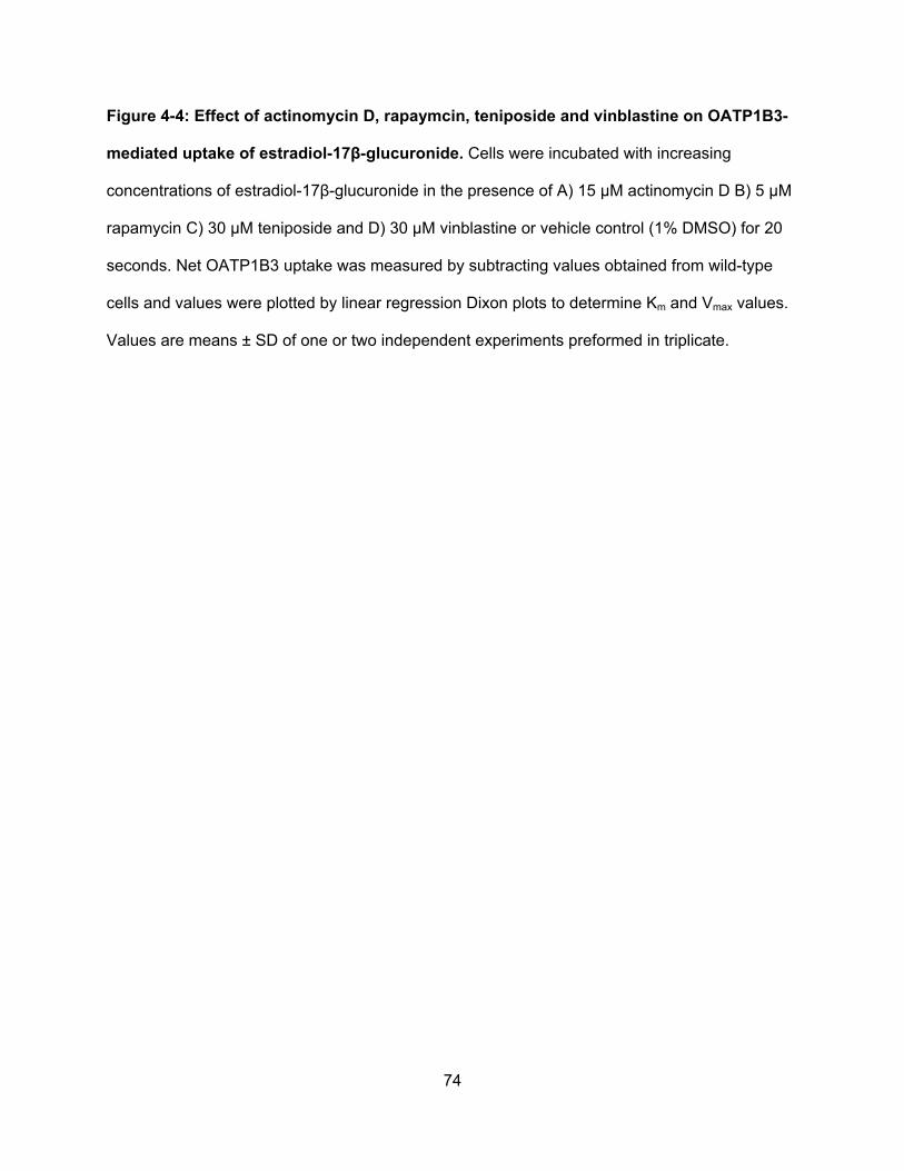
74
Figure 4-4: Effect of actinomycin D, rapaymcin, teniposide and vinblastine on OATP1B3-
mediated uptake of estradiol-17β-glucuronide. Cells were incubated with increasing
concentrations of estradiol-17β-glucuronide in the presence of A) 15 µM actinomycin D B) 5 µM
rapamycin C) 30 µM teniposide and D) 30 µM vinblastine or vehicle control (1% DMSO) for 20
seconds. Net OATP1B3 uptake was measured by subtracting values obtained from wild-type
cells and values were plotted by linear regression Dixon plots to determine Km and Vmax values.
Values are means ± SD of one or two independent experiments preformed in triplicate.
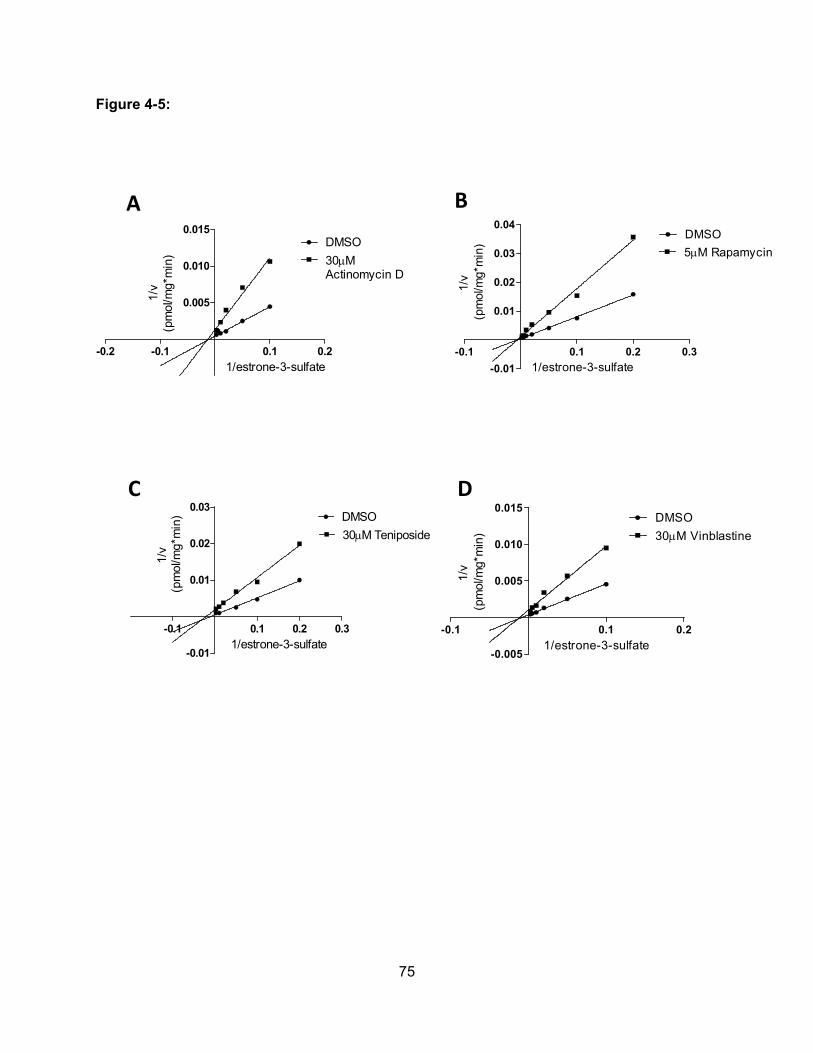
75
Figure 4-5:
-0.2 -0.1 0.1 0.2
0.005
0.010
0.015DMSO
30MActinomycin D
1/estrone-3-sulfate
1/v
(pm
ol/m
g*m
in)
-0.1 0.1 0.2 0.3
-0.01
0.01
0.02
0.03
0.04DMSO
5M Rapamycin
1/estrone-3-sulfate
1
/v
(p
mol
/mg*
min
)
-0.1 0.1 0.2
-0.005
0.005
0.010
0.015DMSO
30M Vinblastine
1/estrone-3-sulfate
1/v
(pm
ol/m
g*m
in)
-0.1 0.1 0.2 0.3
-0.01
0.01
0.02
0.03DMSO
30M Teniposide
1/estrone-3-sulfate
1/v
(p
mol
/mg*
min
)
A B
D C
76
Figure 4-5: Effect of actinomycin D, rapaymcin, teniposide and vinblastine on OATP1B3-
mediated uptake of estrone-3-sulfate. Cells were incubated with increasing concentrations of
estrone-3-sulfate in the presence of A) 30 µM actinomycin D B) 5 µM rapamycin C) 30 µM
teniposide and D) 30 µM vinblastine or vehicle control (1% DMSO) for 20 seconds. Net
OATP1B3 uptake was measured by subtracting values obtained from wild-type cells and values
were plotted by linear regression Dixon plots to determine Km and Vmax values. Values are
means ± SD of one or two independent experiments preformed in triplicate.
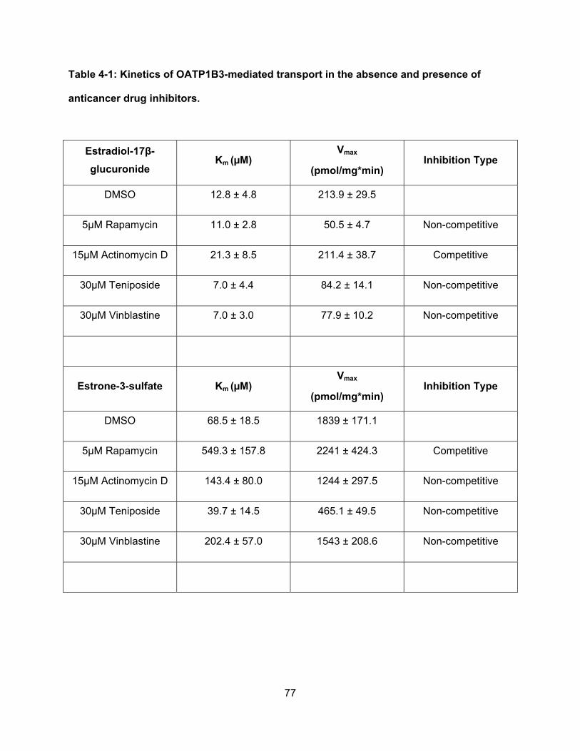
77
Table 4-1: Kinetics of OATP1B3-mediated transport in the absence and presence of
anticancer drug inhibitors.
Estradiol-17β-
glucuronide Km (µM)
Vmax
(pmol/mg*min) Inhibition Type
DMSO 12.8 ± 4.8 213.9 ± 29.5
5µM Rapamycin 11.0 ± 2.8 50.5 ± 4.7 Non-competitive
15µM Actinomycin D 21.3 ± 8.5 211.4 ± 38.7 Competitive
30µM Teniposide 7.0 ± 4.4 84.2 ± 14.1 Non-competitive
30µM Vinblastine 7.0 ± 3.0 77.9 ± 10.2 Non-competitive
Estrone-3-sulfate Km (µM) Vmax
(pmol/mg*min) Inhibition Type
DMSO 68.5 ± 18.5 1839 ± 171.1
5µM Rapamycin 549.3 ± 157.8 2241 ± 424.3 Competitive
15µM Actinomycin D 143.4 ± 80.0 1244 ± 297.5 Non-competitive
30µM Teniposide 39.7 ± 14.5 465.1 ± 49.5 Non-competitive
30µM Vinblastine 202.4 ± 57.0 1543 ± 208.6 Non-competitive
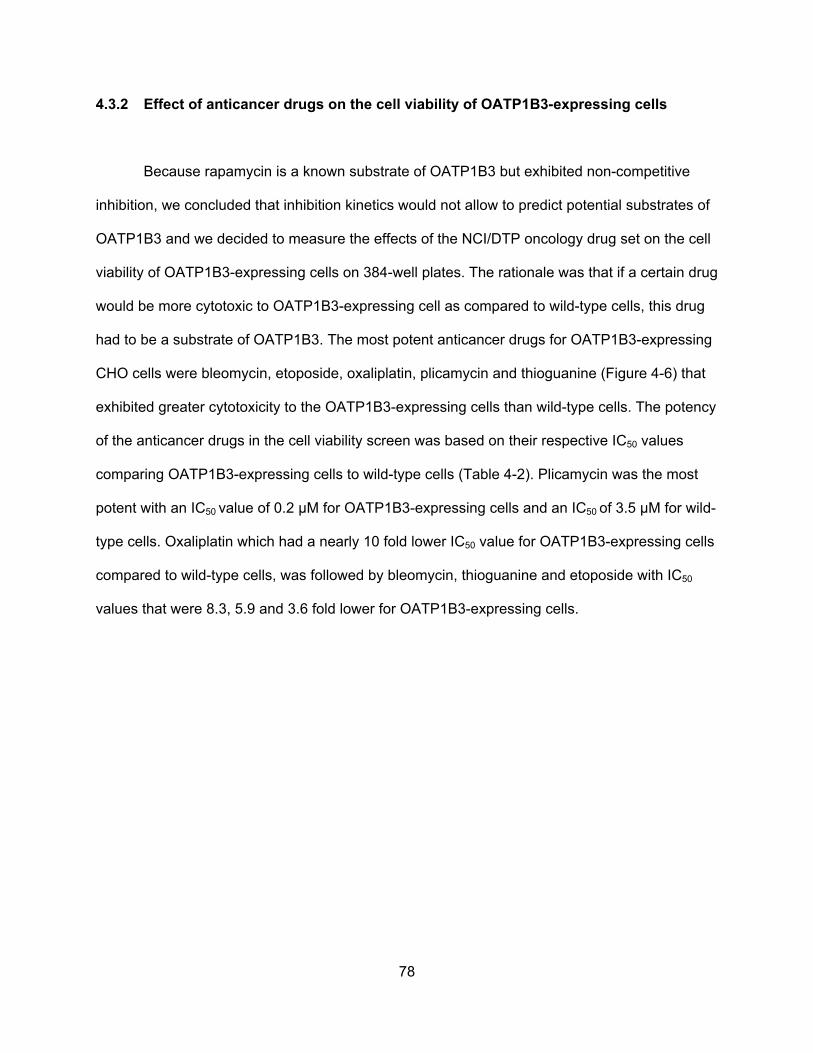
78
4.3.2 Effect of anticancer drugs on the cell viability of OATP1B3-expressing cells
Because rapamycin is a known substrate of OATP1B3 but exhibited non-competitive
inhibition, we concluded that inhibition kinetics would not allow to predict potential substrates of
OATP1B3 and we decided to measure the effects of the NCI/DTP oncology drug set on the cell
viability of OATP1B3-expressing cells on 384-well plates. The rationale was that if a certain drug
would be more cytotoxic to OATP1B3-expressing cell as compared to wild-type cells, this drug
had to be a substrate of OATP1B3. The most potent anticancer drugs for OATP1B3-expressing
CHO cells were bleomycin, etoposide, oxaliplatin, plicamycin and thioguanine (Figure 4-6) that
exhibited greater cytotoxicity to the OATP1B3-expressing cells than wild-type cells. The potency
of the anticancer drugs in the cell viability screen was based on their respective IC50 values
comparing OATP1B3-expressing cells to wild-type cells (Table 4-2). Plicamycin was the most
potent with an IC50 value of 0.2 µM for OATP1B3-expressing cells and an IC50 of 3.5 µM for wild-
type cells. Oxaliplatin which had a nearly 10 fold lower IC50 value for OATP1B3-expressing cells
compared to wild-type cells, was followed by bleomycin, thioguanine and etoposide with IC50
values that were 8.3, 5.9 and 3.6 fold lower for OATP1B3-expressing cells.
79
Figure 4-6:
0.1 1 10 100 1000 100000
20
40
60
80
100
Wild-typeOATP1B3
Etoposide (M)
0.1 1 10 100 1000 100000
20
40
60
80
100
120
Oxaliplatin (M)
Cel
l via
bilit
y at
48
hour
s(%
of
cont
rol)
0.01 0.1 1 10 100 10000
20
40
60
80
100
Plicamycin (M)
0.1 1 10 100 1000 100000
20
40
60
80
100
Bleomycin (M)
Cel
l via
bilit
y at
48
hour
s(%
of
cont
rol)
0.01 0.1 1 10 100 10000
20
40
60
80
100
120
Thioguanine (M)
Cel
l via
bilit
y at
48
hour
s(%
of
cont
rol)
A
C
B
D
E
80
Figure 4-6: Effect of anticancer drugs on cell viability of wild-type and OATP1B3-
expressing CHO cells. Cell viability was measured with increasing concentrations of A)
bleomycin, B) etoposide, C) oxaliplatin, D) plicamycin and E) thioguanine for 48 hours on 384-
well plates of wild-type (circles) and OATP1B3- expressing (triangles) CHO cells. Values are
given as percent of control and are means SD of at least three independent experiments.
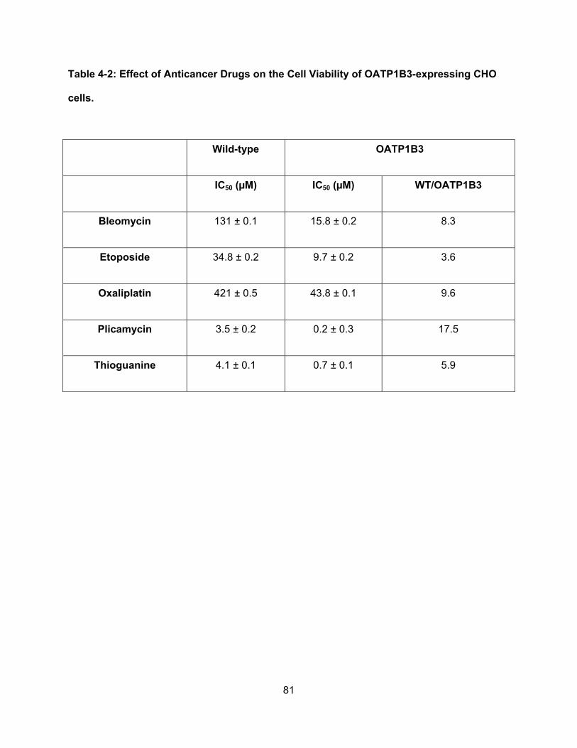
81
Table 4-2: Effect of Anticancer Drugs on the Cell Viability of OATP1B3-expressing CHO
cells.
Wild-type OATP1B3
IC50 (µM) IC50 (µM) WT/OATP1B3
Bleomycin 131 ± 0.1 15.8 ± 0.2 8.3
Etoposide 34.8 ± 0.2 9.7 ± 0.2 3.6
Oxaliplatin 421 ± 0.5 43.8 ± 0.1 9.6
Plicamycin 3.5 ± 0.2 0.2 ± 0.3 17.5
Thioguanine 4.1 ± 0.1 0.7 ± 0.1 5.9
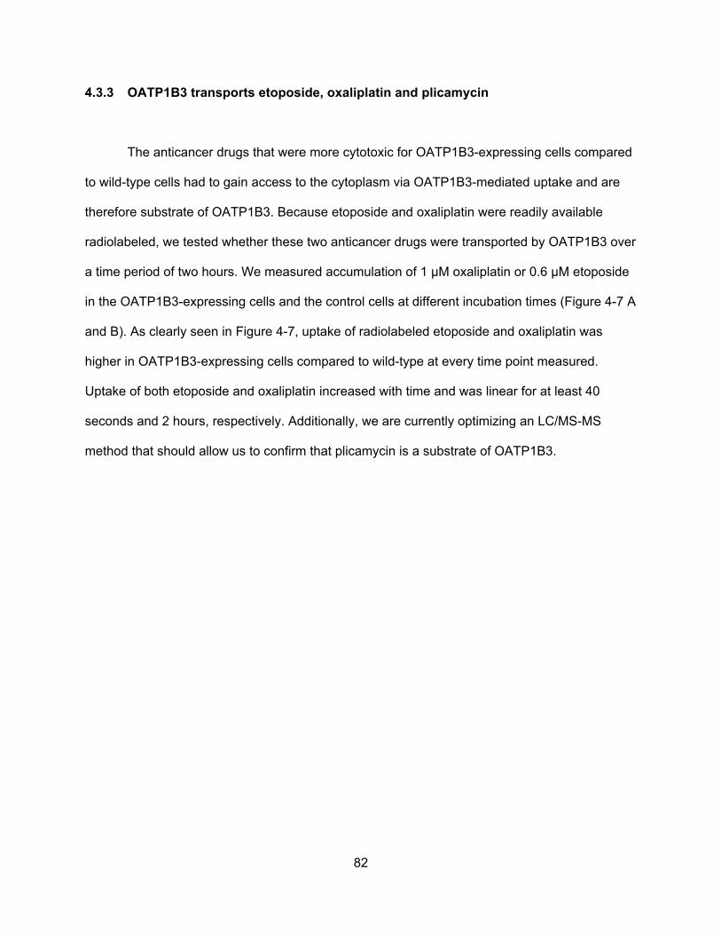
82
4.3.3 OATP1B3 transports etoposide, oxaliplatin and plicamycin
The anticancer drugs that were more cytotoxic for OATP1B3-expressing cells compared
to wild-type cells had to gain access to the cytoplasm via OATP1B3-mediated uptake and are
therefore substrate of OATP1B3. Because etoposide and oxaliplatin were readily available
radiolabeled, we tested whether these two anticancer drugs were transported by OATP1B3 over
a time period of two hours. We measured accumulation of 1 µM oxaliplatin or 0.6 µM etoposide
in the OATP1B3-expressing cells and the control cells at different incubation times (Figure 4-7 A
and B). As clearly seen in Figure 4-7, uptake of radiolabeled etoposide and oxaliplatin was
higher in OATP1B3-expressing cells compared to wild-type at every time point measured.
Uptake of both etoposide and oxaliplatin increased with time and was linear for at least 40
seconds and 2 hours, respectively. Additionally, we are currently optimizing an LC/MS-MS
method that should allow us to confirm that plicamycin is a substrate of OATP1B3.
83
Figure 4-7:
5 30 60 1200
5
10
15
Time (min)
Upt
ake
of 1
M O
xalip
latin
(pm
ol/m
g pr
otei
n)
A
1 5 30 600
1
2
3
4
5Wild-typeOATP1B3
Time (min)U
ptak
e of
0.6
M E
topo
side
(pm
ol/m
g pr
otei
n)
B
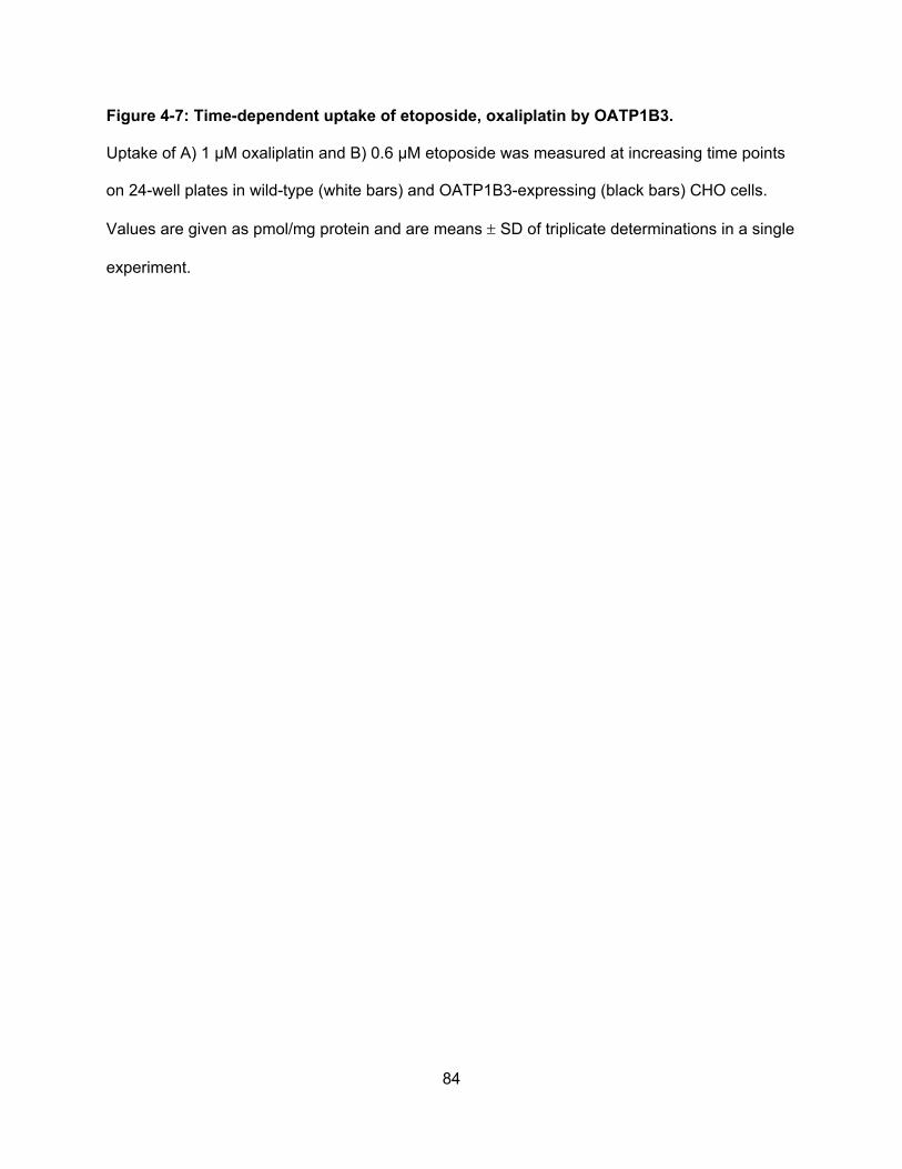
84
Figure 4-7: Time-dependent uptake of etoposide, oxaliplatin by OATP1B3.
Uptake of A) 1 μM oxaliplatin and B) 0.6 μM etoposide was measured at increasing time points
on 24-well plates in wild-type (white bars) and OATP1B3-expressing (black bars) CHO cells.
Values are given as pmol/mg protein and are means SD of triplicate determinations in a single
experiment.
85
4.4: Discussion
In the present study we identified five known anticancer drugs as substrates of
OATP1B3 using a cytotoxicity assay that compared the toxicity of the drugs in OATP1B3-
expressing CHO cells to wild-type CHO cells. Bleomycin, etoposide, oxaliplatin, plicamycin and
thioguanine were all more cytotoxic in OATP1B3-expressing cells and we therefore conclude
that these five anticancer drugs are transported into the CHO cells via OATP1B3. Two of these
drugs are readily available as radiolabeled compounds and we could confirm that 3H-etoposide
and 14C-oxaliplatin are taken up into OATP1B3-expressing cells more efficiently than into wild-
type CHO cells in a time dependent manner.
A recent study suggested that etoposide uptake into the liver would occur primarily via
OATP1B1 and OATP2B1 with very little transport by OATP1B3 using stable transfected
HEK293 cells (Fahrmayr et al., 2012). Our results are in agreement with that study because
uptake was slow and increased over one hour (Figure 4-7). Furthermore, another study
demonstrated that both mouse and human OCTN2 are able to transport etoposide (Hu et al.,
2012). The fact that etoposide is a substrate of several OATPs and of OCTN2 demonstrates
that there are overlaps in the substrate specificities of members of the OATP and the OCT
families. This suggestion is further supported by our finding that oxaliplatin is a substrate for
OATP1B3 because previous studies demonstrated that members of the OCT family including
OCTN1, OCTN2, OCT1, OCT2 and OCT3 (Ciarimboli et al., 2005; Yonezawa et al., 2005;
Zhang et al., 2006; Filipski et al., 2008; Filipski et al., 2009; Burger et al., 2010; Ciarimboli et al.,
2010; Jong et al., 2011) are involved in oxaliplatin transport.
Thioguanine which has been suggested to be a substrate of CNT3 and ENT2 (Fotoohi et
al., 2006) seems to be a substrate of OATP1B3 in our assay. This is one of the first documented
86
overlaps in the substrate specificities of nucleoside transporters and OATPs and further
demonstrates that the different xenobiotic transporters are quite promiscuous and have
overlapping substrate specificities that go beyond the respective families.
It is important to note that although etoposide and oxaliplatin had no effect on OATP1B3-
mediated estrone-3-sulfate and estradiol-17β-glucuronide uptake, they are nevertheless
substrates of OATP1B3. This can be explained by differences in the affinities of the different
substrates to the transporter or to the fact that they use different substrate binding or
translocation pathways and confirm that substrates of OATPs are not necessarily inhibitors of
OATP-mediated uptake of all substrates.
Plicamycin, an antitumor antibiotic used to treat testicular cancer and hypercalcemia, is
considered the most hepatotoxic chemotherapeutic drug used in the clinic and is therefore
rarely used in treatment regimens (Kennedy, 1970; King and Perry, 2001). Our finding that
plicamycin is transported by OATP1B3 can explain the disposition of plicamcyin into the liver
and thus OATP1B3-mediated uptake of plicamycin into hepatocytes can be considered a
mechanism that contributes to its hepatotoxicity in patients treated with this drug.
Our interaction results demonstrate that the majority of the anticancer drugs in the
oncology drug set did not affect OATP1B3-mediated substrate uptake while several compounds
inhibited, and only four compounds stimulated uptake. Furthermore, we found that the effects
were substrate dependent, especially evident for the stimulation of OATP1B3-mediated uptake.
While irinotecan and vorinostat stimulated uptake of estradiol-17β-glucuronide, they had no
effect on estrone-3-sulfate transport. Similar, letrozole and sorafenib stimulated estrone-3-
sulfate uptake but had no effect on estradiol-17β-glucuronide uptake. These results confirm that
it is important to test multiple substrates of OATP1B3 when screening for inhibitors and supports
87
findings from other studies that demonstrated similar evidence of multiple substrate binding
sites in OATPs (Gui et al., 2008; Roth et al., 2011a; Roth et al., 2011b).
In conclusion, we have presented strong evidence that bleomycin, etoposide, oxaliplatin,
plicamycin and thioguanine are substrates of OATP1B3 and supported these findings with
uptake studies involving radiolabeled substrates for etoposide and oxaliplatin. Our data suggest
that OATP1B3-mediated transport of plicamycin is a potential mechanism contributing to its
hepatotoxicity seen in patients. We also demonstrated that the effect of anticancer drugs on
OATP1B3-mediated transport is substrate dependent confirming previous reports from our and
from other groups. Because OATP1B3 is expressed in several cancers and because OATP1B3
can transport anticancer drugs, our results suggest that anticancer therapy could potentially be
improved by using OATP1B3 as a potential target for the delivery of anticancer drug substrates
into OATP1B3-expressing cancers.
88
Chapter 5
Identification of Cytotoxic Kansas Plant Compounds that are Substrates of
Organic Anion Transporting Polypeptide 1B3
5.1: Abstract
Organic Anion Transporting Polypeptide 1B3 (OATP1B3) is a multispecific membrane
transport protein that mediates the uptake of numerous drugs and xenobiotics. Normally,
OATP1B3 is exclusively expressed in hepatocytes but recent studies have shown that it is also
expressed in several different cancers. Cytotoxic compounds that are transported by OATP1B3
could potentially be used to target OATP1B3-expressing cancer cells. Therefore, in this study,
we used bioassay-guided fractionation to identify novel cytotoxic compounds from Kansas plant
extracts. Crude extracts of Kansas plants were screened in a cytotoxicity assay for their ability
to selectively kill OATP1B3-expressing cells. After two rounds of screening we have identified
two butanol sub-fractions of the related plants Rhus aromatica and Rhus glabra that contain
cytotoxic compounds that are substrates of OATP1B3. Through further fractionation-screening
assays, we plan to isolate pure compounds that are responsible for the cytotoxicity seen in the
OATP1B3-expressing cells. Thus far, these results demonstrate that the established
fractionation and cytotoxicity screening assay is a good tool for the identification of novel plant-
based cytotoxic OATP1B3 substrates and could provide lead compounds for the development
of novel anticancer drugs.
89
5.2: Introduction
Organic anion transporting polypeptides (OATPs) are multispecific drug transporters that
mediate the uptake of numerous endogenous and exogenous compounds into cells. OATP1B3,
a member of this superfamily of drug transporters, is normally exclusively expressed at the
basolateral membrane of human hepatocytes (Hagenbuch and Meier, 2004). Recent studies
have demonstrated altered expression of OATP1B3 in numerous different cancer tissues
(Obaidat et al., 2012). For example, OATP1B3 expression has been shown to be decreased in
hepatocellular carcinoma (Vavricka et al., 2004; Vander Borght et al., 2005; Tsuboyama et al.,
2010), while OATP1B3 expression is increased in colon (Lee et al., 2008), prostate (Hamada et
al., 2008), breast (Muto et al., 2007) and even bone cancer (Liedauer et al., 2009). Among the
multitude of substrates transported by OATP1B3 are the anticancer drugs methotrexate (Abe et
al., 2001), paclitaxel, docetaxel (Smith et al., 2005) and imatinib (Hu et al., 2008). Based on the
up-regulated expression of OATP1B3 in several cancers and based on its ability to transport
anticancer drugs, OATP1B3 has been suggested as a potential therapeutic target for the
transport of anticancer drugs into cancer cells.
One way to improve OATP1B3-mediated anticancer therapy would be the use of small
molecule stimulators of OATP1B3-mediated anticancer drug transport to increase the uptake of
cytotoxic anticancer drugs into cancer cells. Another way could be based on the identification of
novel anticancer drugs that are substrates of OATP1B3 and could be used to treat OATP1B3-
expressing cancers (Obaidat et al., 2012). Cytotoxic compounds isolated from plants have been
previously used to develop anticancer drug treatments. Several anti-microtubule agents
including vinblastine and vincristine that were isolated from the plant Vinca rosea L (Johnson et
al., 1963) or paclitaxel and docetaxel that were isolated from the bark of a Pacific yew tree
(Taxus brevifolia) (Pazdur et al., 1993) are currently used clinical anticancer drugs for the
90
treatment of multiple different cancers. Thus, plants are a rich source for the identification of
novel anticancer drugs that can be used in the clinic. A previous study using bioassay-guided
fractionation isolated and characterized modulators of OATPs from an extract from the plant
Rollinia emarginata Schlecht (Roth et al., 2011a). In this study, we used a similar approach
using cytotoxicity assays as guidance for the fractionation assays to screen a library of Kansas
plants for novel cytotoxic substrates of OATP1B3 that potentially could be used as lead
compounds to develop anticancer drugs.
5.3: Results
5.3.1: The green tea compound epigallocatechin gallate is toxic for OATP-
expressing CHO cells
The most abundant component of green tea, epigallocatechin gallate (EGCG), has been
shown to have anti-cancer effects. A recent study identified EGCG as a substrate of OATP1B3
(Roth et al., 2011b). Therefore, we tested whether expression of OATP1B3 would confer toxicity
to CHO cells exposed to increasing concentrations of EGCG. As can be seen in Figure 5-1, the
viability of CHO cells was dramatically decreased if they expressed either OATP1B1 or
OATP1B3. The calculated IC50 values were 271 ± 1 µM for wild-type CHO cells, 7.7 ± 0.1 µM for
OATP1B1- and 3.1 ±0.1 µM for OATP1B3-expressing CHO cells. Therefore, we used EGCG as
a positive control for the screening of the plant extracts.
91
5.3.2: Identification of Kansas plants with effects on the cell viability of OATP1B3-
expressing CHO cells
To identify Kansas plants that are cytotoxic to OATP1B3-expressing cells, we compared
the cell viability of OATP1B3-expressing to wild-type CHO cells in the absence or presence of a
number of Kansas plant extracts. As can be seen in Figure 5-2, there were plant extracts that
had hardly any effect on both CHO wild-type and OATP1B3-expressing cells (e.g. V001 –
V012). There were also extracts that killed wild-type and OATP1B3-expressing CHO cells
completely (e.g. V361 – V365) and there were extracts like V021, V345, V570, V576, V579,
V581 and V585 that were preferentially cytotoxic to OATP1B3-expressing cells.
To confirm the preferentially OATP1B3 cytotoxic plant extracts, we measured cell viability of
wild-type and OATP1B3-expressing cells after 48 hrs of incubation in the presence of increasing
concentrations of the extracts. We confirmed that the butanol fractions of Rhus aromatica
(V345) and Rhus glabra (V585) were preferentially cytotoxic to OATP1B3-expressing CHO cells
(Figure 5-3).
5.3.3: Sub-fractions of Rhus aromatica and Rhus glabra contain potent cytotoxic
substrates of OATP1B3
To identify cytotoxic components of the butanol fractions of Rhus aromatica and Rhus glabra,
the extracts were further separated according to their hydrophobicity using MCI GEL® CHP20P
resin and increasing concentrations of methanol. Five fractions of Rhus aromatica and six
fractions for Rhus glabra were tested using the same cytotoxicity assay and the results are
summarized in Figure 5-4. Most of the tested sub-fractions exhibited preferential cytotoxicity for
the OATP1B3-expressing cells. Rhus aromatica sub-fraction 4 and Rhus glabra sub-fractions 2
and 4 were the most potent cytotoxic fractions with IC50 values of 9 μg/mL, 2.6 μg/mL and 3

92
μg/mL, respectively (Table 5-1). These three sub-fractions will be further sub-fractionated by a
combination of gel filtration, thin-layer chromatography and HPLC to identify pure compounds
that will be characterized using the same cytotoxicity assay.
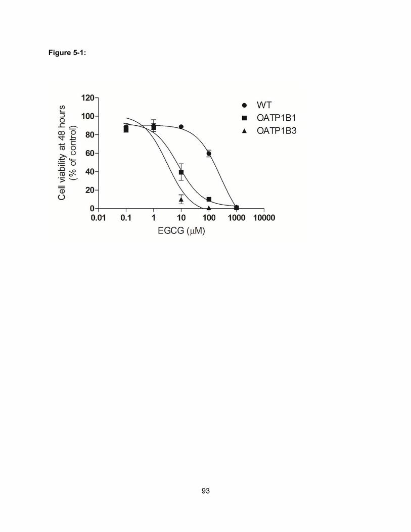
93
Figure 5-1:
94
Figure 5-1: Cell viability assay of wild-type and OATP-expressing CHO cells in the
absence and presence of EGCG. Cell viability of wild-type CHO cells (circles), OATP1B1-
(squares) or OATP1B3-expressing CHO cells (triangles) was measured after a 48 hour
incubation in the absence or presence of increasing concentrations of EGCG. Values obtained
are given as percent of control and are means ± SD of triplicate determinations of four to six
experiments.
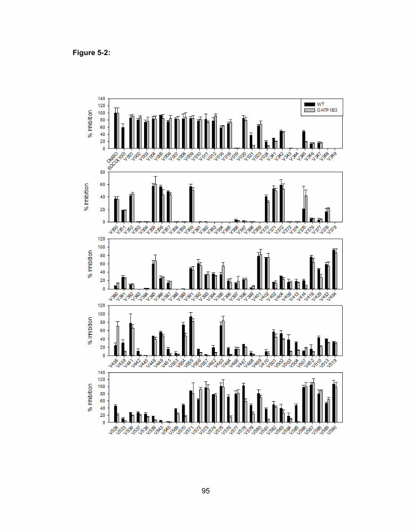
95
Figure 5-2:
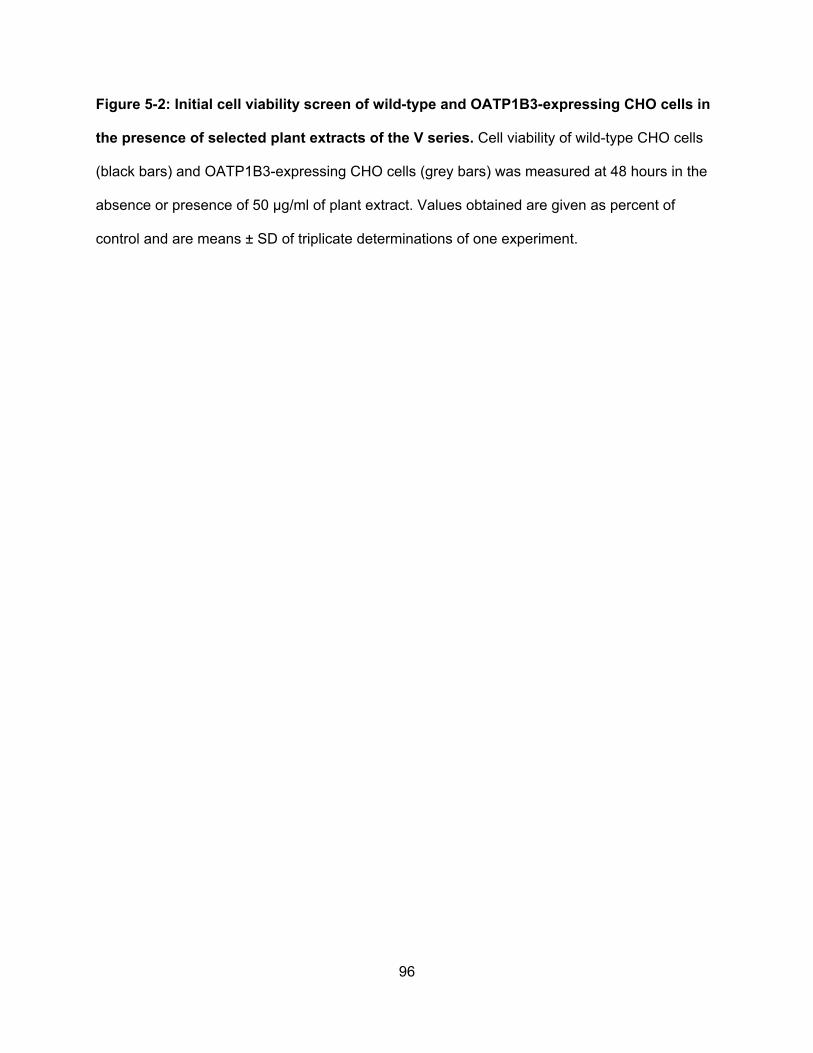
96
Figure 5-2: Initial cell viability screen of wild-type and OATP1B3-expressing CHO cells in
the presence of selected plant extracts of the V series. Cell viability of wild-type CHO cells
(black bars) and OATP1B3-expressing CHO cells (grey bars) was measured at 48 hours in the
absence or presence of 50 µg/ml of plant extract. Values obtained are given as percent of
control and are means ± SD of triplicate determinations of one experiment.
97
Figure 5-3:
1 10 1000
20
40
60
80
100
120
Rhus aromatica (µg/ml)
Cel
l via
bilit
y (%
of
cont
rol)
1 10 1000
20
40
60
80
100
120Wild-type
OATP1B3
Rhus glabra (µg/ml)
98
Figure 5-3: Cell viability of wild-type and OATP1B3-expressing CHO cells in the presence
of the butanol extracts of Rhus aromatica (V345) and Rhus glabra (V585). Cell viability of
wild-type CHO cells (circles) and OATP1B3-expressing CHO cells (triangles) was measured at
48 hours in the presence of increasing concentrations of plant extract. Values obtained are
given as percent of control and are means ± SD of three independent experiments.
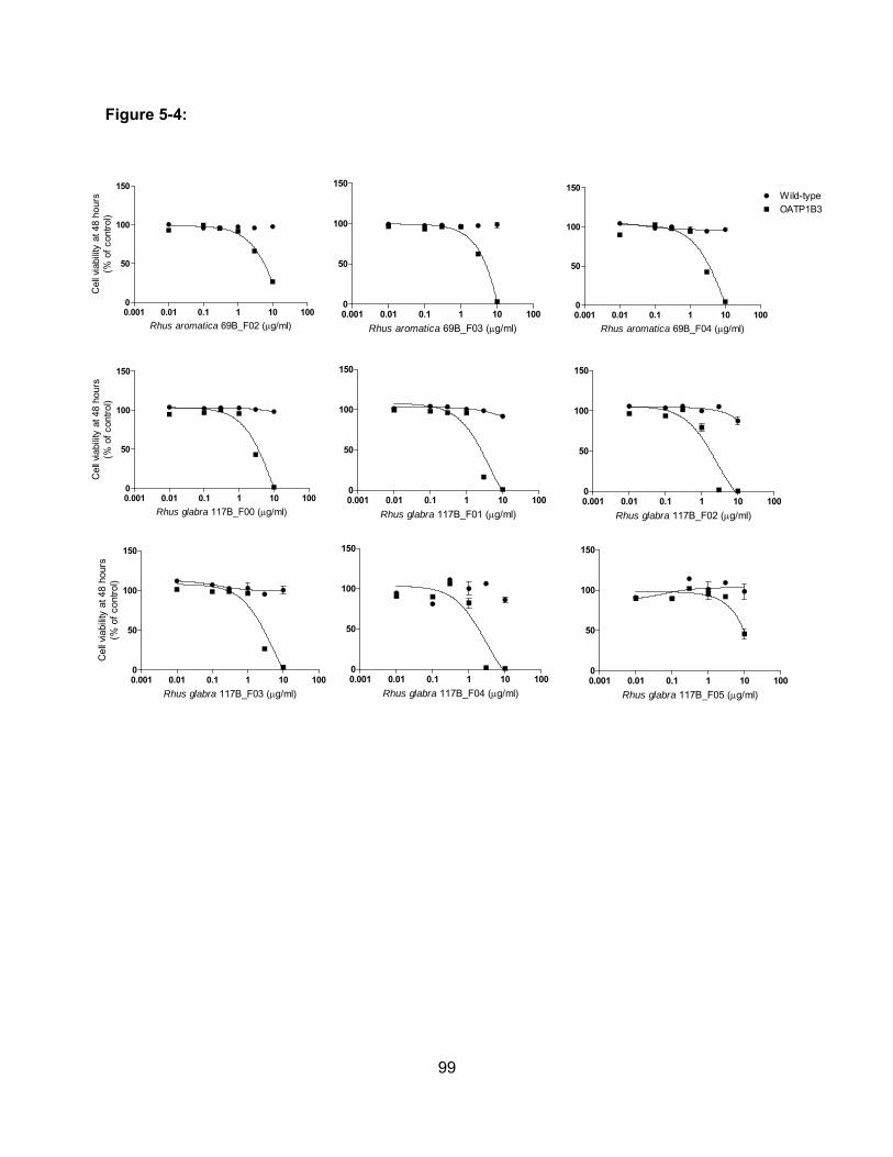
99
Figure 5-4:
0.001 0.01 0.1 1 10 1000
50
100
150
Rhus aromatica 69B_F02 (g/ml)
Cel
l via
bilit
y at
48
hour
s(%
of
cont
rol)
0.001 0.01 0.1 1 10 1000
50
100
150
Rhus aromatica 69B_F03 (g/ml)0.001 0.01 0.1 1 10 1000
50
100
150Wild-typeOATP1B3
Rhus aromatica 69B_F04 (g/ml)
0.001 0.01 0.1 1 10 1000
50
100
150
Rhus glabra 117B_F02 (g/ml)
0.001 0.01 0.1 1 10 1000
50
100
150
Rhus glabra 117B_F01 (g/ml)
0.001 0.01 0.1 1 10 1000
50
100
150
Rhus glabra 117B_F00 (g/ml)
Cel
l via
bilit
y at
48
hour
s(%
of
cont
rol)
0.001 0.01 0.1 1 10 1000
50
100
150
Rhus glabra 117B_F03 (g/ml)
Cel
l via
bilit
y at
48
hour
s(%
of
cont
rol)
0.001 0.01 0.1 1 10 1000
50
100
150
Rhus glabra 117B_F04 (g/ml)0.001 0.01 0.1 1 10 1000
50
100
150
Rhus glabra 117B_F05 (g/ml)
100
Figure 5-4: Cell viability of wild-type and OATP1B3-expressing CHO cells in the presence
of sub-fractions of Rhus aromatica and Rhus glabra. Cell viability of wild-type CHO cells
(circles) and OATP1B3-expressing CHO cells (squares) was measured at 48 hours in the
presence of increasing concentrations of plant extract. Values obtained are given as percent of
control and are means ± SD of two independent experiments.
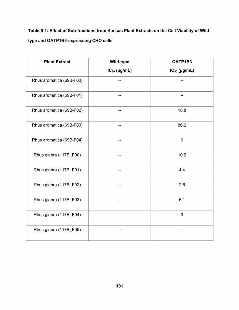
101
Table 5-1: Effect of Sub-fractions from Kansas Plant Extracts on the Cell Viability of Wild-
type and OATP1B3-expressing CHO cells
Plant Extract Wild-type
IC50 (µg/mL)
OATP1B3
IC50 (µg/mL)
Rhus aromatica (69B-F00) -- --
Rhus aromatica (69B-F01) -- --
Rhus aromatica (69B-F02) -- 18.8
Rhus aromatica (69B-F03) -- 86.2
Rhus aromatica (69B-F04) -- 9
Rhus glabra (117B_F00) -- 10.2
Rhus glabra (117B_F01) -- 4.4
Rhus glabra (117B_F02) -- 2.6
Rhus glabra (117B_F03) -- 5.1
Rhus glabra (117B_F04) -- 3
Rhus glabra (117B_F05) -- --
102
5.4: Discussion
Recent studies showing the expression of OATP1B3 in different cancers suggests its
potential use as a therapeutic target for the transport of cytotoxic anticancer drugs into cancer
cells. In this study, we used a bioassay-guided approach along with cytotoxicity assays to
identify novel cytotoxic compounds from Kansas plants that are substrates of OATP1B3. We
demonstrated that a few butanol fractions from the plants Rhus aromatica and Rhus glabra
preferentially kill OATP1B3-expressing cells compared with wild-type cells. We plan to continue
this study by further sub-fractionating the most potent Rhus aromatica and Rhus glabra butanol
fractions until we identify pure compounds from both plants that are responsible for the
cytotoxicity of OATP1B3-expressing cells. These results demonstrate that Kansas plants are a
good source for identifying cytotoxic OATP1B3 substrates that can eventually be developed into
novel anticancer drugs used in the clinic.
103
Chapter 6
Summary and Discussion of Dissertation
6.1: Significance
Organic anion transporting polypeptides (OATPs) are multispecific transporters that
mediate the uptake of numerous endogenous and exogenous compounds into cells. Some
OATPs can be expressed in the epithelium of many different tissues in the body and the
expression of some OATPs can be restricted to a single tissue. OATP1B3 is an example of a
tissue-specific OATP that is exclusively expressed in the liver, specifically at the basolateral
membrane of human hepatocytes. Aside from their normal expression profiles, expression of
OATPs has also been shown to be up- or down-regulated in diseased tissues including
numerous different cancers. Because OATPs are responsible for the transport of a wide range
of compounds their contribution to the disposition of drugs in the body is important.
Consequently, polymorphisms in these transporters can alter pharmacokinetics of drugs and
lead to ineffective therapy or even to toxicities. Because OATPs are expressed in different
cancers and they are able to transport anticancer drugs, they are considered potential targets
for cancer diagnostics and therapy.
Due to the limited information regarding OATPs and cancer, little is known about the role
they play in cancer and about the potential uses to improve cancer therapy. Furthermore,
without a detailed understanding of the expression and function of OATPs in cancer cells, it will
be difficult to utilize OATPs as therapeutic targets. The studies presented in this dissertation
were designed to address this lack of understanding by characterizing OATP expression in
cancer and anticancer drug transport by OATP1B3. To do this I formulated the central
hypothesis that expression of OATPs in cancer combined with their ability to transport cytotoxic
104
anticancer drugs makes them potential targets for improving cancer diagnosis and therapy. This
hypothesis was based on recent studies that demonstrated altered expression of OATPs in
different cancers and on the ability of OATPs to transport some anticancer drugs. Additionally,
experiments in our laboratory have demonstrated that small molecules can allosterically
stimulate OATP-mediated transport. I tested this hypothesis via three specific aims: 1)
identification and characterization of cancer cells that express OATPs, 2) identification and
functional characterization of anticancer drug uptake mediated by OATPs and 3) identification of
cytotoxic compounds from plant extracts that can be developed into anticancer drugs that target
OATP-expressing cancers.
6.2: Specific Aim 1
In the first specific aim, I examined the expression of OATPs in human pancreatic ductal
adenocarcinoma tissues. Initial experiments using a quantigene multiplex assay lead to the
detection of transcripts of four OATPs in all the tested pancreatic tissues. Expression of these
OATPs was further confirmed at the protein level using immunoflourescence staining.
Expression of OATP1B3 was then characterized on a tissue microarray containing several
different types and stages of pancreatic cancer.
Pancreatic cancer commonly develops drug resistance and remains to be a difficult
cancer to treat. Efflux transporters have been major targets in cancer therapy. However,
recently uptake transporters have gained interest as targets to improve cancer therapy. Initial
experiments revealed expression of four OATPs in pancreatic adenocarcinoma at both
messenger RNA and protein level: OATP1B3, OATP2A1, OATP3A1 and OATP4A1. My results
revealed that expression of OATP1B3, OATP2A1, OATP3A1 and OATP4A1 was increased in
pancreatic adenocarcinoma compared to normal pancreas tissue. OATP2A1, OATP3A1 and
105
OATP4A1, under normal conditions, are expressed in a multitude of different tissues in the body.
However, OATP1B3 is exclusively expressed in the liver suggesting that upon treatment with
anticancer drug substrates of OATP1B3, other normal tissues that do not express OATP1B3
would not be affected by these toxic drugs. Therefore, OATP1B3 is clearly a good target for
anticancer therapy using drugs that kill rapidly dividing cells. Thus, OATP1B3 expression was
further characterized in different stages and types of pancreatic cancer.
Because expression of OATP2A1, OATP3A1 and OATP4A1 increases from normal
pancreas tissue to pancreatic adenocarcinoma tissues, these OATPs may serve as potential
therapeutic targets for the delivery of anticancer drugs into pancreatic cancer tumors. However,
none of these three OATPs has been shown to transport any anticancer drugs. OATP1B3 was
also detected in pancreatic adenocarcinoma, however, there was little to no OATP1B3
expression in normal pancreas tissue. Because OATP1B3 has been shown to transport
anticancer drugs like methotrexate, docetaxel and paclitaxel, it is a better candidate for
anticancer therapy than the other three OATPs.
As a final step in this aim, I examined OATP1B3 expression at different stages and types
of pancreatic cancer. The highest expression of OATP1B3 was detected in pancreatic tissues
with mild and chronic inflammation, followed by stage one and stage two pancreatic
adenocarcinomas. OATP1B3 expression was low in stage three pancreatic adenocarcinomas
and absent in metastatic pancreatic cancer. These data suggest that OATP1B3 may serve as a
biomarker for detection of early stage pancreatic cancer and also represents a potential target
for the transport of anticancer drugs early on in pancreatic adenocarcinoma tumor cells. Future
studies should investigate whether circulating OATP1B3 can be detected in pateints with early
stage pancreatic cancer.
106
6.3: Specific Aim 2
As the first specific aim indicated that OATP1B3 is the best target for anticancer therapy,
in the second specific aim I tested the hypothesis that OATP1B3 can transport anticancer drugs
that are used in the clinic. The NCI/DTP anticancer drug library, comprised of 89 FDA approved
anticancer drugs, was screened for substrates of OATP1B3. Initial experiments demonstrated
that several anticancer drugs inhibit OATP1B3-mediated transport of the two model substrates
estradiol-17β-glucuronide and estrone-3-sulfate. Inhibition of model substrate transport
indicated that the anticancer drugs interact with OATP1B3 transport and therefore could be
potential substrates. Therefore, we further confirmed inhibition of OATP1B3-mediated uptake
and determined the mechanism of OATP1B3 inhibition of the four anticancer drugs, actinomycin
D, rapamycin, teniposide and vinblastine. Our results showed that actinomycin D competitively
inhibited OATP1B3-mediated transport of estradiol-17β-glucuronide, but non-competitively
inhibited OATP1B3-mediated transport of estrone-3-sulfate. This suggests that anticancer drug
inhibition of OATP1B3-mediated transport is substrate-dependent. These findings corroborated
that OATP1B3 does indeed have multiple binding sites or translocation pathways for the
different substrates it transports. However, because inhibition kinetics were not consistent and
because even the known substrate rapamycin exhibited non-competitive inhibition it was
impossible to predict novel substrates of OATP1B3 based on inhibition kinetics. It is important to
note that an inhibitor is not necessarily a substrate of OATP1B3, even if it inhibits in a
competitive way, and a compound that does not inhibit should not be ruled out as a potential
substrate for OATP1B3. Therefore, we established a different and unique approach to identify
novel OATP1B3 substrates from the oncology drug set using a cell viability screen.
Using this cell viability approach where drug toxicity was compared between wild-type
and OATP1B3-expressing cells we could demonstrated that the five anticancer drugs bleomycin,
107
etoposide, plicamycin, oxaliplatin and thioguanine indeed preferentially killed OATP1B3-
expressing cells as compared to wild-type cells. These results strongly suggest that these
anticancer drugs are transported by OATP1B3 into the cells to facilitate cell killing. We could
confirm that etoposide and oxaliplatin are indeed substrates of OATP1B3 using radiolabeled
substrates and thus confirmed the validity of the novel approach of screening for cytotoxic
OATP1B3 substrates through the cell viability assay. The results of this specific aim confirmed
that OATP1B3 can transport additional anticancer drugs and suggests that OATP1B3-
expressing cancers could potentially be treated with these cytotoxic anticancer drug substrates
of OATP1B3.
6.4: Specific Aim 3
In the final specific aim, we started with Kansas plant extracts and wanted to identify and
isolate cytotoxic substrates of OATP1B3 that can potentially be developed into novel anticancer
drugs for the treatment of OATP1B3-expressing cancers. In this study we used cell viability
assays that would identify cell extracts or sub-fractions if they contained cytotoxic substrates of
OATP1B3 to screen a series of Kansas plant extracts. Butanol fractions of Rhus aromatica and
Rhus glabra were identified as plant extracts with cytotoxic effects on OATP1B3-expressing
CHO cells. Both extracts were further sub-fractionated and the fractions were again tested for
their effect on OATP1B3-dependent cytotoxicity. Subfraction four from Rhus aromatica and
subfractions two and four from Rhus glabra exhibited the most potent cytotoxic effects and will
be further fractionated until pure cytotoxic substrates are identified. We expect that this study
will identify novel cytotoxic OATP1B3 substrates that could be lead compounds for the
development of novel therapeutic chemicals for the treatment of OATP1B3-expressing cancers.
Such lead compounds would be optimized with the help of medicinal chemists from the
University of Kansas in Lawrence and structure function relationship studies will allow us to
108
identify structural components that are required for OATP1B3 substrates. The long term goal of
these studies is to design novel cytotoxic compounds that are OATP1B3 substrates for the
treatment of OATP1B3-expressing cancers.
6.5: Future Directions
The studies in my dissertation demonstrated that OATPs are up-regulated in pancreatic
adenocarcinoma, are able to transport several anticancer drugs and are able to transport
cytotoxic plant compounds. Knowledge of the expression and function of multispecific drug
transporters, such as OATPs, in cancer has the potential to improve drug targeting and delivery
into cancer cells that exhibit drug resistance. Characterizing the expression and function of
OATPs in cancer is of particular interest, as this could be harnessed to use different approaches
for anticancer therapy. Four approaches can be taken for OATP-mediated anticancer therapy:
(a) OATP-mediated uptake of hormones, hormone conjugates, or unidentified growth promoting
chemicals could be prevented with OATP-selective inhibitors, (b) novel anticancer drugs could
be developed as OATP substrates to increase their uptake into OATP-expressing cancer cells,
(c) uptake of anticancer drugs could be enhanced by allosteric stimulators and (d) expression of
OATPs in the plasma membrane could be modulated to increase or decrease uptake of various
substrates into cancer cells and expression of OATPs could even be used as biomarkers in
cancer development.
The first specific aim identified four major OATPs with increased expression in
pancreatic adenocarcinoma and specifically showed OATP1B3 expression in early stages of
pancreatic adenocarcinoma. By identifying OATPs that are expressed in pancreatic cancer, it
should be possible to screen for anticancer drugs that are substrates of these OATPs. Due to
109
the selective expression of OATP1B3 in the liver, under normal physiological conditions, and its
ability to transport a few anticancer drugs we further investigated the expression of OATP1B3 at
different stages and types of pancreatic cancer and the results suggested that OATP1B3 was
the best candidate for anticancer therapy and improved diagnostics. The second specific aim
identified five anticancer drugs, bleomycin, etoposide, oxaliplatin, plicamycin and thioguanine,
as substrates of OATP1B3. These findings suggest that anyone of these five anticancer drugs
could be used to treat OATP1B3-expressing cancers. Finally, in the third specific aim, we
identified two Kansas plants, Rhus aromatica and Rhus glabra, as plants that contain cytotoxic
compounds that are substrates of OATP1B3 and potentially can be developed into novel
anticancer drugs for the treatment of OATP1B3-expressing cancers. Collectively, these studies
suggest that OATPs can be used as potential targets for anticancer therapy and diagnostics and
propose additional studies than can lead to therapies that can be used in the clinic to improve
future outcomes for cancer patients.
As previously mentioned, one of the approaches that can be taken to improve anticancer
therapy by using OATPs is the development of inhibitors for OATPs to prevent the proliferation
and development of hormone-dependent cancers. Given that OATPs can transport hormones
and their conjugates, one school of thought considers OATPs as contributors to cancer
development for hormone-dependent cancers specifically for breast and prostate cancer.
Therefore, future studies to identify novel OATP inhibitors would be useful to inhibit the uptake
of hormones. High-throughput screening techniques can be employed in identifying specific
OATP inhibitors (Gui et al., 2010). Development of such high-throughput screens is beneficial
for screening large libraries of small molecules in short periods of time. Similarly, high-
throughput screens can be developed to identify stimulators of OATP1B3-mediated anticancer
drug transport. However, it is important to note that modulation of OATP-mediated transport is
110
substrate dependent and this should be taken into consideration when designing such
experiments.
Some OATPs are expressed in many different tissues in the body; their expression in
these tissues can contribute to the distribution of drugs and xenobiotics into the tissues that
harbor their expression. The studies of this dissertation suggest OATP1B3 as the more practical
candidate for OATP-mediated anticancer therapy for the reason that aside from the expression
of OATP1B3 in cancer tissues it is normally exclusively expressed in the liver. Therefore, upon
systemic treatment with anticancer drug substrates of OATP1B3, distribution of these anticancer
drugs will theoretically be targeted to the cancer cells and the liver. It is important to consider
that OATP1B3-mediated anticancer therapy can lead to hepatotoxicity. However, if the
anticancer drugs that are used for treatment target rapidly-dividing cells they should not affect
the liver since it is not a rapidly dividing organ. Additionally, the liver is considered the ultimate
detoxifying organ in the body and expresses numerous detoxifying enzymes that metabolize
drugs and prepare them for excretion from the body, whereas cancer cells lack such enzymes.
It is also important to understand that efflux transporters like MDR1 and MRPs of the ABC
family of transporters are highly up-regulated in cancer cells. The rapid activity of these efflux
transporters could negate the stimulation of OATP-mediated transport by rapidly pumping out
the anticancer drugs from the cancer cells. One approach to resolve this would be to identify a
small molecule that stimulates OATP1B3-mediated anticancer uptake but also inhibits MDR-1-
mediated anticancer efflux. An example of such a small molecule is the green tea component
epigallocatechin gallate (EGCG), which also has anti-proliferative properties. EGCG stimulates
OATP1B3-mediated estrone-3-sulfate transport, has no effect on estradiol-17β-glucuronide
uptake and inhibits Fluo-3 uptake (Roth et al., 2011b), while it also has been shown to inhibit
MDR-1 (Mei et al., 2004). Thus, EGCG may serve as a candidate for OATP-mediated
111
anticancer therapy if it can stimulate OATP1B3-mediated anticancer drug uptake and inhibit
MDR-1 efflux of the same anticancer drug.
In the studies of this dissertation, we used an in-vitro stable expression model of
OATP1B3-expressing Chinese hamster ovary (CHO) cell lines. Our stable expression cell line
was beneficial in identifying novel anticancer compounds transported by OATP1B3. However, in
order to complete these studies, it is important to repeat some experiments in a more
appropriate model using cancer cell lines that express OATP1B3. Several studies have shown
OATP1B3 expression in numerous different cancer cell lines at the mRNA level. Although these
studies are helpful as initial experiments, it is important to identify expression of OATP1B3 at
the protein level. To our knowledge, OATP1B3 expression at the protein level has only been
shown in very few cancer cell lines, one of which is the colorectal cancer cell line HCT-8 (Lee et
al., 2008). Additionally, it is important to consider the differences between established cell lines
and tumor tissues. Differences acquired by the cultured cancer cell lines cause them to not
completely resemble their respective tumor tissues that they are modeled for. This was
especially evidenced in the study done by Gillet et al. where they compared established cancer
cell lines to clinical samples and showed that the cell lines have more resemblance to each
other regardless of the tissue of origin than to the clinical samples they are supposed to model
(Gillet et al., 2011). Therefore, the more appropriate model system to pursue while considering
OATP-mediated cancer therapy would be primary tumor models as they more closely mimic
tumor cells.
Although in-vitro studies can be helpful to investigate many aspects of cancer biology, it
is important to confirm these observations using in-vivo cell models that take into consideration
systemic effects of experiments. Gene duplication events and species divergence caused
human OATPs to have multiple orthologs compared to rodent Oatps. For example, Oatp1b2 is
112
the mouse ortholog of human OATP1B1 and OATP1B3. When considering OATP-mediated
anticancer therapy, it is important to consider Oatp1b2 when preforming in-vivo experiments
using mouse models. Therefore, experiments remain to be done that determine whether
Oatp1b2 transport is modulated the same way as OATP1B3. One way to get around these
experiments is the use of transgenic knockout mouse models to study the role of OATPs in
anticancer drug disposition (van de Steeg et al., 2009; van de Steeg et al., 2011). Another
beneficial in-vivo model system includes the use of nude mice. Experiments should be done
where nude mice are implanted with OATP1B3-expressing cancer cells and treated with
cytotoxic anticancer drugs that are OATP1B3 substrates. Taken together, there are several
considerations that need to be taken when choosing the appropriate model system for these
experiments.
Recent studies identified a novel OATP1B3 isoform in colon and lung tumor tissues. The
isoform identified, named ct-OATP1B3 (cancer type OATP1B3), has a novel transcription start
site and an alternative promoter region (Nagai et al., 2012) which results in a splice variant of
OATP1B3 that lacks the amino terminus and the first transmembrane domain. Because this
study did not investigate whether ct-OATP1B3 is functional, it is imperative that we assess the
function of ct-OATP1B3 before moving forward with studies of OATP1B3-mediated anticancer
therapy.
In conclusion, the studies in this dissertation suggest that OATPs are potentially targets
for improving diagnostics and therapy in cancer. Further research is necessary to determine the
exact role of OATPs in cancer and to elucidate how they can be used in anticancer therapy.
113
References
Abe T, Kakyo M, Tokui T, Nakagomi R, Nishio T, Nakai D, Nomura H, Unno M, Suzuki M,
Naitoh T, Matsuno S, and Yawo H (1999) Identification of a novel gene family encoding
human liver-specific organic anion transporter LST-1. J Biol Chem 274:17159-17163.
Abe T, Unno M, Onogawa T, Tokui T, Kondo TN, Nakagomi R, Adachi H, Fujiwara K, Okabe M,
Suzuki T, Nunoki K, Sato E, Kakyo M, Nishio T, Sugita J, Asano N, Tanemoto M, Seki M,
Date F, Ono K, Kondo Y, Shiiba K, Suzuki M, Ohtani H, Shimosegawa T, Iinuma K,
Nagura H, Ito S, and Matsuno S (2001) LST-2, a human liver-specific organic anion
transporter, determines methotrexate sensitivity in gastrointestinal cancers.
Gastroenterology 120:1689-1699.
Adachi H, Suzuki T, Abe M, Asano N, Mizutamari H, Tanemoto M, Nishio T, Onogawa T,
Toyohara T, Kasai S, Satoh F, Suzuki M, Tokui T, Unno M, Shimosegawa T, Matsuno S,
Ito S, and Abe T (2003) Molecular characterization of human and rat organic anion
transporter OATP-D. Am J Physiol Renal Physiol 285:F1188-1197.
Al Sarakbi W, Mokbel R, Salhab M, Jiang WG, Reed MJ, and Mokbel K (2006) The role of STS
and OATP-B mRNA expression in predicting the clinical outcome in human breast
cancer. Anticancer Res 26:4985-4990.
Badagnani I, Castro RA, Taylor TR, Brett CM, Huang CC, Stryke D, Kawamoto M, Johns SJ,
Ferrin TE, Carlson EJ, Burchard EG, and Giacomini KM (2006) Interaction of
methotrexate with organic-anion transporting polypeptide 1A2 and its genetic variants. J
Pharmacol Exp Ther 318:521-529.
Ballestero MR, Monte MJ, Briz O, Jimenez F, Gonzalez-San Martin F, and Marin JJ (2006)
Expression of transporters potentially involved in the targeting of cytostatic bile acid
derivatives to colon cancer and polyps. Biochem Pharmacol 72:729-738.
114
Banerjee N, Allen C, and Bendayan R (2012) Differential Role of OATPs in Estrone-3-Sulphate
Uptake by Breast Epithelial Cells and Breast Cancer Cells. J Pharmacol Exp Ther
342:510-519.
Boyle P and Levin B (2008) World Cancer Report 2008. International Agency for Research on
Cancer, Lyon Cedex 08, France.
Briz O, Serrano MA, Rebollo N, Hagenbuch B, Meier PJ, Koepsell H, and Marin JJ (2002)
Carriers involved in targeting the cytostatic bile acid-cisplatin derivatives cis-diammine-
chloro-cholylglycinate-platinum(II) and cis-diammine-bisursodeoxycholate-platinum(II)
toward liver cells. Mol Pharmacol 61:853-860.
Bronger H, Konig J, Kopplow K, Steiner HH, Ahmadi R, Herold-Mende C, Keppler D, and Nies
AT (2005) ABCC drug efflux pumps and organic anion uptake transporters in human
gliomas and the blood-tumor barrier. Cancer Res 65:11419-11428.
Burger H, Zoumaro-Djayoon A, Boersma AW, Helleman J, Berns EM, Mathijssen RH, Loos WJ,
and Wiemer EA (2010) Differential transport of platinum compounds by the human
organic cation transporter hOCT2 (hSLC22A2). Br J Pharmacol 159:898-908.
Choi JH, Murray JW, and Wolkoff AW (2011) PDZK1 binding and serine phosphorylation
regulate subcellular trafficking of organic anion transport protein 1a1. Am J Physiol
Gastrointest Liver Physiol 300:G384-393.
Choi K, Zhuang H, Crain B, and Dore S (2008) Expression and localization of prostaglandin
transporter in Alzheimer disease brains and age-matched controls. J Neuroimmunol
195:81-87.
Ciarimboli G, Deuster D, Knief A, Sperling M, Holtkamp M, Edemir B, Pavenstadt H, Lanvers-
Kaminsky C, am Zehnhoff-Dinnesen A, Schinkel AH, Koepsell H, Jurgens H, and
Schlatter E (2010) Organic cation transporter 2 mediates cisplatin-induced oto- and
nephrotoxicity and is a target for protective interventions. Am J Pathol 176:1169-1180.
115
Ciarimboli G, Ludwig T, Lang D, Pavenstadt H, Koepsell H, Piechota HJ, Haier J, Jaehde U,
Zisowsky J, and Schlatter E (2005) Cisplatin nephrotoxicity is critically mediated via the
human organic cation transporter 2. Am J Pathol 167:1477-1484.
Cui Y, Konig J, Nies AT, Pfannschmidt M, Hergt M, Franke WW, Alt W, Moll R, and Keppler D
(2003) Detection of the human organic anion transporters SLC21A6 (OATP2) and
SLC21A8 (OATP8) in liver and hepatocellular carcinoma. Lab Invest 83:527-538.
Daenen S, van der Holt B, Verhoef GE, Lowenberg B, Wijermans PW, Huijgens PC, van
Marwijk Kooy R, Schouten HC, Kramer MH, Ferrant A, van den Berg E, Steijaert MM,
Verdonck LF, and Sonneveld P (2004) Addition of cyclosporin A to the combination of
mitoxantrone and etoposide to overcome resistance to chemotherapy in refractory or
relapsing acute myeloid leukaemia: a randomised phase II trial from HOVON, the Dutch-
Belgian Haemato-Oncology Working Group for adults. Leuk Res 28:1057-1067.
Fahrmayr C, Konig J, Auge D, Mieth M, and Fromm MF (2012) Identification of drugs and drug
metabolites as substrates of multidrug resistance protein 2 (MRP2) using triple-
transfected MDCK-OATP1B1-UGT1A1-MRP2 cells. Br J Pharmacol 165:1836-1847.
Feng B, Xu JJ, Bi YA, Mireles R, Davidson R, Duignan DB, Campbell S, Kostrubsky VE, Dunn
MC, Smith AR, and Wang HF (2009) Role of hepatic transporters in the disposition and
hepatotoxicity of a HER2 tyrosine kinase inhibitor CP-724,714. Toxicol Sci 108:492-500.
Filipski KK, Loos WJ, Verweij J, and Sparreboom A (2008) Interaction of Cisplatin with the
human organic cation transporter 2. Clin Cancer Res 14:3875-3880.
Filipski KK, Mathijssen RH, Mikkelsen TS, Schinkel AH, and Sparreboom A (2009) Contribution
of organic cation transporter 2 (OCT2) to cisplatin-induced nephrotoxicity. Clin
Pharmacol Ther 86:396-402.
Fotoohi AK, Lindqvist M, Peterson C, and Albertioni F (2006) Involvement of the concentrative
nucleoside transporter 3 and equilibrative nucleoside transporter 2 in the resistance of T-
lymphoblastic cell lines to thiopurines. Biochem Biophys Res Commun 343:208-215.
116
Friedenberg WR, Rue M, Blood EA, Dalton WS, Shustik C, Larson RA, Sonneveld P, and
Greipp PR (2006) Phase III study of PSC-833 (valspodar) in combination with vincristine,
doxorubicin, and dexamethasone (valspodar/VAD) versus VAD alone in patients with
recurring or refractory multiple myeloma (E1A95): a trial of the Eastern Cooperative
Oncology Group. Cancer 106:830-838.
Fujiwara K, Adachi H, Nishio T, Unno M, Tokui T, Okabe M, Onogawa T, Suzuki T, Asano N,
Tanemoto M, Seki M, Shiiba K, Suzuki M, Kondo Y, Nunoki K, Shimosegawa T, Iinuma
K, Ito S, Matsuno S, and Abe T (2001) Identification of thyroid hormone transporters in
humans: different molecules are involved in a tissue-specific manner. Endocrinology
142:2005-2012.
Gao B, Hagenbuch B, Kullak-Ublick GA, Benke D, Aguzzi A, and Meier PJ (2000) Organic
anion-transporting polypeptides mediate transport of opioid peptides across blood-brain
barrier. J Pharmacol Exp Ther 294:73-79.
Gao B, Huber RD, Wenzel A, Vavricka SR, Ismair MG, Reme C, and Meier PJ (2005)
Localization of organic anion transporting polypeptides in the rat and human ciliary body
epithelium. Exp Eye Res 80:61-72.
Gillet JP, Calcagno AM, Varma S, Marino M, Green LJ, Vora MI, Patel C, Orina JN, Eliseeva TA,
Singal V, Padmanabhan R, Davidson B, Ganapathi R, Sood AK, Rueda BR, Ambudkar
SV, and Gottesman MM (2011) Redefining the relevance of established cancer cell lines
to the study of mechanisms of clinical anti-cancer drug resistance. Proc Natl Acad Sci U
S A 108:18708-18713.
Glaeser H, Bailey DG, Dresser GK, Gregor JC, Schwarz UI, McGrath JS, Jolicoeur E, Lee W,
Leake BF, Tirona RG, and Kim RB (2007) Intestinal drug transporter expression and the
impact of grapefruit juice in humans. Clin Pharmacol Ther 81:362-370.
Gottesman MM, Fojo T, and Bates SE (2002) Multidrug resistance in cancer: role of ATP-
dependent transporters. Nat Rev Cancer 2:48-58.
117
Grube M, Kock K, Oswald S, Draber K, Meissner K, Eckel L, Bohm M, Felix SB, Vogelgesang S,
Jedlitschky G, Siegmund W, Warzok R, and Kroemer HK (2006) Organic anion
transporting polypeptide 2B1 is a high-affinity transporter for atorvastatin and is
expressed in the human heart. Clin Pharmacol Ther 80:607-620.
Gui C and Hagenbuch B (2008) Amino acid residues in transmembrane domain 10 of organic
anion transporting polypeptide 1B3 are critical for cholecystokinin octapeptide transport.
Biochemistry 47:9090-9097.
Gui C and Hagenbuch B (2009) Role of transmembrane domain 10 for the function of organic
anion transporting polypeptide 1B1. Protein Sci 18:2298-2306.
Gui C, Miao Y, Thompson L, Wahlgren B, Mock M, Stieger B, and Hagenbuch B (2008) Effect of
pregnane X receptor ligands on transport mediated by human OATP1B1 and OATP1B3.
Eur J Pharmacol 584:57-65.
Gui C, Obaidat A, Chaguturu R, and Hagenbuch B (2010) Development of a cell-based high-
throughput assay to screen for inhibitors of organic anion transporting polypeptides 1B1
and 1B3. Curr Chem Genomics 4:1-8.
Guo GL and Klaassen CD (2001) Protein kinase C suppresses rat organic anion transporting
polypeptide 1- and 2-mediated uptake. J Pharmacol Exp Ther 299:551-557.
Hagenbuch B (2007) Cellular entry of thyroid hormones by organic anion transporting
polypeptides. Best Pract Res Clin Endocrinol Metab 21:209-221.
Hagenbuch B and Gui C (2008) Xenobiotic transporters of the human organic anion transporting
polypeptides (OATP) family. Xenobiotica 38:778-801.
Hagenbuch B and Meier PJ (2003) The superfamily of organic anion transporting polypeptides.
Biochim Biophys Acta 1609:1-18.
Hagenbuch B and Meier PJ (2004) Organic anion transporting polypeptides of the OATP/
SLC21 family: phylogenetic classification as OATP/ SLCO superfamily, new
nomenclature and molecular/functional properties. Pflugers Arch 447:653-665.
118
Hamada A, Sissung T, Price DK, Danesi R, Chau CH, Sharifi N, Venzon D, Maeda K, Nagao K,
Sparreboom A, Mitsuya H, Dahut WL, and Figg WD (2008) Effect of SLCO1B3
haplotype on testosterone transport and clinical outcome in caucasian patients with
androgen-independent prostatic cancer. Clin Cancer Res 14:3312-3318.
Hanggi E, Grundschober AF, Leuthold S, Meier PJ, and St-Pierre MV (2006) Functional
analysis of the extracellular cysteine residues in the human organic anion transporting
polypeptide, OATP2B1. Mol Pharmacol 70:806-817.
Hayeshi R, Hilgendorf C, Artursson P, Augustijns P, Brodin B, Dehertogh P, Fisher K, Fossati L,
Hovenkamp E, Korjamo T, Masungi C, Maubon N, Mols R, Mullertz A, Monkkonen J,
O'Driscoll C, Oppers-Tiemissen HM, Ragnarsson EG, Rooseboom M, and Ungell AL
(2008) Comparison of drug transporter gene expression and functionality in Caco-2 cells
from 10 different laboratories. Eur J Pharm Sci 35:383-396.
Heuer H and Visser TJ (2009) Minireview: Pathophysiological importance of thyroid hormone
transporters. Endocrinology 150:1078-1083.
Hidalgo M (2010) Pancreatic cancer. N Engl J Med 362:1605-1617.
Holla VR, Backlund MG, Yang P, Newman RA, and DuBois RN (2008) Regulation of
prostaglandin transporters in colorectal neoplasia. Cancer Prev Res (Phila) 1:93-99.
Hsiang B, Zhu Y, Wang Z, Wu Y, Sasseville V, Yang WP, and Kirchgessner TG (1999) A novel
human hepatic organic anion transporting polypeptide (OATP2). Identification of a liver-
specific human organic anion transporting polypeptide and identification of rat and
human hydroxymethylglutaryl-CoA reductase inhibitor transporters. J Biol Chem
274:37161-37168.
Hu C, Lancaster CS, Zuo Z, Hu S, Chen Z, Rubnitz JE, Baker SD, and Sparreboom A (2012)
Inhibition of OCTN2-mediated transport of carnitine by etoposide. Mol Cancer Ther
11:921-929.
119
Hu S, Franke RM, Filipski KK, Hu C, Orwick SJ, de Bruijn EA, Burger H, Baker SD, and
Sparreboom A (2008) Interaction of imatinib with human organic ion carriers. Clin
Cancer Res 14:3141-3148.
Huber RD, Gao B, Sidler Pfandler MA, Zhang-Fu W, Leuthold S, Hagenbuch B, Folkers G,
Meier PJ, and Stieger B (2007) Characterization of two splice variants of human organic
anion transporting polypeptide 3A1 isolated from human brain. Am J Physiol Cell Physiol
292:C795-806.
Ichihara S, Kikuchi R, Kusuhara H, Imai S, Maeda K, and Sugiyama Y (2010) DNA methylation
profiles of organic anion transporting polypeptide 1B3 in cancer cell lines. Pharm Res
27:510-516.
Ismair MG, Stieger B, Cattori V, Hagenbuch B, Fried M, Meier PJ, and Kullak-Ublick GA (2001)
Hepatic uptake of cholecystokinin octapeptide by organic anion-transporting
polypeptides OATP4 and OATP8 of rat and human liver. Gastroenterology 121:1185-
1190.
Jemal A, Siegel R, Ward E, Hao Y, Xu J, and Thun MJ (2009) Cancer statistics, 2009. CA
Cancer J Clin 59:225-249.
Jigorel E, Le Vee M, Boursier-Neyret C, Bertrand M, and Fardel O (2005) Functional expression
of sinusoidal drug transporters in primary human and rat hepatocytes. Drug Metab
Dispos 33:1418-1422.
Johnson IS, Armstrong JG, Gorman M, and Burnett JP, Jr. (1963) The Vinca Alkaloids: A New
Class of Oncolytic Agents. Cancer Res 23:1390-1427.
Jong NN, Nakanishi T, Liu JJ, Tamai I, and McKeage MJ (2011) Oxaliplatin transport mediated
by organic cation/carnitine transporters OCTN1 and OCTN2 in overexpressing human
embryonic kidney 293 cells and rat dorsal root ganglion neurons. J Pharmacol Exp Ther
338:537-547.
120
Kang J, Chapdelaine P, Parent J, Madore E, Laberge PY, and Fortier MA (2005) Expression of
human prostaglandin transporter in the human endometrium across the menstrual cycle.
J Clin Endocrinol Metab 90:2308-2313.
Kennedy BJ (1970) Metabolic and toxic effects of mithramycin during tumor therapy. Am J Med
49:494-503.
Kindla J, Rau TT, Jung R, Fasching PA, Strick R, Stoehr R, Hartmann A, Fromm MF, and Konig
J (2011a) Expression and localization of the uptake transporters OATP2B1, OATP3A1
and OATP5A1 in non-malignant and malignant breast tissue. Cancer Biol Ther 11.
Kindla J, Rau TT, Jung R, Fasching PA, Strick R, Stoehr R, Hartmann A, Fromm MF, and Konig
J (2011b) Expression and localization of the uptake transporters OATP2B1, OATP3A1
and OATP5A1 in non-malignant and malignant breast tissue. Cancer Biol Ther 11:584-
591.
King PD and Perry MC (2001) Hepatotoxicity of chemotherapy. Oncologist 6:162-176.
Kobayashi D, Nozawa T, Imai K, Nezu J, Tsuji A, and Tamai I (2003) Involvement of human
organic anion transporting polypeptide OATP-B (SLC21A9) in pH-dependent transport
across intestinal apical membrane. J Pharmacol Exp Ther 306:703-708.
Kock K, Koenen A, Giese B, Fraunholz M, May K, Siegmund W, Hammer E, Volker U,
Jedlitschky G, Kroemer HK, and Grube M (2010) Rapid modulation of the organic anion
transporting polypeptide 2B1 (OATP2B1, SLCO2B1) function by protein kinase C-
mediated internalization. J Biol Chem 285:11336-11347.
Konig J (2011) Uptake transporters of the human OATP family: molecular characteristics,
substrates, their role in drug-drug interactions, and functional consequences of
polymorphisms. Handb Exp Pharmacol:1-28.
Konig J, Cui Y, Nies AT, and Keppler D (2000a) Localization and genomic organization of a new
hepatocellular organic anion transporting polypeptide. J Biol Chem 275:23161-23168.
121
Konig J, Cui Y, Nies AT, and Keppler D (2000b) A novel human organic anion transporting
polypeptide localized to the basolateral hepatocyte membrane. Am J Physiol
Gastrointest Liver Physiol 278:G156-164.
Kounnis V, Ioachim E, Svoboda M, Tzakos A, Sainis I, Thalhammer T, Steiner G, and Briasoulis
E (2011) Expression of organic anion-transporting polypeptides 1B3, 1B1, and 1A2 in
human pancreatic cancer reveals a new class of potential therapeutic targets. Onco
Targets Ther 4:27-32.
Kullak-Ublick GA, Hagenbuch B, Stieger B, Schteingart CD, Hofmann AF, Wolkoff AW, and
Meier PJ (1995) Molecular and functional characterization of an organic anion
transporting polypeptide cloned from human liver. Gastroenterology 109:1274-1282.
Kullak-Ublick GA, Ismair MG, Stieger B, Landmann L, Huber R, Pizzagalli F, Fattinger K, Meier
PJ, and Hagenbuch B (2001) Organic anion-transporting polypeptide B (OATP-B) and its
functional comparison with three other OATPs of human liver. Gastroenterology
120:525-533.
Lee SY, Williamson B, Caballero OL, Chen YT, Scanlan MJ, Ritter G, Jongeneel CV, Simpson
AJ, and Old LJ (2004) Identification of the gonad-specific anion transporter SLCO6A1 as
a cancer/testis (CT) antigen expressed in human lung cancer. Cancer Immun 4:13.
Lee W, Belkhiri A, Lockhart AC, Merchant N, Glaeser H, Harris EI, Washington MK, Brunt EM,
Zaika A, Kim RB, and El-Rifai W (2008) Overexpression of OATP1B3 confers apoptotic
resistance in colon cancer. Cancer Res 68:10315-10323.
Lee W, Glaeser H, Smith LH, Roberts RL, Moeckel GW, Gervasini G, Leake BF, and Kim RB
(2005) Polymorphisms in human organic anion-transporting polypeptide 1A2
(OATP1A2): implications for altered drug disposition and central nervous system drug
entry. J Biol Chem 280:9610-9617.
Letschert K, Faulstich H, Keller D, and Keppler D (2006) Molecular characterization and
inhibition of amanitin uptake into human hepatocytes. Toxicol Sci 91:140-149.
122
Libra A, Fernetti C, Lorusso V, Visigalli M, Anelli PL, Staud F, Tiribelli C, and Pascolo L (2006)
Molecular determinants in the transport of a bile acid-derived diagnostic agent in tumoral
and nontumoral cell lines of human liver. J Pharmacol Exp Ther 319:809-817.
Liedauer R, Svoboda M, Wlcek K, Arrich F, Ja W, Toma C, and Thalhammer T (2009) Different
expression patterns of organic anion transporting polypeptides in osteosarcomas, bone
metastases and aneurysmal bone cysts. Oncol Rep 22:1485-1492.
Lockhart AC, Harris E, Lafleur BJ, Merchant NB, Washington MK, Resnick MB, Yeatman TJ,
and Lee W (2008) Organic anion transporting polypeptide 1B3 (OATP1B3) is
overexpressed in colorectal tumors and is a predictor of clinical outcome. Clin Exp
Gastroenterol 1:1-7.
Maeda T, Irokawa M, Arakawa H, Kuraoka E, Nozawa T, Tateoka R, Itoh Y, Nakanishi T, and
Tamai I (2010) Uptake transporter organic anion transporting polypeptide 1B3
contributes to the growth of estrogen-dependent breast cancer. J Steroid Biochem Mol
Biol 122:180-185.
Mandery K, Bujok K, Schmidt I, Wex T, Treiber G, Malfertheiner P, Rau TT, Amann KU, Brune
K, Fromm MF, and Glaeser H (2010) Influence of cyclooxygenase inhibitors on the
function of the prostaglandin transporter organic anion-transporting polypeptide 2A1
expressed in human gastroduodenal mucosa. J Pharmacol Exp Ther 332:345-351.
Mei Y, Qian F, Wei D, and Liu J (2004) Reversal of cancer multidrug resistance by green tea
polyphenols. J Pharm Pharmacol 56:1307-1314.
Meyer zu Schwabedissen HE, Tirona RG, Yip CS, Ho RH, and Kim RB (2008) Interplay
between the nuclear receptor pregnane X receptor and the uptake transporter organic
anion transporter polypeptide 1A2 selectively enhances estrogen effects in breast
cancer. Cancer Res 68:9338-9347.
Miki Y, Suzuki T, Kitada K, Yabuki N, Shibuya R, Moriya T, Ishida T, Ohuchi N, Blumberg B,
and Sasano H (2006) Expression of the steroid and xenobiotic receptor and its possible
123
target gene, organic anion transporting polypeptide-A, in human breast carcinoma.
Cancer Res 66:535-542.
Mikkaichi T, Suzuki T, Onogawa T, Tanemoto M, Mizutamari H, Okada M, Chaki T, Masuda S,
Tokui T, Eto N, Abe M, Satoh F, Unno M, Hishinuma T, Inui K, Ito S, Goto J, and Abe T
(2004) Isolation and characterization of a digoxin transporter and its rat homologue
expressed in the kidney. Proc Natl Acad Sci U S A 101:3569-3574.
Miyagawa M, Maeda K, Aoyama A, and Sugiyama Y (2009) The eighth and ninth
transmembrane domains in organic anion transporting polypeptide 1B1 affect the
transport kinetics of estrone-3-sulfate and estradiol-17beta-D-glucuronide. J Pharmacol
Exp Ther 329:551-557.
Monks NR, Liu S, Xu Y, Yu H, Bendelow AS, and Moscow JA (2007) Potent cytotoxicity of the
phosphatase inhibitor microcystin LR and microcystin analogues in OATP1B1- and
OATP1B3-expressing HeLa cells. Mol Cancer Ther 6:587-598.
Muto M, Onogawa T, Suzuki T, Ishida T, Rikiyama T, Katayose Y, Ohuchi N, Sasano H, Abe T,
and Unno M (2007) Human liver-specific organic anion transporter-2 is a potent
prognostic factor for human breast carcinoma. Cancer Sci 98:1570-1576.
Nagai M, Furihata T, Matsumoto S, Ishii S, Motohashi S, Yoshino I, Ugajin M, Miyajima A,
Matsumoto S, and Chiba K (2012) Identification of a new organic anion transporting
polypeptide 1B3 mRNA isoform primarily expressed in human cancerous tissues and
cells. Biochem Biophys Res Commun 418:818-823.
Ni W, Ji J, Dai Z, Papp A, Johnson AJ, Ahn S, Farley KL, Lin TS, Dalton JT, Li X, Jarjoura D,
Byrd JC, Sadee W, Grever MR, and Phelps MA (2010) Flavopiridol pharmacogenetics:
clinical and functional evidence for the role of SLCO1B1/OATP1B1 in flavopiridol
disposition. PLoS One 5:e13792.
Nozawa T, Minami H, Sugiura S, Tsuji A, and Tamai I (2005a) Role of organic anion transporter
OATP1B1 (OATP-C) in hepatic uptake of irinotecan and its active metabolite, 7-ethyl-10-
124
hydroxycamptothecin: in vitro evidence and effect of single nucleotide polymorphisms.
Drug Metab Dispos 33:434-439.
Nozawa T, Suzuki M, Takahashi K, Yabuuchi H, Maeda T, Tsuji A, and Tamai I (2004)
Involvement of estrone-3-sulfate transporters in proliferation of hormone-dependent
breast cancer cells. J Pharmacol Exp Ther 311:1032-1037.
Nozawa T, Suzuki M, Yabuuchi H, Irokawa M, Tsuji A, and Tamai I (2005b) Suppression of cell
proliferation by inhibition of estrone-3-sulfate transporter in estrogen-dependent breast
cancer cells. Pharm Res 22:1634-1641.
Obaidat A, Roth M, and Hagenbuch B (2012) The expression and function of organic anion
transporting polypeptides in normal tissues and in cancer. Annu Rev Pharmacol Toxicol
52:135-151.
Okabe M, Szakacs G, Reimers MA, Suzuki T, Hall MD, Abe T, Weinstein JN, and Gottesman
MM (2008) Profiling SLCO and SLC22 genes in the NCI-60 cancer cell lines to identify
drug uptake transporters. Mol Cancer Ther 7:3081-3091.
Oostendorp RL, van de Steeg E, van der Kruijssen CM, Beijnen JH, Kenworthy KE, Schinkel
AH, and Schellens JH (2009) Organic anion-transporting polypeptide 1B1 mediates
transport of Gimatecan and BNP1350 and can be inhibited by several classic ATP-
binding cassette (ABC) B1 and/or ABCG2 inhibitors. Drug Metab Dispos 37:917-923.
Oswald S, Nassif A, Modess C, Keiser M, Hanke U, Engel A, Lutjohann D, Weitschies W, and
Siegmund W (2010) Pharmacokinetic and pharmacodynamic interactions between the
immunosuppressant sirolimus and the lipid-lowering drug ezetimibe in healthy volunteers.
Clin Pharmacol Ther 87:663-667.
Pazdur R, Kudelka AP, Kavanagh JJ, Cohen PR, and Raber MN (1993) The taxoids: paclitaxel
(Taxol) and docetaxel (Taxotere). Cancer Treat Rev 19:351-386.
125
Pizzagalli F, Hagenbuch B, Stieger B, Klenk U, Folkers G, and Meier PJ (2002) Identification of
a novel human organic anion transporting polypeptide as a high affinity thyroxine
transporter. Mol Endocrinol 16:2283-2296.
Pizzagalli F, Varga Z, Huber RD, Folkers G, Meier PJ, and St-Pierre MV (2003) Identification of
steroid sulfate transport processes in the human mammary gland. J Clin Endocrinol
Metab 88:3902-3912.
Pressler H, Sissung TM, Venzon D, Price DK, and Figg WD (2011) Expression of OATP family
members in hormone-related cancers: potential markers of progression. PLoS One
6:e20372.
Pusztai L, Wagner P, Ibrahim N, Rivera E, Theriault R, Booser D, Symmans FW, Wong F,
Blumenschein G, Fleming DR, Rouzier R, Boniface G, and Hortobagyi GN (2005) Phase
II study of tariquidar, a selective P-glycoprotein inhibitor, in patients with chemotherapy-
resistant, advanced breast carcinoma. Cancer 104:682-691.
Reichert M and Rustgi AK (2011) Pancreatic ductal cells in development, regeneration, and
neoplasia. J Clin Invest 121:4572-4578.
Roberts LM, Woodford K, Zhou M, Black DS, Haggerty JE, Tate EH, Grindstaff KK, Mengesha
W, Raman C, and Zerangue N (2008) Expression of the thyroid hormone transporters
monocarboxylate transporter-8 (SLC16A2) and organic ion transporter-14 (SLCO1C1) at
the blood-brain barrier. Endocrinology 149:6251-6261.
Roth M, Araya JJ, Timmermann BN, and Hagenbuch B (2011a) Isolation of modulators of the
liver-specific organic anion-transporting polypeptides (OATPs) 1B1 and 1B3 from
Rollinia emarginata Schlecht (Annonaceae). J Pharmacol Exp Ther 339:624-632.
Roth M, Timmermann BN, and Hagenbuch B (2011b) Interactions of green tea catechins with
organic anion-transporting polypeptides. Drug Metab Dispos 39:920-926.
Saeki T, Nomizu T, Toi M, Ito Y, Noguchi S, Kobayashi T, Asaga T, Minami H, Yamamoto N,
Aogi K, Ikeda T, Ohashi Y, Sato W, and Tsuruo T (2007) Dofequidar fumarate (MS-209)
126
in combination with cyclophosphamide, doxorubicin, and fluorouracil for patients with
advanced or recurrent breast cancer. J Clin Oncol 25:411-417.
Sasaki M, Suzuki H, Aoki J, Ito K, Meier PJ, and Sugiyama Y (2004) Prediction of in vivo biliary
clearance from the in vitro transcellular transport of organic anions across a double-
transfected Madin-Darby canine kidney II monolayer expressing both rat organic anion
transporting polypeptide 4 and multidrug resistance associated protein 2. Mol Pharmacol
66:450-459.
Sato K, Sugawara J, Sato T, Mizutamari H, Suzuki T, Ito A, Mikkaichi T, Onogawa T, Tanemoto
M, Unno M, Abe T, and Okamura K (2003) Expression of organic anion transporting
polypeptide E (OATP-E) in human placenta. Placenta 24:144-148.
Schuster VL (2002) Prostaglandin transport. Prostaglandins Other Lipid Mediat 68-69:633-647.
Smith NF, Acharya MR, Desai N, Figg WD, and Sparreboom A (2005) Identification of
OATP1B3 as a high-affinity hepatocellular transporter of paclitaxel. Cancer Biol Ther
4:815-818.
Smith NF, Marsh S, Scott-Horton TJ, Hamada A, Mielke S, Mross K, Figg WD, Verweij J,
McLeod HL, and Sparreboom A (2007) Variants in the SLCO1B3 gene: interethnic
distribution and association with paclitaxel pharmacokinetics. Clin Pharmacol Ther
81:76-82.
St-Pierre MV, Hagenbuch B, Ugele B, Meier PJ, and Stallmach T (2002) Characterization of an
organic anion-transporting polypeptide (OATP-B) in human placenta. J Clin Endocrinol
Metab 87:1856-1863.
Steckelbroeck S, Nassen A, Ugele B, Ludwig M, Watzka M, Reissinger A, Clusmann H,
Lutjohann D, Siekmann L, Klingmuller D, and Hans VH (2004) Steroid sulfatase (STS)
expression in the human temporal lobe: enzyme activity, mRNA expression and
immunohistochemistry study. J Neurochem 89:403-417.
127
Suzuki T, Onogawa T, Asano N, Mizutamari H, Mikkaichi T, Tanemoto M, Abe M, Satoh F,
Unno M, Nunoki K, Suzuki M, Hishinuma T, Goto J, Shimosegawa T, Matsuno S, Ito S,
and Abe T (2003) Identification and characterization of novel rat and human gonad-
specific organic anion transporters. Mol Endocrinol 17:1203-1215.
Svoboda M, Riha J, Wlcek K, Jaeger W, and Thalhammer T (2011a) Organic anion transporting
polypeptides (OATPs): regulation of expression and function. Curr Drug Metab 12:139-
153.
Svoboda M, Wlcek K, Taferner B, Hering S, Stieger B, Tong D, Zeillinger R, Thalhammer T, and
Jager W (2011b) Expression of organic anion-transporting polypeptides 1B1 and 1B3 in
ovarian cancer cells: relevance for paclitaxel transport. Biomed Pharmacother 65:417-
426.
Szakacs G, Paterson JK, Ludwig JA, Booth-Genthe C, and Gottesman MM (2006) Targeting
multidrug resistance in cancer. Nat Rev Drug Discov 5:219-234.
Takane H, Kawamoto K, Sasaki T, Moriki K, Kitano H, Higuchi S, Otsubo K, and Ieiri I (2009)
Life-threatening toxicities in a patient with UGT1A1*6/*28 and SLCO1B1*15/*15
genotypes after irinotecan-based chemotherapy. Cancer Chemother Pharmacol
63:1165-1169.
Tamai I, Nezu J, Uchino H, Sai Y, Oku A, Shimane M, and Tsuji A (2000) Molecular
identification and characterization of novel members of the human organic anion
transporter (OATP) family. Biochem Biophys Res Commun 273:251-260.
Tsuboyama T, Onishi H, Kim T, Akita H, Hori M, Tatsumi M, Nakamoto A, Nagano H, Matsuura
N, Wakasa K, and Tomoda K (2010) Hepatocellular carcinoma: hepatocyte-selective
enhancement at gadoxetic acid-enhanced MR imaging--correlation with expression of
sinusoidal and canalicular transporters and bile accumulation. Radiology 255:824-833.
van de Steeg E, van der Kruijssen CM, Wagenaar E, Burggraaff JE, Mesman E, Kenworthy KE,
and Schinkel AH (2009) Methotrexate pharmacokinetics in transgenic mice with liver-
128
specific expression of human organic anion-transporting polypeptide 1B1 (SLCO1B1).
Drug Metab Dispos 37:277-281.
van de Steeg E, van Esch A, Wagenaar E, van der Kruijssen CM, van Tellingen O, Kenworthy
KE, and Schinkel AH (2011) High impact of Oatp1a/1b transporters on in vivo disposition
of the hydrophobic anticancer drug paclitaxel. Clin Cancer Res 17:294-301.
van Montfoort JE, Hagenbuch B, Groothuis GM, Koepsell H, Meier PJ, and Meijer DK (2003)
Drug uptake systems in liver and kidney. Curr Drug Metab 4:185-211.
Vander Borght S, Libbrecht L, Blokzijl H, Faber KN, Moshage H, Aerts R, Van Steenbergen W,
Jansen PL, Desmet VJ, and Roskams TA (2005) Diagnostic and pathogenetic
implications of the expression of hepatic transporters in focal lesions occurring in normal
liver. J Pathol 207:471-482.
Vavricka SR, Jung D, Fried M, Grutzner U, Meier PJ, and Kullak-Ublick GA (2004) The human
organic anion transporting polypeptide 8 (SLCO1B3) gene is transcriptionally repressed
by hepatocyte nuclear factor 3beta in hepatocellular carcinoma. J Hepatol 40:212-218.
Wang X, Wolkoff AW, and Morris ME (2005) Flavonoids as a novel class of human organic
anion-transporting polypeptide OATP1B1 (OATP-C) modulators. Drug Metab Dispos
33:1666-1672.
Wlcek K, Svoboda M, Riha J, Zakaria S, Olszewski U, Dvorak Z, Sellner F, Ellinger I, Jager W,
and Thalhammer T (2011) The analysis of organic anion transporting polypeptide
(OATP) mRNA and protein patterns in primary and metastatic liver cancer. Cancer Biol
Ther 11:801-811.
Wlcek K, Svoboda M, Thalhammer T, Sellner F, Krupitza G, and Jaeger W (2008) Altered
expression of organic anion transporter polypeptide (OATP) genes in human breast
carcinoma. Cancer Biol Ther 7:1450-1455.
Wright JL, Kwon EM, Ostrander EA, Montgomery B, Line DW, Vessella RL, Stanford JL, and
Mostaghel EA (2011) Expression of SLCO transport genes in castration resistant
129
prostate cancer and impact of genetic variation in SCLO1B3 and SLCO2B1 on prostate
cancer outcomes. Cancer Epidemiol Biomarkers Prev.
Xiang X, Jada SR, Li HH, Fan L, Tham LS, Wong CI, Lee SC, Lim R, Zhou QY, Goh BC, Tan
EH, and Chowbay B (2006) Pharmacogenetics of SLCO1B1 gene and the impact of *1b
and *15 haplotypes on irinotecan disposition in Asian cancer patients. Pharmacogenet
Genomics 16:683-691.
Yamaguchi H, Kobayashi M, Okada M, Takeuchi T, Unno M, Abe T, Goto J, Hishinuma T, and
Mano N (2008) Rapid screening of antineoplastic candidates for the human organic
anion transporter OATP1B3 substrates using fluorescent probes. Cancer Lett 260:163-
169.
Yonezawa A, Masuda S, Nishihara K, Yano I, Katsura T, and Inui K (2005) Association between
tubular toxicity of cisplatin and expression of organic cation transporter rOCT2 (Slc22a2)
in the rat. Biochem Pharmacol 70:1823-1831.
Zafar SF and El-Rayes BF (2012) Chemotherapeutic Strategies in Advanced or Metastatic
Pancreatic Adenocarcinoma. Am J Clin Oncol.
Zhang S, Lovejoy KS, Shima JE, Lagpacan LL, Shu Y, Lapuk A, Chen Y, Komori T, Gray JW,
Chen X, Lippard SJ, and Giacomini KM (2006) Organic cation transporters are
determinants of oxaliplatin cytotoxicity. Cancer Res 66:8847-8857.
Zollner G, Wagner M, Fickert P, Silbert D, Fuchsbichler A, Zatloukal K, Denk H, and Trauner M
(2005) Hepatobiliary transporter expression in human hepatocellular carcinoma. Liver Int
25:367-379.
130
Appendices:
Appendix I: Citations of published papers
Hays, A., Apte, U. and B. Hagenbuch. Organic anion transporting polypeptides are expressed
in pancreatic cancer and may serve as potential diagnostic markers and therapeutic
targets for early stage adenocarcinomas [Submitted, under review]
De Graan, A.M., Lancaster, C.S., Obaidat, A., Hagenbuch, B., Elens, L., Friberg, L.E., de
Bruijn, P., Hu, S., Gibson, A.A., Bruun, G.H., Mikkelsen, T.S., Walker, A.L., Du, G., Loos,
W.J., van Schaik, R.H.N., Baker, S.D., Mathijssen, R.H.J. and A. Sparreboom (2012)
Influence of Polymorphic OATP1B-Type Carriers on the Disposition of Docetaxel. Clin
Cancer Res June 18 [Epub ahead of print]
Roth, M., Obaidat, A. and B. Hagenbuch (2012) OATPs, OATs and OCTs: The organic anion
and cation transporters of the SLCO and SLC22A gene superfamilies. Br J Pharmacol.
165: 1260-87.
Obaidat, A., Roth, M. and B. Hagenbuch (2012) The expression and function of Organic Anion
Transporting Polypeptides in normal tissues and in cancer. Annu Rev Pharmacol
Toxicol. 52: 135-151.
Obaidat, A., Weiss, J., Wahlgren, B., Manam, R.R., Macherla, V., Chao, T., Palladino, M.,
Lloyd, G.K., Potts, B., Enna, S.J., Neuteboom, S. and B. Hagenbuch (2011) Proteasome
regulator marizomib (NPI-0052) exhibits prolonged inhibition, attenuated efflux, and
greater cytotoxicity than its reversible analogs. J Pharmacol Exp Ther. 337: 479-486.
Gui, C., Obaidat, A., Chagaturu, R. and B. Hagenbuch (2010) A cell-based high-throughput
assay to identify inhibitors of organic anion transporting polypeptides 1B1 and 1B3. Curr
Chem Genomics. 4: 1-8.
131
Appendix II: List of license agreements for copyrighted materials
License Agreement for Figure 1-1:
License number: 2721520866868
License date: Aug 03, 2011
Licensed content publisher: Springer
Licensed content publication: Pflügers Archiv
Licensed content title: Organic anion transporting polypeptides of the
OATP/SLC21 family: phylogenetic classification as OATP/SLCO superfamily, new
nomenclature and molecular/functional properties
Licensed content author: Bruno Hagenbuch
Licensed content date: Feb 1, 2004
Volume number: 447
Issue number: 5
Type of use: Thesis/Dissertation
Portion: Figures