Embed Size (px)
Citation preview

INTRODUCTION
It has long been recognized that intestinal mucins are highlyheterogeneous in their glycosylation, since this is readilydetectable by a variety of biochemical and immunologicaltechniques (Wesley et al., 1985; Podolsky, 1985a,b; Podol-sky et al., 1986). In contrast, less is known about the syn-thesis and regulation of expression of apomucins or theirstructure and heterogeneity. Recent advances in two areas,namely the cloning of cDNAs encoding apomucins and theisolation of permanent lines of cultured mucus-secretingcell populations, have facilitated the analysis of this prob-lem.
It is now known that several distinct genes encode the
apomucins. MUC1 (Gendler et al., 1987; Lan et al., 1990)encodes a transmembrane mucin-like glycoproteinexpressed in many epithelial cell types. MUC2 and MUC3cDNA clones have been isolated from a human small intes-tine cDNA library (Gum et al., 1989, 1990, 1992) and fromtracheobronchial libraries (Gerard et al., 1990; Jany et al.,1991). MUC4 and a family of other partial cDNAs includedunder the name of MUC5 have been isolated from a tra-cheobronchial library (Crepin et al., 1990; Nguyen VanCong et al., 1990; Porchet et al., 1991; Aubert et al., 1991;Dufosse et al., 1993). The only full-length cDNA sequencecurrently available is that of MUC1 (Gendler et al., 1990;Lan et al., 1990), although most of the MUC2 cDNA hasbeen sequenced (Gum et al., 1992). Mucin-encoding genes
771Journal of Cell Science 106, 771-783 (1993)Printed in Great Britain © The Company of Biologists Limited 1993
Mucin expression was analysed, in relation to cellgrowth, in parental HT-29 cells and in two populationsof mucus-secreting HT-29 cells selected by adaptationto methotrexate (HT29-MTX) or 5-fluorouracil (HT29-FU). These two populations express mature mucins thatdiffer in their immunoreactivity to antibodies againstgastric (HT29-MTX) or colonic mucins (HT29-FU). Inthe parental population, at late confluency, only veryfew cells produce mucins or the MUC1 glycoprotein, thisbeing consistent with the low level of expression of themRNAs corresponding to the MUC1 to MUC5C mucingenes. In the HT29-MTX and HT29-FU populations, theappearance of mucus droplets, as shown by histochem-istry and immunofluorescence, starts a few days afterconfluency, progressively involving a greater proportionof cells and reaching a steady state at late confluency.The MUC1 glycoprotein appears earlier, already beingdetectable in preconfluent cells. Its distribution isrestricted to the apical surface of the cells and is dis-
tinct from that of the mucus droplets. In both popula-tions the growth-related levels of MUC1 mRNA are con-cordant with the apparent levels of expression of theMUC1 glycoprotein. The levels of MUC2, MUC3, MUC4and MUC5C mRNAs differ from one population toanother and, within each population, according to thestage of the culture. The highest levels of MUC2 andMUC4 mRNAs are found in the HT29-FU cells, whereasthe highest levels of MUC3 and MUC5C are found inthe HT29-MTX cells, suggesting that the differencesobserved in the mature mucins expressed by either pop-ulation may be related to which MUC genes areexpressed. In both populations significant or even highlevels of MUC mRNAs are already present in early cul-tures, i.e. at a stage when the mature mucins are not yetdetectable, suggesting that mucin maturation is a laterevent.
Key words: mucins, MUC genes, HT-29
SUMMARY
Differential expression of the human mucin genes MUC1 to MUC5 in
relation to growth and differentiation of different mucus-secreting HT-29
cell subpopulations
Thécla Lesuffleur1,*, Nicole Porchet2, Jean-Pierre Aubert2, Dallas Swallow3, James R. Gum4, Young S.Kim4, Francisco X. Real5 and Alain Zweibaum1
1INSERM U178, 16 avenue Paul-Vaillant Couturier, 94807 Villejuif Cedex, France2INSERM U16, Lille, France3MRC Human Biochemical Genetics Unit, London, UK4Gastroenterology Research Laboratory, VA Hospital, San Francisco, CA, USA5Institut Municipal d’Investigacio Mèdica, Barcelona, Spain
*Author for correspondence at Unité de Recherches sur la Différenciation Cellulaire Intestinale

772
seem to be characterized by a high level of polymorphismdue to the presence of variable numbers of tandem repeatsequences (Swallow et al., 1987; Gendler et al., 1988; Grif-fiths et al., 1990; Fox et al., 1992; Nguyen Van Cong etal., 1990; Porchet et al., 1991; Toribara et al., 1991; Grosset al., 1992). The identification of several distinct mucin-encoding genes raises a number of general questions regard-ing the biology of mucus-secreting cells. (1) How manyapomucin genes can be expressed in one tissue? (2) Aremultiple mucin genes expressed by a single cell? (3) Whatis the relationship between morphological differentiationand mucin mRNA and protein synthesis? (4) what is therelationship between apomucin structure and glycosylation?
The mucus-secreting subpopulations isolated from theHT-29 cell line should help in answering these questions.This cell line is of particular interest with regard to mucins.Indeed, from the original population, which contains a smallproportion (<0.5%) of goblet cells (Augeron and Laboisse,1984; Lesuffleur et al., 1990, 1991c), several groups haveisolated, through selective pressure, clones or subpopula-tions of mucus-secreting cells (Augeron and Laboisse,1984; Huet et al., 1987; Hafez et al., 1990; Kreusel et al.,1991; Devine et al., 1991). There is some evidence that themucins secreted by HT-29 cells are heterogeneous as to theoligosaccharide epitopes that they express. Mucins secretedby the clones isolated from galactose-adapted cells (Huetet al., 1987) were found to share epitopes with normalcolonic mucins (Phillips et al., 1988), whereas thosesecreted by the clones isolated from sodium butyrate-treatedcells (Augeron and Laboisse, 1984) are characterized bysialylated oligosaccharide chains of gastric type (Capon etal., 1992). Accordingly, the small proportion of goblet cellspresent in the parental line were shown to share antigenicproperties with either colonic or gastric mucins as disclosedby their immunoreactivity to polyclonal antibodies raisedagainst crude colonic or gastric mucins (Lesuffleur et al.,1990).
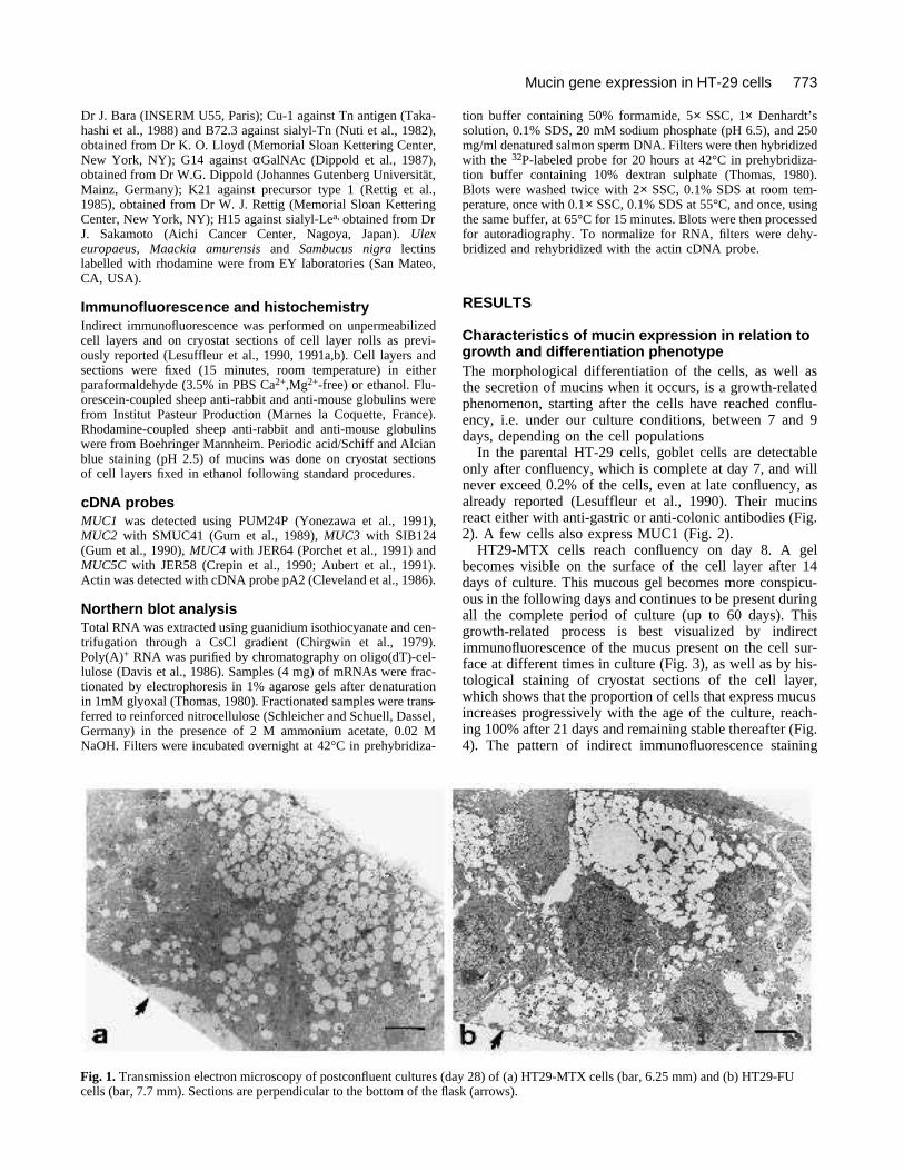
We have recently isolated, through selective pressurewith methotrexate (MTX) (Lesuffleur et al., 1990, 1991a;Dahyia et al., 1992) or 5-fluorouracil (FU) (Lesuffleur etal., 1991c), two distinct subpopulations of mucus-secretingHT-29 cells that maintain their ability to differentiate undernormal culture conditions. These two populations differ asto the morphology of the cells and to the type of immunore-activity of their mucins. Cells adapted to 10−6 M MTX forma homogeneous monolayer of polarized goblet cells (Fig.1a) that exhibit a discrete apical brush border endowed withintestinal hydrolases and secrete mucins of gastricimmunoreactivity (Lesuffleur et al., 1990, 1991a), twocharacteristics that are reminiscent of the differentiation ofthe human fetal colonic epithelium (Zweibaum et al., 1991;Bara et al., 1986). Cells adapted to 10−5 M FU form a mixedpopulation involving a majority (80%) of polarized dome-forming cells, devoid of a brush border, and a minority(20%) of mucus-secreting cells that, in contrast to MTX-adapted cells, are organized into multilayered foci of non-polarized goblet cells that accumulate mucins within theintercellular spaces (Fig. 1b); these mucins being of colonicimmunoreactivity (Lesuffleur et al., 1991c). These differ-entiation characteristics, like those reported by the othergroups, have mainly been analysed in late post-confluent
cultures and nothing is known of the time-course of thegoblet cell differentiation process in relation to cell growth,a relationship that has been well documented in enterocyte-like differentiated cultured cells like Caco-2 cells or otherHT-29 cell populations (Zweibaum et al., 1991).
The aims of this study were to analyse the time courseof differentiation of mucus-secreting MTX and FU cells, toexamine the expression of the mucin gene mRNAs in rela-tion to the growth and to the phenotype of these two pop-ulations compared with the parental line, and to determinewhether the differences in phenotype observed betweenMTX and FU cells are associated with differences in themucin genes that are expressed in these cells.
MATERIALS AND METHODS
Cell cultureThe parental HT-29 cell line (Fogh and Trempe, 1975), obtainedfrom late Dr Jorgen Fogh (Memorial Sloan Kettering CancerCenter, Rye, NY), was established from a blood group A patient(J. Fogh, personal communication). The cell line was usedbetween passages 169 and 182 and is referred to as HT-29. Thesubpopulations obtained by adaptation to 10−5 M 5-fluorouracil(Lesuffleur et al., 1991c) and 10−6 M methotrexate (Lesuffleur etal., 1990) are referred to as HT29-FU and HT29-MTX, respec-tively. Drug-adapted cells were used after several weekly passages(4 to 20) in the absence of drug. They were routinely grown in25 or 75 cm 2 plastic T-flasks (Corning Glassworks, Corning, NY)in Dulbecco’s modified Eagles’s minimum essential medium sup-plemented with 10% heat-inactivated (56°C, 30 minutes) fetalbovine serum (Boehringer Mannheim Biochemicals, Mannheim,Germany) at 37°C in a 10% CO2/90% air atmosphere. For main-tenance or experimental purposes all cells were seeded at 2×10⁴
cells per cm2. The medium was changed daily.
Antibodies and lectinsRabbit polyclonal antibodies against human gastric (L56) andcolonic (L53) mucins were prepared as reported (Lesuffleur et al.,1990); a pool of mAbs against mucin M1 epitopes that specifi-cally react with gastric mucins (Bara et al., 1986) was obtainedfrom Dr J. Bara (INSERM U55, Paris); mAb ZE4 was raisedagainst normal human small intestinal mucosa and was found toreact with colonic and small intestinal mucins, but not with gas-tric mucins (D. Swallow, unpublished results); mAbs BC1, BC2and BC3 (Xing et al., 1989), and LICR LON M8 (McIlhinney etal., 1985), which recognize the tandem repeat sequence of theMUC1 gene product were obtained from Dr McKenzie (Univer-sity of Melbourne, Australia) and The Ludwig Institute for CancerResearch, Sutton, UK, respectively. Dipeptidylpeptidase-IV (DPP-IV) was detected using mAb HBB 3/775/42 (Hauri et al., 1985)raised against the human small intestinal enzyme (obtained fromDr H. P. Hauri, Biocenter, Basel, Switzerland) and a rabbitimmune serum raised against the porcine kidney enzyme (Dar-moul et al., 1991) (obtained from Dr G. Trugnan, INSERM U239,Paris). Polyclonal rabbit antibodies against porcine villin (Robineet al., 1985) were obtained from Dr D. Louvard (Institut Pasteur,Paris). The following monoclonal antibodies were used for study-ing the saccharide epitopes associated with mucins: mAbs againstAB blood group antigens from the Second International Work-shop and Symposium on Monoclonal Antibodies against HumanRed Blood Cells and Related Antigens (Oriol et al., 1990),obtained from Dr R. Oriol (INSERM U178, Villejuif); 7LE againstLea antigen (Bara et al., 1987), 2-25LE against Leb (Bara et al.,1986), and 12-4LE against Ley (Bara et al., 1988), obtained from
T. Lesuffleur and others
773Mucin gene expression in HT-29 cells
Dr J. Bara (INSERM U55, Paris); Cu-1 against Tn antigen (Taka-hashi et al., 1988) and B72.3 against sialyl-Tn (Nuti et al., 1982),obtained from Dr K. O. Lloyd (Memorial Sloan Kettering Center,New York, NY); G14 against αGalNAc (Dippold et al., 1987),obtained from Dr W.G. Dippold (Johannes Gutenberg Universität,Mainz, Germany); K21 against precursor type 1 (Rettig et al.,1985), obtained from Dr W. J. Rettig (Memorial Sloan KetteringCenter, New York, NY); H15 against sialyl-Lea, obtained from DrJ. Sakamoto (Aichi Cancer Center, Nagoya, Japan). Ulexeuropaeus, Maackia amurensis and Sambucus nigra lectinslabelled with rhodamine were from EY laboratories (San Mateo,CA, USA).
Immunofluorescence and histochemistryIndirect immunofluorescence was performed on unpermeabilizedcell layers and on cryostat sections of cell layer rolls as previ-ously reported (Lesuffleur et al., 1990, 1991a,b). Cell layers andsections were fixed (15 minutes, room temperature) in eitherparaformaldehyde (3.5% in PBS Ca2+,Mg2+-free) or ethanol. Flu-orescein-coupled sheep anti-rabbit and anti-mouse globulins werefrom Institut Pasteur Production (Marnes la Coquette, France).Rhodamine-coupled sheep anti-rabbit and anti-mouse globulinswere from Boehringer Mannheim. Periodic acid/Schiff and Alcianblue staining (pH 2.5) of mucins was done on cryostat sectionsof cell layers fixed in ethanol following standard procedures.
cDNA probesMUC1 was detected using PUM24P (Yonezawa et al., 1991),MUC2 with SMUC41 (Gum et al., 1989), MUC3 with SIB124(Gum et al., 1990), MUC4 with JER64 (Porchet et al., 1991) andMUC5C with JER58 (Crepin et al., 1990; Aubert et al., 1991).Actin was detected with cDNA probe pA2 (Cleveland et al., 1986).
Northern blot analysisTotal RNA was extracted using guanidium isothiocyanate and cen-trifugation through a CsCl gradient (Chirgwin et al., 1979).Poly(A)+ RNA was purified by chromatography on oligo(dT)-cel-lulose (Davis et al., 1986). Samples (4 mg) of mRNAs were frac-tionated by electrophoresis in 1% agarose gels after denaturationin 1mM glyoxal (Thomas, 1980). Fractionated samples were trans-ferred to reinforced nitrocellulose (Schleicher and Schuell, Dassel,Germany) in the presence of 2 M ammonium acetate, 0.02 MNaOH. Filters were incubated overnight at 42°C in prehybridiza-
tion buffer containing 50% formamide, 5× SSC, 1× Denhardt’ssolution, 0.1% SDS, 20 mM sodium phosphate (pH 6.5), and 250mg/ml denatured salmon sperm DNA. Filters were then hybridizedwith the 32P-labeled probe for 20 hours at 42°C in prehybridiza-tion buffer containing 10% dextran sulphate (Thomas, 1980).Blots were washed twice with 2× SSC, 0.1% SDS at room tem-perature, once with 0.1× SSC, 0.1% SDS at 55°C, and once, usingthe same buffer, at 65°C for 15 minutes. Blots were then processedfor autoradiography. To normalize for RNA, filters were dehy-bridized and rehybridized with the actin cDNA probe.
RESULTS
Characteristics of mucin expression in relation togrowth and differentiation phenotypeThe morphological differentiation of the cells, as well asthe secretion of mucins when it occurs, is a growth-relatedphenomenon, starting after the cells have reached conflu-ency, i.e. under our culture conditions, between 7 and 9days, depending on the cell populations
In the parental HT-29 cells, goblet cells are detectableonly after confluency, which is complete at day 7, and willnever exceed 0.2% of the cells, even at late confluency, asalready reported (Lesuffleur et al., 1990). Their mucinsreact either with anti-gastric or anti-colonic antibodies (Fig.2). A few cells also express MUC1 (Fig. 2).
HT29-MTX cells reach confluency on day 8. A gelbecomes visible on the surface of the cell layer after 14days of culture. This mucous gel becomes more conspicu-ous in the following days and continues to be present duringall the complete period of culture (up to 60 days). Thisgrowth-related process is best visualized by indirectimmunofluorescence of the mucus present on the cell sur-face at different times in culture (Fig. 3), as well as by his-tological staining of cryostat sections of the cell layer,which shows that the proportion of cells that express mucusincreases progressively with the age of the culture, reach-ing 100% after 21 days and remaining stable thereafter (Fig.4). The pattern of indirect immunofluorescence staining
Fig. 1. Transmission electron microscopy of postconfluent cultures (day 28) of (a) HT29-MTX cells (bar, 6.25 mm) and (b) HT29-FUcells (bar, 7.7 mm). Sections are perpendicular to the bottom of the flask (arrows).

774 T. Lesuffleur and others
Fig. 2. Indirect immunofluorescence staining of paraformaldehyde-fixed cryostat sections of late post-confluent cultures (day 28) ofparental HT-29 cells with (a) polyclonal antiserum L-53 against colonic mucins; (b) polyclonal antiserum L-56 against gastric mucins;(c) mAb BC2 against MUC1 peptide. The same results as shown in a and b were obtained with mAb ZE4 and mAb against M1 antigen,respectively (not shown). Note that very few cells express immunoreactive material. Bar, 100 µm.
Fig. 3. Detection by indirect immunofluorescence, with the polyclonal antiserum L-56, of mucus secreted on the cell surface of cultures ofHT29-MTX cells in relation to cell growth; a, b, c, d, e and f correspond to days 7, 10, 12, 14, 21 and 28, respectively. Assays wereperformed on unpermeabilized cultures fixed with ethanol. In order not to remove the mucous gel, the medium was aspirated gently fromthe flask, which was kept horizontal, and ethanol was added without prior rinsing of the cell layer. The same results were obtained withmAbs against M1 antigens. The small mucin granules present in all the pictures do not correspond to intracellular mucins but to smallclusters of mucins present on the cell layer surface as demonstrated by scanning electron microscopy or by their disappearance afterextensive rinsing of the cell layer prior to fixation (not shown). Bar, 100 µm.

775Mucin gene expression in HT-29 cells
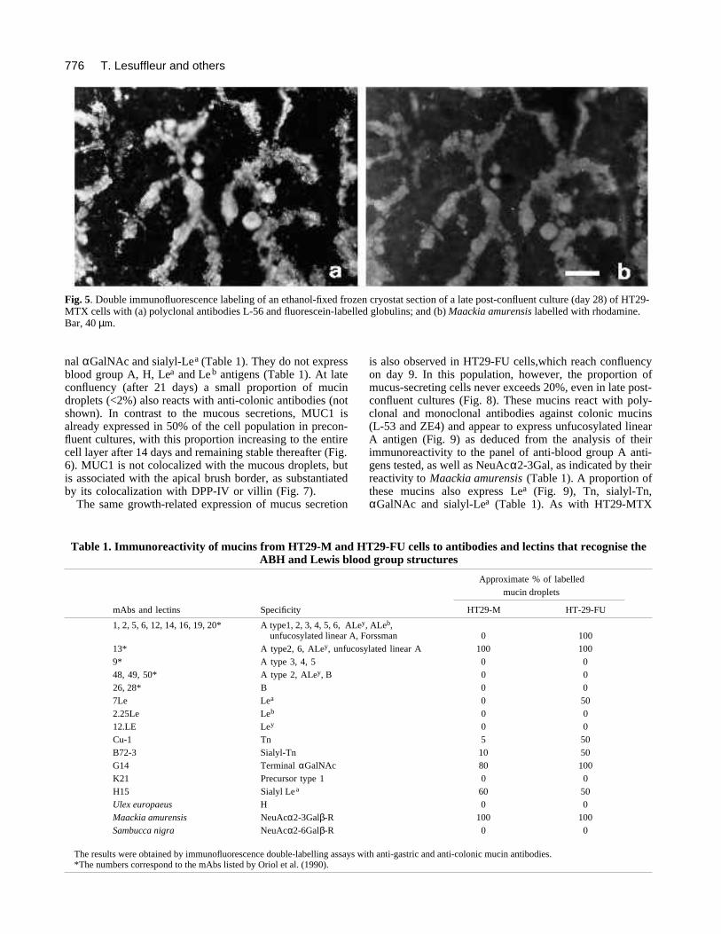
with anti-gastric mucin antibodies exactly corresponds tothe Alcian blue staining. These mucins appear to carryterminal oligosaccharides with the structure NeuAcα2-
3Galβ-, as indicated by double immunofluorescence label-ing with Maackia amurensis lectin (Fig. 5 and Table 1). Ahigh proportion of the mucin droplets also express termi-
Fig. 4. Alcian blue staining of ethanol-fixed cryostat sections of the cell layer of HT29-MTX cells in relation to cell growth. The numbersindicate the day after seeding. Unlike the preparation shown in Fig. 3, the cell layer was rinsed with PBS prior to harvesting and freezing,so that the mucus stained corresponds to the intracellular mucus, as in Fig. 1a. The same pattern of mucus expression was observed byPAS staining or indirect immunofluorescence with mAbs against M1 antigens or polyclonal antiserum L-56 (not shown). Bar, 100 µm.
776
nal αGalNAc and sialyl-Lea (Table 1). They do not expressblood group A, H, Lea and Le b antigens (Table 1). At lateconfluency (after 21 days) a small proportion of mucindroplets (<2%) also reacts with anti-colonic antibodies (notshown). In contrast to the mucous secretions, MUC1 isalready expressed in 50% of the cell population in precon-fluent cultures, with this proportion increasing to the entirecell layer after 14 days and remaining stable thereafter (Fig.6). MUC1 is not colocalized with the mucous droplets, butis associated with the apical brush border, as substantiatedby its colocalization with DPP-IV or villin (Fig. 7).
The same growth-related expression of mucus secretion
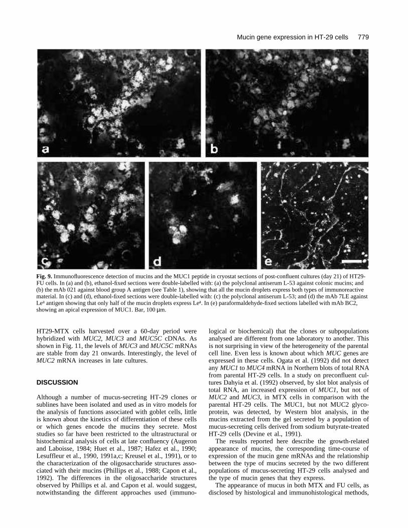
is also observed in HT29-FU cells,which reach confluencyon day 9. In this population, however, the proportion ofmucus-secreting cells never exceeds 20%, even in late post-confluent cultures (Fig. 8). These mucins react with poly-clonal and monoclonal antibodies against colonic mucins(L-53 and ZE4) and appear to express unfucosylated linearA antigen (Fig. 9) as deduced from the analysis of theirimmunoreactivity to the panel of anti-blood group A anti-gens tested, as well as NeuAcα2-3Gal, as indicated by theirreactivity to Maackia amurensis (Table 1). A proportion ofthese mucins also express Lea (Fig. 9), Tn, sialyl-Tn,αGalNAc and sialyl-Lea (Table 1). As with HT29-MTX
T. Lesuffleur and others
Fig. 5. Double immunofluorescence labeling of an ethanol-fixed frozen cryostat section of a late post-confluent culture (day 28) of HT29-MTX cells with (a) polyclonal antibodies L-56 and fluorescein-labelled globulins; and (b) Maackia amurensis labelled with rhodamine.Bar, 40 µm.
Table 1. Immunoreactivity of mucins from HT29-M and HT29-FU cells to antibodies and lectins that recognise theABH and Lewis blood group structures
Approximate % of labelledmucin droplets
mAbs and lectins Specificity HT29-M HT-29-FU
1, 2, 5, 6, 12, 14, 16, 19, 20* A type1, 2, 3, 4, 5, 6, ALey, ALeb,unfucosylated linear A, Forssman 0 100
13* A type2, 6, ALey, unfucosylated linear A 100 1009* A type 3, 4, 5 0 048, 49, 50* A type 2, ALey, B 0 026, 28* B 0 07Le Lea 0 502.25Le Leb 0 012.LE Ley 0 0Cu-1 Tn 5 50B72-3 Sialyl-Tn 10 50G14 Terminal αGalNAc 80 100K21 Precursor type 1 0 0H15 Sialyl Lea 60 50Ulex europaeus H 0 0Maackia amurensis NeuAcα2-3Galβ-R 100 100Sambucca nigra NeuAcα2-6Galβ-R 0 0
The results were obtained by immunofluorescence double-labelling assays with anti-gastric and anti-colonic mucin antibodies.*The numbers correspond to the mAbs listed by Oriol et al. (1990).

777Mucin gene expression in HT-29 cells
Fig. 6. Immunofluorescence detection of MUC1 peptide with mAb BC2 in paraformaldehyde-fixed cryostat sections of cultures of HT29-MTX cells in relation to cell growth; a, b and c correspond to days 7, 14 and 21, respectively, and are from the same culture as shown inFig. 3. Note that MUC1 is already expressed at day 7, i.e. when no mucins can be detected by Alcian blue staining. Bar, 100 µm.
Fig. 7. Phase-contrast microscopy and double immunofluorescence staining of paraformaldehyde-fixed cryostat sections of late post-confluent cultures (day 21) of HT29-MTX cells; (a), (b) and (c), the same section labelled with: (b) the polyclonal antiserum L-56 againstgastric mucins; and (c) the mAb BC2 against MUC1 peptide; (d), (e) and (f), same section labelled with: (e) the polyclonal antiserumagainst DPP-IV; and (f) the mAb BC2. Note the difference in distribution of MUC1 and mucins and the apical colocalization of MUC1and DPP-IV. The same results as in (c) and (f) were obtained with the mAbs BC1, BC3 and LICR LON M8 (not shown). The sameresults as in (e) were obtained with anti-villin antibodies (not shown). Bar, 40 µm.

778
cells, MUC1 is already expressed at the apical surface of ahigh proportion of cells in preconfluent cultures (day 7, notshown), i.e. when mucous secretions are not yet detectableby histological or immunological techniques. The propor-tion of cells expressing MUC1 increases during the daysfollowing and is stable after day 14 (Fig. 9). Expression ofMUC1, as demonstrated by double-immunofluorescencestaining (not shown) is restricted to these polarized cells,organized into a monolayer, which form the majority of theHT29-FU population.
Differential and growth-related expression ofmucin genes mRNAsDays 7, 14 and 21 were chosen for a comparative analysisof the expression of the apomucin mRNAs in the three cellpopulations, since these represent stages at which no, littleor extensive, mucus is produced by the cultures of MTXand FU cells. As shown in Fig. 10, the presence and thelevels of expression of the MUC mRNAs vary with the timein culture and the phenotype of the cells.
Two transcripts of 6.5 kb and 4 kb of MUC1, presum-ably corresponding to the two MUC1 alleles, are expressedin all three populations. They are already expressed at ahigh level in early cultures (day 7) in all populations. Exceptfor HT-29 cells, which show a decrease of these transcripts
with time, their levels remain high in the two differentiatedpopulations.
MUC2 mRNA is expressed at a low level in the parentalHT-29 cells and its level decreases with time. In the HT29-FU cells it is expressed at much higher levels, but alsodecreases with time. In the HT29-MTX cells the level ofexpression is low in early cultures but increases with time.
The highest levels of MUC3 mRNA are found in theMTX cells. In these cells the transcript is already detectableat day 7; it greatly increases at day 14, remaining stablethereafter. It is poorly expressed in HT-29 and notdetectable in the FU cells.
MUC4 mRNA is the most highly expressed in the HT29-FU cells, with its level of expression decreasing from day7 to day 21. It is poorly expressed in HT-29 and HT29-MTX cells.
The highest levels of MUC5C mRNA are found in theHT29-MTX cells, with the level increasing dramaticallybetween 7 and 14 days of culture. A similar increase inexpression with time was not detected in the other cell pop-ulations where the expression remains low.
Because of the stable expression of mucins in the latepost-confluent cultures of HT29-MTX cells and theexpression of some mucins of colonic phenotype at verylate confluency, poly(A)+ RNAs prepared from cultures of
T. Lesuffleur and others
Fig. 8. Alcian blue staining of ethanol-fixed cryostat sections of HT29-FU cells in relation to cell growth. The numbers indicate the daysafter seeding. The same pattern of mucin expression was observed with PAS staining or indirect immunofluorescence with the mAb ZE4or the polyclonal antiserum L-53 against colonic mucins (not shown). Bar, 100 µm.
779Mucin gene expression in HT-29 cells
HT29-MTX cells harvested over a 60-day period werehybridized with MUC2, MUC3 and MUC5C cDNAs. Asshown in Fig. 11, the levels of MUC3 and MUC5C mRNAsare stable from day 21 onwards. Interestingly, the level ofMUC2 mRNA increases in late cultures.
DISCUSSION
Although a number of mucus-secreting HT-29 clones orsublines have been isolated and used as in vitro models forthe analysis of functions associated with goblet cells, littleis known about the kinetics of differentiation of these cellsor which genes encode the mucins they secrete. Moststudies so far have been restricted to the ultrastructural orhistochemical analysis of cells at late confluency (Augeronand Laboisse, 1984; Huet et al., 1987; Hafez et al., 1990;Lesuffleur et al., 1990, 1991a,c; Kreusel et al., 1991), or tothe characterization of the oligosaccharide structures asso-ciated with their mucins (Phillips et al., 1988; Capon et al.,1992). The differences in the oligosaccharide structuresobserved by Phillips et al. and Capon et al. would suggest,notwithstanding the different approaches used (immuno-
logical or biochemical) that the clones or subpopulationsanalysed are different from one laboratory to another. Thisis not surprising in view of the heterogeneity of the parentalcell line. Even less is known about which MUC genes areexpressed in these cells. Ogata et al. (1992) did not detectany MUC1 to MUC4 mRNA in Northern blots of total RNAfrom parental HT-29 cells. In a study on preconfluent cul-tures Dahyia et al. (1992) observed, by slot blot analysis oftotal RNA, an increased expression of MUC1, but not ofMUC2 and MUC3, in MTX cells in comparison with theparental HT-29 cells. The MUC1, but not MUC2 glyco-protein, was detected, by Western blot analysis, in themucins extracted from the gel secreted by a population ofmucus-secreting cells derived from sodium butyrate-treatedHT-29 cells (Devine et al., 1991).
The results reported here describe the growth-relatedappearance of mucins, the corresponding time-course ofexpression of the mucin gene mRNAs and the relationshipbetween the type of mucins secreted by the two differentpopulations of mucus-secreting HT-29 cells analysed andthe type of mucin genes that they express.
The appearance of mucus in both MTX and FU cells, asdisclosed by histological and immunohistological methods,
Fig. 9. Immunofluorescence detection of mucins and the MUC1 peptide in cryostat sections of post-confluent cultures (day 21) of HT29-FU cells. In (a) and (b), ethanol-fixed sections were double-labelled with: (a) the polyclonal antiserum L-53 against colonic mucins; and(b) the mAb 021 against blood group A antigen (see Table 1), showing that all the mucin droplets express both types of immunoreactivematerial. In (c) and (d), ethanol-fixed sections were double-labelled with: (c) the polyclonal antiserum L-53; and (d) the mAb 7LE againstLea antigen showing that only half of the mucin droplets express Lea. In (e) paraformaldehyde-fixed sections labelled with mAb BC2,showing an apical expression of MUC1. Bar, 100 µm.

780
appears to be a growth-related process that starts at con-fluency and progressively involves an increasing proportionof cells. This process differs in its time-course with eachof the mucins examined, the epithelial mucin MUC1 beingdetected much earlier than the mucous droplets. It is alsoassociated with quantitative and qualitative differences inMUC gene expression.
In HT29-MTX and FU cells the MUC1 protein isexpressed on the apical surface of the cells, like in othersystems of polarized cells (Ormerod et al., 1981; Corcoranand Walker, 1990), and its localization is distinct from thatof the mucous droplets as disclosed by immunofluores-cence. In contrast to the late appearance of mucous dropletsin these cells, the MUC1 protein is already present in pre-confluent cultures. This high expression of the MUC1 pro-tein is consistent with the observation that the levels ofMUC1 mRNA are high and almost constant during cellgrowth in the two populations. In the parental line, how-ever, unlike in the differentiated populations, the level ofMUC1 mRNA is high in early cultures, but decreases withthe time in culture. What mechanism is responsible for thedecrease in the steady-state level of MUC1 mRNA in thesecells remains to be elucidated.
The expression of MUC2, MUC3, MUC4 and MUC5CmRNAs differs from one population to another and, withineach population, according to the stage of the culture.
MUC2 and MUC4 are expressed at the highest levels inFU cells, the cells that express mucins of colonic
T. Lesuffleur and others
Fig. 10. Northern blot hybridization of MUC1, MUC2, MUC3,MUC4 and MUC5C cDNAs with poly(A)+ RNAs from cultures ofthe parental HT-29 (P), HT29-FU (F) and HT29-MTX (M) cellsin relation to cell growth; (a), (b) and (c) correspond to days 7, 14and 21, respectively. The results presented for MUC2, MUC3,MUC4 and MUC5C represent successive hybridisation of a singlefilter. The MUC1 results shown correspond to another filter.Similar results were obtained from three different Northern blotscorresponding to experiments with cells at different passages.
MUC1
MUC2
MUC3
MUC4
MUC5C
-actin
-actin
Fig. 11. Growth-related expression of MUC2, MUC3 and MUC5CmRNAs in long-term cultures of HT29-MTX cells; cDNAs werehybridized with poly(A)+ RNAs using the same filter for allhybridizations; a, b, c, d, e, f and g correspond to days 7, 14, 21,28, 40, 50 and 60, respectively.

781Mucin gene expression in HT-29 cells
immunoreactivity, whereas the highest levels of MUC3 andMUC5C are found in the MTX cells, the cells that expressmucins of gastric immunoreactivity. It has been postulatedthat the type of mucin peptide may determine the type ofcarbohydrate present in the final mature mucin (Gum et al.,1989). Although the epitopes recognized by the polyclonaland monoclonal antibodies against gastric and colonicmucins used in this study have not been identified, it isclear that mucins from MTX and FU cells carry differentoligosaccharide structures. There is indirect evidence thatthe oligosaccharide species associated with the mucins fromMTX cells are the same as those that have been character-ized on the mucins produced by clone 16E (Capon et al.,1992): they react with the lectin from Maackia amurensis,which binds to the major oligosaccharide species found inCl.16E mucins, and we have previously shown that mucinsfrom Cl.16E immunoreact with the polyclonal antibodiesagainst gastric mucins (L-56) used in this study (Lesuffleuret al., 1991b). Although further compositional studies arerequired to determine precisely which oligosaccharidestructures are associated with the mucins from each popu-lation, it is clear that MUC2 and MUC4 are expressed con-comitantly with the mucins of colonic-like phenotype (fromFU cells), while MUC3 and MUC5C are expressed con-comitantly with the mucins of gastric-like phenotype (fromMTX cells). These results are interesting in view of recentstudies using in situ hybridization (Audié et al., 1993) andapomucin-specific antibodies (Gambus et al., 1993; Ho etal., 1993; and unpublished observations) which indicate thathigh levels of MUC2 are expressed in the colon, but not inthe stomach, and the reciprocal pattern is observed withMUC5C.
It is noteworthy that the expression of the MUC mRNAsoccurs earlier than the production of mucus (detected his-tologically) in the differentiated cells. This discrepancywould suggest that the process of mucin biosynthesis inthese cells involves a growth-related time-lag between theactivation of the mucin genes, which are responsible for thebiosynthesis of the mucin peptides, and the onset of theirglycosylation. It would therefore be of interest to detect thenewly synthesized apomucins in early cultures. However,using the monoclonal antibody LDQ10 (Gambus et al.,1993) against MUC2 peptide, or polyclonal antibody M3Pagainst MUC3 repeat peptide (Gum et al., 1990) we wereunable to detect either of these these peptide antigens byimmunofluorescence, in 7-day-old cultures of FU and MTXcells, respectively (unpublished results). This will be fur-ther investigated by electron microscopy and immunogoldlabeling, since Egea et al. (1993) have successfully demon-strated MUC2 in the RER in post-confluent MTX cells bythis method, even though it is undetectable by immunoflu-orescence.
It is also possible and likely that other mucin genes areexpressed in these cells, such as the human mucin geneMUC6, which has been identified recently (Toribara et al.,1993), or other unidentified mucin genes.
Thus, notwithstanding the fact that they are malignant,and may show abnormal apomucin synthesis and glycosy-lation, these two different populations of mucus-secretingcells isolated from the HT-29 cell line appear to be rele-vant in vitro models for studying the sequence of regula-
tory events that are associated with the onset and mainte-nance of the biosynthesis of mucins, and the relationshipbetween the type of mucin gene expressed and the type ofglycosylation of the mature mucin.
This work was supported in part by Association pour laRecherche sur le Cancer, Fondation pour la Recherche MédicaleFrançaise, INSERM-CSIC Cooperation Agreement, grant SAL90-0853 from the Plan Nacional de I+D (Ministerio de Educacion,Madrid), the Veterans Affairs Medical Research Service, andNATO grant 0789/88. Part of this work has been presented at the2nd International Workshop on Carcinoma-Associated Mucins(Cambridge, August 1992).
REFERENCES
Aubert, J. P., Porchet, N., Crepin, M., Duterque-Coquillaud, M.,Vergnes, G., Mazzuca, M., Debuire, B., Petitprez, D. and Degand, P.(1991). Evidence for different human tracheobronchial mucin peptidesdeduced from nucleotide cDNA sequences. Amer. J. Resp. Cell. Mol.Biol. 5, 178-185.
Audié, J. P., Janin, A., Porchet, N., Copin, M. C., Gosselin, B. andAubert, J. P. (1993). Expression of human mucin genes in respiratory,digestive and reproductive tracts ascertained by in situ hybridization. J.Histochem. Cytochem. (in press).
Augeron, C. and Laboisse, C. (1984). Emergence of permanentlydifferentiated cell clones in a human colonic cancer cell line in cultureafter treatment with sodium butyrate. Cancer Res. 44, 3961-3969.
Bara, J., Daher, N., Mollicone, R. and Oriol, R. (1987).Immunohistological pattern of 20 monoclonal antibodies against non-Anon-B glycoconjugates in normal human pyloric and duodenal mucosae.Blood Transf. Immunohaematol. 30, 685-692.
Bara, J., Gautier, R., Daher, N., Zaghouani, H. and Burtin, P. (1986).Monoclonal antibodies against oncofetal mucin M1 antigens associatedwith precancerous colonic mucosae. Cancer Res. 46, 3983-3989.
Bara, J., Mollicone, R., Herrero-Zabeleta, E., Gautier, R., Daher, N.and Oriol, R. (1988). Ectopic expression of the Y (Ley) antigen definedby monoclonal antibody 12-4LE in distal colonic adenocarcinomas. Int. J.Cancer 41, 683-689.
Capon, C., Laboisse, C. L., Wieruszeki, J. M., Maoret, J. J., Augeron, C.and Fournet, B. (1992). Oligosaccharide structures of mucins secretedby the human colonic cancer cell line CL. 16E. J. Biol. Chem. 267, 19248-19257.
Chirgwin, J. M., Przybyla, A. E., MacDonald, R. J. and Rutter, W. J.(1979). Isolation of biologically active ribonucleic acid from sourcesenriched in ribonuclease. Biochemistry 18, 5294-5299.
Cleveland, D. W., Lopata, M. A., McDonald, R. J., Cowan, N. J., Rutter,W. J. and Kirschner, M. W. (1986). Number and evolutionnaryconservation of α- and β-tubulin and cytoplasmic β- and α-actine genesusing specific cloned cDNA probes. Cell 20, 95-105.
Corcoran, D. and Walker, R. A. (1990). Ultrastructural localization ofmilk fat globule membrane antigens in human breast carcinomas. J.Pathol. 161, 161-166.
Crepin, M., Porchet, N., Aubert, J. P. and Degand, P. (1990). Diversity ofthe peptide moiety of human airway mucins. Biorheology 27, 471-484.
Dahiya, R., Lesuffleur, T., Kwak, K. S., Byrd, J. C., Barbat, A.,Zweibaum, A. and Kim, Y. S. (1992). Expression and characterizationof mucins associated with the resistance to methotrexate of HT-29 humancolonic adenocarcinoma cell line. Cancer Res. 52, 4655-4662.
Darmoul, D., Rouyer-Fessard, C., Blais, A., Voisin, T., Sapin, C.,Baricault, L., Couvineau, A., Laburthe, M. and Trugnan, G. (1991).Dipeptidylpeptidase IV expression in rat jejunal crypt-villus axis iscontrolled at mRNA level. Amer. J. Physiol. 261 (Gastrointest. LiverPhysiol. 24), G763-G769.
Davis, L. G., Dibner, M. D. and Battey, J. F. (1986). Basic Methods inMolecular Biology. Elsevier Science, New York.
Devine, P. L., Layton, G. T., Clark, B. A., Birrell, G. W., Ward, B. G.,Xing, P. and McKenzie, I. F. C. (1991). Production of MUC1 andMUC2 mucins by human tumor cell lines. Biochem. Biophys. Res.Commun. 178, 593-599.
782
Dippold, W. G., Klingel, R., Bernhard, H., Dienes, H. P., Knuth, A. andMeyer Zum Buschenfelde, K. H. (1987). Secretory epithelial cellmarker on gastrointestinal tumors and in human secretions defined by amonoclonal antibody. Cancer Res. 47, 2092-2097.
Dufosse, J., Porchet, N., Audie, J. P., Guyonnet-Duperat, V., Laine, A.,Van-Seuningen, I., Marrakchi, S., Degand, P. and Aubert, J. P.(1993). Degenerate 87 base pair tandem repeats createhydrophilic/hydrophobic alternating domains in human mucin peptidesmapped to 11p15. Biochem. J. (in press).
Egea, G., Franci, C., Gambus, G., Lesuffleur, T., Zweibaum, A andReal, F. X. (1993). cis-Golgi resident proteins and O-glycans areabnormally compartmentalized in the RER of colon cancer cells. J. CellSci. (in press).
Fogh, J. and Trempe, G. (1975). New human tumor cell lines. In HumanTumor Cells In vitro (ed. J. Fogh), pp. 115-141. Plenum Press, New York.
Fox, M. F., Lahbib, F. Pratt, W., Attwood, J., Gum, J., Kim, Y. andSwallow, D. M. (1992). Regional localization of the intestinal mucinMUC3 to chromosome 7q22. Ann. Hum. Genet. 56, 281-287.
Gambus, G., de Bolos, C., Andreu, D., Franci, C., Egea, G. and Real, F.X. (1993). Detection of a peptide epitope of the MUC2 gene product witha mouse monoclonal antibody. Gastroenterology 104, 93-102.
Gendler, S. J., Burchell, J. M., Duhig, T., Lamport, D., White, R.,Parker, M. and Taylor-Papidimitriou, J. (1987). Cloning of partialcDNA encoding differentiation and tumor-associated mucinglycoproteins expressed by human mammary epithelium. Proc. Nat.Acad. Sci. USA 84, 6060-6064.
Gendler, S. J., Lancaster, C. A., Taylor-Papadimitrou, J., Duhig, T.,Peat, N., Burchell, J., Pemberton, L., Lalani, E. and Wilson, D.(1990). Molecular cloning and expression of the human tumour-associated polymorphic epithelial mucin, PEM. J. Biochem. 265, 15286-15293.
Gendler, S. J., Taylor-Papadimitriou, J., Duhig, T., Rothbard, J. andBurchell, J. (1988). A highly immunogenic region of a humanpolymorphic epithelial mucin expressed by carcinomas is made up oftandem repeats. J. Biol. Chem. 263, 12820-12823.
Gerard, C., Eddy, R. L., Shows, T. B. (1990). The core polypeptide ofcystic fibrosis tracheal mucin contains a tandem repeat structure.Evidence for a common mucin in airway and gastrointestinal tissue. J.Clin. Invest. 86, 1921-1927.
Griffiths, B., Mathews, D. J., West, L., Attwood, J., Povey, S., Swallow,D. M., Gum, J. R. and Kim, Y. S. (1990). Assignment of thepolymorphic intestinal mucin gene (MUC2) to chromosome 11p15. Ann.Hum. Gen. 54, 277-285.
Gross, M. S., Guyonnet-Duperat, V., Porchet, N., Bernheim, A., Aubert,J. P. and Nguyen, V. C. (1992). Mucin 4 (MUC4) gene: regionalassignment (3q29) and RFLP analysis. Ann. Genet. 35, 21-26.
Gum, J. R., Byrd, J. C., Hicks, J. W., Toribara, N. W., Lamport, D. T. A.and Kim, Y. S. (1989). Molecular cloning of human intestinal mucincDNAs. J. Biol. Chem. 264, 6480-6487.
Gum, J. R., Hicks, J. W., Swallow, D. M., Lagace, R. L., Byrd, J. C.,Lamport, D. T. A., Siddiki, B. and Kim, Y. S. (1990). Molecularcloning of cDNAs derived from a novel human intestinal mucin gene.Biochem. Biophys. Res. Commun. 171, 407-415.
Gum, J. R., Hicks, J. W., Toribara, N. W., Rothe, E. M., Lagace, R. E.and Kim, Y. S. (1992). The human MUC2 intestinal mucin has cysteine-rich subdomains located both upstream and downstream of its centralrepetitive region. J. Biol. Chem. 267, 21375-21383.
Hafez, M. M., Infante, D., Winawer, S. and Friedman, E. (1990).Transforming growth factor β1 acts as an autocrine-negative growthregulator in colon enterocytic differentiation but not in goblet cellmaturation. Cell Growth Differ. 1, 617-626.
Hauri, H. P., Sterchi, E. E., Bienz, D., Fransen, J. A. M. and Marxer, A.(1985). Expression and intracellular transport of microvillus membranehydrolases in human intestinal epithelial cells. J. Cell Biol. 101, 838-851.
Ho, S. B., Niehans, G. A., Lyftogt, C., Yan, P. S., Cherwitz, D. L., Gum,E. T., Dahyia, R. and Kim, Y. S. (1993). Heterogeneity of mucin geneexpression in normal and neoplastic tissues. Cancer Res. 53, 641-651.
Huet, C., Sahuquillo-Mérino, C., Coudrier, E. and Louvard, D. (1987).Absorptive and mucus-secreting subclones isolated from a multipotententintestinal cell line (HT-29) provide new models for cell polarity andterminal differentiation. J. Cell Biol. 105, 345-358.
Jany, B. H., Gallup, M. W., Yan, P. S., Gum, J. R., Kim, Y. S. andBasbaum, C. B. (1991). Human bronchus and intestine express the samemucin gene. J. Clin. Invest. 87, 77-82.
Kreusel, K. M., Fromm, M., Schulze, J. D. and Hegel, U. (1991). Cl−
secretion in epithelial monolayers of mucus-forming human colon cells(HT-29/B6). Amer. J. Physiol. 261 (Cell Physiol. 30), C574-C582.
Lan, M. S., Batra, S. K., Qi, W., Metzgard, R. S. and Hollingsworth, M.A. (1990). Cloning and sequencing of a human pancreatic tumor mucincDNA. J. Biol. Chem. 265, 15294-15299.
Lesuffleur, T., Barbat, A., Dussaulx, E. and Zweibaum, A. (1990).Growth adaptation to methotrexate of HT-29 human colon carcinomacells is associated with their ability to differentiate into columnarabsorptive and mucus-secreting cells. Cancer Res. 50, 6334-6343.
Lesuffleur, T., Barbat, A., Luccioni, C., Beaumatin, J., Claire, M.,Kornowski, A., Dussaulx, E., Dutrillaux, B. and Zweibaum, A.(1991a). Dihydrofolate reductase gene amplification-associated shift ofdifferentiation in methotrexate-adapted HT-29 cells. J. Cell. Biol. 115,1409-1418.
Lesuffleur, T., Kornowski, A., Augeron, C., Dussaulx, E., Barbat, A.,Laboisse, C. and Zweibaum, A. (1991b). Increased growth adaptabilityto 5-fluorouracil and methotrexate of HT-29 sub-populations selected fortheir commitment to differentiation. Int. J. Cancer 49, 731-737.
Lesuffleur, T., Kornowski, A., Luccioni, C., Muleris, M., Barbat, A.,Beaumatin, J., Dussaulx, E., Dutrillaux, B. and Zweibaum, A.(1991c). Adaptation to 5-fluorouracil of the heterogeneous human colontumor cell line HT-29 results in the selection of cells committed todifferentiation. Int. J. Cancer 49, 721-730.
Mc. Ilhinney, R. A. J., Patel, S. and Gore, M. E. (1985). Monoclonalantibodies recognizing epitopes carried on both glycolipids andglycoproteins of the human fat globule membrane. Biochem. J. 227, 155-162.
Nguyen, Van Cong, Aubert, J. P., Gross, M. S., Porchet, N., Degand, P.and Frezal, J. (1990). Assignment of human tracheobronchial mucingene(s) to 11p15 and a tracheobronchial mucin-related sequence tochromosome 13. Hum. Genet. 86, 167-172.
Nuti, M., Teramoto, Y. A., Mariani-Constantini, R., Horan Hand, P.,Colcher, D. and Schlom, J. A. (1982). A monoclonal antibody (B72. 3)defines patterns of distribution of a novel tumor-associated antigen inhuman mammary carcinoma cell populations. Int. J. Cancer29, 539-545.
Ogata, S., Uehara, H., Chen, A. and Itzkowitz, S. H. (1992). Mucin geneexpression in colonic tissues and cell lines. Cancer Res. 52, 5971-5978.
Oriol, R., Samuelsson, B. E. and Messeter, L. (1990). ABO antibodies:serological behaviour and immuno-chemical characterization. J.Immunogenet. 17, 279-299.
Ormerod, M. G., Monoghan, P., Easty, D. and Easty, G. C. (1981).Asymmetrical distribution of epithelial membrane antigen on the plasmamembranes of human breast cell lines in culture. Diagn. Histopathol. 4,89-93.
Phillips, T. E., Huet, C., Bilbo, P. R., Podolsky, D. K., Louvard, D. andNeutra, M. (1988). Human intestinal goblet cells in monolayer culture:characterization of a mucus-secreting subclone derived from the HT29colon adenocarcinoma cell line. Gastroenterology 94, 1390-1403.
Podolsky, D. K. (1985a). Oligosaccharide structures of human colonicmucin. J. Biol. Chem. 260, 8262-8271.
Podolsky, D. K. (1985b). Oligosaccharide structures of isolated humancolonic mucin species. J. Biol. Chem. 260, 15510-15515.
Podolsky, D. K., Fournier, D. A. and Lynch, K. E. (1986). Human colonicgoblet cells. Demonstration of distinct subpopulations defined by mucin-specific monoclonal antibodies. J. Clin. Invest. 77, 1263-1271.
Porchet, N., Nguyen, V. C., Dufosse, J., Audie, J. P., Guyonnet-Duperat,V., Gross, M. S., Denis, C., Degand, P., Bernheim, A. and Aubert, J. P.(1991). Molecular cloning and chromosomal localization of a novelhuman tracheobronchial mucin cDNA containing tandemly repeatedsequences of 48 base pairs. Biochem. Biophys. Res. Commun. 175, 414-422.
Rettig, W. J., Cordon-Cardo, C., No, J. S. C., Oettgen, H. F., Old, L. J.and Lloyd, K. O. (1985). High molecular weight glycoproteins of humanteratocarcinoma defined by monoclonal antibodies to carbohydratedeterminants. Cancer Res. 45, 815-821.
Robine, S., Huet, C., Moll, R., Sahuquillo-Merino, C., Coudrier, E.,Zweibaum, A. and Louvard, D. (1985). Can villin be used to identifymalignant and undifferentiated normal digestive epithelial cells? Proc.Nat. Acad. Sci. USA 82, 8488-8492.
Swallow, D. M., Gendler, S., Griffiths, B., Corney, G., Taylor-Papadimitriou, J. and Bramwell, M. E. (1987). The human tumour-associated epithelial mucins are coded by an hypervariable gene locusPUM. Nature 328, 82-84.
T. Lesuffleur and others

783Mucin gene expression in HT-29 cells
Takahashi, H. K., Metoki, R. and Hakomori, S. (1988). ImmunoglobulinG3 monoclonal antibody directed to Tn antigen (tumor-associated α-N-acetylgalactosaminyl epitope) that does not cross-react with blood groupA antigen. Cancer Res. 48, 4361-4367.
Thomas, P. S. (1980). Hybridization of denaturel mRNA and small DNAfragments transferred to nitrocellulose. Proc. Natl. Acad. Sci. USA 77,5201-5205.
Toribara, N. W., Gum, J. R., Culhane, P. J., Lagace, R. E., Hicks, J. W.,Petersen, G. M. and Kim, Y. S. (1991). MUC2 human small intestinalmucin gene structure. J. Clin. Invest. 88, 1005-1013.
Toribara, N. W., Roberton, A. M., Ho, S. B., Kuo, W. L., Gum, E.,Hicks, J. W., Gum, J. R., Byrd, J. C., Siddiki, B. and Kim, Y. S.(1993). Human gastric mucin. Identification of a unique species byexpression cloning. J. Biol. Chem. 268, 5879-5885.
Wesley, A., Mantle, M., Man, D., Qureshi, R., Forstner, G. andForstner, J. (1985). Neutral and acidic species of human intestinalmucin. J. Biol. Chem. 260, 7955-7959.
Xing, P. X., Tjandra, J. J., Reynolds, K., McLaughlin, P. J., Purcell, D.F. J. and McKenzie, I. (1989). Reactivity of anti-human milk fat globuleantibodies with synthetic peptides. J. Immunol. 142, 3503-3509.
Yonezawa, S., Byrd, J. C., Dahiya, R., Ho, J. J. L., Gum, J. J., Griffiths,B., Swallow, D. M. and Kim, Y. S. (1991). Differential mucin geneexpression in human pancreatic and colon cancer cells. Biochem. J. 276,599-605.
Zweibaum, A., Laburthe, M., Grasset, E. and Louvard, D. (1991). Useof cultured cell lines in studies of intestinal cell differentiation andfunciton. In Intestinal Absorption and Secretion. Handbook ofPhysiology, section 6, The Gastrointestinal System, vol. IV (ed. M. Fieldand R. A. Frizzell), pp. 223-255. Amer. Physiol. Soc. Bethesda, MD.
(Received 27 May 1993 - Accepted 27 July 1993)