Embed Size (px)
Citation preview
TOPICAL COLLECTIONONMYELODYSPLASTIC SYNDROMES ANDMPN/MDS OVERLAP (M PATNAIK,
SECTION EDITOR)
Differential Diagnosis and Workup of Monocytosis: A SystematicApproach to a Common Hematologic Finding
Abhishek A. Mangaonkar1 & Aaron J. Tande2& Delamo I. Bekele3
Accepted: 23 February 2021# The Author(s), under exclusive licence to Springer Science+Business Media, LLC, part of Springer Nature 2021
AbstractPurpose of Review Monocytosis is a frequently encountered clinical condition that needs appropriate investigation due to a broadrange of differential diagnoses. This review is meant to summarize the latest literature in the diagnostic testing and interpretationand offer a stepwise diagnostic approach for a patient presenting with monocytosis.Recent Findings Basic studies have highlighted the phenotypic and functional heterogeneity in the monocyte compartment.Studies, both translational and clinical, have provided insights into why monocytosis occurs and how to distinguish the differentetiologies. Flow cytometry studies have illustrated that monocyte repartitioning can distinguish chronic myelomonocytic leuke-mia, a prototypical neoplasm with monocytosis from other reactive or neoplastic causes.Summary In summary, we provide an algorithmic approach to the diagnosis of a patient presenting with monocytosis and expectthis document to serve as a reference guide for clinicians.
Keywords Monocytosis .Monocytes . Classical . Intermediate . Non-classical . ChronicMyelomonocytic Leukemia
Introduction
Monocytes and their tissue-specific mature counterparts,macrophages, are key components of the mononuclearphagocyte system involved in ingestion of microorgan-isms and foreign material in various tissues. They alsoregulate inflammatory and immune responses byinteracting with lymphocytes and serve as antigen-presenting cells by differentiating into dendritic cells[1]. Upon response to activating signals such aschemokines and cytokines, activated monocytes adhereand migrate to the sites of infection or inflammationthrough diapedesis. In addition, monocytes have a
poorly defined interaction with the coagulation cascade(Fig. 1).
Based on expression of lipopolysaccharide co-receptor,CD14, and FcγIII receptor, CD16, at least three functionallydistinct populations of monocytes are known to exist in thebody [2]. Classical monocytes (MO1, CD14++, CD16−) rep-resent the largest subgroup involved in phagocytosis throughthe glycolytic and pentose phosphate pathway [2].Conversely, the non-classical (MO3, CD14dim, CD16+)monocyte fraction is associated with oxidative phosphoryla-tion pathway and protein metabolism, and the intermediate(MO2, CD14+, CD16+) subset is associated with antigen pre-sentation to other immune cells [2, 3]. These associations withdistinct pathways suggest diverse physiological roles formonocytes in healthy individuals. Recently, many groupshave shown existence of transitional and intermediary subsetsof monocytes with diverse functions, reflecting significantfunctional heterogeneity in the monocyte compartment [4, 5,6••].
Several disease states are associated with monocytosis withdiverse clinical presentations. This review is focused on howto accurately identify the underlying etiology in adult patientspresenting with an elevated monocyte count. Readers are alsoreferred to another comprehensive review on the topic [7•].
This article is part of the Topical Collection on MyelodysplasticSyndromes and MPN/MDS Overlap
* Abhishek A. [email protected]
1 Division of Hematology, Mayo Clinic, Rochester, MN, USA2 Division of Infectious Diseases, Mayo Clinic, Rochester, MN, USA3 Division of Rheumatology, Department of Medicine, Mayo Clinic,
Rochester, MN, USA
https://doi.org/10.1007/s11899-021-00618-4
/ Published online: 20 April 2021
Current Hematologic Malignancy Reports (2021) 16:267–275

Monocytosis Definition
The normal absolute monocyte count ranges in adults ranges inbetween 0.2–0.8 × 10⁹/L, with values varying significantly withage and sex. Under normal physiology conditions, monocytecounts are usually slightly higher in males; however total whiteblood cell count and otherWBC types such as lymphocytes andgranulocytes are not significantly different [8]. Further, mono-cytes are more sensitive to inflammatory stimuli in men thanwomen likely secondary to sex hormone differences [8, 9].Racial differences are onlyminimal, with some studies showingno significant differences [10], while others indicating a slightlylower absolute monocyte counts in Blacks and Asians com-pared to Caucasians [11, 12]. The World Health Organizationdefines persistent monocytosis as an absolute monocyte count> 1 × 10⁹/L with monocytes accounting for > 10% of leuko-cytes persisting for > 3 months [13].
Differential Diagnoses of Monocytosis
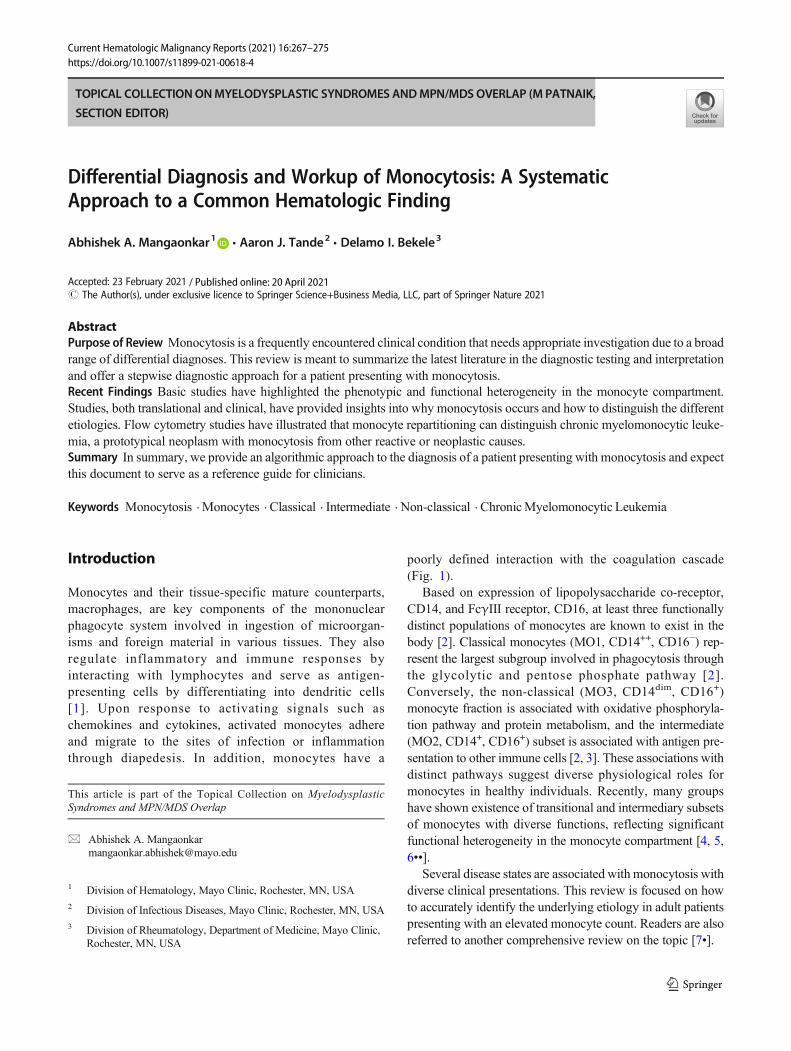
The various causes of monocytosis can be divided into twobroad categories: reactive or clonal (Table 1).
Reactive Causes
Transient causes:Monocytosis can been seen in a patient withrecovering bone marrow after cytotoxic chemotherapy [14].Stressful events such as splenectomy [15], myocardial infarc-tion [16], and exercise [17] can also result in monocytosis andare often transient and reversible [7•]. However, monocytosisis also a frequent occurrence in acutely ill patients, with oneprospective study observing this finding in over a quarter ofpatients seen in a Swiss emergency department [18]. In thisstudy, patients presenting with infections or respiratory illnesswere significantly more likely to present with monocytosisthan other illnesses. Certain acute bacterial infections, suchas leptospirosis [19] or listeriosis [20], can be classically as-sociated with monocytosis. Recently, SARS-CoV-2–associat-ed COVID-19 disease has been associated with increase ininflammatory HLA-DR++ CD11c++ CD14+ monocytes inmild cases, and dysfunctional HLA-DRdimCD163++ andHLA-DRdimS100A++ monocytes, disappearance of non-classical CD14dimCD16+ monocytes in severe cases, furtherhighlighting the important role of monocytes in regulatingsystemic inflammatory responses [21, 22]. However, the find-ing of monocytosis in the acute setting is neither sensitive nor
Fig. 1 The diverse functions of monocytes in humans. Monocytes arecharacterized by expression of several cell surface receptors. This figurehighlights some of those receptors and the key monocyte-associatedfunctions such as macrophage-associated phagocytosis, dendritic cell-
mediated antigen presentation, wound healing, interactions withcoagulation cascade, and regulation of inflammatory responses (createdwith BioRender.com)
268 Curr Hematol Malig Rep (2021) 16:267–275
specific for any particular etiology. The most appropriate andcost-effective diagnostic evaluation in the acute setting is torepeat monocyte count once the acute inflammatory responsehas resolved.
Persistent causes: A diverse group of subacute or chronicinfections have been associated with monocytosis. These in-clude syphilis [23], brucellosis [24], malaria [25], tuberculosis[26], visceral leishmaniasis [27], and rickettsial infection[28].While these case reports and case series are intriguing, theyshould be interpreted with caution. Rather than suggesting aunique laboratory finding in these disparate infections, theymore likely reflect a non-specific finding among a constella-tion of other clinical features.
Monocytosis can also be seen in chronic auto-inflammatory disorders, primarily divided into two major cat-egories: (1) granulomatous disorders: sarcoidosis and inflam-matory bowel disease (ulcerative colitis) and (2) systemicrheumatologic disorders including rheumatoid arthritis (RA),systemic lupus erythematous (SLE), and immune-mediatedthrombocytopenia. Vasculitis (polyarteritis nodosa) and myo-sitis [7•, 29], SLE and RA commonly present with leukopenia,including lymphopenia and neutropenia.
Medications can also result in monocytosis. Whileneutrophillia is more commonly seen with corticosteroid ther-apy, monocytosis has been reported [30]. Drugs such asziprasidone [31], granulocyte colony stimulating factor (G-CSF, [32]), radiation therapy [33], and anti-thymocyte globu-lin [34] have all been associated with monocytosis [7•].
When evaluating for causes of a persistent reactivemonocytosis, the most important approach begins with a thor-ough history of illness and physical examination, including anappropriate infectious diseases exposure history which includesassessment of tuberculosis risk, international travel history, andrisk of zoonotic infections. Recurrent fevers, night sweats, and/or weight loss are supportive but are not specific for an occultinfection since chronic inflammatory disorders and neoplasticcauses can also manifest with these symptoms. Additionalsymptoms suggestive of a chronic systemic illness should war-rant a review of systems covering features of rheumatic disor-ders. If clinically indicated, further testing should include auto-antibodies and imaging studies. Given the rarity of monocytosisas the presenting finding of rheumatic disease, attention mayneed to be focused on the differential diagnosis including infec-tions and clonal etiologies. Additional/associated symptoms,temporal trends of the leukocyte and monocyte counts alongwith pharmacotherapy should be explored.
Clonal Causes
Based on acuity of presentation, clonal causes can be dividedacute and chronic neoplasms (Table 1). The acute neoplasmsinclude acute myeloid leukemia (AML) and dendritic cell leu-kemia. Historically, the French-American-British (FAB) clas-sification categorized monocytic acute leukemia into two sub-types: acute myelomonocytic leukemia (M4) and acute
Table 1 Table showing thedifferential diagnosis ofmonocytosis
Reactive Clonal
Transient
Bone marrow recovery
Exercise-induced
Acute infections
Splenectomy
Medication-induced
Myocardial infarction
Stress-induced
Acute neoplasms
Acute monocytic or myelomonocytic leukemia
Dendritic cell leukemia
Chronic neoplasms
Chronic myelomonocytic leukemia
Juvenile myelomonocytic leukemia
Myeloid neoplasms with PDGFRB rearrangement
(Can mimic CMML)*
Myeloid neoplasms with PDGFRA rearrangement*
Persistent
Chronic infections
Rheumatologic conditions
Medication-induced
Myeloid neoplasms with FGFR1 rearrangement*
Myeloid neoplasms with PCM1-JAK2 fusion*
Chronic myeloid leukemia
Myeloproliferative neoplasms with monocytosis:
a. Essential thrombocythemia
b. Polycythemia vera
c. Primary myelofibrosis
Systemic mastocytosis
B and T cell malignancies
Solid tumors
*Associated with concomitant eosinophilia
269Curr Hematol Malig Rep (2021) 16:267–275

monoblastic/monocytic leukemia (M5) [35]. In the 2017World Health Organization classification, monocytic leuke-mias have been classified under AML, not otherwise specified(NOS) [13]. Chronic myeloid neoplasms with monocytosisinclude chronic myelomonocytic leukemia (CMML) and itspediatric counterpart also known as juvenile myelomonocyticleukemia (JMML), myeloproliferative neoplasms withmonocytosis, myeloid neoplasms with PDGFRB (can mimicCMML), PDGFRA or FGFR1 rearrangement or the provi-sional PCM1-JAK2 fusion, and systemic mastocytosis.
CMML is the prototypical myeloid neoplasm withmonocytosis defined by the presence of monocytosis (> 1 ×10⁹/L) with monocytes accounting for > 10% of leukocytes inthe absence of WHO-criteria for BCR/ABL1-positive chronicmyeloid leukemia and myeloproliferative neoplasms, and ab-sence of PDGFRA, PDGFB, and FGFR1 rearrangements orPCM1-JAK2 fusions, blast count < 20% of cells in both bonemarrow and peripheral blood, and presence of dysplasia inone or more lineages (absence of dysplasia does not excludethe diagnosis of CMML if there is evidence of clonal molec-ular or cytogenetic changes, and monocytosis is persistent (>3 months) [13, 36, 37]. Morphologically, it may be difficultto distinguish blasts from promonocytes or monoblasts and iscounted together clinically when calculating total blast per-centage in both peripheral blood and bone marrow assess-ment. Biologically, the combination of somatic variants inepigenetic (TET2, ASXL1, IDH1/2, orDNMT3A) and splicingfactor genes (SRSF2, ZRSR2, or SF3B1) or signaling pathwaygenes (RAS, JAK2 or CBL) results in monocytosis [38, 39].The precise mechanism of monocytosis is unclear but may beat least partly explained by hypersensitivity to granulocyte-macrophage-colony stimulating factor (GM-CSF) signaling[40], which is also a relevant therapeutic target in CMML[41]. CMML is often associated by variants in the aforemen-tioned group of genes; however it is also genetically heterog-enous, which indicates the existence of other mechanisms ofmonocytosis. On the contrary, JMML is more genomicallyhomogenous and associated with RAS pathway genes(PTPN11, KRAS, NRAS, NF1, or CBL) but also characterizedby GM-CSF hypersensitivity of myeloid progenitors [42].Monocytosis can be seen in other chronic myeloid neoplasmssuch as myeloproliferative neoplasms (MPN), either at diag-nosis or during the course of the disease. The presence ofMPN features in the bone marrow or MPN-associated vari-ants (JAK2/CALR/MPL) supports the diagnosis of MPNwith monocytosis rather than CMML [13]. A minority ofpatients with myelodysplastic syndromes can also presentwith monocytosis (not otherwise meeting criteria forCMML); however they are likely at an early stage of evolu-t ion toward CMML [43] or can be classif ied asoligomonocytic CMML which is considered a pre-CMMLcondition due to similar clinico-pathologic and molecular fea-tures [44, 45••].
Some independent studies have assessed monocyterepartitioning as a diagnostic tool in CMML. The origi-nal study by Selimoglu-Buet D et al. established that theexpansion of the classical fraction of monocytes (> 94%)has been shown to be associated with a sensitivity of90.6% and specificity of 95.1% in diagnosing chronicmyelomonocytic leukemia [46••]. Subsequent studiesreaffirmed this finding [47, 48]. One study noted thatdecreased percentage of the MO3 fraction (< 1.13% inperipheral blood, < 2.42% in bone marrow) has a bettersensitivity and specificity when compared with MO1fraction expansion (> 94%) for the diagnosis of CMML[49]. However, there are some studies that advise cautionwhen using this approach [50]. In the presence of co-existing rheumatologic or other reactive conditions, thereis a preferential expansion of MO2 fraction, and there-fore the aforementioned diagnostic cut-offs for the MO1or MO3 fraction may not be as sensitive or specific forthe diagnosis of CMML [50, 51]. This could be partlyrelated to the transcriptomic heterogeneity in the mono-cyte compartment, especially in the MO2 compartment[6••], indicating the existence of additional functionallydistinct subtypes beyond the three fractions. Although werecommend the use of this assay in distinguishingCMML from other neoplastic causes of monocytosis,caution should be exercise in patients with co-existingrheumatologic conditions or disorders. Recent techniquesin cell phenotyping such as time of flight mass cytometry(CyTOF) can be potentially used to help further refinethe diagnostic accuracy of monocyte repartitioning inCMML, by employing markers other than CD14 andCD16 such as CD36, CCR2, HLA-D4 and CD11c [50,52•, 53••, 54].
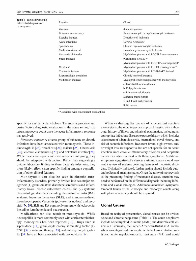
Approach to Evaluation of a Patientwith Monocytosis
When approaching a patient with monocytosis, we recom-mend assessing chronicity and pattern of monocytosis,along with a comprehensive history and physical exam.Morphology of the monocytes should be confirmed on aperipheral smear to assess dysplasia. Monocytosis plusdysplasia or additional complete blood count abnormalitiessuch as anemia, thrombocytopenia, eosinophilia, or baso-philia, or clinical indicators of malignancy such as spleno-megaly, bone pain, or weight loss among others indicate aclonal etiology and in these cases a bone marrow aspirate/core biopsy assessment should be performed. In addition tostandard morphological and cytogenetic/molecular assess-ment, dual staining with alpha-naphthyl butyrate-esteraseand naphthol AS-D chloroacetate esterase may help indistinguishing the monocytic component and also in
270 Curr Hematol Malig Rep (2021) 16:267–275

distinguishing monocytes from monoblasts and/orpromonocytes (Figs. 2 and 3) [55]. Dual staining with boththe aforementioned dyes indicates the existence ofdysmyelopoietic cells, whereas pure monocytic cells stainpositive only for alpha-naphthyl butyrate-esterase [55]. Inthe absence of dysplasia and isolated monocytosis, a repeatcomplete blood count in 3 months can be considered.Resolution of monocytosis at that time-point indicates areactive etiology. However, if the monocytosis is persis-tent, then a comprehensive clinical assessment for reactivecauses should be undertaken, sometimes in coordinationwith other sub-specialist experts such as rheumatology orinfectious diseases. Monocyte compartment flow cytome-try assessment can be considered. However, if there areconcomitant rheumatic conditions, then we recommendproceeding straight to bone marrow evaluation due tothe aforementioned limitations of monocyte repartioning.The expansion of CD16+ fraction of monocytes oftenindicates a non-neoplastic etiology; however it shouldbe considered that clonal disorders such as CMMLand MDS have an established association with rheumat-ic conditions [56, 57]. In other words, both reactive and
clonal causes could exist in the same individual andappropriate judgment should be employed in each clin-ical situation (Fig. 4).
MO1
MO2
MO3
a
b
c
Fig. 3 Figure showing flow cytometry-based monocyte repartioningpatterns in normal individuals, reactive monocytosis and chronicmyelomonocytic leukemia (CMML). In CMML, there is a preferentialexpansion of the classical (> 94%, MO1, CD14++, CD16−) monocytefraction as opposed to intermediate (MO2, CD14+, CD16+) and non-classical fraction (MO3, CD14dim, CD16+) (Courtesy:MichaelM. Timm)Fig. 2 Bone marrow aspirate evaluation of a patient with chronic
myelomonocytic leukemia. a Shows hematoxylin and eosin staining at× 1000magnification. Arrow points to monocytes. b Shows dual stainingof CMML bone marrow aspirate with alpha-naphthyl butyrate-esteraseand naphthol AS-D chloroacetate esterase indicating increasedmonocyticcells (staining only alpha-naphthyl butyrate-esterase) for shown at × 1000magnification (Courtesy: Kaaren K Reichard, MD)
271Curr Hematol Malig Rep (2021) 16:267–275
Conclusion
In summary, monocytosis is a common hematologicfinding with a broad range of etiologic possibilities.We provide an algorithmic approach for investigating
monocytosis and highlight the recent advances in phe-notypic characterization of monocytes. Future areas ofresearch should involve characterization of monocytecompartment heterogeneity, both in normal anddisease-states through novel technologies such as
Fig. 4 Figure outlining a stepwiseapproach for a patient presentingwith monocytosis. Abbreviations:CBC = complete blood count;CMML = chronicmyelomonocytic leukemia
272 Curr Hematol Malig Rep (2021) 16:267–275

CyTOF. This would not only increase the precision ofmonocyte repartioning as a diagnostic tool but also offernovel insights into disease biology.
Acknowledgements Authors would like to acknowledge Kaaren K.Reichard, MD, for the bone marrow aspirate (Fig. 2) images andMichael M. Timm for the flow cytometry (Fig. 3) images.
Declarations
Conflict of Interest The authors declare no competing interest.
Human and Animal Rights and Informed Consent This article does notcontain any studies with human or animal subjects performed by any ofthe authors.
References
Papers of particular interest, published recently, have beenhighlighted as:• Of importance•• Of major importance
1. Kenneth KaushanskyMAL, Prchal JT, LeviMM, Press OW, BurnsLJ, Caligiuri MA. Williams Hematology. 9th ed; 2016. p. 1045–100.
2. Schmidl C, Renner K, Peter K, Eder R, Lassmann T, Balwierz PJ,et al. Transcription and enhancer profiling in human monocytesubsets. Blood. 2014;123(17):e90–9. https://doi.org/10.1182/blood-2013-02-484188.
3. Ziegler-Heitbrock L, Hofer TP. Toward a refined definition ofmonocyte subsets. Front Immunol. 2013;4:23. https://doi.org/10.3389/fimmu.2013.00023.
4. Teh YC, Ding JL, Ng LG, Chong SZ. Capturing the fantastic voy-age of monocytes through time and space. Front Immunol.2019;10:834. https://doi.org/10.3389/fimmu.2019.00834.
5. Chong SZ, EvrardM, Devi S, Chen J, Lim JY, See P, et al. CXCR4identifies transitional bone marrow premonocytes that replenish themature monocyte pool for peripheral responses. J Exp Med.2016;213(11):2293–314. https://doi.org/10.1084/jem.20160800.
6.•• Villani AC, Satija R, Reynolds G, Sarkizova S, Shekhar K, FletcherJ, et al. Single-cell RNA-seq reveals new types of human blooddendritic cells, monocytes, and progenitors. Science.2017;356(6335). https://doi.org/10.1126/science.aah4573. This isan important paper highlighting the heterogeneity in humanmonocyte compartment.
7.• Lynch DT, Hall J, Foucar K. How I investigate monocytosis. Int JLab Hematol. 2018;40(2):107–14. https://doi.org/10.1111/ijlh.12776. This is a comprehensive review on how to investigatemonocytosis.
8. Bouman A, Schipper M, Heineman MJ, Faas MM. Gender differ-ence in the non-specific and specific immune response in humans.Am J Reprod Immunol. 2004;52(1):19–26. https://doi.org/10.1111/j.1600-0897.2004.00177.x.
9. Saxena S, Wong ET. Heterogeneity of common hematologic pa-rameters among racial, ethnic, and gender subgroups. Arch PatholLab Med. 1990;114(7):715–9.
10. Tollerud DJ, Clark JW, Brown LM, Neuland CY, Pankiw-TrostLK, Blattner WA, et al. The influence of age, race, and gender on
peripheral bloodmononuclear-cell subsets in healthy nonsmokers. JClin Immunol. 1989;9(3):214–22. https://doi.org/10.1007/bf00916817.
11. Freedman DS, Gates L, Flanders WD, Van Assendelft OW,Barboriak JJ, Joesoef MR, et al. Black/white differences in leuko-cyte subpopulations in men. Int J Epidemiol. 1997;26(4):757–64.https://doi.org/10.1093/ije/26.4.757.
12. Lim E, Miyamura J, Chen JJ. Racial/ethnic-specific reference inter-vals for common laboratory tests: a comparison among Asians,Blacks, Hispanics, and White. Hawaii J Med Public Health.2015;74(9):302–10.
13. Swerdlow SH, Campo E, Harris NL. WHO classification of tumorsof haematopoietic and lymphoid tissues. Revised 4th ed. Lyon:International Agency for Research on Cancer (IARC); 2017.
14. Liu L, Yang L, Yan W, Zhai J, Pizzo DP, Chu P, et al.Chemotherapy induces breast cancer stemness in association withdysregulated monocytosis. Clin Cancer Res. 2018;24(10):2370–82. https://doi.org/10.1158/1078-0432.Ccr-17-2545.
15. Bearman RM, Kjeldsberg CR, Pangalis GA, Rappaport H. Chronicmonocytic leukemia in adults. Cancer. 1981;48(10):2239–55.https://doi.org/10.1002/1097-0142(19811115)48:10<2239::aid-cncr2820481020>3.0.co;2-x.
16. Liebetrau C, Hoffmann J, Dörr O, Gaede L, Blumenstein J,Biermann H, et al. Release kinetics of inflammatory biomarkersin a clinical model of acute myocardial infarction. Circ Res.2015;116(5):867–75. https://doi.org/10.1161/circresaha.116.304653.
17. Shin YO, Lee JB. Leukocyte chemotactic cytokine and leukocytesubset responses during ultra-marathon running. Cytokine.2013;61(2):364–9. https://doi.org/10.1016/j.cyto.2012.11.019.
18. Hensel M, Grädel L, Kutz A, Haubitz S, Huber A, Mueller B, et al.Peripheral monocytosis as a predictive factor for adverse outcomein the emergency department: survey based on a register study.Medicine (Baltimore). 2017;96(28):e7404. https://doi.org/10.1097/md.0000000000007404.
19. Raffray L, Giry C, Vandroux D, Fayeulle S, MoitonMP, Gerber A,et al. The monocytosis during human leptospirosis is associatedwith modest immune cell activation states. Med MicrobiolImmunol. 2019;208(5):667–78. https://doi.org/10.1007/s00430-018-0575-9.
20. Whitelock-Jones L, Carswell J, Rasmussen KC. Listeria pneumo-nia. A case report. S Afr Med J. 1989;75(4):188–9.
21. Silvin A, Chapuis N, Dunsmore G, Goubet AG, Dubuisson A,Derosa L, et al. Elevated calprotectin and abnormal myeloid cellsubsets discriminate severe from mild COVID-19. Cell.2020;182(6):1401–18.e18. https://doi.org/10.1016/j.cell.2020.08.002.
22. Schulte-Schrepping J, Reusch N, Paclik D, Baßler K, SchlickeiserS, Zhang B, et al. Severe COVID-19 is marked by a dysregulatedmyeloid cell compartment. Cell. 2020;182(6):1419–40.e23. https://doi.org/10.1016/j.cell.2020.08.001.
23. Karayalcin G, Khanijou A, Kim KY, Aballi AJ, Lanzkowsky P.Monocytosis in congenital syphilis. Am J Dis Child. 1977;131(7):782–3. https://doi.org/10.1001/archpedi.1977.02120200064013.
24. Tsolia M, Drakonaki S, Messaritaki A, Farmakakis T, Kostaki M,Tsapra H, et al. Clinical features, complications and treatment out-come of childhood brucellosis in central Greece. J Infect.2002;44(4):257–62. https://doi.org/10.1053/jinf.2002.1000.
25. Abdalla SH. Peripheral blood and bone marrow leucocytes inGambian children with malaria: numerical changes and evaluationof phagocytosis. Ann Trop Paediatr. 1988;8(4):250–8. https://doi.org/10.1080/02724936.1988.11748582.
26. Slavin RE, Walsh TJ, Pollack AD. Late generalized tuberculosis: aclinical pathologic analysis and comparison of 100 cases in thepreantibiotic and antibiotic eras. Medicine (Baltimore).1980;59(5):352–66.
273Curr Hematol Malig Rep (2021) 16:267–275

27. Kawakami A, Fukunaga T, Usui M, Asaoka H, Noda M, NakajimaT, et al. Visceral leishmaniasis misdiagnosed as malignant lympho-ma. Intern Med. 1996;35(6):502–6. https://doi.org/10.2169/internalmedicine.35.502.
28. Dalugama C, Gawarammana IB. Rare presentation of rickettsialinfection as purpura fulminans: a case report. J Med Case Rep.2018;12(1):145. https://doi.org/10.1186/s13256-018-1672-5.
29. Jameson JL, Fauci AS, Kasper DL, Hauser SL, Longo DL,Loscalzo J. Leukocytosis and leukopenia. Harrison's Manual ofMedicine, 20e. McGraw-Hill Education: New York; 2020.
30. Barker S, Scott M, Chan GT. Corticosteroids and monocytosis. N ZMed J. 2012;125(1360):76–8.
31. Thöne J, Kessler E. Monocytosis subsequent to ziprasidone treat-ment: a possible side effect. Prim Care Companion J ClinPsychiatry. 2007;9(6):465–6. https://doi.org/10.4088/pcc.v09n0611b.
32. Liu CZ, Persad R, Inghirami G, Sen F, Amorosi E, Goldenberg A,et al. Transient atypical monocytosis mimic acute myelomonocyticleukemia in post-chemotherapy patients receiving G-CSF: report oftwo cases. Clin Lab Haematol. 2004;26(5):359–62. https://doi.org/10.1111/j.1365-2257.2004.00628.x.
33. Rotman M, Ansley H, Rogow L, Stowe S. Monocytosis: a newobservation during radiotherapy. Int J Radiat Oncol Biol Phys.1977;2(1-2):117–21. https://doi.org/10.1016/0360-3016(77)90016-5.
34. de Planque MM, Brand A, Kluin-Nelemans HC, Eernisse JG, vander Burgh F, Natarajan AT, et al. Haematopoietic and immunologicabnormalities in severe aplastic anaemia patients treated with anti-thymocyte globulin. Br J Haematol. 1989;71(3):421–30. https://doi.org/10.1111/j.1365-2141.1989.tb04301.x.
35. Walter RB, Othus M, Burnett AK, Löwenberg B, Kantarjian HM,Ossenkoppele GJ, et al. Significance of FAB subclassification of"acute myeloid leukemia, NOS" in the 2008 WHO classification:analysis of 5848 newly diagnosed patients. Blood. 2013;121(13):2424–31. https://doi.org/10.1182/blood-2012-10-462440.
36. Patnaik MM, Tefferi A. Chronic myelomonocytic leukemia: 2018update on diagnosis, risk stratification and management. Am JHematol. 2018;93(6):824–40. https://doi.org/10.1002/ajh.25104.
37. Mangaonkar AA, Patnaik MM. Advances in chronicmyelomonocytic leukemia and future prospects: lessons learnedfrom precision genomics. Adv Cell Gene Ther. 2019;2(2).https://doi.org/10.1002/acg2.48.
38. Yoshimi A, Lin KT, Wiseman DH, RahmanMA, Pastore A,WangB, et al. Coordinated alterations in RNA splicing and epigeneticregulation drive leukaemogenesis. Nature. 2019;574(7777):273–7.https://doi.org/10.1038/s41586-019-1618-0.
39. Kunimoto H, Meydan C, Nazir A, Whitfield J, Shank K, RapaportF, et al. Cooperative epigenetic remodeling by TET2 loss andNRAS mutation drives myeloid transformation and MEK inhibitorsensitivity. Cancer Cell. 2018;33(1):44–59.e8. https://doi.org/10.1016/j.ccell.2017.11.012.
40. Padron E, Painter JS, Kunigal S, Mailloux AW, McGraw K,McDaniel JM, et al. GM-CSF-dependent pSTAT5 sensitivity is afeature with therapeutic potential in chronic myelomonocytic leu-kemia. Blood. 2013;121(25):5068–77. https://doi.org/10.1182/blood-2012-10-460170.
41. Patnaik MM, Sallman DA, Mangaonkar AA, Heuer R, Hirvela J,Zblewski D, et al. Phase 1 study of lenzilumab, a recombinant anti-human GM-CSF antibody, for chronic myelomonocytic leukemia.Blood. 2020;136(7):909–13. https://doi.org/10.1182/blood.2019004352.
42. Emanuel PD, Bates LJ, Castleberry RP, Gualtieri RJ, ZuckermanKS. Selective hypersensitivity to granulocyte-macrophage colony-stimulating factor by juvenile chronic myeloid leukemia hemato-poietic progenitors. Blood. 1991;77(5):925–9.
43. Selimoglu-Buet D, Badaoui B, Benayoun E, Toma A, Fenaux P,Quesnel B, et al. Accumulation of classical monocytes defines a sub-group of MDS that frequently evolves into CMML. Blood.2017;130(6):832–5. https://doi.org/10.1182/blood-2017-04-779579.
44. Geyer JT, Tam W, Liu Y-C, Chen Z, Wang SA, Bueso-Ramos C,et al. Oligomonocytic chronic myelomonocytic leukemia (chronicmyelomonocytic leukemia without absolute monocytosis) displaysa similar clinicopathologic andmutational profile to classical chron-ic myelomonocytic leukemia. Mod Pathol. 2017;30(9):1213–22.https://doi.org/10.1038/modpathol.2017.45.
45.•• Valent P, Orazi A, Savona MR, Patnaik MM, Onida F, van deLoosdrecht AA, et al. Proposed diagnostic criteria for classicalchronic myelomonocytic leukemia (CMML), CMML variantsand pre-CMML conditions. Haematologica. 2019;104(10):1935–49. https://doi.org/10.3324/haematol.2019.222059. This is apaper on classification and diagnosis of CMML.
46.•• Selimoglu-Buet D, Wagner-Ballon O, Saada V, Bardet V, ItzyksonR, Bencheikh L, et al. Characteristic repartition of monocyte sub-sets as a diagnostic signature of chronic myelomonocytic leukemia.Blood. 2015;125(23):3618–26. https://doi.org/10.1182/blood-2015-01-620781. This is an important study on monocyterepartioning in CMML.
47. Patnaik MM, Timm MM, Vallapureddy R, Lasho TL, KetterlingRP, Gangat N, et al. Flow cytometry based monocyte subset anal-ysis accurately distinguishes chronic myelomonocytic leukemiafrom myeloproliferative neoplasms with associated monocytosis.Blood Cancer J. 2017;7(7):e584. https://doi.org/10.1038/bcj.2017.66.
48. Feng R, Bhatt VR, Fu K, Pirruccello S, Yuan J. Application ofimmunophenotypic analysis in distinguishing chronicmyelomonocytic leukemia from reactive monocytosis. CytometryB Clin Cytom. 2018;94(6):901–9. https://doi.org/10.1002/cyto.b.21721.
49. Hudson CA, Burack WR, Leary PC, Bennett JM. Clinical utility ofclassical and nonclassical monocyte percentage in the diagnosis ofchronic myelomonocytic leukemia. Am J Clin Pathol. 2018;150(4):293–302. https://doi.org/10.1093/ajcp/aqy054.
50. Pophali PA, Timm MM, Mangaonkar AA, Shi M, Reichard K,Tefferi A, et al. Practical limitations of monocyte subsetrepartitioning by multiparametric flow cytometry in chronicmyelomonocytic leukemia. Blood Cancer J. 2019;9(9):65. https://doi.org/10.1038/s41408-019-0231-7.
51. Murali A, Cross D, Mollee P. The use of monocyte subsetrepartitioning by flow cytometry for diagnosis of chronicmyelomonocytic leukaemia. Blood Cancer J. 2021;11(1):6.https://doi.org/10.1038/s41408-020-00401-3.
52.• Mangaonkar AA, Reichard KK, Binder M, Coltro G, Lasho TL,Carr RM, et al. Bone marrow dendritic cell aggregates associatewith systemic immune dysregulation in chronic myelomonocyticleukemia. Blood Adv. 2020;4(21):5425–30. https://doi.org/10.1182/bloodadvances.2020002415. This is an important paperdescribing for the first time, the use of CyTOF in CMML.
53.• Thomas GD, Hamers AAJ, Nakao C, Marcovecchio P, Taylor AM,McSkimming C, et al. Human blood monocyte subsets: a newgating strategy defined using cell surface markers identified bymass cytometry. Arterioscler Thromb Vasc Biol. 2017;37(8):1548–58. https://doi.org/10.1161/atvbaha.117.309145. This is animportant study providing evidence that novel markers can beused to better differentiate monocyte subsets.
54. Ong SM, Teng K, Newell E, Chen H, Chen J, Loy T, et al. A Novel,Five-marker alternative to CD16-CD14 gating to identify the threehuman monocyte subsets. Front Immunol. 2019;10:1761. https://doi.org/10.3389/fimmu.2019.01761.
55. Swerdlow SHCE, Harris NL, Jaffe ES, Pileri SA, Stein H, Thiele J,et al. WHO classification of tumours of haematopoietic and lym-phoid tissues; 2018. p. 82–6.
274 Curr Hematol Malig Rep (2021) 16:267–275

56. Grignano E, Mekinian A, Braun T, Liozon E, HamidouM, DecauxO, et al. Autoimmune and inflammatory diseases associated withchronic myelomonocytic leukemia: a series of 26 cases and litera-ture review. Leuk Res. 2016;47:136–41. https://doi.org/10.1016/j.leukres.2016.05.013.
57. Mekinian A, Grignano E, Braun T, Decaux O, Liozon E,Costedoat-Chalumeau N, et al. Systemic inflammatory and auto-immune manifestations associated with myelodysplastic
syndromes and chronic myelomonocytic leukaemia: a Frenchmulticentre retrospective study. Rheumatology (Oxford).2016;55(2):291–300. https://doi.org/10.1093/rheumatology/kev294.
Publisher’s Note Springer Nature remains neutral with regard to jurisdic-tional claims in published maps and institutional affiliations.
275Curr Hematol Malig Rep (2021) 16:267–275