Embed Size (px)
Citation preview

© AGO e.V. in der DGGG e.V.
sowie
in der DKG e.V.
Die neue FIGO-Klassifikation
Ivo Meinhold-Heerlein, Aachen
AGO State of the Art 2013
22. Juni 2013
Berlin
© AGO e.V. in der DGGG e.V.
sowie
in der DKG e.V.
Cancer of Ovary, Fallopian Tube and Peritoneum: FIGO staging
Deliberations on October 7th, 2012 in Roma at Fieri Di Roma
FIGO GYNAECOLOGY ONCOLOGY COMMITTEE in collaboration with ESGO, UICC, AJCC, SGO, GCIG, IGCS
© AGO e.V. in der DGGG e.V.
sowie
in der DKG e.V.
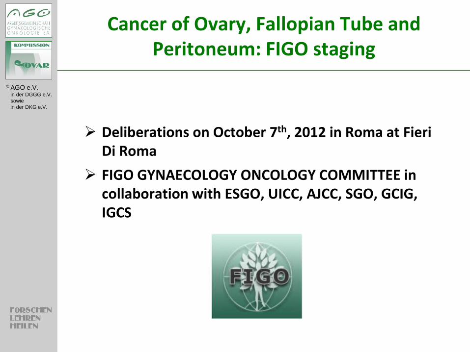
The process of the proposed changes to staging of Ovarian Cancer (OV), Fallopian Tube (FT) and Primary Peritoneal Cancer (PPC) started 3 years ago with a proposal that was sent to all relevant gynaecology oncology organisations and societies throughout the world and input was collated, evaluated and formulated into the staging that is presented below.
All suggestions are based on the best AVAILABLE evidence. The committee acknowledges that there are areas that are not supported by strong evidence and have been careful to ensure that changes are not made without quality evidence.
The new staging below was reached by consensus of all participating in the meeting on October 7th, 2012, some of whom were representatives of their organisations. The new staging was presented to the FIGO EXCECUTIVE BOARD on Friday 12th October 2012 and approved two weeks later.
The proposal will now be presented to the boards of UICC and AJCC. Only once approval has been given from these organisations will FIGO be able to publish the new staging globally.
Cancer of Ovary, Fallopian Tube and Peritoneum: FIGO staging
© AGO e.V. in der DGGG e.V.
sowie
in der DKG e.V.
Cancer of Ovary, Fallopian Tube and Peritoneum: FIGO staging
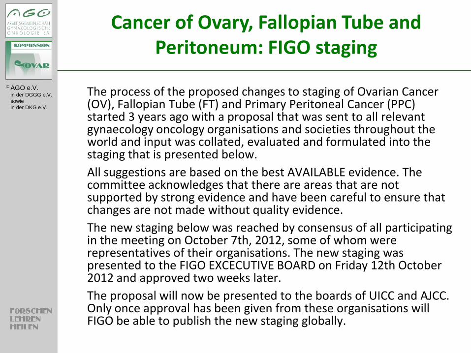
FIGO TNM
OV Primary tumor, ovary Tov
FT Primary tumor, fallopian tube Tft P Primary tumor, peritoneum Tp
X Primary tumor cannot be assessed Tx
© AGO e.V. in der DGGG e.V.
sowie
in der DKG e.V.
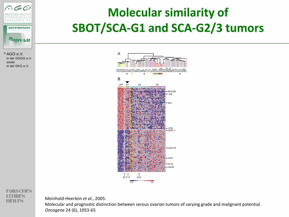
Molecular similarity of SBOT/SCA-G1 and SCA-G2/3 tumors
Meinhold-Heerlein et al., 2005: Molecular and prognostic distinction between serous ovarian tumors of varying grade and malignant potential. Oncogene 24 (6), 1053-65
© AGO e.V. in der DGGG e.V.
sowie
in der DKG e.V.
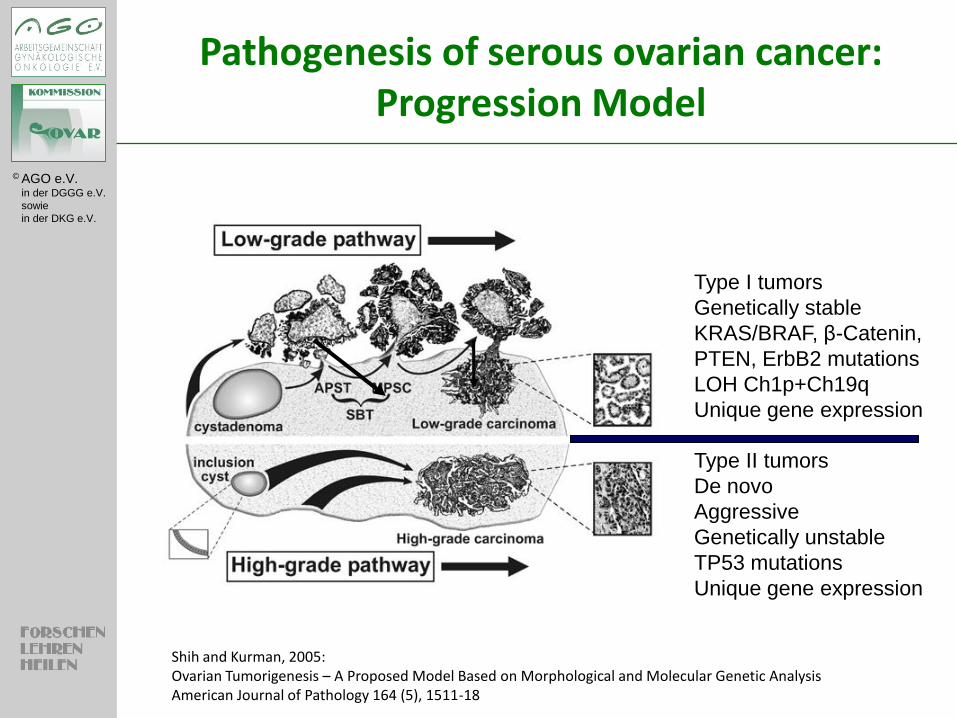
Type II tumors
De novo
Aggressive
Genetically unstable
TP53 mutations
Unique gene expression
Type I tumors
Genetically stable
KRAS/BRAF, β-Catenin,
PTEN, ErbB2 mutations
LOH Ch1p+Ch19q
Unique gene expression
Shih and Kurman, 2005: Ovarian Tumorigenesis – A Proposed Model Based on Morphological and Molecular Genetic Analysis American Journal of Pathology 164 (5), 1511-18
Pathogenesis of serous ovarian cancer: Progression Model
© AGO e.V. in der DGGG e.V.
sowie
in der DKG e.V.
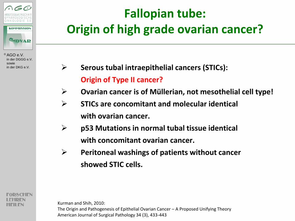
Fallopian tube: Origin of high grade ovarian cancer?
Serous tubal intraepithelial cancers (STICs):
Origin of Type II cancer?
Ovarian cancer is of Müllerian, not mesothelial cell type!
STICs are concomitant and molecular identical
with ovarian cancer.
p53 Mutations in normal tubal tissue identical
with concomitant ovarian cancer.
Peritoneal washings of patients without cancer
showed STIC cells.
Kurman and Shih, 2010: The Origin and Pathogenesis of Epithelial Ovarian Cancer – A Proposed Unifying Theory American Journal of Surgical Pathology 34 (3), 433-443
© AGO e.V. in der DGGG e.V.
sowie
in der DKG e.V.
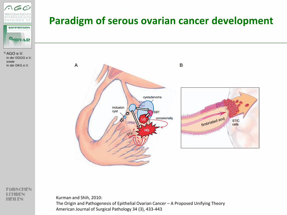
Paradigm of serous ovarian cancer development
Kurman and Shih, 2010: The Origin and Pathogenesis of Epithelial Ovarian Cancer – A Proposed Unifying Theory American Journal of Surgical Pathology 34 (3), 433-443
© AGO e.V. in der DGGG e.V.
sowie
in der DKG e.V.
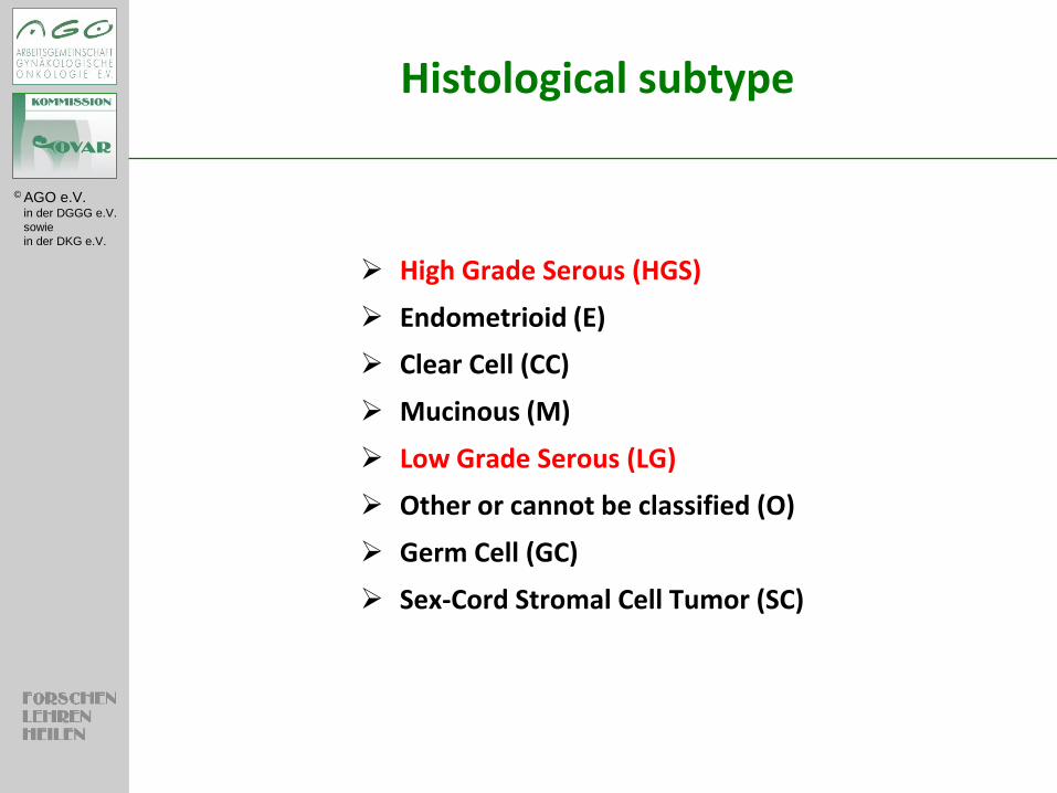
Histological subtype
High Grade Serous (HGS)
Endometrioid (E)
Clear Cell (CC)
Mucinous (M)
Low Grade Serous (LG)
Other or cannot be classified (O)
Germ Cell (GC)
Sex-Cord Stromal Cell Tumor (SC)
© AGO e.V. in der DGGG e.V.
sowie
in der DKG e.V.
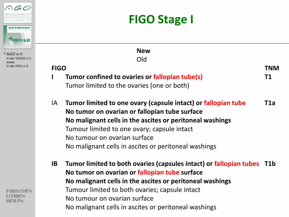
FIGO Stage I
New Old FIGO TNM I Tumor confined to ovaries or fallopian tube(s) T1 Tumor limited to the ovaries (one or both) IA Tumor limited to one ovary (capsule intact) or fallopian tube T1a No tumor on ovarian or fallopian tube surface No malignant cells in the ascites or peritoneal washings Tumour limited to one ovary; capsule intact No tumour on ovarian surface No malignant cells in ascites or peritoneal washings IB Tumor limited to both ovaries (capsules intact) or fallopian tubes T1b No tumor on ovarian or fallopian tube surface No malignant cells in the ascites or peritoneal washings Tumour limited to both ovaries; capsule intact No tumour on ovarian surface No malignant cells in ascites or peritoneal washings
© AGO e.V. in der DGGG e.V.
sowie
in der DKG e.V.
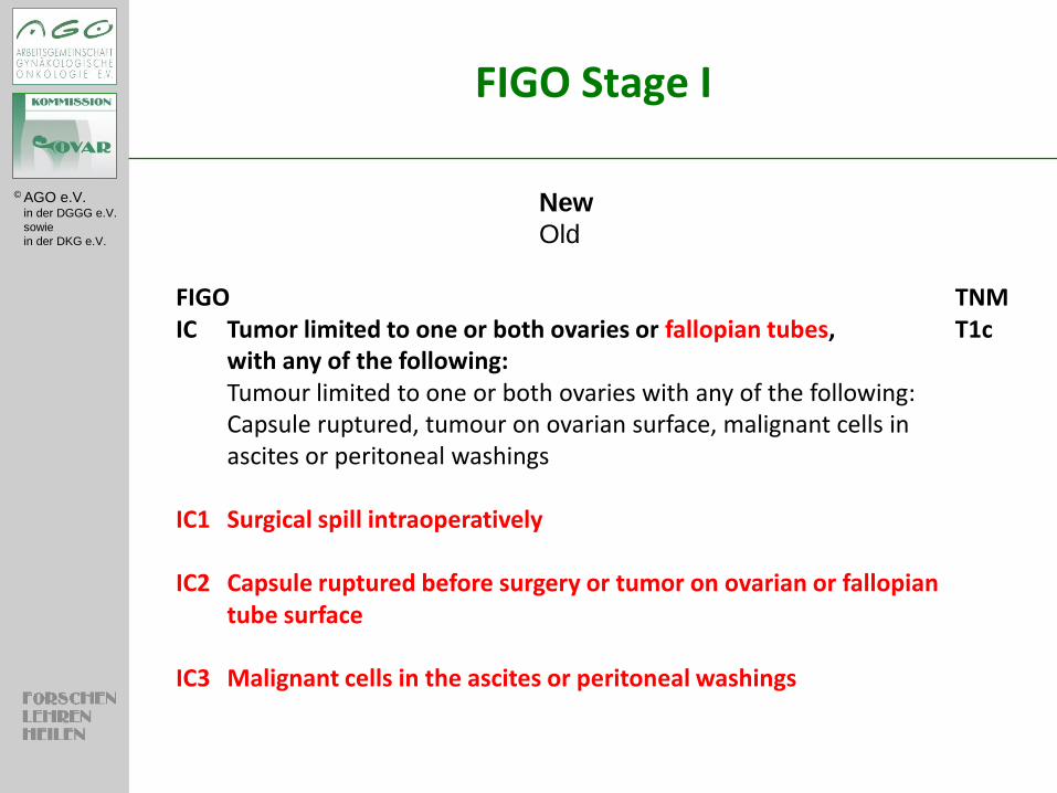
FIGO Stage I
New
Old FIGO TNM IC Tumor limited to one or both ovaries or fallopian tubes, T1c with any of the following: Tumour limited to one or both ovaries with any of the following: Capsule ruptured, tumour on ovarian surface, malignant cells in
ascites or peritoneal washings IC1 Surgical spill intraoperatively IC2 Capsule ruptured before surgery or tumor on ovarian or fallopian
tube surface IC3 Malignant cells in the ascites or peritoneal washings
© AGO e.V. in der DGGG e.V.
sowie
in der DKG e.V.
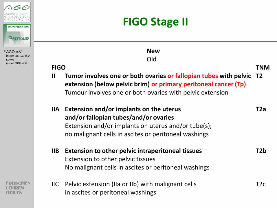
FIGO Stage II
New Old FIGO TNM II Tumor involves one or both ovaries or fallopian tubes with pelvic T2 extension (below pelvic brim) or primary peritoneal cancer (Tp) Tumour involves one or both ovaries with pelvic extension IIA Extension and/or implants on the uterus T2a and/or fallopian tubes/and/or ovaries Extension and/or implants on uterus and/or tube(s); no malignant cells in ascites or peritoneal washings IIB Extension to other pelvic intraperitoneal tissues T2b Extension to other pelvic tissues No malignant cells in ascites or peritoneal washings IIC Pelvic extension (IIa or IIb) with malignant cells T2c in ascites or peritoneal washings
© AGO e.V. in der DGGG e.V.
sowie
in der DKG e.V.
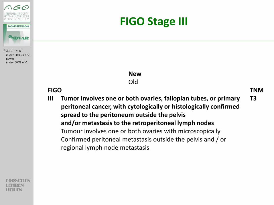
FIGO Stage III
New Old FIGO TNM III Tumor involves one or both ovaries, fallopian tubes, or primary T3 peritoneal cancer, with cytologically or histologically confirmed spread to the peritoneum outside the pelvis and/or metastasis to the retroperitoneal lymph nodes Tumour involves one or both ovaries with microscopically Confirmed peritoneal metastasis outside the pelvis and / or regional lymph node metastasis
© AGO e.V. in der DGGG e.V.
sowie
in der DKG e.V.
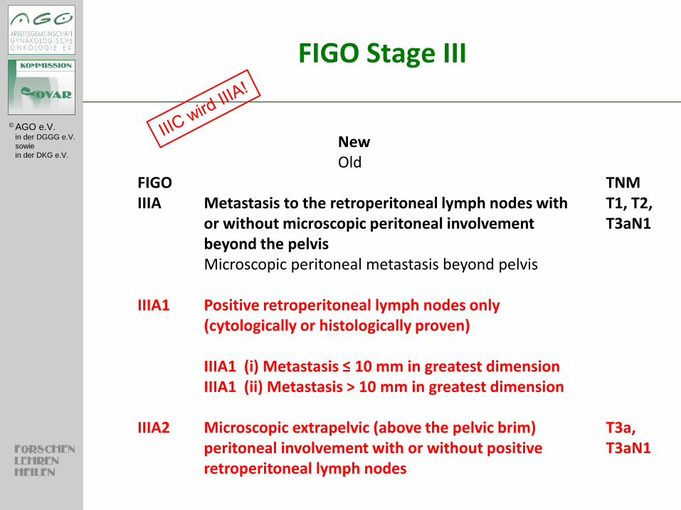
FIGO Stage III
New Old FIGO TNM IIIA Metastasis to the retroperitoneal lymph nodes with T1, T2, or without microscopic peritoneal involvement T3aN1 beyond the pelvis Microscopic peritoneal metastasis beyond pelvis IIIA1 Positive retroperitoneal lymph nodes only (cytologically or histologically proven) IIIA1 (i) Metastasis ≤ 10 mm in greatest dimension IIIA1 (ii) Metastasis > 10 mm in greatest dimension IIIA2 Microscopic extrapelvic (above the pelvic brim) T3a, peritoneal involvement with or without positive T3aN1 retroperitoneal lymph nodes
© AGO e.V. in der DGGG e.V.
sowie
in der DKG e.V.
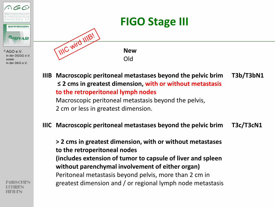
FIGO Stage III
New Old IIIB Macroscopic peritoneal metastases beyond the pelvic brim T3b/T3bN1 ≤ 2 cms in greatest dimension, with or without metastasis to the retroperitoneal lymph nodes Macroscopic peritoneal metastasis beyond the pelvis, 2 cm or less in greatest dimension. IIIC Macroscopic peritoneal metastases beyond the pelvic brim T3c/T3cN1 > 2 cms in greatest dimension, with or without metastases to the retroperitoneal nodes (includes extension of tumor to capsule of liver and spleen without parenchymal involvement of either organ) Peritoneal metastasis beyond pelvis, more than 2 cm in greatest dimension and / or regional lymph node metastasis
© AGO e.V. in der DGGG e.V.
sowie
in der DKG e.V.
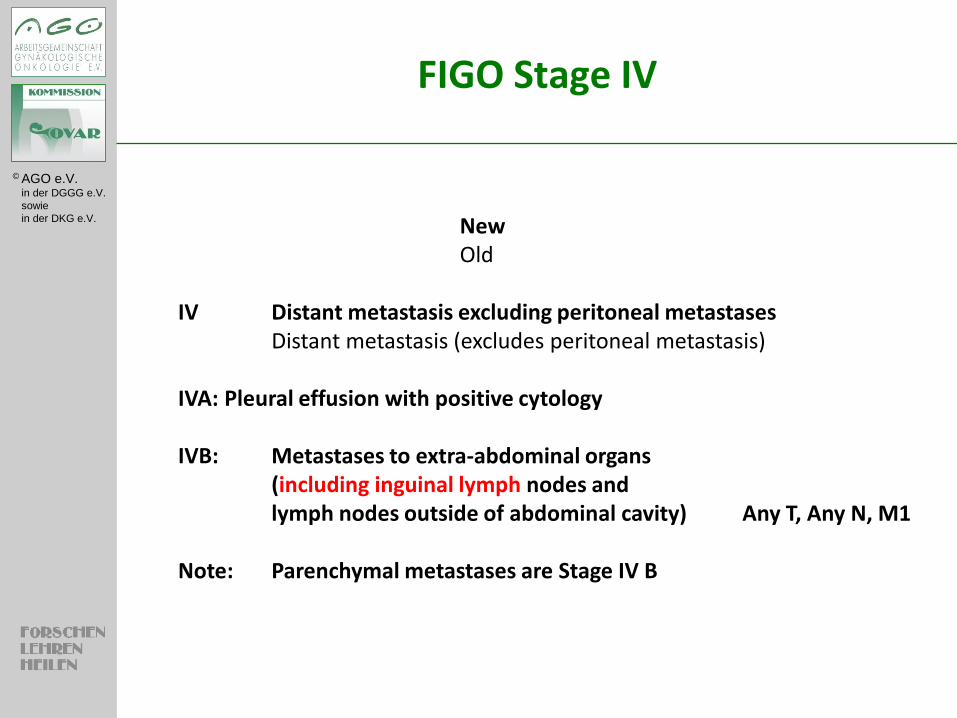
FIGO Stage IV
New Old IV Distant metastasis excluding peritoneal metastases Distant metastasis (excludes peritoneal metastasis) IVA: Pleural effusion with positive cytology IVB: Metastases to extra-abdominal organs (including inguinal lymph nodes and lymph nodes outside of abdominal cavity) Any T, Any N, M1 Note: Parenchymal metastases are Stage IV B
© AGO e.V. in der DGGG e.V.
sowie
in der DKG e.V.
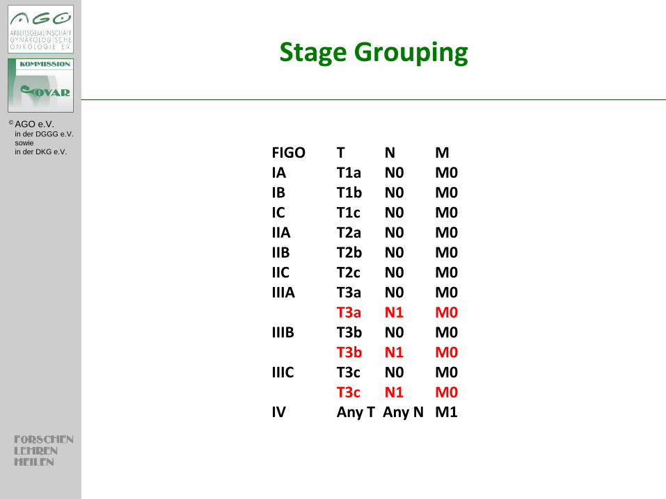
Stage Grouping
FIGO T N M
IA T1a N0 M0
IB T1b N0 M0
IC T1c N0 M0
IIA T2a N0 M0
IIB T2b N0 M0
IIC T2c N0 M0
IIIA T3a N0 M0
T3a N1 M0
IIIB T3b N0 M0
T3b N1 M0
IIIC T3c N0 M0
T3c N1 M0
IV Any T Any N M1
© AGO e.V. in der DGGG e.V.
sowie
in der DKG e.V.
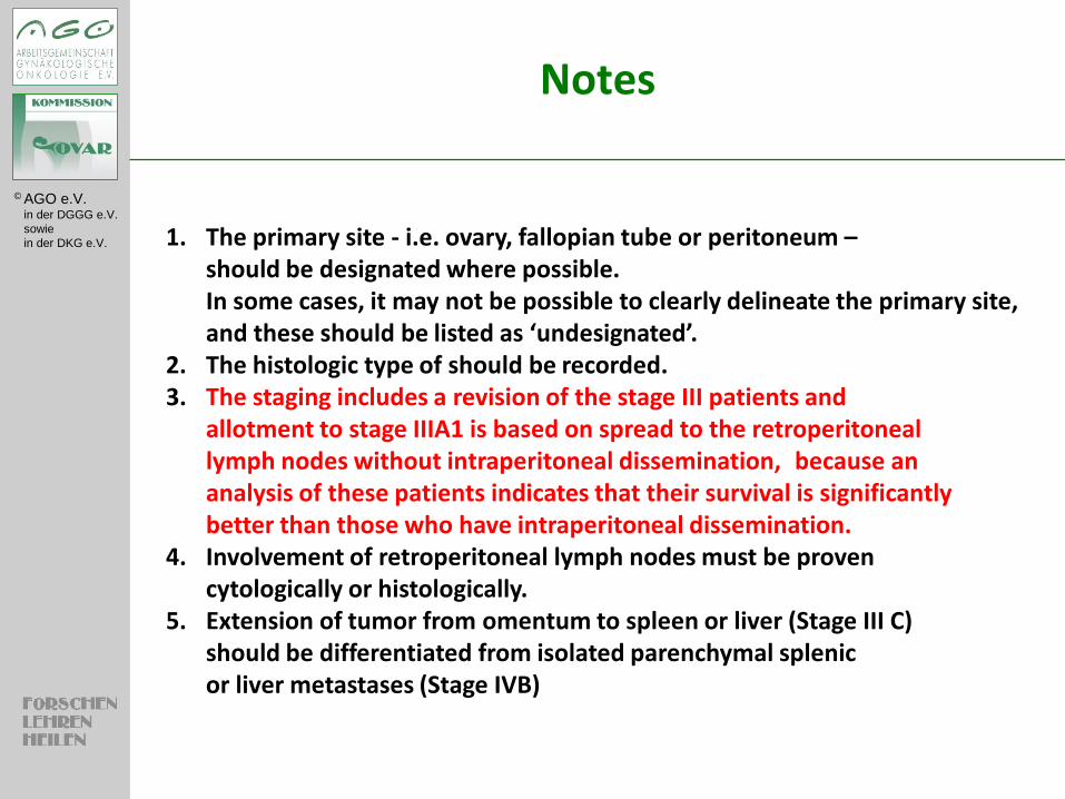
Notes
1. The primary site - i.e. ovary, fallopian tube or peritoneum – should be designated where possible. In some cases, it may not be possible to clearly delineate the primary site, and these should be listed as ‘undesignated’. 2. The histologic type of should be recorded. 3. The staging includes a revision of the stage III patients and allotment to stage IIIA1 is based on spread to the retroperitoneal lymph nodes without intraperitoneal dissemination, because an analysis of these patients indicates that their survival is significantly better than those who have intraperitoneal dissemination. 4. Involvement of retroperitoneal lymph nodes must be proven cytologically or histologically. 5. Extension of tumor from omentum to spleen or liver (Stage III C) should be differentiated from isolated parenchymal splenic or liver metastases (Stage IVB)

© AGO e.V. in der DGGG e.V.
sowie
in der DKG e.V.
Vielen Dank für die Aufmerksamkeit!