Embed Size (px)
Citation preview

Helen Young is Lymphoedema Nurse Specialist and Clinical Manager, Lymphoedema Clinic, St Giles’ Hospice, Whittington, Staffordshire, UK
The effective management of lymphoedema requires accurate diagnosis and the exclusion or
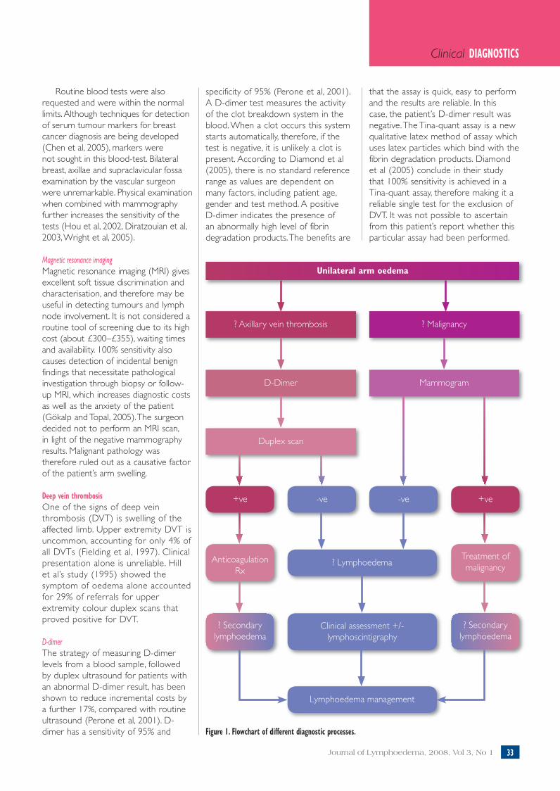
treatment of any underlying factors that may cause impairment of lymphatic drainage. Sudden onset unilateral limb oedema may be caused by malignant or life-threatening disorders (Weissleder and Schuchhardt, 2001) and therefore must be investigated fully. This article presents a case report of a patient with a history of unilateral arm oedema of unknown cause and will illustrate the concept of differential diagnosis (Figure 1). It will then describe the lymphoedema management strategies appropriate for this patient.
Case reportA woman was referred by her vascular consultant to the lymphoedema service for assessment and management of chronic arm swelling. The patient was
56 years old and had noticed a sudden onset of swelling to her non-dominant left arm 30 months previously. Since then the swelling had been persistent and was aggravated by strenuous or repetitive activity. The patient described the arm as ‘feeling very heavy and uncomfortable at times’, but there had been no alteration in sensation or range of movement since onset. Diagnostic tests including mammography, duplex ultrasound and chest X-ray had been requested by the vascular consultant and performed prior to referral. At initial assessment a clinical history was taken, and a physical examination performed in order that potential causes could be considered (Table 1). Injury or trauma causes cellular death and dissolution. The cell proteins that are released increase lymphatic load and, therefore, it could be argued that repeated trauma may cause a state of chronic overload of the lymphatic system, resulting in the lymphatic valves becoming widened and the development of valvular insufficiency. This would cause backflow of lymph and ultimately lymphostasis.
Investigating the potential causes of oedemaMalignancy When investigating sudden onset arm swelling, underlying malignancy must be considered. Tumour mass in the chest wall, the apex of the axilla, infraclavicular or supraclavicular regions may all impair venous return
and lymphatic drainage through the root of the limb, causing oedema. However, studies show that only 1–2% of secondary lymphoedema is due to previously undiagnosed tumours (Weissleder and Schuchhardt, 2001).
MammographyA mammogram had been performed six months prior to the onset of swelling according to NHS protocol (NHS Breast Screening Programme, 2005), and the results were screen negative. The specificity of mammography screening has been shown to be 96.8% in post-menopausal women, such as this patient, and the technique has greatest sensitivity for this patient group due to their reduced tissue density (Banks et al, 2004).
Mammography had been performed within a routine population screening programme; however, it was necessary to review the results and films were relevant as cancers diagnosed in the inter-screening interval may account for around 19% of the total incidence of breast cancer. This may be due to false-negative readings because of reader error in perception or interpretation; 25–45% of interval cancers (a cancer that occurs within three years of a negative screening test) may fall into this category (Houssami et al, 2006). The films were reviewed and were found to support the original results of ‘nothing abnormal to detect’.
Oedema of the upper limb may be attributable to several factors, including malignancy, venous or lymphatic abnormality. This article illustrates the concept of differential diagnosis for lymphoedema, by critically reviewing the diagnostic tests and examinations performed to investigate unilateral arm swelling in a 51-year-old woman. It reveals the pitfalls of using non-standardised diagnostic tests and suggests that evidence-based protocols for diagnostic procedures are necessary if we are to correctly diagnose the causes of oedema and provide patients with effective treatment for the condition.
Helen Young
Key words
OedemaDifferential diagnosisVenous abnormalityMalignancy
Diagnostic investigations for oedema of the upper limb
32 Journal of Lymphoedema, 2008, Vol 3, No 1
Clinical DIAGNOSTICS
Young FinalC.indd 14 28/3/08 10:48:30
33Journal of Lymphoedema, 2008, Vol 3, No 1
Clinical DIAGNOSTICS
Routine blood tests were also requested and were within the normal limits. Although techniques for detection of serum tumour markers for breast cancer diagnosis are being developed (Chen et al, 2005), markers were not sought in this blood-test. Bilateral breast, axillae and supraclavicular fossa examination by the vascular surgeon were unremarkable. Physical examination when combined with mammography further increases the sensitivity of the tests (Hou et al, 2002, Diratzouian et al, 2003, Wright et al, 2005).
Magnetic resonance imagingMagnetic resonance imaging (MRI) gives excellent soft tissue discrimination and characterisation, and therefore may be useful in detecting tumours and lymph node involvement. It is not considered a routine tool of screening due to its high cost (about £300–£355), waiting times and availability. 100% sensitivity also causes detection of incidental benign findings that necessitate pathological investigation through biopsy or follow-up MRI, which increases diagnostic costs as well as the anxiety of the patient (Gökalp and Topal, 2005). The surgeon decided not to perform an MRI scan, in light of the negative mammography results. Malignant pathology was therefore ruled out as a causative factor of the patient’s arm swelling.
Deep vein thrombosis One of the signs of deep vein thrombosis (DVT) is swelling of the affected limb. Upper extremity DVT is uncommon, accounting for only 4% of all DVTs (Fielding et al, 1997). Clinical presentation alone is unreliable. Hill et al’s study (1995) showed the symptom of oedema alone accounted for 29% of referrals for upper extremity colour duplex scans that proved positive for DVT.
D-dimerThe strategy of measuring D-dimer levels from a blood sample, followed by duplex ultrasound for patients with an abnormal D-dimer result, has been shown to reduce incremental costs by a further 17%, compared with routine ultrasound (Perone et al, 2001). D-dimer has a sensitivity of 95% and
specificity of 95% (Perone et al, 2001). A D-dimer test measures the activity of the clot breakdown system in the blood. When a clot occurs this system starts automatically, therefore, if the test is negative, it is unlikely a clot is present. According to Diamond et al (2005), there is no standard reference range as values are dependent on many factors, including patient age, gender and test method. A positive D-dimer indicates the presence of an abnormally high level of fibrin degradation products. The benefits are
that the assay is quick, easy to perform and the results are reliable. In this case, the patient’s D-dimer result was negative. The Tina-quant assay is a new qualitative latex method of assay which uses latex particles which bind with the fibrin degradation products. Diamond et al (2005) conclude in their study that 100% sensitivity is achieved in a Tina-quant assay, therefore making it a reliable single test for the exclusion of DVT. It was not possible to ascertain from this patient’s report whether this particular assay had been performed.
Unilateral arm oedema
? Axillary vein thrombosis ? Malignancy
D-Dimer Mammogram
Duplex scan
+ve -ve +ve-ve
AnticoagulationRx
Treatment ofmalignancy
? Secondarylymphoedema
? Secondarylymphoedema
? Lymphoedema
Clinical assessment +/- lymphoscintigraphy
Lymphoedema management
Figure 1. Flowchart of different diagnostic processes.
Young FinalC.indd 15 28/3/08 10:48:33
Duplex scanning (colour Doppler sonovenography)The vascular consultant referred the patient for Duplex scanning (colour Doppler sonovenography), as this is considered the ‘gold standard’ for diagnosing DVT non-invasively (Diamond et al, 2005). It is highly accurate in diagnosing DVT and carries essentially no risk to the patient. It has a sensitivity of 97% and specificity of 94% in proximal limb oedema (Chunilal and Ginsberg, 2000). However,
it has poor sensitivity for distal DVT (Perone et al, 2001). Benefits of this test include detection of DVT of the upper extremities and capability of identifying other soft-tissue pathologies (Merli, 2005). The cost benefit is great, at about one-third of the cost of the MRI technique (£90–£130). Ultrasound allows visualisation within the vessels as well as surrounding soft tissue with real time visualisation. Doppler colour flow mapping will visualise vessel calibre and flow velocity, giving insight into whether there is intrinsic occlusion within the venous system of the arm, indicative of thrombi. The results from this patient’s scan also came back negative, supporting the results of her D-dimer test.
Contrast venographyAscending contrast venography has also been declared the ‘gold standard’ in diagnosing DVT, with 99% sensitivity and specificity (Merli, 2005), and it has the ability to investigate the distal venous system. However, it is an invasive procedure, it is difficult to perform in the presence of oedema and it is not always readily available (Chunilal and Ginsberg, 2000). It is used when non-invasive results are inconclusive. In this case, the results of the ultrasound scan stated that the left subclavian, axillary and brachial veins appeared patent and that contrast venography was not requested. Clinically, the oedema did not present as venous in nature, with no evidence of pitting, no reduction with elevation and no signs of collateral venous drainage.
Initial patient treatment planVolume measurements of both limbs, using the 4cm interval circumferential technique (Stanton et al, 2006) showed an excess of 240ml — an 11.9% excess in the oedematous limb when compared to the unaffected limb. A plan of management in accordance with the findings from clinical examination was implemented. Since DVT had been excluded and there were no other contraindications for the application of hosiery, the patient was fitted with a class 2 compression sleeve (C–G), as the oedema extended from the wrist to the root of the limb, and she had no previous history of hand or
34 Journal of Lymphoedema, 2008, Vol 3, No 1
Clinical DIAGNOSTICS
digit swelling. Graduated compression facilitates both venous return and lymphatic drainage to the root of the limb (Hampton, 2003). Skin care was also discussed as a precaution to minimise the risk of infection in the potentially compromised limb (Mortimer, 2000).
The patient stated that she was relieved that something positive was now being done to help her swelling. She was informed that her swelling appeared to be consistent with a failure of the lymphatic drainage system, but further investigation of the lymphatics would confirm clinical findings. She was reviewed four weeks after fitting the compression hosiery. On re-measurement, the limb volume difference had reduced by 160ml and the difference between limb volumes was now only 4%, compared with the 11.9% initial excess. Symptoms of discomfort had resolved and the patient was pleased with the response to treatment.
LymphoedemaShe was informed that her swelling appeared to be consistent with
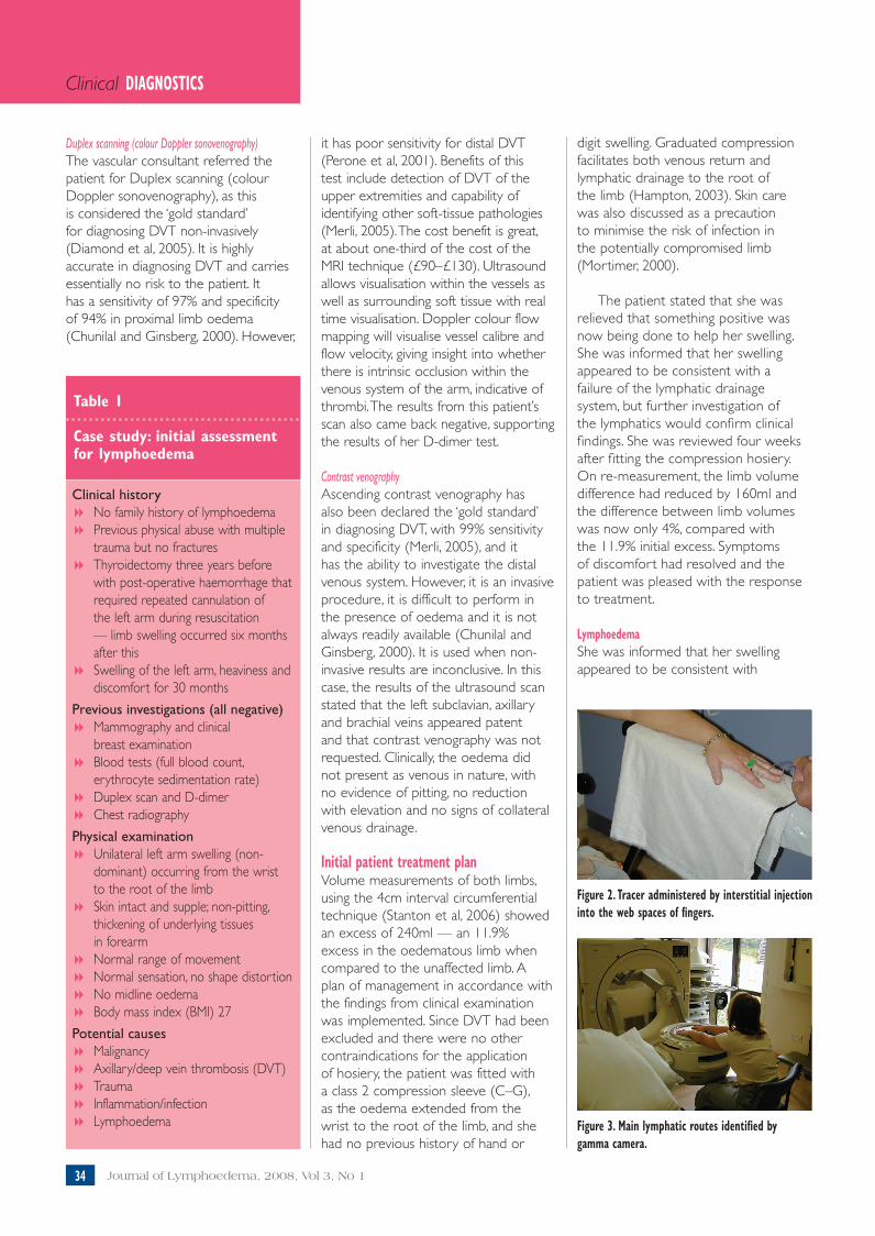
Figure 2. Tracer administered by interstitial injection into the web spaces of fingers.
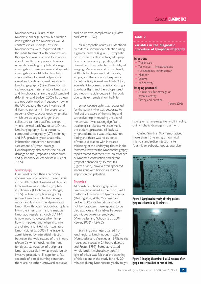
Figure 3. Main lymphatic routes identified by gamma camera.
Clinical history 8 No family history of lymphoedema 8 Previous physical abuse with multiple trauma but no fractures8 Thyroidectomy three years before with post-operative haemorrhage that required repeated cannulation of the left arm during resuscitation — limb swelling occurred six months after this8 Swelling of the left arm, heaviness and discomfort for 30 months
Previous investigations (all negative) 8 Mammography and clinical breast examination8 Blood tests (full blood count, erythrocyte sedimentation rate)8 Duplex scan and D-dimer8 Chest radiography
Physical examination8 Unilateral left arm swelling (non- dominant) occurring from the wrist to the root of the limb8 Skin intact and supple; non-pitting, thickening of underlying tissues in forearm8 Normal range of movement8 Normal sensation, no shape distortion8 No midline oedema8 Body mass index (BMI) 27
Potential causes 8 Malignancy8 Axillary/deep vein thrombosis (DVT)8 Trauma8 Inflammation/infection8 Lymphoedema
Table 1
Case study: initial assessment for lymphoedema
Young FinalC.indd 16 28/3/08 10:48:34
35Journal of Lymphoedema, 2008, Vol 3, No 1
Clinical DIAGNOSTICS
lymphoedema, a failure of the lymphatic drainage system, but further investigation of the lymphatics would confirm clinical findings. Tests for lymphoedema were requested after the initial treatment with compression therapy. She was reviewed four weeks after fitting the compression hosiery while still awaiting lymphatic drainage investigation. There are several diagnostic investigations available for lymphatic abnormalities. To visualise lymphatic vessel and node abnormalities, direct lymphangiography (‘direct’ injection of radio-opaque material into a lymphatic) and lymphography are the gold standard (Mortimer and Badger, 2005), but these are not performed as frequently now in the UK because they are invasive and difficult to perform in the presence of oedema. Only subcutaneous lymphatics which are as large, or larger than collectors can be opacified, except where dermal backflow occurs. Direct lymphangiography, like ultrasound, computed tomography (CT) scanning and MRI provides gross anatomical information rather than functional assessment of lymph drainage. Lymphography also carries the risk of damage to the lymphatic endothelium and pulmonary oil embolism (Liu et al, 2005).
LymphoscintigraphyFunctional rather than anatomical information is considered more useful in the differential diagnosis of chronic limb swelling as it detects lymphatic insufficiency (Mortimer and Badger, 2005). Indirect lymphoscintigraphy (indirect injection into the dermis) more readily shows the dynamics of lymph flow through radiocolloid uptake from the interstitium and transit via lymphatic vessels, although 3D MRI is now used to detect when lymph flow is impaired and when channels are dilated and filled with stagnated lymph (Liu et al, 2005). The tracer is administered by interstitial injection between the web spaces of the fingers (Figure 2), which obviates the need for direct cannulation of peripheral lymphatic vessels in what would be an invasive procedure. Except for a few seconds of a mild burning sensation, there are no other untoward sequelae
and no known complications (Hafez and Wolfe, 1996).
Main lymphatic routes are identified by external scintillation detection using a gamma camera (Figure 3). Lymphatic obstruction results in retrograde lymph flow to cutaneous lymphatics, called dermal backflow, detected with delayed imaging (Weissleder and Schuchhardt, 2001). Advantages are that it is safe, simple, and the amount of exposure to radioactivity is small — 18–40 MBq, equivalent to cosmic radiation during a two-hour flight, and the isotope used, technetium, rapidly decays in the body due to its extremely short half-life.
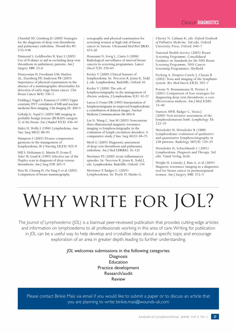
Lymphoscintigraphy was requested for the patient who was desperate to find out the cause of the swelling and to receive help in reducing the size of her arm, as it was causing significant psychological distress. At assessment, the oedema presented clinically as lymphoedema as it was unilateral, non-pitting and there was no evidence of venous dilatation with increased thickening of the underlying tissues in the forearm. However, the lymphoscintigraphy report stated that there was ‘no evidence of lymphatic obstruction and patent lymphatic channels by 15 minutes’ (Figures 4 and 5), however, this appeared inconsistent with her clinical history, inspection and palpation.
DiscussionAlthough lymphoscintigraphy has become established as the most useful method of diagnosis of lymphoedema (Pecking et al, 2002, Mortimer and Badger, 2005), its limitations should not be forgotten. There appear to be discrepancies and variables between techniques currently employed (Weissleder and Schuchhardt, 2001, Keeley, 2006) (Table 2).
Scanning parameters varied from ‘until regional lymph nodes imaged’ (Weissleder and Weissleder, 1998), to ‘six hours, and repeat in 24 hours’ (Larcos and Foster, 1995). Some advocated ‘whole body lymphoscintigraphy’. In light of this, it was felt that the scanning of this patient in the study for only 20 minutes during lymphoscintigraphy might
have given a false-negative result in ruling out lymphatic drainage impairment.
Casley-Smith (1997) emphasised more than 10 years ago how vital it is to standardise injection site (dermis or subcutaneous), exercise,
Injections8 Tracer type8 Technique — intracutaneous, subcutaneous, intramuscular8 Number8 Volume 8 Radioactivity
Imaging protocol8 At rest or after massage or physical activity8 Timing and duration
(Keeley, 2006)
Table 2
Variables in the diagnostic procedure of lymphoscintigraphy
Figure 5. Imaging discontinued at 20 minutes after lymph nodes visualised at root of limb.
Figure 4. Lymphoscintigraphy showing patient lymphatic channels by 15 minutes.
Young FinalC.indd 17 28/3/08 10:48:34

36 Journal of Lymphoedema, 2008, Vol 3, No 1
Clinical DIAGNOSTICS
any massage, and the duration of monitoring. He stated that many radiologists are performing this technique with inadequate or no training, and some centres do not use controls to establish normal clearance rates and lymph speeds. This may create false negative reports from lymphoscintigraphy, and mean that conclusions or comparisons are dubious. It appears that little has altered and the lack of international standardisation has continued.
In light of the patient’s lymphoscintigraphy results indicating a normal uptake, even though clinical examination supported a diagnosis of lymphoedema, contact was made with the reporting radiologist to establish their current protocol and possible reasons for this anomaly. Guidelines were being used rather than a protocol, without a documented evidence base, and imaging was discontinued immediately after lymph nodes were visualised at the root of the affected limb. This took 20 minutes on this occasion.
The anomaly lead the author to obtain protocols from other local nuclear medicine departments that perform lymphoscintigraphy, and they were found to vary greatly. Some departments were unwilling to share protocols and guidelines, and declined to discuss their practice. One department only had guidelines for lymphoscintigraphy for sentinel node localisation, as ‘leg lymphoscintigraphy was no longer used, as this was outdated’ and MRI was being used instead.
Following lymphoscintigraphy results, the patient was seen for review and the results were discussed, as well as the potential reasons for anomalies. The maintenance treatment phase of skin care and compression hosiery continued, as this had achieved a significant reduction in her oedema and an improvement in comfort. In addition, the patient was being taught to perform simple lymphatic drainage and given a three-monthly review, and the lymphoedematous arm continued to
remain stable at 4% excess compared with the unaffected arm.
ConclusionThere is general agreement in the literature that clinical examination, including history, inspection, palpation and volume measurement may be sufficient to diagnose lymphoedema. However, the appropriate application of diagnostic imaging investigations can rule out underlying malignant disorders, where clinical examination procedures fail to provide an unambiguous diagnosis (Weissleder and Schuchhardt, 2001). This case study highlights flaws in relying solely on diagnostic investigation reports, as in the author’s opinion the patient should have been diagnosed as having lymphoedema, but this was ruled out by lymphoscintigraphy.
The sensitivity and specificity, even if 100%, is only applicable if the diagnostic test is performed accurately and interpreted correctly. Misdiagnosis is more likely to occur in the absence of standardised procedures, and if those performing and interpreting the investigation are not appropriately trained. Standardisation of evidence-based lymphatic imaging practised nationally should help to ensure that patients receive an equitable service, are diagnosed accurately and are, therefore, more likely to be treated appropriately.
A combination of the diagnostic measures discussed alongside clinical assessment are essential for effective diagnosis and informed management of lymphoedema. The issue of non-standardisation of some of the investigations, however, will have significant implications for future practice. The risk of causing fur ther trauma and infection for a patient undergoing multiple injections for lymphoscintigraphy, and the cost of the procedure in both time and money cannot be justified until local procedures are carried out according to evidence-based recommendations.
Future considerationsLymphoscintigraphy has the potential to guide decisions about the
management of oedema. This means that valid, standardised procedure and interpretation processes need to be established to ensure accuracy of reporting. Anomalies in practice need to be highlighted, and the implications of a lack of evidence-based standardisation put back into the international arena for future consideration.
References Banks E, Reeves G, Beral V, et al (2004) Influence of personal characteristics of individual women on sensitivity and specificity of mammography in the Million Women Study: cohort study. BMJ 329(7464): 477
Chen CC, Hou MF, Wang JY, et al (2005) Simultaneous detection of multiple mRNA markers CK19, CEA, c-Met, Her2/neu and hMAM with membrane array, an innovative technique with a great potential for breast cancer diagnosis. Cancer Lett 240(2): 279–88
JL
Key points
8 The effective management of lymphoedema requires accurate diagnosis and the exclusion or treatment of any underlying factors that may cause impairment of lymphatic drainage.
8 Clinical examination, including history, inspection, palpation and volume measurement may be sufficient to diagnose lymphoedema, however, appropriate application of diagnostic imaging investigations can rule out underlying malignant disorders.
8 A combination of diagnostic measures alongside clinical assessment is central for effective diagnosis and informed management of lymphoedema.
8 Misdiagnosis is more likely to occur in the absence of standardised procedures.
Young FinalC.indd 18 28/3/08 10:48:35
37Journal of Lymphoedema, 2008, Vol 3, No 1
Clinical DIAGNOSTICS
Chunilal SD, Ginsberg JS (2000) Strategies for the diagnosis of deep vein thrombosis and pulmonary embolism. Thromb Res 97: V33–V48
Diamond S, Goldbweber R, Katz S (2005) Use of D-dimer to aid in excluding deep vein thrombosis in ambulatory patients. Am J Surgery 189: 23–6
Diratzouian H, Freedman GM, Hanlon AL, Eisenberg DF, Anderson PR (2003) Importance of physical examination in the absence of a mammographic abnormality for detection of early-stage breast cancer. Clin Breast Cancer 6(4): 330–3
Fielding J, Nagel S, Pomeroy O (1997) Upper extremity DVT correlation of MR and nuclear medicine flow imaging. Clin Imaging 21: 260–3
Gökalp G, Topal U (2005) MR imaging in probably benign lesions (BI-RADS category 3) of the breast. Eur J Radiol 57(3): 436–44
Hafez H, Wolfe J (1996) Lymphedema. Ann Vasc Surg 10(1): 88–95
Hampton S (2003) Elvarex compression garments in the management of lymphoedema. Br J Nursing 12(15): 925–9
Hill S, Holtzman G, Martin D, Evans P, Toler W, Goad K (1995) Selective use of the Duplex scan in diagnosis of deep venous thrombosis. Am J Surg 170: 201–5
Hou M, Chuang H, Ou-Yang F, et al (2002) Comparison of breast mammography,
sonography and physical examination for screening women at high risk of breast cancer in Taiwan. Ultrasound Med Biol 28(4): 415–20
Houssami N, Irwig L, Ciatto S (2006) Radiological surveillance of interval breast cancers in screening programmes. Lancet Oncol 7(3): 259–65
Keeley V (2000) Clinical features of lymphoedema. In: Twycross R, Jenns K, Todd J, eds. Lymphoedema. Radcliffe, Oxford: 61
Keeley V (2006) The role of lymphoscintigraphy in the management of chronic oedema. J Lymphoedema 1(1): 42–57
Larcos G Foster DR (1995) Interpretation of lymphoscintigrams in suspected lymphoedema: a contribution of delayed images. Nuclear Medicine Communications 16: 683–6
Liu N, Wang C, Sun M (2005) Noncontrast three-dimensional magnetic resonance imaging vs lymphoscintigraphy in the evaluation of lymph circulation disorders: A comparative study. J Vasc Surg 41(1): 69–75
Merli G (2005) Diagnostic assessment of deep vein thrombosis and pulmonary embolism. Am J Med 118(8A): 3S–12S
Mortimer PS (2000) Acute inflammatory episodes. In: Twycross R, Jenns K, Todd J, eds. Lymphoedema. Radcliffe, Oxford: 134
Mortimer P, Badger C (2005) Lymphoedema. In: Doyle D, Hanks G,
Cherny N, Calman K, eds. Oxford Textbook of Palliative Medicine. 3rd edn. Oxford University Press, Oxford: 640–7
National Health Service (2005) Breast Screening Programme. Consolidated Guidance on Standards for the NHS Breast Screening Programme. NHS Cancer Screening Programmes, Sheffield
Pecking A, Desprez-Curely J, Cluzan R (2002) Tests and imaging of the lymphatic system. Rev Med Intern 23(3): 391–7
Perone N, Bounameaux H, Perrier A (2001) Comparison of four strategies for diagnosing deep vein thrombosis: a cost-effectiveness analysis. Am J Med 1(10): 33–40
Stanton AWB, Badger C, Sitzia J (2000) Non-invasive assessment of the lymphoedematous limb. Lymphology 33: 122–35
Weissleder H, Weissleder R (1988) Lymphoedema: evaluation of qualitative and quantitative lymphoscintigraphy in 238 patients. Radiology 167(3): 729–35
Weissleder H, Schuchhardt C (2001) Lymphoedema. Diagnosis and Therapy. 3rd edn. Viatal Verlag, Koln
Wright H, Litinsky J, Rim A, et al (2005) Magnetic resonance imaging as a diagnostic tool for breast cancer in premenopausal women. Am J Surgery 190: 572–5
Why write for JOL?The Journal of Lymphoedema (JOL) is a biannual peer-reviewed publication that provides cutting-edge articles
and information on lymphoedema to all professionals working in this area of care. Writing for publication in JOL can be a useful way to help develop and crystallise ideas about a specific topic and encourage
exploration of an area in greater depth, leading to further understanding.
JOL welcomes submissions in the following categories:DiagnosisEducation
Practice developmentResearch/audit
Review
Please contact Binkie Mais via email if you would like to submit a paper or to discuss an article that you are planning to write: [email protected]
Young FinalC.indd 19 28/3/08 10:48:35