Embed Size (px)
Citation preview
©2013 MFMER | slide-1
Diagnostic Criteria for Neuromyelitis Optica 2014 Brian G. Weinshenker, MD, FRCP(C)
©2013 MFMER | slide-2
Disclosures • Royalties related to patent for discovery of NMO-IgG
• licensed to RSR Ltd; Oxford University
• Consulting contracts related to NMO clinical research (within past 5 years): • GlaxoSmithKline Pharmaceuticals • Elan Pharmaceuticals • Chord Pharmaceuticals • Chugai Pharmaceuticals • Novartis Pharmaceuticals • Alexion Pharmaceuticals
• Member DSMB • Biogen Idec (Chair) • Novartis • Mitsubishi (Chair)
• Member Attack Adjudication Committee: • MedImmune (Chair)
©2013 MFMER | slide-3
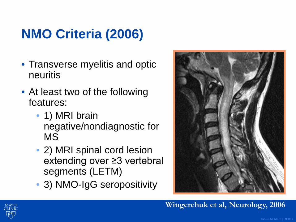
NMO Criteria (2006)
• Transverse myelitis and optic neuritis
• At least two of the following features:
• 1) MRI brain negative/nondiagnostic for MS
• 2) MRI spinal cord lesion extending over ≥3 vertebral segments (LETM)
• 3) NMO-IgG seropositivity
Wingerchuk et al, Neurology, 2006
©2013 MFMER | slide-4
Why are current diagnostic criteria inadequate in 2014? • Discovery of NMO-IgG
• NMO can be recognized reliably at an earlier point
• Limited versions of NMO • recurrent myelitis or recurrent optic neuritis
• Brain lesions may occur • may be the presenting manifestations • may be highly suggestive or diagnostic
• Co-association of other autoimmune conditions: • Do they exclude NMO?
©2013 MFMER | slide-5
International Panel for NMO Diagnosis (IPND) • Convened October, 2011 • Co-chairs:
• Dean Wingerchuk • Brian Weinshenker
• Overall objective: • To revise NMO diagnostic criteria to reflect
advances in: • Clinical and radiological spectrum • Serological testing
©2013 MFMER | slide-6
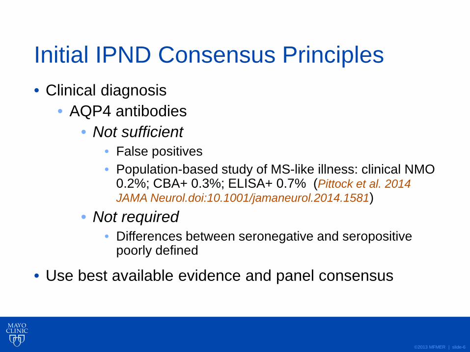
Initial IPND Consensus Principles • Clinical diagnosis
• AQP4 antibodies • Not sufficient
• False positives • Population-based study of MS-like illness: clinical NMO
0.2%; CBA+ 0.3%; ELISA+ 0.7% (Pittock et al. 2014 JAMA Neurol.doi:10.1001/jamaneurol.2014.1581)
• Not required • Differences between seronegative and seropositive
poorly defined
• Use best available evidence and panel consensus

©2013 MFMER | slide-7
Methods • 18 members from 9 countries • 6 Working Groups
• Specific charges relevant to NMO diagnosis
©2013 MFMER | slide-8
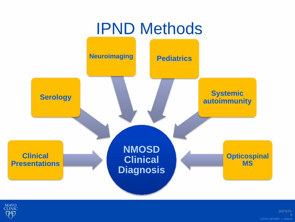
IPND Methods
3007678-8
NMOSD Clinical
Diagnosis Clinical
Presentations
Serology
Neuroimaging Pediatrics
Systemic autoimmunity
Opticospinal MS
©2013 MFMER | slide-9
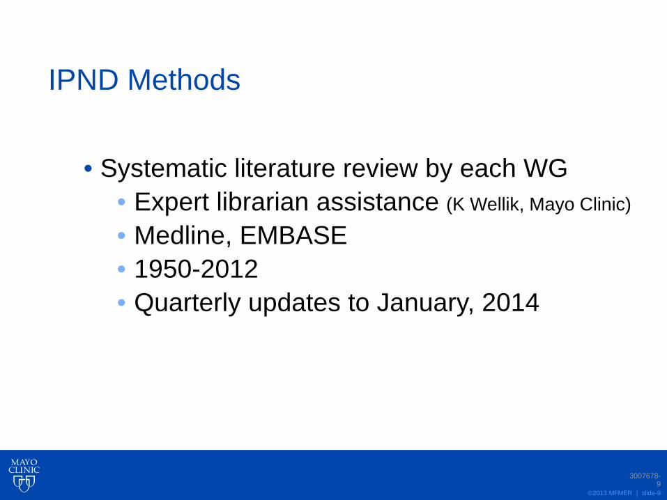
IPND Methods
• Systematic literature review by each WG • Expert librarian assistance (K Wellik, Mayo Clinic)
• Medline, EMBASE • 1950-2012 • Quarterly updates to January, 2014
3007678-9
©2013 MFMER | slide-10
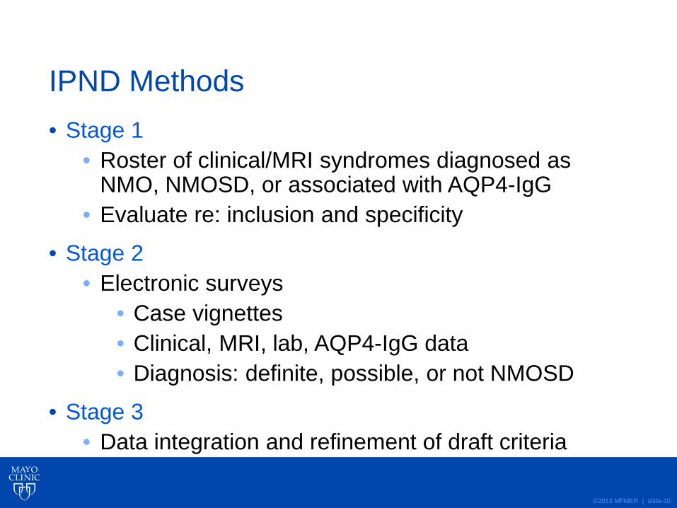
IPND Methods • Stage 1
• Roster of clinical/MRI syndromes diagnosed as NMO, NMOSD, or associated with AQP4-IgG
• Evaluate re: inclusion and specificity
• Stage 2 • Electronic surveys
• Case vignettes • Clinical, MRI, lab, AQP4-IgG data • Diagnosis: definite, possible, or not NMOSD
• Stage 3 • Data integration and refinement of draft criteria
©2013 MFMER | slide-11
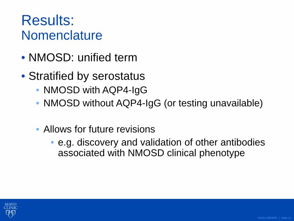
Results: Nomenclature • NMOSD: unified term • Stratified by serostatus
• NMOSD with AQP4-IgG • NMOSD without AQP4-IgG (or testing unavailable)
• Allows for future revisions
• e.g. discovery and validation of other antibodies associated with NMOSD clinical phenotype
©2013 MFMER | slide-12
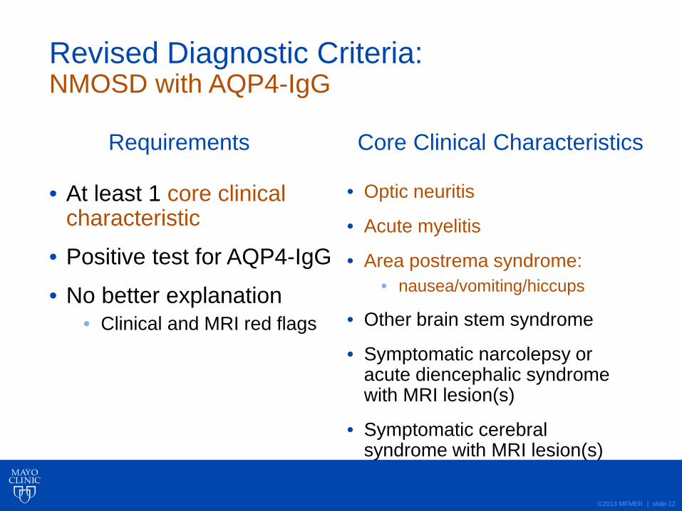
Revised Diagnostic Criteria: NMOSD with AQP4-IgG
• At least 1 core clinical characteristic
• Positive test for AQP4-IgG
• No better explanation • Clinical and MRI red flags
• Optic neuritis
• Acute myelitis
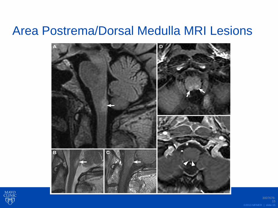
• Area postrema syndrome: • nausea/vomiting/hiccups
• Other brain stem syndrome
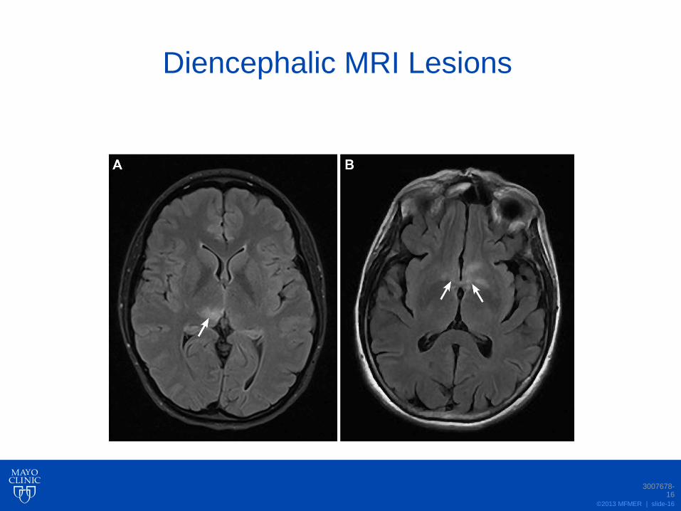
• Symptomatic narcolepsy or acute diencephalic syndrome with MRI lesion(s)
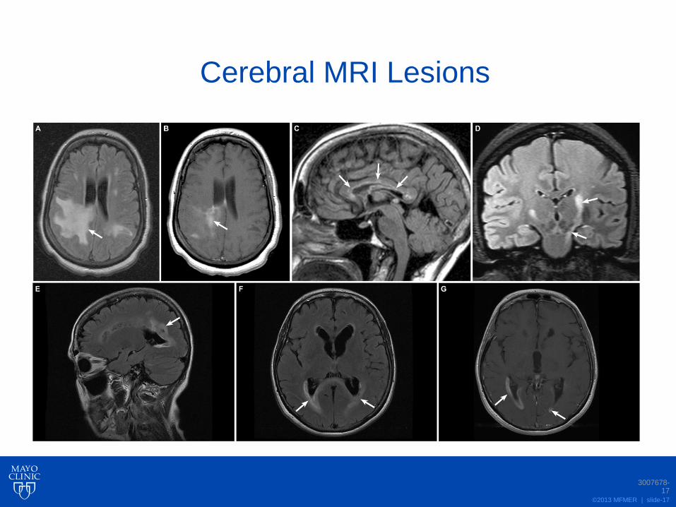
• Symptomatic cerebral syndrome with MRI lesion(s)
Requirements Core Clinical Characteristics
©2013 MFMER | slide-13
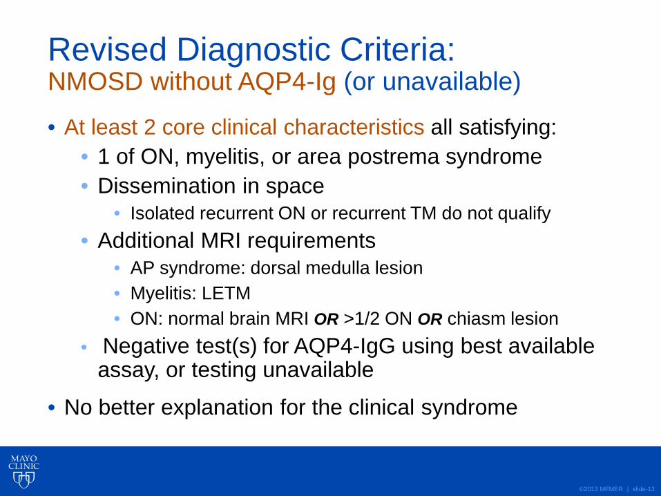
Revised Diagnostic Criteria: NMOSD without AQP4-Ig (or unavailable)
• At least 2 core clinical characteristics all satisfying: • 1 of ON, myelitis, or area postrema syndrome • Dissemination in space
• Isolated recurrent ON or recurrent TM do not qualify • Additional MRI requirements
• AP syndrome: dorsal medulla lesion • Myelitis: LETM • ON: normal brain MRI OR >1/2 ON OR chiasm lesion
• Negative test(s) for AQP4-IgG using best available assay, or testing unavailable
• No better explanation for the clinical syndrome
©2013 MFMER | slide-14
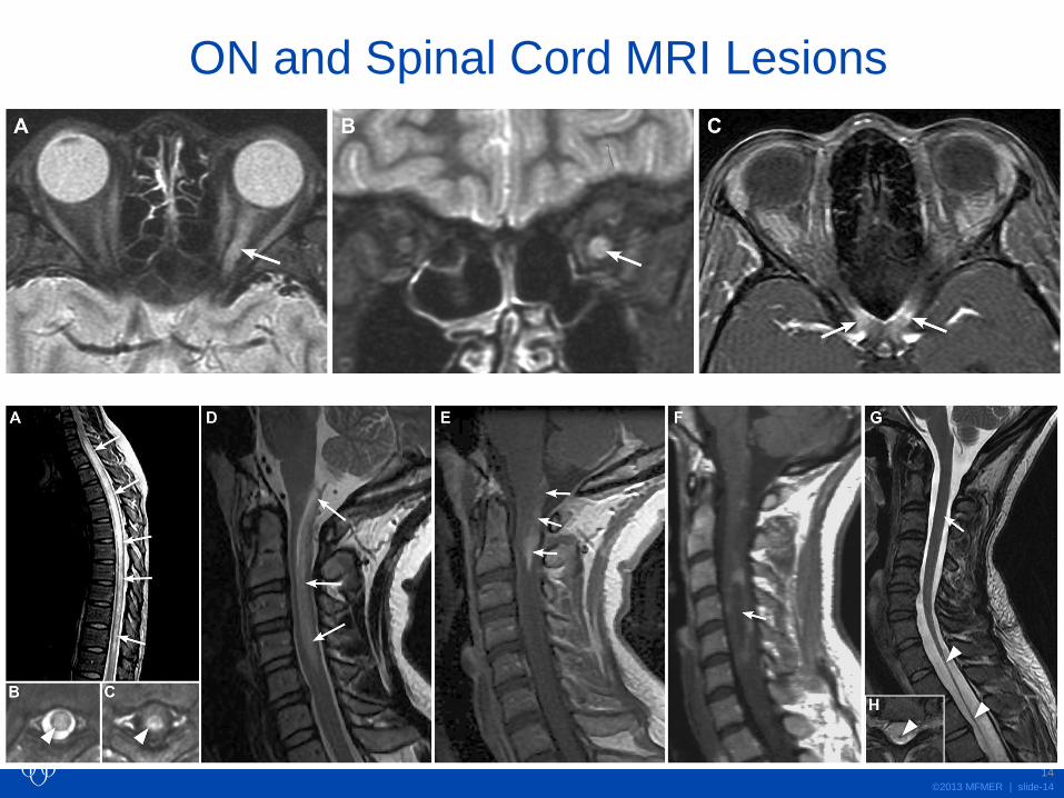
ON and Spinal Cord MRI Lesions
3007678-14
©2013 MFMER | slide-15
Area Postrema/Dorsal Medulla MRI Lesions
3007678-15
©2013 MFMER | slide-16
Diencephalic MRI Lesions
3007678-16
©2013 MFMER | slide-17
Cerebral MRI Lesions
3007678-17
©2013 MFMER | slide-18
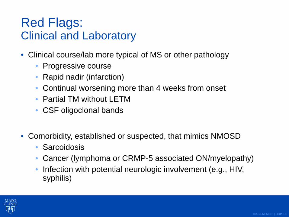
Red Flags: Clinical and Laboratory • Clinical course/lab more typical of MS or other pathology
• Progressive course • Rapid nadir (infarction) • Continual worsening more than 4 weeks from onset • Partial TM without LETM • CSF oligoclonal bands
• Comorbidity, established or suspected, that mimics NMOSD • Sarcoidosis • Cancer (lymphoma or CRMP-5 associated ON/myelopathy) • Infection with potential neurologic involvement (e.g., HIV,
syphilis)
©2013 MFMER | slide-19
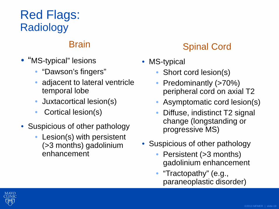
Red Flags: Radiology
Brain
• “MS-typical” lesions • “Dawson’s fingers” • adjacent to lateral ventricle
temporal lobe • Juxtacortical lesion(s) • Cortical lesion(s)
• Suspicious of other pathology • Lesion(s) with persistent
(>3 months) gadolinium enhancement
Spinal Cord • MS-typical
• Short cord lesion(s) • Predominantly (>70%)
peripheral cord on axial T2 • Asymptomatic cord lesion(s) • Diffuse, indistinct T2 signal
change (longstanding or progressive MS)
• Suspicious of other pathology • Persistent (>3 months)
gadolinium enhancement • “Tractopathy” (e.g.,
paraneoplastic disorder)

©2013 MFMER | slide-20
Pediatric NMOSD • Same criteria as adult NMOSD • Cautions:
• LETM in pediatric MS • Greater incidence of cerebral presentations
©2013 MFMER | slide-21
Opticospinal MS • Historically important • Confusing terminology
• a form of MS versus NMO versus something unique?
• Similarly defined in Asia, patients have the same disease
• “Superseded” terminology
©2013 MFMER | slide-22
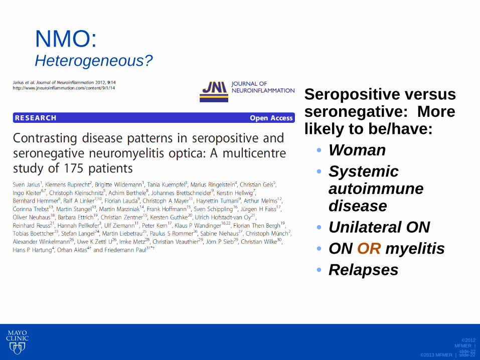
NMO: Heterogeneous?
• Seropositive versus seronegative: More likely to be/have:
• Woman • Systemic
autoimmune disease
• Unilateral ON • ON OR myelitis • Relapses
©2012 MFMER |
slide-22
©2013 MFMER | slide-23
©2013 MFMER | slide-24
NMO: Beginnings of Classification of Heterogeneity?
• Seropositive NMO
• Seronegative NMO • False negative serologic assay • MS mimicking NMO • Other disorders that mimic NMO:
• Sarcoidosis • Paraneoplastic
• “True seronegative NMO” • Younger patients • Monophasic course • Less severe clinical manifestations • MOG antibodies?

©2013 MFMER | slide-25
Summary
• NMOSD is the unified term for NMO/NMOSD • AQP4-IgG Seropositive:
• Requires at least 1 of the 6 core clinical characteristics
• AQP4-IgG Seronegative or Unavailable: • At least 2 core clinical characteristics
• 1 must be ON, acute myelitis or area postrema syndrome
• MRI support • Dissemination in space
3007678-25

©2013 MFMER | slide-26
Implications to Clinical Trials • “New disease”?
• Estimate 2X number of cases • facilitates enrolment and enhances study
feasibility • Historical data on prognosis/outcome:
• unreliable?
• Heterogeneity in prognosis, behavior between • seropositive and seronegative
©2013 MFMER | slide-27
Acknowledgments • Funding and Administrative Support:
• Guthy-Jackson Charitable Foundation • Dr. Katja van Herle, Dr. Valerie Pasquetto, Dr. Michael Yeaman, Ms.
Jacinta Behne
• Library Science (Mayo Clinic) • Kay Wellik, MLS
• Administrative Support (Mayo Clinic) • Amy Clayton • Katrina Rivera
• Contribution to optic-spinal MS discussion • Jun-ichi Kira, MD
©2013 MFMER | slide-28
IPND Members • Brenda Banwell, MD
• Jeff Bennett, MD, PhD
• Philippe Cabre, MD
• Bill Carroll, MD
• Tanuja Chitnis, MD
• Jerome de Seze, MD
• Kazuo Fujihara, MD
• Ben Greenberg, MD
• Anu Jacob, MD
• Sven Jarius, MD
• Marco Lana-Peixoto, MD
• Michael Levy, MD, PhD
• Jack Simon, MD
• Silvia Tenembaum, MD
• Tony Traboulsee, MD
• Paddy Waters, PhD
• Brian Weinshenker, MD
• Dean Wingerchuk, MD

©2013 MFMER | slide-29
©2013 MFMER | slide-30
Systemic Autoimmune Disease • NMO diagnosis allowable • Concurrence with SLE, SS, MG increases
likelihood of a diagnosis of NMO • Association with systemic autoimmune disease
more likely reflects concurrence than causation
©2013 MFMER | slide-31
Pathology for Diagnosis • Not routine • AQP4-IgG obviates need for biopsy in most
cases • Atypical cases:
• Astrocytopathy and selective AQP4 loss may • establish diagnosis • exclude other pathologies
©2013 MFMER | slide-32
A1 A2 A3 A4
B1 B2 B3 B4
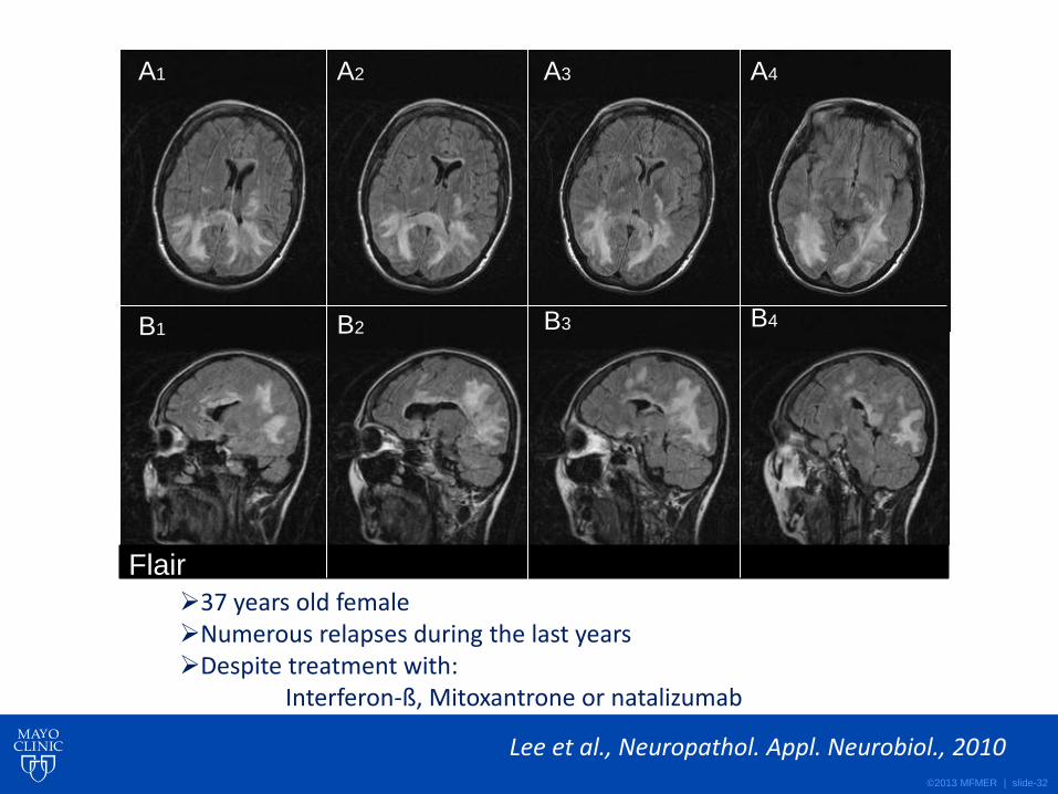
Flair 37 years old female Numerous relapses during the last years Despite treatment with: Interferon-ß, Mitoxantrone or natalizumab
Lee et al., Neuropathol. Appl. Neurobiol., 2010
©2013 MFMER | slide-33
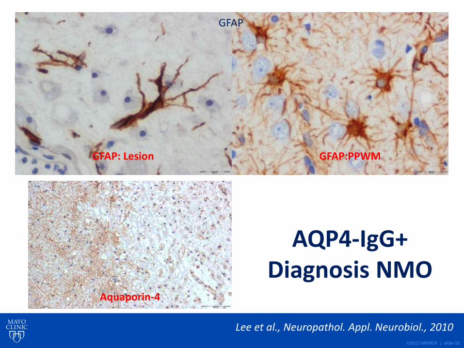
GFAP
GFAP:PPWM GFAP: Lesion
Aquaporin-4
AQP4-IgG+ Diagnosis NMO
Lee et al., Neuropathol. Appl. Neurobiol., 2010
©2013 MFMER | slide-34
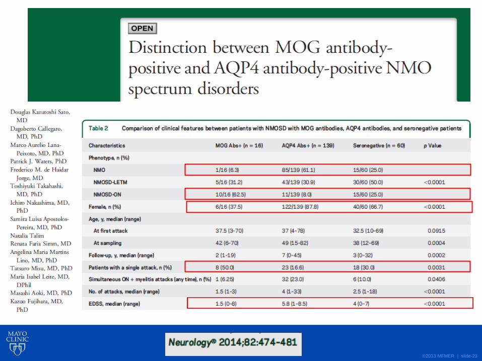
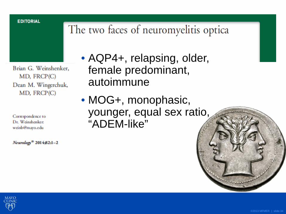
• AQP4+, relapsing, older, female predominant, autoimmune
• MOG+, monophasic, younger, equal sex ratio, “ADEM-like”