Embed Size (px)
Citation preview

DOI 10.1212/CPJ.0000000000000044 published online June 18, 2014Neurol Clin Pract
J.D. Bartleson and Timothy P. Mausinterventions
Diagnostic and therapeutic spinal interventions: Facet joint
This information is current as of June 18, 2014
http://cp.neurology.org/content/early/2014/06/18/CPJ.0000000000000044.full.htmllocated on the World Wide Web at:
The online version of this article, along with updated information and services, is
Academy of Neurology. All rights reserved. Print ISSN: 2163-0402. Online ISSN: 2163-0933.continuously since 2011, it is now a bimonthly with 6 issues per year. Copyright © 2014 American
® is an official journal of the American Academy of Neurology. PublishedNeurol Clin Pract
Neurology® Clinical Practice
Diagnostic and therapeuticspinal interventionsFacet joint interventionsJ.D. Bartleson, MD
Timothy P. Maus, MD
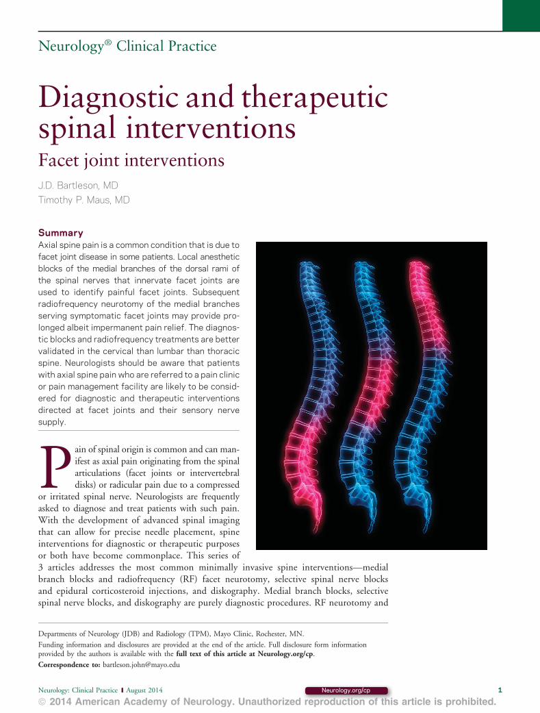
SummaryAxial spine pain is a common condition that is due tofacet joint disease in some patients. Local anestheticblocks of the medial branches of the dorsal rami ofthe spinal nerves that innervate facet joints areused to identify painful facet joints. Subsequentradiofrequency neurotomy of the medial branchesserving symptomatic facet joints may provide pro-longed albeit impermanent pain relief. The diagnos-tic blocks and radiofrequency treatments are bettervalidated in the cervical than lumbar than thoracicspine. Neurologists should be aware that patientswith axial spine pain who are referred to a pain clinicor pain management facility are likely to be consid-ered for diagnostic and therapeutic interventionsdirected at facet joints and their sensory nervesupply.
Pain of spinal origin is common and can man-ifest as axial pain originating from the spinalarticulations (facet joints or intervertebraldisks) or radicular pain due to a compressed
or irritated spinal nerve. Neurologists are frequentlyasked to diagnose and treat patients with such pain.With the development of advanced spinal imagingthat can allow for precise needle placement, spineinterventions for diagnostic or therapeutic purposesor both have become commonplace. This series of3 articles addresses the most common minimally invasive spine interventions—medialbranch blocks and radiofrequency (RF) facet neurotomy, selective spinal nerve blocksand epidural corticosteroid injections, and diskography. Medial branch blocks, selectivespinal nerve blocks, and diskography are purely diagnostic procedures. RF neurotomy and
Departments of Neurology (JDB) and Radiology (TPM), Mayo Clinic, Rochester, MN.
Funding information and disclosures are provided at the end of the article. Full disclosure form informationprovided by the authors is available with the full text of this article at Neurology.org/cp.
Correspondence to: [email protected]
Neurology: Clinical Practice |||||||||||| August 2014 Neurology.org/cp 1
ª 2014 American Academy of Neurology. Unauthorized reproduction of this article is prohibited.
epidural steroid injections have therapeutic intent. While some neurologists perform facetinterventions and epidural injections, more often they refer patients to interventionalists(typically anesthesiologists, physiatrists, or radiologists) who work in specialized pain cen-ters. This article covers indications for medial branch blocks and RF facet neurotomy, howthe interventions are performed and interpreted, and risks and benefits.
Indications, contraindications, and patient and segment selectionfor medial branch blocks and RF neurotomyThe facet or zygapophyseal joints are a considerable source of axial spine pain.1,2 Lumbarregion facet-mediated pain is rare in young adults (about 15% of chronic back pain) butincreases in prevalence up to age 70 and accounts overall for about 30% of patients withchronic low back pain.3 Cervical facet joints are responsible for 25%–65% of nontraumaticand 50%–60% of trauma-induced cases of chronic neck pain.4 The medial branches of thedorsal rami of the spinal nerves innervate the facet joints. Medial branch blocks are indi-cated in patients with chronic (.6 months) axial spine pain that is inadequately explainedand poorly controlled. If diagnostic blocks of the nerves that supply specific facet jointsrelieve the patient’s pain, RF lesioning of the same nerves can be offered to provideprolonged benefit. Contraindications to the performance of medial branch blocks or sub-sequent RF neurotomy include systemic infection, local infection at the procedure site,substantial bleeding diathesis, and pregnancy.
The facet joints targeted for medial branch blocks are chosen based on a combination of clin-ical and prevalence data. Physical and neurologic examination does not identify symptomaticfacet joints. Structural findings of facet joint arthrosis on plain radiographs, CT, or MRI are notpredictive of facet joint origin pain.5 MRI T2 hyperintensity or increased uptake on SPECTor SPECT/CT are purported to identify painful joints, but these findings have not beenconfirmed. Facet joint localization can be facilitated by comparing the patient’s pain to facetjoint pain referral maps.1,2,4 Prevalence data are also used.2 Upper cervical pain with headacheis most likely attributable to the C2-3 facet. Lower cervical pain is more likely to be C5-6than C6-7 in origin. Lumbar pain is overwhelmingly due to the L4-5 or L5-S1 facet joints.
How the interventions are performed and interpretedMedial branch blocks Each facet joint is innervated by 2 vertically adjacent spinal me-
dial branches; the only exception is the C2-3 facet, innervated solely by the superficial me-dial branch of the C3 dorsal ramus known as the third occipital nerve.1,2 The medialbranches can be targeted with image guidance because anatomic studies have shownconsistent relationships between the medial branches and bony landmarks visible onfluoroscopy. Under fluoroscopic x-ray guidance, with sterile technique and without se-dation, 25-gauge spinal needles are sequentially placed at the predicted position of medialbranches of the dorsal rami of the spinal nerves, which innervate the targeted facet joints.Injection of a small amount of contrast material (0.3 mL) excludes vascular uptake.A small volume of local anesthetic (0.5 mL) is then instilled. Because of frequentfalse-positive responses, the diagnostic criterion best supported by the literature is thatof dual, comparative medial branch blocks in which the targeted medial branchesare blocked on separate occasions using local anesthetics with different durations ofaction (e.g., lidocaine and bupivacaine). For a positive medical branch block, the patientmust have substantial (80%–100% pain relief for an appropriate duration with eachanesthetic agent. Such a concordant response should provide a specificity of 88%.2
Spine interventions have a higher risk whenperformed in the cervical or thoracic more than thelumbar spine.
2 © 2014 American Academy of Neurology
J.D. Bartleson and Timothy P. Maus
ª 2014 American Academy of Neurology. Unauthorized reproduction of this article is prohibited.
Many patients only receive a single local anesthetic injection directed at each suspectfacet joint’s medial branches. Because single injections have a false-positive rate of 27%–
34%, the specificity of single blocks is much reduced.2,6
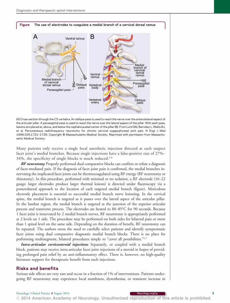
RF neurotomy Properly performed dual comparative blocks can confirm or refute a diagnosisof facet-mediated pain. If the diagnosis of facet joint pain is confirmed, the medial branches in-nervating the implicated facet joints can be thermocoagulated using RF energy (RF neurotomy orrhizotomy). In this procedure, performed with minimal or no sedation, a RF electrode (16–22gauge; larger electrodes produce larger thermal lesions) is directed under fluoroscopy via aposterolateral approach to the location of each targeted medial branch (figure). Meticulouselectrode placement is essential to successful medial branch nerve lesioning. In the cervicalspine, the medial branch is targeted as it passes over the lateral aspect of the articular pillar.In the lumbar region, the medial branch is targeted at the junction of the superior articularprocess and transverse process. The electrodes are heated to 80–85°C for 90 seconds. Because1 facet joint is innervated by 2 medial branch nerves, RF neurotomy is appropriately performedat 2 levels on 1 side. The procedure may be performed on both sides for bilateral pain or morethan 1 spinal level on the same side. Depending on the duration of benefit, RF neurotomy canbe repeated. The authors stress the need to carefully select patients and identify symptomaticfacet joints using dual comparative diagnostic medial branch blocks. There is no place forperforming multisegment, bilateral procedures simply to “cover all possibilities.”1,2
Intra-articular corticosteroid injections Separately, or coupled with a medial branchblock, patients may receive intra-articular facet joint injections of a steroid in hopes of provid-ing prolonged pain relief by an anti-inflammatory effect. There is, however, no high-qualityliterature support for therapeutic benefit from such injections.
Risks and benefitsSerious side effects are very rare and occur in a fraction of 1% of interventions. Patients under-going RF neurotomy may experience local numbness, dysesthesias, or transient increase in
Figure The use of electrodes to coagulate a medial branch of a cervical dorsal ramus
(A) Cross-section through the C5 vertebra. An oblique pass is used to reach the nerve over the anterolateral aspect ofthe articular pillar. A parasagittal pass is used to reach the nerve over the lateral aspect of the pillar. With each pass,lesions are placed at, above, and below the cephalocaudad center of the pillar (B). From Lord SM, Barnsley L, Wallis BJ,et al. Percutaneous radiofrequency neurotomy for chronic cervical zygapophyseal joint pain. N Engl J Med1996;335:1721–1726. Copyright © Massachusetts Medical Society. Reprinted with permission from Massachu-setts Medical Society.
Neurology: Clinical Practice |||||||||||| August 2014 Neurology.org/cp 3
Diagnostic and therapeutic spinal interventions
ª 2014 American Academy of Neurology. Unauthorized reproduction of this article is prohibited.

pain. Temporary neuritis may occur (2%).2 Third occipital neurotomy patients often expe-rience cutaneous numbness and mild ataxia due to this nerve’s role in cervical propriocep-tion.2 Allergic reactions are extremely rare but can occur with local anesthetics,corticosteroids, or contrast media. If corticosteroids are injected, systemic side effects canoccur. Pretreatment imaging and fluoroscopy during the procedure expose the patient toradiation. Complications common to any needle procedure can occur and include infection,dural puncture, spinal nerve or cord injury, and vascular injury. Spine interventions have ahigher risk when performed in the cervical or thoracic more than the lumbar spine. Themedial branches can regenerate with return of pain.
Benefits include discovery of the patient’s pain mechanism, improvement in their chronicpain, improvement in physical functioning, return to work, and decreased use of other healthcare. In the cervical spine, RF neurotomy may be especially helpful for pain followingwhiplash-type injury.7 If obtained, pain relief may not be complete or permanent. Additionalimprovement often follows repeat intervention, but benefit may wane over time. In additionto the facet joints, the medial branches supply some paraspinal muscles (chiefly the multif-idus), soft tissues, and at some spinal levels, paraspinal skin. As RF neurotomy is designed tointerrupt sensory signals, not treat or modify a disease process, the ultimate pathologic causeof the patient’s pain is not known or directly treated.
What is the evidence that medial branch interventions are beneficial? A Cochrane systematicreview concluded that there is “limited evidence that RF denervation offers short-term relieffor chronic neck pain of zygapophyseal joint origin and for chronic cervicobrachial pain” and“conflicting evidence on the short-term effect of RF lesioning on pain and disability inchronic low-back pain of zygapophyseal joint origin.”8 Chou et al.9 in an evidence-basedAmerican Pain Society Clinical Practice Guideline found “insufficient evidence to adequatelyevaluate benefits of” therapeutic medial branch blocks and RF denervation for low back pain.
A challenge to such reviews is that they evaluate and include studies based on research meth-odology, not whether the procedures were performed with evidence-based patient selection andprocedural technique. Studies performed without rigorous patient selection, or without proce-dural technique based on proven anatomic principles, will not achieve good outcomes and willobscure the benefit that can be achieved with proper technique. In contrast to these reviews,high-quality studies using meticulous technique report that the majority of patients experi-enced 80%–100% pain relief for months to a year or more following RF neurotomy forchronic neck pain.7,10,11 With optimal technique, 66% of cervical RF patients achievedcomplete pain relief, restoration of activities of daily living, return to work, and no needfor other health care for neck pain for a median duration of 17–20 months.11 Prolonged reliefcould be restored by repeat neurotomy.7,10,11 In a study of low back pain patients using dualcomparative medial branch blocks and correct RF technique, 60% of patients achieved atleast 90% pain relief at 1 year follow-up; 87% of patients obtained at least 60% pain reliefafter 1 year.12 In another rigorous study of lumbar RF neurotomy, the majority of patientshad complete relief of low back pain, restoration of function, return to work, and no otherhealth care needed for back pain for a median duration of 15 months from the first procedureand 13 months for repeat treatment.13 Outcomes are better, and the evidence is stronger, inthe cervical spine than in the lumbar spine. This in part reflects the lower prevalence of facet-mediated pain in the lumbar region and hence greater challenges in patient selection. There is
Outcomes are better, and the evidence isstronger, in the cervical spine than in thelumbar spine.
4 © 2014 American Academy of Neurology
J.D. Bartleson and Timothy P. Maus
ª 2014 American Academy of Neurology. Unauthorized reproduction of this article is prohibited.

little evidence to support facet interventions in the thoracic spine. When appropriately eval-uated, only a minority of patients with axial spine pain will be candidates for RF neurotomy.
The discrepancy between the less favorable results in metadata and guidelines analyses andthe excellent results reported in series using optimal diagnostic and RF technique implies con-siderable provider variability. Skilled interventionalists who select patients using established cri-teria will have much better outcomes than less skilled providers who are willing to use theseprocedures on anyone with neck or low back pain. With proper technique, these interventionsare beneficial. However, the paucity of data regarding provider skill levels and outcomes makesit difficult to select interventionalists for referral.
REFERENCES1. Slipman CW, Derby R, Simeone FA, Mayer TG, editors. Interventional Spine: An Algorithmic
Approach. Philadelphia: Saunders; 2008.2. Bogduk N, editor. Practice Guidelines for Spinal Diagnostic and Treatment Procedures, 2nd ed. San
Francisco, CA: International Spine Intervention Society; 2013.3. DePalma MJ, Ketchum JM, Saullo T. What is the source of chronic low back pain and does age play a
role? Pain Med 2011;12:224–233.4. Gellhorn AC. Cervical facet-mediated pain. Phys Med Rehabil Clin N Am 2011;22:447–458.5. Bogduk N. Degenerative joint disease of the spine. Radiol Clin North Am 2012;50:613–628.6. Barnsley L, Lord S, Wallis B, Bogduk N. False-positive rates of cervical zygapophyseal joint blocks.
Clin J Pain 1993;9:124–130.7. Lord SM, Barnsley L, Wallis BJ, McDonald GJ, Bogduk N. Percutaneous radio-frequency neurotomy
for chronic cervical zygapophyseal-joint pain. N Engl J Med 1996;335:1721–1726.8. Niemisto L, Kalso EA, Malmivaara A, Seitsalo S, Hurri H. Radiofrequency denervation for neck and
back pain. Cochrane Database Syst Rev 2003;1:CD004058.9. Chou R, Loeser JD, Owens DK, et al. Interventional therapies, surgery, and interdisciplinary reha-
bilitation for low back pain. Spine 2009;14:1066–1077.10. McDonald GJ, Lord SM, Bogduk N. Long-term follow-up of patients treated with cervical radiofre-
quency neurotomy for chronic neck pain. Neurosurgery 1999;45:61–67; discussion 67–68.11. MacVicar J, Borowczyk JM, MacVicar AM, Loughnan BM, Bogduk N. Cervical medial branch
radiofrequency neurotomy in New Zealand. Pain Med 2012;13:647–654.12. Dreyfuss P, Halbrook B, Pauza K, et al. Efficacy and validity of radiofrequency neurotomy for chronic
lumbar zygapophysial joint pain. Spine 2000;25:1270–1277.13. MacVicar J, Borowczyk JM, MacVicar AM, Loughnan BM, Bogduk N. Lumbar medial branch
radiofrequency neurotomy in New Zealand. Pain Med 2013;14:639–645.
STUDY FUNDINGNo targeted funding reported.
DISCLOSURESJ.D. Bartleson receives publishing royalties for Spine Disorders: Medical and Surgical Management (Cam-bridge University Press, 2009). T. Maus has received travel compensation as member of the ExecutiveBoard of the International Spine Intervention Society and practices interventional pain management(80% clinical effort) at Mayo Clinic. Full disclosure form information provided by the authors isavailable with the full text of this article at Neurology.org/cp.
Neurology: Clinical Practice |||||||||||| August 2014 Neurology.org/cp 5
Diagnostic and therapeutic spinal interventions
ª 2014 American Academy of Neurology. Unauthorized reproduction of this article is prohibited.

DOI 10.1212/CPJ.0000000000000044 published online June 18, 2014Neurol Clin Pract
J.D. Bartleson and Timothy P. MausDiagnostic and therapeutic spinal interventions: Facet joint interventions
This information is current as of June 18, 2014
ServicesUpdated Information &
0000044.full.htmlhttp://cp.neurology.org/content/early/2014/06/18/CPJ.000000000including high resolution figures, can be found at:
Supplementary Material
000000000044.DC1.htmlhttp://cp.neurology.org/neurclinpract/suppl/2014/06/23/CPJ.0000Supplementary material can be found at:
Subspecialty Collections
http://cp.neurology.org//cgi/collection/all_spinal_cordAll Spinal Cord
http://cp.neurology.org//cgi/collection/all_painAll Painfollowing collection(s): This article, along with others on similar topics, appears in the
Permissions & Licensing
http://cp.neurology.org/misc/about.xhtml#permissionsor in its entirety can be found online at:Information about reproducing this article in parts (figures,tables)
Reprints
http://cp.neurology.org/misc/addir.xhtml#reprintsusInformation about ordering reprints can be found online:

DOI 10.1212/CPJ.0000000000000043 published online June 18, 2014Neurol Clin Pract
J.D. Bartleson and Timothy P. Mausinjections
Diagnostic and therapeutic spinal interventions: Epidural
This information is current as of June 18, 2014
http://cp.neurology.org/content/early/2014/06/18/CPJ.0000000000000043.full.htmllocated on the World Wide Web at:
The online version of this article, along with updated information and services, is
Academy of Neurology. All rights reserved. Print ISSN: 2163-0402. Online ISSN: 2163-0933.continuously since 2011, it is now a bimonthly with 6 issues per year. Copyright © 2014 American
® is an official journal of the American Academy of Neurology. PublishedNeurol Clin Pract

Neurology® Clinical Practice
Diagnostic and therapeuticspinal interventionsEpidural injectionsJ.D. Bartleson, MD
Timothy P. Maus, MD
SummaryEpidural injections of local anesthetic or a corticoste-roid are frequently given to diagnose and treatpatients with radicular pain originating from any spi-nal level. The best-quality evidence supports a trans-foraminal approach in the lumbar spine. Manypatients experience substantial benefit from a singletherapeutic injection. Depending upon the benefitobtained, additional injections may be administered.Selective nerve blocks with local anesthetic alonecan identify the spinal nerve mediating the patient’spain. Serious short-term risks are rare but occur;long-term risks are infrequent and can be due tosystemic effects of multiple corticosteroid injec-tions. Patients who have failed conservative therapyor are not candidates for surgical intervention canbe considered for epidural steroid injections to re-lieve their radicular pain temporarily.
Radicular pain, with or without radiculopa-thy, is a common clinical condition mostfrequently due to disk herniation or fixedlateral recess or foraminal stenosis. In the
lumbar region, lifetime prevalence estimates rangefrom 12% to 43% with annual prevalence estimatesof 2%–34%.1 Cervical radicular pain is less common,and more likely due to fixed stenosis than disk her-niation.2 Clinical and experimental studies of thepathophysiology of radicular pain suggest that both mechanical compression and an inflam-matory response are needed for pain production.3 The inflammatory process supports thetargeted injection of corticosteroids as a therapy for radicular pain. Local anesthetics alonemay be used diagnostically to identify the spinal nerve mediating the patient’s pain.
Departments of Neurology (JDB) and Radiology (TPM), Mayo Clinic, Rochester, MN.
Funding information and disclosures are provided at the end of the article. Full disclosure form informationprovided by the authors is available with the full text of this article at Neurology.org/cp.
Correspondence to: [email protected]
Neurology: Clinical Practice |||||||||||| August 2014 Neurology.org/cp 1
ª 2014 American Academy of Neurology. Unauthorized reproduction of this article is prohibited.
Corticosteroids can be delivered to the dorsal epidural space using an interlaminar approach,or via the sacral hiatus with a caudal approach. However, these midline techniques place theinjected drug some distance from the site of inflammation. The transforaminal approachdelivers the corticosteroid directly to a symptomatic spinal nerve in the intervertebral foramenwhere pharmaceuticals are much more likely to reach the site of inflammation in the ventralepidural space. While some neurologists perform epidural injections, more often they aredone by interventionalists (usually anesthesiologists, physiatrists, or radiologists) who work inspecialized pain center or hospital settings. This article covers 1) indications for epiduralinjections, 2) how epidural injections are performed and interpreted, and 3) risks andbenefits.
Indications and contraindications for epidural injectionsThese injections are indicated for the diagnosis and especially treatment of significant radicularpain with or without radiculopathy in patients who have failed a trial of conservative therapy.4,5
Epidural steroid injections as a therapeutic procedure are much better supported in thelumbar spine than in the cervical or thoracic spine. Although epidural injections are some-times given to patients with axial spine pain and neurogenic claudication, there is no high-quality evidence supporting their use in these conditions. The spinal nerve to be targeted isselected based on a synthesis of imaging, clinical, and neurophysiologic findings. Advancedspinal imaging with MRI, plain CT, or CT myelography is considered a prerequisite forepidural injections by most interventionalists. Preprocedure imaging allows the injectionroute to be planned to optimize medication delivery to the epidural space and minimize riskto neural or vascular structures.6 Such imaging also excludes underlying tumor and infection.Local anesthetics alone are injected for diagnostic purposes; this is properly termed a selectivespinal nerve block. The response to a selective spinal nerve block can aid in diagnosis andlocalization. Injections of a corticosteroid with or without a local anesthetic are given fortherapeutic purposes. Contraindications include systemic or local infection at the injectionsite, allergies to the medications used, pregnancy, or significant coagulopathy. Anticoagulantuse is currently being reevaluated; the risk of hemorrhagic complications must be weightedagainst the real risk of systemic thrombotic complications from withholding anticoagulants.Aspirin and nonsteroidal anti-inflammatories are not contraindications.7 Anticoagulants (war-farin, antiplatelet, and antithrombin agents) should be stopped for interlaminar injections; theissue is less clear for transforaminal injections and the reader is referred to published guide-lines for specific recommendations.5,7
How epidural injections are performed and interpretedInterlaminar epidural steroid injections can be given in the lower cervical, thoracic, and lumbarspine; caudal epidural injections are administered via the sacral hiatus.4,5 There is less evidenceof benefit from lumbar interlaminar and caudal epidural steroid injections, and they are beingsupplanted by transforaminal injections. Lumbar interlaminar and caudal injections may beused if the transforaminal route is not feasible (due to postoperative changes or anatomiclimitations) or with multilevel, bilateral radicular symptoms and findings. Interlaminar injec-tions are appropriate in the low cervical region, as the literature support for cervical trans-foraminal injections is much less robust than for lumbar transforaminal injections.Interlaminar procedures are performed on prone patients, without sedation, and with fluo-roscopic guidance, using 18- to 22-gauge needles. The dorsal epidural space is entered by aloss of resistance technique, but exclusion of vascular or intrathecal flow via contrast injection
Epidural steroid injections as a therapeutic procedureare much better supported in the lumbar spine than inthe cervical or thoracic spine.
2 © 2014 American Academy of Neurology
J.D. Bartleson and Timothy P. Maus
ª 2014 American Academy of Neurology. Unauthorized reproduction of this article is prohibited.

is mandatory. Typically only corticosteroids are administered with interlaminar and caudalinjections, which are not target-specific and have no immediate diagnostic value.
A transforaminal procedure is performed without sedation and with the patient supine or onhis or her side (cervical) or prone (thoracic and lumbar). Under fluoroscopic guidance, a 22- to25-gauge needle is advanced into the targeted epidural space. Contrast medium is injected tomake certain that the injection is not intrathecal or intravascular, and to ensure that the injec-tate spreads appropriately along the targeted neural sleeve (figure). If the injection of contrastmedium is intrathecal or intra-arterial, the procedure should be terminated and postponeduntil the dura or artery has healed. In transforaminal epidural steroid injections, patients areusually given both local anesthetic and a corticosteroid, administered sequentially. Thepatient’s vital signs, neurologic status, and pain level are monitored. The patient may obtainimmediate benefit if a local anesthetic was administered, or it may take several days for theinjected corticosteroid to provide pain relief. Due to potential spread of the agents to adjacentspinal nerves, transforaminal epidural injections have limited diagnostic utility. Improvement2 weeks after the injection when the corticosteroid anti-inflammatory effect is engagedpredicts long-term benefit.
There is a historical practice of providing a series of epidural injections (e.g., weekly for 3weeks), but this is unsupported by the literature and has no place in current practice.5 Adecision to repeat an injection should be based on the response to previous injections. Ifbenefit was substantial (e.g., .50% pain reduction) but incomplete, or has waned after aconsiderable period of relief (e.g., .1 month), then it is reasonable to perform additional
Figure Left L5 radicular pain due to an L4-5 disk extrusion with caudal migration into thelateral recess in a 32-year-old man
Left L5 transforaminal epidural steroid injection; final needle position (A, C) is immediately superior and lateral to theexiting L5 nerve. Contrast injection (B, D) confirms that medication will reach the interface of the disk and L5 nerve inthe lateral recess (arrows).
Neurology: Clinical Practice |||||||||||| August 2014 Neurology.org/cp 3
Diagnostic and therapeutic spinal interventions
ª 2014 American Academy of Neurology. Unauthorized reproduction of this article is prohibited.

injections in hopes of obtaining greater benefit, or recovery of prior benefit. Most patients donot require repeated injections. In a systematic literature review of lumbar transforaminalinjections, 94% of patients achieving 50% or greater reduction in pain did so with a singleinjection.8 Although there is insufficient evidence to identify an absolute maximum numberof injections in a year, a consensus statement by the North American Spine Society (NASS)suggests that a potential need for more than 4 injections in 1 year should prompt consider-ation of alternative treatment, including possible surgical intervention.9
Risks and benefitsVery rare catastrophic spinal cord and vertebrobasilar infarction can occur with transforaminalepidural injections, presumably due to injection into or damage to an artery supplying thespinal cord in the thoracolumbar region (artery of Adamkiewicz or another large radiculo-medullary artery) or the vertebral artery in the cervical spine.4,5 The risk of catastrophic spinalcord and vertebrobasilar infarction may be mitigated by meticulous technique includingcontrast injection, minimal or no patient sedation, use of a test dose of local anesthetic,and use of a nonparticulate steroid (e.g., dexamethasone). Interlaminar epidural injectionsrisk injury to the cervical or thoracic spinal cord by direct needle trauma or epiduralhematoma formation. Risks of all epidural injections are greater in the cervical spine. Thereare also risks inherent in any spine-related interventional procedure that include allergy tomedications, radiation exposure, dural puncture, needle injury to neural or vascular structures,and infection (e.g., epidural abscess and recently reported iatrogenic fungal meningitis andspinal and paraspinal infections).4,5,10 Multiple corticosteroid injections can cause systemic sideeffects including suppression of the hypothalamic-pituitary-adrenal axis and worsening ofosteoporosis.
Benefits of epidural steroid injections include pain relief, functional recovery, avoidance ofsurgical intervention, and identification of the patient’s pain generator. There is more evidenceof temporary symptomatic benefit than avoidance of surgery. While there is support for theiruse in the cervical spine,11,12 there is much stronger evidence of benefit in the lumbarspine.5,8,9 Epidural injections in the thoracic spine are poorly studied. In 2007, the AmericanAcademy of Neurology Therapeutics and Technology Assessment Subcommittee concludedthat “epidural steroid injections may result in some improvement in radicular lumbosacralpain when assessed between 2 and 6 weeks following the injection, compared to controltreatments (Level C, Class I–III evidence). The average magnitude of effect is small and . ingeneral .does not impact average impairment of function, need for surgery, or provide long-term pain relief beyond 3 months.”13 A 2009 review found “fair evidence that epidural steroidinjection is moderately effective for short-term (but not long-term) symptom relief” forsciatica.14
Recent reviews provide stronger support for major benefit from lumbar transforaminal ep-idural steroid injections.8,9 The systematic analysis by MacVicar et al.8 suggests that forradicular pain from lumbar disk herniations, “60% of patients seem to achieve at least50% relief of pain at between 1 and 2 months, but only about 40% maintain this outcomefor 12 months.” The NASS position statement reports that .50% of patients experience painrelief for .1 month, with half still benefitting after 1 year.9 A recent comparative effectivenessstudy in disk herniations demonstrated that 70% of patients achieved 50% pain relief and
Benefits of epidural steroid injections includepain relief, functional recovery, avoidance ofsurgical intervention, and identification of thepatient’s pain generator.
4 © 2014 American Academy of Neurology
J.D. Bartleson and Timothy P. Maus
ª 2014 American Academy of Neurology. Unauthorized reproduction of this article is prohibited.
50% functional restoration 6 months after lumbar transforaminal injections.15 In disk her-niation patients, temporary pain relief from the anti-inflammatory effect of the injectedcorticosteroid may last until the disk herniation and its associated pain have improved orresolved. There is evidence that patients with more recent onset of pain,16 with lesser degreesof neural compression,17 and with optimal delivery of corticosteroid to the ventral epiduralspace18 have better outcomes. The pain relief provided by epidural injections should enablesome patients to avoid diskectomy. A surgical sparing effect has been demonstrated withlumbar transforaminal injections in some studies,19 but not in others; this issue is unresolved.For radicular pain with lumbar spinal stenosis, uncontrolled studies report benefit, but NASSstates that there is insufficient evidence to make a recommendation regarding the use ofepidural injections in this setting.9
Epidural steroid injections are popular and will continue to be recommended by providers topatients for relief of radicular pain. They should be used judiciously and in the context of properindications.
REFERENCES1. Konstantinou K, Dunn KM. Sciatica: review of epidemiological studies and prevalence estimates.
Spine 2008;33:2464–2472.2. Radhakrishnan K, Litchy WJ, O’Fallon WM, Kurland LT. Epidemiology of cervical radiculop-
athy: a population-based study from Rochester, Minnesota, 1976 through 1990. Brain 1994;117:325–335.
3. Mulleman D, Mammou S, Griffoul I, Watier H, Goupille P. Pathophysiology of disk-related sciatica:I: evidence supporting a chemical component. Joint Bone Spine 2006;73:151–158.
4. Slipman CW, Derby R, Simeone FA, Mayer TG, eds. Interventional Spine: An Algorithmic Ap-proach. Philadelphia: Saunders; 2008.
5. Bogduk N, ed. Practice Guidelines for Spinal Diagnostic and Treatment Procedures, 2nd ed. SanFrancisco, CA: International Spine Intervention Society; 2013.
6. Cohen SP, Maus T, Kennedy DJ. The need for magnetic resonance imaging before epidural corti-costeroid injection. Pm & R 2013;5:230–237.
7. Horlocker TT, Wedel DJ, Rowlingson JC, et al. Regional anesthesia in the patient receiving antith-rombotic or thrombolytic therapy: American Society of Regional Anesthesia and Pain Medicineevidence-based guidelines (3rd ed). Reg Anesth Pain Med 2010;35:64–101.
8. MacVicar J, King W, Landers MH, Bogduk N. The effectiveness of lumbar transforaminal injectionof steroids: a comprehensive review with systematic analysis of the published data. Pain Med 2013;14:14–28.
9. North American Spine Society. Lumbar Transforaminal Epidural Steroid Injections Review andRecommendation Statement. 2013. Available at: www.spine.org/Documents/ResearchClinicalCare/LTFESIReviewRecStatement.pdf. Accessed June 10, 2014.
10. Chiller TM, Roy M, Nguyen D, et al. Clinical findings for fungal infections caused by methylpred-nisolone injections. N Engl J Med 2013;369:1610–1619.
11. Carette S, Fehlings MG. Cervical radiculopathy. N Engl J Med 2005;353:392–399.12. Carragee EJ, Hurwitz EL, Cheng I, et al. Treatment of neck pain. Spine 2008;33:S153–S169.13. Armon C, Argoff CE, Samuels J, Backonja M-M. Assessment: use of epidural steroid injections to
treat radicular lumbosacral pain; report of the Therapeutics and Technology Assessment Subcommit-tee of the American Academy of Neurology. Neurology 2007;68:723–279.
14. Chou R, Atlas S, Stanos SP, Rosenquist RW. Nonsurgical interventional therapies for low back pain: areview of the evidence for an American Pain Society Clinical Practice Guideline. Spine 2009;34:1078–1093.
15. Kennedy DJ, Plastaras C, Casey E, et al. Comparative effectiveness of lumbar transforaminal ep-idural steroid injections with particulate versus non-particulate corticosteroids for lumbar radicularpain due to intervertebral disc herniation: a prospective, randomized, double-blind trial. Pain Med2014;15:548–555.
16. Kaufmann TJ, Geske JR, Murthy NS, et al. Clinical effectiveness of single lumbar transforaminalepidural steroid injections. Pain Med 2013;14:1126–1133.
17. Ghahreman A, Bogduk N. Predictors of a favorable response to transforaminal injection of steroids inpatients with lumbar radicular pain due to disc herniation. Pain Med 2011;12:871–879.
18. Desai MJ, Shah B, Sayal PK. Epidural contrast flow patterns of transforaminal epidural steroidinjections stratified by commonly used final needle-tip position. Pain Med 2011;12:864–870.
Neurology: Clinical Practice |||||||||||| August 2014 Neurology.org/cp 5
Diagnostic and therapeutic spinal interventions
ª 2014 American Academy of Neurology. Unauthorized reproduction of this article is prohibited.

19. Riew KD, Yin Y, Gilula L, et al. The effect of nerve-root injections on the need for operativetreatment of lumbar radicular pain: a prospective, randomized, controlled, double-blind study.J Bone Joint Surg Am 2000;82-A:1589–1593.
STUDY FUNDINGNo targeted funding reported.
DISCLOSURESJ.D. Bartleson receives publishing royalties for Spine Disorders: Medical and Surgical Management (Cam-bridge University Press, 2009). T. Maus has received travel compensation as member of the ExecutiveBoard of the International Spine Intervention Society and practices interventional pain management(80% clinical effort) at Mayo Clinic. Full disclosure form information provided by the authors isavailable with the full text of this article at Neurology.org/cp.
6 © 2014 American Academy of Neurology
J.D. Bartleson and Timothy P. Maus
ª 2014 American Academy of Neurology. Unauthorized reproduction of this article is prohibited.
DOI 10.1212/CPJ.0000000000000043 published online June 18, 2014Neurol Clin Pract
J.D. Bartleson and Timothy P. MausDiagnostic and therapeutic spinal interventions: Epidural injections
This information is current as of June 18, 2014
ServicesUpdated Information &
0000043.full.htmlhttp://cp.neurology.org/content/early/2014/06/18/CPJ.000000000including high resolution figures, can be found at:
Supplementary Material
000000000043.DC1.htmlhttp://cp.neurology.org/neurclinpract/suppl/2014/06/23/CPJ.0000Supplementary material can be found at:
Permissions & Licensing
http://cp.neurology.org/misc/about.xhtml#permissionsor in its entirety can be found online at:Information about reproducing this article in parts (figures,tables)
Reprints
http://cp.neurology.org/misc/addir.xhtml#reprintsusInformation about ordering reprints can be found online:

DOI 10.1212/CPJ.0000000000000045 published online June 18, 2014Neurol Clin Pract
Timothy P. Maus and J.D. BartlesonDiagnostic and therapeutic spinal interventions: Diskography
This information is current as of June 18, 2014
http://cp.neurology.org/content/early/2014/06/18/CPJ.0000000000000045.full.htmllocated on the World Wide Web at:
The online version of this article, along with updated information and services, is
Academy of Neurology. All rights reserved. Print ISSN: 2163-0402. Online ISSN: 2163-0933.continuously since 2011, it is now a bimonthly with 6 issues per year. Copyright © 2014 American
® is an official journal of the American Academy of Neurology. PublishedNeurol Clin Pract

Neurology® Clinical Practice
Diagnostic and therapeuticspinal interventionsDiskographyTimothy P. Maus, MD
J.D. Bartleson, MD
SummaryDiskography (provocation diskography, disk stimula-tion) is an invasive diagnostic test performed to con-firm or exclude internal disk disruption as the causeof axial spine pain. Diskography involves injectingfluid into the nucleus of the disk under manometriccontrol; a positive response is reproduction of typicalpain. Extensive but indirect literature validatesdiskography in the lumbar spine; it is less well-supported in the cervical or thoracic spine. Risks in-clude rare instances of infection and neural injury;lumbar diskography may be associated with acceler-ated disk degeneration. Diskography has utility inestablishing a diagnosis of diskogenic pain. Whennegative, it prevents inappropriate interventions di-rected at the disk; when positive, it prevents inter-ventions directed at other axial pain generators. Itsrole in selecting patients for therapies directed atdiskogenic pain is limited by the lack of available val-idated treatments.
Axial spine pain is common; low back pain isestimated to affect 66%–80% and neckpain 67% of people during their life.1,2
The most common cause of chronic lowback pain is internal disk disruption (IDD) or disko-genic pain. Distinct from radicular pain or radiculop-athy, this describes pain originating from the diskitself. It accounts for approximately 40% of chronic low back pain; the differential diagnosisalso includes facet joint pain (;30%), sacroiliac joint pain (;20%), pain from vertebral insuf-ficiency fractures (;3%–5%), and myofascial pain.3 Diskogenic pain is more prevalent in youngadults and declines with age while pain of facet and sacroiliac origin increases with age.3
Departments of Radiology (TPM) and Neurology (JDB), Mayo Clinic, Rochester, MN.
Funding information and disclosures are provided at the end of the article. Full disclosure form informationprovided by the authors is available with the full text of this article at Neurology.org/cp.
Correspondence to: [email protected]
Neurology: Clinical Practice |||||||||||| August 2014 Neurology.org/cp 1
ª 2014 American Academy of Neurology. Unauthorized reproduction of this article is prohibited.
The pathophysiology of diskogenic pain is complex, and supported by a synthesis of biome-chanical, histologic, and physiologic studies.4 The process in the lumbar disk can be summarizedas follows: degradation of the nuclear matrix of the disk, likely precipitated by endplate fractures,reduces the nucleus pulposus’ ability to bear axial load. Load bearing is transferred to theposterior annulus. The posterior annulus may undergo structural failure, resulting in radiallydirected fissures extending from the nucleus to the outer annulus. This creates the anatomicsubstrate for diskogenic pain. The native nociceptive innervation of the disk is confined to theouter annulus; reparative granulation tissue in annular fissures results in neovascularization andingrowth of additional nociceptive afferents that may extend deep into the disk. The native andacquired innervation is subject to mechanical stimulation by the increased load on the posteriorannulus; inflammatory cytokines from nuclear degradation are present within radial fissures andmay provoke chemical stimulation of nociceptors. By these mechanisms, a disk may becomepainful. The purpose of diskography (also termed provocation diskography or disk stimulation)is to make the diagnosis of IDD or diskogenic pain. Diskography is most commonly performedby interventional pain specialists, typically anesthesiologists, physiatrists, or radiologists.Neurologists will encounter patients with chronic axial pain, and should be familiar with theindications, contraindications, procedure, validity, utility, and risks of diskography.
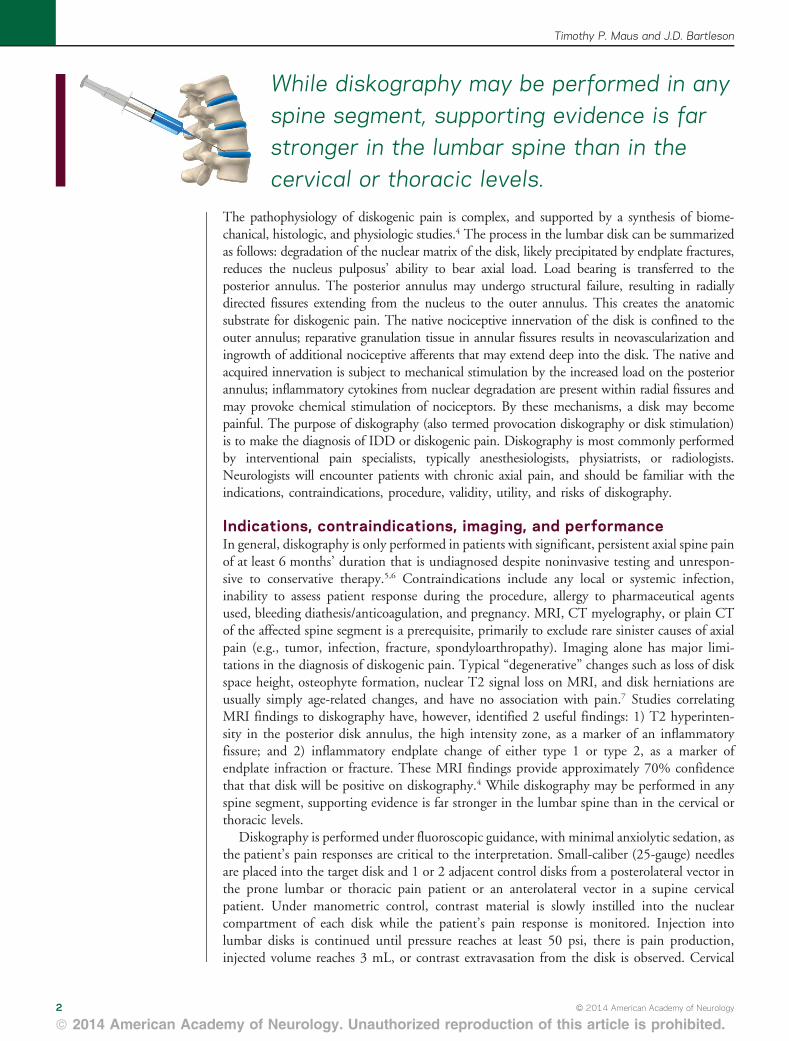
Indications, contraindications, imaging, and performanceIn general, diskography is only performed in patients with significant, persistent axial spine painof at least 6 months’ duration that is undiagnosed despite noninvasive testing and unrespon-sive to conservative therapy.5,6 Contraindications include any local or systemic infection,inability to assess patient response during the procedure, allergy to pharmaceutical agentsused, bleeding diathesis/anticoagulation, and pregnancy. MRI, CT myelography, or plain CTof the affected spine segment is a prerequisite, primarily to exclude rare sinister causes of axialpain (e.g., tumor, infection, fracture, spondyloarthropathy). Imaging alone has major limi-tations in the diagnosis of diskogenic pain. Typical “degenerative” changes such as loss of diskspace height, osteophyte formation, nuclear T2 signal loss on MRI, and disk herniations areusually simply age-related changes, and have no association with pain.7 Studies correlatingMRI findings to diskography have, however, identified 2 useful findings: 1) T2 hyperinten-sity in the posterior disk annulus, the high intensity zone, as a marker of an inflammatoryfissure; and 2) inflammatory endplate change of either type 1 or type 2, as a marker ofendplate infraction or fracture. These MRI findings provide approximately 70% confidencethat that disk will be positive on diskography.4 While diskography may be performed in anyspine segment, supporting evidence is far stronger in the lumbar spine than in the cervical orthoracic levels.
Diskography is performed under fluoroscopic guidance, with minimal anxiolytic sedation, asthe patient’s pain responses are critical to the interpretation. Small-caliber (25-gauge) needlesare placed into the target disk and 1 or 2 adjacent control disks from a posterolateral vector inthe prone lumbar or thoracic pain patient or an anterolateral vector in a supine cervicalpatient. Under manometric control, contrast material is slowly instilled into the nuclearcompartment of each disk while the patient’s pain response is monitored. Injection intolumbar disks is continued until pressure reaches at least 50 psi, there is pain production,injected volume reaches 3 mL, or contrast extravasation from the disk is observed. Cervical
While diskography may be performed in anyspine segment, supporting evidence is farstronger in the lumbar spine than in thecervical or thoracic levels.
2 © 2014 American Academy of Neurology
Timothy P. Maus and J.D. Bartleson
ª 2014 American Academy of Neurology. Unauthorized reproduction of this article is prohibited.

and thoracic disks accept much lower volumes (0.5 mL) and injection is terminated with painproduction, extravasation, or resistance to further injection. Local anesthetics may be injectedinto the disk after pain provocation to avoid confounding the results of testing additionaldisks or to alleviate provoked pain. However, diskography is not a therapeutic intervention.There is as yet no clearly defined role for purely analgesic diskography. The patient’s responseto the injection is monitored and documented. The patient is asked to compare any provokedpain with their typical spine pain in terms of its intensity, location, and character. In lumbardiskography, CT is performed after the procedure to better define the presence and grading ofannular fissures (figure). Cervical and thoracic disks are structurally unique from lumbardisks and postdiskography imaging has no role in diagnosis. Provocation of the patient’scharacteristic spine pain is the key outcome measure and the lumbar target disk is positive ifthe pain is significant ($6/10), concordant with the patient’s baseline pain, caused by nomore than moderate pressure (#50 psi), not brought on by stimulation of at least 1 adjacent
Figure Diskography
(A) Sagittal T2-weighted MRI shows desiccation (decreased T2 signal) in L4 disk and an annular fissure associated withbulging disk at L4-5 (arrow). (B) Lateral and (C) anteroposterior views of a 3-level diskogram. The L3 and L5 levels are nor-mal, but at L4 there is a posterior radial fissure with a small disk protrusion (white arrow) and a greater than 180-degreecircumferential fissure anteriorly, which tracks the annular laminae (black arrows). (D) Axial CT image after instillation ofcontrast material into the abnormal L4 intervertebral disk and (E) the normal L3 disk. The arrows in (D) show contrastmaterial in the superficial layers of the annulus fibrosuswell beyond the bounds of the nucleus pulposus, while the contrastin (E) is confined to the nucleus pulposus. From Bartleson JD, Deen HG. Spine Disorders: Medical and Surgical Manage-ment, 2009. Copyrighted by and used with permission of Mayo Foundation for Medical Education and Research.
Neurology: Clinical Practice |||||||||||| August 2014 Neurology.org/cp 3
Diagnostic and therapeutic spinal interventions
ª 2014 American Academy of Neurology. Unauthorized reproduction of this article is prohibited.
control disk, and occurs in the presence of high-grade fissures on postdiskography CT.5 In thecervical or thoracic regions, the diagnosis is dependent on production of concordant pain.5
Validity and utility of diskographyValidation of any test for the diagnosis of diskogenic pain is hampered by the lack of a path-oanatomic reference standard; there is no histologic marker of a painful disk. Conventional sen-sitivity and specificity cannot be assessed. There is, however, a substantial body of evidencesupporting the assertion that diskography can identify a painful disk, and that the false-positive rate for lumbar diskography, as assessed by studies of asymptomatic volunteers, islow.4 This has been challenged in several studies,8,9 but a systematic review of the originaldata for all studies of diskography in asymptomatic volunteers, with the meticulous applica-tion of manometric, patient response, and imaging criteria, found that the false-positive ratefor diskography did not exceed 10%, and may be as low as 6%.10 Interpretation of diskog-raphy responses may be more challenging in patients with multiple positive disks and in thepresence of somatization disorders.
Diskography can identify a painful disk, but what is its utility in clinical practice? Diskog-raphy might be used to identify patients who would benefit from a proven treatment, but thereis no consensus on an appropriate interventional therapy. Surgical fusion as a treatment for dis-kogenic pain remains highly controversial. A recent meta-analysis of the randomized controlledtrials of surgical arthrodesis vs conservative care did not show a significant difference in func-tional recovery in surgical vs conservative care patients.11 Only one study has provided modestevidence that diskography improves surgical outcomes through better patient selection.12 Inthe cervical spine, only observational trials are available, the evidence is more tenuous, and therole of diskography is less certain. Diskography is often utilized to define the extent of aproposed fusion, or qualify patients for disk arthroplasty, but there is no high-quality evidenceto support these uses. A host of destructive techniques (lasers, conductive heating, plasmafield or radiofrequency lesioning, ozone or methylene blue instillation) have been applied todiskogenic pain patients, with generally disappointing results. Regenerative disk therapies areon the horizon, but are not yet validated.
The lack of a proven therapy does not discount the value of reaching a diagnosis; neurolo-gists pursue the diagnosis of amyotrophic lateral sclerosis or multiple sclerosis despite limitedtherapeutic options. A diagnosis via diskography will allow diskogenic pain patients to partic-ipate in trials of emerging treatments. There is also utility in that a diagnosis provides closure,identifying the patient as having a medical condition, and protecting the patient from beinglabeled as malingering or having a psychological disorder. Negative diskography protects thepatient from inappropriate therapies directed at diskogenic pain. Positive diskography, byestablishing a diagnosis, protects the patient from futile, invasive, and expensive diagnosticand therapeutic endeavors directed to other causes of axial spine pain.
RisksThe primary risk of disk stimulation is bacterial diskitis, which is very low. In a systematic re-view, the prevalence of diskitis without prophylactic antibiotics was 0.24%; the prevalence ofdiskitis with antibiotics was stated to be 0%, but with confidence intervals (CI) of 0%–2.9%.13
Antibiotic prophylaxis is typically delivered into the disk, mixed with the contrast media.Meticulous aseptic technique is mandatory. Infection risks are higher in the cervical spine.There are very rare risks of spinal nerve or spinal cord injury, dural puncture, pneumothorax
Cervical and thoracic disks are structurallyunique from lumbar disks and postdiskographyimaging has no role in diagnosis.
4 © 2014 American Academy of Neurology
Timothy P. Maus and J.D. Bartleson
ª 2014 American Academy of Neurology. Unauthorized reproduction of this article is prohibited.
in the thoracic spine, or vascular injury in the cervical spine. Recent evidence based on MRI7–10 years postdiskography has raised concern that diskography may accelerate degenerativechanges and increase the incidence of disk herniation.14 Disks undergoing diskography had agreater proportion with progression of degenerative change (35%) vs controls (14%),although the CI overlap.14,4 There were also more new disk herniations in injected disksthan in control disks. Although the CI for disk herniations also overlap in the small samples,the twofold to fivefold increase in foraminal disk herniations and extrusions over controlsraises concern. Larger studies are needed to better define this apparent risk. Diskographyshould always be applied selectively and thoughtfully as a means of establishing the diagnosisof internal disk disruption or diskogenic pain.
REFERENCES1. Deyo RA. Low back pain. N Engl J Med 2001;344:363–370.2. Rubin DI. Epidemiology, and risk factors for spine pain. Neurol Clin 2007;25:353–371.3. DePalma MJ, Ketchum JM, Saullo T. What is the source of chronic low back pain and does age play a
role? Pain Med 2011;12:224–233.4. Bogduk N, Aprill C, Derby R. Lumbar discogenic pain: state-of-the-art review. Pain Med 2013;14:
813–836.5. Bogduk N, ed. Practice Guidelines for Spinal Diagnostic and Treatment Procedures, 2nd ed. San
Francisco, CA: International Spine Intervention Society; 2013.6. Slipman CW, Derby R, Simeone FA, Mayer TG, editors. Interventional Spine: An Algorithmic
Approach. Philadelphia: Saunders; 2008.7. Bogduk N. Degenerative joint disease of the spine. Radiol Clin North Am 2012;50:613–628.8. Carragee EJ, Tanner CM, Khurana S, et al. The rates of false-positive lumbar discography in select
patients without low back symptoms. Spine 2000;25:1373–1380; discussion 1381.9. Carragee EJ, Alamin TF, Carragee JM. Low-pressure positive discography in subjects asymptomatic of
significant low back pain illness. Spine 2006;31:505–509.10. Wolfer LR, Derby R, Lee JE, Lee SH. Systematic review of lumbar provocation discography in
asymptomatic subjects with a meta-analysis of false-positive rates. Pain Physician 2008;11:513–538.11. Bydon M, De la Garza-Ramos R, Macki M, Baker A, Gokaslan ZL, Bydon A. Lumbar fusion versus
non-operative management for treatment of discogenic low back pain: a systematic review and meta-analysis of randomized controlled trials. J Spinal Disord Tech Epub 2014 Feb 25.
12. Colhoun E, McCall IW, Williams L, Cassar Pullicino VN. Provocation discography as a guide toplanning operations on the spine. J Bone Joint Surg Br 1988;70:267–271.
13. Willems PC, Jacobs W, Duinkerke ES, De Kleuver M. Lumbar discography: should we use prophy-lactic antibiotics? A study of 435 consecutive discograms and a systematic review of the literature.J Spinal Disord Tech 2004;17:243–247.
14. Carragee EJ, Don AS, Hurwitz EL, Cuellar JM, Carrino JA, Herzog R. 2009 ISSLS prize winner: doesdiscography cause accelerated progression of degeneration changes in the lumbar disc: a ten-yearmatched cohort study. Spine 2009;34:2338–2345.
STUDY FUNDINGNo targeted funding reported.
DISCLOSUREST. Maus has received travel compensation as member of the Executive Board of the International SpineIntervention Society and practices interventional pain management (80% clinical effort) at Mayo Clinic.J.D. Bartleson receives publishing royalties for Spine Disorders: Medical and Surgical Management (Cam-bridge University Press, 2009). Full disclosure form information provided by the authors is availablewith the full text of this article at Neurology.org/cp.
Neurology: Clinical Practice |||||||||||| August 2014 Neurology.org/cp 5
Diagnostic and therapeutic spinal interventions
ª 2014 American Academy of Neurology. Unauthorized reproduction of this article is prohibited.
DOI 10.1212/CPJ.0000000000000045 published online June 18, 2014Neurol Clin Pract
Timothy P. Maus and J.D. BartlesonDiagnostic and therapeutic spinal interventions: Diskography
This information is current as of June 18, 2014
ServicesUpdated Information &
0000045.full.htmlhttp://cp.neurology.org/content/early/2014/06/18/CPJ.000000000including high resolution figures, can be found at:
Supplementary Material
000000000045.DC1.htmlhttp://cp.neurology.org/neurclinpract/suppl/2014/06/23/CPJ.0000Supplementary material can be found at:
Permissions & Licensing
http://cp.neurology.org/misc/about.xhtml#permissionsor in its entirety can be found online at:Information about reproducing this article in parts (figures,tables)
Reprints
http://cp.neurology.org/misc/addir.xhtml#reprintsusInformation about ordering reprints can be found online: