Embed Size (px)
Citation preview
CHEST SupplementDIAGNOSIS AND MANAGEMENT OF LUNG CANCER, 3RD ED: ACCP GUIDELINES
journal.publications.chestnet.org CHEST / 143 / 5 / MAY 2013 SUPPLEMENT e251S
3.1.1. In individuals with pleural-based tumors, a designated limited panel of histochemical and immunohistochemical assays or ultrastructural analysis is recommended to distinguish between pleural adenocarcinoma and malignant meso-thelioma in order to increase diagnostic accu-racy (Grade 1B) .
4.1.1. In individuals with parenchymal-based tu mors, distinguishing between small cell
Background: This article provides evidence-based background and recommendations for the devel-opment of American College of Chest Physicians guidelines for the diagnosis and management of lung cancer. Specifi c population, intervention, comparison, and outcome questions were addressed to arrive at consensus recommendations. Methods: A systematic search of the medical and scientifi c literature using MEDLINE and PubMed was performed for the years 1990 to 2011 and limited to literature on humans and arti-cles written in English. Our approach to examining the evidence and formulating recommen-dations is described in the “Methodology for Lung Cancer Evidence Review and Guideline Development: American College of Chest Physicians Evidence-Based Clinical Practice Guide-lines (2nd Edition)” and updated in “Methodology for Development of Guidelines for Lung Cancer: Diagnosis and Management of Lung Cancer, 3rd ed: American College of Chest Physicians Evidence-Based Clinical Practice Guidelines.” Results: Pathologic examination results of lung cancers should be recorded in a synoptic form to include important prognostic features of histologic type, tumor size and location, involvement of visceral pleura, extension to regional and distant lymph nodes, and metastatic spread to visceral organs and bone to increase completeness of recording. It is important for the surgical patholo-gist to make distinctions between malignant mesothelioma and pleural adenocarcinomas, small cell and non-small cell carcinomas, adenocarcinomas and squamous cell carcinomas, and primary and metastatic carcinomas of the lung. In challenging cases of pathologic differential diagnosis, additional studies may enable the separation of distinct tumor types. Conclusions: Pathologic assessment of lung cancers is a crucial component for the diagnosis, man-agement, and prognosis of lung cancer, making the pathologist a critical member of the clinical and management team. Selective diagnostic techniques, including limited designed immunohis-tochemical panels, and decision analysis will increase diagnostic accuracy. CHEST 2013; 143(5)(Suppl):e251S–e262S
Abbreviations: AIS 5 adenocarcinoma in situ; CK 5 cytokeratin; MIA 5 minimally invasive adenocarcinoma; NSCLC 5 non-small cell lung carcinoma; SCLC 5 small cell lung carcinoma; TTF-1 5 thyroid transcription factor-1
Diagnostic Surgical Pathology in Lung Cancer Diagnosis and Management of Lung Cancer, 3rd ed: American College of Chest Physicians Evidence-Based Clinical Practice Guidelines
Arnold M. Schwartz , MD , PhD , FCCP ; and M. Katayoon Rezaei , MD
Summary of Recommendations
2.1.1. When pathologically diagnosing patients with lung cancer, the synoptic reporting of his-tologic type, tumor size and location, tumor grade (if appropriate), lymphovascular inva-sion, pleural involvement, surgical margins, and status and location of lymph nodes by station is recommended (Grade 1B) .
Downloaded From: http://journal.publications.chestnet.org/ by Cesar Saenz on 05/14/2013
e252S Diagnostic Surgical Pathology
immunohistochemical assays is recommended to increase the diagnostic accuracy (Grade 1C) .
The histopathologic identifi cation of lung tumors includes clinical history and presentation, radio-
graphic features and patterns, diagnostic surgical pathology and cytopathology, and immunohistochem-ical and molecular studies. A multidisciplinary team approach ensures accurate and meaningful diagnos-tic information, appropriate staging, and relevant prog-nostic information for disease management. 2
The goal of pathologic examination is to identify and characterize a specifi c histopathologic tumor diagno-sis that satisfactorily correlates with the radiographic images and provides important ancillary data. Infor-mation from the pathologic examination should be organized in a form usable by the treating team of clinicians and may be presented in the pathology report as a synoptic database. The synoptic database provides pathologic information to help the treating physician deduce the pathologic tumor stage and appreciate histopathologic features that inform tumor biology and provide insights into therapeutic options and management decisions. The pathologist must also con-sider a range of diagnostic studies to eliminate benign, tumor-like conditions, such as infections, infl amma-tory masses, immunologic disorders, developmental anomalies, and pneumoconiosis, that may mimic a neoplastic condition. In certain cases, the pathologic examinations may be enhanced by histochemical and immunohistochemical assays as well as by electron microscopic ultrastructural, cytogenetic, and molecular studies. The need for selective studies should be communicated to the pathologist in advance of spec-imen acquisition so that special handling and processing techniques are administered in a timely manner. Many of these studies may be elected following initial review of the cytologic or histologic sections. Collaborative approaches to establish tumor banks for research and protocol studies should be encouraged.
This article on the pathology of lung cancer is an update of the fi ndings and recommendations of the “Diagnostic Surgical Pathology in Lung Cancer: ACCP Evidence-Based Clinical Practice Guidelines (2nd Edition)” article of the second edition of the American College of Chest Physicians (ACCP) Lung Cancer Guidelines. 3 In addition to the previous topics about the pathologic differential diagnosis of pleural-based malignancies and the distinction of small cell from non-small cell carcinoma, the current article also addresses new population, intervention, compar-ison, and outcome questions and discusses new terms, such as “adenocarcinoma in situ” (AIS) and “minimally invasive adenocarcinoma” (MIA). Given the results of new chemotherapeutic trials, differentiation among
carcinoma and non-small cell carcinoma of the lung is recommended. For challenging cases, a diagnostic panel of immunohistochemical assays or ultrastructural analysis is recommended to increase the diagnostic accuracy (Grade 1B) .
5.1.1. For individuals with glandular produc-ing tumors, distinguishing adenocarcinoma in situ and minimally invasive adenocarcinomas from invasive adenocarcinomas is recommended (Grade 1C) .
Remark : Pathologic discrimination among these diag-nostic entities are made on complete review of the tumor and not on needle biopsies.
6.1.1. In individuals with pathologically diag-nosed non-small cell lung cancer, additional discrimination between adenocarcinoma and squamous cell carcinoma, even on cytologic mate-rial or small tissue samples, is recommended (Grade 1B) .
Remark : The precise subclassifi cation is achieved in most cases by conventional histo- and cytomorphology. Immunohistochemical assays are recommended in cases where routine histopathologic differentiation is diffi cult to ascertain.
7.1.1. For individuals with lung tumors whose differential includes primary lung carcinoma vs metastatic carcinoma, a directed panel of
Manuscript received September 24, 2012; revision accepted November 30, 2012. Affi liations: From the Department of Pathology, The George Washington University Medical Center, Washington, DC. Funding/Sponsors: The overall process for the development of these guidelines, including matters pertaining to funding and con-fl icts of interest, are described in the methodology article. 1 The development of this guideline was supported primarily by the American College of Chest Physicians. The lung cancer guide-lines conference was supported in part by a grant from the Lung Cancer Research Foundation. The publication and dissemination of the guidelines was supported in part by a 2009 independent educational grant from Boehringer Ingelheim Pharmaceuticals, Inc. COI grids refl ecting the confl icts of interest that were current as of the date of the conference and voting are posted in the online supplementary materials. Disclaimer: American College of Chest Physicians guidelines are intended for general information only, are not medical advice, and do not replace professional medical care and physician advice, which always should be sought for any medical condition. The complete disclaimer for this guideline can be accessed at http://dx.doi.org/10.1378/chest.1435S1. Correspondence to: Arnold M. Schwartz, MD, PhD, FCCP, Department of Pathology, The George Washington University Medical Center, Kaiser Bldg, Fifth Floor, 2100-W Pennsylvania Ave NW, Washington, DC 20037; e-mail: [email protected] © 2013 American College of Chest Physicians . Reproduction of this article is prohibited without written permission from the American College of Chest Physicians. See online for more details. DOI: 10.1378/chest.12-2356
Downloaded From: http://journal.publications.chestnet.org/ by Cesar Saenz on 05/14/2013
journal.publications.chestnet.org CHEST / 143 / 5 / MAY 2013 SUPPLEMENT e253S
such as salivary gland type, mesothelial, hematopoietic, and mesenchymal tumors, have been described, and their histologic subtyping can be accurately rendered. The precise designation of the histopathologic type is encouraged and may be achieved by routine techniques and special studies. The designation of carcinoma not otherwise specifi ed or non-small cell carcinoma should only be rendered in a minority of cases. In addition to the identifi cation of tumor histology, further inter-pretation of tumor grade, differentiation, architectural pattern, nuclear characteristics, cytoplasmic expres-sion, host stromal, and infl ammatory response should be noted. 5,7,8
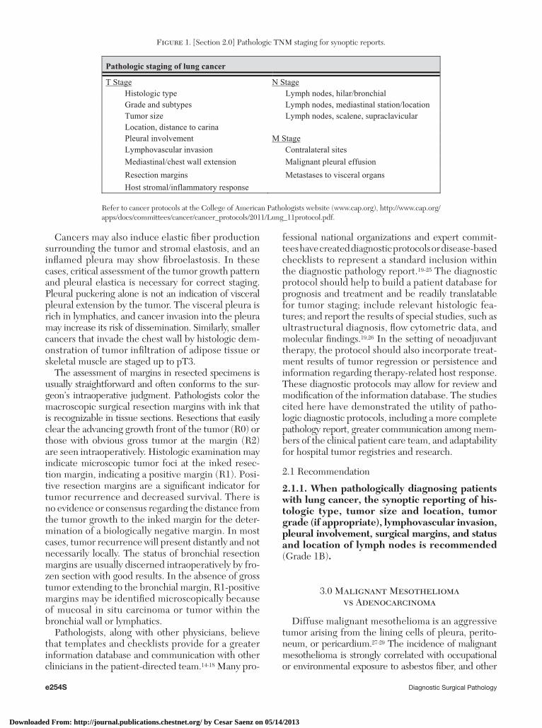
Macroscopic and microscopic examination provides diagnostic and prognostic staging information regard-ing the tumor size and location, permeation of the visceral pleura, presence of lymphovascular and perineu-ral invasion, and spread to hilar and mediastinal lymph nodes. 9-12 Mediastinal lymph nodes should be desig-nated according to their surgical station, as defi ned by the surgeon ( Fig 1 ). Histologic assessment of non-tumorous areas of lung may reveal underlying patho-logic conditions, such as smoking-related pathologic changes; pneumoconioses; parenchymal scarring; and secondary effects of the tumor, such as obstructive pneumonia. Additional studies, such as immunohis-tochemistry, in situ hybridization, and molecular bio-logic techniques, may amplify the diagnostic precision and supply prognostic information for therapeutic management. Routine histologic assessment and spe-cial diagnostic studies may be seen as a multiparam-eter system that provides increasingly prognostic and predictive information of tumor biology and clinical behavior. The categorical TNM staging of lung can-cers provides prognostic stratifi cation, criteria for patient inclusion and exclusion in protocol studies and treatment and management subgroups, and improved communication among members of the multidisci-plinary team.
Certain important issues in pathologic staging are worthy of consideration. The size of the tumor, when surrounded by lung, should be measured in the unfi xed state because formalin fi xation tends to cause a slight shrinkage of the tumor size. Tumors that are , 1 cm may also be measured microscopically, and the mac-roscopic and microscopic sizes may be presented in the synoptic report. Although the size of the tumor is a major factor in the T stage , smaller tumors that penetrate the visceral pleura (pT2) or extend into the parietal pleura and invade the chest wall (pT3) are upstaged despite their size . For cancers that appear to approach the visceral pleura, elastic tissue histochemical staining (van Gieson stain) will readily identify the wavy elastic fi bers of the pleura and dem-onstrate interruption or permeation of the fi bers by infi ltrating tumor. 8,13
non-small cell carcinomas, particularly the separation of squamous cell carcinomas from adenocarcinomas, is emphasized. The article also discusses the separa-tion of primary cancers from metastases from other organ sites. There has been considerable information regarding molecular aspects of lung cancer and the targeted therapy of particular pathways and their receptors or enzymes; these topics have been chan-neled into a separate article by Nana-Sinkam and Powell, “Molecular Biology of Lung Cancer,” 4 in the ACCP Lung Cancer Guidelines.
1.0 Methods
A systematic search of the medical and scientifi c literature using Embase, MEDLINE, and Cochrane Library search engines was performed for the years 1990 to 2011, and results were lim-ited to literature on humans and articles written in English. The search was performed as a review and update of the “Diagnostic Surgical Pathology of Lung Cancer: ACCP Evidence-Based Clin-ical Practice Guidelines (2nd Edition).” 3
The searches were performed by an ACCP methodologist, and the authors supplemented these searches with a review of refer-ences from relevant reviews and other pertinent literature. 1 The searches were performed to respond to the following population, intervention, comparison, and outcome questions:
1. Among patients with lung cancer, what pathologic fi ndings should be reported?
2. Among pleural-based malignancies, what approach and tests should be performed for diagnostic accuracy?
3. Among lung cancers, what approach and tests should be performed to distinguish small cell and non-small cell carcinoma?
4. Among glandular malignancies, what diagnostic criteria should be performed to separate AIS, MIA, and invasive adenocarcinoma?
5. Among non-small cell cancers, what approach and tests should be performed to distinguish squamous cell carcinoma from adenocarcinoma?
6. Among cancers of the lung, what approach and tests should be performed to distinguish primary vs metastatic cancers.
The recommendations in this article were graded by a standard-ized method and critically assessed and reviewed by the entire lung cancer panel, the Thoracic Oncology NetWork review com-mittee, the Guidelines Oversight Committee, selected Editorial Board members of CHEST , and the Board of Regents of the ACCP.
2.0 Pathologic Staging
Tumors within the lung represent a frequent diag-nostic challenge and include both primary and meta-static neoplasms. Overwhelmingly, primary malignant lung tumors are carcinomas, namely epithelial neo-plasms, and have historically been divided into small cell and non-small cell carcinomas. 5,6
Non-small cell carcinomas may be further sepa-rated into squamous cell carcinomas with keratin expression, adenocarcinomas with glandular expres-sion, and large cell carcinomas without distinctive cellular features. The classifi cation of other carcinomas,
Downloaded From: http://journal.publications.chestnet.org/ by Cesar Saenz on 05/14/2013
e254S Diagnostic Surgical Pathology
fessional national organizations and expert commit-tees have created diagnostic protocols or disease-based checklists to represent a standard inclusion within the diagnostic pathology report. 19-25 The diagnostic protocol should help to build a patient database for prognosis and treatment and be readily translatable for tumor staging; include relevant histologic fea-tures; and report the results of special studies, such as ultrastructural diagnosis, fl ow cytometric data, and molecular fi ndings. 19,26 In the setting of neoadjuvant therapy, the protocol should also incorporate treat-ment results of tumor regression or persistence and information regarding therapy-related host response. These diagnostic protocols may allow for review and modifi cation of the information database. The studies cited here have demonstrated the utility of patho-logic diagnostic protocols, including a more complete pathology report, greater communication among mem-bers of the clinical patient care team, and adaptability for hospital tumor registries and research .
2.1 Recommendation
2.1.1. When pathologically diagnosing patients with lung cancer, the synoptic reporting of his-tologic type, tumor size and location, tumor grade (if appropriate), lymphovascular invasion, pleural involvement, surgical margins, and status and location of lymph nodes is recommended (Grade 1B) .
3.0 Malignant Mesothelioma vs Adenocarcinoma
Diffuse malignant mesothelioma is an aggressive tumor arising from the lining cells of pleura, perito-neum, or pericardium. 27-29 The incidence of malignant mesothelioma is strongly correlated with occupational or environmental exposure to asbestos fi ber, and other
Cancers may also induce elastic fi ber production surrounding the tumor and stromal elastosis, and an infl amed pleura may show fi broelastosis. In these cases, critical assessment of the tumor growth pattern and pleural elastica is necessary for correct staging. Pleural puckering alone is not an indication of visceral pleural extension by the tumor. The visceral pleura is rich in lymphatics, and cancer invasion into the pleura may increase its risk of dissemination. Similarly, smaller cancers that invade the chest wall by histologic dem-onstration of tumor infi ltration of adipose tissue or skeletal muscle are staged up to pT3.
The assessment of margins in resected specimens is usually straightforward and often conforms to the sur-geon’s intraoperative judgment. Pathologists color the macroscopic surgical resection margins with ink that is recognizable in tissue sections. Resections that easily clear the advancing growth front of the tumor (R0) or those with obvious gross tumor at the margin (R2) are seen intraoperatively. Histologic examination may indicate microscopic tumor foci at the inked resec-tion margin, indicating a positive margin (R1). Posi-tive resection margins are a signifi cant indicator for tumor recurrence and decreased survival. There is no evidence or consensus regarding the distance from the tumor growth to the inked margin for the deter-mination of a biologically negative margin. In most cases, tumor recurrence will present distantly and not necessarily locally. The status of bronchial resection margins are usually discerned intraoperatively by fro-zen section with good results. In the absence of gross tumor extending to the bronchial margin, R1-positive margins may be identifi ed microscopically because of mucosal in situ carcinoma or tumor within the bronchial wall or lymphatics.
Pathologists, along with other physicians, believe that templates and checklists provide for a greater information database and communication with other clinicians in the patient-directed team. 14-18 Many pro-
Figure 1. [Section 2.0] Pathologic TNM staging for synoptic reports.
Refer to cancer protocols at the College of American Pathologists website (www.cap.org), http://www.cap.org/apps/docs/committees/cancer/cancer_protocols/2011/Lung_11protocol.pdf.
Downloaded From: http://journal.publications.chestnet.org/ by Cesar Saenz on 05/14/2013
journal.publications.chestnet.org CHEST / 143 / 5 / MAY 2013 SUPPLEMENT e255S
toid type, appears as a mesenchymal spindle cell malignancy with features resembling a fi brosarcoma.
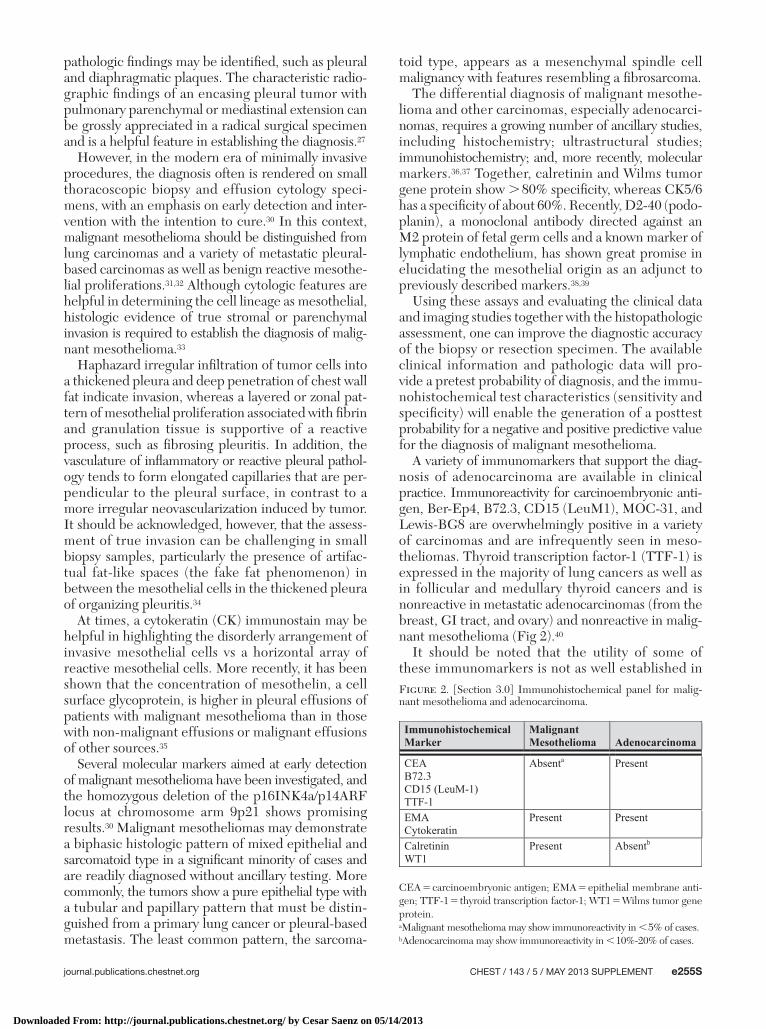
The differential diagnosis of malignant mesothe-lioma and other carcinomas, especially adenocarci-nomas, requires a growing number of ancillary studies, includ ing histochemistry; ultrastructural studies; immunohistochemistry; and, more recently, molecular markers. 36,37 Together, calretinin and Wilms tumor gene protein show . 80% specifi city, whereas CK5/6 has a specifi city of about 60%. Recently, D2-40 (podo-planin), a monoclonal antibody directed against an M2 protein of fetal germ cells and a known marker of lymphatic endothelium, has shown great promise in elucidating the mesothelial origin as an adjunct to previously described markers. 38,39
Using these assays and evaluating the clinical data and imaging studies together with the histopathologic assessment, one can improve the diagnostic accuracy of the biopsy or resection specimen. The available clinical information and pathologic data will pro-vide a pretest probability of diagnosis, and the immu-nohistochemical test characteristics (sensitivity and specifi city) will enable the generation of a posttest probability for a negative and positive predictive value for the diagnosis of malignant mesothelioma.
A variety of immunomarkers that support the diag-nosis of adenocarcinoma are available in clinical practice. Immunoreactivity for carcinoembryonic anti-gen, Ber-Ep4, B72.3, CD15 (LeuM1), MOC-31, and Lewis-BG8 are overwhelmingly positive in a variety of carcinomas and are infrequently seen in meso-theliomas. Thyroid transcription factor-1 (TTF-1) is expressed in the majority of lung cancers as well as in follicular and medullary thyroid cancers and is nonreactive in metastatic adenocarcinomas (from the breast, GI tract, and ovary) and nonreactive in malig-nant mesothelioma ( Fig 2 ). 40
It should be noted that the utility of some of these immunomarkers is not as well established in
pathologic fi ndings may be identifi ed, such as pleural and diaphragmatic plaques. The characteristic radio-graphic fi ndings of an encasing pleural tumor with pulmonary parenchymal or mediastinal extension can be grossly appreciated in a radical surgical specimen and is a helpful feature in establishing the diagnosis. 27
However, in the modern era of minimally invasive procedures, the diagnosis often is rendered on small thoracoscopic biopsy and effusion cytology speci-mens, with an emphasis on early detection and inter-vention with the intention to cure. 30 In this context, malignant mesothelioma should be distinguished from lung carcinomas and a variety of metastatic pleural-based carcinomas as well as benign reactive mesothe-lial proliferations. 31,32 Although cytologic features are helpful in determining the cell lineage as mesothelial, histologic evidence of true stromal or parenchymal invasion is required to establish the diagnosis of malig-nant mesothelioma. 33
Haphazard irregular infi ltration of tumor cells into a thickened pleura and deep penetration of chest wall fat indicate invasion, whereas a layered or zonal pat-tern of mesothelial proliferation associated with fi brin and granulation tissue is supportive of a reactive process, such as fi brosing pleuritis. In addition, the vasculature of infl ammatory or reactive pleural pathol-ogy tends to form elongated capillaries that are per-pendicular to the pleural surface, in contrast to a more irregular neovascularization induced by tumor. It should be acknowledged, however, that the assess-ment of true invasion can be challenging in small biopsy samples, particularly the presence of artifac-tual fat-like spaces (the fake fat phenomenon) in between the mesothelial cells in the thickened pleura of organizing pleuritis. 34
At times, a cytokeratin (CK) immunostain may be helpful in highlighting the disorderly arrangement of invasive mesothelial cells vs a horizontal array of reactive mesothelial cells. More recently, it has been shown that the concentration of mesothelin, a cell surface glycoprotein, is higher in pleural effusions of patients with malignant mesothelioma than in those with non-malignant effusions or malignant effusions of other sources. 35
Several molecular markers aimed at early detection of malignant mesothelioma have been investigated, and the homozygous deletion of the p16INK4a/p14ARF locus at chromosome arm 9p21 shows promising results. 30 Malignant mesotheliomas may demonstrate a biphasic histologic pattern of mixed epithelial and sarcomatoid type in a signifi cant minority of cases and are readily diagnosed without ancillary testing. More commonly, the tumors show a pure epithelial type with a tubular and papillary pattern that must be distin-guished from a primary lung cancer or pleural-based metastasis. The least common pattern, the sarcoma-
Figure 2. [Section 3.0] Immunohistochemical panel for malig-nant mesothelioma and adenocarcinoma.
CEA 5 carcinoembryonic antigen; EMA 5 epithelial membrane anti-gen; TTF-1 5 thyroid transcription factor-1; WT1 5 Wilms tumor gene protein.aMalignant mesothelioma may show immunoreactivity in , 5% of cases.bAdenocarcinoma may show immunoreactivity in , 10%-20% of cases.
Downloaded From: http://journal.publications.chestnet.org/ by Cesar Saenz on 05/14/2013
e256S Diagnostic Surgical Pathology
per year in the United States. 5 Almost all patients are heavy smokers, and the majority present with a peri-hilar mass with subsequent peribronchial compres-sion and obstruction. Nearly all patients present in advanced stages with disseminated disease, and the diagnosis relies primarily on small transbronchial biopsy samples or cytologic material. Despite the limitation of small sample size, a diagnosis can be determined by morphologic examination in the majority of cases. At times, cytologic preparations offer better preservation of cellular details, a key feature in the diagnosis. SCLCs are high-grade, mitotically active carcinomas with exten-sive necrosis and nuclear molding and chromatic baso-philic smearing. As the name implies, the cells are small in size, generally two to three times the size of small lymphocytes. Key morphologic features of SCLCs include scant cytoplasm, high nuclear-to-cytoplasmic ratio, nuclear molding, fi nely granular chromatin, and absent or inconspicuous nucleoli. Crush artifact and perivascular basophilic condensation (Azzopardi effect) often are seen. Architecturally, the tumor grows in large sheets and may be associated with vague organoid nesting, a ribbon-like pattern, and rosettes. The interobserver agreement is . 95% when these criteria are satisfi ed. Although the mor-phologic criteria often are diagnostic, in certain prob-lematic cases, immunohistochemistry is of value in the differential diagnosis. 37,47 TTF-1, although expressed in the majority of SCLCs, cannot be used as evidence of pulmonary origin because it is also expressed in small cell carcinomas of extrapulmonary sites. 48
In most cases, differentiating SCLC from non-small cell lung cancer (NSCLC) is achieved by routine mor-phologic examination. The tumor cells of NSCLCs generally are larger with a moderate amount of cyto-plasm, vesicular or coarse chromatin pattern, and prominent nucleoli. Nuclear molding and smearing are not usually seen. Glandular or squamous dif-ferentiation, either morphologically or by specifi c immunoreactivity to napsin A or p63, respectively, will aid in the diagnosis of NSCLC. One should also keep in mind that a small percentage of SCLCs belong to the category of combined SCLC, where at least 10% of the tumor shows morphologic features of a large cell carcinoma and rarely adenocarcinoma or squamous cell carcinoma. This distinction is particu-larly problematic in small tissue samples. Small cell carcinomas may be distinguished from non-Hodgkin lymphoma by identifying immunoreactive TTF-1 and neuroendocrine markers in SCLC and their absence combined with lymphoid immunophenotyping in lymphoma. 49
SCLCs may show overlapping morphologic fea-tures with a spectrum of pulmonary neuroendocrine tumors with diverse epidemiologic associations, bio-logic behavior, and survival rates. The high-grade, large
differentiating mesotheliomas from nonpulmonary ade-nocarcinomas (peritoneal and ovarian serous carcinomas) and other types of carcinomas, including squamous cell carcinomas. Similarly, the diagnosis of sarcoma-toid and desmoplastic mesotheliomas remains chal-lenging because the majority of these tumors show negative staining with mesothelial markers. Although a positive CK AE1/AE3 stain may be useful in this set-ting, it should be interpreted with caution and in the context of clinical and radiographic fi ndings because sarcomatoid carcinomas and synovial sarcoma involv-ing the pleural cavity show similar immunophenotypic profi les.
Ultrastructural analysis by transmission electron microscopy, although not being used as frequently in clinical practice, offers diagnostic clues to the meso-thelial vs epithelial nature of the malignant process. Mesothelial cells have numerous surface microvilli that are long and slender without associated core rootlets, fi laments, or terminal bars. Conversely, epithelial cells have few short and blunted microvilli clustering along the luminal border. 41-43
3.1 Recommendation
3.1.1. In individuals with pleural-based tumors, a designated limited panel of histochemical and immunohistochemical assays or ultrastructural analysis is recommended to distinguish between pleural adenocarcinoma and malignant meso-thelioma in order to increase diagnostic accu-racy (Grade 1B) .
4.0 Small Cell Vs Non-small Cell Carcinoma
Bronchogenic carcinomas of the lung have histori-cally been divided into small cell and non-small cell carcinomas. 5,44,45 The small cell carcinomas are high grade and mitotically active with neuroendocrine dif-ferentiation and usually present with thoracic and extrathoracic dissemination. 46 The cells are derived from endogenous, endodermally derived neuroen-docrine cells and are characterized by dense-core neurosecretory granules identifi ed ultrastructurally. The neurosecretory granules contain bioactive amines and peptides and are the source of paraneoplastic syndromes. With all the recent advances in the sub-typing of lung cancer, the distinction between small cell and non-small carcinoma remains signifi cant. This separation underscores the major differences in their characteristic clinical presentation, behavior, and prog-nosis as well as in their therapeutic approaches and response.
Small cell lung cancer (SCLC) comprises 14% of all lung cancers, with . 30,000 new cases diagnosed
Downloaded From: http://journal.publications.chestnet.org/ by Cesar Saenz on 05/14/2013
journal.publications.chestnet.org CHEST / 143 / 5 / MAY 2013 SUPPLEMENT e257S
nar cells with resemblance to reactive type 2 pneu-mocytes and have been termed “atypical alveolar hyperplasia” (AAH), “bronchioloalveolar adenoma,” and “alveolar epithelial hyperplasia.” The lesions are easily recognized microscopically, and their periph-eral growth pattern may blend with adjacent normal lung. The alveolar septae may be thin or thickened, and the atypical cells are noninvasive without mitotic activity. Although AAH tends to show variable cyto-logic atypia, when the lesion appears . 5 mm, shows continuous and uniform cytologic atypia with surface (or lepidic) septal growth, and becomes well demar-cated from the adjacent normal lung, the term “bron-chioloalveolar carcinoma” (BAC) had historically been used. The current change in nomenclature designates pure BAC as AIS. 52,53,55 It was judged by a panel of experts that the diagnosis of BAC was rendered for a variety of lesions dominated by lepidic tumor growth with and without stromal invasion and that the extent of invasion was not quantifi ed. 52,53,55
Those lesions with a predominant lepidic growth pat-tern and central stromal invasion (defi ned next) were designated as MIAs. Histologic distinction between AAH and AIS may be diffi cult, and immunohisto-chemical assays are not necessarily helpful. AIS may appear radiographically as ground glass opacity with-out central densities, and radiologic evaluation of AAH is often diffi cult.
AIS (formerly BAC) can only be diagnosed when the entire lesion is evaluated histologically and there is no invasive component, no lymphovascular per-meation, no pleural extension, and no nodal spread. 56 When individuals have these solitary noninvasive tumors excised, they are associated with an excellent 5- and 10-year survival. 55,57-61
Tumor cells in the lepidic growth pattern may com-prise columnar mucinous goblet-like cells or hob-nailed nonmucinous serous-like cells. The mucinous pattern has an immunophenotypic profi le resembling colonic differentiation with CK7-negative, CK20-positive, and TTF-1-negative phenotype. In contrast, the non-mucinous type, comprising type 2 alveolar pneu-mocytes or bronchiolar cell differentiation, shows an immunophenotype of CK7 and TTF-1 positivity and CK20 negativity. AIS may grow along alveolar sur-faces or exhibit airway dissemination. Consequently, the carcinoma may appear as a single peripheral mass, as multicentric tumor nodules, or as a pneumonic pattern.
Some adenocarcinomas demonstrate a central core of fi broblastic proliferation with cancer invasion that is in the range of few millimeters. 55,57-61 The image on CT scan may show a central density surrounded by a ground glass pattern. These tumors have also been associated with an excellent patient survival when com-pletely excised. These mixed-type adenocarcinomas
cell neuroendocrine carcinoma is cytologically similar to other NSCLCs but shows more classic morphologic patterns of neuroendocrine architecture, such as organoid, trabecular, or palisading, and immunore-activity to at least one neuroendocrine marker. The intermediate-grade atypical carcinoid and the low-grade typical carcinoid tend to show relatively bland and uniform cytologic characteristics with moderate granular cytoplasm and fi nely granular chromatin and are histologically low grade, with only punctate necro-sis and minimally elevated mitosis (2-10 per 2 mm 2 ) seen in atypical carcinoids. In small biopsy samples, a Ki-67 proliferation index may offer a more accu-rate distinction between SCLCs with a high index of . 50% to 70% and carcinoids with a low index of 5% to 15%. 50,51
4.1 Recommendation
4.1.1 . In individuals with parenchymal-based tumors, distinguishing between small cell carci-noma and non-small cell carcinoma of the lung is recommended. For challenging cases, a diagnos-tic panel of immunohistochemical assays or ultra-structural analy sis is recommended to increase the diagnostic accuracy (Grade 1B) .
5.0 Adenocarcinoma, AIS (Bronchioloalveolar Carcinoma), and MIAs
The invasive adenocarcinomas are malignant tumors characterized by glandular or acinar differentiation, papillary structures, and cytoplasmic mucin vacu-oles. 5,52,53 Histologic subclassifi cation of these tumors include acinar, papillary, micropapillary, and solid types, although frequently, the carcinomas comprise composite subtypes. Histologic grading of adenocar-cinomas appears to have prognostic information for the likelihood of nodal metastases and overall sur-vival. Grading has been performed on the basis of architectural and cytologic features or histologic sub-typing, with acinar and papillary being low grade and solid adenocarcinoma being high grade. 54
Historically, adenocarcinomas were designated as scar carcinomas arising peripherally in a fi broblastic background. Some of these cancers have developed within inflammatory scars; however, it is currently understood that adenocarcinomas promote a host stromal desmoplastic response similar to adenocarci-nomas of the breast and pancreas and, consequently, induce the stromal background rather than arise within it. Invasive adenocarcinoma resection specimens may demonstrate, in a minority of cases, small separate peripheral lesions that can be multiple, small ( , 5 mm), and remote from the main tumor. 52,53 These lesions are characterized by atypical cuboidal to low colum-
Downloaded From: http://journal.publications.chestnet.org/ by Cesar Saenz on 05/14/2013
e258S Diagnostic Surgical Pathology
associated with the two most common subtypes of NSCLC, namely adenocarcinoma and squamous cell carcinoma, have played a critical role in guiding the individualized targeted therapy. Particular molecular targets tend to be preferentially associated with ade-nocarcinoma rather than squamous cell carcinoma, and certain chemotherapeutic agents show either favorable outcomes (adenocarcinoma) or greater side effects (squamous cell carcinoma) between these histo-logic types. The clinical demand for precise classifi -cation of adenocarcinoma vs squamous cell carcinoma is especially heightened in patients who present with advanced staged disease, where cytologic material and small tissue biopsy samples are the basis of select-ing the most effective chemotherapy regimen while avoiding life-threatening side effects.
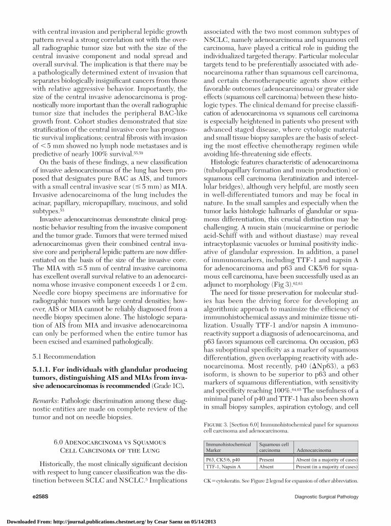
Histologic features characteristic of adenocarcinoma (tubulopapillary formation and mucin production) or squamous cell carcinoma (keratinization and intercel-lular bridges), although very helpful, are mostly seen in well-differentiated tumors and may be focal in nature. In the small samples and especially when the tumor lacks histologic hallmarks of glandular or squa-mous differentiation, this crucial distinction may be challenging. A mucin stain (mucicarmine or periodic acid-Schiff with and without diastase) may reveal intracytoplasmic vacuoles or luminal positivity indic-ative of glandular expression. In addition, a panel of immunomarkers, including TTF-1 and napsin A for adenocarcinoma and p63 and CK5/6 for squa-mous cell carcinoma, have been successfully used as an adjunct to morphology ( Fig 3 ). 62,63
The need for tissue preservation for molecular stud-ies has been the driving force for developing an algorithmic approach to maximize the effi ciency of immunohistochemical assays and minimize tissue uti-lization. Usually TTF-1 and/or napsin A immuno-reactivity support a diagnosis of adenocarcinoma, and p63 favors squamous cell carcinoma. On occasion, p63 has suboptimal specifi city as a marker of squamous differentiation, given overlapping reactivity with ade-nocarcinoma. Most recently, p40 ( D Np63), a p63 isoform, is shown to be superior to p63 and other markers of squamous differentiation, with sensitivity and specifi city reaching 100%. 64,65 The usefulness of a minimal panel of p40 and TTF-1 has also been shown in small biopsy samples, aspiration cytology, and cell
with central invasion and peripheral lepidic growth pattern reveal a strong correlation not with the over-all radiographic tumor size but with the size of the central invasive component and nodal spread and overall survival. The implication is that there may be a pathologically determined extent of invasion that separates biologically insignifi cant cancers from those with relative aggressive behavior. Importantly, the size of the central invasive adenocarcinoma is prog-nostically more important than the overall radiographic tumor size that includes the peripheral BAC-like growth front. Cohort studies demonstrated that size stratifi cation of the central invasive core has prognos-tic survival implications; central fi brosis with invasion of , 5 mm showed no lymph node metastases and is predictive of nearly 100% survival. 55,59
On the basis of these fi ndings, a new classifi cation of invasive adenocarcinomas of the lung has been pro-posed that designates pure BAC as AIS, and tumors with a small central invasive scar ( � 5 mm) as MIA. Invasive adenocarcinoma of the lung includes the acinar, papillary, micropapillary, mucinous, and solid subtypes. 53
Invasive adenocarcinomas demonstrate clinical prog-nostic behavior resulting from the invasive component and the tumor grade. Tumors that were termed mixed adenocarcinomas given their combined central inva-sive core and peripheral lepidic pattern are now differ-entiated on the basis of the size of the invasive core. The MIA with � 5 mm of central invasive carcinoma has excellent overall survival relative to an adenocarci-noma whose invasive component exceeds 1 or 2 cm. Needle core biopsy specimens are informative for radiographic tumors with large central densities; how-ever, AIS or MIA cannot be reliably diagnosed from a needle biopsy specimen alone. The histologic separa-tion of AIS from MIA and invasive adenocarcinoma can only be performed when the entire tumor has been excised and examined pathologically.
5.1 Recommendation
5.1.1. For individuals with glandular producing tumors, distinguishing AIS and MIAs from inva-sive adenocarcinomas is recommended (Grade 1C) .
Remarks : Pathologic discrimination among these diag-nostic entities are made on complete review of the tumor and not on needle biopsies.
6.0 Adenocarcinoma vs Squamous Cell Carcinoma of the Lung
Historically, the most clinically signifi cant decision with respect to lung cancer classifi cation was the dis-tinction between SCLC and NSCLC. 5 Implications
Figure 3. [Section 6.0] Immunohistochemical panel for squamous cell carcinoma and adenocarcinoma.
CK 5 cytokeratin. See Figure 2 legend for expansion of other abbreviation.
Downloaded From: http://journal.publications.chestnet.org/ by Cesar Saenz on 05/14/2013
journal.publications.chestnet.org CHEST / 143 / 5 / MAY 2013 SUPPLEMENT e259S
cinoma, clear cell (renal) carcinoma, and hepatocellular carcinoma, may have their own unique histopathologic features. These tumors may be suggested as meta-static based on their cytologic and histologic patterns, and their primary diagnosis should be pursued. Over-whelmingly, lymphomas and sarcomas in the lung are metastatic tumors.
Immunohistochemical analysis has greatly assisted the surgical pathologist in the differential diagnosis of primary vs metastatic carcinoma and has increased the ability to identify those metastatic tumors of unknown origin. 68-72 The currently preferred immuno-histochemical marker for the identifi cation of primary lung carcinoma is TTF-1. This factor is selectively expressed embryologically in the thyroid follicular cells and in airway and parenchymal cells of the lung. Papillary, follicular, and medullary carcinomas of the thyroid show strong immunoreactivity for TTF-1. In addition, primary lung cancers of adenocarcinoma and small cell carcinoma show diffuse strong immu-noreactivity. Squamous cell carcinoma of the lung is nonimmunoreactive for TTF-1. Adenocarcinomas from other sites, such as the GI tract and the breast, are nonreactive for TTF-1. Micropapillary adenocar-cinomas may be present in a variety of visceral sites, and their differentiation may be ascertained by sev-eral immunohistochemical panels. 73
Although the carcinomas of the lung are immuno-reactive for a set of cellular CKs, the specifi c CK com-ponents from the large family of these cytoplasmic fi laments are somewhat unique to each tumor type. By identifying the immunoreactivity to CK7 and CK20, additional information may provide support for the differential diagnosis of primary vs metastatic carcinomas. 72,74
Most helpful in the analysis is the differential between primary lung adenocarcinoma and metastatic adenocarcinoma of colorectal origin. These carcinomas may appear identical histologically yet have oppo-site immunohistochemical profi les. In the majority of cases, primary lung adenocarcinomas are TTF-1 positive, CK7 positive, and CK20 negative; colo-rectal adenocarcinomas have the opposite fi ndings of TTF-1- and CK7-negative and CK20-positive immu-noreactivity. Colorectal carcinomas are also CDX-2 immunoreactive; primary lung adenocarcinomas are nonreactive. 75,76
The diagnosis of tumors of unknown origin may also be elucidated by their selective expression of CK7 and CK20 immunohistochemistry. Carcinomas immuno-reactive for CK7 and CK20 tend to be those from the urinary bladder. Carcinomas nonimmunoreactive for CK7 and CK20 may be metastases from the liver, kidney, and prostate. In addition, some tumors have specifi c immunoidentifying markers, such as prostate-specifi c antigen for prostate carcinoma, thyroglobulin
block samples. 65,66 The feasibility of molecular testing on cytologic material has also been addressed in sev-eral studies, with encouraging results. 62,67
6.1 Recommendation
6.1.1. In individuals with pathologically diagnosed NSCLC, additional discrimination between ade-nocarcinoma and squamous cell carcinoma, even on cytologic material or small tissue samples, is recommended (Grade 1B) .
Remark : The precise subclassifi cation is achieved in most cases by conventional histo- and cytomorphol-ogy. Immunohistochemical assays are recommended in cases where routine histopathologic differentiation is diffi cult to ascertain.
7.0 Primary vs Metastatic Lung Cancer
The distinction between primary lung cancers and metastatic lung cancers is made through clinical his-tory and patient presentation, radiographic and imag-ing techniques, and optimal specimen acquisition and evaluation. Primary tumors of the lung may present in a typical clinical, radiologic, and pathologic pattern. Although lung cancers may occur in any lobe, either peripherally or centrally, squamous cell carcinomas tend to present as near-hilar masses and are associated with bronchial metaplasia and squamous dysplasia. The cases are found in cigarette smokers, and radio-logic imaging of COPD and histologic features of chronic bronchitis and emphysematous changes are seen. Adenocarcinomas tend to present in a peripheral location, show retraction or invasion of the visceral pleura, and are associated with tumor desmoplasia or scar. Large cell and small cell carcinomas of the lung also have their typical settings and presentations. Meta-static tumors from the epidemiologically most typical sites (breast, colon, and prostate) tend to show more expansile growth in the lung rather than infi ltrative growth, with adjacent lung retraction more typical of primary lung cancers.
The diffi cult differential diagnosis occurs with the identifi cation of a single metastatic site of adenocarci-noma or squamous cell carcinoma in the absence of a known primary carcinoma. Squamous cell carcinomas of the head and neck and adenocarcinomas of the GI tract may mimic primary lung cancers. Gross and microscopic features of the tumor may provide clues to its primary origin. Lung tumors tend to arise in bronchogenic squamous metaplasia and squamous dysplasia, show infi ltrative rather than pushing growth margins, and retract rather than bulge the visceral pleura. Adenocarcinomas from other visceral sites, such as endometrial carcinoma, papillary thyroid car-
Downloaded From: http://journal.publications.chestnet.org/ by Cesar Saenz on 05/14/2013
e260S Diagnostic Surgical Pathology
Dr Schwartz: contributed to the literature search and writing and editing of the manuscript. Dr Rezaei: contributed to the literature search and writing and editing of the manuscript. Financial/nonfi nancial disclosures: The authors have reported to CHEST that no potential confl icts of interest exist with any companies/organizations whose products or services may be dis-cussed in this article . Role of Sponsors: The American College of Chest Physicians was solely responsible for the development of these guidelines. The remaining supporters played no role in the development process. External supporting organizations cannot recommend panelists or topics, nor are they allowed prepublication access to the manuscripts and recommendations. Further details on the Confl ict of Interest Policy are available online at http://chestnet.org. Endorsements: This guideline is endorsed by the European Society of Thoracic Surgeons, Oncology Nursing Society, Ameri-can Association for Bronchology and Interventional Pulmonology, and the Society of Thoracic Surgeons.
References 1 . Lewis SZ , Diekemper R , Addrizzo-Harris DJ . Methodology
for development of guidelines for lung cancer: diagnosis and management of lung cancer, 3rd ed: American College of Chest Physicians evidence-based clinical practice guidelines . Chest . 2013 ;143(5)(suppl):41S-50S.
2 . Cagle PT , Allen TC , Dacic S , et al . Revolution in lung cancer: new challenges for the surgical pathologist . Arch Pathol Lab Med . 2011 ; 135 ( 1 ): 110 - 116 .
3 . Schwartz AM , Henson DE ; American College of Chest Phy-sicians . Diagnostic surgical pathology in lung cancer: ACCP evidence-based clinical practice guidelines (2nd edition) . Chest . 2007 ; 132 ( suppl 3 ): 78S - 93S .
4 . Nana-Sinkam SP , Powell CA . Molecular biology of lung cancer: diagnosis and management of lung cancer, 3rd ed: American College of Chest Physicians evidence-based clinical practice guidelines. Chest . 2013 ;143(5)(suppl):e30S-e39S.
5 . Travis WD . Pathology of lung cancer . Clin Chest Med . 2002 ; 23 ( 1 ): 65 - 81 .
6 . Franklin WA . Diagnosis of lung cancer: pathology of inva-sive and preinvasive neoplasia . Chest . 2000 ; 117 ( 4 )( suppl 1 ): 80S - 89S .
7 . Chamberlain DW , Wenckebach GF , Alexander F , Fraser RS , Kolin A , Newman T . Pathological examination and the report-ing of lung cancer specimens . Clin Lung Cancer . 2000 ; 1 ( 4 ): 261 - 268 .
8 . Marchevsky AM . Problems in pathologic staging of lung can-cer . Arch Pathol Lab Med . 2006 ; 130 ( 3 ): 292 - 302 .
9 . Detterbeck FC , Boffa DJ , Tanoue LT . The new lung cancer staging system . Chest . 2009 ; 136 ( 1 ): 260 - 271 .
10 . Tanoue LT , Detterbeck FC . New TNM classifi cation for non-small-cell lung cancer . Expert Rev Anticancer Ther . 2009 ; 9 ( 4 ): 413 - 423 .
11 . Buccheri G , Ferrigno D . Prognostic value of stage grouping and TNM descriptors in lung cancer . Chest . 2000 ; 117 ( 5 ): 1247 - 1255 .
12 . Flieder DB . Commonly encountered diffi culties in pathologic staging of lung cancer . Arch Pathol Lab Med . 2007 ; 131 ( 7 ): 1016 - 1026 .
13 . Butnor KJ , Vollmer RT , Blaszyk H , Glatz K . Interobserver agreement on what constitutes visceral pleural invasion by non-small cell lung carcinoma: an internet-based assessment of international current practices . Am J Clin Pathol . 2007 ; 128 ( 4 ): 638 - 647 .
14 . Leslie KO , Rosai J . Standardization of the surgical pathology report: formats, templates, and synoptic reports . Semin Diagn Pathol . 1994 ; 11 ( 4 ): 253 - 257 .
for thyroid carcinoma, a -fetoprotein and human cho-rionic gonadotropin for certain germ cell tumors, Hep-1 for hepatocellular carcinoma, estrogen receptor for some breast and gynecologic cancers, and MART-1 and Melan-A for malignant melanoma. Although there are abundant immunoreactive assays, the diagnostic approach should be focused by the clinical history, radiologic information, and histopathologic pattern.
7.1 Recommendation
7.1.1. For individuals with lung tumors whose differential includes primary lung carcinoma vs metastatic carcinoma, a directed panel of immunohistochemical assays is recommended to increase the diagnostic accuracy (Grade 1C) .
8.0 Conclusion
In a multidisciplinary approach to the treatment of patients with lung cancer, pathologic examination provides precise diagnosis, staging information, and histologic correlation with clinical behavior; manage-ment options; and biologic insights of the tumor. The surgical pathology report should contain a synoptic database to inform the clinical team about the multi-parameter aspect of the tumor diagnosis. The sur-gical pathologist should address, within a collaborative team effort, challenging diagnostic issues, such as differentiating pleural-based adenocarcinoma from malignant mesothelioma, separating small cell carcinoma from non-small cell carcinoma, discriminating between invasive adenocarcinoma and favorable subtypes of AIS and MIA, distinguishing between adenocarci-noma and squamous cell carcinoma, and identifying primary vs metastatic carcinomas. The pathologist should be provided with the clinical information and radiographic fi ndings and incorporate these data with the histopathologic evaluation. The pathologist should also collaborate with the clinical team to enhance specimen acquisition and testing so that optimal infor-mation can be obtained from the available tissue. Much information can be determined from routine histopathologic analysis; additional information will be gained from a limited panel of immunohistochem-ical reactivity. New molecular biologic pathways and prognostic factors will amplify the pathologic conclu-sions and provide avenues toward directed therapy of a tumor’s proliferative activity, invasiveness, angio-genesis, and metastatic potential.
Acknowledgments Author contributions: Dr Schwartz had full access to all of the data in the study and takes responsibility for the integrity of the data and the accuracy of the data analysis .
Downloaded From: http://journal.publications.chestnet.org/ by Cesar Saenz on 05/14/2013
journal.publications.chestnet.org CHEST / 143 / 5 / MAY 2013 SUPPLEMENT e261S
cells from malignant mesothelioma in cytologic effusions . Cancer Cytopathol . 2010 ; 118 ( 2 ): 90 - 96 .
33 . Churg A , Colby TV , Cagle P , et al . The separation of benign and malignant mesothelial proliferations . Am J Surg Pathol . 2000 ; 24 ( 9 ): 1183 - 1200 .
34 . Churg A , Cagle P , Colby TV , et al ; US-Canadian Mesothelioma Reference Panel . The fake fat phenomenon in organizing pleuritis: a source of confusion with desmoplastic malignant mesotheliomas . Am J Surg Pathol . 2011 ; 35 ( 12 ): 1823 - 1829 .
35 . Yamada S , Tabata C , Tabata R , Fukuoka K , Nakano T . Clinical signifi cance of pleural effusion mesothelin in malignant pleural mesothelioma . Clin Chem Lab Med . 2011 ; 49 ( 10 ): 1721 - 1726 .
36 . Klebe S , Nurminen M , Leigh J , Henderson DW . Diagnosis of epithelial mesothelioma using tree-based regression anal-ysis and a minimal panel of antibodies . Pathology . 2009 ; 41 ( 2 ): 140 - 148 .
37 . Mani H , Zander DS . Immunohistochemistry: applications to the evaluation of lung and pleural neoplasms: part 2 . Chest . 2012 ; 142 ( 5 ): 1324 - 1333 .
38 . Kao SC , Griggs K , Lee K , et al . Validation of a minimal panel of antibodies for the diagnosis of malignant pleural mesothe-lioma . Pathology . 2011 ; 43 ( 4 ): 313 - 317 .
39 . Hanna A , Pang Y , Bedrossian CW , Dejmek A , Michael CW . Podoplanin is a useful marker for identifying mesothelioma in malignant effusions . Diagn Cytopathol . 2010 ; 38 ( 4 ): 264 - 269 .
40. Marchevsky AM . Application of immunohistochemistry to the diagnosis of malignant mesothelioma. Arch Pathol Lab Med. 2008 ;132(3):397-401.
41 . Bedrossian CW , Bonsib S , Moran C . Differential diagnosis between mesothelioma and adenocarcinoma: a multimodal approach based on ultrastructure and immunocytochemistry . Semin Diagn Pathol . 1992 ; 9 ( 2 ): 124 - 140 .
42 . Dardick I , Al-Jabi M , McCaughey WT , Srigley JR , van Nostrand AW , Ritchie AC . Ultrastructure of poorly differ-entiated diffuse epithelial mesotheliomas . Ultrastruct Pathol . 1984 ; 7 ( 2-3 ): 151 - 160 .
43 . Sidhu GS . The ultrastructure of malignant epithelial neoplasms of the lung . Pathol Annu . 1982 ; 17 ( pt 1 ): 235 - 266 .
44 . Travis WD . Update on small cell carcinoma and its differ-entiation from squamous cell carcinoma and other non-small cell carcinomas. Mod Pathol . 2012 ; 25 ( suppl 1 ): S18 - S30 .
45. Dubinski W , Leighl NB , Tsao MS, Hwang DM . Ancillary test-ing in lung cancer diagnosis. Pulm Med . 2012 ;2012:249082.
46 . Nicholson SA , Beasley MB , Brambilla E , et al . Small cell lung carcinoma (SCLC): a clinicopathologic study of 100 cases with surgical specimens . Am J Surg Pathol . 2002 ; 26 ( 9 ): 1184 - 1197 .
47 . Sigel CS , Moreira AL , Travis WD , et al . Subtyping of non-small cell lung carcinoma: a comparison of small biopsy and cytology specimens . J Thorac Oncol . 2011 ; 6 ( 11 ): 1849 - 1856 .
48 . Agoff SN , Lamps LW , Philip AT , et al . Thyroid transcription factor-1 is expressed in extrapulmonary small cell carcinomas but not in other extrapulmonary neuroendocrine tumors. Mod Pathol . 2000 ; 13 ( 3 ): 238 - 242 .
49 . Johansson L . Histopathologic classifi cation of lung cancer: Relevance of cytokeratin and TTF-1 immunophenotyping . Ann Diagn Pathol . 2004 ; 8 ( 5 ): 259 - 267 .
50. Skov BG , Holm B , Erreboe A , Skov T, Mellemgaard A . ERCC1 and Ki67 in small cell lung carcinoma and other neu-roendocrine tumors of the lung: distribution and impact on survival. J Thorac Oncol . 2010 ;5(4):453-459.
51 . Thunnissen E , Kerr KM , Herth FJ , et al . The challenge of NSCLC diagnosis and predictive analysis on small samples. Practical approach of a working group . Lung Cancer . 2012 ;76(1):1-18.
52 . Travis WD , Brambilla E , Noguchi M , et al ; American Thoracic Society . International Association for the Study of Lung
15 . Markel SF , Hirsch SD . Synoptic surgical pathology reporting . Hum Pathol . 1991 ; 22 ( 8 ): 807 - 810 .
16 . Qu Z , Ninan S , Almosa A , Chang KG , Kuruvilla S , Nguyen N . Synoptic reporting in tumor pathology: advantages of a web-based system . Am J Clin Pathol . 2007 ; 127 ( 6 ): 898 - 903 .
17 . Srigley JR , McGowan T , Maclean A , et al . Standardized synop-tic cancer pathology reporting: a population-based approach . J Surg Oncol . 2009 ; 99 ( 8 ): 517 - 524 .
18 . Dworak O . Synoptic surgical pathology reporting . Hum Pathol . 1992 ; 23 ( 1 ): 85 - 86 .
19 . Zarbo RJ . Interinstitutional assessment of colorectal car-cinoma surgical pathology report adequacy. A College of American Pathologists Q-Probes study of practice patterns from 532 laboratories and 15,940 reports . Arch Pathol Lab Med . 1992 ; 116 ( 11 ): 1113 - 1119 .
20 . Austin R , Thompson B , Coory M , Walpole E , Francis G , Fritschi L . Histopathology reporting of breast cancer in Queensland: the impact on the quality of reporting as a result of the introduction of recommendations . Pathology . 2009 ; 41 ( 4 ): 361 - 365 .
21 . Beattie GC , McAdam TK , Elliott S , Sloan JM , Irwin ST . Improve-ment in quality of colorectal cancer pathology reporting with a standardized proforma—a comparative study . Colorectal Dis . 2003 ; 5 ( 6 ): 558 - 562 .
22 . Gill AJ , Johns AL , Eckstein R , et al ; New South Wales Pan-creatic Cancer Network (NSWPCN) . Synoptic reporting improves histopathological assessment of pancreatic resec-tion specimens . Pathology . 2009 ; 41 ( 2 ): 161 - 167 .
23 . Haydu LE , Holt PE , Karim RZ , et al . Quality of histopatho-logical reporting on melanoma and infl uence of use of a syn-optic template . Histopathology . 2010 ; 56 ( 6 ): 768 - 774 .
24 . Kang HP , Devine LJ , Piccoli AL , Seethala RR , Amin W , Parwani AV . Usefulness of a synoptic data tool for reporting of head and neck neoplasms based on the College of American Pathologists cancer checklists . Am J Clin Pathol . 2009 ; 132 ( 4 ): 521 - 530 .
25 . Karim RZ , van den Berg KS , Colman MH , McCarthy SW , Thompson JF , Scolyer RA . The advantage of using a syn-optic pathology report format for cutaneous melanoma . Histopathology . 2008 ; 52 ( 2 ): 130 - 138 .
26 . Idowu MO , Bekeris LG , Raab S , Ruby SG , Nakhleh RE . Adequacy of surgical pathology reporting of cancer: a College of American Pathologists Q-Probes study of 86 institutions . Arch Pathol Lab Med . 2010 ; 134 ( 7 ): 969 - 974 .
27 . Miller BH , Rosado-de-Christenson ML , Mason AC , Fleming MV , White CC , Krasna MJ . From the archives of the AFIP. Malig-nant pleural mesothelioma: radiologic-pathologic correlation . Radiographics . 1996 ; 16 ( 3 ): 613 - 644 .
28 . McCaughey WT , Colby TV , Battifora H , et al . Diagnosis of dif-fuse malignant mesothelioma: experience of a US/Canadian Mesothelioma Panel . Mod Pathol . 1991 ; 4 ( 3 ): 342 - 353 .
29 . Butnor KJ , Beasley MB , Kong F-M , et al . Protocol for the examination of specimens from patients with thymoma or thymic carcinoma. 2009. College of American Pathologists website. http://www.cap.org/apps/docs/committees/cancer/cancer_protocols/2012/Mesothelioma_12protocol.pdf. Accessed March 4, 2013 .
30 . Tsujimura T , Torii I , Sato A , et al . Pathological and molec-ular biological approaches to early mesothelioma . Int J Clin Oncol . 2012 ; 17 ( 1 ): 40 - 47 .
31 . Rakha EA , Patil S , Abdulla K , Abdulkader M , Chaudry Z , Soomro IN . The sensitivity of cytologic evaluation of pleu-ral fl uid in the diagnosis of malignant mesothelioma . Diagn Cytopathol . 2010 ; 38 ( 12 ): 874 - 879 .
32 . Hasteh F , Lin GY , Weidner N , Michael CW . The use of immunohistochemistry to distinguish reactive mesothelial
Downloaded From: http://journal.publications.chestnet.org/ by Cesar Saenz on 05/14/2013
e262S Diagnostic Surgical Pathology
Cancer/American Thoracic Society/European Respiratory Society: international multidisciplinary classifi cation of lung adenocarcinoma: executive summary . Proc Am Thorac Soc . 2011 ; 8 ( 5 ): 381 - 385 .
53 . Travis WD , Brambilla E , Noguchi M , et al . International Association for the Study of Lung Cancer/American Thoracic Society/European Respiratory Society international multi-disciplinary classifi cation of lung adenocarcinoma . J Thorac Oncol . 2011 ; 6 ( 2 ): 244 - 285 .
54 . Barletta JA , Yeap BY , Chirieac LR . Prognostic signifi cance of grading in lung adenocarcinoma . Cancer . 2010 ; 116 ( 3 ): 659 - 669 .
55 . Travis WD , Brambilla E , Van Schil P , et al . Paradigm shifts in lung cancer as defi ned in the new IASLC/ATS/ERS lung ade-nocarcinoma classifi cation . Eur Respir J . 2011 ; 38 ( 2 ): 239 - 243 .
56 . Borczuk AC . Assessment of invasion in lung adenocarcinoma classifi cation, including adenocarcinoma in situ and minimally invasive adenocarcinoma . Mod Pathol . 2012 ; 25 ( suppl 1 ): S1 - S10 .
57 . Noguchi M , Morikawa A , Kawasaki M , et al . Small adenocar-cinoma of the lung. Histologic characteristics and prognosis . Cancer . 1995 ; 75 ( 12 ): 2844 - 2852 .
58 . Van Schil PE , Asamura H , Rusch VW , et al . Surgical impli-cations of the new IASLC/ATS/ERS adenocarcinoma classi-fi cation . Eur Respir J . 2012 ; 39 ( 2 ): 478 - 486 .
59 . Yoshizawa A , Motoi N , Riely GJ , et al . Impact of proposed IASLC/ATS/ERS classifi cation of lung adenocarcinoma: prog-nostic subgroups and implications for further revision of stag-ing based on analysis of 514 stage I cases . Mod Pathol . 2011 ; 24 ( 5 ): 653 - 664 .
60 . Yokose T , Suzuki K , Nagai K , Nishiwaki Y , Sasaki S , Ochiai A . Favorable and unfavorable morphological prognostic factors in peripheral adenocarcinoma of the lung 3 cm or less in diameter . Lung Cancer . 2000 ; 29 ( 3 ): 179 - 188 .
61 . Suzuki K , Yokose T , Yoshida J , et al . Prognostic signifi cance of the size of central fi brosis in peripheral adenocarcinoma of the lung . Ann Thorac Surg . 2000 ; 69 ( 3 ): 893 - 897 .
62. Hasanovic A , Rekhtman N , Sigel CS, Moreira AL . Advances in fi ne needle aspiration cytology for the diagnosis of pulmo-nary carcinoma. Patholog Res Int . 2011 ;2011:897292.
63 . Rekhtman N , Ang DC , Sima CS , Travis WD , Moreira AL . Immunohistochemical algorithm for differentiation of lung adenocarcinoma and squamous cell carcinoma based on large series of whole-tissue sections with validation in small speci-mens . Mod Pathol . 2011 ; 24 ( 10 ): 1348 - 1359 .
64. Bishop JA , Teruya-Feldstein J , Westra WH , Pelosi G , Travis WD, Rekhtman N . p40 ( D Np63) is superior to p63 for the diagnosis of pulmonary squamous cell carcinoma. Mod Pathol . 2012 ;25(3):405-415.
65. Pelosi G , Fabbri A , Bianchi F, et al. D Np63 (p40) and thyroid transcription factor-1 immunoreactivity on small biopsies or
cellblocks for typing non-small cell lung cancer: a novel two-hit, sparing-material approach. J Thorac Oncol . 2012 ;7(2):281-290.
66 . Righi L , Graziano P , Fornari A , et al . Immunohistochemical subtyping of nonsmall cell lung cancer not otherwise specifi ed in fi ne-needle aspiration cytology: a retrospective study of 103 cases with surgical correlation . Cancer . 2011 ; 117 ( 15 ): 3416 - 3423 .
67. Rekhtman N , Brandt SM , Sigel CS, et al. Suitability of tho-racic cytology for new therapeutic paradigms in non-small cell lung carcinoma: high accuracy of tumor subtyping and feasibility of EGFR and KRAS molecular testing. J Thorac Oncol . 2011 ;6(3):451-458.
68 . Bishop JA , Sharma R , Illei PB . Napsin A and thyroid transcrip-tion factor-1 expression in carcinomas of the lung, breast, pancreas, colon, kidney, thyroid, and malignant mesothelioma . Hum Pathol . 2010 ; 41 ( 1 ): 20 - 25 .
69 . Dennis JL , Hvidsten TR , Wit EC , et al . Markers of adeno-carcinoma characteristic of the site of origin: development of a diagnostic algorithm . Clin Cancer Res . 2005 ; 11 ( 10 ): 3766 - 3772 .
70 . Ikeda S , Fujimori M , Shibata S , et al . Combined immuno-histochemistry of beta-catenin, cytokeratin 7, and cytokeratin 20 is useful in discriminating primary lung adenocarcinomas from metastatic colorectal cancer . BMC Cancer . 2006 ; 6 : 31 .
71 . Park SY , Kim BH , Kim JH , Lee S , Kang GH . Panels of immunohistochemical markers help determine primary sites of metastatic adenocarcinoma . Arch Pathol Lab Med . 2007 ; 131 ( 10 ): 1561 - 1567 .
72 . Yang M , Nonaka D . A study of immunohistochemical differ-ential expression in pulmonary and mammary carcinomas . Mod Pathol . 2010 ; 23 ( 5 ): 654 - 661 .
73 . Lotan TL , Ye H , Melamed J , Wu XR , Shih IeM , Epstein JI . Immunohistochemical panel to identify the primary site of invasive micropapillary carcinoma . Am J Surg Pathol . 2009 ; 33 ( 7 ): 1037 - 1041 .
74 . Ciampa A , Fanger G , Khan A , Rock KL , Xu B . Mammaglobin and CRxA-01 in pleural effusion cytology: potential utility of distinguishing metastatic breast carcinomas from other cytok-eratin 7-positive/cytokeratin 20-negative carcinomas . Cancer . 2004 ; 102 ( 6 ): 368 - 372 .
75 . Saad RS , Cho P , Silverman JF , Liu Y . Usefulness of Cdx2 in separating mucinous bronchioloalveolar adenocarcinoma of the lung from metastatic mucinous colorectal adenocarci-noma . Am J Clin Pathol . 2004 ; 122 ( 3 ): 421 - 427 .
76 . Saad RS , Essig DL , Silverman JF , Liu Y . Diagnostic utility of CDX-2 expression in separating metastatic gastrointes-tinal adenocarcinoma from other metastatic adenocarcinoma in fi ne-needle aspiration cytology using cell blocks . Cancer . 2004 ; 102 ( 3 ): 168 - 173 .
Downloaded From: http://journal.publications.chestnet.org/ by Cesar Saenz on 05/14/2013