Embed Size (px)
Citation preview

RSC Advances
PAPER
Ope
n A
cces
s A
rtic
le. P
ublis
hed
on 0
6 A
ugus
t 201
9. D
ownl
oade
d on
12/
10/2
021
10:1
8:04
AM
. T
his
artic
le is
lice
nsed
und
er a
Cre
ativ
e C
omm
ons
Attr
ibut
ion-
Non
Com
mer
cial
3.0
Unp
orte
d L
icen
ce.
View Article OnlineView Journal | View Issue
DFT study of the
aDepartment of Chemistry, University of A
E-mail: [email protected]; Javed.IbFaisalabad Institute of Cardiology, FaisalacChildren Hospital Faisalabad, PakistandUniversity of Engineering and Technology,eDepartment of Chemistry, COMSAT Univers
22060, PakistanfPunjab Bio-energy Institute, University of A
Cite this: RSC Adv., 2019, 9, 24325
Received 12th April 2019Accepted 14th July 2019
DOI: 10.1039/c9ra02778e
rsc.li/rsc-advances
This journal is © The Royal Society of C
therapeutic potential ofphosphorene as a new drug-delivery system totreat cancer
Amina Tariq,a Sidra Nazir,b Ahmad Wahab Arshad,c Faisal Nawaz,d Khurshid Ayub*e
and Javed Iqbal *af
In this study, the therapeutic potential of phosphorene as a drug-delivery system for chlorambucil to treat
cancer was evaluated. The geometric, electronic and excited state properties of chlorambucil,
phosphorene and the phosphorene–chlorambucil complex were evaluated to explore the efficiency of
phosphorene as a drug-delivery system. The nature of interaction between phosphorene and
chlorambucil is illustrated through a non-covalent interaction (NCI) plot, which illustrated that weak
forces of interaction are present between phosphorene and chlorambucil. These weak intermolecular
forces are advantageous for an easy offloading of the drug at the target. Frontier molecular orbital
analysis revealed that charge was transferred from chlorambucil to phosphorene during excitation from
the HOMO to LUMO. The charge transfer was further supplemented by charge-decomposition analysis
(CDA). Excited-state calculations showed that the lmax was red-shifted by 79 nm for the phosphorene–
chlorambucil complexes. The photo-induced electron-transfer (PET) process was observed for different
excited states, which could be well explained visually based on the electron–hole theory. The photo-
induced electron transfer suggests that a quenching of fluorescence occurs upon interaction. This study
confirmed that phosphorene possesses significant therapeutic potential as a drug-delivery system for
chlorambucil to treat cancer. This study will also motivate further exploration of other 2D materials for
drug-delivery applications.
Introduction
Typical therapeutic treatments still experience impeded targetspecicity, poor bioavailability and organ toxicity.1,2 To improvethe pharmacological properties and bioavailability of manydrugs, drug-delivery systems are introduced to releasea controlled amount of drug at the intended target sites.3 In theeld of medicine, nanotechnology has played a revolutionaryrole in which nanostructures are used to deliver drugs tospecic disease-centred cells. The application of nanotech-nology in the eld of medicine is developing rapidly. In thisregard, scientists are focused on developing drug-deliverysystems based on nanostructures that can deliver a drug ata specic target site.4 These nanostructures are smaller in size,and therefore, they can cross biological membranes and
griculture, Faisalabad, 38040, Pakistan.
bad, Pakistan
Lahore, Faisalabad Campus, Pakistan
ity Islamabad, Abbottabad Campus, KPK,
griculture, Faisalabad, 38040, Pakistan
hemistry 2019
biological barriers and also increase the life span of the drug inthe body. These nanostructures are designed in such a way thatthey can selectively interact with the diseased cells and aid theirdirect treatment, therefore, the side-effects of the drug on theother healthy body organs, tissues and cells can be reduced.Also, they control the amount of drug release at the target site.4–6
Drug intake can also be reduced due to the controlled drugrelease and long-time circulation in the body.7
Interest in drug release through a drug-delivery system wasinitiated aer the discovery of nanostructures, such as fuller-enes and nanotubes.8 Functionalized carbon nanotubes andfullerenes have been used for drug delivery to specic targetsites in the body.6,9 Two-dimensional nanomaterials, such asmolybdenum disulde (MoS2),10 tungsten diselenide (WSe2),11
hexagonal boron nitride (h-BN),12 bismuth selenide (Bi2Se3)13
and graphene,14 have also been used as nanocarriers because oftheir good physio-chemical properties. The recent discovery ofblack phosphorus (so-called phosphorene) has introduced newpossibilities for designing a sensible candidate for drug deliverybecause of its relatively low cytotoxicity and good biocompati-bility.15 Owing to the wrinkled lattice conguration of blackphosphorus, it has a much higher surface to volume ratio,which can increase the drug-loading capacity as compared toother two-dimensional materials such as h-BN and graphene.16
RSC Adv., 2019, 9, 24325–24332 | 24325

Fig. 1 Optimized geometry of phosphorene and the phosphorene–chlorambucil complex.
RSC Advances Paper
Ope
n A
cces
s A
rtic
le. P
ublis
hed
on 0
6 A
ugus
t 201
9. D
ownl
oade
d on
12/
10/2
021
10:1
8:04
AM
. T
his
artic
le is
lice
nsed
und
er a
Cre
ativ
e C
omm
ons
Attr
ibut
ion-
Non
Com
mer
cial
3.0
Unp
orte
d L
icen
ce.
View Article Online
The two-dimensional black phosphorus shows a layer-dependent band gap, high charge mobility and tangibleanisotropy in optoelectronics as well as phononic proper-ties.17–20 The biodegradation of phosphorene inside the humanbody produces non-toxic intermediates, such as phosphite,phosphate and other PxOy, therefore, BP is not harmful, espe-cially for the treatment of cancer for in vivo applications.16
Because of its tuneable band gap (depending on the number oflayers from �0.3 in the bulk to �2.0 in the monolayer) andbroad absorption range in the UV-vis region, black phosphorusis superior to WSe2 andMoS2.21 Due to the promising propertiesof black phosphorus, it can be used as a carrier by loading thedrug into the nanostructure cavities or by loading on thenanostructure by making a complex. The drugs are captured bythe nanostructure by weak attraction forces (i.e. Londondispersion forces).
The photo-induced charge (PCT) and electron-transfer (PET)processes are of high importance in biological systems becausethese processes affect the dynamics of other phenomena, suchas phosphorescence and uorescence.22–24 For example, thetransfer of electrons or charge from a chelator to a uorophorecauses uorescence quenching.25,26 In drug-delivery systemsalong with uorescence detection, the optical detection abilityof the drug and drug carrier is also of high importance.27,28 Anexcitation wavelength (electron-transfer wavelength) in thevisible region is preferred because ultraviolet light is harmfulfor living organisms.29
Many anticancer therapeutic drugs are effective for cancertreatment, but their use is limited because of their toxicities.30
For example, chlorambucil has a number of side-effects likeacute bone marrow suppression, azoospermia, teratogenicityand anovulation.31 In addition, other potential side-effectsinclude gastrointestinal and hepato-toxicity, seizures, drugfever, interstitial pneumonia and pulmonary brosis.32,33 Toimprove the therapeutic index and to reduce the side-effects,researchers are focused on developing nanoscale anticancerdrug carriers.34,35 Herein, the drug delivery potential of phos-phorene for chlorambucil was evaluated through density func-tional theory (DFT) and TD-DFT. Chlorambucil isa chemotherapeutic medicine used to treat various types ofcancers, such as chronic lymphocytic leukaemia (CLL)36 andHodgkin and non-Hodgkin lymphoma.37 The mode of action ofthe drug involves its interaction with DNA.
Computational details
All calculations were performed by using density functionaltheory (DFT)38 with Gaussian 09 soware package.39 The B3LYPfunctional with the 6-31 G** basis set was used for the opti-mization of the geometries.40 Several possible orientations ofchlorambucil on phosphorene were considered in order tosearch for the most stable structure. The optimized structureswere conrmed as true minima through frequency calculationsat the same level of theory (lack of any imaginary frequency).Excited state calculations were performed by time-dependentDFT at TD-B3LYP/6-31G**. The HOMO and LUMO orbitalenergies and some important parameters, such as the dipole
24326 | RSC Adv., 2019, 9, 24325–24332
moment, chemical potential (m), chemical hardness (h), chem-ical soness (s) and global electrophilicity index (u), werecalculated at the ground-state optimized geometries of thedrug, phosphorene and the complex molecule. Electron-localization function (ELF) electron-density images wereplotted to analyse the change in the electron density aercomplex formation.41,42
The charge-decomposition analysis (CDA) was performed toanalyse the splitting of the ligand eld for the phosphorene–chlorambucil complex systems and the contribution of theenergy levels of the two components in the formation of thecomplex.43 NBO analysis is an important parameter to analysethe intermolecular orbital interactions in complexes, particu-larly the charge transfer between the two components ofa complex.44,45 Electron transitions from the drug to phos-phorene and phosphorene to the drug in the complex and thetransition in phosphorene itself were also observed. The UV-visabsorption spectra of phosphorene, chlorambucil and phos-phorene–chlorambucil were obtained in the gaseous phase. Thenon-covalent interaction (NCI) analysis and the photo-inducedelectron-transfer process (PET) were studied at the B3LYP/6-31G** level of theory.
Results and discussion
The optimized geometry of phosphorene and the phosphorene–chlorambucil complex are shown in Fig. 1. The optimizedgeometries of the complex (Fig. 1) clearly illustrate that the drugmolecule interacts with phosphorene through P/H non-bonding interactions, where the drug molecule acts asa hydrogen bond donor. There are ve P/H interactionspresent when chlorambucil is adsorbed on the surface ofphosphorene.
Two aromatic protons interact with phosphorene atdistances of 3.46 and 3.64 A. The aliphatic protons (CH2) of thedrug also interact with the phosphorus atoms of phosphorenewith similar interaction distances (3.50–3.60 A). The dipolemoment of the optimized phosphorene was negligible. Aer theformation of the phosphorene–chlorambucil complex, thedipole moment is increased to 2.49 D. The increase in the dipolemoment of this complex is quite useful for its solubility in polarsolvents (i.e. water). The increase in the hydrophilicity aer theformation of the complex is helpful for the movement of thedrug in living systems.
This journal is © The Royal Society of Chemistry 2019
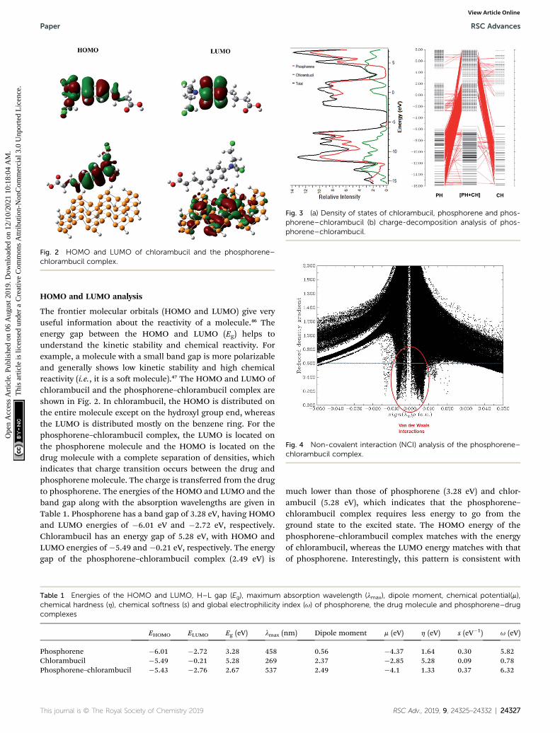
Fig. 2 HOMO and LUMO of chlorambucil and the phosphorene–chlorambucil complex.
Fig. 3 (a) Density of states of chlorambucil, phosphorene and phos-phorene–chlorambucil (b) charge-decomposition analysis of phos-phorene–chlorambucil.
Fig. 4 Non-covalent interaction (NCI) analysis of the phosphorene–chlorambucil complex.
Paper RSC Advances
Ope
n A
cces
s A
rtic
le. P
ublis
hed
on 0
6 A
ugus
t 201
9. D
ownl
oade
d on
12/
10/2
021
10:1
8:04
AM
. T
his
artic
le is
lice
nsed
und
er a
Cre
ativ
e C
omm
ons
Attr
ibut
ion-
Non
Com
mer
cial
3.0
Unp
orte
d L
icen
ce.
View Article Online
HOMO and LUMO analysis
The frontier molecular orbitals (HOMO and LUMO) give veryuseful information about the reactivity of a molecule.46 Theenergy gap between the HOMO and LUMO (Eg) helps tounderstand the kinetic stability and chemical reactivity. Forexample, a molecule with a small band gap is more polarizableand generally shows low kinetic stability and high chemicalreactivity (i.e., it is a so molecule).47 The HOMO and LUMO ofchlorambucil and the phosphorene–chlorambucil complex areshown in Fig. 2. In chlorambucil, the HOMO is distributed onthe entire molecule except on the hydroxyl group end, whereasthe LUMO is distributed mostly on the benzene ring. For thephosphorene–chlorambucil complex, the LUMO is located onthe phosphorene molecule and the HOMO is located on thedrug molecule with a complete separation of densities, whichindicates that charge transition occurs between the drug andphosphorene molecule. The charge is transferred from the drugto phosphorene. The energies of the HOMO and LUMO and theband gap along with the absorption wavelengths are given inTable 1. Phosphorene has a band gap of 3.28 eV, having HOMOand LUMO energies of �6.01 eV and �2.72 eV, respectively.Chlorambucil has an energy gap of 5.28 eV, with HOMO andLUMO energies of �5.49 and �0.21 eV, respectively. The energygap of the phosphorene–chlorambucil complex (2.49 eV) is
Table 1 Energies of the HOMO and LUMO, H–L gap (Eg), maximumchemical hardness (h), chemical softness (s) and global electrophilicity icomplexes
EHOMO ELUMO Eg (eV) lmax (
Phosphorene �6.01 �2.72 3.28 458Chlorambucil �5.49 �0.21 5.28 269Phosphorene–chlorambucil �5.43 �2.76 2.67 537
This journal is © The Royal Society of Chemistry 2019
much lower than those of phosphorene (3.28 eV) and chlor-ambucil (5.28 eV), which indicates that the phosphorene–chlorambucil complex requires less energy to go from theground state to the excited state. The HOMO energy of thephosphorene–chlorambucil complex matches with the energyof chlorambucil, whereas the LUMO energy matches with thatof phosphorene. Interestingly, this pattern is consistent with
absorption wavelength (lmax), dipole moment, chemical potential(m),ndex (u) of phosphorene, the drug molecule and phosphorene–drug
nm) Dipole moment m (eV) h (eV) s (eV�1) u (eV)
0.56 �4.37 1.64 0.30 5.822.37 �2.85 5.28 0.09 0.782.49 �4.1 1.33 0.37 6.32
RSC Adv., 2019, 9, 24325–24332 | 24327

Fig. 5 Orbital overlapping between the drug molecule and phos-phorene in phosphorene–chlorambucil.
Table 2 Results of the NBO calculation for phosphorene before andafter the formation of a complex with chlorambucil; the energies aregiven in kcal mol�1
CH / PHS Energies PHS / PHS Before Aer
pC65�C70/s*P10�P17 0.11 sP1�P59/s*
P3�P4 1.83 1.85
sC65�H84/s*P18�P23 0.06 sP1�P61/s*
P3�P7 1.66 1.69
sC71�H89/s*P7�P9 0.09 sP31�P32/s*
P34�P35 1.55 1.56
LPð1ÞCl83/s*P26�H42
0.07 sP31�P32/s*P27�P28 1.66 1.69
LPð2ÞCl83/s*P26�H42
0.75 LPð1ÞP9/s*P16�P20 3.32 3.29
LPð2ÞCl83/s*P33�H44
0.08 LPð1ÞP3/s*P7�P9 2.92 2.97
LPð3ÞCl83/s*P33�H44
0.29 LPð1ÞP4/s*P1�P3 3.13 3.17
PHS / CH LPð1ÞP6/s*P7�P9 3.15 3.19
LP(1)P3 / sC73–H93 0.21 LPð1ÞP5/s*P13�P14 4.23 4.28
LP(1)P4 / sC70–H87 0.15 LPð1ÞP55/s*P8�P53 5.28 5.30
LP(1)P7 / sC71–H89 0.12 LPð1ÞP35/s*P28�P31 2.13 2.15
LP(1)P17 / sC70–H87 0.16 LPð1ÞP5/s*P4�P11 3.56 3.58
LP(1)P18 / sC65–H84 0.06 LPð1ÞP3/s*P4�P11 2.96 2.98
RSC Advances Paper
Ope
n A
cces
s A
rtic
le. P
ublis
hed
on 0
6 A
ugus
t 201
9. D
ownl
oade
d on
12/
10/2
021
10:1
8:04
AM
. T
his
artic
le is
lice
nsed
und
er a
Cre
ativ
e C
omm
ons
Attr
ibut
ion-
Non
Com
mer
cial
3.0
Unp
orte
d L
icen
ce.
View Article Online
the densities in the HOMO and LUMO; the HOMO is present onchlorambucil, whereas the LUMO is present on phosphorene.The lowering of the energy gap suggests that the excitationwavelength is red-shied for the chlorambucil–phosphorenecomplex. The maximum absorption (lmax) for phosphorene wasobserved at 458 nm in the UV-visible region, which was red-shied by 79 nm for the chlorambucil–phosphorene complex(lmax ¼ 537 nm).
Fig. 7 UV-vis absorption spectra of chlorambucil, phosphorene andphosphorene–chlorambucil.
Charge decomposition analysis (CDA) and density of states(DOS)
The smaller HOMO–LUMO gap of the complex facilitates thecharge transfer within the complex molecule. The decrease inthe band gap is due to the addition of molecular energy levels inthe complex MOs by the component fragments, as can be seenin the CDA diagram. The CDA diagram clearly shows how thephosphorene–chlorambucil complex orbitals are formed by themixing of each of the fragment's orbitals (phosphorene andchlorambucil). The addition of a molecular energy level byphosphorene and chlorambucil causes an increase in theenergy of the HOMO (�5.43 eV) and a decrease in the energy ofthe LUMO (�2.76 eV) of the complex, which favourablycontributes to a decrease in the band gap, which furthercontributes to the charge-transfer process. These results alsoshow that the drug was successfully attached to the
Fig. 6 Electron-density images of some phosphorus atoms present in phin phosphorene–chlorambucil.
24328 | RSC Adv., 2019, 9, 24325–24332
phosphorene molecule. In the CDA analysis, the orbital inter-action diagram was plotted for the complex, in which the orbitalcontributions of the drug and phosphorene in the formation of
osphorene: (A) ELF for phosphorene, (B) ELF for phosphorene present
This journal is © The Royal Society of Chemistry 2019

Fig. 8 Electron–hole orbitals and PET process for n-state ¼ 1–5 ofphosphorene–chlorambucil.
Paper RSC Advances
Ope
n A
cces
s A
rtic
le. P
ublis
hed
on 0
6 A
ugus
t 201
9. D
ownl
oade
d on
12/
10/2
021
10:1
8:04
AM
. T
his
artic
le is
lice
nsed
und
er a
Cre
ativ
e C
omm
ons
Attr
ibut
ion-
Non
Com
mer
cial
3.0
Unp
orte
d L
icen
ce.
View Article Online
the complex were observed. The matching DOS diagrams can beseen in Fig. 3, which is in good agreement with the CDA results.It can be seen that the MOs of phosphorene and chlorambucil
This journal is © The Royal Society of Chemistry 2019
are mixed together to form the molecular energy level of thecomplex and this phenomenon causes the decrease in the bandgap aer the complex formation (2.67 eV), which contributes tothe charge-transfer process.
Dipole moment, chemical potential, chemical hardness,chemical soness and global electrophilicity index
The dipole moment, chemical potential (m), chemical hardness(h), chemical soness (s) and global electrophilicity index (u)are also given in Table 1 for the drug and phosphorenemoleculeand for the complex. Interesting properties were observed forthe complex. The chemical potential of the complex (�4.1 eV)was lower than that of phosphorene (�4.37 eV); however, it washigher than the drug molecule (�2.85 eV). The chemical hard-ness (h), chemical soness (s) and global electrophilicity index(u) follow a uniform pattern, where the properties of thecomplex are at either end of the spectrum. The complex wassoer than phosphorene or the drug molecule, as reected bythe lower hardness and higher soness values. The soness(hardness) values of the complex were 1.33 eV (0.37 eV�1)compared to 1.64 (0.30) and 5.28 (0.09) for phosphorene and thedrug, respectively. These parameters give information about themolecular stability and electron transfer in the molecule. Theglobal electrophilicity index (u) determines the stabilization inenergy when a molecular system gains additional charge fromits surroundings. The negative chemical potential values of thecomplex (�4.1 eV) indicated the stability of the complex. Theintended target site can be determined based on the soness,hardness and electronegativity of the drug and target site.
Non-covalent interaction (NCI) analysis
The NCI analysis gives useful information about the non-covalent interactions within a molecule.48 The strong direc-tional attractions associated with localized atom–atom contactsand the molecular regions having weak interactions can bedistinguished by an NCI plot. The scatter graphs plottedbetween the reduced density gradient and electron density r,oriented by the sign of l2 and the 3D color-lled reduced densitygradient isosurface are shown in Fig. 4. From the graph, it canbe seen that weak forces of interaction are present between thecomplex molecules, which can also be seen in the color-lledinter-molecular isosurface image (Fig. 5).
The green regions between the components of the complex(i.e. phosphorene and drug) depict van der Waals-type interac-tions, which are weak intermolecular forces. This inference isconsistent with the geometric analysis, whereby weak interac-tions are realized between H atoms of the drug and phosphorusatoms of phosphorene. These weak interactions are quiteadvantageous because the drug molecule can easily be removedfrom phosphorene at its target site (destination). The orbitaloverlap between the drug and phosphorene in the complexdepicts the load exchange between the drug and phosphorene.
Electron localization function (ELF) and NBO analysis
The ELF electron-density images of phosphorene and thephosphorene–drug complex are shown in Fig. 6A and B,
RSC Adv., 2019, 9, 24325–24332 | 24329

Table 3 Analysis of hole–electrons for first five excited states of phosphorene–chlorambucil
Excited states of phosphorene–chlorambucil 1 2 3 4 5
lmax (nm) 537 534 485 475 464DE (eV) 2.31 2.32 2.55 2.61 2.67F 0.002 0.0002 0.0001 0.0003 0.00D (A) 6.81 6.79 6.65 6.85 6.90S 0.006 0.003 0.004 0.003 0.002Transition mode CT CT CT CT CT
RSC Advances Paper
Ope
n A
cces
s A
rtic
le. P
ublis
hed
on 0
6 A
ugus
t 201
9. D
ownl
oade
d on
12/
10/2
021
10:1
8:04
AM
. T
his
artic
le is
lice
nsed
und
er a
Cre
ativ
e C
omm
ons
Attr
ibut
ion-
Non
Com
mer
cial
3.0
Unp
orte
d L
icen
ce.
View Article Online
respectively. The ELF shows slight changes in the electrondensity aer the formation of the complex. The electron densityof phosphorene is affected aer the formation of the phos-phorene–chlorambucil complex.
These ELF images help to understand the transition occur-ing from the drug to phosphorene and from phosphorene to thedrug in the NBO analysis of the complexes. These transitionscause the changes in the electron density of phosphorus atomspresent in phosphorene. The changes in energies for internaltransition occuring in phosphorene before and aer theformation of complex can be seen in Table 2.
Photophysical properties
The UV-vis absorption spectra of chlorambucil, phosphoreneand the chlorambucil–phosphorene complex are shown inFig. 7. The chlorambucil–phosphorene complex absorbs ata much higher wavelength (537 nm) than chlorambucil (269nm), which allows a broad absorption in the UV-vis region. Theabsorption at 537 nm for the complex corresponds to 2.30 eV,which is very much consistent with the HOMO–LUMO gap. Thiscorrelation suggests that the excitation at 537 nm correspondsto the excitation from the HOMO to LUMO, which in turn isa transfer of charge from the drug to phosphorene. Noticeablechanges in the visible region of the absorption spectrum can beapplied for the colorimetric and uorescence detection ofvarious types of bioanalytes (e.g. proteins, DNA and inorganicions), which promotes the target specicity.49
Photoinduced electron transfer analysis
Investigation of electron excitation for a drug delivery systemgives information about its biological activity.16 The electron-transfer process and the contribution of electron or holeorbitals can easily be recognized by the electron–hole theory.The electron–hole orbitals for the rst ve excited states areshown in Fig. 8 for phosphorene–chlorambucil. As shown inFig. 8, the electron and hole orbitals are separated with goodapproximation, which represents that the PET phenomenonoccurs between the complex components. The PET and PCTprocesses affect the uorescence process. In other words, theseprocesses cause quenching in the uorescence emission spec-trum. This can give very useful information about thebiochemical system. Analysis results of the hole–electrons forthe rst ve excited states of phosphorene–chlorambucil aregiven in Table 3. The greater the distance (D) between theorbitals, the longer the charge-transfer length; however, for the
24330 | RSC Adv., 2019, 9, 24325–24332
localized excitation this distance is small. A smaller value of thehole–electron overlap integral (S) represents a high charge-transfer rate. The distances between the orbitals (D) for therst ve excited states of the complex were 6.81 A, 6.79 A, 6.65 A,6.85 A and 6.90 A. The distance between the orbitals (D) wasgreater for all rst excited states, which shows a high charge-transfer length. The hole–electron overlap integrals (S) for therst ve excited states were 0.006, 0.003, 0.004, 0.003 and 0.002.A smaller value of the hole–electron overlap integral (S) showsa large charge-transfer rate. This analysis also showed thata charge-transfer process occurs between the complex compo-nents, which causes a quenching in the uorescence emissionspectrum. The decrease in the uorescence intensity can givemolecular information as well as the location of the drug ina biological system.
For example, the energies of excitation LPð1ÞP9/s*P16�P20
reached 3.29 kcal mol�1 from 3.32 kcal mol�1 aer the complexformation. Similarly, for sP1�P61/s*
P3�P7, the excitation energywas raised to 2.69 kcal mol�1 from 2.66 kcal mol�1 and forLPð1ÞP6/s*
P7�P9, the excitation energy was raised to3.19 kcal mol�1 from 3.15 kcal mol�1.
Conclusions
In this study, the therapeutic potential of phosphorene asa drug delivery system for chlorambucil to treat cancer wasevaluated. The geometric, electronic and excited state proper-ties of chlorambucil, phosphorene and the phosphorene–chlorambucil complex were evaluated to explore the efficiencyof phosphorene as a drug-delivery system. The complex betweenchlorambucil and phosphorene was established through weakP/H bonds. The phosphorene–chlorambucil complex hada dipole moment value of 2.49 D, which was signicantly higherthan that of phosphorene. The increase in the dipole momentaer the formation of the complex suggests an increased solu-bility in the polar solvent (water). The nature of interactionbetween phosphorene and chlorambucil was illustratedthrough a non-covalent interaction (NCI) plot, which illustratedthat weak forces of interaction were present between phos-phorene and chlorambucil. These weak intermolecular forcesare advantageous for an easy offloading of the drug at the target.The HOMO in the phosphorene–chlorambucil complex waslocalized on the chlorambucil, whereas the LUMO was centeredon phosphorene. Frontier molecular orbital analysis revealedthat the charge was transferred from chlorambucil to
This journal is © The Royal Society of Chemistry 2019
Paper RSC Advances
Ope
n A
cces
s A
rtic
le. P
ublis
hed
on 0
6 A
ugus
t 201
9. D
ownl
oade
d on
12/
10/2
021
10:1
8:04
AM
. T
his
artic
le is
lice
nsed
und
er a
Cre
ativ
e C
omm
ons
Attr
ibut
ion-
Non
Com
mer
cial
3.0
Unp
orte
d L
icen
ce.
View Article Online
phosphorene during excitation from the HOMO to LUMO. Thecharge transfer was further supplemented by a charge-decomposition analysis (CDA). The energies for different tran-sitions from the donor to acceptor (drug to phosphorene andphosphorene to drug) in NBO analysis also indicated a chargetransfer between the drugs and phosphorene molecules.Excited-state calculations showed that the lmax was red-shiedby 79 nm for the phosphorene–chlorambucil complexes. Thephoto-induced electron-transfer (PET) process was observed fordifferent excited states, which could be well explained visuallybased on the electron–hole theory. The photo-induced electrontransfer suggests a quenching of uorescence upon interaction.In summary, phosphorene possesses signicant therapeuticpotential as a drug-delivery system for chlorambucil to treatcancer.
Conflicts of interest
The authors declare that they have no conicts of interest.
Acknowledgements
The computations/simulations/SIMILAR were performed onresources provided by the Swedish National Infrastructure forComputing (SNIC) at Umea University, 901 87, Umea, Sweden.The authors acknowledge the nancial and technical supportfrom Punjab Bio-energy Institute (PBI), University of AgricultureFaisalabad (UAF).
References
1 Z. Sheng, D. Hu, M. Zheng, P. Zhao, H. Liu, D. Gao, P. Gong,G. Gao, P. Zhang and Y. Ma, ACS Nano, 2014, 8, 12310–12322.
2 S. K. Misra, G. Ghoshal, M. R. Gartia, Z. Wu, A. K. De, M. Ye,C. R. Bromeld, E. M. Williams, K. Singh and K. V. Tangella,ACS Nano, 2015, 9, 10695–10718.
3 T. M. Allen and P. R. Cullis, Science, 2004, 303, 1818–1822.4 Y. Liu, H. Miyoshi and M. Nakamura, Int. J. Cancer, 2007,120, 2527–2537.
5 X. Sun, Z. Liu, K. Welsher, J. T. Robinson, A. Goodwin,S. Zaric and H. Dai, Nano Res., 2008, 1, 203–212.
6 O. M. Koo, I. Rubinstein and H. Onyuksel, Nanomedicine,2005, 1, 193–212.
7 N. Nasongkla, E. Bey, J. Ren, H. Ai, C. Khemtong, J. S. Guthi,S.-F. Chin, A. D. Sherry, D. A. Boothman and J. Gao, NanoLett., 2006, 6, 2427–2430.
8 O. C. Farokhzad and R. Langer, ACS Nano, 2009, 3, 16–20.9 A. Bianco, K. Kostarelos and M. Prato, Curr. Opin. Chem.Biol., 2005, 9, 674–679.
10 T. Liu, C. Wang, X. Gu, H. Gong, L. Cheng, X. Shi, L. Feng,B. Sun and Z. Liu, Adv. Mater., 2014, 26, 3433–3440.
11 L. Cheng, C. Yuan, S. Shen, X. Yi, H. Gong, K. Yang andZ. Liu, ACS Nano, 2015, 9, 11090–11101.
12 Q. Weng, B. Wang, X. Wang, N. Hanagata, X. Li, D. Liu,X. Wang, X. Jiang, Y. Bando and D. Golberg, ACS Nano,2014, 8, 6123–6130.
This journal is © The Royal Society of Chemistry 2019
13 L. Cheng, S. Shen, S. Shi, Y. Yi, X. Wang, G. Song, K. Yang,G. Liu, T. E. Barnhart and W. Cai, Adv. Funct. Mater., 2016,26, 2185–2197.
14 L. Chen, X. Zhong, X. Yi, M. Huang, P. Ning, T. Liu, C. Ge,Z. Chai, Z. Liu and K. Yang, Biomaterials, 2015, 66, 21–28.
15 D. L. Childers, J. Corman, M. Edwards and J. J. Elser,Bioscience, 2011, 61, 117–124.
16 J. R. Choi, K. W. Yong, J. Y. Choi, A. Nilghaz, Y. Lin, J. Xu andX. Lu, Theranostics, 2018, 8, 1005.
17 C. Zhang, J. Lian, W. Yi, Y. Jiang, L. Liu, H. Hu, W. Xiao,S. Du, L. Sun and H. Gao, J. Phys. Chem. C, 2009, 113,18823–18826.
18 Y. Cai, G. Zhang and Y.-W. Zhang, Sci. Rep., 2014, 4, 6677.19 S. Zhang, J. Yang, R. Xu, F. Wang, W. Li, M. Ghufran,
Y.-W. Zhang, Z. Yu, G. Zhang and Q. Qin, ACS Nano, 2014,8, 9590–9596.
20 J. Kim, S. S. Baik, S. H. Ryu, Y. Sohn, S. Park, B.-G. Park,J. Denlinger, Y. Yi, H. J. Choi and K. S. Kim, Science, 2015,349, 723–726.
21 Q. H. Wang, K. Kalantar-Zadeh, A. Kis, J. N. Coleman andM. S. Strano, Nat. Nanotechnol., 2012, 7, 699.
22 A. P. De Silva, H. N. Gunaratne, T. Gunnlaugsson,A. J. Huxley, C. P. McCoy, J. T. Rademacher and T. E. Rice,Chem. Rev., 1997, 97, 1515–1566.
23 B. An, H. Yuan, Q. Zhu, Y. Li, X. Guo and J. Zhang,Spectrochim. Acta, Part A, 2017, 175, 36–42.
24 A. P. De Silva, D. B. Fox, A. J. Huxley and T. S. Moody, Coord.Chem. Rev., 2000, 205, 41–57.
25 A. A. Taherpour, M. Jamshidi and O. Rezaei, J. Mol. Struct.,2017, 1147, 815–820.
26 R. Kramer, Angew. Chem., Int. Ed., 1998, 37, 772–773.27 P. Jiang and Z. Guo, Coord. Chem. Rev., 2004, 248, 205–229.28 E. Reisner, V. B. Arion, B. K. Keppler and A. J. Pombeiro,
Inorg. Chim. Acta, 2008, 361, 1569–1583.29 S. C. Burdette and S. J. Lippard, Coord. Chem. Rev., 2001, 216,
333–361.30 J. W. Park, E. H. Chae, S. H. Kim, J. H. Lee, J. W. Kim,
S. M. Yoon and J.-Y. Choi, Mater. Chem. Phys., 2006, 97,371–378.
31 P. A. Sinoway and J. P. Callen, Arthritis Rheum., 1993, 36, 319–324.
32 J. H. Hoofnagle, G. L. Davis, D. F. Schafer, M. Peters,M. I. Avigan, S. C. Pappas, R. G. Hanson, G. Y. Minuk,G. M. Dusheiko and G. Campbell, Gastroenterology, 1986,91, 1327–1334.
33 R. P. Rapini, R. E. Jordan and S. E. Wolverton, Cytotoxicagents, Systemic Drugs for Skin Diseases, ed. S. E. Wolvertonand J. K. Wilkins, WB Saunders, Philadelphia, 1991, pp.125–151.
34 M.-Y. Hua, H.-W. Yang, H.-L. Liu, R.-Y. Tsai, S.-T. Pang,K.-L. Chuang, Y.-S. Chang, T.-L. Hwang, Y.-H. Chang andH.-C. Chuang, Biomaterials, 2011, 32, 8999–9010.
35 J. Shi, Y. Liu, L. Wang, J. Gao, J. Zhang, X. Yu, R. Ma, R. Liuand Z. Zhang, Acta Biomater., 2014, 10, 1280–1291.
36 K. R. Rai, B. L. Peterson, F. R. Appelbaum, J. Kolitz, L. Elias,L. Shepherd, J. Hines, G. A. Threatte, R. A. Larson andB. D. Cheson, N. Engl. J. Med., 2000, 343, 1750–1757.
RSC Adv., 2019, 9, 24325–24332 | 24331

RSC Advances Paper
Ope
n A
cces
s A
rtic
le. P
ublis
hed
on 0
6 A
ugus
t 201
9. D
ownl
oade
d on
12/
10/2
021
10:1
8:04
AM
. T
his
artic
le is
lice
nsed
und
er a
Cre
ativ
e C
omm
ons
Attr
ibut
ion-
Non
Com
mer
cial
3.0
Unp
orte
d L
icen
ce.
View Article Online
37 M. J. Lacher and J. R. Durant, Ann. Intern. Med., 1965, 62,468–476.
38 R. Patel and Y. P. Singh, J. Mol. Struct., 2018, 1153, 162–169.39 M. J. Frisch, G. W. Trucks, H. B. Schlegel, G. E. Scuseria,
M. A. Robb, J. R. Cheeseman, G. Scalmani, V. Barone,B. Mennucci, G. A. Petersson, H. Nakatsuji, M. Caricato,X. Li, H. P. Hratchian, A. F. Izmaylov, J. Bloino, G. Zheng,J. L. Sonnenberg, M. Hada, M. Ehara, K. Toyota,R. Fukuda, J. Hasegawa, M. Ishida, T. Nakajima, Y. Honda,O. Kitao, H. Nakai, T. Vreven, J. A. Montgomery Jr,J. E. Peralta, F. Ogliaro, M. Bearpark, J. J. Heyd,E. Brothers, K. N. Kudin, V. N. Staroverov, R. Kobayashi,J. Normand, K. Raghavachari, A. Rendell, J. C. Burant,S. S. Iyengar, J. Tomasi, M. Cossi, N. Rega, J. M. Millam,M. Klene, J. E. Knox, J. B. Cross, V. Bakken, C. Adamo,J. Jaramillo, R. Gomperts, R. E. Stratmann, O. Yazyev,A. J. Austin, R. Cammi, C. Pomelli, J. W. Ochterski,R. L. Martin, K. Morokuma, V. G. Zakrzewski, G. A. Voth,P. Salvador, J. J. Dannenberg, S. Dapprich, A. D. Daniels,O. Farkas, J. B. Foresman, J. V. Ortiz, J. Cioslowski andD. J. Fox, Gaussian 09, Revision B.01, Gaussian Inc.,Wallingford, 2010.
24332 | RSC Adv., 2019, 9, 24325–24332
40 A. Becke, J. Chem. Phys., 1993, 98, 5648.41 M. Jamshidi, O. Rezaei, A. R. Belverdi, S. Malekian and
A. R. Belverdi, J. Mol. Struct., 2016, 1123, 111–115.42 X. Su, P. Si, Q. Hou, X. Kong and W. Cheng, Phys. B, 2009,
404, 1794–1798.43 F. R. Wagner, V. Bezugly, M. Kohout and Y. Grin, Chem.–Eur.
J., 2007, 13, 5724–5741.44 P. Rajesh, P. Kandan, S. Sathish, A. Manikandan,
S. Gunasekaran, T. Gnanasambandan and S. B. Abirami, J.Mol. Struct., 2017, 1137, 277–291.
45 R. P. Gangadharan and S. S. Krishnan, Acta Phys. Pol., A,2014, 125, 18–22.
46 J. M. Seminario, Recent developments and applications ofmodern density functional theory, Elsevier, 1996.
47 K. Fukui, Angew. Chem., Int. Ed. Engl., 1982, 21, 801–809.48 E. R. Johnson, S. Keinan, P. Mori-Sanchez, J. Contreras-
Garcia, A. J. Cohen and W. Yang, J. Am. Chem. Soc., 2010,132, 6498–6506.
49 X. Ling, H. Wang, S. Huang, F. Xia and M. S. Dresselhaus,Proc. Natl. Acad. Sci. U. S. A., 2015, 112, 4523–4530.
This journal is © The Royal Society of Chemistry 2019