Embed Size (px)
Citation preview
Development/Plasticity/Repair
Cellular Form of Prion Protein Inhibits Reelin-MediatedShedding of Caspr from the Neuronal Cell Surface toPotentiate Caspr-Mediated Inhibition of Neurite Outgrowth
Vasudharani Devanathan,1,2 Igor Jakovcevski,1* Antonella Santuccione,1* Shen Li,1 Hyun Joon Lee,1 Elior Peles,5
Iryna Leshchyns’ka,1,3 Vladimir Sytnyk,1,3 and Melitta Schachner1,4
1Zentrum fur Molekulare Neurobiologie, Universitat Hamburg, 20246 Hamburg, Germany, 2Department of Pharmacology and Experimental Therapy,Institute of Experimental and Clinical Pharmacology and Toxicology, Eberhard Karls University Hospitals and Clinics and Interfaculty Center ofPharmacogenomics and Pharmaceutical Research, University of Tubingen, 72074 Tubingen, Germany, 3School of Biotechnology and BiomolecularSciences, University of New South Wales, Sydney, New South Wales 2052, Australia, 4Keck Center for Collaborative Neuroscience, Rutgers University,Piscataway, New Jersey 08854-8082, and 5Department of Molecular Cell Biology, Weizmann Institute of Science, Rehovot 76100, Israel
Extension of axonal and dendritic processes in the CNS is tightly regulated by outgrowth-promoting and -inhibitory cues to assureprecision of synaptic connections. We identify a novel role for contactin-associated protein (Caspr) as an inhibitory cue that reducesneurite outgrowth from CNS neurons. We show that proteolysis of Caspr at the cell surface is regulated by the cellular form of prionprotein (PrP), which directly binds to Caspr. PrP inhibits Reelin-mediated shedding of Caspr from the cell surface, thereby increasingsurface levels of Caspr and potentiating the inhibitory effect of Caspr on neurite outgrowth. PrP deficiency results in reduced levels ofCaspr at the cell surface, enhanced neurite outgrowth in vitro, and more efficient regeneration of axons in vivo following spinal cordinjury. Thus, we reveal a previously unrecognized role for Caspr and PrP in inhibitory modulation of neurite outgrowth in CNS neurons,which is counterbalanced by the proteolytic activity of Reelin.
IntroductionCellular prion protein (PrP) is an adhesion molecule impli-cated in neuronal survival, neurite outgrowth (Chen et al.,2003; Santuccione et al., 2005), and synaptic function (Collinge etal., 1994). This functional feature of PrP is consistent with its associ-ation with several cell surface proteins involved in regulation of neu-ral development, including the 67 kDa laminin receptor, the 37kDa laminin receptor precursor protein, and the extracellularmatrix glycoprotein laminin (Rieger et al., 1997; Graner et al.,2000; Gauczynski et al., 2001). PrP also occurs in a complex withthe neural cell adhesion molecule (NCAM) (Schmitt-Ulms et al.,
2001) that results from cis- and trans-interactions between PrPand NCAM at the neural cell surface (Santuccione et al., 2005).PrP promotes recruitment of NCAM to lipid rafts and therebyregulates activation of fyn kinase, an enzyme implicated inNCAM- and PrP-mediated intracellular signaling (Beggs et al.,1994, 1997; Mouillet-Richard et al., 2000; Bodrikov et al., 2005,2008). PrP also associates with the �2/�2-Na�/K�-ATPase,which in turn associates with the Ig superfamily adhesion mole-cule basigin (Kleene et al., 2007). This complex between PrP,�2/�2-ATPase, and basigin regulates the transport of lactate inastrocytes via the monocarboxylate transporter 1, which is acti-vated by the synaptic GluR2 subunit-containing AMPA receptorwith which PrP also interacts (Kleene et al., 2007).
It is conceivable that reduction in the functions of PrP by itsconversion into the pathological form (PrP scrapie) renders thecellular form of PrP less available to the normal cellular func-tions, thereby contributing to the pathology of prion diseases.Since an understanding of prion diseases also depends on theidentification of binding partners for the cellular form of prionprotein and the characterization of their functional properties,we searched for other binding partners of PrP with the aim ofwidening our understanding of the functions of PrP in the con-text of its interactions with molecules at the cell surface and/or inthe extracellular matrix.
Here, we report on the interaction of PrP and contactin-associated protein (Caspr) (Peles et al., 1997). Caspr is most wellcharacterized as an adhesion molecule that is required for theformation of axoglial paranodal junctions surrounding the nodes
Received Nov. 15, 2009; revised April 7, 2010; accepted May 16, 2010.We are grateful to Dr. Martin H. Groschup (Institute for Novel and Emerging Infectious Diseases, Greifswald,
Germany) and Dr. Charles Weissmann (Department of Infectology, Scripps Florida, Jupiter, FL) for PrP �/� mice, Dr.Michael Frotscher and Dr. Shanting Zhao (Institute for Anatomy and Cell Biology, Albert-Ludwigs-University ofFreiburg, Freiburg, Germany) for Reelin �/� and Reelin �/� brains, Dr. Man Sun Sy (Case Western Reserve Univer-sity, Cleveland, OH) for monoclonal antibody 8H4 against PrP, Dr. Catherine Faivre-Sarrailh (Institut Jean Roche,Marseille, France) for antibodies against Caspr, Dr. Tom Curran (St. Jude Children’s Research Hospital, Memphis, TN)for the Reelin plasmid, Dr. Gianfranco Gennarini (University of Bari, Bari, Italy) for the contactin plasmid, and Dr.Sylvain Lehmann (Institut de Genetique Humaine du Centre National de la Recherche Scientifique, Montpellier,France) for the PrP plasmid.
*I.J. and A.S. contributed equally to this work.Correspondence should be addressed to either of the following: Melita Schachner, Keck Center for Collaborative
Neuroscience, Rutgers, University, 604 Allison Road, Piscataway, NJ 08854-8082, E-mail: [email protected]; or Vladimir Sytnyk, School of Biotechnology and Biomolecular Sciences, University of New South Wales,Sydney, New South Wales 2052, Austrlia, E-mail: [email protected].
A. Santuccione’s present address: Division of Psychiatry Research, University of Zurich, August-Forel-Strasse 1,8008 Zurich, Switzerland.
DOI:10.1523/JNEUROSCI.5657-09.2010Copyright © 2010 the authors 0270-6474/10/309292-14$15.00/0
9292 • The Journal of Neuroscience, July 7, 2010 • 30(27):9292–9305
of Ranvier in myelinated axons (Einheber et al., 1997; Scherer,1999; Peles and Salzer, 2000). We now show a novel function forCaspr as a negative regulator of neurite outgrowth in CNS neu-rons. While it was shown previously that Caspr associates incis-interaction with the glycosylphosphatidyl inositol (GPI)-anchored neural cell adhesion molecule contactin/F3 (Peles et al.,1997), which is required for the transport of Caspr to the cellsurface (Faivre-Sarrailh et al., 2000), we provide evidence that theinteraction with GPI-anchored PrP prevents Caspr from shedding atthe cell surface. Furthermore, we identified Reelin, an extracellularmatrix glycoprotein with protease activity (Quattrocchi et al., 2002),as an enzyme involved in shedding of Caspr. Thus, we reveal a pre-viously unrecognized role for Caspr and PrP in inhibitory modula-tion of neurite outgrowth in neurons, which is counterbalanced bythe proteolytic activity of Reelin.
Materials and MethodsAntibodies. Mouse monoclonal antibody 8H4 against PrP, a generous giftfrom Dr. Man Sun Sy (Case Western Reserve University, Cleveland,OH), was used for immunocytochemistry and immunohistochemsitry.The specificity of the antibody was verified in PrP-transfected Chinesehamster ovary (CHO) cells (supplemental Fig. S1, available at www.jneurosci.org as supplemental material) and PrP �/� brains. Rat mono-clonal antibody 555 against L1 (Appel et al., 1993) was used forimmunocytochemistry and immunoblotting. Rabbit polyclonal anti-bodies against the extracellular domain of Caspr (L23, L17), a kind giftfrom Catherine Faivre-Sarrailh (Institut Jean-Roche, Marseille, France),were used for immunocytochemistry and neurite outgrowth experi-ments. Polyclonal goat antibodies against the N terminus of Caspr (N15)from Santa Cruz Biotechnology were used for immunocytochemistry,Western blot, and neurite outgrowth experiments. Goat polyclonal anti-bodies against PrP (M20) (Santa Cruz Biotechnology) were used forimmunoprecipitation and Western blot analysis. Mouse monoclonal an-tibody against Reelin (Millipore) was used for Western blot analysis andimmunohistochemistry. Polyclonal rabbit and monoclonal mouseantibodies against calbindin (Sigma) were used for immunohisto-chemistry. For Western blot we also used polyclonal rabbit antibodiesagainst the FGF receptor (Flg) (Santa Cruz Biotechnology), poly-clonal rabbit antibodies against CHL1 (Leshchyns’ka et al., 2006),polyclonal rabbit antibodies against actin and mouse monoclonalantibodies against tubulin (Sigma), polyclonal goat antibodiesagainst contactin (R & D Systems), mouse monoclonal antibodyagainst glyceraldehyde 3-phosphate dehydrogenase (GAPDH), andrabbit polyclonal antibodies against 5-hydroxytryptamine trans-porter (5-HT) and tyrosine hydroxylase (TH) (Millipore). Secondaryantibodies against goat, rabbit, rat, and mouse Igs coupled to Cy2,Cy3, Cy5, or HRP were from Dianova.
Production of the polyclonal antibodies against the intracellular domainof Caspr. Polyclonal antibodies against the intracellular domain of Casprwere generated by immunizing rabbits with a peptide derived from theCaspr intracellular domain (H-APGPRDQNLPQILEESRSEC-OH).Imject Maleimide Activated McKLH kit (Pierce) was used for conjugat-ing the peptide to a carrier protein for animal injections. Rabbits werekilled after five boosts and the blood was collected and processed tocollect the serum. Antiserum thus obtained was purified further by affin-ity chromatography using protein G-Sepharose 4 fast flow column.Bound antibodies were eluted using 0.1 M glycine (pH 2.7–3.0). Elutedmaterial was neutralized with 1 M Tris-HCl, pH 8.8, and dialyzed exten-sively against PBS, pH 7.3. The specificity of the purified antibodies wasconfirmed in Caspr transfected CHO cells (supplemental Fig. S1, avail-able at www.jneurosci.org as supplemental material). The antibodieswere used for biochemical, histochemical, and cytochemical studies.
Animals. PrP �/� mice (Bueler et al., 1992) were generously providedby Dr. Martin H. Groschup (Institute for Novel and Emerging InfectiousDiseases, Greifswald, Germany). Caspr �/� mice were as described pre-viously (Gollan et al., 2003). Young (10 d old) or adult (6 –10 week old)wild-type, PrP �/�, and Caspr �/� animals were used as indicated in the
text. Brains of 10-d-old Reelin �/� and Reelin �/� mice were generouslyprovided by Dr. Michael Frotscher and Dr. Shanting Zhao (Institute forAnatomy and Cell Biology, Albert-Ludwigs-University of Freiburg,Freiburg, Germany).
Plasmids. The Reelin plasmid was a kind gift from Dr. Tom Curran (St.Jude Children’s Research Hospital, Memphis, TN). The full-length Casprplasmid was as described previously (Peles et al., 1997). Caspr-Fc encod-ing the extracellular domain of rat Caspr fused to human Fc (Gollan et al.,2003) was subcloned into pcDNA3 and used to transfect CHO cells thatsecrete this protein into the culture medium. The contactin plasmid wasa kind gift from Dr. Gianfranco Gennarini (University of Bari, Bari,Italy). PrP plasmids were kind gifts of Dr. Sylvain Lehmann (Institut deGenetique Humaine du CNRS, Montpellier, France).
Purification of Caspr. Caspr was purified from brain homogenates us-ing polyclonal antibodies against the intracellular domain of Caspr withthe Bio-Rad Econo system. Antibodies were coupled to cyanogenbromide-activated Sepharose according to the manufacturer’s instruc-tions (GE Healthcare). Brains from adult mice were homogenized inbuffer containing 5 mM Tris, pH 7.5, 0.32 M sucrose, 1 mM CaCl2, 1 mM
MgCl2, and 1 mM NaHCO3, 0.5% Nonidet P-40, and EDTA-free Com-plete Protease Inhibitor Cocktail (Roche) and centrifuged at 2000 � g for15 min at 4°C. The supernatant was then collected and lysed overnight at4°C by adding N-octylglucoside and Triton X-100 to a final concentra-tion of 1 and 0.5%, respectively. The lysate was centrifuged at 50,000 � gfor 1 h at 4°C. The resulting supernatant was collected and applied to thecolumn containing immobilized polyclonal antibodies against Caspr.Bound proteins were eluted with 50 mM ethanolamine, pH 11.5, 150 mM
NaCl,and0.2%3-[(3-cholamidopropyl)dimethylammonio]-1-propane-sulfonate and immediately neutralized with 1 M Tris-HCl, pH 6.8.Protein-containing fractions were collected, dialyzed overnight at 4°Cagainst TBS, and verified for the presence of Caspr by SDS-PAGE, fol-lowed by silver staining.
ELISA. L1-Fc (2 �g/ml) (Chen et al., 1999), PrP-Fc (2 �g/ml) (Chen etal., 2003), and BSA (2 �g/ml) were immobilized on the polyvinylchloridesurface of a 96-well microtiter plate in PBS overnight at 4°C. Nonab-sorbed proteins were removed and wells were washed five times for 5 minat room temperature (RT) with PBS containing 0.05% Tween 20 (PBS-T). Wells were blocked for 1 h at RT with 1% BSA in PBS and subse-quently incubated with different concentrations of Caspr (0 –2 �g/ml) inPBS-T containing 3% BSA for 2 h at RT. Wells were then washed fivetimes for 5 min each at RT with PBS-T, and bound Caspr was detectedwith Caspr antibodies followed by HRP-conjugated secondary antibodiesvisualized by detecting HRP with 2,2�-azino-bis(3-ethylbenzthiazoline-6-sulfonic acid reagent, which resulted in a colored product that was quantifiedusing an ELISA reader at 405 nm.
Brain and spinal cord homogenate preparation. Whole brains or the5-mm-long segments from the spinal cord lesion scar were taken foranalysis. Homogenates were prepared using a Potter homogenizer in 50mM Tris-HCl buffer, pH 7.5, containing 0.32 M sucrose, 1 mM CaCl2, 1mM MgCl2, 1 mM NaHCO3, and Complete Protease Inhibitor Cocktail(Roche).
Isolation of the total membrane fraction. Brain homogenates were pre-pared using a Potter homogenizer in HOMO buffer (1 mM MgCl2, 1 mM
CaCl2, 1 mM NaHCO3, 5 mM Tris, 0.32 M sucrose, pH 7.4) and centri-fuged at 700 � g for 10 min at 4°C. The resulting supernatants werecentrifuged at 100,000 � g at 4°C for 30 min. Pellets containing the totalmembrane fraction were used for further analyses.
Isolation of growth cones. Growth cones were isolated as describedpreviously (Pfenninger et al., 1983). Brain homogenates from 1- to 3-d-old mice were centrifuged at 1660 � g for 15 min at 4°C. The supernatantwas collected and centrifuged on a discontinuous density gradient of0.75/1.0/2.66 M sucrose at 242,000 � g for 40 min at 4°C. The interfacebetween the load and 0.75 M sucrose containing growth cones was col-lected, resuspended in 10 ml of HOMO buffer, and pelleted by centrifu-gation at 100,000 � g for 40 min at 4°C to obtain purified growth cones.
Coimmunoprecipitation. Homogenates (1 mg of protein) were lysedfor 30 min in 50 mM Tris-HCl buffer, pH 7.5, containing 150 mM NaCl,1% Nonidet P-40, 1% octyl-D-glucopyranoside or 0.5% SDS, 1 mM NaF,2 mM Na3VO4, 0.1 mM PMSF, and EDTA-free Protease Inhibitor Cock-
Devanathan et al. • PrP Inhibits Caspr Shedding J. Neurosci., July 7, 2010 • 30(27):9292–9305 • 9293
tail (Roche). The lysis buffer containing this combination of detergentscompletely solubilizes lipid rafts and has been used in a number of studiesinvolving coimmunoprecipitation of lipid raft components (Trupp et al.,1999; Paratcha et al., 2001; Santuccione et al., 2005). Samples were thencentrifuged for 15 min at 20,000 � g and 4°C. Supernatants were clearedwith protein A/G-agarose beads (Santa Cruz Biotechnology) for 3 h at4°C and incubated with respective primary antibodies overnight at 4°C,followed by precipitation with protein A/G-agarose beads applied for 3 hat 4°C. The beads were washed three times with lysis buffer and once withPBS, and bound proteins analyzed by immunoblotting.
Western blot analysis. Samples were mixed with sample buffer andboiled for 5–7 min. Proteins were separated by electrophoresis on 8, 10,or 12% SDS-polyacrylamide gels, depending on the molecular weight ofthe proteins that were analyzed, and transferred to a nitrocellulose mem-brane (GE Healthcare). Membranes were blocked with 5% milk in PBS,incubated with primary antibodies overnight at 4°C (or for 3 d at 4°C forsupernatant analysis) with shaking and washed with PBS. Primary anti-bodies were detected with corresponding HRP-conjugated secondaryantibodies applied for 1 h in 5% milk in PBS. After washing, secondaryantibodies were visualized with chemiluminescence.
In vitro proteolytic processing by Reelin. Caspr-Fc, or Caspr or L1 puri-fied from mouse brain were incubated at 37°C for 2 h with Reelin-containing culture medium from CHO cells transfected with full-lengthReelin or medium from CHO cells transfected with pcDNA3 for control.Samples were then resuspended in sample buffer, boiled for 8 min, andanalyzed by Western blot.
Cerebellar granule cell cultures. Dissociated granule cell cultures wereprepared from the cerebellum of 6- to 8-d-old wild-type, PrP �/�, andCaspr �/� mice as described previously (Chen et al., 1999).
CHO cell culture. CHO cells were maintained in Glasgow modifiedEagle’s medium containing 10% fetal calf serum and transfected usingLipofectamine with Plus reagent (Invitrogen) following the manufac-turer’s instructions.
Immunocytochemistry. Indirect immunofluorescence staining wasperformed as described previously (Sytnyk et al., 2002; Leshchyns’ka etal., 2003). Neurons on glass coverslips were washed in PBS and fixed in4% formaldehyde in PBS for 15 min at RT. Neurons were then washedthree times with PBS, permeabilized with 0.25% Triton X-100 in PBSapplied for 5 min, blocked in 1% BSA in PBS, and incubated with pri-mary antibodies diluted in 1% BSA in PBS for 1 h. Primary antibodieswere detected with corresponding secondary antibodies applied in 1%BSA in PBS for 30 min. To detect Caspr at the cell surface, polyclonalantibodies against the extracellular domain of Caspr were applied to liveneurons for 20 min in culture medium in a CO2 incubator, followed bysecondary antibodies applied for 15 min in culture medium in a CO2
incubator. In cocapping experiments, antibodies against the extracellulardomains of PrP and Caspr were applied together in culture medium for15 min, followed by secondary antibodies applied in culture medium for15 min in a CO2 incubator. Coverslips were embedded in Aqua-Poly/Mount (Polysciences). Images were acquired at RT using a confocallaser-scanning microscope TCS-SP2 (Leica) using a 63� objective. Theimages were exported using LSM510 software (version 3; Zeiss).
Neurite outgrowth assay. Cells were maintained in 12-well plates(Nunc) on glass coverslips (15 mm in diameter; Hecht) substrate coatedwith poly-L-lysine (Sigma) followed, when indicated, with Caspr anti-bodies (1 �g/ml), laminin (Sigma; 1 �g/ml), or PrP-Fc (1 �g/ml) andcultured in chemically defined serum-free medium (Chen et al., 1999).Where indicated, aprotinin (1 �M) was added to the medium. Twenty-two hours after seeding, cells were fixed for 15 min with 2.5% glutaral-dehyde in PBS and stained with toluidine blue/trypan blue. The totallength of all neurites per neuron was measured by the KS image analysissystem (Kontron-Zeiss). At least three coverslips for each group weretaken in each experiment, and at least 100 cells from one coverslip wereanalyzed. Experiments were repeated three times.
Analysis of Caspr shedding into the culture medium. Inhibitors [aproti-nin (1 �M; Sigma), pepstatin (1 �M; Sigma), phenanthrolamine (10 �M;Sigma), GM6001 (1 �M; Merck)] or PrP-Fc (1 �g/ml) were applied 6 hafter plating of cells. Cells were maintained for 5 d by replacing mediumand inhibitors or PrP-Fc every second day. Culture medium was then
collected. Proteins in the culture medium were concentrated by Amiconcolumns with a 10 kDa molecular weight cutoff (Millipore) and used forWestern blot analysis.
Cell surface biotinylation. Surface biotinylation was performed as de-scribed previously (Schmidt et al., 1997) with modifications. Briefly, 48 hafter transfection cells were washed twice with ice-cold PBS supple-mented with 0.5 mM CaCl2 and 2 mM MgCl2 (PBSCM). Surface proteinswere biotinylated by incubating cells with 0.5 mg/ml sulfo-NHS-LS-biotin (Pierce) in PBSCM for 10 min at 4°C. Biotinylation was termi-nated by incubation with 20 mM glycine in PBSCM at 4°C for 10 min,followed by extensive washing with PBSCM. Biotinylated cells were thenlysed for 30 min in 50 mM Tris-HCl buffer, pH 7.5, containing 150 mM
NaCl, 0.5% Triton X-100, 1 mM NaF, 2 mM Na3VO4, 0.1 mM PMSF, 1 mM
Na4P2O7, 1 mM EDTA, and EDTA-free Protease Inhibitor Cocktail(Roche) and centrifuged at 700 � g for 15 min at 4°C. The supernatantswere collected and protein concentrations were determined using theBCA kit (Pierce). The amounts of surface-localized proteins were deter-mined by precipitating biotinylated proteins with streptavidin-coupledagarose beads (Pierce) applied to the supernatants at 4°C overnight. Aga-rose beads were pelleted by centrifugation and washed twice with the lysisbuffer. Precipitated proteins were solubilized by the addition of 2�Laemmli buffer to the agarose beads and analyzed by Western blot.
Subcellular fractionation and isolation of lipid-enriched microdomains.Lipid rafts were prepared from the crude membrane fraction of brainhomogenates of 2- to 3-month-old mice as described previously(Leshchyns’ka et al., 2003).
Immunohistochemistry. Tissue preparation and immunohistochemis-try on cerebella of young C57BL/6 mice was done as described previously(Jakovcevski et al., 2009).
Spinal cord injury. Three-month-old female mice were anesthetized byintraperitoneal injection of ketamine and xylazine [100 mg of Ketanest(Parke-Davis/Pfizer) and 5 mg of Rompun (Bayer, Leverkusen, Ger-many) per kilogram of body weight]. Laminectomy was performed at theT7–T9 level with mouse laminectomy forceps (Fine Science Tools). Amouse spinal cord compression device was used for compression injury(Curtis et al., 1993). Compression force (degree of closure of the forceps)and duration were controlled by an electromagnetic device. The spinalcord was maximally compressed [100%, according to the operationaldefinition of Curtis et al. (1993)] for 1 s by a time-controlled current flowthrough the electromagnetic device. The skin was then surgically closedusing 6-0 nylon stitches (Ethicon). After surgery, mice were kept in aheated room (35°C) for several hours to prevent hypothermia and thenhoused singly in a temperature-controlled (22°C) room with water andstandard food provided ad libitum. After the surgery, bladders of theanimals were manually voided twice daily.
Histological analysis of the spinal cords. Mice were transcardially per-fused with fixative consisting of 4% formaldehyde and 0.1% CaCl2 in 0.1M cacodylate buffer, pH 7.3, for 15 min at room temperature. Afterperfusion, the spinal cords were dissected out and postfixed overnight at4°C in the same solution as used for perfusion and then cryoprotected in15% sucrose solution in 0.1 M cacodylate buffer, pH 7.3, for 2 d at 4°C.Pieces of spinal cords 1 cm in length centered at the lesion site wereembedded in Tissue Tek (Sakura Finetek), and frozen by 2 min immer-sion into 2-methyl-butane (isopentane) precooled to �80°C. Seriallongitudinal sections were cut in a cryostat (CM3050; Leica), and25-�m-thick sections were collected on SuperFrost Plus glass slides(Roth). Spaced serial sections 250 �m apart were stained with cresylviolet/luxol fast blue and used for estimations of the scar volumeusing the Cavalieri principle. Areas of the scar required for volumeestimation were measured directly under the microscope using Neu-rolucida software (MicroBrightField). Immunofluorescence labelingof the spinal cord tissue with anti-5-HT and anti-TH antibodies wasperformed as described previously (Jakovcevski et al., 2007).
Analysis of locomotor function. The recovery of ground locomotion wasevaluated using the Basso, Beattie, Bresnahan (BBB) rating scale (Basso etal., 1995) modified for mice (Joshi and Fehlings, 2002) and a novel single-frame motion analysis (Apostolova et al., 2006). This method includesevaluation of four parameters in three different tests: beam walking(foot-stepping angle and rump-weight index), voluntary movements
9294 • J. Neurosci., July 7, 2010 • 30(27):9292–9305 Devanathan et al. • PrP Inhibits Caspr Shedding
without body weight support (extension-flexion ratio), and inclined lad-der climbing (number of correct steps). Assessment was performed be-fore and at 1, 3, and 6 weeks after the injury. Values for the left and rightlegs were averaged.
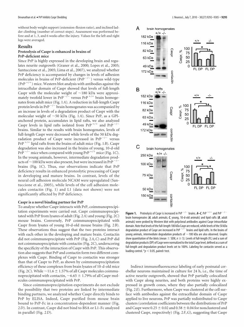
ResultsProteolysis of Caspr is enhanced in brains ofPrP-deficient miceSince PrP is highly expressed in the developing brain and regu-lates neurite outgrowth (Graner et al., 2000; Lopes et al., 2005;Santuccione et al., 2005; Lima et al., 2007), we analyzed whetherPrP deficiency is accompanied by changes in levels of adhesionmolecules in brains of PrP-deficient (PrP�/�) versus wild-type(PrP�/�) mice. Western blot analysis with antibodies against theintracellular domain of Caspr showed that levels of full-lengthCaspr with the molecular weight of �180 kDa were approxi-mately twofold lower in PrP�/� versus PrP�/� brain homoge-nates from adult mice (Fig. 1A). A reduction in full-length Casprprotein levels in PrP�/� brain homogenates was accompanied byan increase in levels of a degradation product of Caspr with themolecular weight of �50 kDa (Fig. 1A). Since PrP, as a GPI-anchored protein, accumulates in lipid rafts, we also analyzedCaspr levels in lipid rafts isolated from PrP�/� and PrP�/�
brains. Similar to the results with brain homogenates, levels offull-length Caspr were decreased while levels of the 50 kDa deg-radation product of Caspr were increased in PrP�/� versusPrP�/� lipid rafts from the brains of adult mice (Fig. 1B). Casprdegradation was also increased in the brains of young, 10-d-oldPrP�/� mice when compared with young PrP�/� mice (Fig. 1C).In the young animals, however, intermediate degradation prod-ucts of �100 kDa were also present, but were increased in PrP�/�
brains (Fig. 1C). Thus, our observations indicate that PrPdeficiency results in enhanced proteolytic processing of Casprin developing and mature brains. In contrast, levels of theneural cell adhesion molecule NCAM were upregulated (San-tuccione et al., 2005), while levels of the cell adhesion mole-cules contactin (Fig. 1) and L1 (data not shown) were notsignificantly affected by PrP deficiency.
Caspr is a novel binding partner for PrPTo analyze whether Caspr interacts with PrP, coimmunoprecipi-tation experiments were carried out. Caspr coimmunoprecipi-tated with PrP from lysates of adult (Fig. 2A) and young (Fig. 2C)mouse brains. Conversely, PrP coimmunoprecipitated withCaspr from lysates of young 10-d-old mouse brains (Fig. 2B).These observations thus suggest that the two proteins interactwith each other in the developing and mature brain. Contactindid not coimmunoprecipitate with PrP (Fig. 2A,C) and PrP didnot coimmunoprecipitate with contactin (Fig. 2C), underscoringthe specificity of the interaction of Caspr with PrP. This observa-tion also suggests that PrP and contactin form two different com-plexes with Caspr. Binding of Caspr to contactin was strongerthan that of Caspr to PrP, as shown by coimmunoprecipitationefficiency of these complexes from brain lysates of 10-d-old mice(Fig. 2C). While �11.6 � 1.57% of all Caspr molecules coimmu-noprecipitated with contactin, �6.65 � 1.79% of all Caspr mol-ecules coimmunoprecipitated with PrP.
Since coimmunoprecipitation experiments do not excludethe possibility that two proteins are linked by intermediatebinding partners, we analyzed whether Caspr directly binds toPrP by ELISA. Indeed, Caspr purified from mouse brainbound to PrP-Fc in a concentration-dependent manner (Fig.2 D). In contrast, Caspr did not bind to BSA or L1-Fc analyzedin parallel (Fig. 2 D).
Indirect immunofluorescence labeling of early postnatal cer-ebellar neurons maintained in culture for 24 h, i.e., the time ofactive neurite outgrowth, showed that PrP partially colocalizedwith Caspr along neurites, and both proteins were highly ex-pressed in growth cones, where they also partially colocalized(Fig. 2E). Furthermore, when Caspr was clustered at the cell sur-face with antibodies against the extracellular domain of Casprapplied to live neurons, PrP was partially redistributed to Casprclusters (correlation coefficients between the distributions of PrPand Caspr were 0.25 � 0.02 and 0.58 � 0.04 for nonclustered andclustered Caspr, respectively) (Fig. 2F,G), suggesting that Caspr
Figure 1. Proteolysis of Caspr is increased in PrP �/� brains. A–C, PrP �/� and PrP �/�
brain homogenates (A, adult animals; C, young, 10-d-old animals) and lipid rafts (B, adultanimals) were probed by Western blot with polyclonal antibodies against Caspr intracellulardomain. Note that levels of the full-length 180 kDa Caspr are reduced, while levels of the 50 kDadegradation product of Caspr are increased in PrP �/� brains and lipid rafts. In the brains ofyoung animals, intermediate degradation products of �100 kDa are also observed. Graphsshow quantitation of the blots (mean � SEM, n � 5). Levels of full-length (FL) and a sum ofdegradation products (DP) of Caspr were normalized to the total Caspr level, defined as a sum offull-length and degradation product levels set to 100%. Labeling for contactin served as aloading control. *p � 0.05, paired t test.
Devanathan et al. • PrP Inhibits Caspr Shedding J. Neurosci., July 7, 2010 • 30(27):9292–9305 • 9295
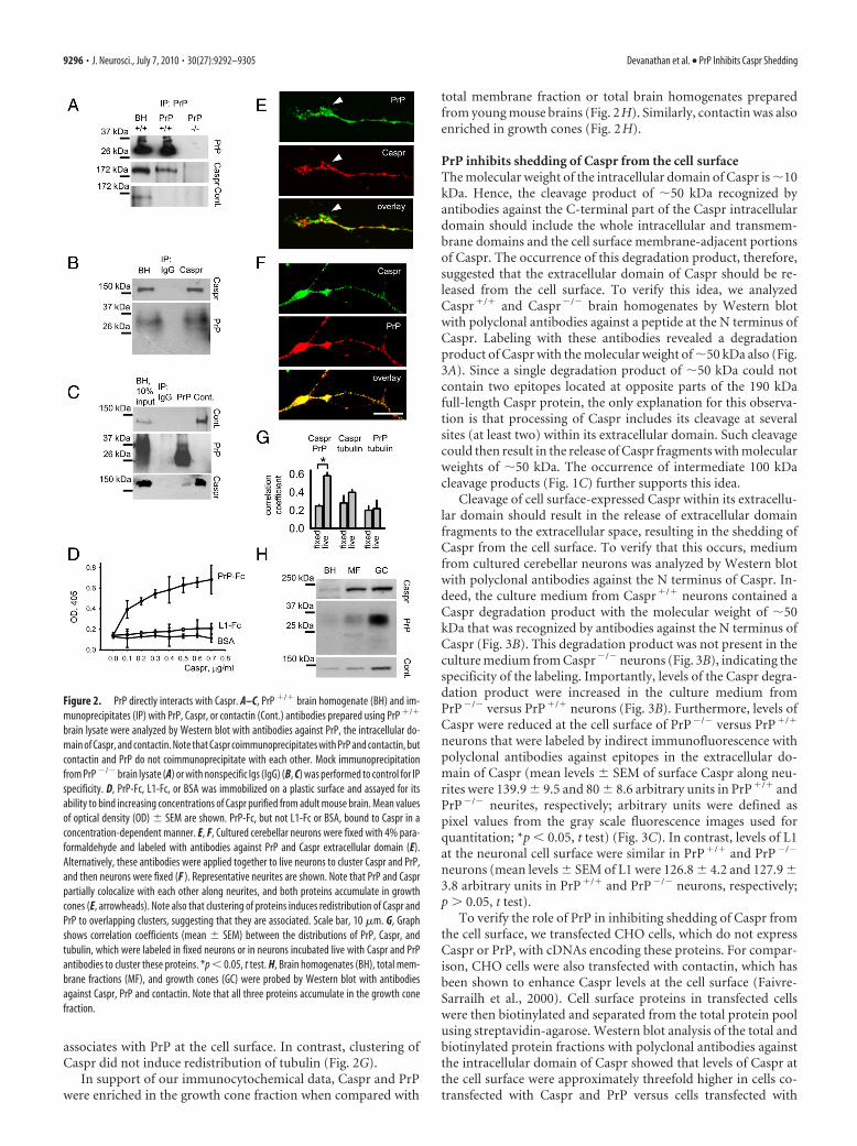
associates with PrP at the cell surface. In contrast, clustering ofCaspr did not induce redistribution of tubulin (Fig. 2G).
In support of our immunocytochemical data, Caspr and PrPwere enriched in the growth cone fraction when compared with
total membrane fraction or total brain homogenates preparedfrom young mouse brains (Fig. 2H). Similarly, contactin was alsoenriched in growth cones (Fig. 2H).
PrP inhibits shedding of Caspr from the cell surfaceThe molecular weight of the intracellular domain of Caspr is �10kDa. Hence, the cleavage product of �50 kDa recognized byantibodies against the C-terminal part of the Caspr intracellulardomain should include the whole intracellular and transmem-brane domains and the cell surface membrane-adjacent portionsof Caspr. The occurrence of this degradation product, therefore,suggested that the extracellular domain of Caspr should be re-leased from the cell surface. To verify this idea, we analyzedCaspr�/� and Caspr�/� brain homogenates by Western blotwith polyclonal antibodies against a peptide at the N terminus ofCaspr. Labeling with these antibodies revealed a degradationproduct of Caspr with the molecular weight of �50 kDa also (Fig.3A). Since a single degradation product of �50 kDa could notcontain two epitopes located at opposite parts of the 190 kDafull-length Caspr protein, the only explanation for this observa-tion is that processing of Caspr includes its cleavage at severalsites (at least two) within its extracellular domain. Such cleavagecould then result in the release of Caspr fragments with molecularweights of �50 kDa. The occurrence of intermediate 100 kDacleavage products (Fig. 1C) further supports this idea.
Cleavage of cell surface-expressed Caspr within its extracellu-lar domain should result in the release of extracellular domainfragments to the extracellular space, resulting in the shedding ofCaspr from the cell surface. To verify that this occurs, mediumfrom cultured cerebellar neurons was analyzed by Western blotwith polyclonal antibodies against the N terminus of Caspr. In-deed, the culture medium from Caspr�/� neurons contained aCaspr degradation product with the molecular weight of �50kDa that was recognized by antibodies against the N terminus ofCaspr (Fig. 3B). This degradation product was not present in theculture medium from Caspr�/� neurons (Fig. 3B), indicating thespecificity of the labeling. Importantly, levels of the Caspr degra-dation product were increased in the culture medium fromPrP�/� versus PrP�/� neurons (Fig. 3B). Furthermore, levels ofCaspr were reduced at the cell surface of PrP�/� versus PrP�/�
neurons that were labeled by indirect immunofluorescence withpolyclonal antibodies against epitopes in the extracellular do-main of Caspr (mean levels � SEM of surface Caspr along neu-rites were 139.9 � 9.5 and 80 � 8.6 arbitrary units in PrP�/� andPrP�/� neurites, respectively; arbitrary units were defined aspixel values from the gray scale fluorescence images used forquantitation; *p � 0.05, t test) (Fig. 3C). In contrast, levels of L1at the neuronal cell surface were similar in PrP�/� and PrP�/�
neurons (mean levels � SEM of L1 were 126.8 � 4.2 and 127.9 �3.8 arbitrary units in PrP�/� and PrP�/� neurons, respectively;p � 0.05, t test).
To verify the role of PrP in inhibiting shedding of Caspr fromthe cell surface, we transfected CHO cells, which do not expressCaspr or PrP, with cDNAs encoding these proteins. For compar-ison, CHO cells were also transfected with contactin, which hasbeen shown to enhance Caspr levels at the cell surface (Faivre-Sarrailh et al., 2000). Cell surface proteins in transfected cellswere then biotinylated and separated from the total protein poolusing streptavidin-agarose. Western blot analysis of the total andbiotinylated protein fractions with polyclonal antibodies againstthe intracellular domain of Caspr showed that levels of Caspr atthe cell surface were approximately threefold higher in cells co-transfected with Caspr and PrP versus cells transfected with
Figure 2. PrP directly interacts with Caspr. A–C, PrP �/� brain homogenate (BH) and im-munoprecipitates (IP) with PrP, Caspr, or contactin (Cont.) antibodies prepared using PrP �/�
brain lysate were analyzed by Western blot with antibodies against PrP, the intracellular do-main of Caspr, and contactin. Note that Caspr coimmunoprecipitates with PrP and contactin, butcontactin and PrP do not coimmunoprecipitate with each other. Mock immunoprecipitationfrom PrP �/� brain lysate (A) or with nonspecific Igs (IgG) (B, C) was performed to control for IPspecificity. D, PrP-Fc, L1-Fc, or BSA was immobilized on a plastic surface and assayed for itsability to bind increasing concentrations of Caspr purified from adult mouse brain. Mean valuesof optical density (OD) � SEM are shown. PrP-Fc, but not L1-Fc or BSA, bound to Caspr in aconcentration-dependent manner. E, F, Cultured cerebellar neurons were fixed with 4% para-formaldehyde and labeled with antibodies against PrP and Caspr extracellular domain (E).Alternatively, these antibodies were applied together to live neurons to cluster Caspr and PrP,and then neurons were fixed (F ). Representative neurites are shown. Note that PrP and Casprpartially colocalize with each other along neurites, and both proteins accumulate in growthcones (E, arrowheads). Note also that clustering of proteins induces redistribution of Caspr andPrP to overlapping clusters, suggesting that they are associated. Scale bar, 10 �m. G, Graphshows correlation coefficients (mean � SEM) between the distributions of PrP, Caspr, andtubulin, which were labeled in fixed neurons or in neurons incubated live with Caspr and PrPantibodies to cluster these proteins. *p � 0.05, t test. H, Brain homogenates (BH), total mem-brane fractions (MF), and growth cones (GC) were probed by Western blot with antibodiesagainst Caspr, PrP and contactin. Note that all three proteins accumulate in the growth conefraction.
9296 • J. Neurosci., July 7, 2010 • 30(27):9292–9305 Devanathan et al. • PrP Inhibits Caspr Shedding
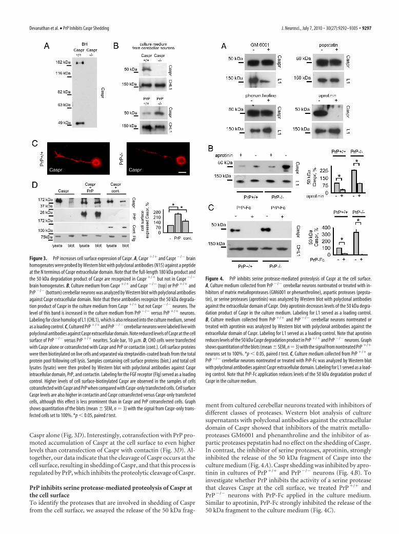
Caspr alone (Fig. 3D). Interestingly, cotransfection with PrP pro-moted accumulation of Caspr at the cell surface to even higherlevels than cotransfection of Caspr with contactin (Fig. 3D). Al-together, our data indicate that the cleavage of Caspr occurs at thecell surface, resulting in shedding of Caspr, and that this process isregulated by PrP, which inhibits the proteolytic cleavage of Caspr.
PrP inhibits serine protease-mediated proteolysis of Caspr atthe cell surfaceTo identify the proteases that are involved in shedding of Casprfrom the cell surface, we assayed the release of the 50 kDa frag-
ment from cultured cerebellar neurons treated with inhibitors ofdifferent classes of proteases. Western blot analysis of culturesupernatants with polyclonal antibodies against the extracellulardomain of Caspr showed that inhibitors of the matrix metallo-proteases GM6001 and phenanthroline and the inhibitor of as-partic proteases pepstatin had no effect on the shedding of Caspr.In contrast, the inhibitor of serine proteases, aprotinin, stronglyinhibited the release of the 50 kDa fragment of Caspr into theculture medium (Fig. 4A). Caspr shedding was inhibited by apro-tinin in cultures of PrP�/� and PrP�/� neurons (Fig. 4B). Toinvestigate whether PrP inhibits the activity of a serine proteasethat cleaves Caspr at the cell surface, we treated PrP�/� andPrP�/� neurons with PrP-Fc applied in the culture medium.Similar to aprotinin, PrP-Fc strongly inhibited the release of the50 kDa fragment to the culture medium (Fig. 4C).
Figure 3. PrP increases cell surface expression of Caspr. A, Caspr �/� and Caspr �/� brainhomogenates were probed by Western blot with polyclonal antibodies (N15) against a peptideat the N terminus of Caspr extracellular domain. Note that the full-length 180 kDa product andthe 50 kDa degradation product of Caspr are recognized in Caspr �/� but not in Caspr �/�
brain homogenates. B, Culture medium from Caspr �/� and Caspr �/� (top) or PrP �/� andPrP �/� (bottom) cerebellar neurons was analyzed by Western blot with polyclonal antibodiesagainst Caspr extracellular domain. Note that these antibodies recognize the 50 kDa degrada-tion product of Caspr in the culture medium from Caspr �/� but not Caspr �/� neurons. Thelevel of this band is increased in the culture medium from PrP �/� versus PrP �/� neurons.Labeling for close homolog of L1 (CHL1), which is also released into the culture medium, servedas a loading control. C, Cultured PrP �/� and PrP �/� cerebellar neurons were labeled live withpolyclonal antibodies against Caspr extracellular domain. Note reduced levels of Caspr at the cellsurface of PrP �/� versus PrP �/� neurites. Scale bar, 10 �m. D, CHO cells were transfectedwith Caspr alone or cotransfected with Caspr and PrP or contactin (cont.). Cell surface proteinswere then biotinylated on live cells and separated via streptavidin-coated beads from the totalprotein pool following cell lysis. Samples containing cell surface proteins (biot.) and total celllysates (lysate) were then probed by Western blot with polyclonal antibodies against Casprintracellular domain, PrP, and contactin. Labeling for the FGF receptor (Flg) served as a loadingcontrol. Higher levels of cell surface-biotinylated Caspr are observed in the samples of cellscotransfected with Caspr and PrP when compared with Caspr-only transfected cells. Cell surfaceCaspr levels are also higher in contactin and Caspr cotransfected versus Caspr-only transfectedcells, although this effect is less prominent than in Caspr and PrP cotransfected cells. Graphshows quantitation of the blots (mean � SEM, n 3) with the signal from Caspr-only trans-fected cells set to 100%. *p � 0.05, paired t test.
Figure 4. PrP inhibits serine protease-mediated proteolysis of Caspr at the cell surface.A, Culture medium collected from PrP �/� cerebellar neurons nontreated or treated with in-hibitors of matrix metalloproteases (GM6001 or phenanthroline), aspartic proteases (pepsta-tin), or serine proteases (aprotinin) was analyzed by Western blot with polyclonal antibodiesagainst the extracellular domain of Caspr. Only aprotinin decreases levels of the 50 kDa degra-dation product of Caspr in the culture medium. Labeling for L1 served as a loading control.B, Culture medium collected from PrP �/� and PrP �/� cerebellar neurons nontreated ortreated with aprotinin was analyzed by Western blot with polyclonal antibodies against theextracellular domain of Caspr. Labeling for L1 served as a loading control. Note that aprotininreduces levels of the 50 kDa Caspr degradation product in PrP �/� and PrP �/� neurons. Graphshows quantitation of the blots (mean�SEM, n3) with the signal from nontreated PrP �/�
neurons set to 100%. *p � 0.05, paired t test. C, Culture medium collected from PrP �/� orPrP �/� cerebellar neurons nontreated or treated with PrP-Fc was analyzed by Western blotwith polyclonal antibodies against Caspr extracellular domain. Labeling for L1 served as a load-ing control. Note that PrP-Fc application reduces levels of the 50 kDa degradation product ofCaspr in the culture medium.
Devanathan et al. • PrP Inhibits Caspr Shedding J. Neurosci., July 7, 2010 • 30(27):9292–9305 • 9297
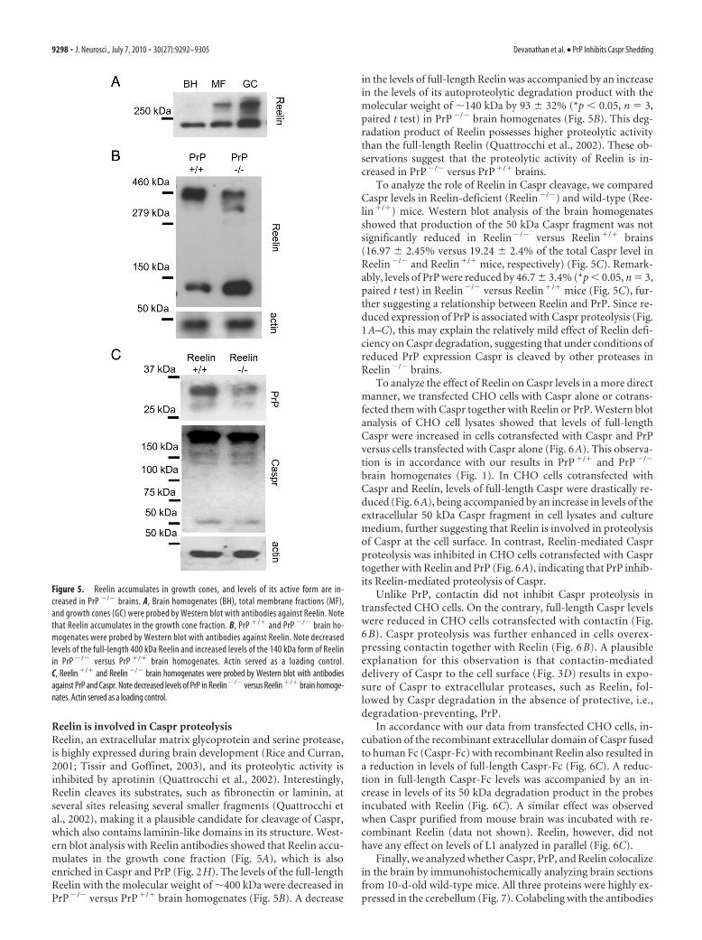
Reelin is involved in Caspr proteolysisReelin, an extracellular matrix glycoprotein and serine protease,is highly expressed during brain development (Rice and Curran,2001; Tissir and Goffinet, 2003), and its proteolytic activity isinhibited by aprotinin (Quattrocchi et al., 2002). Interestingly,Reelin cleaves its substrates, such as fibronectin or laminin, atseveral sites releasing several smaller fragments (Quattrocchi etal., 2002), making it a plausible candidate for cleavage of Caspr,which also contains laminin-like domains in its structure. West-ern blot analysis with Reelin antibodies showed that Reelin accu-mulates in the growth cone fraction (Fig. 5A), which is alsoenriched in Caspr and PrP (Fig. 2H). The levels of the full-lengthReelin with the molecular weight of �400 kDa were decreased inPrP�/� versus PrP�/� brain homogenates (Fig. 5B). A decrease
in the levels of full-length Reelin was accompanied by an increasein the levels of its autoproteolytic degradation product with themolecular weight of �140 kDa by 93 � 32% (*p � 0.05, n 3,paired t test) in PrP�/� brain homogenates (Fig. 5B). This deg-radation product of Reelin possesses higher proteolytic activitythan the full-length Reelin (Quattrocchi et al., 2002). These ob-servations suggest that the proteolytic activity of Reelin is in-creased in PrP�/� versus PrP�/� brains.
To analyze the role of Reelin in Caspr cleavage, we comparedCaspr levels in Reelin-deficient (Reelin�/�) and wild-type (Ree-lin�/�) mice. Western blot analysis of the brain homogenatesshowed that production of the 50 kDa Caspr fragment was notsignificantly reduced in Reelin�/� versus Reelin�/� brains(16.97 � 2.45% versus 19.24 � 2.4% of the total Caspr level inReelin�/� and Reelin�/� mice, respectively) (Fig. 5C). Remark-ably, levels of PrP were reduced by 46.7 � 3.4% (*p � 0.05, n 3,paired t test) in Reelin�/� versus Reelin�/� mice (Fig. 5C), fur-ther suggesting a relationship between Reelin and PrP. Since re-duced expression of PrP is associated with Caspr proteolysis (Fig.1A–C), this may explain the relatively mild effect of Reelin defi-ciency on Caspr degradation, suggesting that under conditions ofreduced PrP expression Caspr is cleaved by other proteases inReelin�/� brains.
To analyze the effect of Reelin on Caspr levels in a more directmanner, we transfected CHO cells with Caspr alone or cotrans-fected them with Caspr together with Reelin or PrP. Western blotanalysis of CHO cell lysates showed that levels of full-lengthCaspr were increased in cells cotransfected with Caspr and PrPversus cells transfected with Caspr alone (Fig. 6A). This observa-tion is in accordance with our results in PrP�/� and PrP�/�
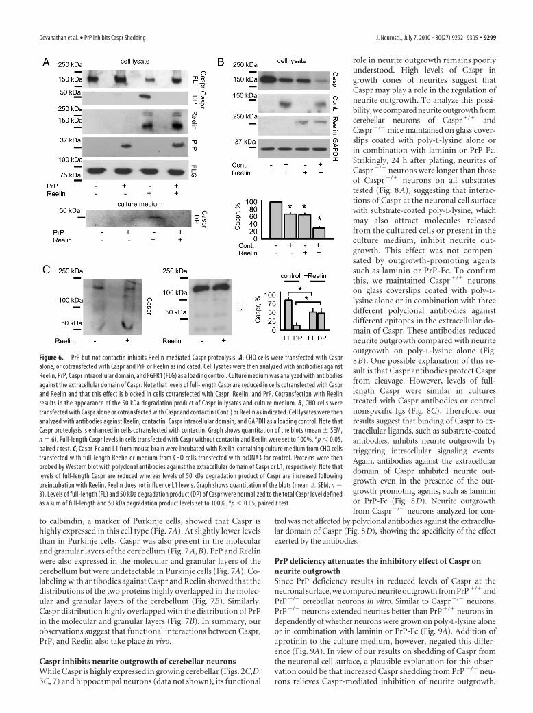
brain homogenates (Fig. 1). In CHO cells cotransfected withCaspr and Reelin, levels of full-length Caspr were drastically re-duced (Fig. 6A), being accompanied by an increase in levels of theextracellular 50 kDa Caspr fragment in cell lysates and culturemedium, further suggesting that Reelin is involved in proteolysisof Caspr at the cell surface. In contrast, Reelin-mediated Casprproteolysis was inhibited in CHO cells cotransfected with Casprtogether with Reelin and PrP (Fig. 6A), indicating that PrP inhib-its Reelin-mediated proteolysis of Caspr.
Unlike PrP, contactin did not inhibit Caspr proteolysis intransfected CHO cells. On the contrary, full-length Caspr levelswere reduced in CHO cells cotransfected with contactin (Fig.6 B). Caspr proteolysis was further enhanced in cells overex-pressing contactin together with Reelin (Fig. 6 B). A plausibleexplanation for this observation is that contactin-mediateddelivery of Caspr to the cell surface (Fig. 3D) results in expo-sure of Caspr to extracellular proteases, such as Reelin, fol-lowed by Caspr degradation in the absence of protective, i.e.,degradation-preventing, PrP.
In accordance with our data from transfected CHO cells, in-cubation of the recombinant extracellular domain of Caspr fusedto human Fc (Caspr-Fc) with recombinant Reelin also resulted ina reduction in levels of full-length Caspr-Fc (Fig. 6C). A reduc-tion in full-length Caspr-Fc levels was accompanied by an in-crease in levels of its 50 kDa degradation product in the probesincubated with Reelin (Fig. 6C). A similar effect was observedwhen Caspr purified from mouse brain was incubated with re-combinant Reelin (data not shown). Reelin, however, did nothave any effect on levels of L1 analyzed in parallel (Fig. 6C).
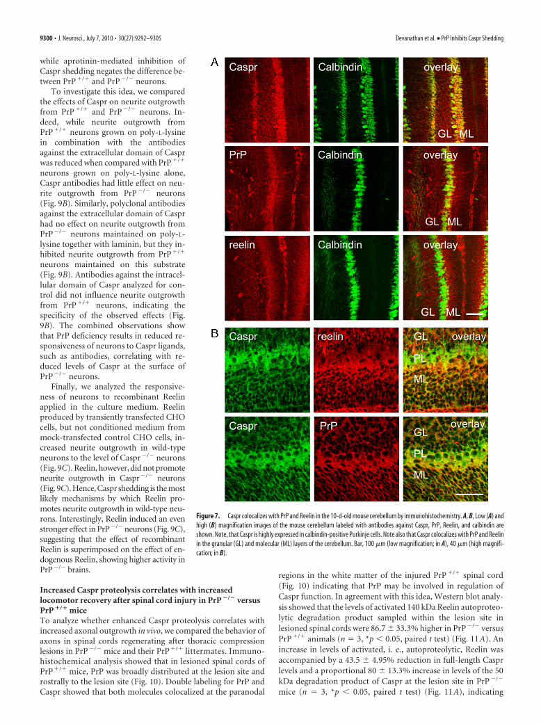
Finally, we analyzed whether Caspr, PrP, and Reelin colocalizein the brain by immunohistochemically analyzing brain sectionsfrom 10-d-old wild-type mice. All three proteins were highly ex-pressed in the cerebellum (Fig. 7). Colabeling with the antibodies
Figure 5. Reelin accumulates in growth cones, and levels of its active form are in-creased in PrP �/� brains. A, Brain homogenates (BH), total membrane fractions (MF),and growth cones (GC) were probed by Western blot with antibodies against Reelin. Notethat Reelin accumulates in the growth cone fraction. B, PrP �/� and PrP �/� brain ho-mogenates were probed by Western blot with antibodies against Reelin. Note decreasedlevels of the full-length 400 kDa Reelin and increased levels of the 140 kDa form of Reelinin PrP �/� versus PrP �/� brain homogenates. Actin served as a loading control.C, Reelin �/� and Reelin �/� brain homogenates were probed by Western blot with antibodiesagainst PrP and Caspr. Note decreased levels of PrP in Reelin �/� versus Reelin �/� brain homoge-nates. Actin served as a loading control.
9298 • J. Neurosci., July 7, 2010 • 30(27):9292–9305 Devanathan et al. • PrP Inhibits Caspr Shedding
to calbindin, a marker of Purkinje cells, showed that Caspr ishighly expressed in this cell type (Fig. 7A). At slightly lower levelsthan in Purkinje cells, Caspr was also present in the molecularand granular layers of the cerebellum (Fig. 7A,B). PrP and Reelinwere also expressed in the molecular and granular layers of thecerebellum but were undetectable in Purkinje cells (Fig. 7A). Co-labeling with antibodies against Caspr and Reelin showed that thedistributions of the two proteins highly overlapped in the molec-ular and granular layers of the cerebellum (Fig. 7B). Similarly,Caspr distribution highly overlapped with the distribution of PrPin the molecular and granular layers (Fig. 7B). In summary, ourobservations suggest that functional interactions between Caspr,PrP, and Reelin also take place in vivo.
Caspr inhibits neurite outgrowth of cerebellar neuronsWhile Caspr is highly expressed in growing cerebellar (Figs. 2C,D,3C, 7) and hippocampal neurons (data not shown), its functional
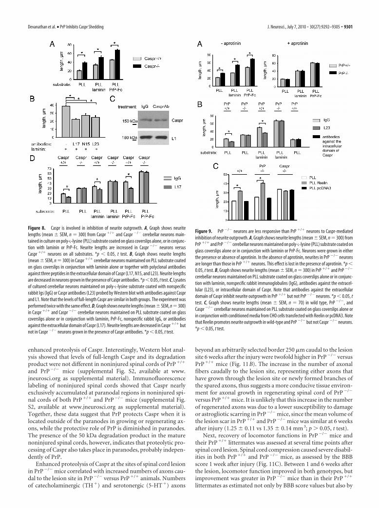
role in neurite outgrowth remains poorlyunderstood. High levels of Caspr ingrowth cones of neurites suggest thatCaspr may play a role in the regulation ofneurite outgrowth. To analyze this possi-bility, we compared neurite outgrowth fromcerebellar neurons of Caspr�/� andCaspr�/� mice maintained on glass cover-slips coated with poly-L-lysine alone orin combination with laminin or PrP-Fc.Strikingly, 24 h after plating, neurites ofCaspr�/� neurons were longer than thoseof Caspr�/� neurons on all substratestested (Fig. 8A), suggesting that interac-tions of Caspr at the neuronal cell surfacewith substrate-coated poly-L-lysine, whichmay also attract molecules releasedfrom the cultured cells or present in theculture medium, inhibit neurite out-growth. This effect was not compen-sated by outgrowth-promoting agentssuch as laminin or PrP-Fc. To confirmthis, we maintained Caspr�/� neuronson glass coverslips coated with poly-L-lysine alone or in combination with threedifferent polyclonal antibodies againstdifferent epitopes in the extracellular do-main of Caspr. These antibodies reducedneurite outgrowth compared with neuriteoutgrowth on poly-L-lysine alone (Fig.8B). One possible explanation of this re-sult is that Caspr antibodies protect Casprfrom cleavage. However, levels of full-length Caspr were similar in culturestreated with Caspr antibodies or controlnonspecific Igs (Fig. 8C). Therefore, ourresults suggest that binding of Caspr to ex-tracellular ligands, such as substrate-coatedantibodies, inhibits neurite outgrowth bytriggering intracellular signaling events.Again, antibodies against the extracellulardomain of Caspr inhibited neurite out-growth even in the presence of the out-growth promoting agents, such as lamininor PrP-Fc (Fig. 8D). Neurite outgrowthfrom Caspr�/� neurons analyzed for con-
trol was not affected by polyclonal antibodies against the extracellu-lar domain of Caspr (Fig. 8D), showing the specificity of the effectexerted by the antibodies.
PrP deficiency attenuates the inhibitory effect of Caspr onneurite outgrowthSince PrP deficiency results in reduced levels of Caspr at theneuronal surface, we compared neurite outgrowth from PrP�/� andPrP�/� cerebellar neurons in vitro. Similar to Caspr�/� neurons,PrP�/� neurons extended neurites better than PrP�/� neurons in-dependently of whether neurons were grown on poly-L-lysine aloneor in combination with laminin or PrP-Fc (Fig. 9A). Addition ofaprotinin to the culture medium, however, negated this differ-ence (Fig. 9A). In view of our results on shedding of Caspr fromthe neuronal cell surface, a plausible explanation for this obser-vation could be that increased Caspr shedding from PrP�/� neu-rons relieves Caspr-mediated inhibition of neurite outgrowth,
Figure 6. PrP but not contactin inhibits Reelin-mediated Caspr proteolysis. A, CHO cells were transfected with Caspralone, or cotransfected with Caspr and PrP or Reelin as indicated. Cell lysates were then analyzed with antibodies againstReelin, PrP, Caspr intracellular domain, and FGFR1 (FLG) as a loading control. Culture medium was analyzed with antibodiesagainst the extracellular domain of Caspr. Note that levels of full-length Caspr are reduced in cells cotransfected with Casprand Reelin and that this effect is blocked in cells cotransfected with Caspr, Reelin, and PrP. Cotransfection with Reelinresults in the appearance of the 50 kDa degradation product of Caspr in lysates and culture medium. B, CHO cells weretransfected with Caspr alone or cotransfected with Caspr and contactin (Cont.) or Reelin as indicated. Cell lysates were thenanalyzed with antibodies against Reelin, contactin, Caspr intracellular domain, and GAPDH as a loading control. Note thatCaspr proteolysis is enhanced in cells cotransfected with contactin. Graph shows quantitation of the blots (mean � SEM,n 6). Full-length Caspr levels in cells transfected with Caspr without contactin and Reelin were set to 100%. *p � 0.05,paired t test. C, Caspr-Fc and L1 from mouse brain were incubated with Reelin-containing culture medium from CHO cellstransfected with full-length Reelin or medium from CHO cells transfected with pcDNA3 for control. Proteins were thenprobed by Western blot with polyclonal antibodies against the extracellular domain of Caspr or L1, respectively. Note thatlevels of full-length Caspr are reduced whereas levels of 50 kDa degradation product of Caspr are increased followingpreincubation with Reelin. Reelin does not influence L1 levels. Graph shows quantitation of the blots (mean � SEM, n 3). Levels of full-length (FL) and 50 kDa degradation product (DP) of Caspr were normalized to the total Caspr level definedas a sum of full-length and 50 kDa degradation product levels set to 100%. *p � 0.05, paired t test.
Devanathan et al. • PrP Inhibits Caspr Shedding J. Neurosci., July 7, 2010 • 30(27):9292–9305 • 9299
while aprotinin-mediated inhibition ofCaspr shedding negates the difference be-tween PrP�/� and PrP�/� neurons.
To investigate this idea, we comparedthe effects of Caspr on neurite outgrowthfrom PrP�/� and PrP�/� neurons. In-deed, while neurite outgrowth fromPrP�/� neurons grown on poly-L-lysinein combination with the antibodiesagainst the extracellular domain of Casprwas reduced when compared with PrP�/�
neurons grown on poly-L-lysine alone,Caspr antibodies had little effect on neu-rite outgrowth from PrP�/� neurons(Fig. 9B). Similarly, polyclonal antibodiesagainst the extracellular domain of Casprhad no effect on neurite outgrowth fromPrP�/� neurons maintained on poly-L-lysine together with laminin, but they in-hibited neurite outgrowth from PrP�/�
neurons maintained on this substrate(Fig. 9B). Antibodies against the intracel-lular domain of Caspr analyzed for con-trol did not influence neurite outgrowthfrom PrP�/� neurons, indicating thespecificity of the observed effects (Fig.9B). The combined observations showthat PrP deficiency results in reduced re-sponsiveness of neurons to Caspr ligands,such as antibodies, correlating with re-duced levels of Caspr at the surface ofPrP�/� neurons.
Finally, we analyzed the responsive-ness of neurons to recombinant Reelinapplied in the culture medium. Reelinproduced by transiently transfected CHOcells, but not conditioned medium frommock-transfected control CHO cells, in-creased neurite outgrowth in wild-typeneurons to the level of Caspr�/� neurons(Fig. 9C). Reelin, however, did not promoteneurite outgrowth in Caspr�/� neurons(Fig. 9C). Hence, Caspr shedding is the mostlikely mechanisms by which Reelin pro-motes neurite outgrowth in wild-type neu-rons. Interestingly, Reelin induced an evenstronger effect in PrP�/� neurons (Fig. 9C),suggesting that the effect of recombinantReelin is superimposed on the effect of en-dogenous Reelin, showing higher activity inPrP�/� brains.
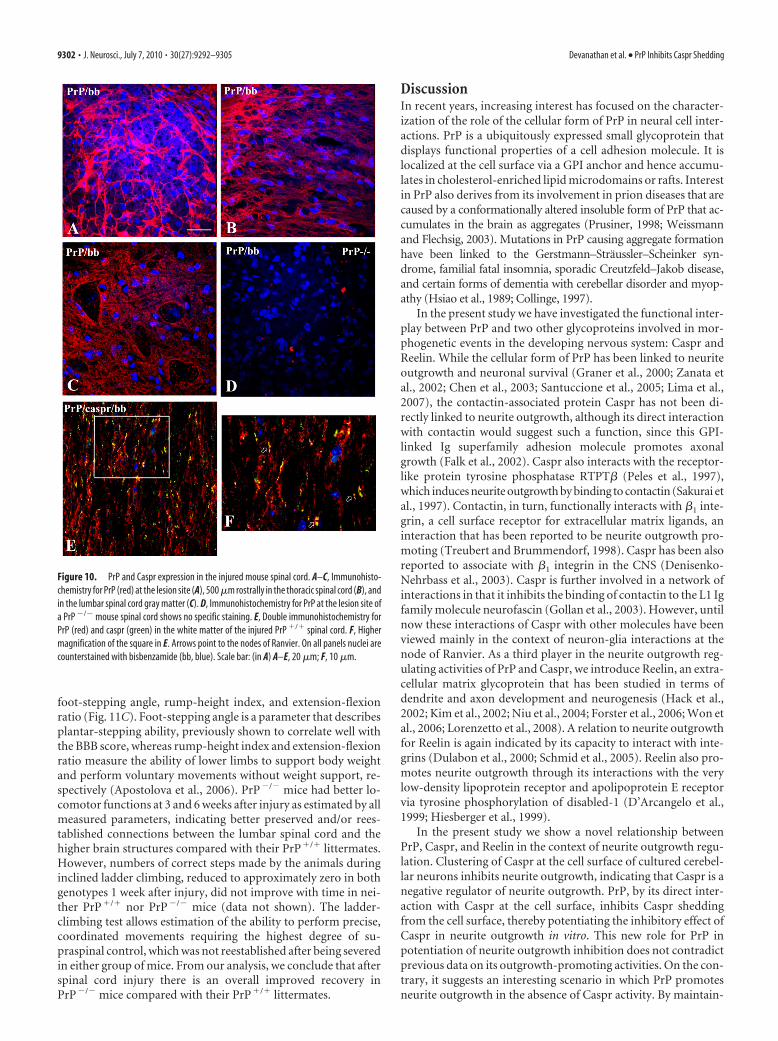
Increased Caspr proteolysis correlates with increasedlocomotor recovery after spinal cord injury in PrP �/� versusPrP �/� miceTo analyze whether enhanced Caspr proteolysis correlates withincreased axonal outgrowth in vivo, we compared the behavior ofaxons in spinal cords regenerating after thoracic compressionlesions in PrP�/� mice and their PrP�/� littermates. Immuno-histochemical analysis showed that in lesioned spinal cords ofPrP�/� mice, PrP was broadly distributed at the lesion site androstrally to the lesion site (Fig. 10). Double labeling for PrP andCaspr showed that both molecules colocalized at the paranodal
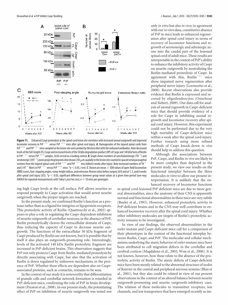
regions in the white matter of the injured PrP�/� spinal cord(Fig. 10) indicating that PrP may be involved in regulation ofCaspr function. In agreement with this idea, Western blot analy-sis showed that the levels of activated 140 kDa Reelin autoproteo-lytic degradation product sampled within the lesion site inlesioned spinal cords were 86.7 � 33.3% higher in PrP�/� versusPrP�/� animals (n 3, *p � 0.05, paired t test) (Fig. 11A). Anincrease in levels of activated, i. e., autoproteolytic, Reelin wasaccompanied by a 43.5 � 4.95% reduction in full-length Casprlevels and a proportional 80 � 13.3% increase in levels of the 50kDa degradation product of Caspr at the lesion site in PrP�/�
mice (n 3, *p � 0.05, paired t test) (Fig. 11A), indicating
Figure 7. Caspr colocalizes with PrP and Reelin in the 10-d-old mouse cerebellum by immunohistochemistry. A, B, Low (A) andhigh (B) magnification images of the mouse cerebellum labeled with antibodies against Caspr, PrP, Reelin, and calbindin areshown. Note, that Caspr is highly expressed in calbindin-positive Purkinje cells. Note also that Caspr colocalizes with PrP and Reelinin the granular (GL) and molecular (ML) layers of the cerebellum. Bar, 100 �m (low magnification; in A), 40 �m (high magnifi-cation; in B).
9300 • J. Neurosci., July 7, 2010 • 30(27):9292–9305 Devanathan et al. • PrP Inhibits Caspr Shedding
enhanced proteolysis of Caspr. Interestingly, Western blot anal-ysis showed that levels of full-length Caspr and its degradationproduct were not different in noninjured spinal cords of PrP�/�
and PrP�/� mice (supplemental Fig. S2, available at www.jneurosci.org as supplemental material). Immunofluorescencelabeling of noninjured spinal cords showed that Caspr nearlyexclusively accumulated at paranodal regions in noninjured spi-nal cords of both PrP�/� and PrP�/� mice (supplemental Fig.S2, available at www.jneurosci.org as supplemental material).Together, these data suggest that PrP protects Caspr when it islocated outside of the paranodes in growing or regenerating ax-ons, while the protective role of PrP is diminished in paranodes.The presence of the 50 kDa degradation product in the maturenoninjured spinal cords, however, indicates that proteolytic pro-cessing of Caspr also takes place in paranodes, probably indepen-dently of PrP.
Enhanced proteolysis of Caspr at the sites of spinal cord lesionin PrP�/� mice correlated with increased numbers of axons cau-dal to the lesion site in PrP�/� versus PrP�/� animals. Numbersof catecholaminergic (TH�) and serotonergic (5-HT�) axons
beyond an arbitrarily selected border 250 �m caudal to the lesionsite 6 weeks after the injury were twofold higher in PrP�/� versusPrP�/� mice (Fig. 11B). The increase in the number of axonalfibers caudally to the lesion site, representing either axons thathave grown through the lesion site or newly formed branches ofthe spared axons, thus suggests a more conducive tissue environ-ment for axonal growth in regenerating spinal cord of PrP�/�
versus PrP�/� mice. It is unlikely that this increase in the numberof regenerated axons was due to a lower susceptibility to damageor astrogliotic scarring in PrP�/� mice, since the mean volume ofthe lesion scar in PrP�/� and PrP�/� mice was similar at 6 weeksafter injury (1.25 � 0.11 vs 1.35 � 0.14 mm 3; p � 0.05, t test).
Next, recovery of locomotor functions in PrP�/� mice andtheir PrP�/� littermates was assessed at several time points afterspinal cord lesion. Spinal cord compression caused severe disabil-ities in both PrP�/� and PrP�/� mice, as assessed by the BBBscore 1 week after injury (Fig. 11C). Between 1 and 6 weeks afterthe lesion, locomotor function improved in both genotypes, butimprovement was greater in PrP�/� mice than in their PrP�/�
littermates as estimated not only by BBB score values but also by
Figure 8. Caspr is involved in inhibition of neurite outgrowth. A, Graph shows neuritelengths (mean � SEM, n 300) from Caspr �/� and Caspr �/� cerebellar neurons main-tained in culture on poly-L-lysine (PLL) substrate coated on glass coverslips alone, or in conjunc-tion with laminin or PrP-Fc. Neurite lengths are increased in Caspr �/� neurons versusCaspr �/� neurons on all substrates. *p � 0.05, t test. B, Graph shows neurite lengths(mean � SEM, n 300) in Caspr �/� cerebellar neurons maintained on PLL substrate coatedon glass coverslips in conjunction with laminin alone or together with polyclonal antibodiesagainst three peptides in the extracellular domain of Caspr (L17, N15, and L23). Neurite lengthsare decreased in neurons grown in the presence of Caspr antibodies. *p � 0.05, t test. C, Lysatesof cultured cerebellar neurons maintained on poly-L-lysine substrate coated with nonspecificrabbit Igs (IgG) or Caspr antibodies (L23) probed by Western blot with antibodies against Casprand L1. Note that the levels of full-length Caspr are similar in both groups. The experiment wasperformed twice with the same effect. D, Graph shows neurite lengths (mean � SEM, n 300)in Caspr �/� and Caspr �/� cerebellar neurons maintained on PLL substrate coated on glasscoverslips alone or in conjunction with laminin, PrP-Fc, nonspecific rabbit IgG, or antibodiesagainst the extracellular domain of Caspr (L17). Neurite lengths are decreased in Caspr �/� butnot in Caspr �/� neurons grown in the presence of Caspr antibodies. *p � 0.05, t test.
Figure 9. PrP �/� neurons are less responsive than PrP �/� neurons to Caspr-mediatedinhibition of neurite outgrowth. A, Graph shows neurite lengths (mean � SEM, n 300) fromPrP �/� and PrP �/� cerebellar neurons maintained on poly-L-lysine (PLL) substrate coated onglass coverslips alone or in conjunction with laminin or PrP-Fc. Neurons were grown in eitherthe presence or absence of aprotinin. In the absence of aprotinin, neurites in PrP �/� neuronsare longer than those in PrP �/� neurons. This effect is lost in the presence of aprotinin. *p �0.05, t test. B, Graph shows neurite lengths (mean � SEM, n 300) in PrP �/� and PrP �/�
cerebellar neurons maintained on PLL substrate coated on glass coverslips alone or in conjunc-tion with laminin, nonspecific rabbit immunoglobulins (IgG), antibodies against the extracel-lular (L23), or intracellular domain of Caspr. Note that antibodies against the extracellulardomain of Caspr inhibit neurite outgrowth in PrP �/� but not PrP �/� neurons. *p � 0.05, ttest. C, Graph shows neurite lengths (mean � SEM, n 70) in wild type, PrP �/�, andCaspr �/� cerebellar neurons maintained on PLL substrate coated on glass coverslips alone orin conjunction with conditioned media from CHO cells transfected with Reelin or pcDNA3. Notethat Reelin promotes neurite outgrowth in wild-type and PrP �/� but not Caspr �/� neurons.*p � 0.05, t test.
Devanathan et al. • PrP Inhibits Caspr Shedding J. Neurosci., July 7, 2010 • 30(27):9292–9305 • 9301
foot-stepping angle, rump-height index, and extension-flexionratio (Fig. 11C). Foot-stepping angle is a parameter that describesplantar-stepping ability, previously shown to correlate well withthe BBB score, whereas rump-height index and extension-flexionratio measure the ability of lower limbs to support body weightand perform voluntary movements without weight support, re-spectively (Apostolova et al., 2006). PrP�/� mice had better lo-comotor functions at 3 and 6 weeks after injury as estimated by allmeasured parameters, indicating better preserved and/or rees-tablished connections between the lumbar spinal cord and thehigher brain structures compared with their PrP�/� littermates.However, numbers of correct steps made by the animals duringinclined ladder climbing, reduced to approximately zero in bothgenotypes 1 week after injury, did not improve with time in nei-ther PrP�/� nor PrP�/� mice (data not shown). The ladder-climbing test allows estimation of the ability to perform precise,coordinated movements requiring the highest degree of su-praspinal control, which was not reestablished after being severedin either group of mice. From our analysis, we conclude that afterspinal cord injury there is an overall improved recovery inPrP�/� mice compared with their PrP�/� littermates.
DiscussionIn recent years, increasing interest has focused on the character-ization of the role of the cellular form of PrP in neural cell inter-actions. PrP is a ubiquitously expressed small glycoprotein thatdisplays functional properties of a cell adhesion molecule. It islocalized at the cell surface via a GPI anchor and hence accumu-lates in cholesterol-enriched lipid microdomains or rafts. Interestin PrP also derives from its involvement in prion diseases that arecaused by a conformationally altered insoluble form of PrP that ac-cumulates in the brain as aggregates (Prusiner, 1998; Weissmannand Flechsig, 2003). Mutations in PrP causing aggregate formationhave been linked to the Gerstmann–Straussler–Scheinker syn-drome, familial fatal insomnia, sporadic Creutzfeld–Jakob disease,and certain forms of dementia with cerebellar disorder and myop-athy (Hsiao et al., 1989; Collinge, 1997).
In the present study we have investigated the functional inter-play between PrP and two other glycoproteins involved in mor-phogenetic events in the developing nervous system: Caspr andReelin. While the cellular form of PrP has been linked to neuriteoutgrowth and neuronal survival (Graner et al., 2000; Zanata etal., 2002; Chen et al., 2003; Santuccione et al., 2005; Lima et al.,2007), the contactin-associated protein Caspr has not been di-rectly linked to neurite outgrowth, although its direct interactionwith contactin would suggest such a function, since this GPI-linked Ig superfamily adhesion molecule promotes axonalgrowth (Falk et al., 2002). Caspr also interacts with the receptor-like protein tyrosine phosphatase RTPT� (Peles et al., 1997),which induces neurite outgrowth by binding to contactin (Sakurai etal., 1997). Contactin, in turn, functionally interacts with �1 inte-grin, a cell surface receptor for extracellular matrix ligands, aninteraction that has been reported to be neurite outgrowth pro-moting (Treubert and Brummendorf, 1998). Caspr has been alsoreported to associate with �1 integrin in the CNS (Denisenko-Nehrbass et al., 2003). Caspr is further involved in a network ofinteractions in that it inhibits the binding of contactin to the L1 Igfamily molecule neurofascin (Gollan et al., 2003). However, untilnow these interactions of Caspr with other molecules have beenviewed mainly in the context of neuron-glia interactions at thenode of Ranvier. As a third player in the neurite outgrowth reg-ulating activities of PrP and Caspr, we introduce Reelin, an extra-cellular matrix glycoprotein that has been studied in terms ofdendrite and axon development and neurogenesis (Hack et al.,2002; Kim et al., 2002; Niu et al., 2004; Forster et al., 2006; Won etal., 2006; Lorenzetto et al., 2008). A relation to neurite outgrowthfor Reelin is again indicated by its capacity to interact with inte-grins (Dulabon et al., 2000; Schmid et al., 2005). Reelin also pro-motes neurite outgrowth through its interactions with the verylow-density lipoprotein receptor and apolipoprotein E receptorvia tyrosine phosphorylation of disabled-1 (D’Arcangelo et al.,1999; Hiesberger et al., 1999).
In the present study we show a novel relationship betweenPrP, Caspr, and Reelin in the context of neurite outgrowth regu-lation. Clustering of Caspr at the cell surface of cultured cerebel-lar neurons inhibits neurite outgrowth, indicating that Caspr is anegative regulator of neurite outgrowth. PrP, by its direct inter-action with Caspr at the cell surface, inhibits Caspr sheddingfrom the cell surface, thereby potentiating the inhibitory effect ofCaspr in neurite outgrowth in vitro. This new role for PrP inpotentiation of neurite outgrowth inhibition does not contradictprevious data on its outgrowth-promoting activities. On the con-trary, it suggests an interesting scenario in which PrP promotesneurite outgrowth in the absence of Caspr activity. By maintain-
Figure 10. PrP and Caspr expression in the injured mouse spinal cord. A–C, Immunohisto-chemistry for PrP (red) at the lesion site (A), 500 �m rostrally in the thoracic spinal cord (B), andin the lumbar spinal cord gray matter (C). D, Immunohistochemistry for PrP at the lesion site ofa PrP �/� mouse spinal cord shows no specific staining. E, Double immunohistochemistry forPrP (red) and caspr (green) in the white matter of the injured PrP �/� spinal cord. F, Highermagnification of the square in E. Arrows point to the nodes of Ranvier. On all panels nuclei arecounterstained with bisbenzamide (bb, blue). Scale bar: (in A) A–E, 20 �m; F, 10 �m.
9302 • J. Neurosci., July 7, 2010 • 30(27):9292–9305 Devanathan et al. • PrP Inhibits Caspr Shedding
ing high Caspr levels at the cell surface, PrP allows neurites torespond promptly to Caspr activation that would arrest neuriteoutgrowth when the proper targets are reached.
In the present study, we confirmed Reelin’s function as a pro-tease rather than as a ligand for integrins or lipoprotein receptors.This proteolytic activity of Reelin (Quattrocchi et al., 2002) ap-pears to play a role in regulating the Caspr-dependent inhibitionof neurite outgrowth of cerebellar neurons; in the absence of PrP,Reelin proteolytically cleaves the extracellular domain of Caspr,thus reducing the capacity of Caspr to decrease neurite out-growth. The functions of the extracellular 50 kDa fragment ofCaspr produced by Reelin are not known, but it is possible that byitself it also plays an outgrowth-promoting role. Interestingly,levels of the activated 140 kDa Reelin proteolytic fragment areincreased in PrP-deficient brains. This observation suggests thatPrP not only protects Caspr from Reelin-mediated proteolysis bydirectly associating with Caspr, but also that the activation ofReelin is down regulated by unknown mechanisms in the pres-ence of PrP. Whether these effects are dependent also on Caspr-associated proteins, such as contactin, remains to be seen.
In the context of our study it is noteworthy that differentiationof granule cells and cerebellar network formation is retarded inPrP-deficient mice, confirming the role of PrP in brain develop-ment (Prestori et al., 2008). In our present study, the potentiatingeffect of PrP on inhibition of neurite outgrowth was tested not
only in vitro but also in vivo; in agreementwith our in vitro data, constitutive absenceof PrP in mice leads to enhanced regener-ation after spinal cord injury in terms ofrecovery of locomotor functions and re-growth of serotonergic and adrenergic ax-ons into the caudal part of the lesionedspinal cord of adult mice. These results areinterpretable in the context of PrP’s abilityto enhance the inhibitory activity of Caspron neurite outgrowth by neutralizing theReelin-mediated proteolysis of Caspr. Inagreement with this, Reelin�/� miceshow impaired nerve regeneration afterperipheral nerve injury (Lorenzetto et al.,2008). Recent observations also provideevidence that Reelin is expressed and se-creted by oligodendrocytes (Osterhoutand Siebert, 2009). Our data call for anal-ysis of axonal regrowth in Caspr-deficientmice that should provide evidence of arole for Caspr in inhibiting axonal re-growth and locomotor recovery after spi-nal cord injury. However, this experimentcould not be performed due to the veryhigh mortality of Caspr-deficient micewithin a week after the spinal cord injury.Further research using more refinedmethods of Caspr knock-down in vivoshould help to address this question.
Although the associations betweenPrP, Caspr, and Reelin in vivo are likely tobe more complex than depicted in thepresent study, we view our results on thefunctional interplay between the threemolecules in vitro to allow our present in-terpretation. It is unlikely that the en-hanced recovery of locomotor functions
in spinal cord-lesioned PrP deficient mice are due to more gen-eral abnormalities, since the anatomy of their CNS is apparentlynormal and functional abnormalities in these mice are very subtle(Bueler et al., 1992). However, enhanced proteolytic activity inPrP-deficient brains and in the CNS may well contribute to en-hanced locomotor recovery after the spinal cord injury. Whetherother inhibitory molecules are targets of Reelin’s proteolytic ac-tivity remains to be investigated.
In view of our findings, the observed abnormalities in thereeler mutant and Caspr-deficient mice call for a comparison oftheir phenotypes in the context of the functional interplay be-tween Reelin, Caspr, and PrP. The molecular and cellular mech-anisms underlying the ataxic behavior of reeler mutant mice havebeen attributed to cell migration defects in the cerebellar andcerebral cortices (Magdaleno et al., 2002; Won et al., 2006). It isnot known, however, how these relate to the absence of the pro-teolytic activity of Reelin. The ataxic defects of Caspr-deficientmice have been mostly related to the abnormal structure of nodesof Ranvier in the central and peripheral nervous systems (Bhat etal., 2001), but they also could be related in view of our presentobservations in the context of an altered balance between neuriteoutgrowth-promoting and neurite outgrowth-inhibitory cues.The relation of these molecules to transmitter receptors, ionchannels, and ion transporters that have emerged recently as im-
Figure 11. Enhanced Caspr proteolysis at the spinal cord lesion site correlates with increased axonal outgrowth and improvedlocomotor recovery in PrP �/� versus PrP �/� mice after spinal cord injury. A, Homogenates of the injured spinal cords fromPrP �/� and PrP �/� mice sampled at the lesion site were probed by Western blot with the indicated antibodies. Note decreasedlevels of the full-length (FL) Caspr and increased levels of the 50 kDa degradation product (DP) of Caspr and 140 kDa form of Reelinin PrP �/� versus PrP �/� samples. Actin served as a loading control. B, Graph shows numbers of catecholaminergic (TH �) andserotonergic (5HT �) axons projecting beyond a line drawn 250 �m caudally to the lesion site counted in spaced serial parasagittalsections from the injured spinal cords of PrP �/� and PrP �/� mice killed 6 weeks after injury. Note increased numbers of TH �
and 5-HT � fibers in PrP �/� versus PrP �/� mice. *p � 0.05, t test. C, Shown are mean � SEM values of open-field locomotion(BBB) scores, foot-stepping angles, rump-height indices, and extension-flexion ratios before surgery (d 0) and at 1, 3, and 6 weeksafter spinal cord injury (SCI). *p � 0.05, significant differences between group mean values at a given time period (one-wayANOVA for repeated measurements with Tukey’s post hoc test; n 10 mice per genotype).
Devanathan et al. • PrP Inhibits Caspr Shedding J. Neurosci., July 7, 2010 • 30(27):9292–9305 • 9303
portant partners in recognition molecule function also needs tobe investigated.
ReferencesApostolova I, Irintchev A, Schachner M (2006) Tenascin-R restricts post-
traumatic remodeling of motoneuron innervation and functional recov-ery after spinal cord injury in adult mice. J Neurosci 26:7849 –7859.
Appel F, Holm J, Conscience JF, Schachner M (1993) Several extracellulardomains of the neural cell adhesion molecule L1 are involved in neuriteoutgrowth and cell body adhesion. J Neurosci 13:4764 – 4775.
Basso DM, Beattie MS, Bresnahan JC (1995) A sensitive and reliable loco-motor rating scale for open field testing in rats. J Neurotrauma 12:1–21.
Beggs HE, Soriano P, Maness PF (1994) NCAM-dependent neurite out-growth is inhibited in neurons from Fyn-minus mice. J Cell Biol127:825– 833.
Beggs HE, Baragona SC, Hemperly JJ, Maness PF (1997) NCAM140 inter-acts with the focal adhesion kinase p125(fak) and the SRC-related ty-rosine kinase p59(fyn). J Biol Chem 272:8310 – 8319.
Bhat MA, Rios JC, Lu Y, Garcia-Fresco GP, Ching W, St Martin M, Li J,Einheber S, Chesler M, Rosenbluth J, Salzer JL, Bellen HJ (2001) Axon-glia interactions and the domain organization of myelinated axons re-quires neurexin IV/Caspr/Paranodin. Neuron 30:369 –383.
Bodrikov V, Leshchyns’ka I, Sytnyk V, Overvoorde J, den Hertog J, SchachnerM (2005) RPTPalpha is essential for NCAM-mediated p59fyn activa-tion and neurite elongation. J Cell Biol 168:127–139.
Bodrikov V, Sytnyk V, Leshchyns’ka I, den Hertog J, Schachner M (2008)NCAM induces CaMKIIalpha-mediated RPTPalpha phosphorylation toenhance its catalytic activity and neurite outgrowth. J Cell Biol182:1185–1200.
Bueler H, Fischer M, Lang Y, Bluethmann H, Lipp HP, DeArmond SJ,Prusiner SB, Aguet M, Weissmann C (1992) Normal development andbehaviour of mice lacking the neuronal cell-surface PrP protein. Nature356:577–582.
Chen S, Mantei N, Dong L, Schachner M (1999) Prevention of neuronal celldeath by neural adhesion molecules L1 and CHL1. J Neurobiol38:428 – 439.
Chen S, Mange A, Dong L, Lehmann S, Schachner M (2003) Prion proteinas trans-interacting partner for neurons is involved in neurite outgrowthand neuronal survival. Mol Cell Neurosci 22:227–233.
Collinge J (1997) Human prion diseases and bovine spongiform encepha-lopathy (BSE). Hum Mol Genet 6:1699 –1705.
Collinge J, Whittington MA, Sidle KC, Smith CJ, Palmer MS, Clarke AR,Jefferys JG (1994) Prion protein is necessary for normal synaptic func-tion. Nature 370:295–297.
Curtis R, Green D, Lindsay RM, Wilkin GP (1993) Up-regulation ofGAP-43 and growth of axons in rat spinal cord after compression injury.J Neurocytol 22:51– 64.
D’Arcangelo G, Homayouni R, Keshvara L, Rice DS, Sheldon M, Curran T(1999) Reelin is a ligand for lipoprotein receptors. Neuron 24:471– 479.
Denisenko-Nehrbass N, Goutebroze L, Galvez T, Bonnon C, Stankoff B, EzanP, Giovannini M, Faivre-Sarrailh C, Girault JA (2003) Association ofCaspr/paranodin with tumor suppressor schwannomin/merlin and beta1integrin in the central nervous system. J Neurochem 84:209 –221.
Dulabon L, Olson EC, Taglienti MG, Eisenhuth S, McGrath B, Walsh CA,Kreidberg JA, Anton ES (2000) Reelin binds alpha3beta1 integrin andinhibits neuronal migration. Neuron 27:33– 44.
Einheber S, Zanazzi G, Ching W, Scherer S, Milner TA, Peles E, Salzer JL(1997) The axonal membrane protein Caspr, a homologue of neurexinIV, is a component of the septate-like paranodal junctions that assembleduring myelination. J Cell Biol 139:1495–1506.
Faivre-Sarrailh C, Gauthier F, Denisenko-Nehrbass N, Le Bivic A, Rougon G,Girault JA (2000) The glycosylphosphatidyl inositol-anchored adhesionmolecule F3/contactin is required for surface transport of paranodin/contactin-associated protein (Caspr). J Cell Biol 149:491–502.
Falk J, Bonnon C, Girault JA, Faivre-Sarrailh C (2002) F3/contactin, a neu-ronal cell adhesion molecule implicated in axogenesis and myelination.Biol Cell 94:327–334.
Forster E, Jossin Y, Zhao S, Chai X, Frotscher M, Goffinet AM (2006) Recentprogress in understanding the role of Reelin in radial neuronal migration,with specific emphasis on the dentate gyrus. Eur J Neurosci 23:901–909.
Gauczynski S, Peyrin JM, Haik S, Leucht C, Hundt C, Rieger R, Krasemann S,Deslys JP, Dormont D, Lasmezas CI, Weiss S (2001) The 37-kDa/67-
kDa laminin receptor acts as the cell-surface receptor for the cellular prionprotein. EMBO J 20:5863–5875.
Gollan L, Salomon D, Salzer JL, Peles E (2003) Caspr regulates the process-ing of contactin and inhibits its binding to neurofascin. J Cell Biol163:1213–1218.
Graner E, Mercadante AF, Zanata SM, Forlenza OV, Cabral AL, Veiga SS,Juliano MA, Roesler R, Walz R, Minetti A, Izquierdo I, Martins VR, BrentaniRR (2000) Cellular prion protein binds laminin and mediates neuritogen-esis. Brain Res Mol Brain Res 76:85–92.
Hack I, Bancila M, Loulier K, Carroll P, Cremer H (2002) Reelin is a detach-ment signal in tangential chain-migration during postnatal neurogenesis.Nat Neurosci 5:939 –945.
Hiesberger T, Trommsdorff M, Howell BW, Goffinet A, Mumby MC, CooperJA, Herz J (1999) Direct binding of Reelin to VLDL receptor and ApoEreceptor 2 induces tyrosine phosphorylation of disabled-1 and modulatestau phosphorylation. Neuron 24:481– 489.
Hsiao K, Baker HF, Crow TJ, Poulter M, Owen F, Terwilliger JD, Westaway D,Ott J, Prusiner SB (1989) Linkage of a prion protein missense variant toGerstmann-Straussler syndrome. Nature 338:342–345.
Jakovcevski I, Wu J, Karl N, Leshchyns’ka I, Sytnyk V, Chen J, Irintchev A,Schachner M (2007) Glial scar expression of CHL1, the close homologof the adhesion molecule L1, limits recovery after spinal cord injury.J Neurosci 27:7222–7233.
Jakovcevski I, Siering J, Hargus G, Karl N, Hoelters L, Djogo N, Yin S, ZecevicN, Schachner M, Irintchev A. (2009) Close homologue of adhesion mol-ecule L1 promotes survival of Purkinje and granule cells and granule cellmigration during murine cerebellar development. J Comp Neurol 513:496 –510.
Joshi M, Fehlings MG (2002) Development and characterization of a novel,graded model of clip compressive spinal cord injury in the mouse: Part 1.Clip design, behavioral outcomes, and histopathology. J Neurotrauma19:175–190.
Kim HM, Qu T, Kriho V, Lacor P, Smalheiser N, Pappas GD, Guidotti A,Costa E, Sugaya K (2002) Reelin function in neural stem cell biology.Proc Natl Acad Sci U S A 99:4020 – 4025.
Kleene R, Loers G, Langer J, Frobert Y, Buck F, Schachner M (2007) Prionprotein regulates glutamate-dependent lactate transport of astrocytes.J Neurosci 27:12331–12340.
Leshchyns’ka I, Sytnyk V, Morrow JS, Schachner M (2003) Neural cell ad-hesion molecule (NCAM) association with PKCbeta2 via betaI spectrin isimplicated in NCAM-mediated neurite outgrowth. J Cell Biol161:625– 639.
Leshchyns’ka I, Sytnyk V, Richter M, Andreyeva A, Puchkov D, Schachner M(2006) The adhesion molecule CHL1 regulates uncoating of clathrin-coated synaptic vesicles. Neuron 52:1011–1025.
Lima FR, Arantes CP, Muras AG, Nomizo R, Brentani RR, Martins VR(2007) Cellular prion protein expression in astrocytes modulates neuro-nal survival and differentiation. J Neurochem 103:2164 –2176.
Lopes MH, Hajj GN, Muras AG, Mancini GL, Castro RM, Ribeiro KC, BrentaniRR, Linden R, Martins VR (2005) Interaction of cellular prion and stress-inducible protein 1 promotes neuritogenesis and neuroprotection by distinctsignaling pathways. J Neurosci 25:11330–11339.
Lorenzetto E, Panteri R, Marino R, Keller F, Buffelli M (2008) Impairednerve regeneration in reeler mice after peripheral nerve injury. Eur J Neu-rosci 27:12–19.
Magdaleno S, Keshvara L, Curran T (2002) Rescue of ataxia and preplatesplitting by ectopic expression of Reelin in reeler mice. Neuron33:573–586.
Mouillet-Richard S, Ermonval M, Chebassier C, Laplanche JL, Lehmann S,Launay JM, Kellermann O (2000) Signal transduction through prionprotein. Science 289:1925–1928.
Niu S, Renfro A, Quattrocchi CC, Sheldon M, D’Arcangelo G (2004) Reelinpromotes hippocampal dendrite development through the VLDLR/ApoER2-Dab1 pathway. Neuron 41:71– 84.
Osterhout DJ, Siebert JR (2009) Oligodendrocyte progenitor cells expressand secrete reelin. Soc Neurosci Abstract 35:126.10.
Paratcha G, Ledda F, Baars L, Coulpier M, Besset V, Anders J, Scott R, IbanezCF (2001) Released GFRalpha1 potentiates downstream signaling, neu-ronal survival, and differentiation via a novel mechanism of recruitmentof c-Ret to lipid rafts. Neuron 29:171–184.
Peles E, Salzer JL (2000) Molecular domains of myelinated axons. CurrOpin Neurobiol 10:558 –565.
9304 • J. Neurosci., July 7, 2010 • 30(27):9292–9305 Devanathan et al. • PrP Inhibits Caspr Shedding
Peles E, Nativ M, Lustig M, Grumet M, Schilling J, Martinez R, Plowman GD,Schlessinger J (1997) Identification of a novel contactin-associatedtransmembrane receptor with multiple domains implicated in protein-protein interactions. EMBO J 16:978 –988.
Pfenninger KH, Ellis L, Johnson MP, Friedman LB, Somlo S (1983) Nervegrowth cones isolated from fetal rat brain: subcellular fractionation andcharacterization. Cell 35:573–584.
Prestori F, Rossi P, Bearzatto B, Laine J, Necchi D, Diwakar S, Schiffmann SN,Axelrad H, D’Angelo E (2008) Altered neuron excitability and synapticplasticity in the cerebellar granular layer of juvenile prion protein knock-out mice with impaired motor control. J Neurosci 28:7091–7103.
Prusiner SB (1998) Prions. Proc Natl Acad Sci U S A 95:13363–13383.Quattrocchi CC, Wannenes F, Persico AM, Ciafre SA, D’Arcangelo G, Farace
MG, Keller F (2002) Reelin is a serine protease of the extracellular ma-trix. J Biol Chem 277:303–309.
Rice DS, Curran T (2001) Role of the reelin signaling pathway in centralnervous system development. Annu Rev Neurosci 24:1005–1039.
Rieger R, Edenhofer F, Lasmezas CI, Weiss S (1997) The human 37-kDalaminin receptor precursor interacts with the prion protein in eukaryoticcells. Nat Med 3:1383–1388.
Sakurai T, Lustig M, Nativ M, Hemperly JJ, Schlessinger J, Peles E, Grumet M(1997) Induction of neurite outgrowth through contactin and Nr-CAMby extracellular regions of glial receptor tyrosine phosphatase beta. J CellBiol 136:907–918.
Santuccione A, Sytnyk V, Leshchyns’ka I, Schachner M (2005) Prion pro-tein recruits its neuronal receptor NCAM to lipid rafts to activate p59fynand to enhance neurite outgrowth. J Cell Biol 169:341–354.
Scherer SS (1999) Nodes, paranodes, and incisures: from form to function.Ann NY Acad Sci 883:131–142.
Schmid RS, Jo R, Shelton S, Kreidberg JA, Anton ES (2005) Reelin, integrin
and DAB1 interactions during embryonic cerebral cortical development.Cereb Cortex 15:1632–1636.
Schmidt A, Hannah MJ, Huttner WB (1997) Synaptic-like microvesicles ofneuroendocrine cells originate from a novel compartment that is contin-uous with the plasma membrane and devoid of transferrin receptor. J CellBiol 137:445– 458.
Schmitt-Ulms G, Legname G, Baldwin MA, Ball HL, Bradon N, Bosque PJ,Crossin KL, Edelman GM, DeArmond SJ, Cohen FE, Prusiner SB (2001)Binding of neural cell adhesion molecules (N-CAMs) to the cellular prionprotein. J Mol Biol 314:1209 –1225.
Sytnyk V, Leshchyns’ka I, Delling M, Dityateva G, Dityatev A, Schachner M(2002) Neural cell adhesion molecule promotes accumulation of TGNorganelles at sites of neuron-to-neuron contacts. J Cell Biol 159:649 – 661.
Tissir F, Goffinet AM (2003) Reelin and brain development. Nat Rev Neu-rosci 4:496 –505.
Treubert U, Brummendorf T (1998) Functional cooperation of �1-integrinsand members of the Ig superfamily in neurite outgrowth induction.J Neurosci 18:1795–1805.
Trupp M, Scott R, Whittemore SR, Ibanez CF (1999) Ret-dependent and-independent mechanisms of glial cell line-derived neurotrophic factorsignaling in neuronal cells. J Biol Chem 274:20885–20894.
Weissmann C, Flechsig E (2003) PrP knock-out and PrP transgenic mice inprion research. Br Med Bull 66:43– 60.
Won SJ, Kim SH, Xie L, Wang Y, Mao XO, Jin K, Greenberg DA (2006)Reelin-deficient mice show impaired neurogenesis and increased strokesize. Exp Neurol 198:250 –259.
Zanata SM, Lopes MH, Mercadante AF, Hajj GN, Chiarini LB, Nomizo R,Freitas AR, Cabral AL, Lee KS, Juliano MA, de Oliveira E, Jachieri SG,Burlingame A, Huang L, Linden R, Brentani RR, Martins VR (2002)Stress-inducible protein 1 is a cell surface ligand for cellular prion thattriggers neuroprotection. EMBO J 21:3307–3316.
Devanathan et al. • PrP Inhibits Caspr Shedding J. Neurosci., July 7, 2010 • 30(27):9292–9305 • 9305