Embed Size (px)
Citation preview

Development/Plasticity/Repair
Sensory Axon-Derived Neuregulin-1 Is Required for AxoglialSignaling and Normal Sensory Function But Not for Long-Term Axon Maintenance
Florence R. Fricker,1 Ning Zhu,1 Christoforos Tsantoulas,1 Bjarke Abrahamsen,2 Mohammed A. Nassar,2
Matthew Thakur,1 Alistair N. Garratt,3 Carmen Birchmeier,3 Stephen B. McMahon,1 John N. Wood,2 andDavid L. H. Bennett1
1Wolfson Centre for Age-Related Diseases, King’s College London, Guy’s Campus, London SE1 1UL, United Kingdom, 2Molecular Nociception Group,University College London, London WC1E6BT, United Kingdom, and 3Max Delbrueck Center for Molecular Medicine, 13092 Berlin, Germany
Neuregulin-1 has a key role in mediating signaling between axons and Schwann cells during development. A limitation to studying its rolein adulthood is the embryonic lethality of global Nrg1 gene deletion. We used the Cre-loxP system to generate transgenic mice in whichneuregulin-1 is conditionally ablated in the majority of small-diameter and a proportion of large-diameter sensory neurons that haveaxons conducting in the C- and A�-fiber range, respectively. Sensory neuron-specific neuregulin-1 ablation resulted in abnormally largeRemak bundles with axons clustered in “polyaxonal” pockets. The total number of axons in the sural nerve was unchanged, but a greaterproportion was unmyelinated. In addition, we observed large-diameter axons that were in a 1:1 relationship with Schwann cells, sur-rounded by a basal lamina but not myelinated. There was no evidence of DRG or Schwann cell death; the markers of different DRG cellpopulations and cutaneous innervation were unchanged. These anatomical changes were reflected in a slowing of conduction velocity atthe lower end of the A-fiber conduction velocity range and a new population of more rapidly conducting C-fibers that are likely torepresent large-diameter axons that have failed to myelinate. Conditional neuregulin-1 ablation resulted in a reduced sensitivity tonoxious mechanical stimuli. These findings emphasize the importance of neuregulin-1 in mediating the signaling between axons andboth myelinating and nonmyelinating Schwann cells required for normal sensory function. Sensory neuronal survival and axonalmaintenance, however, are not dependent on axon-derived neuregulin-1 signaling in adulthood.
IntroductionNeuregulins (NRGs) are structurally related glycoproteins, andfour genes have been identified encoding NRGs, Nrg1, Nrg2,Nrg3, and Nrg4 (Holmes et al., 1992; Peles et al., 1992; Wen et al.,1992; Falls et al., 1993; Goodearl et al., 1993; Marchionni et al.,1993; Carraway et al., 1997; Zhang et al., 1997; Harari et al., 1999).NRG1 is the most widely studied of the NRGs and is known tohave a major role in neural, mammary, and cardiac development(Wen et al., 1994; Nave and Salzer, 2006). Alternative splicing anddifferential promoter usage gives rise to at least 15 differentNRG1 isoforms that demonstrate different spatiotemporal pat-terns of expression (Meyer et al., 1997; Falls, 2003). All isoformspossess an epidermal growth factor (EGF)-like signaling domainthat is essential for mediating biological activity and can be clas-sified into subgroups according to the structure of their N ter-
mini. Type I and II isoforms have Ig-like domains and after pro-teolytic cleavage can be released as soluble proteins from the cellsurface. Type III isoforms possess a CRD (cysteine-rich domain)and have two transmembrane domains. This isoform undergoesproteolytic cleavage by BACE1, a process required for some as-pects of its biological activity (Hu et al., 2006; Willem et al., 2006).
During development, axonally derived NRG1 binds to erbB2and erbB3 receptors expressed on Schwann cells (and their pre-cursors); this signaling pathway is critical for the survival of gliaand peripheral neurons, Schwann cell proliferation, motility, andfinally myelination (Garratt et al., 2000b). The ensheathment fateof the axon depends on a threshold level of NRG1 type III beingexpressed on the axonal surface (Taveggia et al., 2005). Further-more, myelin thickness depends on the amount of NRG1 type IIIexpressed on the axolemma (Michailov et al., 2004). Increasingthe levels of NRG1 type III produces thicker myelin sheaths,whereas haploinsufficiency of NRG1 type III (Taveggia et al.,2005) or targeted ablation of the erbB2 receptor in myelinatingSchwann cells results in thinner myelin sheaths (Garratt et al.,2000a).
NRG-erbB signaling is not only required for establishing thenormal interaction of large-diameter axons with myelinatingSchwann cells but may have a role in signaling between unmyeli-nated axons and nonmyelinating Schwann cells (NMSCs), thecell types that form the Remak bundle. A dominant-negative
Received Dec. 19, 2008; revised May 5, 2009; accepted May 11, 2009.This work was supported by the Medical Research Council (J.N.W.) and the German Science Foundation (SFB 665)
(A.N.G., C.B.). F.R.F. is a Biotechnology and Biological Sciences Research Council Collaborative Awards in Science andEngineering PhD student in part funded by Acorda Therapeutics, Inc. D.L.H.B. is a Wellcome Intermediate ClinicalScientist (Grant 077074/z/05/z). C.T. and M.T. are PhD students of the Wellcome Trust-funded London Pain Consor-tium. We thank Drs. S. Hall and B. Murinson for advice on the analysis of peripheral nerve anatomy.
Correspondence should be addressed to Dr. David L. H. Bennett, Wolfson Centre for Age-Related Diseases, King’sCollege London, Guy’s Campus, London SE1 1UL, UK. E-mail: [email protected].
DOI:10.1523/JNEUROSCI.6053-08.2009Copyright © 2009 Society for Neuroscience 0270-6474/09/297667-12$15.00/0
The Journal of Neuroscience, June 17, 2009 • 29(24):7667–7678 • 7667
erbB4 receptor has been used to disrupt erbB2/3 signaling inNMSCs, resulting in reduced numbers of axons per Remak bun-dle as well as Schwann cell death and sensory axon loss (Chen etal., 2003).
One difficulty in studying NRG1 function in postnatal devel-opment and adulthood has been that the ablation of all NRG1isoforms results in embryonic lethality because of defective car-diogenesis. Blockade of neuregulin-erbB-mediated axoglial sig-naling throughout the peripheral nervous system results in peri-natal mortality because of respiratory failure (Meyer et al., 1997;Morris et al., 1999; Woldeyesus et al., 1999; Wolpowitz et al.,2000; Falls, 2003). We therefore crossed animals in which Cre isexpressed under control elements of the Nav1.8 gene (Nav1.8-Cre) with animals in which exons 7–9 of the Neuregulin gene(Nrg1) have been floxed (Nrg1f/f). This approach results in Nrg1gene deletion in the majority of small DRG cells (which normallyhave unmyelinated axons) and a population of large-diameterDRG cells (which normally have thinly myelinated axons), en-abling us to study NRG1 function in axoglial signaling betweensensory axons and both myelinating and NMSCs.
Materials and MethodsAnimalsAll work performed conformed to United Kingdom Home Office legis-lation (Scientific Procedures Act 1986). All behavior, surgery, immuno-histochemistry, and electron microscopy was performed on animals ortissue taken from 10-week-old animals unless stated otherwise.
The generation and genotyping of mutant mice with floxed alleles ofNrg1 (Nrg1f/f) and Nav1.8-Cre mice have been described previously(Meyer and Birchmeier, 1995; Yang et al., 2001; Stirling et al., 2005;Brinkmann et al., 2008). In Nav1.8-Cre mice, the onset of Cre expressionis at embryonic day 13 (E13), and it is expressed selectively in nociceptorsconstituting the majority of sensory neurons with unmyelinated axons(C-fibers) and a small group with thin myelinated axons (A�-fibers)(Stirling et al., 2005; Abrahamsen et al., 2008). Nav1.8-Cre mice havepreviously been thoroughly characterized. The Nav1.8 sodium channelcontributes to the tetrodotoxin-resistant (TTX-r) sodium current, andalthough these animals are haplotype insufficent for Nav1.8 DRG cellsdemonstrate a normal TTX-r current density and normal nociceptivebehavior (Stirling et al., 2005). Both colonies were on a C57BL/6 back-ground. Nrg1f/f mice have loxP sites flanking the essential EGF domain(Li et al., 2002; Brinkmann et al., 2008). Floxed alleles are genetically nullfor �-NRG1 isoforms but have normal peripheral nerve developmentand can be considered controls. Nrg1f/f and Nrg1f/f;Nav1.8-Cre mice wereborn in expected ratios. In each experiment, Nrg1f/f;Nav1.8-Cre animalswere compared with Nrg1f/f littermate controls. We included equal num-bers of animals of each gender in experimental groups where possible.
Cre-mediated recombination in DRG cells was assessed using PCR ofgenomic DNA; the primers used to detect the presence of the first loxP sitewere as follows: 5�-TTTGGTGGACTGGGTTTCTC-3� and 5�-CTGACTGGCCTTTCTTCCAG-3�. The primers used to detect the recom-bined allele were as follows: 5�-TTTGGTGGACTGGGTTTCTC-3� and 5�-TCACTATGTAGCTCTGGCTGGCATC-3� (both reactions wereperformed as follows: heating at 94°C for 2 min, 34 cycles of 94°C for 30 s60°C for 30 s and 72°C for 50 s, followed by a final extension at 72°C for 8min). For genotyping, ear-derived DNA was analyzed by PCR to identifyNrg1f/f;Nav1.8-Cre experimental mice. Primers used to identify the Nav1.8-Cre allele were as follows: 5�-TGTAGATGGACTGCAGAGGATGGA-3�and 5�-AAATGTTGCTGGATAGTTTTTACTGCC-3� (94°C for 3 min, 30cycles of 94°C for 30 s, 62°C for 30 s 72°C for 1 min, followed by a finalextension at 72°C for 10 min).
In situ hybridization combined with fluorescence histochemistryAll solutions and materials used in tissue preparation and processing forin situ hybridization were RNase-free or DEPC-treated. Histochemistrywas performed before in situ hybridization. After a series of washes inDEPC-treated PBS, samples were incubated overnight at room temper-
ature with rabbit anti-calcitonin gene-related peptide (CGRP) (1:2000;Sigma-Aldrich) and biotin-conjugated IB4 (1:50; Sigma-Aldrich). Afterincubation, samples were washed and incubated with donkey anti-rabbitIgG-conjugated Alexa Fluor 546 (1:1000; Invitrogen) and extravidin-AMCA (7-amino-4-methylcoumarin-3-acetic acid) (1:400; Vector Lab-oratories) for 3 h. All antibody dilutions were in DEPC-PBS containing0.2% Triton X-100, 0.1% sodium azide, and 100 U/ml RNasin Plus ribo-nuclease inhibitor (Promega). Finally, samples were washed in DEPC-treated PBS.
Prehybridization treatment included acetylation in 0.25 M acetic an-hydride/0.1 M triethanolamine, dehydration in graded alcohol (70 –100%ethanols), and delipidation in 100% chloroform. In situ hybridizationwas performed using 34-nt-long radioactive probes as described previ-ously. Oligonucleotides were designed using NetPrimer software, sub-jected to BLAST analysis to ensure specificity of hybridization (http://www.ncbi.nlm.nih.gov/blast/Blast.cgi), synthesized (Sigma-Aldrich),and radioactively end-labeled with 35S-dATP (PerkinElmer Life and An-alytical Sciences) using terminal deoxynucleotidyl transferase (Pro-mega). The NRG1 probe sequence was CTGGTGATCGTTGC-CAAAACTACGTAATGGCCAGC, designed to detect the �EGF domainof mouse NRG1 mRNA (accession number NM_178591). Hybridizationwith the oligonucleotide probe was performed overnight at 37°C. Afterhybridization, sections were washed in standard saline citrate solutionswith increasing stringencies. Slides were dehydrated rapidly throughgraded alcohols, air-dried, dipped in autoradiographic emulsion (LM1;GE Healthcare), and exposed for 3– 4 weeks before development. Slideswere either counterstained with toluidine blue and coverslipped withDPX, or mounted with Vectashield (Vector Laboratories) when fluores-cence was present. Controls included competition of specific labeling byaddition of a 20-fold excess of unlabeled oligonucleotide in the hybrid-ization reaction, as well as identical labeling patterns when using differ-ent probes.
DRG cell cultureNeuronal dissociated DRG cultures were prepared as previously de-scribed (Golding et al., 1999). Postnatal day 3 (P3) mice were killedaccording to institutional (King’s College London) and United KingdomHome Office regulations. The DRGs were dissected, trimmed, and placedin Ca 2�- and Mg 2�-free HBSS (Invitrogen). DRGs were then incubatedin 0.125% collagenase (Sigma-Aldrich) at 37°C in Ham’s F12 mediumfor 40 min followed by 0.25% trypsin/EDTA (Sigma-Aldrich) for 15 min,washed in Ham’s F12 containing 10% fetal calf serum (Sigma-Aldrich),and tritrurated with a P1000 Gilson pipette in 500 �l of modified Bot-tenstein and Sato’s culture medium (BS) in Ham’s F12. The cell suspen-sion was then centrifuged at 600 � g for 8 min through a cushion of 15%BSA (Sigma-Aldrich) to remove myelin debris. Dissociated neurons wereresuspended in Bottenstein and Sato’s medium containing 50 ng/mlNGF (Alomone) and 2% FCS and plated onto coverslips coated withpoly-L-lysine (10 �g/ml; Sigma-Aldrich) and laminin (10 �g/ml; Sigma-Aldrich). Medium was changed daily, and on alternate days 20 �M FUDR(fluorodeoxyuridine) (Sigma-Aldrich) was added to reduce the growthof non-neuronal cells.
Western blottingCells from DRG culture were washed with PBS, scraped off the coverslip,lysed, and centrifuged at 12,000 rpm for 15 min. The supernatant wasused to determine the protein concentration using the BCA protein assaykit (Pierce; 23227). A total of 50 �g of protein was mixed with SDS gelsample buffer and electrophoresed on 8% SDS-polyacrylamide gels,transferred onto nitrocellulose membrane, blocked in 10% milk, andimmunoblotted with antibodies against NRG1 [Neuregulin-1�/�1/2 (C-20); Santa Cruz; SC-348] at a dilution of 1:500 and �-III-tubulin (Pro-mega; G7121) at a dilution of 1:2000. Secondary antibodies were anti-rabbit IgG horseradish peroxidase linked (GE Healthcare; NA9340V)and anti-mouse IgG horseradish peroxidase linked (GE Healthcare;NA931V). ECL plus Western blotting detection system (GE Healthcare)was used to visualize the immunoreactive band on chemiluminescencefilm (GE Healthcare), which was developed on a Fuji x-ray imager (Me-diphot 937).
7668 • J. Neurosci., June 17, 2009 • 29(24):7667–7678 Fricker et al. • Sensory NRG1 Is Required for Axoglial Signaling
HistologyImmunofluorescence microscopy. Animals were deeply anesthetized withpentobarbitone and transcardially perfused with 5 ml of saline followedby 25 ml of paraformaldehyde (4% in 0.1 M phosphate buffer).
Skin biopsies were taken from the hindpawglabrous skin proximal to the pad below thefirst digit. Skin, L4 DRG, sciatic and sural nervetissue were postfixed in paraformaldehyde (4%in 0.1 M phosphate buffer) for 2 h, and thentransferred to 20% sucrose overnight (4°C).Tissue was then blocked in OCT embeddingcompound on dry ice and stored at �80°C.Transverse sections of skin were cut at 14 �mon a cryostat onto chrome alum gelatin-covered slides. DRG sections were cut at 10 �mand longitudinal nerve sections at 5 �m on acryostat onto SuperFrost Ultra Plus slides.E14.5 and E16.5 embryos were obtained bymating and observing a vaginal plug; the day ofthe vaginal plug was defined as embryonic day0.5. Embryos were postfixed in paraformalde-hyde (4% in 0.1 M phosphate buffer) overnightand transferred sequentially to 10, 20, and 30%sucrose for 1 d at each concentration (4°C) be-fore being embedded in OCT compound on dryice and stored at �80°C. Twenty-micrometer-thick sections of whole embryos were cuttransversely.
C-fibers in the epidermis were visualized by im-munostaining with the pan-neuronal markerprotein gene product (PGP), polyclonal rabbitPGP9.5 (Ultraclone; 1:800; 14 h) or polyclonalsheep CGRP (BIOMOL; CA1137; 1:800; 14 h).Secondary antibodies used were anti-rabbit Cy3(Stratech; 711-166-152; 1:500; 2.5 h) and anti-sheep Cy3 (Stratech; 713-166-147; 1:400; 2.5 h),respectively. DRG cell profiles were visualized bystaining for either IB4 Isolectin B4 biotin conju-gate (BSI-B4; 10 �g/ml; Sigma-Aldrich; L2140;14 h), CGRP (1:800; 14 h), or monoclonal mouseanti-neurofilament 200 clone N52 [NF200 (N52);1:500; Sigma-Aldrich; N0142; 14 h], all of whichwere double stained with PGP9.5 to aid countingthe total number of profiles. Secondary antibodiesused were as follows: ExtrAvidin-FITC (Sigma-Aldrich; E 2761; 1:500; 2.5 h) with anti-rabbit Cy3for IB4 double staining; anti-sheep Cy3 with goatanti-rabbit FITC (Stratech; 111-096-003; 1:200;2.5 h) for CGRP double staining; donkey anti-mouse FITC (Stratech; 715-095-150; 1:200; 2.5 h)with anti-rabbit Cy3 for NF200 (N52) doublestaining. Incubation of primary antibodies wasovernight and was preceded by an incubationwith normal donkey serum (Millipore BioscienceResearch Reagents; S30; 1:10; 30 min). All re-agents were diluted in PBS/0.2% Triton X-100/0.1% sodium azide.
Immunofluorescence was visualized under aZeiss Imager.Z1 microscope. Epidermal fiberswere counted at a 40� magnification live on themicroscope according to rules set out by Lauriaet al. (2005). Only nerve fibers that can be seento cross the basement membrane were counted.If the nerve fiber diverged into two fibers beforecrossing the membrane, it was counted as twofibers, whereas if the fiber diverged after cross-ing the membrane it was counted as only onefiber. Nerve fragments in the epidermis that didnot cross the basement membrane in the sec-tion were not counted. Skin sections were num-
bered and then selected randomly using randomly generated numbers.Four sections per animal were counted blind, twice. Photographs weretaken using the Axio Cam at 10� magnification in a mosaic. The length
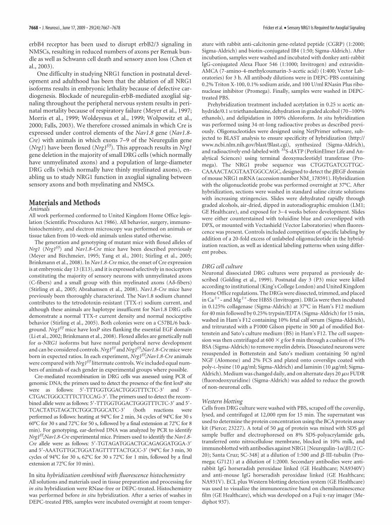
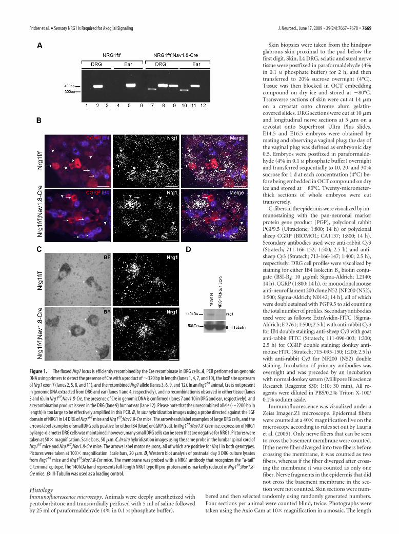
Figure 1. The floxed Nrg1 locus is efficiently recombined by the Cre recombinase in DRG cells. A, PCR performed on genomicDNA using primers to detect the presence of Cre with a product of �320 bp in length (lanes 1, 4, 7, and 10), the loxP site upstreamof Nrg1 exon 7 (lanes 2, 5, 8, and 11), and the recombined Nrg1 allele (lanes 3, 6, 9, and 12). In an Nrg1f/f animal, Cre is not presentin genomic DNA extracted from DRG and ear (lanes 1 and 4, respectively), and no recombination is observed in either tissue (lanes3 and 6). In Nrg1f/f;Nav1.8-Cre, the presence of Cre in genomic DNA is confirmed (lanes 7 and 10 in DRG and ear, respectively), anda recombination product is seen in the DRG (lane 9) but not ear (lane 12). Please note that the unrecombined allele (�2200 bp inlength) is too large to be effectively amplified in this PCR. B, In situ hybridization images using a probe directed against the EGFdomain of NRG1 in L4 DRG of Nrg1f/f mice and Nrg1f/f;Nav1.8-Cre mice. The arrowheads label examples of large DRG cells, and thearrows label examples of small DRG cells positive for either IB4 (blue) or CGRP (red). In Nrg1f/f;Nav1.8-Cre mice, expression of NRG1by large-diameter DRG cells was maintained; however, many small DRG cells can be seen that are negative for NRG1. Pictures weretaken at 50� magnification. Scale bars, 50 �m. C, In situ hybridization images using the same probe in the lumbar spinal cord ofNrg1f/f mice and Nrg1f/f;Nav1.8-Cre mice. The arrows label motor neurons, all of which are positive for Nrg1 in both genotypes.Pictures were taken at 100� magnification. Scale bars, 20 �m. D, Western blot analysis of postnatal day 3 DRG culture lysatesfrom Nrg1f/f mice and Nrg1f/f;Nav1.8-Cre mice. The membrane was probed with a NRG1 antibody that recognizes the “a-tail”C-terminal epitope. The 140 kDa band represents full-length NRG1 type III pro-protein and is markedly reduced in Nrg1f/f;Nav1.8-Cre mice. �-III-Tubulin was used as a loading control.
Fricker et al. • Sensory NRG1 Is Required for Axoglial Signaling J. Neurosci., June 17, 2009 • 29(24):7667–7678 • 7669

of the epidermis was measured using Image Jand used to calculate intraepidermal nerve fiberdensity (IENFD) (number of fibers per milli-meter). Photographs of DRG sections weretaken using the Axio Cam at 40� magnificationin a mosaic. Two sections per animal per stain-ing were counted blind from a photographtaken using AxioVision LE, release 4.2, soft-ware. Positive cell profiles were counted as wellas negative to get a percentage of total cell pro-files for each marker. The same software wasused to measure the diameter of DRG profilesfor cell size distributions.
Terminal deoxynucleotidyl transferase-mediated biotinylated UTP nick end labeling(TUNEL) analysis staining was performed us-ing an ApopTag Fluorescein In Situ ApoptosisDetection kit (Millipore Bioscience ResearchReagents; S7110) according to the manufactur-er’s instructions. Nerve sections were counter-stained with 4�,6�-diamidino-2-phenylindole(DAPI) mounting medium (Vector Laborato-ries; H-1200) to identify nuclei for total counts.Photographs of all of the fields covering thewhole of each longitudinal nerve section weretaken at 40� magnification. The number offields varied depending on the nerve and the ageof the animal; at least two nerve sections wereanalyzed per animal. More than 1000 cells werecounted blind per genotype at each time point,and TUNEL-positive cells were represented as apercentage of total cell number. TUNEL-positive embryonic DRG cells were countedblind in at least six L4/5 DRG sections peranimal.
Electron microscopy. Animals were deeplyanesthetized with pentobarbitone and transcar-dially perfused with 5 ml of saline followed by25 ml of 4% paraformaldehyde, 3% glutaralde-hyde in 0.1 M phosphate buffer. Sural nerveswere dissected and a 5 mm length taken as itreaches the gastrocnemius muscle. Nerves werepostfixed in 3% glutaraldehyde at 4°C over-night, washed in 0.1 M PB, osmicated, dehy-drated, and embedded in epoxy resin (TAABEmbedding Materials). Sections 1 �m thickwere cut on a microtome and stained with tolu-idine blue before being examined on a light mi-croscope. Ultrathin sections were cut on an ul-tramicrotome and stained with lead uranylacetate by the Centre for Ultrastructural Imag-ing (King’s College London). Sections weremounted on unsupported 100 mesh grids, anduninhibited sections were visualized on a Hita-chi H7600 transmission electron microscope.
For analysis, photographs of the entire sectionof one nerve from each animal were taken at amagnification of 15,000�. Total counts of my-elinated and unmyelinated axons as well asSchwann cell nuclei were performed of the wholecross section of the nerve. The g ratios of 100 ax-ons and C-fiber diameters of 300 axons, chosenusing randomly generated numbers, were mea-sured per animal using AxioVision LE, release 4.2, software.
NeurophysiologyThe conduction velocity (CV) of individual A- and C-fibers projectingthrough the sciatic nerve was measured in urethane-anesthetized animals(terminal urethane anesthesia; 1.25 g/kg, i.p.). A tracheal cannula was in-
serted and core body temperature was maintained at 37°C using a feedback-controlled heating blanket. The spinal cord was exposed by an L2–L5 dorsallaminectomy and the sciatic nerve was exposed at the midthigh level andimmersed in mineral oil. The sciatic nerve was placed on a pair of silver hookstimulating electrodes and was cut distally. Fine jeweler’s forceps were usedto dissect microfilaments from the L4 dorsal root close to the dorsal rootentry zone and these were mounted on a recording electrode. It was then
Figure 2. NRG1 is necessary for normal Remak bundle structure and myelination. A–F, Electron micrographs of transversesections of adult mouse sural nerve (10 weeks of age). A, C, E, Sural nerves of Nrg1f/f mice. B, D, F, Sural nerves of Nrg1f/f;Nav1.8-Cre mice. Magnification: A, B, 15,000�; C, D, 25,000�; E, F, 60,000�. A–D, Note the difference in Remak bundles (arrowheads)between the Nrg1f/f mice (A, C) in which each unmyelinated axon is separated by Schwann cell cytoplasm and are of similar sizes,and the Nrg1f/f;Nav1.8-Cre mice (B, D) in which Remak bundles are greatly enlarged.� labels an abnormally large fiber still withinthe Remak bundle. A large fiber of diameter of 1.976 �m, which would usually be myelinated (arrow), is completely amyelinated;at high magnification (D), it is clearly invested in Schwann cell cytoplasm and a basement membrane can be seen to surround thefiber. The separation of unmyelinated axons by Schwann cell cytoplasm can be seen clearly at high magnification (C–F ); arrow-heads highlight the distance between unmyelinated fibers. F, Note in Nrg1f/f;Nav1.8-Cre mice many axons are not separated bySchwann cell cytoplasm; note also the type II axon/Schwann cell relationship in which the mesaxon is breached and the axon isdirectly exposed to the endoneurium (arrows). Scale bars, 500 nm.
7670 • J. Neurosci., June 17, 2009 • 29(24):7667–7678 Fricker et al. • Sensory NRG1 Is Required for Axoglial Signaling

possible to evoke activity in sensory axons by applying graded electrical stim-uli to the sciatic nerve and recording the individual action potentials as theypassed over the recording electrodes. Dividing the distance between thesesets of electrodes by the conduction time taken for action potentials yields theconduction velocity of individual axons. Groups of myelinated and unmy-elinated axons (typically 30–50 per animal) were studied. To evoke activityin A-fibers (CV, �2 m/s), the sciatic nerve was stimulated using a squarewave current pulse of duration 100 �S. The evoked activity on the rootfilament was amplified and filtered by conventional means. The stimulatingcurrent was gradually increased up to a maximum of 500 �A, which issufficient to recruit all A-fibers in the nerve. This procedure typically resultedin a group of 3–10 axons being progressively recruited with increasing cur-rent strengths in each strand. To stimulate C-fibers (CV, �2 m/s), a squarewave current pulse of 1 mA for 1 ms was used. When recording activity inC-fibers, 32 responses were averaged (to reveal relatively small potentials).Because of the relative slowness of conduction in these fibers, the actionpotentials evoked are dispersed by the time they reach the recording elec-trodes and they generally are not superimposed. It was therefore possible toidentify action potentials from individual C axons in these recordings, fromwhich the latency (and hence conduction velocity) was determined (see Fig.4A,B). There were usually 5–10 A and 3–10 C-fibers per strand (and typi-cally 10–15 strands would be studied, resulting in a total of �50 A and 50C-fibers being collected per animal). Conduction velocity distributions wereexpressed as cumulative sums (Qsums) and then compared statistically us-ing the Kolmogorov–Smirnov test.
Behavioral testsAll behavioral testing was performed with the observer blind to the ge-notype of the animal.
Mechanical withdrawal threshold assessed using von Frey hairs. Staticmechanical withdrawal thresholds were assessed by applying von Freyhairs (Somedic) to the plantar surface of the hindpaw. Unrestrainedanimals were acclimatized in acrylic cubicles (8 � 5 � 10 cm) atop a wiremesh grid for up to 60 min before testing. Calibrated von Frey hairs wereapplied to the plantar surface of the hindpaw until the fiber bent. The50% withdrawal threshold was determined using the up– down methoddescribed previously (Dixon, 1980).
Randall–Selitto test of mechanosensation. Animals were placed in a re-strainer and left to settle for a few minutes. Force was applied approxi-mately midway along the tail. The force at which the animal attempted towithdraw the tail, vocalize, or struggle was recorded (Takesue et al.,1969). The test was repeated three times for each animal. Results wereexpressed as the mean weight tolerated for each group.
Hargreaves test of thermal nociception. Heat–pain threshold of thehindpaw was ascertained with the Hargreaves method using the PlantarTest (7370; active intensity, 20%; Ugo Basile) (Hargreaves et al., 1988).Unrestrained animals were acclimatized in acrylic cubicles (8 � 5 � 10cm) atop a uniform glass surface for up to 60 min before testing. Aninfrared light source was directed onto the plantar surface of the hind-paw, and the latency to paw withdrawal was measured in seconds. Four
responses were recorded for each hindpaw on each testing occasion withat least 2 min between stimuli. To avoid tissue injury, the maximumstimulus latency was 20.0 s.
Hot-plate test. The response to thermal stimuli was tested using a hot-plate analgesia meter (Ugo Basile). Mice were acclimatized to the hot-plate chamber for 15 min before the plate was heated. The latency forhindpaw withdrawal at two temperatures, 50 and 55°C, was measured.The latency was measured once for each animal, and the tests at differenttemperatures were separated by at least 24 h. A cutoff latency time of 60and 30 s for 50 and 55°C, respectively, was imposed in each measurementto avoid lesions to the skin and unnecessary suffering to the animals.
Cold-plate test. An incremental hot– cold plate analgesia meter(IITC Life Science) was used to assess noxious cold sensitivity on theplantar surface of the paw as previously described (Kwan et al., 2006).Mice were placed on the surface maintained at 0 0.5°C, and painwas assessed by counting the number of hindpaw lifts or jumpingduring a 5 min period. The mean number of the nocifensive responseswas determined by counting over two separate trials conducted withat least a 60 min interval to prevent thermal sensitization and/orbehavioral interferences. Results were expressed as the mean nocifen-sive responses for each group.
Rotarod. Mice were placed on the rotarod (Jones and Roberts, 1968) asit was rotating at 20 rpm. After 1 min, the rate of revolution was increasedand reached a maximum of 36 rpm within 90 s. The duration that eachanimal spent on the rod was measured, with a cutoff time of 5 min. Thetest was performed three times for each animal with an interval of at least15 min between each test.
Statistical analysisDifferences between Nrg1f/f;Nav1.8-Cre animals and Nrg1f/f littermatecontrols were determined using the Student’s t test for a single time pointand two-way repeated-measures ANOVA using the Tukey’s post hoc test,with genotype and time points as factors. Results are reported as meanvalues SEM. Conduction velocity cumulative sum plots were com-pared statistically using the Kolmogorov–Smirnov test, as were cell size,and axon diameter frequency distributions.
ResultsEfficient ablation of NRG1 in sensory neuronsIn this study, we used mice expressing the Cre recombinase trans-gene under the control of the Nav1.8 promoter (Stirling et al.,2005). When crossed with mice homozygous for the conditionalNrg1 allele (Nrg1f/f), which have loxP sites flanking the essentialEGF domain (Meyer and Birchmeier, 1995; Yang et al., 2001),this sequence, which is present in all Nrg1 isoforms, is condition-ally ablated in the majority of small-diameter as well as a popu-lation of large-diameter DRG cells. Genomic DNA from controlNrg1f/f and knock-out Nrg1f/f;Nav1.8-Cre DRGs show recombina-tion of the allele in the presence of Cre recombinase expression(Fig. 1A). In situ hybridization using a probe directed against the� isoform of the EGF domain confirms the specificity of NRG1ablation at the mRNA level specifically within small-diameterDRG cells (Fig. 1B, arrows). In Nrg1f/f animals, the highest level ofexpression of NRG1 is seen in the large-diameter DRG cells (Fig.1B, arrowheads) as has previously been demonstrated(Bermingham-McDonogh et al., 1997); however, expression isseen at a low level in small-diameter DRG cells identified by themarkers CGRP and IB4. Expression of NRG1 in the CGRP andIB4 populations is mostly absent in Nrg1f/f;Nav1.8-Cre animals;however, large-diameter DRG cells (which are CGRP and IB4negative) continue to show a normal pattern of expression. InNav1.8-Cre animals, Cre is selectively expressed in sensory neu-rons, and therefore, as expected, the normal expression of NRG1in motoneurons is observed in Nrg1f/f;Nav1.8-Cre animals (Fig.1C, arrows). No signal was seen after addition of a 20-fold excessof unlabeled oligonucleotide in the hybridization reaction dem-
Table 1. Quantification of sural nerve morphometry using electron microscopy
Nrg1f/f Nrg1f/f;Nav1.8-Cre
Total fibers 3452.5 100.19 3581.25 197.01% Myelinated fibers 19.92 0.52 16.44 0.28**% Unmyelinated fibers 80.08 0.52 83.56 0.28**No. myelinating Schwann cell nuclei 21.25 2.17 17.5 1.5No. nonmyelinating Schwann cell nuclei 11.75 0.75 12.25 1.11Total no. Schwann cell nuclei 33 1.78 29.75 2.5No. of Remak bundles 260.5 15.39 221.5 13.76C-Fibers per Remak bundle 10.73 0.75 13.54 0.4*No. bundles with polyaxonal pockets 23 2.42 74.25 7.34***% Bundles with polyaxonal pockets 9.06 1.42 34.2 4.81**Average g ratio 0.687 0.003 0.718 0.003***Unmyelinated fiber diameter 0.487 0.004 0.537 0.008***Myelinated fiber diameter 2.36 0.04 2.54 0.04**
Quantification of sural nerve morphometry using electron microscopy. Values are shown as SEM.
*p � 0.05, **p � 0.01, and ***p � 0.001, respectively, by Student’s t test.
Fricker et al. • Sensory NRG1 Is Required for Axoglial Signaling J. Neurosci., June 17, 2009 • 29(24):7667–7678 • 7671

onstrating the specificity of the radiola-beled probe (supplemental Fig. 1, availableat www.jneurosci.org as supplemental ma-terial). Additional evidence of NRG1 abla-tion in small-diameter sensory neuronswas shown at the protein level by Westernblotting analysis of protein lysate of post-natal DRG cell cultures that are grown inthe presence of NGF (which selects forsmall-diameter DRG cells) (Fig. 1D). Theamount of NRG1 detected in DRG cell cul-tures from Nrg1f/f;Nav1.8-Cre mice ismarkedly reduced.
Loss of NRG1 in sensory neurons leadsto altered Remak bundle structure aswell as myelination defectsThe sural nerve was chosen for electronmicroscopic analysis as it is a cutaneoussensory nerve and consequently is en-riched in A- and C-fibers affected by theablation of Nrg1 in these animals. Nrg1f/f;Nav1.8-Cre animals demonstrated an ob-vious phenotype in relation to both Remakbundle structure and myelination (Fig. 2).Strikingly large Remak bundles were ob-served with many more axons present perbundle than in Nrg1f/f animals. In Nrg1f/f
animals, unmyelinated axons were indi-vidually ensheathed by extensions ofSchwann cell cytoplasm so that they wereseparated from each other; however, incontrast, the C-fibers within the Remakbundles of Nrg1f/f;Nav1.8-Cre mice weremuch more closely packed, and often notseparated by Schwann cell cytoplasm, re-sulting in “polyaxonal” pockets that arenormally only rarely seen in peripheralnerve (Murinson and Griffin, 2004) (Fig.2E,F, arrowheads). In addition, in Nrg1f/f;Nav1.8-Cre mice we observed examples oftype II axon/Schwann cell relationship inwhich the mesaxon is breached and theaxon is directly exposed to the endo-neurium (Fig. 2F, arrows) (Murakawa etal., 2002). There was a much greater rangeof sizes of C-fibers within the Remak bun-dles of Nrg1f/f;Nav1.8-Cre mice (Fig. 2B).
Electron microscopy was used to countthe total number of axons within the suralnerve, which was unchanged in Nrg1f/f;Nav1.8-Cre mice. However, a greater pro-portion of axons were unmyelinated (Table1). Interestingly, there was a population oflarge-diameter fibers in a 1:1 relationshipwith a Schwann cell, surrounded by a basal lamina but having failedto myelinate (Figs. 2D, 3B). The presence of such fibers indicates thatamyelination was not solely attributable to impaired ensheathment.A percentage of 30.8 2.8% of the amyelinated fibers �1 �m indiameter were in a 1:1 relationship with a Schwann cell, the rest beingpresent in Remak bundles. There was therefore a change at both endsof the frequency distribution when plotting the number of unmyeli-nated axons per Schwann cell unit (Fig. 3C). The proportion of
unmyelinated axons associated with a Schwann cell in a 1:1 ratioincreased from 14.70.8% (meanSEM) in Nrg1f/f mice to 28.81.3% in Nrg1f/f;Nav1.8-Cre mice (Fig. 3C) (Sharghi-Namini et al.,2006). As described above, Nrg1f/f;Nav1.8-Cre mice also had agreater proportion of Schwann cell units containing large numbersof unmyelinated axons resulting in a significant change in the fre-quency distribution (Fig. 3C) ( p � 0.001, Kolmogorov–Smirnovtest). We also noted a small population of larger myelinated fibers
Figure 3. Analysis of effect of NRG1 ablation on myelinated and unmyelinated fiber diameter. Morphometric data calculatedfrom electron micrographs of sural nerves from 10-week-old mice (n 4). A, Frequency distribution of g ratios. Nrg1f/f mice havean average g ratio of 0.68738 0.00329 and Nrg1f/f;Nav1.8-Cre have an average g ratio of 0.71841 0.00337 (Student’s t test,p � 0.001). Note the shift to higher g ratios in Nrg1f/f;Nav1.8-Cre mice (Kolmogorov–Smirnov test, p � 0.001). B, Quantitativeanalysis of the axon diameter of unmyelinated fibers. Note that, in Nrg1f/f; Nav1.8-Cre mice, there is a new population of large-diameter axons (�1 �m diameter) that are unmyelinated compared with the Nrg1f/f mice (there was a significant increase in theproportion of unmyelinated axons with a diameter �1 �m; � 2 analysis, p � 0.001). C, Frequency distribution showing thenumber of unmyelinated axons/nonmyelinating Schwann cell. Note that the percentage frequency of unmyelinating axons in a1:1 ratio doubles in Nrg1f/f;Nav1.8-Cre mice compared with the Nrg1f/f mice. This increase is likely to be secondary to thoselarge-diameter axons that are in 1:1 relationship with a Schwann cell but that have failed to myelinate. Nrg1f/f;Nav1.8-Cre micealso have an increase in the Schwann cell units containing large numbers of axons. These changes lead to a significant change inthe frequency distribution (Kolmogorov–Smirnov test, p � 0.001). Error bars indicate SEM.
7672 • J. Neurosci., June 17, 2009 • 29(24):7667–7678 Fricker et al. • Sensory NRG1 Is Required for Axoglial Signaling

that appear to have proportionately thinner myelin in relation to theaxon diameter reflected by an increase in the average g ratio and asignificant shift in the frequency distribution (Fig. 3A) ( p � 0.001,Kolmogorov–Smirnov test) (supplemental Fig. 2, available at ww-w.jneurosci.org as supplemental material). The mean axon diameterof both unmyelinated and myelinated axons increased, representingthe fact that a population of small-diameter myelinated axons havebecome large-diameter unmyelinated axons in the Nrg1f/f;Nav1.8-Cre mice (Table 1).
There was no decrease in the number ofSchwann cell nuclei of either myelinatingor NMSCs (Table 1), and no apoptoticSchwann cells were seen by electron mi-croscopy. Using TUNEL staining, we ex-amined Schwann cell apoptosis in postna-tal (P3) and adult sciatic and sural nerve.As previously described, Schwann cell ap-optosis was observed in the postnatal butnot adult nerve (Grinspan et al., 1996).There was no significant difference in theproportion of TUNEL-positive Schwanncell nuclei in the Nrg1f/f;Nav1.8-Cre com-pared with Nrg1f/f animals (Fig. 4). Nav1.8is not expressed in sympathetic neurons,and therefore, as expected, the appearanceof unmyelinated axons in the lumbar sym-pathetic plexus of Nrg1f/f;Nav1.8-Cre ani-mals was normal (supplemental Fig. 3,available at www.jneurosci.org as supple-mental material). The anatomical pheno-type in peripheral sensory nerves in NRG1knock-out animals strongly suggests a roleof NRG1 in signaling between unmyeli-nated axons and NMSCs as well as inmyelination.
Ablation of NRG1 leads to alteredconduction in sensory axonsThe shape of action potentials recordedfrom primary afferents in the Nrg1f/f;Nav1.8-Cre mice appeared normal, butthere were changes in conduction veloc-ity (Fig. 5). The distribution of CVs ofindividual myelinated fibers (i.e., con-ducting at �2 m/s) is shown as cumula-tive sum plots in Figure 5C. There was asignificant slowing of CV in Nrg1f/f;Nav1.8-Cre mice compared with Nrg1f/f
control mice (Fig. 5C) ( p � 0.05, Kol-mogorov–Smirnov test), which was ap-parent for the more slowly conductingA-fibers. This observation is consistentwith the known expression of Nav1.8 insome small myelinated axons, and theloss of neuregulin expression form thosein the Nrg1f/f;Nav1.8-Cre mice. Therewas also a greater electrical threshold foractivation in this population of units.Whereas in Nrg1f/f animals only 1 of 208units recorded with a CV �2 m/s re-quired stimulation using the C-fiberrather than A-fiber parameters (as de-scribed in Materials and Methods), this
was the case in 51 of 262 such units recorded in Nrg1f/f;Nav1.8-Cre mice ( p � 10 �5, � 2 test). The CV slowing and increasedthresholds seen in the Nrg1f/f;Nav1.8-Cre mice is likely to rep-resent the electrophysiological consequences of failure/im-pairment of myelination of a population of large-diameteraxons corresponding to our findings on morphometric anal-ysis. Fibers conducting �12 m/s have a virtually identical CVdistribution when comparing Nrg1f/f;Nav1.8-Cre and Nrg1f/f
mice.
Figure 4. There is no difference in apoptosis of Schwann cells between Nrg1f/f and Nrg1f/f;Nav1.8-Cre mice. A, Photomicro-graphs of longitudinal sections of sciatic and sural nerve stained for apoptosis using the TUNEL system and DAPI as a nuclear stainat P3 and adult. No apoptotic nuclei were seen in adult sural or sciatic nerve. B, Quantitative analysis of TUNEL-positive cells at P3in sciatic and sural nerve. There was no significant difference between Nrg1f/f and Nrg1f/f;Nav1.8-Cre mice. Error bars indicate SEM.Images are at 40� magnification. Scale bars, 20 �m.
Fricker et al. • Sensory NRG1 Is Required for Axoglial Signaling J. Neurosci., June 17, 2009 • 29(24):7667–7678 • 7673

A cumulative sum plot of fibers re-corded with a CV of �2 m/s (i.e., C-fiberconduction range) is shown in Figure 5D.There was a significant increase in CV inNrg1f/f;Nav1.8-Cre mice compared withcontrol Nrg1f/f mice particularly in therange of 1–2 m/s ( p � 0.05, Kolmogorov–Smirnov test). This is likely to represent apopulation of large-diameter axons(which would normally be A�-fibers) thathave failed to myelinate and therefore nowconduct in the C-fiber range. There was nodifference in CV distribution in the slowerpart of the range (0 – 0.8 m/s) and in par-ticular no slowing of CV in Nrg1f/f;Nav1.8-Cre animals.
Epidermal innervation and DRG cellhistochemistry are unaltered afterNRG1 ablationWe investigated whether NRG1 ablationled to increased DRG cell apoptosis dur-ing development. Naturally occurringDRG cell death occurs between E12.5and E16 in the mouse (White et al.,1998). The TUNEL-positive DRG cellprofile counts were unchanged in Nrg1f/
f;Nav1.8-Cre animals at E14.5 and E16.5,indicating no difference in the rate ofDRG neuron apoptosis before birth(supplemental Fig. 4, available atwww.jneurosci.org as supplemental ma-terial). The innervation of the epidermiswas analyzed to investigate whether ab-normal axoglial signaling caused byNRG1 ablation could lead to a reductionin target innervation by sensory neu-rons. There was no significant differencein IENFD (fibers per millimeter) whenstained for either PGP9.5, a pan-neuronal marker (IENFD, 35.2 6.2and 31.5 4.4 in Nrg1f/f and Nrg1f/f;Nav1.8-Cre animals, respectively), orCGRP (IENFD, 8.6 1 and 6.9 2.7 in Nrg1f/f and Nrg1f/f;Nav1.8-Cre animals, respectively). There remained no differ-ence in IENFD at the age of 1 year (27.7 2 and 29.6 2.7 inNrg1f/f and Nrg1f/f;Nav1.8-Cre animals, respectively). This in-dicates that, after E13, axonally derived NRG1 is not requiredfor C-fibers to reach their target of the epidermis or maintaintarget innervation (Fig. 6). There was no difference in the DRGcell size distribution after NRG1 ablation (supplemental Fig.5, available at www.jneurosci.org as supplemental material).In addition, there was no evidence for an alteration in the con-stituent DRG cell populations as defined histochemically:counts of DRG cell profiles stained for NF200 (a marker forlarge-diameter DRG cells), CGRP, and IB4 binding (markers forpeptidergic and nonpeptidergic C-fibers, respectively) were un-changed (Table 2; supplemental Fig. 5, available at www.jneuro-sci.org as supplemental material).
Altered nociceptive behavior in the absence of NRG1To assess the impact of NRG1 ablation on nociceptive behavior, thethermal and mechanical thresholds of Nrg1f/f;Nav1.8-Cre and Nrg1f/f
mice were assessed at intervals over a period of 1 year (Fig. 7; supple-mental Fig. 6, available at www.jneurosci.org as supplemental mate-rial). There was no difference in sensitivity to noxious thermal stim-uli assessed using Hargreaves method at any time point. Similarly,the mechanical threshold tested by von Frey hairs was unchanged.There was an increased threshold in Nrg1f/f;Nav1.8-Cre mice afterapplication of a ramp pressure mechanical stimulus (using themethod of Randall–Selitto), which became more apparent with age.The performance of Nrg1f/f;Nav1.8-Cre on the rotarod as well astheir response to the hot-plate test and noxious cold were unchanged(supplemental Fig. 6, available at www.jneurosci.org as supplemen-tal material).
DiscussionWe found that the selective ablation of NRG1 within sensoryneurons results in profound changes in the structural relation-ship between axons and both nonmyelinating and myelinatingSchwann cells: Large numbers of clustered unmyelinated ax-ons are observed within Remak bundles and myelination isimpaired as demonstrated by a reduction in the proportion of
Figure 5. The CV of individual sensory axons projecting through the sciatic nerve was measured by stimulation of the nerveelectrically and recording and averaging activity in fine strands of the L4 dorsal root. A and B show representative recordings fromNrg1f/f and Nrg1f/f;Nav1.8-Cre mice, respectively, after stimulation using C-fiber parameters (1 mA; 1 ms). The arrows showexamples of individual C-fiber potentials occurring in response to stimulation. The arrowheads denote a population of unitsessentially only seen in Nrg1f/f;Nav1.8-Cre mice that conduct in what is normally the slower part of the A-fiber range but thatrequire C-fiber stimulation parameters. C, Cumulative sum plot of average CV distribution in units with CV �2 m/s (Nrg1f/f, n 4; Nrg1f/f;Nav1.8-Cre, n 5; error bars show SEM). There is a significant slowing of these units in the Nrg1f/f;Nav1.8-Cre animals(seen as a leftward shift in Qsum plots), particularly in the CV range of 2–10 m/s. D, Cumulative sum plot of average CV distributionin units with CV �2 m/s. These units are significantly faster in the Nrg1f/f;Nav1.8-Cre animals (seen as a rightward shift in Qsumplots), particularly in the CV range of 1–2 m/s.
7674 • J. Neurosci., June 17, 2009 • 29(24):7667–7678 Fricker et al. • Sensory NRG1 Is Required for Axoglial Signaling

myelinated axons and an increased g ratio. These structuralchanges are reflected in a slowing of conduction velocity in theA-fiber conduction range and a reduced sensitivity to high-threshold mechanical stimuli. In contrast to animals that lack
NRG1 at earlier stages of development,there was no evidence of DRG cell oraxon loss.
The role of NRG1 in signaling betweensmall-diameter sensory axons andnonmyelinating Schwann cellsNrg1f/f;Nav1.8-Cre mice demonstratedimpaired sorting and ensheathment of un-myelinated axons: We found an increase inthe number of axons per Remak bundle.Normally unmyelinated axons in periph-eral nerves are isolated from each other bySchwann cell processes (Murinson andGriffin, 2004). After conditional NRG1ablation there was poor segregation of un-myelinated axons that were clustered inpolyaxonal pockets. We also observed in-stances of type 2 axon/Schwann cell rela-tionships in which the axolemma is di-rectly exposed to the endoneurium(Murakawa et al., 2002). A similar mor-phological phenotype is seen within theperipheral nerves of animals that are hap-loinsufficient for NRG1 type III (Taveggiaet al., 2005) or in animals lacking BACE1,an enzyme that proteolytically cleaves thismolecule (Willem et al., 2006). In contrast,Chen et al. (2003) used a dominant-negative erbB4 receptor expressed inNMSCs to block Neuregulin signaling anddescribed increased Schwann cell prolifer-ation, apoptosis, and reduced numbers ofaxons per Remak bundle. These differ-ences may be attributable to the fact that inthe study of Chen et al., NRG1 signaling isblocked in the postnatal period/adulthoodas opposed to late embryonic develop-ment. Second, the Nrg1f/f;Nav1.8-Cre miceselectively lack sensory axon-derivedNRG1; however, there is a possibility thatNRG1 may have an autocrine action onSchwann cells that would be impaired inthe dominant-negative erbB4 receptormice but not in our animals (Raabe et al.,1996; Rosenbaum et al., 1997); direct evi-dence for an autocrine NRG1 loop in
Schwann cells is, however, lacking (Meier et al., 1999). There isalso a possibility that the dominant-negative erbB4 receptor mayblock erbB signaling from ligands other than NRG1 within pe-ripheral nerve.
The survival and migration of Schwann cell precursors aredependent on axonally derived NRG1 type III (Dong et al., 1995;Meyer et al., 1997; Wolpowitz et al., 2000). At later stages ofdevelopment, Schwann cells develop autocrine survival circuits(Jessen and Mirsky, 2005). As expected, we observed Schwanncell apoptosis in postnatal but not in the adult nerve (Grinspan etal., 1996); however, there was no increase in the rate of Schwanncell apoptosis (at either time point) in Nrg1f/f;Nav1.8-Cre mice.Consistent with this observation, there was no significant changein the number of Schwann cell nuclei (either nonmyelinating ormyelinating) within the adult sural nerve of Nrg1f/f;Nav1.8-Cremice. In these animals in which the number of sural axons re-
Figure 6. Cutaneous innervation is unchanged in NRG1 knock-out mice. Photomicrographs of skin sections stained for PGP9.5,a pan-neuronal marker. There is no difference in the density of fibers (arrows) in the epidermal layer in Nrg1f/f;Nav1.8-Cre mice (B)compared with Nrg1f/f littermates (A). The images are at 20� magnification; scale bars, 50 �m. The inset panel images are at40� magnification; scale bars, 20 �m.
Table 2. Percentage counts of L4 DRG cell profiles immunopositive for CGRP, IB4,and NF200
% Count of immunopositive DRG profiles
Nrg1f/fNrg1f/f;Nav1.8-Cre
IB4 34.35 2.1 30.04 1.4CGRP 30.65 2.2 33.00 1.5NF200 (N52) 31.90 2.3 32.66 2.3
Note that there was no difference between in Nrg1f/f and Nrg1f/f;Nav1.8-Cre mice in the percentage of DRG cellprofile percentages immunopositive for these markers; n 4. Data are presented as mean SEM.
Fricker et al. • Sensory NRG1 Is Required for Axoglial Signaling J. Neurosci., June 17, 2009 • 29(24):7667–7678 • 7675

mains the same and there are Remak bun-dles containing increased numbers of un-myelinated axons, one may have expecteda reduction in the number of Schwanncells. One explanation for this apparentparadox is that these animals demonstratea new population of large-diameter axonsthat are in a 1:1 relationship with aSchwann cell but that have failed to be my-elinated (see below). Large-diameter ax-ons that have failed to myelinate would beexpected to be associated with moreSchwann cells along the length of the axonbecause of the shorter internuclear dis-tance between adjacent Schwann cells as-sociated with unmyelinated axons (Griffinand Thompson, 2008). The occurrence ofsuch axons alongside the increase in thesize of Remak bundles appears to be in bal-ance such that the total number ofSchwann cells is unchanged in Nrg1f/f;Nav1.8-Cre mice.
The role of NRG1 in the myelination oflarge-diameter sensory axonsDuring the development of the peripheralnervous system, the level of axonally de-rived NRG1 type III determines the en-sheathment fate of axons and the thicknessof the myelin sheath (Michailov et al.,2004; Taveggia et al., 2005). Large-diameter DRG cells that are myelinatedshow a higher level of NRG1 type III ex-pression (Bermingham-McDonogh et al.,1997). NRG1 does not appear to have sucha critical instructive role in myelination ofaxons within the CNS (Taveggia et al.,2005, 2008; Roy et al., 2007; Brinkmann etal., 2008). Consistent with a role of NRG1 in the myelination ofsensory axons, we found an increased proportion of unmyeli-nated axons in Nrg1f/f;Nav1.8-Cre mice as well as an increase inthe g ratio of nerve fibers within the sural nerve. Previous studiesof animals haploinsufficient for NRG1 type III (Taveggia et al.,2005) or with a dominant-negative erbB4 receptor expressed inmyelinating glia (Chen et al., 2003) also found an increased gratio. The key role of NRG1 in myelination is emphasized by theobservation in Nrg1f/f;Nav1.8-Cre mice of large-diameter sensoryaxons in a 1:1 relationship with a Schwann cell and surroundedby a basal lamina but that have failed to myelinate.
The consequences of NRG1 ablation for the survival andfunctional properties of sensory neuronsIt has been suggested that NMSCs support axons in a number ofways, for instance in the provision of neurotrophic factors (Grif-fin and Thompson, 2008). We had therefore speculated that theincreased numbers of axons per Remak bundle and the arrange-ment of C-fibers in polyaxonal pockets seen in Nrg1f/f;Nav1.8-Cremice may lead to defective axonal maintenance. Althoughchanges in the functional properties of axons were observed,there was no evidence of axonal degeneration in these animals.
During early development of the peripheral nervous system(between E12.5 and 14.5), DRG cell survival is dependent onNRG1-mediated survival of Schwann cell precursors (Riethma-
cher et al., 1997). In animals with a dominant-negative erbB4receptor expressed in NMSCs in the postnatal period, Chen et al.(2003) described DRG cell death, reduced expression of themarkers trkA and IB4 within DRG cells, and progressive reduc-tion in sensitivity to noxious thermal stimuli (all these changeswere apparent by P60). In Nrg1f/f;Nav1.8-Cre mice, there was noincrease in DRG cell apoptosis and the total number of axonswithin the sural nerve was unchanged. The proportion of DRGcell profiles expressing histochemical markers for different DRGcell populations [NF200, CGRP, and IB4 (Snider and McMahon,1998)] were the same. In addition, IENFD, which is reduced inthe context of small-fiber neuropathy both in rodents (Johnsonet al., 2008) and humans (McArthur et al., 1998), was unchanged.Therefore, although sensory axonally derived NRG1 is requiredfor the formation of the normal relationship between axons andSchwann cells in peripheral nerve, unlike Chen et al. (2003) wedid not find a reciprocal relationship in which it drives a signalfrom the NMSC back to the sensory axon required for the struc-tural integrity and long-term survival of sensory neurons. Thereare clearly situations in which reciprocal interactions betweenC-fibers and NMSCs are critical for the maintenance of axonalintegrity, as for example seen in L1-deficient mice in which de-fective ensheathment of unmyelinated axons leads to progressiveaxonal degeneration (Haney et al., 1999).
The altered axoglial relationships within Nrg1f/f;Nav1.8-Cre
Figure 7. The nociceptive behavior of a cohort of Nrg1f/f and Nrg1f/f;Nav1.8-Cre mice was followed over the course of 48 weeks (n8per group). There was no significant difference in the response to noxious thermal stimuli as measured by Hargreaves apparatus (A) or theresponse threshold to mechanical stimulation using von Frey hairs (B) (analyzed by two-way repeated-measures ANOVA). C, There was areduced sensitivity to a high-threshold mechanical pressure stimulus applied to the tail using the Randall–Sellito apparatus, which wassignificant at the age of 36 and 48 weeks (*p � 0.05 by two-way repeated-measures ANOVA Tukey’s post hoc).
7676 • J. Neurosci., June 17, 2009 • 29(24):7667–7678 Fricker et al. • Sensory NRG1 Is Required for Axoglial Signaling

mice were reflected in the electrophysiological findings in theseanimals. As expected (given the myelination defect in a subset oflarger-diameter axons), there was a slowing in conduction veloc-ity particularly at the lower part of the A-fiber CV range. Whenrecording from C-fibers in Nrg1f/f;Nav1.8-Cre mice, there was apopulation of faster conducting C-fibers. These were found par-ticularly in the range of 1–2 m/s and are likely to represent large-diameter axons that have failed to be myelinated and so nowconduct in the C-fiber range. The poor segregation of axons bySchwann cell cytoplasm may be expected to lead to reduced elec-trical insulation of C-fibers. But we did not observe any slowingof conduction or altered action potential morphology in C-fibers.On examining sensory function in Nrg1 f/f;Nav1.8-Cre mice, wefound that the sensitivity of these animals to noxious heat andcold were unchanged. The response to punctuate mechanicalstimulation with von Frey hairs was also unaltered; however, theresponse to a noxious pressure stimulus was reduced. This mayrepresent deficits in encoding by either A�- (some of which havemyelination deficits) or C-fibers. Our failure to see a change inthreshold to von Frey hair stimulation is not unexpected giventhat in animals in which the same population of Nav1.8 Cre-expressing DRG cells are ablated the von Frey threshold is alsounaltered (Abrahamsen et al., 2008).
In conclusion, axon-derived NRG1 has a key role in axoglialsignaling between sensory axons and both nonmyelinating andmyelinating Schwann cells necessary for normal sensory func-tion, however is not required for the long-term maintenance ofsensory axons in adulthood.
ReferencesAbrahamsen B, Zhao J, Asante CO, Cendan CM, Marsh S, Martinez-Barbera
JP, Nassar MA, Dickenson AH, Wood JN (2008) The cell and molecularbasis of mechanical, cold, and inflammatory pain. Science 321:702–705.
Bermingham-McDonogh O, Xu YT, Marchionni MA, Scherer SS (1997)Neuregulin expression in PNS neurons: isoforms and regulation by targetinteractions. Mol Cell Neurosci 10:184 –195.
Brinkmann BG, Agarwal A, Sereda MW, Garratt AN, Muller T, Wende H,Stassart RM, Nawaz S, Humml C, Velanac V, Radyushkin K, Goebbels S,Fischer TM, Franklin RJ, Lai C, Ehrenreich H, Birchmeier C, Schwab MH,Nave KA (2008) Neuregulin-1/ErbB signaling serves distinct functionsin myelination of the peripheral and central nervous system. Neuron59:581–595.
Carraway KL 3rd, Weber JL, Unger MJ, Ledesma J, Yu N, Gassmann M, Lai C(1997) Neuregulin-2, a new ligand of ErbB3/ErbB4-receptor tyrosine ki-nases. Nature 387:512–516.
Chen S, Rio C, Ji RR, Dikkes P, Coggeshall RE, Woolf CJ, Corfas G (2003)Disruption of ErbB receptor signaling in adult non-myelinating Schwanncells causes progressive sensory loss. Nat Neurosci 6:1186 –1193.
Dixon WJ (1980) Efficient analysis of experimental observations. Annu RevPharmacol Toxicol 20:441– 462.
Djouhri L, Fang X, Okuse K, Wood JN, Berry CM, Lawson SN (2003) TheTTX-resistant sodium channel Nav1.8 (SNS/PN3): expression and corre-lation with membrane properties in rat nociceptive primary afferent neu-rons. J Physiol 550:739 –752.
Dong Z, Brennan A, Liu N, Yarden Y, Lefkowitz G, Mirsky R, Jessen KR(1995) Neu differentiation factor is a neuron-glia signal and regulatessurvival, proliferation, and maturation of rat Schwann cell precursors.Neuron 15:585–596.
Falls DL (2003) Neuregulins: functions, forms, and signaling strategies. ExpCell Res 284:14 –30.
Falls DL, Rosen KM, Corfas G, Lane WS, Fischbach GD (1993) ARIA, aprotein that stimulates acetylcholine receptor synthesis, is a member ofthe neu ligand family. Cell 72:801– 815.
Garratt AN, Voiculescu O, Topilko P, Charnay P, Birchmeier C (2000a) Adual role of erbB2 in myelination and in expansion of the Schwann cellprecursor pool. J Cell Biol 148:1035–1046.
Garratt AN, Britsch S, Birchmeier C (2000b) Neuregulin, a factor with manyfunctions in the life of a Schwann cell. Bioessays 22:987–996.
Golding JP, Bird C, McMahon S, Cohen J (1999) Behaviour of DRG sensoryneurites at the intact and injured adult rat dorsal root entry zone: postna-tal neurites become paralysed, whilst injury improves the growth of em-bryonic neurites. Glia 26:309 –323.
Goodearl AD, Davis JB, Mistry K, Minghetti L, Otsu M, Waterfield MD,Stroobant P (1993) Purification of multiple forms of glial growth factor.J Biol Chem 268:18095–18102.
Griffin JW, Thompson WJ (2008) Biology and pathology of nonmyelinat-ing Schwann cells. Glia 56:1518 –1531.
Grinspan JB, Marchionni MA, Reeves M, Coulaloglou M, Scherer SS (1996)Axonal interactions regulate Schwann cell apoptosis in developing pe-ripheral nerve: neuregulin receptors and the role of neuregulins. J Neu-rosci 16:6107– 6118.
Haney CA, Sahenk Z, Li C, Lemmon VP, Roder J, Trapp BD (1999) Hetero-philic binding of L1 on unmyelinated sensory axons mediates Schwanncell adhesion and is required for axonal survival. J Cell Biol146:1173–1184.
Harari D, Tzahar E, Romano J, Shelly M, Pierce JH, Andrews GC, Yarden Y(1999) Neuregulin-4: a novel growth factor that acts through the ErbB-4receptor tyrosine kinase. Oncogene 18:2681–2689.
Hargreaves K, Dubner R, Brown F, Flores C, Joris J (1988) A new and sen-sitive method for measuring thermal nociception in cutaneous hyperal-gesia. Pain 32:77– 88.
Holmes WE, Sliwkowski MX, Akita RW, Henzel WJ, Lee J, Park JW, YansuraD, Abadi N, Raab H, Lewis GD (1992) Identification of heregulin, aspecific activator of p185erbB2. Science 256:1205–1210.
Hu X, Hicks CW, He W, Wong P, Macklin WB, Trapp BD, Yan R (2006)Bace1 modulates myelination in the central and peripheral nervous sys-tem. Nat Neurosci 9:1520 –1525.
Jessen KR, Mirsky R (2005) The origin and development of glial cells inperipheral nerves. Nat Rev Neurosci 6:671– 682.
Johnson MS, Ryals JM, Wright DE (2008) Early loss of peptidergic intraepi-dermal nerve fibers in an STZ-induced mouse model of insensate diabeticneuropathy. Pain 140:35– 47.
Jones BJ, Roberts DJ (1968) A rotarod suitable for quantitative measure-ments of motor incoordination in naive mice. Naunyn SchmiedebergsArch Exp Pathol Pharmakol 259:211.
Kwan KY, Allchorne AJ, Vollrath MA, Christensen AP, Zhang DS, Woolf CJ,Corey DP (2006) TRPA1 contributes to cold, mechanical, and chemicalnociception but is not essential for hair-cell transduction. Neuron50:277–289.
Lauria G, Cornblath DR, Johansson O, McArthur JC, Mellgren SI, Nolano M,Rosenberg N, Sommer C (2005) EFNS guidelines on the use of skinbiopsy in the diagnosis of peripheral neuropathy. Eur J Neurol12:747–758.
Li L, Cleary S, Mandarano MA, Long W, Birchmeier C, Jones FE (2002) Thebreast proto-oncogene, HRGalpha regulates epithelial proliferation andlobuloalveolar development in the mouse mammary gland. Oncogene21:4900 – 4907.
Marchionni MA, Goodearl AD, Chen MS, Bermingham-McDonogh O, KirkC, Hendricks M, Danehy F, Misumi D, Sudhalter J, Kobayashi K (1993)Glial growth factors are alternatively spliced erbB2 ligands expressed inthe nervous system. Nature 362:312–318.
McArthur JC, Stocks EA, Hauer P, Cornblath DR, Griffin JW (1998) Epi-dermal nerve fiber density: normative reference range and diagnostic ef-ficiency. Arch Neurol 55:1513–1520.
Meier C, Parmantier E, Brennan A, Mirsky R, Jessen KR (1999) Devel-oping Schwann cells acquire the ability to survive without axons byestablishing an autocrine circuit involving insulin-like growth factor,neurotrophin-3, and platelet-derived growth factor-BB. J Neurosci19:3847–3859.
Meyer D, Birchmeier C (1995) Multiple essential functions of neuregulin indevelopment. Nature 378:386 –390.
Meyer D, Yamaai T, Garratt A, Riethmacher-Sonnenberg E, Kane D, TheillLE, Birchmeier C (1997) Isoform-specific expression and function ofneuregulin. Development 124:3575–3586.
Michailov GV, Sereda MW, Brinkmann BG, Fischer TM, Haug B, BirchmeierC, Role L, Lai C, Schwab MH, Nave KA (2004) Axonal neuregulin-1regulates myelin sheath thickness. Science 304:700 –703.
Morris JK, Lin W, Hauser C, Marchuk Y, Getman D, Lee KF (1999) Rescueof the cardiac defect in ErbB2 mutant mice reveals essential roles of ErbB2in peripheral nervous system development. Neuron 23:273–283.
Fricker et al. • Sensory NRG1 Is Required for Axoglial Signaling J. Neurosci., June 17, 2009 • 29(24):7667–7678 • 7677

Murakawa Y, Zhang W, Pierson CR, Brismar T, Ostenson CG, Efendic S,Sima AA (2002) Impaired glucose tolerance and insulinopenia in theGK-rat causes peripheral neuropathy. Diabetes Metab Res Rev18:473– 483.
Murinson BB, Griffin JW (2004) C-fiber structure varies with location inperipheral nerve. J Neuropathol Exp Neurol 63:246 –254.
Nave KA, Salzer JL (2006) Axonal regulation of myelination by neuregulin1. Curr Opin Neurobiol 16:492–500.
Peles E, Bacus SS, Koski RA, Lu HS, Wen D, Ogden SG, Levy RB, Yarden Y(1992) Isolation of the neu/HER-2 stimulatory ligand: a 44 kd glycopro-tein that induces differentiation of mammary tumor cells. Cell69:205–216.
Raabe TD, Clive DR, Neuberger TJ, Wen D, DeVries GH (1996) Culturedneonatal Schwann cells contain and secrete neuregulins. J Neurosci Res46:263–270.
Riethmacher D, Sonnenberg-Riethmacher E, Brinkmann V, Yamaai T, LewinGR, Birchmeier C (1997) Severe neuropathies in mice with targeted mu-tations in the ErbB3 receptor. Nature 389:725–730.
Rosenbaum C, Karyala S, Marchionni MA, Kim HA, Krasnoselsky AL, Hap-pel B, Isaacs I, Brackenbury R, Ratner N (1997) Schwann cells expressNDF and SMDF/n-ARIA mRNAs, secrete neuregulin, and show consti-tutive activation of erbB3 receptors: evidence for a neuregulin autocrineloop. Exp Neurol 148:604 – 615.
Roy K, Murtie JC, El-Khodor BF, Edgar N, Sardi SP, Hooks BM, Benoit-Marand M, Chen C, Moore H, O’Donnell P, Brunner D, Corfas G (2007)Loss of erbB signaling in oligodendrocytes alters myelin and dopaminer-gic function, a potential mechanism for neuropsychiatric disorders. ProcNatl Acad Sci U S A 104:8131– 8136.
Sharghi-Namini S, Turmaine M, Meier C, Sahni V, Umehara F, Jessen KR,Mirsky R (2006) The structural and functional integrity of peripheralnerves depends on the glial-derived signal desert hedgehog. J Neurosci26:6364 – 6376.
Snider WD, McMahon SB (1998) Tackling pain at the source: new ideasabout nociceptors. Neuron 20:629 – 632.
Stirling LC, Forlani G, Baker MD, Wood JN, Matthews EA, Dickenson AH,Nassar MA (2005) Nociceptor-specific gene deletion using heterozy-gous NaV1.8-Cre recombinase mice. Pain 113:27–36.
Takesue EI, Schaefer W, Jukniewicz E (1969) Modification of the Randall-Selitto analgesic apparatus. J Pharm Pharmacol 21:788 –789.
Taveggia C, Zanazzi G, Petrylak A, Yano H, Rosenbluth J, Einheber S, Xu X,Esper RM, Loeb JA, Shrager P, Chao MV, Falls DL, Role L, Salzer JL(2005) Neuregulin-1 type III determines the ensheathment fate of axons.Neuron 47:681– 694.
Taveggia C, Thaker P, Petrylak A, Caporaso GL, Toews A, Falls DL, EinheberS, Salzer JL (2008) Type III neuregulin-1 promotes oligodendrocyte my-elination. Glia 56:284 –293.
Wen D, Peles E, Cupples R, Suggs SV, Bacus SS, Luo Y, Trail G, Hu S, SilbigerSM, Levy RB (1992) Neu differentiation factor: a transmembrane glyco-protein containing an EGF domain and an immunoglobulin homologyunit. Cell 69:559 –572.
Wen D, Suggs SV, Karunagaran D, Liu N, Cupples RL, Luo Y, Janssen AM,Ben-Baruch N, Trollinger DB, Jacobsen VL (1994) Structural and func-tional aspects of the multiplicity of Neu differentiation factors. Mol CellBiol 14:1909 –1919.
White FA, Keller-Peck CR, Knudson CM, Korsmeyer SJ, Snider WD (1998)Widespread elimination of naturally occurring neuronal death in Bax-deficient mice. J Neurosci 18:1428 –1439.
Willem M, Garratt AN, Novak B, Citron M, Kaufmann S, Rittger A,DeStrooper B, Saftig P, Birchmeier C, Haass C (2006) Control of pe-ripheral nerve myelination by the beta-secretase BACE1. Science314:664 – 666.
Woldeyesus MT, Britsch S, Riethmacher D, Xu L, Sonnenberg-RiethmacherE, Abou-Rebyeh F, Harvey R, Caroni P, Birchmeier C (1999) Peripheralnervous system defects in erbB2 mutants following genetic rescue of heartdevelopment. Genes Dev 13:2538 –2548.
Wolpowitz D, Mason TB, Dietrich P, Mendelsohn M, Talmage DA, Role LW(2000) Cysteine-rich domain isoforms of the neuregulin-1 gene are re-quired for maintenance of peripheral synapses. Neuron 25:79 –91.
Yang X, Arber S, William C, Li L, Tanabe Y, Jessell TM, Birchmeier C, BurdenSJ (2001) Patterning of muscle acetylcholine receptor gene expression inthe absence of motor innervation. Neuron 30:399 – 410.
Zhang D, Sliwkowski MX, Mark M, Frantz G, Akita R, Sun Y, Hillan K,Crowley C, Brush J, Godowski PJ (1997) Neuregulin-3 (NRG3): a novelneural tissue-enriched protein that binds and activates ErbB4. Proc NatlAcad Sci U S A 94:9562–9567.
7678 • J. Neurosci., June 17, 2009 • 29(24):7667–7678 Fricker et al. • Sensory NRG1 Is Required for Axoglial Signaling