Embed Size (px)
Citation preview
Development/Plasticity/Repair
Opposing Roles for Hoxa2 and Hoxb2 in HindbrainOligodendrocyte Patterning
Andres Miguez,1,2,3 Sebastien Ducret,4 Thomas Di Meglio,4 Carlos Parras,1,2,3 Hatem Hmidan,1,2,3 Celine Haton,1,2,3
Sowmya Sekizar,1,2,3 Abdelkrim Mannioui,1,2,3 Marie Vidal,1,2,3 Aurelien Kerever,1,2,3 Omar Nyabi,5 Jody Haigh,5
Bernard Zalc,1,2,3,6 Filippo M. Rijli,4,7 and Jean-Leon Thomas1,2,3,6,8
1Universite Pierre and Marie Curie–Paris 6, Centre de Recherche de l’Institut du Cerveau et de la Moelle epiniere, 75013 Paris, France, 2Institut National dela Sante et de la Recherche Medicale, Unite Mixte de Recherche S 975, 75013 Paris, France, 3Centre National de la Recherche Scientifique, Unite Mixte deRecherche 7225, 75013 Paris, France, 4Friedrich Miescher Institute for Biomedical Research, 4058 Basel, Switzerland, 5Department for MolecularBiomedical Research, Vlaams Instituut voor Biotechnologie, and Department of Biomedical Molecular Biology, Ghent University, B-9052 Ghent, Belgium,6Assistance Publique—Hopitaux de Paris, Groupe Hospitalier Pitie-Salpetriere, 75013 Paris, France, 7University of Basel, CH-4003 Basel, Switzerland, and8Yale School of Medicine, Department of Neurology, New Haven, Connecticut 06520-8018
Oligodendrocytes are the myelin-forming cells of the vertebrate CNS. Little is known about the molecular control of region-specific oligoden-drocyte development. Here, we show that oligodendrogenesis in the mouse rostral hindbrain, which is organized in a metameric series ofrhombomere-derived (rd) territories, follows a rhombomere-specific pattern, with extensive production of oligodendrocytes in the pontineterritory (r4d) and delayed and reduced oligodendrocyte production in the prepontine region (r2d, r3d). We demonstrate that segmentalorganization of oligodendrocytes is controlled by Hox genes, namely Hoxa2 and Hoxb2. Specifically, Hoxa2 loss of function induced a dorsoven-tral enlargement of the Olig2/Nkx2.2-expressing oligodendrocyte progenitor domain, whereas conditional Hoxa2 overexpression in the Olig2�
domain inhibited oligodendrogenesis throughout the brain. In contrast, Hoxb2 deletion resulted in a reduction of the pontine oligodendrogenicdomain. Compound Hoxa2�/�/Hoxb2�/� mutant mice displayed the phenotype of Hoxb2�/� mutants in territories coexpressing Hoxa2 andHoxb2 (rd3, rd4), indicating that Hoxb2 antagonizes Hoxa2 during rostral hindbrain oligodendrogenesis. This study provides the first in vivoevidence that Hox genes determine oligodendrocyte regional identity in the mammalian brain.
IntroductionOligodendrocytes are the myelin-forming cells of the CNS. De-spite an apparent similitude of the myelin sheath, oligodendro-cytes and their precursors differ by a number of criteria, such asphysiological properties, growth-factor dependency, migration
pathways, and their different sites of origin along the neural tube(Spassky et al., 1998, 2001; Kessaris et al., 2006; Karadottir et al.,2008; Tripathi et al., 2011). During development, multiple sub-populations of oligodendrocyte precursor cells (OPCs) are gen-erated in successive waves from different domains along thedorsoventral (DV) axis of the neural tube (Richardson et al.,2006; Rowitch and Kriegstein, 2010). In the spinal cord and hind-brain, OPCs first arise ventrally, adjacent to the midline (Noll andMiller, 1993; Davies and Miller, 2001). In the mouse, the ventralwave of oligodendrogenesis starts at embryonic day (E) 11.5–E13.5 (Timsit et al., 1995; Pringle et al., 1996). From E13.5, addi-tional subpopulations of OPCs develop in the lateral and dorsalplates following a ventrodorsal temporal sequence (Cai et al.,2005; Fogarty et al., 2005; Vallstedt et al., 2005; Sugimori et al.,2007).
The identity of OPCs is determined, at least in part, by theexpression of transcription factors, such as Olig1/2, Nkx2.2, orSox10, that have been characterized along the DV axis of theneural tube (Rowitch, 2004; Wegner, 2008). Dorsally, the Olig2�
domain is abutting the Pax6� progenitor territory, whereas ven-trally it is bordered by the Nkx2.2� progenitor domain, except inthe rostral hindbrain where Olig2� and Nkx2.2� progenitor do-mains almost completely overlap (Vallstedt et al., 2005).
A role for rostrocaudal (RC) positional determinants in oligo-dendroglial identity has not yet been explored. We have chosenthe mouse hindbrain, which is transiently compartmentalized
Received Feb. 23, 2012; revised Oct. 4, 2012; accepted Oct. 5, 2012.Author contributions: F.M.R. and J.-L.T. designed research; A. Miguez, S.D., T.D.M., C.P., H.H., C.H., S.S.,
A. Mannioui, and M.V. performed research; A.K., O.N., and J.H. contributed unpublished reagents/analytic tools; A.Miguez and B.Z. analyzed data; A. Miguez, F.M.R., and J.-L.T. wrote the paper.
This work was supported by Institut National de la Sante et de la Recherche Medicale (J.-L.T.), Novartis ResearchFoundation (F.M.R.), Federation pour la Recherche sur le Cerveau (J.-L.T., F.M.R.), Fondation pour L’Aide a la Recher-che sur la Sclerose En Plaques (J.-L.T., F.M.R., A. Miguez), Agence Nationale Recherche (08-BLAN-0162-01, J.-L.T.,C.H.), Swiss National Science Foundation (Sinergia CRSI33_127440) (F.M.R.), Boehringer Ingelheim Fonds (A. Mi-guez), and Neuroscience pole of research in Ile de France (C.H.). T.D.M. is recipient of a European Molecular BiologyOrganization long-term fellowship. A. Miguez was a recipient of a grant from a European Cooperation in Science andTechnology network while he was working at F.M.R.’s laboratory. We thank D. Rowitch for the gift of the Olig2-tva-Cre mouse line. Nkx2.2 and Islet1/2 antibodies, developed by T.M. Jessell and S. Brenner-Morton, and Nkx6.1antibody, developed by O.D. Madsen, were obtained from the Developmental Studies Hybridoma Bank developedunder the auspices of the National Institute of Child Health and Human Development and maintained by theUniversity of Iowa, Department of Biological Sciences, Iowa City, Iowa. Probes for in situ hybridization were kindlyprovided by the following colleagues: D. Rowitch (Olig1/2), R. DiLauro (Nkx2.2), P. Gruss (Pax6 ), and F. Guillemot(Ngn2).
Correspondence should be addressed to any of the following: Dr. Bernard Zalc, Institut National de la Sante et dela Recherche Medicale, Unite Mixte de Recherche S 975, 75013 Paris, France, E-mail: [email protected]; Dr.Filippo M. Rijli, Friedrich Miescher Institute for Biomedical Research, Maulberstrasse 66, 4058 Basel, Switzerland,E-mail: [email protected]; or Dr. Jean-Leon Thomas, Yale School of Medicine, Department of Neurology, NewHaven, CT 06520-8018, E-mail: [email protected].
DOI:10.1523/JNEUROSCI.0885-12.2012Copyright © 2012 the authors 0270-6474/12/3217172-14$15.00/0
17172 • The Journal of Neuroscience, November 28, 2012 • 32(48):17172–17185
along the RC axis into seven or eight rhombomeres (r) (Lumsdenand Krumlauf, 1996). Hindbrain oligodendrocytes have been thefocus of only a few reports describing similarities between hind-brain and spinal cord oligodendrogenesis (Ono et al., 1997; Da-vies and Miller, 2001; Vallstedt et al., 2005).
Hox transcription factors show spatially restricted expressionpatterns during early neurogenesis and appear as prime candidatesas RC positional determinants (Lumsden and Krumlauf, 1996). TheHox gene family comprises 39 members, which are organized in fourclusters. Each cluster contains a series of paralog genes (Hox PG1–Hox PG13). Hox PG2 genes, namely Hoxa2 and Hoxb2, are activatedin neural progenitors during hindbrain segmentation (for review,see Narita and Rijli, 2009). Functional studies in the mouse haveshown that Hox PG2 genes are involved in the control ofrhombomere-specific neuronal patterning, migration, and connec-tivity (Davenne et al., 1999; Oury et al., 2006; Geisen et al., 2008).
Here, to investigate a possible role of Hox PG2 genes in oligo-dendrogenesis, we focus on the rostral hindbrain, where Hox PG2genes display their main patterning role (Narita and Rijli, 2009).We provide in vivo evidence that Hox PG2 genes confer an RCsegmental identity to rostral hindbrain OPCs. Moreover, wefound that Hoxa2 and Hoxb2 have opposing roles, namely re-pressing and promoting oligodendrogenesis, respectively. Thesefindings indicate that a tight regulation of the balance betweenHox PG2 gene activities is required for proper RC oligodendro-cyte patterning in the rostral hindbrain.
Materials and MethodsGeneration of the ROSA::(lox-STOP-lox)Hoxa2-IRES-EGFP mouse line. Theconditional Hoxa2 overexpression mouse line was generated by using theGateway-compatible ROSA26 locus targeting vector as previously described(Nyabi et al., 2009). LR reactions were performed between the plasmidpENTR-FLAG-Hoxa2 (containing the Hoxa2 cDNA coding sequence with a5�FLAG tag) and the destination vector pROSA26-DV1 to obtain the target-ing vector pROSA26-FLAG-Hoxa2-IRES-EGFP. This vector was linearizedwith PvuI and electroporated into the E14 embryonic stem (ES) cell line. Thepositive ES cell clones, selected by G418 resistance and screened by PCR,were aggregated with morula-stage embryos obtained from inbred (C57BL/6 � DBA/2) F1 mice. Germline transmission of the ROSA26::(lox-STOP-lox)Hoxa2-IRES-EGFP allele was obtained. Heterozygous andhomozygous mice were viable and fertile.
Generation of a conditional Hoxb2 mutant allele and Hoxb2-null mouseline. To generate a Hoxb2 mutant allele suitable for conditional knock-out studies, we used a strategy based on both Cre-mediated (Gu et al.,1993) and Flp-mediated (Dymecki, 1996) recombination to generate aselection marker-free locus flanked by recombinase-specific sites. A neo-cassette driven by the phosphoglycerate kinase (PGK) promoter was in-serted in the Hoxb2 intron, 950 bp downstream of the translational startcodon. The PGK-neo cassette was flanked by two Frt sites for Flp-mediated excision and a loxP site immediately following the 3� Frt site. Asecond loxP site was also introduced in the Hoxb2 3�UTR, 468 bp down-stream of the Hoxb2 translational stop codon, for Cre-mediated condi-tional deletion. Thus, neither the loxP sites nor the Frt-flanked PGK-neocassette interrupted the Hoxb2 coding sequence. Homologous recombi-nation of the targeting vector was obtained in ES cells and confirmed bySouthern blot, PCR, and sequencing analysis. Germline transmission ofthe Hoxb2(Frt-neo-Frt)lox allele was obtained, and heterozygous mutantmice were viable and fertile. In vivo Flp-mediated excision of the PGK-neo cassette was obtained by mating the Hoxb2(Frt-neo-Frt)lox allele to theACTB:FLPe deleter to obtain the Hoxb2flox allele (Rodríguez et al., 2000).Following germline transmission of the Hoxb2flox allele to the prog-eny, correct excision of the selection marker cassette was confirmedby PCR and sequencing. Hoxb2flox /� and Hoxb2flox /flox mutant micewere produced at Mendelian frequency, had no obvious abnormali-ties, were fertile, and survived a normal lifespan. To obtain a Hoxb2-null allele, a CMV::Cre transgenic deleter (Dup e et al., 1997) was
mated to Hoxb2flox /� mutant mice. The ensuing Hoxb2del/� progenyhad no obvious abnormalities, were fertile, and survived a normallifespan. Hoxb2del/del homozygous-null mutants were generated bycrossing Hoxb2del/� heterozygotes.
Generation of Hoxa2/Hoxb2-deficient mice. Hoxb2del/� mice werecrossed with Hoxa2EGFP�/� mice to obtain double heterozygous mutantanimals. Hoxb2del/�;Hoxa2EGFP�/� mice were mated to obtain Hoxa2/Hoxb2 homozygous-null mutants.
Mouse strains, tamoxifen treatment, and genotyping. OF1 wild-typemice were obtained from Charles River. The following mouse strainswere used: Hoxa2EGFP(lox-neo-lox), Hoxa2EGFP�/�, and Hoxa2flox (Pasqual-etti et al., 2002; Ren et al., 2002); CMV-�actin-CreERT2 (Santagati et al.,2005); Olig2-tva-Cre (Schuller et al., 2008; gift from D. Rowitch, Univer-sity of California, San Francisco). For tamoxifen (Tx) treatments, Tx wasprovided by oral gavage (10 mg/30 – 40 g of body weight) into pregnantmice at day 10.5 of gestation, according to Santagati et al. (2005). Allgenotypes were confirmed by PCR of tail DNA using specific primers.Noon of the plug day was considered as E0.5 and mice of either sex wereused. All animal studies were performed in accordance with the guide-lines issued by the French Ministry of Agriculture and Swiss cantonalauthorities.
Reagents. Antibodies used were anti-Pax6 (polyclonal rabbit, 1:500,Covance), anti-Olig2 (polyclonal rabbit, 1:200, Millipore), anti-Nkx2.2[monoclonal mouse, 1:2, Developmental Studies Hybridoma Bank(DSHB)], anti-Nkx6.1 (mouse, 1:400, DSHB), anti-Islet 1:2 (mouse,1:200, DSHB), anti-PDGF receptor � (anti-PDGFR�) (rat, 1:400, BDBiosciences), anti-phospho-histone 3 (mouse, 1:500, Millipore), anti-GFP (chick, 1:2000, Aves Labs), anti-CNPase (mouse, 1:500, Millipore),anti-5HT (rabbit, 1:1000, Sigma-Aldrich), anti-Sox9 (rabbit, 1:1000, giftfrom M. Wegner), anti-Sox10 (guinea pig, 1:1000, gift from M. Wegner),anti-Pax3 (mouse, 1:100, gift from F. Relaix), anti-Pax7 (mouse, 1:10, giftfrom F. Relaix), and anti-O4 (mouse, 1:4, gift from I. Sommer). Fluores-cent secondary antibodies were Alexa Fluor 488, Alexa Fluor 594, andAlexa Fluor 647 Ig (1:1000, Invitrogen). Hoxa2 and Hoxb2 riboprobeswere previously described (Hunt et al., 1991).
Immunohistochemistry and in situ hybridization. Embryonic brains werefixed by immersion in 4% paraformaldehyde in 0.12 M phosphate buffer, pH7.4 (PFA). Brain cryosections were blocked in 10% fetal bovine serum in PBScontaining 0.3% Triton X-100 and incubated overnight at room tempera-ture with primary antibodies, followed by species-specific secondary anti-bodies. Whole hindbrain explants were dissected out in a PBS/0.6% glucosesolution, opened dorsally, and fixed in PBS containing 4% PFA before im-munostaining. For cryosections, cell nuclei were stained by incubation withHoechst and slides were mounted with Fluoromount (Southern Biotechnol-ogies Associates). Hindbrain explants were mounted with ProLong (Invit-rogen). In situ hybridizations were performed as described in Spassky et al.(1998).
Image acquisition and quantifications. Image acquisition was per-formed using a fluorescence macroscope (MacroFluo Z6/Z16 APO,Leica), a fluorescence microscope equipped with the apotome system(AxioImager Z1, Zeiss), and a brightfield microscope equipped with aCCD camera (DFC 360FX/420C, Leica). The size of the Olig2 ventriculardomain on hindbrain flat-mounts was measured using ImageJ softwarefor digital tracing. The number of ventral Olig2 � and PDGFR� � cells onsagittal cryosections was evaluated as the average number of labeled cellscounted on five successive sagittal sections (20 �m each) on one side ofthe floor plate (see Figs. 3, 4, 6). The number of PH3 � and PH3 �/PDGFR� � cells were counted similarly. The number of ventralPDGFR� � cells in the forebrain was counted on six successive coronalsections (overall thickness, 300 �m) in the anterior entopeduncular area/medial ganglionic eminence region (see Fig. 3). The percentage ofOlig2 � migrating cells (see Figs. 3, 6) was calculated with Prism softwareby comparing integrated intensity of the migrating zone with that of theventricular zone, as previously described (Di Lullo et al., 2011).
Statistical analyses. GraphPad Prism software was used for statisticalanalysis. Data were compared using Student’s t test, unless otherwisespecified. Significant p values are indicated in figure legends. All errorbars represent SEM.
Miguez et al. • Hox Genes in Brainstem Oligodendrogenesis J. Neurosci., November 28, 2012 • 32(48):17172–17185 • 17173
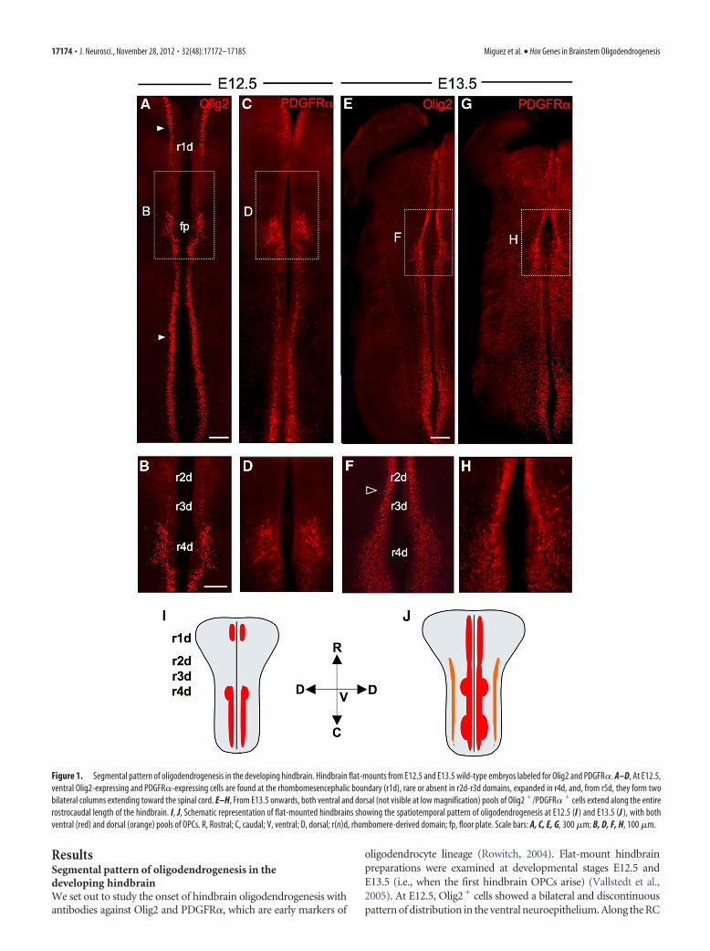
ResultsSegmental pattern of oligodendrogenesis in thedeveloping hindbrainWe set out to study the onset of hindbrain oligodendrogenesis withantibodies against Olig2 and PDGFR�, which are early markers of
oligodendrocyte lineage (Rowitch, 2004). Flat-mount hindbrainpreparations were examined at developmental stages E12.5 andE13.5 (i.e., when the first hindbrain OPCs arise) (Vallstedt et al.,2005). At E12.5, Olig2� cells showed a bilateral and discontinuouspattern of distribution in the ventral neuroepithelium. Along the RC
Figure 1. Segmental pattern of oligodendrogenesis in the developing hindbrain. Hindbrain flat-mounts from E12.5 and E13.5 wild-type embryos labeled for Olig2 and PDGFR�. A–D, At E12.5,ventral Olig2-expressing and PDGFR�-expressing cells are found at the rhombomesencephalic boundary (r1d), rare or absent in r2d-r3d domains, expanded in r4d, and, from r5d, they form twobilateral columns extending toward the spinal cord. E–H, From E13.5 onwards, both ventral and dorsal (not visible at low magnification) pools of Olig2 �/PDGFR� � cells extend along the entirerostrocaudal length of the hindbrain. I, J, Schematic representation of flat-mounted hindbrains showing the spatiotemporal pattern of oligodendrogenesis at E12.5 (I ) and E13.5 (J ), with bothventral (red) and dorsal (orange) pools of OPCs. R, Rostral; C, caudal; V, ventral; D, dorsal; r(n)d, rhombomere-derived domain; fp, floor plate. Scale bars: A, C, E, G, 300 �m; B, D, F, H, 100 �m.
17174 • J. Neurosci., November 28, 2012 • 32(48):17172–17185 Miguez et al. • Hox Genes in Brainstem Oligodendrogenesis
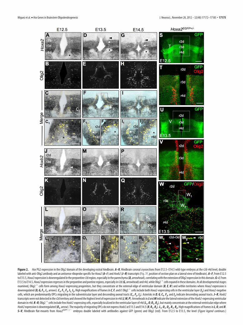
Figure 2. Hox PG2 expression in the Olig2 domain of the developing rostral hindbrain. A–R, Hindbrain coronal cryosections from E12.5–E14.5 wild-type embryos at the r2d-r4d level, doublelabeled with anti-Olig2 antibody and an antisense riboprobe specific for Hoxa2 (A–I ) and Hoxb2 (J–R) transcripts (Fig. 7F, position of section plan on a lateral view of hindbrain). A–F, From E12.5to E13.5, Hoxa2 expression is downregulated in the prepontine r2d region, especially in the parenchyma (D, arrowhead), correlating with the extension of Olig2 expression in this domain. G–I, FromE13.5 to E14.5, Hoxa2 expression regresses in the prepontine and pontine regions, especially in r2d (G, arrowhead) and r4d, while Olig2 � cells expand in these domains. At all developmental stagesexamined, Olig2 � cells form among Hoxa2-expressing progenitors, but they concentrate at the external edge of ventricular domain (B, E, H ) and within territories where Hoxa2 expression isdownregulated (D, G, F1, I1, arrows). C1, F1, F2, I1, I2, High magnifications of frames in C, F, and I. Olig2 � cells include both Hoxa2-expressing cells in the ventricular layer (I2) and Hoxa2-negativecells, which are predominantly OPCs migrating in the subventricular layer and descending axonal tracts (C1, F2, I2). Asterisks in D, G, C1, F2, and I2 indicate descending axonal tracts. J–R, Hoxb2transcripts were not detected in the r2d territory and showed the highest level of expression in r4d (J, M, P). Arrowheads in J and M indicate the lateral extension of the Hoxb2-expressing ventriculardomain in r4d. K–R, Olig2 � cells include few Hoxb2-expressing cells, especially localized in the ventricular layer of r4d (L1, O, O1, O2), but mainly concentrate at the external ventricular edge whereHoxb2 expression is downregulated (O1, arrow). The majority of migrating OPCs do not express Hoxb2 at E13.5 and E14.5 (R, R1, R2). L1, O1, O2, R1, R2, High magnifications of frames in L, O, and R.S–V, Hindbrain flat-mounts from Hoxa2EGFP�/� embryos double labeled with antibodies against GFP (green) and Olig2 (red). From E12.5 to E13.5, the level (Figure legend continues.)
Miguez et al. • Hox Genes in Brainstem Oligodendrogenesis J. Neurosci., November 28, 2012 • 32(48):17172–17185 • 17175
axis, Olig2� cells localized at the rhombomesencephalic boundary(rld) were rare or absent in r2d-r3d domains, expanded dorsoven-trally in the r4d domain, which corresponds to the presumptivepontine region, and from the r5d territory formed two bilateral col-umns extending toward the spinal cord (Fig. 1A,B). At E13.5, theOlig2-expressing domain had become continuous along the ventralhindbrain, although still larger in r4d than in r2d-r3d (Fig. 1E,F). Asimilar spatiotemporal pattern of expression was displayed byother specific oligodendroglial markers, such as PDGFR� (Fig.1C,D,G,H), Sox10, Olig1, and O4 (data not shown). Therefore, be-tween E12.5 and E13.5, oligodendrocytes form in the rostral hind-brain with rhombomere-specific patterns, with a 1 d delay in r2d-r3dand in larger number in r4d.
Hox PG2 expression in the Olig2 domain of the developingrostral hindbrainAmong Hox family members, Hox PG2 genes display the rostral-most expression domains in the CNS. Along the hindbrain RCaxis, Hoxa2 and Hoxb2 have overlapping expression domainswith offset rostral boundaries mapping in r2 and r3 for Hoxa2and Hoxb2, respectively. Moreover, progenitors along the DVaxis display distinct Hoxa2 and Hoxb2 expression levels (Davenneet al., 1999).
We compared Hoxa2 and Olig2 expression in the rostral hind-brain from E12.5 to E14.5, first on coronal cryosections doublelabeled with anti-Olig2 antibody and an antisense riboprobe spe-cific for Hoxa2 transcripts. We observed that (1) Hoxa2 expres-sion is progressively downregulated in the prepontine r2d region(Fig. 2A,D,G, arrowheads in D and G), while Olig2 expressionextends into this domain (Fig. 2B,C,E,F,H,I); (2) Olig2� cellsappear at the dorsal edge of the ventricular progenitor domain inr3d-r4d (Fig. 2B,C,E,F,H,I) and localize in low-expressingHoxa2� sites, while the ventricular domain expresses fairly highHoxa2 transcript levels (Fig. 2F1,I1, arrows); and (3) Olig2� cellsmigrate through territories where Hoxa2 expression is down-regulated, such as in the descending axonal tracts in r4d (Fig.2C1,F2,I2, asterisks).
We next analyzed the expression pattern of Olig2 inHoxa2EGFP�/� mice, which express the EGFP knocked-in at theHoxa2 locus, thus faithfully mimicking the endogenous Hoxa2expression pattern (Pasqualetti et al., 2002). Hindbrain flat-mounts from Hoxa2EGFP�/� embryos were double labeled withantibodies against GFP and Olig2. From E12.5 to E13.5, bilateralcolumns of Olig2� cells emerged at the dorsal edge of the ven-tralmost GFP� progenitor domain (i.e., the Hoxa2�/Nkx2.2�
domain) in r3d-r4d, while GFP expression levels decreased in theventral and alar plates of r2d, similar to the distribution of en-dogenous Hoxa2 transcripts (Fig. 2S–V). At the cellular level,more ventricular Olig2� cells expressed GFP in the r3d than inthe r2d domain (Fig. 2W1,W2). At E18.5, EGFP expression wasexpressed by the majority of CNP� mature oligodendrocytes in
prepontine and pontine territories (data not shown). Altogether,these data indicate that low levels of Hoxa2 expression correlatewith the expansion of Olig2� progenitors at the onset ofoligodendrogenesis.
Next, we compared the expression of Olig2 with that of theother Hox PG2 gene, Hoxb2. Coronal cryosections of hindbrain,at developmental stages E12.5, E13.5, and E14.5, were colabeledwith anti-Olig2 antibody and an antisense riboprobe againstHoxb2 (Fig. 2 J–R). Hoxb2 transcripts were not detected in the r2dterritory, as expected from its normal expression domain. Hoxb2and Hoxa2 were both expressed in r3d as well as in r4d, whichshowed the highest level of Hoxb2 expression (Fig. 2 J,M,P). No-tably, Hoxb2 expression expanded dorsally in the r4d Nkx2.2�
domain, where Olig2� cells colocalized with Hoxb2-expressingprogenitors, especially at E12.5–E13.5 and unlike Hoxa2 (Fig.2 J,M, arrowheads; Fig. 2O–O2; Davenne et al., 1999). In contrast,the majority of migrating OPCs did not appear to express Hoxb2at E13.5 and E14.5 (Fig. 2O,R).
Altogether, the spatiotemporal patterns of Hoxa2 and Hoxb2expression between E12.5 and E14.5 suggest their potential in-volvement in segment-specific patterning of the ventral Olig2�
oligodendrocyte subpopulation.
Conditional Hoxa2 overexpression in Olig2 � cells inhibitsoligodendrogenesisIn the rostral hindbrain, Hoxa2 is expressed at low levels in Olig2�
progenitors at the onset of oligodendrogenesis (Fig. 2C,F). Thus, onepossibility is that downregulation of Hoxa2 is a prerequisite to allowthe appearance of Olig2 expression at the dorsal edge of the Nkx2.2�
domain and, in turn, to induce ventral oligodendrocyte generation.We therefore examined whether forced Hoxa2 overexpression inOlig2� progenitors could inhibit hindbrain oligodendrogenesis.Conditional Hoxa2 overexpression was induced by mating theOlig2-tva-Cre line (Schuller et al., 2008) with a newly generated alleleROSA26::(lox-STOP-lox)Hoxa2-IRES-EGFP (hereafter referred toas Olig2-tva-Cre;ROSA(lox-stop-lox)Hoxa2 mice), in which Hoxa2 is se-lectively activated only in the Olig2 lineage upon Cre-mediated re-combination (see Materials and Methods). Hindbrain flat-mountsfrom E13.5 Olig2-tva-Cre;ROSA(lox-stop-lox)Hoxa2 embryos were im-munostained to detect oligodendroglial cells with antibodies againstOlig2 and PDGFR�. The dorsoventral extent of the Olig2� ventric-ular domain was reduced in the r2d-r3d region, compared withROSA(lox-stop-lox)Hoxa2 controls (Fig. 3A,C,D,F,G). Quantification ofthe number of PDGFR�� OPCs on sagittal sections confirmed that,in these conditional Hoxa2 gain-of-function mutants, ventral oligo-dendrogenesis was decreased by �2-fold in the prepontine and pon-tine territories (r2d-r4d) (Fig. 3B,C,E,F,H). Therefore, forcedHoxa2 expression in Olig2� cells inhibits ventral hindbrain oligo-dendrogenesis.
Remarkably, the Olig2�/PDGFR�� cell population was alsoreduced by �50% in Olig2-tva-Cre;ROSA(lox-stop-lox)Hoxa2 em-bryos compared with controls in ectopic brain territories whereHoxa2 is not normally expressed during development, such as theventral forebrain [Fig. 3L–O; mean number of PDGFR�� cellsper area (mm 2) � SEM: ROSA(lox-stop-lox)Hoxa2, 156.4 � 6.91;Olig2-tva-Cre;ROSA(lox-stop-lox)Hoxa2, 81.76 � 5.56; p � 0.0001,Student’s t test, n � 3]. As shown in Figure 3P–R, EGFP, which isdirectly translated from the IRES of the allele overexpressingHoxa2 (see Materials and Methods), and Olig2 expression levelsare inversely correlated in ventricular forebrain progenitors.Namely, Olig2 levels are low in ectopic EGFP/Hoxa2 highly ex-pressing cells (Fig. 3Q,R, dark arrowheads), while progenitorsstrongly expressing Olig2 display low levels of EGFP/Hoxa2 ecto-
4
(Figure legend continued.) of Hoxa2/EGFP expression decreases in the basal plate of r2d and r4d(S, U), while bilateral columns of Olig2 � cells enlarge in these domains (T, V). Hoxa2/EGFPexpression is maintained in the ventralmost ventricular domain of r3d. W, Sagittal section of aHoxa2EGFP�/� E13.5 embryo at the r2d-r4d level, labeled for GFP (green) and Olig2 (red).Ventricular Olig2 � cells expressing Hoxa2/EGFP were detected in r3d-r4d, but not in r2d. In thesubventricular layer of r2d-r4d region, both Olig2 �EGFP � (dark arrowheads) andOlig2 �EGFP � (white arrowheads) cells were observed, and the majority of Olig2 � cells mi-grating in the parenchyma were GFP � (W1, W2). r(n)d, rhombomere-derived domain. Scalebars: S, U, 200 �m; A--I, J--R, T, V, W, 100 �m; C1, F1, F2, I1, I2, L1, O1, O2, R1, R2, 50 �m; W1,W2, 25 �m.
17176 • J. Neurosci., November 28, 2012 • 32(48):17172–17185 Miguez et al. • Hox Genes in Brainstem Oligodendrogenesis
Figure 3. Conditional Hoxa2 overexpression in Olig2� cells inhibits oligodendrogenesis. A–I, Hindbrain flat-mounts from ROSA(lox-stop-lox)Hoxa2 (A–C) and Olig2-tva-Cre; ROSA(lox-stop-lox)Hoxa2
(D–F) embryos at E13.5, labeled for Olig2 (green) and PDGFR� (red). Hoxa2 gain of function in Olig2-expressing cells results in a reduced Olig2 � progenitor domain in r2d and r3d (A, C, D, F, G) anda decreased number of PDGFR� � cells in the basal plate of r2d, r3d, and r4d (B, C, E, F, H). No differences between genotypes were found regarding the percentage of Olig2 � migrating cells (I).J, K, Hindbrain flat-mounts labeled for PH3, showing that Olig2-tva-Cre; ROSA(lox-stop-lox)Hoxa2 embryos display a decreased number of PH3 � proliferating cells, compared with control animals. L–R,Coronal sections from ROSA(lox-stop-lox)Hoxa2 (L, M) and Olig2-tva-Cre; ROSA(lox-stop-lox)Hoxa2 (N–R) E13.5 embryos, at the level of the anterior entopeduncular area/medial ganglionic eminence of theforebrain, labeled for Olig2 (blue), PDGFR� (red), and GFP (green). M, O, The number of Olig2� (white arrowheads) and PDGFR�� (black arrowheads) in Olig2-tva-Cre; ROSA(lox-stop-lox)Hoxa2
embryos was reduced by half compared with controls. High levels of EGFP/Hoxa2 and Olig2 expression were mutually exclusive in ventricular progenitors (P). Q, (Figure legend continues.)
Miguez et al. • Hox Genes in Brainstem Oligodendrogenesis J. Neurosci., November 28, 2012 • 32(48):17172–17185 • 17177
pic expression (Fig. 3Q,R, white arrowheads). Thus, Hoxa2 caninhibit Olig2 expression in a cell-autonomous manner in progen-itor cells throughout the brain.
We next analyzed the migration and proliferation of OPCs inhindbrain flat-mounts of E13.5 Hoxa2-gain-of-function mu-tants. At this stage, the Olig2� ventricular zone is observed as twobilateral columns of dense populations of cells next to the ventralmidline, while Olig2� migrating cells disperse laterally in theventroalar plate (Fig. 3A,D). We quantified the ratio of migratingversus ventricular/nonmigrating progenitors as a function of flu-orescence intensity in respective areas, as previously reported (DiLullo et al., 2011). Figure 3I quantifies the percentage of cellslocated in the migration area of the r2d-r3d and r4d domains.The percentage of migrating Olig2� cells was similar betweenOlig2-tva-Cre;ROSA(lox-stop-lox)Hoxa2 gain-of-function mutantsand ROSA(lox-stop-lox)Hoxa2 controls, suggesting that OPCs’ migra-tory properties were not altered by Hoxa2 forced expression. Incontrast, cell proliferation was reduced in the ventroalar plate ofr2d-r4d, as shown by immunolabeling of hindbrain flat-mounts(Fig. 3 J,K) and sagittal sections with anti-phospho-histone H3(PH3) antibody. Hoxa2 overexpression induced a decrease inthe number of PH3 � cells [mean number of PH3 � cells perarea (mm 2) � SEM: ROSA(lox-stop-lox)Hoxa2 (control), 35.7 �1.9, Olig2-tva-Cre;ROSA(lox-stop-lox)Hoxa2: 14.8 � 1.8, p �0.0004; Student’s t test, n � 3], and less OPC proliferation(percentage of PH3 �/PDGFR� � cells/total PDGFR� � cells �SEM: ROSA(lox-stop-lox)Hoxa2, 7.1 � 1.2, Olig2-tva-Cre;ROSA(lox-stop-lox)Hoxa2, 3.1 � 0.4, p � 0.0346; Student’s t test,n � 3). These data suggest that Hoxa2 has a primary effect onthe generation rather than on the migration of OPCs.
Finally, we investigated whether the reduced oligodendrogenesisinduced by Hoxa2-overexpression resulted in an alteration of hind-brain oligodendrocyte population at later stages of development. Wequantified OPCs (PDGFR��) as well as cells expressing 2-cyclicnucleotide 3-phosphodiesterase (CNP), a marker for differentiated oli-godendrocytes (Kim et al., 1984), on sagittal sections of hindbrain atE18.5. The number of OPCs and mature oligodendrocytes in Hoxa2-gain-of-function mutants and controls were similar [mean number ofPDGFR�� cells per area (mm2) � SEM: r2d: ROSA(lox-stop-lox)Hoxa2
(control), 265.2 � 78.4; Olig2-tva-Cre;ROSA(lox-stop-lox)Hoxa2, 319.8 �94.1; r3d: ROSA(lox-stop-lox)Hoxa2, 360.7 � 132.2; Olig2-tva-Cre;ROSA(lox-stop-lox)Hoxa2, 419.3 � 134.7; mean number of CNP� cellsper area (mm2): r2d: ROSA(lox-stop-lox)Hoxa2, 9.1 � 7.6; Olig2-tva-Cre;ROSA(lox-stop-lox)Hoxa2, 12.9 � 14.5; r3d: ROSA(lox-stop-lox)Hoxa2, 9.5 �10.9; Olig2-tva-Cre; ROSA(lox-stop-lox)Hoxa2, 30.6 � 37.8; n � 3]. Thesedata suggest that the reduced oligodendrogenesis observed at E13.5is compensated at later stages, either by additional proliferation or byincreased survival of OPCs. In Hoxa2-GOF mutants, there is lesscompetition between the remaining ventral OPCs for growth fac-tors, such as PDGF-A, which determines the final number and dis-tribution of mature oligodendrocytes (Calver et al., 1998), and OPCsmay expand transiently more rapidly. We have tested this hypothesisat embryonic stage E16.5, between the onset of oligodendrogenesis(E12.5) and the onset of oligodendrocyte differentiation (E17.5–E18.5). We found that Hoxa2-GOF mutants showed more prolifer-
ating OPCs in the prepontine domain than control embryos[percentage of PH3�/PDGFR�� cells/total PDGFR�� cells per sec-tion � SEM (r2d-r3d): Olig2-tva-Cre; ROSA(lox-stop-lox)Hoxa2, 8.5 �0.3; ROSA(lox-stop-lox)Hoxa2, 5.8 � 0.2; p � 0.0152, Student’s t test, 3sections/embryo, n � 3]. Additional immunostaining for cleavedcaspase-3 to detect cells undergoing apoptosis in the r2d-r3d domainrevealed caspase-3� cells, but failed to show caspase-3�/PDGFR��
cells both in controls and Hoxa2-gain-of-function mutants (data notshown), suggesting that OPC survival is likely not affected by Hoxa2overexpression. Thus, the twofold reduction in ventricular progen-itors observed in Hoxa2-GOF is rapidly normalized following anadditional cell cycle of OPCs before they differentiate. However, wecannot rule out the possibility that ectopic OPCs originating fromother rostral and caudal progenitor domains of the neural tube maymigrate into the r2-r3 domain and play a role in compensating forthe early reduction of ventral oligodendrogenesis.
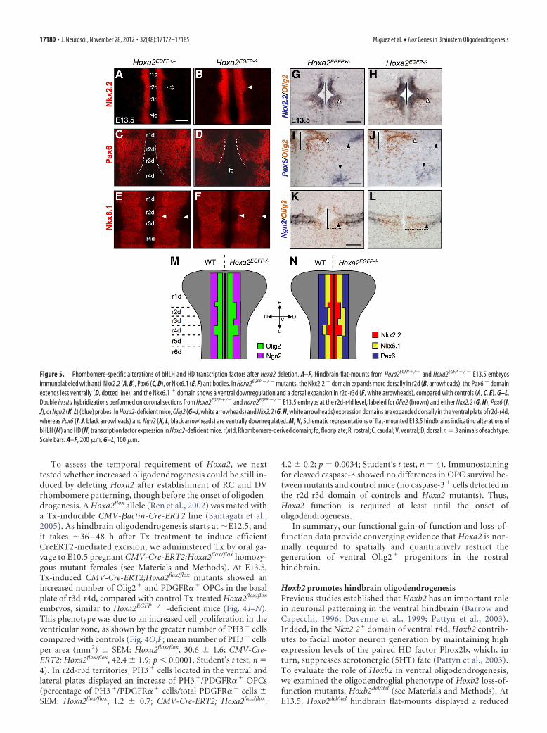
Hoxa2 inactivation results in dorsal expansion of the Olig2progenitor domain and increased oligodendrogenesisTo confirm the inhibitory effect of Hoxa2 on hindbrain oligoden-drogenesis, we next analyzed the effect of Hoxa2 loss of function. AtE13.5, Hoxa2EGFP�/� mutant embryos showed a dorsally expandedventricular domain of Olig2 expression, compared with heterozy-gous littermates (Fig. 4A–D). Motor neuron specification appearednormal, as indicated by the pattern of Islet1/2 expression (data notshown). In contrast, Hoxa2EGFP�/� embryos formed twice as manyPDGFR�� OPCs than controls in the r2d-r4d ventral plate (Fig.4E–H). The number of PDGFR�� OPCs was not altered in r1d, asexpected by the lack of Hoxa2 expression in r1 during normal devel-opment. Similar to Hoxa2 overexpression, Hoxa2 deletion did notsignificantly alter OPC migration in the r2d-r4d region, as shown bythe similar percentage of migrating OPCs in Hoxa2EGFP�/� mutantsand Hoxa2EGFP�/� controls (percentage of Olig2� migrating cells �SEM: r2d-r3d: Hoxa2EGFP�/�, 20.6 � 2.8; Hoxa2EGFP�/�, 14.1 �1.6; p � 0.0690; r4d: Hoxa2EGFP�/�, 14.6 � 1.6; Hoxa2EGFP�/�,11.4 � 2.3; p � 0.291; Student’s t test, n � 4). Moreover, at E18.5,Hoxa2EGFP�/� mutants showed a similar number of OPCs and ma-ture oligodendrocytes than control mice [mean number ofPDGFR�� cells per area (mm2) � SEM: r2d: Hoxa2EGFP�/� (con-trol), 272.1 � 65.4; Hoxa2EGFP�/�, 280.6 � 119.1; r3d:Hoxa2EGFP�/�, 340.5 � 112.2; Hoxa2EGFP�/�, 309.7 � 104.8; meannumber of CNP� cells per area (mm2): r2d: Hoxa2EGFP�/�, 8.9 �5.6; Hoxa2EGFP�/�, 11.2 � 10.1; r3d: Hoxa2EGFP�/�, 11.4 � 10.2;Hoxa2EGFP�/�, 23.8 � 24.1]. Thus, the loss of Hoxa2 function hasan early impact on the onset of oligodendrogenesis, which can becompensated at later developmental stages.
Hoxa2 inhibits ventral oligodendrogenesis by spatially re-stricting the dorsal extent of oligodendrocyte generation inr2d-r4d territories and thus, ultimately, limiting the size of theOlig2 � domain. Indeed, previous studies showed DV rhom-bomere patterning defects in Hoxa2 mutants as early as E10.5(Davenne et al., 1999). We anticipated that in Hoxa2-deficientembryos such DV pattern defects may persist through laterstages and have direct consequences on the patterning of theOlig2 progenitor domain. Thus, we examined the expression ofDV patterning genes implicated in oligodendrogenesis and neu-rogenesis, including Pax6, Nkx2.2, Nkx6.1, and Ngn2. In E13.5Hoxa2EGFP �/ � mutant embryos, the transcript levels and expres-sion patterns of all these genes were altered in the rostral hind-brain, compared with control heterozygotes (Fig. 5A–L).Namely, the expression domains of Nkx2.2 (Fig. 5A,B,G,H) andOlig2 (Fig. 5G–L) in the r2d-r4d ventral plate were dorsally ex-tended. Pax6 expression was decreased in r2d-r4d, with a dorsal
4
(Figure legend continued.) R, Olig2 expression was weak in highly fluorescent cells (dark arrowheads),while progenitors strongly expressing Olig2 displayed a low level of GFP fluorescence (white arrow-heads). r(n)d, rhombomere-derived domain. G, r2d, p � 0.0009; r3d, p � 0.0069; H, r2d, p �0.0034; r3d, p�0.0010; r4d, p�0.0193. n�3 animals of each type. Area, 1 mm 2. Scale bars: A, B,D, E, J, K, L, N, P, 100 �m; C, F, M, O, 50 �m; Q, R, 25 �m. *p � 0.05; **p � 0.01; ***p � 0.001.
17178 • J. Neurosci., November 28, 2012 • 32(48):17172–17185 Miguez et al. • Hox Genes in Brainstem Oligodendrogenesis
shift of its ventral border (Fig. 5C,D,I,J). Similarly, the ventralborder of the Nkx6.1� domain was shifted dorsally in r2d-r3d(Fig. 5E,F). The expression levels of the proneural gene Ngn2were also reduced in r2d-r4d, with a dorsal extension of its ventralventricular domain (Fig. 5K,L). In contrast, no expression ab-normalities were observed for Pax3 and Pax7 in the dorsal plate of
Hoxa2EGFP �/ � embryos (data not shown). Thus, Hoxa2 inhibitsthe dorsal extension of the Olig2�/Nkx2.2� domain in the ventralplate by restricting the dorsoventral extent of bHLH and home-odomain (HD) transcription factors regulating oligodendrogen-esis, such as Pax6, Nkx6.1, and Ngn2 (Fig. 5M,N, summarymodel).
Figure 4. Hoxa2 deletion increases oligodendrogenesis in the rostral hindbrain. A–H, Hindbrain flat-mounts (A–C) and sagittal sections (E–G) from Hoxa2EGFP�/� (B, F) and Hoxa2EGFP �/ � (A,C, G) embryos at E13.5, labeled for GFP (green) and Olig2 or PDGFR� (red). Hoxa2-deficient mice show an enlarged Olig2 � progenitor domain (C, D) and an increased number of PDGFR� � OPCs(G, H) in the basal plate of r2d, r3d, and r4d, compared with controls (B, F). I–L, Sagittal sections from Tx-treated Hoxa2flox/flox (I, J) and CMV-Cre-ERT2; Hoxa2flox/flox (K, L) embryos, induced at E10.5,sacrificed at E13.5, and immunolabeled for Olig2 (green) and PDGFR� (red). Tx-induced Hoxa2 knockout embryos display an increased number of Olig2 � (K, M) and PDGFR� � (L, N) cells in thebasal plate of r3d and r4d, compared with control animals (I, J). White and black arrowheads indicate Olig2 � cells and PDGFR� � cells, respectively, in J and L. O, P, Hindbrain flat-mounts fromTx-treated Hoxa2flox/flox and CMV-Cre-ERT2; Hoxa2flox/flox embryos, induced at E10.5 and sacrificed at E13.5, labeled for PH3. Tx-induced Hoxa2 knockout embryos (P) show an increased number ofPH3 � cells in the basal plate of r2d-r3d, compared with control mice (O). r(n)d, rhombomere-derived domain. D, r2d, p � 0.0009; r3d, p � 0.0044; r4d, p � 0.0001; n � 4 animals of each type;H, r2d, p � 0.00397; r3d, p � 0.000133; r4d, p � 0.0370; n � 6 animals of each type; M, r3d, p � 0.0014; r4d, p � 0.0131; N, r3d, p � 0.011; r4d, p � 0.036; n � 7 animals of each type. Area,1 mm 2. Scale bars: F, G, I, K, 200 �m; A--C, O, P, 100 �m; J, L, 80 �m. *p � 0.05; **p � 0.01; ***p � 0.001.
Miguez et al. • Hox Genes in Brainstem Oligodendrogenesis J. Neurosci., November 28, 2012 • 32(48):17172–17185 • 17179
To assess the temporal requirement of Hoxa2, we nexttested whether increased oligodendrogenesis could be still in-duced by deleting Hoxa2 after establishment of RC and DVrhombomere patterning, though before the onset of oligoden-drogenesis. A Hoxa2flox allele (Ren et al., 2002) was mated witha Tx-inducible CMV-�actin-Cre-ERT2 line (Santagati et al.,2005). As hindbrain oligodendrogenesis starts at �E12.5, andit takes �36 – 48 h after Tx treatment to induce efficientCreERT2-mediated excision, we administered Tx by oral ga-vage to E10.5 pregnant CMV-Cre-ERT2;Hoxa2flox/flox homozy-gous mutant females (see Materials and Methods). At E13.5,Tx-induced CMV-Cre-ERT2;Hoxa2flox/flox mutants showed anincreased number of Olig2� and PDGFR�� OPCs in the basalplate of r3d-r4d, compared with control Tx-treated Hoxa2flox/flox
embryos, similar to Hoxa2EGFP �/ �-deficient mice (Fig. 4 I–N).This phenotype was due to an increased cell proliferation in theventricular zone, as shown by the greater number of PH3� cellscompared with controls (Fig. 4O,P; mean number of PH3� cellsper area (mm 2) � SEM: Hoxa2flox/flox, 30.6 � 1.6; CMV-Cre-ERT2; Hoxa2flox/flox, 42.4 � 1.9; p � 0.0001, Student’s t test, n �4). In r2d-r3d territories, PH3� cells located in the ventral andlateral plates displayed an increase of PH3�/PDGFR�� OPCs(percentage of PH3�/PDGFR�� cells/total PDGFR�� cells �SEM: Hoxa2flox/flox, 1.2 � 0.7; CMV-Cre-ERT2; Hoxa2flox/flox,
4.2 � 0.2; p � 0.0034; Student’s t test, n � 4). Immunostainingfor cleaved caspase-3 showed no differences in OPC survival be-tween mutants and control mice (no caspase-3� cells detected inthe r2d-r3d domain of controls and Hoxa2 mutants). Thus,Hoxa2 function is required at least until the onset ofoligodendrogenesis.
In summary, our functional gain-of-function and loss-of-function data provide converging evidence that Hoxa2 is nor-mally required to spatially and quantitatively restrict thegeneration of ventral Olig2 � progenitors in the rostralhindbrain.
Hoxb2 promotes hindbrain oligodendrogenesisPrevious studies established that Hoxb2 has an important rolein neuronal patterning in the ventral hindbrain (Barrow andCapecchi, 1996; Davenne et al., 1999; Pattyn et al., 2003).Indeed, in the Nkx2.2� domain of ventral r4d, Hoxb2 contrib-utes to facial motor neuron generation by maintaining highexpression levels of the paired HD factor Phox2b, which, inturn, suppresses serotonergic (5HT) fate (Pattyn et al., 2003).To evaluate the role of Hoxb2 in ventral oligodendrogenesis,we examined the oligodendroglial phenotype of Hoxb2 loss-of-function mutants, Hoxb2del/del (see Materials and Methods). AtE13.5, Hoxb2del/del hindbrain flat-mounts displayed a reduced
Figure 5. Rhombomere-specific alterations of bHLH and HD transcription factors after Hoxa2 deletion. A–F, Hindbrain flat-mounts from Hoxa2EGFP�/� and Hoxa2EGFP �/ � E13.5 embryosimmunolabeled with anti-Nkx2.2 (A, B), Pax6 (C, D), or Nkx6.1 (E, F) antibodies. In Hoxa2EGFP �/ � mutants, the Nkx2.2 � domain expands more dorsally in r2d (B, arrowheads), the Pax6 � domainextends less ventrally (D, dotted line), and the Nkx6.1 � domain shows a ventral downregulation and a dorsal expansion in r2d-r3d (F, white arrowheads), compared with controls (A, C, E). G–L,Double in situ hybridizations performed on coronal sections from Hoxa2EGFP�/� and Hoxa2EGFP �/ � E13.5 embryos at the r2d-r4d level, labeled for Olig2 (brown) and either Nkx2.2 (G, H), Pax6 (I,J), or Ngn2 (K, L) (blue) probes. In Hoxa2-deficient mice, Olig2 (G–J, white arrowheads) and Nkx2.2 (G, H, white arrowheads) expression domains are expanded dorsally in the ventral plate of r2d-r4d,whereas Pax6 (I, J, black arrowheads) and Ngn2 (K, L, black arrowheads) are ventrally downregulated. M, N, Schematic representations of flat-mounted E13.5 hindbrains indicating alterations ofbHLH (M) and HD (N) transcription factor expression in Hoxa2-deficient mice. r(n)d, Rhombomere-derived domain; fp, floor plate; R, rostral; C, caudal; V, ventral; D, dorsal. n�3 animals of each type.Scale bars: A–F, 200 �m; G–L, 100 �m.
17180 • J. Neurosci., November 28, 2012 • 32(48):17172–17185 Miguez et al. • Hox Genes in Brainstem Oligodendrogenesis
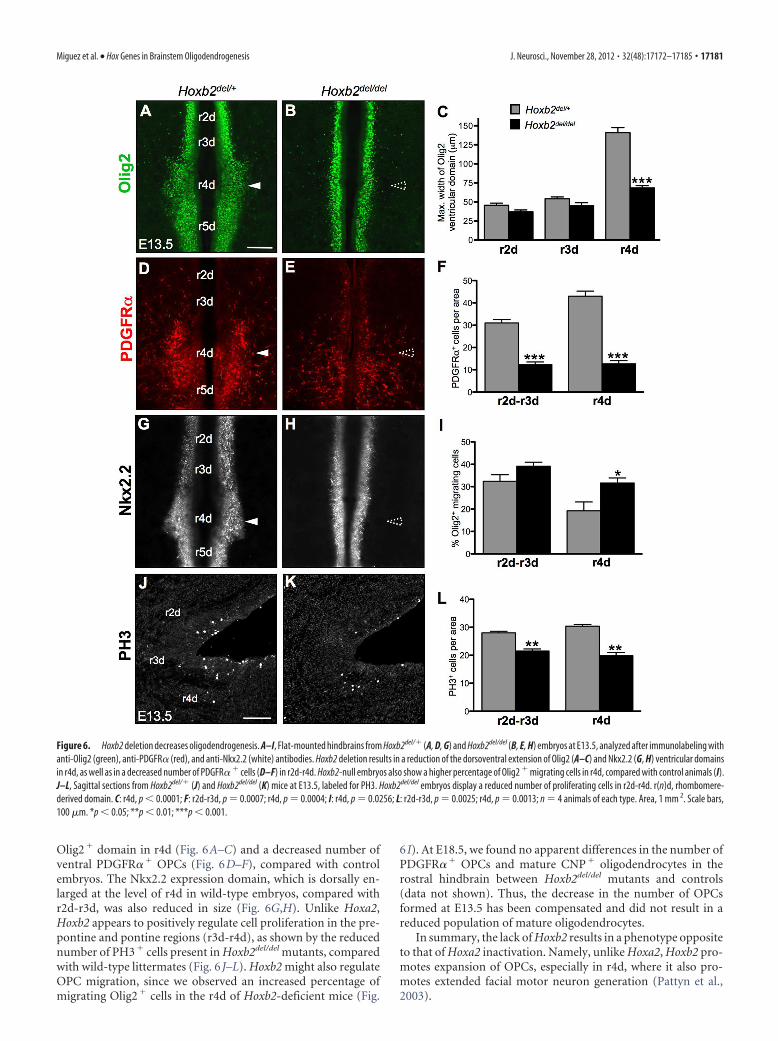
Olig2� domain in r4d (Fig. 6A–C) and a decreased number ofventral PDGFR�� OPCs (Fig. 6D–F), compared with controlembryos. The Nkx2.2 expression domain, which is dorsally en-larged at the level of r4d in wild-type embryos, compared withr2d-r3d, was also reduced in size (Fig. 6G,H). Unlike Hoxa2,Hoxb2 appears to positively regulate cell proliferation in the pre-pontine and pontine regions (r3d-r4d), as shown by the reducednumber of PH3� cells present in Hoxb2del/del mutants, comparedwith wild-type littermates (Fig. 6 J–L). Hoxb2 might also regulateOPC migration, since we observed an increased percentage ofmigrating Olig2� cells in the r4d of Hoxb2-deficient mice (Fig.
6 I). At E18.5, we found no apparent differences in the number ofPDGFR�� OPCs and mature CNP� oligodendrocytes in therostral hindbrain between Hoxb2del/del mutants and controls(data not shown). Thus, the decrease in the number of OPCsformed at E13.5 has been compensated and did not result in areduced population of mature oligodendrocytes.
In summary, the lack of Hoxb2 results in a phenotype oppositeto that of Hoxa2 inactivation. Namely, unlike Hoxa2, Hoxb2 pro-motes expansion of OPCs, especially in r4d, where it also pro-motes extended facial motor neuron generation (Pattyn et al.,2003).
Figure 6. Hoxb2 deletion decreases oligodendrogenesis. A–I, Flat-mounted hindbrains from Hoxb2del/� (A, D, G) and Hoxb2del/del (B, E, H) embryos at E13.5, analyzed after immunolabeling withanti-Olig2 (green), anti-PDGFR� (red), and anti-Nkx2.2 (white) antibodies. Hoxb2 deletion results in a reduction of the dorsoventral extension of Olig2 (A–C) and Nkx2.2 (G, H) ventricular domainsin r4d, as well as in a decreased number of PDGFR� � cells (D–F) in r2d-r4d. Hoxb2-null embryos also show a higher percentage of Olig2 � migrating cells in r4d, compared with control animals (I).J–L, Sagittal sections from Hoxb2del/� (J) and Hoxb2del/del (K) mice at E13.5, labeled for PH3. Hoxb2del/del embryos display a reduced number of proliferating cells in r2d-r4d. r(n)d, rhombomere-derived domain. C: r4d, p � 0.0001; F: r2d-r3d, p � 0.0007; r4d, p � 0.0004; I: r4d, p � 0.0256; L: r2d-r3d, p � 0.0025; r4d, p � 0.0013; n � 4 animals of each type. Area, 1 mm 2. Scale bars,100 �m. *p � 0.05; **p � 0.01; ***p � 0.001.
Miguez et al. • Hox Genes in Brainstem Oligodendrogenesis J. Neurosci., November 28, 2012 • 32(48):17172–17185 • 17181
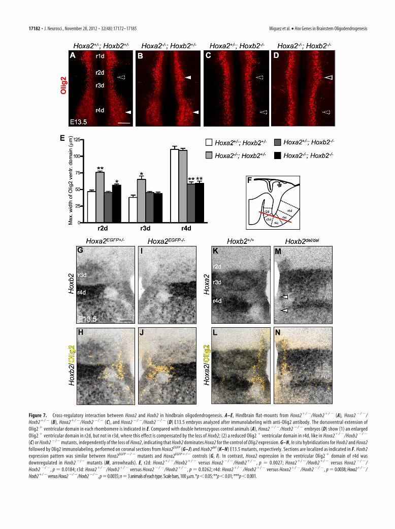
Figure 7. Cross-regulatory interaction between Hoxa2 and Hoxb2 in hindbrain oligodendrogenesis. A–E, Hindbrain flat-mounts from Hoxa2�/ �/Hoxb2�/ � (A), Hoxa2 �/ �/Hoxb2�/ � (B), Hoxa2�/ �/Hoxb2 �/ � (C), and Hoxa2 �/ �/Hoxb2 �/ � (D) E13.5 embryos analyzed after immunolabeling with anti-Olig2 antibody. The dorsoventral extension ofOlig2 � ventricular domain in each rhombomere is indicated in E. Compared with double heterozygous control animals (A), Hoxa2 �/ �/Hoxb2 �/ � embryos (D) show (1) an enlargedOlig2 � ventricular domain in r2d, but not in r3d, where this effect is compensated by the loss of Hoxb2; (2) a reduced Olig2 � ventricular domain in r4d, like in Hoxa2�/ �/Hoxb2 �/ �
(C) or Hoxb2 �/� mutants, independently of the loss of Hoxa2, indicating that Hoxb2 dominates Hoxa2 for the control of Olig2 expression. G–N, In situ hybridizations for Hoxb2 and Hoxa2followed by Olig2 immunolabeling, performed on coronal sections from Hoxa2EGFP (G–J) and Hoxb2del (K–N) E13.5 mutants, respectively. Sections are localized as indicated in F. Hoxb2expression pattern was similar between Hoxa2EGFP �/ � mutants and Hoxa2EGFP�/� controls (G, I). In contrast, Hoxa2 expression in the ventricular Olig2 � domain of r4d wasdownregulated in Hoxb2 �/� mutants (M, arrowheads). E, r2d: Hoxa2�/ �/Hoxb2�/ � versus Hoxa2 �/ �/Hoxb2�/ �, p � 0.0027; Hoxa2�/ �/Hoxb2�/ � versus Hoxa2 �/ �/Hoxb2 �/ �, p � 0.0184; r3d: Hoxa2�/ �/Hoxb2�/ � versus Hoxa2 �/ �/Hoxb2�/ �, p � 0.0262; r4d: Hoxa2�/ �/Hoxb2�/ � versus Hoxa2�/ �/Hoxb2 �/ �, p � 0.0038; Hoxa2�/ �/Hoxb2�/ �versusHoxa2�/ �/Hoxb2�/ �,p�0.0035;n�3animalsofeachtype.Scalebars,100�m.*p�0.05;**p�0.01;***p�0.001.
17182 • J. Neurosci., November 28, 2012 • 32(48):17172–17185 Miguez et al. • Hox Genes in Brainstem Oligodendrogenesis
Cross-regulatory interaction between Hoxa2 and Hoxb2 inhindbrain oligodendrocyte patterningThe phenotypes of single Hoxa2 and Hoxb2 mutants indicate oppos-ing roles for Hoxb2 and Hoxa2 in hindbrain oligodendrogenesis,suggesting that a net balance between Hoxa2 and Hoxb2 expressionlevels in the Olig2� domain may modulate the number of OPCsgenerated in each rhombomere posterior to r1. To test this hypoth-esis, we analyzed the pattern of Olig2� progenitors and PDGFR��
OPCs in compound Hoxa2/Hoxb2 mutants and their littermates atstage E13.5 (Fig. 7A–E). In r2d, where Hoxa2, but not Hoxb2, isexpressed, Hoxa2�/�/Hoxb2�/� mutants showed, as expected, asimilar dorsal expansion of the Olig2� domain as in singleHoxa2�/� mutants. Interestingly, in r3d, the loss of Hoxb2 in doubleHoxa2�/�/Hoxb2�/� homozygotes was sufficient to prevent the in-creased oligodendrogenesis displayed by single Hoxa2�/� mutants.Thus, the Olig2 expression pattern in r3d suggests that the promotingeffect of Hoxb2 on oligodendrogenesis is dominant upon the inhibitoryactionofHoxa2. This was further supported by the finding that in r4dof Hoxa2�/�/Hoxb2�/� mutants, the reduction of the Olig2� ven-tral domain (Fig. 7D) was similar to that of Hoxa2�/�/Hoxb2�/�
(Fig. 7C) and single Hoxb2�/� mutants (Fig. 6B), while singleHoxa2�/� mutants displayed a twofold increase of Olig2� cells inthis domain (Fig. 4C). The Olig2 expression pattern in r4d confirmsthat Hoxb2 antagonizes and dominates Hoxa2 in the regulation ofrhombomere-specific oligodendrogenesis.
To determine whether Hoxa2 and Hoxb2 each regulateseach other’s expression, we examined the hindbrain expres-sion pattern of Hoxa2 in Hoxb2 mutants and, reciprocally, ofHoxb2 in Hoxa2-deficient embryos (Fig. 7F–N). The Hoxb2expression pattern was not changed in Hoxa2EGFP �/ � mu-tants, compared with Hoxa2EGFP�/� controls (Fig. 7G–J). Incontrast, in Hoxb2 �/� mutants, Hoxa2 was significantlydownregulated in the ventricular Olig2 �/Nkx2.2 � progenitordomain of r4d (Fig. 7K–N, arrowheads). This is in agreementwith a direct regulatory role for Hoxb2 in maintaining Hoxa2expression levels in r4 (Lampe et al., 2008) and explains the
similar oligodendrocyte phenotypesof single Hoxb2 �/� and compoundHoxa2 �/ �/Hoxb2 �/ � mutants in theventral r4d domain. Therefore, Hoxa2and Hoxb2 likely act together in ventraloligodendrocyte progenitors, at least inr4d, exerting opposite modulatory effectson the molecular processes regulating OPCspecification and proliferation.
DiscussionIn this study, we found that Hox PG2genes have opposing roles in the regula-tion of oligodendrocyte generation andpatterning in the rostral hindbrain. In thespinal cord, Hox homeoproteins are ex-pressed by oligodendroglial cells (Nicolayet al., 2004a,b; Booth et al., 2007), raisingthe possibility that they may confer RCidentity to oligodendrocytes. However, inthe spinal cord of Hoxa2-deficient mice,no anomalies were detected in the num-ber, migration, and differentiation ofOPCs (Nicolay et al., 2004b). Nonethe-less, many Hox genes are coexpressed atthe level of the spinal cord, thus providingthe potential for functional redundancythat may mask the involvement of indi-
vidual Hox factors. To overcome the redundancy pitfall and in-vestigate the possible involvement of Hox PG2 factors inoligodendrogenesis, we focused on the rostral hindbrain whereHoxa2 and Hoxb2 perform their main functional role. Indeed, weprovide evidence that Hoxa2 and Olig2 expression levels are in-versely correlated in progenitor cells, and that Hoxa2 might beinvolved in restricting the ventral domain of oligodendrocytespecification. From E12.5 to E14.5, ventral oligodendrogenesis isinduced in prepontine and pontine territories in progenitors ex-pressing low levels of Hoxa2. In mice conditionally overexpress-ing Hoxa2 in Olig2� progenitors, the size of the ventral Olig2domain is reduced, especially in r2d-r3d territories. The inhibi-tion of oligodendrogenesis induced by Hoxa2-overexpressionmay occur independently of environmental cues, as it is not onlyevident in the hindbrain but also, more generally, in other brainregions, such as the ventral forebrain. In Hoxa2-deficient mice,there is a dorsal expansion of the ventral Olig2�/Nkx2.2� do-main resulting in increased oligodendrogenesis. This is in partdue to the loss of Hoxa2 expression at the ventral border of theabutting Pax6� ventricular domain, where Hoxa2 is normallyhighly expressed, and to ectopic recruitment of Olig2� progeni-tors. Like Hoxa2, Hoxb2 is expressed in the ventral plate, but,unlike Hoxa2, it promotes Olig2 expression, as indicated by thenarrowing of the Olig2 domain in the r4d ventral plate of Hoxb2-deficient mice and double Hoxa2�/ �/Hoxb2�/ � mutants. Thecoexpression of Hoxa2 and Hoxb2 transcripts in the ventralNkx2.2� ventricular domain and the downregulation of Hoxa2expression in the r4d ventral ventricular domain of Hoxb2�/ �
mutants, suggest a feedforward cross-regulatory inhibitorymechanism between Hoxb2 and Hoxa2 to regulate the numberand the local production of oligodendrocytes in ventral progen-itor cells of the rostral hindbrain.
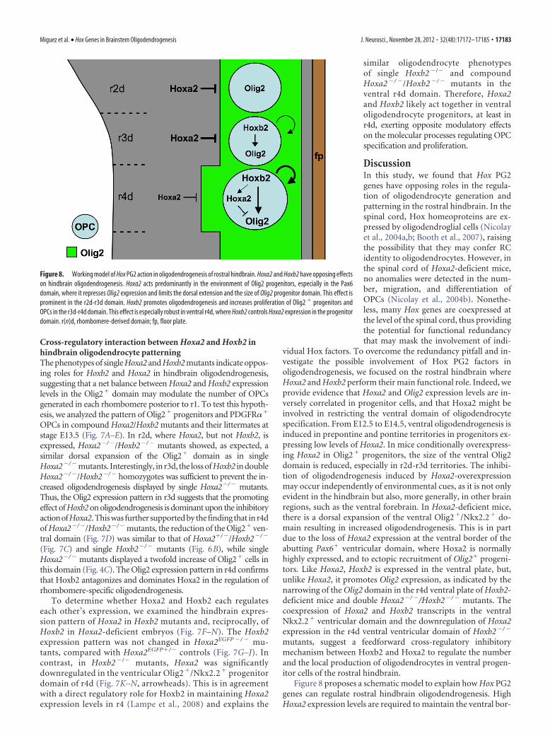
Figure 8 proposes a schematic model to explain how Hox PG2genes can regulate rostral hindbrain oligodendrogenesis. HighHoxa2 expression levels are required to maintain the ventral bor-
Figure 8. Working model of Hox PG2 action in oligodendrogenesis of rostral hindbrain. Hoxa2 and Hoxb2 have opposing effectson hindbrain oligodendrogenesis. Hoxa2 acts predominantly in the environment of Olig2 progenitors, especially in the Pax6domain, where it represses Olig2 expression and limits the dorsal extension and the size of Olig2 progenitor domain. This effect isprominent in the r2d-r3d domain. Hoxb2 promotes oligodendrogenesis and increases proliferation of Olig2 � progenitors andOPCs in the r3d-r4d domain. This effect is especially robust in ventral r4d, where Hoxb2 controls Hoxa2 expression in the progenitordomain. r(n)d, rhombomere-derived domain; fp, floor plate.
Miguez et al. • Hox Genes in Brainstem Oligodendrogenesis J. Neurosci., November 28, 2012 • 32(48):17172–17185 • 17183
der of Pax6 expression, where it contributes to repress Olig2 ac-tivation in ventricular progenitors. Pax6 may directly bind to theOlig2 promoter and repress Olig2 expression, as recently reportedin stem/progenitor cells (Jang and Goldman, 2011). Moreover,Olig2� progenitors may be specified at the dorsal edge of theNkx2.2� domain at a developmental stage when Hoxa2 expres-sion levels are declining. This is suggested by the observation thatforced maintenance of high Hoxa2 expression levels in Olig2�
cells, as in Olig2-tva-Cre;ROSA(lox-stop-lox)Hoxa2 mice, inhibits oli-godendrogenesis. Regarding Hoxb2, our data show that it pro-motes progenitor cell proliferation in the ventroalar plate andacts in Olig2� ventral progenitors to increase oligodendrogen-esis, likely by counteracting Hoxa2-mediated inhibition in r3dand r4d. However, it is noteworthy that in the ventral r4dNkx2.2� domain Hoxb2 is also involved in the maintenance ofhigh Hoxa2 expression levels, which, in turn, inhibit the genera-tion of Olig2� progenitors. Such a transcriptional cross-regulation of Hoxb2 on Hoxa2 may be direct, since a Hoxa2r4-specific enhancer regulated by Hoxb1 and/or Hoxb2 has beenpreviously identified (Tumpel et al., 2007; Lampe et al., 2008).Concerning Hoxb2 regulation of Olig2 expression, it could bedirect, by binding onto Olig2 promoter, or indirect, by modulat-ing the expression of effectors of signaling molecules regulatingOlig2 expression, including those induced by SHH.
The alterations of DV patterning observed in the rostral hind-brain of Hox PG2 mutants suggest that Hox PG2 genes regulatecell fate choices within several progenitor domains along the DVaxis (Davenne et al., 1999). The Nkx2.2� progenitor domain ofr2d and r3d, but not r4d, generates the serotonergic lineage be-fore the onset of oligodendrogenesis (Pattyn et al., 2003). Wefound that the number and distribution of 5HT� cells was notmodified in Hoxa2-deficient or in Hoxa2-overexpressing mice,compared with controls, suggesting that OPCs are not producedat the expense of serotonergic progenitors and that Hoxa2 doesnot regulate serotonergic versus oligodendroglial cell fate balancein Nkx2.2-expressing progenitors (data not shown). Dorsally tothe Nkx2.2�/Olig2� ventral domain, the Pax6 ventricular do-main includes a ventral subset of progenitors committed to gen-erate astroglial cells (Hochstim et al., 2008). In Hoxa2-deficientembryos, a normal number of glial cells expressing Sox9 form inthe rostral hindbrain (data not shown). Since Sox9 is a commonmarker for immature oligodendroglial and astroglial cells (Stoltet al., 2003), this suggests that fewer astrocytes and more OPCsare produced compared with wild-type mice. The excess of OPCsgenerated in the ventral plate of these mutants may be producedat the expense of astrocytes derived from the reduced Pax6 do-main. In contrast, the gain of ventral oligodendrogenesis inHoxa2EGFP �/ � embryos is not associated with a loss or a reduc-tion of dorsal oligodendrogenesis, which later derives from thePax3/Pax7/Gsh1 ventricular domain (Vallstedt et al., 2005), as noalterations of Pax3 and Pax7 expression patterns were detected inthe hindbrain of these mutants.
The observations reported here indicate that rostral hindbrainoligodendrogenesis develops in the ventral plate with a defined spa-tiotemporal pattern involving segment-specific subpopulations ofoligodendroglial progenitors of different size and distribution.Hoxa2 and Hoxb2 appear to contribute to the dorsoventral pattern-ing of ventricular progenitors and to regulate spatially restricted pro-liferation of progenitor and precursor cells in the ventroalar plate.Although additional effects on the migration of OPCs cannot beruled out, especially for Hoxb2 in r4d, Hox PG2 genes appear to bemainly required at an early stage of regional OPC generation fromventral progenitor cells. Moreover, Hoxa2 and Hoxb2 do not appar-
ently contribute to regulate oligodendrocyte differentiation, sinceneither Hoxa2 nor Hoxb2 mutations altered the number ofCNP� oligodendrocytes in the rostral hindbrain. The action ofHox PG2 genes is temporally restricted and the number of OPCsproduced at the onset of oligodendrogenesis in Hoxa2 or Hoxb2mutants is rapidly normalized before birth. In Hoxa2 gain-of-function and Hoxb2-deficient mutants, the reduced number ofventricular progenitors and early born OPCs are likely compen-sated by an additional cell cycle of OPCs before they differentiate.In Hoxa2 loss-of-function mutants, the elimination of extra nu-merous OPCs born at E13.5 probably result from the competi-tion between OPCs to use the limited amount of PDGF-Aavailable in the environment, since PDGF-A-driven cell survivalcontrols override proliferation for determining the final numberand distribution of mature oligodendrocytes (Calver et al., 1998).
Hox PG2 genes participate in rostral hindbrain oligodendro-genesis, but may also regulate OPC production and behavior inmore caudal regions of the neural tube, where such genes areexpressed. It is very likely that, in addition to Hoxb2, other Hoxgenes may modulate the repressing effect of Hoxa2 to control theventral expression of Olig2 and the size of the ventral oligoden-drogenic domain. A more complete picture of Hox gene functionin oligodendroglial development would require global and in-ducible Hox deletion. Hox proteins require common Pbx cofac-tors to achieve their function (Moens and Selleri, 2006).Conditional deletion of Pbx (pre-B cell leukemia transcription)factors in OPCs may provide useful genetic models to better eval-uate the importance of Hox regulation in brainstem and spinalcord oligodendrogenesis.
In patients with multiple sclerosis, not only is the distributionof demyelinating lesions extremely variable, but the capacity ofremyelination varies depending on the lesioned territory. Forinstance, it has been reported that cortical lesions remyelinatemuch more efficiently than periventricular plaques (Patrikios etal., 2006; Albert et al., 2007). This local vulnerability of myelinraises the possibility that it could be related to regional popula-tions of oligodendrocytes with different myelin vulnerabilitiesand/or repair abilities. Investigating the molecules conferring theregional identity of oligodendrocytes may thus have an impor-tant impact on therapeutic strategies aimed at favoring or en-hancing myelin repair following white matter lesions at spatiallyrestricted locations in the CNS. In this respect, it may be of par-ticular interest to examine whether a differential expression ofeither HOXA2 or HOXB2 in multiple sclerosis lesions in thehindbrain may account for the different capacity of some lesionsto repair or not, and whether manipulating HOX PG2 factorsmay contribute to increase myelin repair.
ReferencesAlbert M, Antel J, Bruck W, Stadelmann C (2007) Extensive cortical remy-
elination in patients with chronic multiple sclerosis. Brain Pathol 17:129 –138. CrossRef Medline
Barrow JR, Capecchi MR (1996) Targeted disruption of the Hoxb-2 locus inmice interferes with expression of Hoxb-1 and Hoxb-4. Development122:3817–3828. Medline
Booth J, Nicolay DJ, Doucette JR, Nazarali AJ (2007) Hoxd1 is expressed byoligodendroglial cells and binds to a region of the human myelin oligo-dendrocyte glycoprotein promoter in vitro. Cell Mol Neurobiol 27:641–650. CrossRef Medline
Cai J, Qi Y, Hu X, Tan M, Liu Z, Zhang J, Li Q, Sander M, Qiu M (2005)Generation of oligodendrocyte precursor cells from mouse dorsal spi-nal cord independent of Nkx6 regulation and Shh signaling. Neuron45:41–53. CrossRef Medline
Calver AR, Hall AC, Yu WP, Walsh FS, Heath JK, Betsholtz C, Richardson
17184 • J. Neurosci., November 28, 2012 • 32(48):17172–17185 Miguez et al. • Hox Genes in Brainstem Oligodendrogenesis
WD (1998) Oligodendrocyte population dynamics and the role ofPDGF in vivo. Neuron 20:869 – 882. CrossRef Medline
Davenne M, Maconochie MK, Neun R, Pattyn A, Chambon P, Krumlauf R,Rijli FM (1999) Hoxa2 and Hoxb2 control dorsoventral patterns of neu-ronal development in the rostral hindbrain. Neuron 22:677– 691.CrossRef Medline
Davies JE, Miller RH (2001) Local sonic hedgehog signaling regulates oligo-dendrocyte precursor appearance in multiple ventricular zone domains inthe chick metencephalon. Dev Biol 233:513–525. CrossRef Medline
Di Lullo E, Haton C, Le Poupon C, Volovitch M, Joliot A, Thomas JL, Prochi-antz A (2011) Paracrine Pax6 activity regulates oligodendrocyte precur-sor cell migration in the chick embryonic neural tube. Development 138:4991–5001. CrossRef Medline
Dupe V, Davenne M, Brocard J, Doll e P, Mark M, Dierich A, Chambon P,Rijli FM (1997) In vivo functional analysis of the Hoxa-1 3�retinoic acidresponse element (3�RARE). Development 124:399 – 410. Medline
Dymecki SM (1996) Flp recombinase promotes site-specific DNA recombi-nation in embryonic stem cells and transgenic mice. Proc Natl Acad SciU S A 93:6191– 6196. CrossRef Medline
Fogarty M, Richardson WD, Kessaris N (2005) A subset of oligodendrocytesgenerated from radial glia in the dorsal spinal cord. Development 132:1951–1959. CrossRef Medline
Geisen MJ, Di Meglio T, Pasqualetti M, Ducret S, Brunet JF, Chedotal A, RijliFM (2008) Hox paralog group 2 genes control the migration of mousepontine neurons through Slit-Robo Signaling. PLoS Biol 6:e142. CrossRefMedline
Gu H, Zou YR, Rajewsky K (1993) Independent control of immunoglobulinswitch recombination at individual switch regions evidenced throughCre-loxP-mediated gene targeting. Cell 73:1155–1164. CrossRef Medline
Hochstim C, Deneen B, Lukaszewicz A, Zhou Q, Anderson DJ (2008) Iden-tification of positionally distinct astrocyte subtypes whose identities arespecified by a homeodomain code. Cell 133:510 –522. CrossRef Medline
Hunt P, Gulisano M, Cook M, Sham MH, Faiella A, Wilkinson D, BoncinelliE, Krumlauf R (1991) A distinct Hox code for the branchial region of thevertebrate head. Nature 353:861– 864. CrossRef Medline
Jang ES, Goldman JE (2011) Pax6 expression is sufficient to induce a neu-rogenic fate in glial progenitors of the neonatal subventricular zone. PLoSOne 6:e20894. CrossRef Medline
Karadottir R, Hamilton NB, Bakiri Y, Attwell D (2008) Spiking and non-spiking classes of oligodendrocyte precursor glia in CNS white matter.Nat Neurosci [Erratum (2008) 11:851] 11:450 – 456. CrossRef Medline
Kessaris N, Fogarty M, Iannarelli P, Grist M, Wegner M, Richardson WD(2006) Competing waves of oligodendrocytes in the forebrain and post-natal elimination of an embryonic lineage. Nat Neurosci 9:173–179.CrossRef Medline
Kim SU, McMorris FA, Sprinkle TJ (1984) Immunofluorescence demon-stration of 2�:3�-cyclic-nucleotide 3�-phosphodiesterase in cultured oli-godendrocytes of mouse, rat, calf and human. Brain Res 300:195–199.Medline
Lampe X, Samad OA, Guiguen A, Matis C, Remacle S, Picard JJ, Rijli FM,Rezsohazy R (2008) An ultraconserved Hox-Pbx responsive element re-sides in the coding sequence of Hoxa2 and is active in rhombomere 4.Nucleic Acids Res 36:3214 –3225. CrossRef Medline
Lumsden A, Krumlauf R (1996) Patterning the vertebrate neuraxis. Science274:1109 –1115. CrossRef Medline
Moens CB, Selleri L (2006) Hox cofactors in vertebrate development. DevBiol 291:193–206. CrossRef Medline
Narita Y, Rijli FM (2009) Hox genes in neural patterning and circuit forma-tion in the mouse hindbrain. Curr Top Dev Biol 88:139 –167. CrossRefMedline
Nicolay DJ, Doucette JR, Nazarali AJ (2004a) Hoxb4 in oligodendrogenesis.Cell Mol Neurobiol 24:357–366. CrossRef Medline
Nicolay DJ, Doucette JR, Nazarali AJ (2004b) Early stages of oligodendro-cyte development in the embryonic murine spinal cord proceed normallyin the absence of Hoxa2. Glia 48:14 –26. CrossRef Medline
Noll E, Miller RH (1993) Oligodendrocyte precursors originate at the ven-tral ventricular zone dorsal to the ventral midline region in the embryonicrat spinal cord. Development 118:563–573. Medline
Nyabi O, Naessens M, Haigh K, Gembarska A, Goossens S, Maetens M, DeClercq S, Drogat B, Haenebalcke L, Bartunkova S, De Vos I, De Craene B,Karimi M, Berx G, Nagy A, Hilson P, Marine JC, Haigh JJ (2009) Effi-cient mouse transgenesis using Gateway-compatible ROSA26 locus tar-
geting vectors and F1 hybrid ES cells. Nucleic Acids Res 37:e55. CrossRefMedline
Ono K, Fujisawa H, Hirano S, Norita M, Tsumori T, Yasui Y (1997) Earlydevelopment of the oligodendrocyte in the embryonic chick metenceph-alon. J Neurosci Res 48:212–225. Medline
Oury F, Murakami Y, Renaud JS, Pasqualetti M, Charnay P, Ren SY, Rijli FM(2006) Hoxa2 and rhombomere dependent development of the mousefacial somatosensory map. Science 313:1408 –1413. CrossRef Medline
Pasqualetti M, Ren SY, Poulet M, LeMeur M, Dierich A, Rijli FM (2002) AHoxa2 knockin allele that expresses EGFP upon conditional Cre-mediated recombination. Genesis 32:109 –111. CrossRef Medline
Patrikios P, Stadelmann C, Kutzelnigg A, Rauschka H, Schmidbauer M,Laursen H, Sorensen PS, Bruck W, Lucchinetti C, Lassmann H (2006)Remyelination is extensive in a subset of multiple sclerosis patients. Brain129:3165–3172. CrossRef Medline
Pattyn A, Vallstedt A, Dias JM, Samad OA, Krumlauf R, Rijli FM, Brunet JF,Ericson J (2003) Coordinated temporal and spatial control of motorneuron and serotonergic neuron generation from a common pool of CNSprogenitors. Genes Dev 17:729 –737. CrossRef Medline
Pringle NP, Yu WP, Guthrie S, Roelink H, Lumsden A, Peterson AC, Rich-ardson WD (1996) Determination of neuroepithelial cell fate: inductionof the oligodendrocyte lineage by ventral midline cells and sonic hedge-hog. Dev Biol 177:30 – 42. CrossRef Medline
Ren SY, Pasqualetti M, Dierich A, Le Meur M, Rijli FM (2002) A Hoxa2mutant conditional allele generated by Flp- and Cre-mediated recombi-nation. Genesis 32:105–108. CrossRef Medline
Richardson WD, Kessaris N, Pringle N (2006) Oligodendrocyte wars. NatRev Neurosci 7:11–18. CrossRef Medline
Rodríguez CI, Buchholz F, Galloway J, Sequerra R, Kasper J, Ayala R, StewartAF, Dymecki SM (2000) High-efficiency deleter mice show that FLPe isan alternative to Cre-loxP. Nat Genet 25:139 –140. CrossRef Medline
Rowitch DH (2004) Glial specification in the vertebrate neural tube. NatRev Neurosci 5:409 – 419. CrossRef Medline
Rowitch DH, Kriegstein AR (2010) Developmental genetics of vertebrateglial-cell specification. Nature 468:214 –222. CrossRef Medline
Santagati F, Minoux M, Ren SY, Rijli FM (2005) Temporal requirement ofHoxa2 in cranial neural crest skeletal morphogenesis. Development 132:4927– 4936. CrossRef Medline
Schuller U, Heine VM, Mao J, Kho AT, Dillon AK, Han YG, Huillard E, SunT, Ligon AH, Qian Y, Ma Q, Alvarez-Bylla A, McMahon AP, RowditchDH, Ligon KL (2008) Acquisition of granule neuron precursor identityis a critical determinant of progenitor cell competence to form Shh-induced medulloblastoma. Cancer Cell 14:123–134. CrossRef Medline
Spassky N, Goujet-Zalc C, Parmantier E, Olivier C, Martinez S, Ivanova A,Ikenaka K, Macklin W, Cerruti I, Zalc B, Thomas JL (1998) Multiplerestricted origin of oligodendrocytes. J Neurosci 18:8331– 8343. Medline
Spassky N, Heydon K, Mangatal A, Jankovski A, Olivier C, Queraud-LesauxF, Goujet-Zalc C, Thomas JL, Zalc B (2001) Sonic hedgehog-dependentemergence of oligodendrocytes in the telencephalon: evidence for asource of oligodendrocytes in the olfactory bulb that is independent ofPDGFRalpha signaling. Development 128:4993–5004. Medline
Stolt CC, Lommes P, Sock E, Chaboissier MC, Schedl A, Wegner M (2003)The Sox9 transcription factor determines glial fate choice in the develop-ing spinal cord. Genes Dev 17:1677–1689. CrossRef Medline
Sugimori M, Nagao M, Bertrand N, Parras CM, Guillemot F, Nakafuku M(2007) Combinatorial actions of patterning and HLH transcription fac-tors in the spatiotemporal control of neurogenesis and gliogenesis in thedeveloping spinal cord. Development 134:1617–1629. CrossRef Medline
Timsit S, Martinez S, Allinquant B, Peyron F, Puelles L, Zalc B (1995) Oli-godendrocytes originate in a restricted zone of the embryonic ventralneural tube defined by DM-20 mRNA expression. J Neurosci 15:1012–1024. Medline
Tripathi RB, Clarke LE, Burzomato V, Kessaris N, Anderson PN, Attwell D,Richardson WD (2011) Dorsally and ventrally derived oligodendrocyteshave similar electrical properties but myelinate preferred tracts. J Neuro-sci 31:6809 – 6819. CrossRef Medline
Tumpel S, Cambronero F, Ferretti E, Blasi F, Wiedemann LM, Krumlauf R(2007) Expression of Hoxa2 in rhombomere 4 is regulated by a con-served cross-regulatory mechanism dependent upon Hoxb1. Dev Biol302:646 – 660. CrossRef Medline
Vallstedt A, Klos JM, Ericson J (2005) Multiple dorsoventral origins of oli-
Miguez et al. • Hox Genes in Brainstem Oligodendrogenesis J. Neurosci., November 28, 2012 • 32(48):17172–17185 • 17185

godendrocyte generation in the spinal cord and hindbrain. Neuron 45:55– 67. CrossRef Medline
Wegner M (2008) A matter of identity: transcriptional control in oligoden-drocytes. J Mol Neurosci 35:3–12. CrossRef Medline
17185a • J. Neurosci., November 28, 2012 • 32(48):17172–17185 Miguez et al. • Hox Genes in Brainstem Oligodendrogenesis