Embed Size (px)
Citation preview
Development/Plasticity/Repair
Axon Regeneration Can Facilitate or Suppress HindlimbFunction after Olfactory Ensheathing Glia Transplantation
Aya Takeoka,1 Devin L. Jindrich,1,3 Cintia Munoz-Quiles,4 Hui Zhong,1 Rubia van den Brand,1 Daniel L. Pham,1
Matthias D. Ziegler,1 Almudena Ramon-Cueto,5 Roland R. Roy,1,2 V. Reggie Edgerton,1,2 and Patricia E. Phelps1,2
1Department of Integrative Biology and Physiology and 2Brain Research Institute, UCLA, Los Angeles, California 90095, 3Department of Kinesiology,Arizona State University, Tempe, Arizona 85287, 4Fundacion Investigacion en Regeneracion del Sistema Nervioso, 46019 Valencia, Spain, and 5Instituto deBiomedicina de Valencia, Consejo Superior de Investigaciones Científicas, 46010 Valencia, Spain
Reports based primarily on anatomical evidence suggest that olfactory ensheathing glia (OEG) transplantation promotes axon regener-ation across a complete spinal cord transection in adult rats. Based on functional, electrophysiological, and anatomical assessments, wefound that OEG promoted axon regeneration across a complete spinal cord transection and that this regeneration altered motor re-sponses over time. At 7 months after transection, 70% of OEG-treated rats showed motor-evoked potentials in hindlimb muscles aftertranscranial electric stimulation. Furthermore, a complete spinal cord retransection performed 8 months after injury demonstrated thatthis axon regeneration suppressed locomotor performance and decreased the hypersensitive hindlimb withdrawal response to mechan-ical stimulation. OEG transplantation alone promoted reorganization of lumbosacral locomotor networks and, when combined withlong-term training, enhanced some stepping measures. These novel findings demonstrate that OEG promote regeneration of matureaxons across a complete transection and reorganization of spinal circuitry, both of which contribute to sensorimotor function.
IntroductionAdult rodent spinal cords retain an inherent plasticity to reorga-nize descending connections that directly influence motor out-put after incomplete injuries (Bareyre et al., 2004; Courtine et al.,2008). After a complete spinal cord transection, however,achieving axon regeneration across the lesion that contributesto functional recovery remains a difficult challenge. Olfactoryensheathing glia (OEG) transplantation reportedly promotes tis-sue sparing, axon remyelination, and improvements in motorperformance in incomplete and complete adult spinal cord injury(SCI) models (Imaizumi et al., 1998; Ramon-Cueto et al., 2000;García-Alías et al., 2003; Li et al., 2003; Fouad et al., 2005; Li et al.,2007; Kubasak et al., 2008; Munoz-Quiles et al., 2009). However,the results of SCI studies involving OEG transplantation varysubstantially depending on the injury model, the duration of thestudy, and the source, age, and methods used to prepare andtransplant the OEG (Franssen et al., 2007). Promising transplan-tation candidates that promote axon regeneration are purifiedcultures of olfactory bulb-derived, p75-nerve growth factor re-ceptor (NGFR)-positive OEG (Ramon-Cueto et al., 2000) be-
cause these cells intermingle with astrocytes and migrate wellwithin the reactive astrocytic environment after injury (Lakatoset al., 2000, 2003).
OEG transplantation combined with task-specific trainingimproves the motor function of adult rats with a complete spinalcord transection (i.e., spinal rats) (Ramon-Cueto et al., 2000;Kubasak et al., 2008; Munoz-Quiles et al., 2009), but we knowlittle about how this treatment promotes functional recovery.The spinal cord alone is capable of inducing locomotor activitywithout supraspinal innervation and can be trained to enhancethe efficacy of locomotion (Edgerton et al., 2004). For example,long-term step training promotes reorganization of the lumbo-sacral locomotor circuitry in adult spinal cats by downregulatinginhibitory neurotransmitters associated with interneurons andsomatic motor neurons (Edgerton et al., 1997; de Leon et al.,1999; Tillakaratne et al., 2000, 2002; Rossignol et al., 2001; Coteand Gossard, 2003) and by modifying motor neuron excitabilityin response to hindlimb loading and sensory stimulation (Cote etal., 2003). Combined with pharmacological or epidural electricalstimulation, long-term step training also increases the efficacy ofspecific sensorimotor connections and activates specific neuronsduring locomotion (Ichiyama et al., 2008; Courtine et al., 2009).
We hypothesized that OEG transplantation combined withlong-term step training promotes both axon regeneration acrossthe transection and/or reorganization of the lumbosacral loco-motor circuitry in adult spinal rats. We then asked whether bothregeneration and reorganization attributed to the treatments caninfluence functional outcomes. To test this, we used sensorimo-tor and electrophysiological assessments over 8 months andperformed a complete retransection rostral to the original tran-section 8 months after the initial lesion. OEG transplantation
Received Sept. 22, 2010; revised Nov. 7, 2010; accepted Dec. 22, 2010.This work was supported by National Institute of Neurological Disorders and Stroke Grants R21NS42000-01 and
RO1NS54159.We thank Dr. J. Gornbein for assistance in the statistical analyses, Dr. R. Ichiyama and S. Zdunowski for expert
advice in behavioral data collection and analyses, Dr. G. Lawson and M. Herrera for the aid in postsurgical animalcare, Drs. Ava Udvadia and Pate Skene for advice on GAP-43 experiments, and Allen Cheng, Alisha Aiello, KimberlyMcFarland, Frank Lee, and numerous University of California, Los Angeles undergraduate students for assistancewith animal care and preparation of data for analyses.
Correspondence should be addressed to Dr. Patricia E. Phelps, Department of Integrative Biology and Physiology,UCLA, Box 951606, Los Angeles, CA 90095-1606. E-mail: [email protected].
DOI:10.1523/JNEUROSCI.4967-10.2011Copyright © 2011 the authors 0270-6474/11/314298-13$15.00/0
4298 • The Journal of Neuroscience, March 16, 2011 • 31(11):4298 – 4310
promoted functional axon regeneration across the complete le-sion, and, when combined with long-term step training, it facil-itated reorganization of spinal locomotor networks. Moreover,some of the changes in motor function attributed to OEG trans-plantation and step training point to facilitation, whereas othersresult in suppression of sensorimotor function.
Materials and MethodsOEG culture and transplantationAll procedures followed the National Institutes of Health guidelinesand were approved by the Chancellor’s Animal Research Committeeat UCLA. Methods for OEG culture were adopted from those ofRamon-Cueto et al. (2000) and Munoz-Quiles et al. (2009). OEGwere dissected from the outer two olfactory bulb layers of two 8- to10-week-old male Wistar Hannover rats (Charles River Laboratories)for transplantation into one spinal rat. Primary cultures were immu-nopurified against p75-NGFR antibody (1:5) (Chandler et al., 1984)at 7– 8 d and harvested for transplantation at 14 d. Cultures (n � 10)were 95 � 1% p75-NGFR-positive and expressed S100 and GFAP.
Forty-one female Wistar Hannover rats, 10 –12 weeks old, were anes-thetized with 2–2.5% isoflurane. The spinal cord was completely tran-sected at approximately T9, leaving the ventral and lateral dura intact.Injections of DMEM alone or with OEG (200,000 per stump) were in-jected �1 mm from the transection site at the midline (coordinates fromthe dorsal surface: 1.75, 1.25, 1, and 0.5 �m) into both the rostral andcaudal spinal cord stumps. After transplantation, the spine was stabilizedwith a stainless steel bar (3.2 cm in length and 0.1 cm in diameter)attached to the T7 and T13 vertebral processes.
Step training and behavioral analysesTwo weeks after injury, the rats began manual step training on a treadmillfor 20 min/d, 5 d/week for 7.5 months as described by Kubasak et al.(2008). Stepping ability was evaluated before transection, monthly be-tween 1 and 7 months after transection, and after spinal cord retransec-tion at 8 months at treadmill speeds of 10, 13.5, and 17.5 cm/s and with abody weight support of 85% (i.e., the hindlimbs supported 15% bodyweight). Recording of treadmill stepping was conducted as described byKubasak et al. (2008). Each trial consisted of 90 s of stepping. The highestnumber of plantar steps at three treadmill speeds was sampled for eachrat. At 5 months after transection, spinal rats were injected with quipa-zine (0.3 mg/kg, i.p.) (Ichiyama et al., 2005), a 5-HT2A receptor agonist,and stepping ability was tested 15 min later.
We used principal components analysis (PCA) to measure the con-sistency of the toe trajectories (Fong et al., 2005; Cai et al., 2006).Briefly, stride transitions were identified visually, kinematics trajec-tories of toe markers were resampled every 0.1 s, and correspondingpoints across all measured steps were averaged. The first principalcomponent, similar to an average that explains the greatest amount ofvariance among trajectories, was calculated and the variance ac-counted for by the first component was calculated as a “score.” Alarger PCA score (i.e., closer to 100%) represents more consistentstepping. The best PCA score (percentage) from three treadmillspeeds was sampled for each rat that performed three or more alter-nating steps. A PCA score of 45%, the lowest PCA percentage mea-surable as defined by Cai et al. (2006), was assigned for spinal rats thatperformed two or fewer alternating steps. We also measured meanstance length, step height, and trajectory length to characterize thestep patterns during treadmill testing.
Weight-support testing. Rats were supported using a harness con-nected to an automated body weight-support system (Robomedica)that lowered 10% of the rat’s body weight every 10 s until the ratcollapsed. The percentage body weight support at collapse was deter-mined by an observer blind to the experimental status of the rat.Because ankle joint angles are good indicators of weight-support abil-ity (Timoszyk et al., 2002), data from rats that collapsed forward wereused to calculate the joint angle as described previously (Ichiyama etal., 2009). Ankle angle analyses were performed using the Simi three-dimensional motion.
von Frey test. An experimenter blind to the treatment status of thespinal rats conducted the von Frey up– down test (Chaplan et al., 1994)on both hindpaws at 1, 3, 5, and 7 months after transection and 5 d afterspinal cord retransection to determine any injury-induced changes in thewithdrawal threshold. A series of nine hairs (3.61–5.46 g, starting withthe 4.56 g; North Coast Medical) were used to elicit the hindpaw with-drawal response to determine the 50% withdrawal threshold (grams).
Electrophysiological assessment of hindlimb motor-evokedpotentialsWe performed transcranial electric stimulation at 1, 4, and 7 monthsafter transection of all spinal rats and immediately after and 3 and 5 dafter a retransection on 17 rats. Stimulation of four intact rats also wasstudied before and after acute transection. Rats were anesthetized lightlywith 0.7–1% isoflurane and received stimulation on the primary motorcortex (approximately bregma �3.5 to �2 mm) using a method modi-fied from Lopez-Vales et al. (2006) and on the brainstem (approximatelybregma �6 to �10 mm). Motor-evoked potentials (MEPs) were elicitedby a series of single-pulse stimulations [0.1 ms duration, from 40 to 200mA in 40 mA increments, 30 times/intensity applied at 30 s intervals;constant-current stimulator model DS7AH (Digitimer)]. MEPs were re-corded bilaterally from the tibialis anterior (TA) and medial gastrocne-mius (MG) muscles with subcutaneously inserted needle electrodes[needle: 30 gauge (BD Biosciences); wire: 50 �m in diameter (CaliforniaFine Wire)]. All MEPs were amplified (1000�) and filtered (10 –10,000Hz). Each MEP was defined as a sharp peak with a latency longer than 8ms and an amplitude larger than 0.05 mV. Electrical stimulation withhigh currents as used in this study can potentially introduce artifacts inMEP recordings as a result of volume conduction, stimulation of axialspinal musculature, and/or the activation of cutaneous trunci pathways(Gruner et al., 1993). Direct stimulation artifacts were of very short du-ration (i.e., �1 ms) and were observed in both media and OEG animals.Although we cannot exclude the possibility of extraspinal stimulationartifacts (e.g., via cutaneous trunci pathways), we consider it unlikely thatsuch a reflex would only occur in OEG rats and would be eliminated byretransection.
Morphological analysesRetrograde tracer injections and analyses. Twenty-two chronic spinal rats(media-untrained, n � 5; media-trained, n � 7; OEG-untrained, n � 5;OEG-trained, n � 5) were injected with horseradish peroxidase conju-gated with wheat germ agglutinin (WGA–HRP) 8 months after transec-tion. Additional controls included three intact and three media-injectedspinal rats maintained 1 month after transection. Rats were anesthetizedwith 2% isoflurane, and a 0.65 �l mixture of 5% WGA (Sigma) and 25%HRP (Sigma) was injected 5 mm caudal to the transection in 13 locations(coordinates expressed in mm: midline, 1.8, 1.4, 1.0, 0.8, and 0.4; at 1.2mm to the right or left of the midline, 1.6, 1.2, 0.8, and 0.4) as reportedpreviously (Ramon-Cueto et al., 1998; Munoz-Quiles et al., 2009). After72 h, the rats were perfused for tissue analyses.
We sectioned the primary motor cortex and brainstem and mountedevery third section (40 �m thick) on slides for WGA immunohistochem-istry. Additionally, every eighth spinal cord section (25 �m thick) fromthe cervical enlargement and the transection site of all WGA–HRP-injected spinal rats was processed for WGA immunoreactivity to detectlong propriospinal neurons and to ensure that no tracer leaked into thelesion core and rostral stump, respectively. None of the rats evaluated forretrograde tracing data had WGA–HRP leakage into the lesion core orrostral stump (supplemental Fig. 1C, available at www.jneurosci.org assupplemental material). We used an imaging system (Simple PCI soft-ware; Compix Imaging Systems, Hamamatsu Corporation) to measurethe optical density of WGA immunoreactivity in brainstem and spinalcord neurons. The maximum background level of �20 large neuronsoutside the regions of interest, i.e., neurons that do not project to thespinal cord, were measured first to determine the maximal backgroundlevel of each section. Cells within the nuclei of interest were detected onlyif their optical densities were 5% or greater than the maximal backgroundlevel. Optical densitometry measurements were performed on everythird section (120 �m apart) throughout the red and vestibular nuclei
Takeoka et al. • Functional Regeneration after OEG Transplants J. Neurosci., March 16, 2011 • 31(11):4298 – 4310 • 4299

and the locus coeruleus and every 12th section(480 �m apart) throughout the reticular for-mation and raphe nucleus. All measurementswere performed blind to the transplantation ortraining status of the rats.
Tissue preparation. Rats were anesthetizeddeeply with ketamine (100 mg/kg, i.p.) and xy-lazine (5 mg/kg, i.p.), perfused with 4% para-formaldehyde, and postfixed at 4°C for 4 h.Spinal cords, brains, and brainstems were dis-sected and cryoprotected.
Antibodies. Monoclonal antibodies used in-cluded the following: anti-GFAP (1:1000; BDPharmingen), anti-Neuronal-specific Nuclearprotein (NeuN) (1:100), and anti-GAP43 (1:10,000; Millipore Corporation). Polyclonalantibodies used included the following: goat anti-WGA (1:2000; Vector Laboratories), goat anti-5-HT (1:20,000; ImmunoStar), and rabbitanti-GFAP (1:30,000; Dako).
Immunohistochemistry and lesion site analy-ses. We sectioned the transection site in the sag-ittal plane (25 �m) and mounted the sectionson a series of 16 slides such that every 16thsection was mounted on a slide. All immuno-histochemistry procedures with diamino-benzidine (DAB), followed the monoclonal,polyclonal, or double immunolabeling proto-cols described previously (Takeoka et al., 2009,2010). For immunofluorescence of GFAP andNeuN, we omitted the presoak and TritonX-100 steps. Species appropriate Alexa Fluor488 and 584 (1:200; Invitrogen) were used forlocalization.
Quantification of 5-HT axons and GAP43 so-mata. 5-HT axons were quantified as reportedby Takeoka et al. (2009). GAP43-positive so-mata present in the lower thoracic cord caudalto the original lesion were quantified in everyeighth section of the caudal stump and normal-ized based on tissue volume.
Volume of the lesion core and associated cavi-ties. Approximately 85% of the lesion site wasGFAP labeled to delineate the lesion core andto anatomically confirm that all transectionswere complete. In all rats, the GFAP-negativecore always separated the GFAP-positive ros-tral and caudal stumps as reported in our pre-vious study (Kubasak et al., 2008). The volumeof the lesion core and the associated cavitieswas quantified as reported (Kubasak et al.,2008). We outlined the GFAP-negative tran-section site and the nearby cavities of all ratsinjected with the retrograde tracer (n � 22rats). The GFAP-positive area was subtractedfrom the total section area in every eighth sec-tion (25 �m thick) and normalized based ontissue volume. We focused on differences be-tween the media and OEG groups becausethere were no training effects.
Length of the non-neuronal tissue and lesioncore. We double labeled every eighth section forGFAP and NeuN to estimate the extent oflesion-induced neuronal degeneration in spi-nal cord tissue rostral and caudal to the injury site. We measured (1) theshortest distance between NeuN-labeled neurons and the GFAP-positiveborder adjacent to the GFAP-negative lesion core in both the rostral andcaudal stumps and (2) the total lengths of the non-neuronal region andthe lesion core.
Statistical analyses. The nonparametric Mann–Whitney test wasused for the von Frey analyses. All other statistical comparisons wereconducted with nonparametric resampling analyses to compare groupmean differences and minimize the assumptions on the underlying datadistribution (Efron and Tibshirani, 1991).
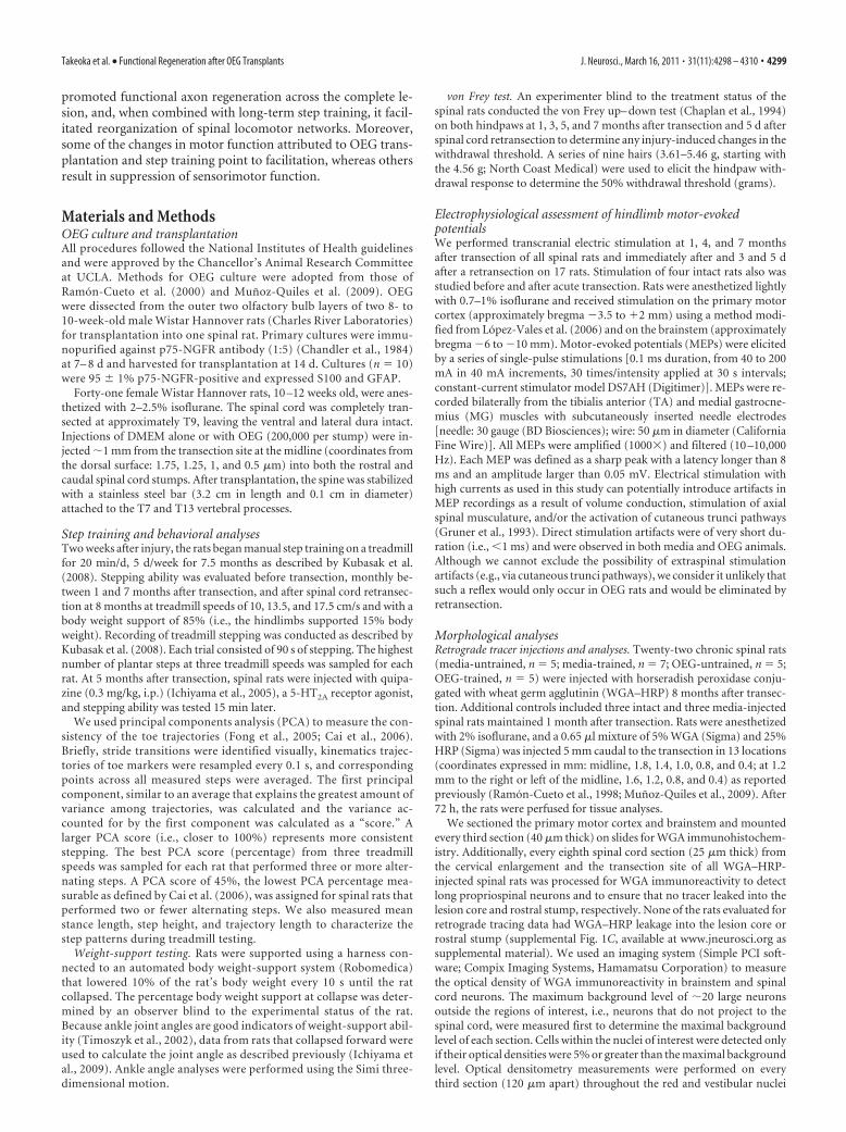
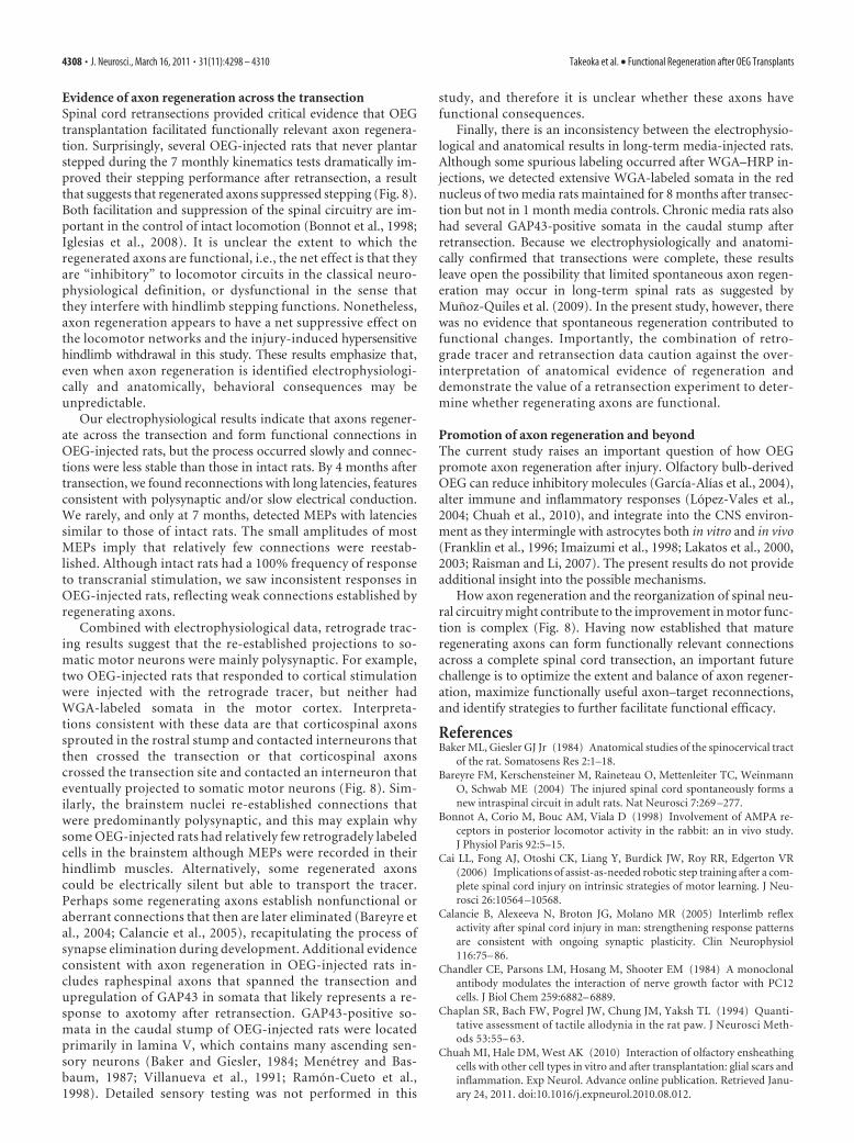
Figure 1. OEG transplantation and long-term step training influence locomotor function in spinal rats. A, Ankle flexion andextension (highlighted in pink) are critical for successful plantar stepping, which is rarely achieved by adult spinal rats. Stepping isoriented toward the left (stick representation is sampled every 0.2 s). Step heights of spinal plantar steppers are usually not as highas those of intact rats. Foot (pink) is placed vertical to the treadmill during successful plantar steps performed by intact rats that stepon their footpads, whereas the foot of nonplantar steppers and draggers is parallel to the treadmill. B, Step trajectories of intact ratsare consistent, whereas those of spinal rats are erratic. Each colored line indicates a single step, and the thick red line represents theaverage trajectory. Step height is lower and stance and step trajectory lengths are shorter in media- and OEG-injected spinal ratsthan intact rats. C, OEG-untrained rats generated more plantar steps than media-untrained rats at 1, 3, and 5 months (*p � 0.05)and more plantar steps than OEG-trained rats at 1 month ( #p � 0.05). D, PCA showed that both OEG groups produced moreconsistent step trajectories than both media groups at 1, 3, and 5 months (*p � 0.05). OEG-untrained rats scored higher thanOEG-trained rats at 1 month ( #p � 0.05). E, OEG and media groups differed in their stance and trajectory lengths at all time points(*p � 0.05). F, Step training improved the number of plantar steps taken in 10 s in response to 5-HT2A receptor activation (*p �0.05; see supplemental videos, available at www.jneurosci.org as supplemental material). Data for individual rats are representedas a circle or square. G, H, OEG injection but not step training enhanced step consistency (G) and stance and trajectory lengths (H )after quipazine injection (*p � 0.05). I, OEG-trained rats exhibit better ankle extension than all other rats when the hindlimbssupport up to 10% of their body weight at 7 months after transection (*p � 0.05). Data are mean � SEM.
4300 • J. Neurosci., March 16, 2011 • 31(11):4298 – 4310 Takeoka et al. • Functional Regeneration after OEG Transplants
ResultsAfter a complete spinal cord transection at T9, spinal rats re-ceived injections of olfactory bulb-derived OEG suspended inmedia or media alone. Long-term step training of one half of thespinal rats started 2 weeks after transection and continued for 20min/d, 5 d/week for 7.5 months. The four experimental groupswere as follows: media-untrained (n � 9), media-trained (n �10), OEG-untrained (n � 11), and OEG-trained (n � 9). Eightmonths after the initial transection, 50% of the spinal rats re-ceived a complete retransection rostral to the original lesion, andthe remaining 50% were injected with a retrograde tracer.
OEG transplantation improves hindlimb step performancePlantar foot placement during hindlimb stepping requires a se-ries of joint movements, i.e., knee and ankle flexion during theswing phase and ankle extension during the stance phase, and israrely achieved spontaneously by adult spinal rats (Fig. 1A) (Edg-erton et al., 2004). Step trajectories of spinal rats measured byPCA are often erratic compared with those of intact rats (Fig. 1B).To determine whether OEG transplantation promotes hindlimbstepping ability in adult spinal rats, we first compared the numberof plantar steps taken by media- and OEG-untrained spinal rats.
OEG-untrained rats generated signifi-cantly more plantar steps than media-untrained rats at 1–5 months aftertransection but not at later time points(Fig. 1C). Neither the media- nor theOEG-untrained rats improved their plan-tar stepping ability over time (Fig. 1C).Next we asked whether or not there was atraining effect. The average number ofplantar steps did not differ statistically be-tween the media-untrained and -trainedgroups throughout the study (Fig. 1C).The OEG-untrained group consistentlyproduced as many or more plantar stepsthan the OEG-trained group, with signif-icant differences detected early in thestudy (Fig. 1C).
We used PCA to calculate the consis-tency of the step trajectories and also com-pared stance lengths, step heights, andtrajectory lengths. PCA analysis assessesthe ability to produce locomotor move-ment, i.e., hindlimb alternation with orwithout hindlimb plantar placement. Thestepping pattern was more consistent,represented by higher PCA scores, forboth OEG-untrained and -trained groupscompared with media groups at 1, 3, and 5but not at 7 months (Fig. 1D). The OEG-untrained rats had higher PCA scores thanOEG-trained rats at 1 month. Both OEGgroups demonstrated longer stance andtrajectory lengths than media groupsthroughout the duration of the study (Fig.1E). Mean step height did not differamong groups at any time point, andthere was no improvement in step consis-tency, step height, or stance and trajectorylengths over time in any group. Together,these results indicate that OEG transplan-tation alone can enhance stepping ability,
and these improvements occurred early in the study with nochange over time.
OEG transplantation and step training improve plantarstepping ability associated with 5-HT2A receptor activationAdministration of quipazine, a 5-HT2A receptor agonist, can ac-tivate and modulate the lumbosacral locomotor networks in spi-nal rats (Gerasimenko et al., 2007; Ichiyama et al., 2008). Wetherefore gave a single injection of quipazine 5 months after tran-section to determine the extent to which OEG transplantationand/or step training affected the locomotor circuitry associatedwith 5-HT2A receptors. The administration of quipazine dramat-ically improved the number of consecutive plantar steps, step-ping consistency, step height, and stance and step trajectorylengths in all four treatment groups compared with their predrugperformance (supplemental Videos 1– 4, available at www.jneurosci.org as supplemental material). Although we did notdetect a training effect on plantar stepping without quipazine,activation of the 5-HT2A receptors by quipazine unmasked a re-finement in plantar stepping ability in media- and OEG-trainedspinal rats (Fig. 1F, 17.5 cm/s). In addition, 5-HT2A receptoractivation resulted in a more consistent stepping pattern (Fig.
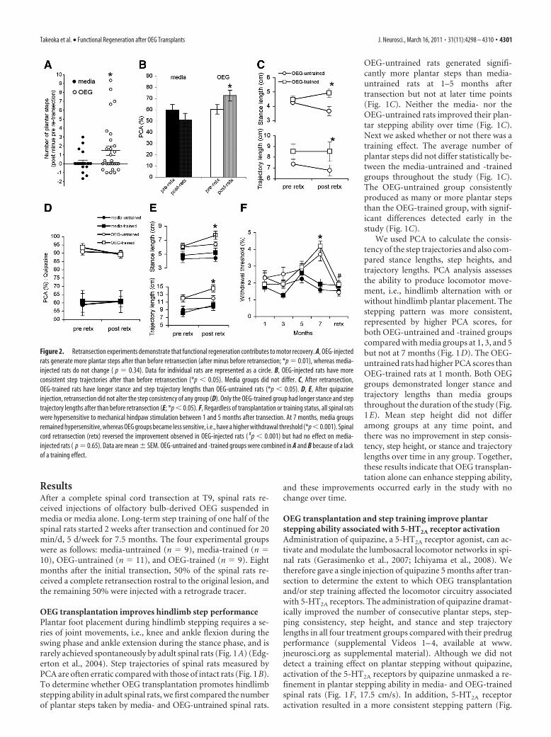
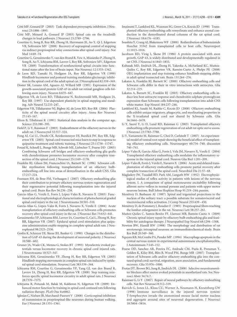
Figure 2. Retransection experiments demonstrate that functional regeneration contributes to motor recovery. A, OEG-injectedrats generate more plantar steps after than before retransection (after minus before retransection; *p � 0.01), whereas media-injected rats do not change ( p � 0.34). Data for individual rats are represented as a circle. B, OEG-injected rats have moreconsistent step trajectories after than before retransection (*p � 0.05). Media groups did not differ. C, After retransection,OEG-trained rats have longer stance and step trajectory lengths than OEG-untrained rats (*p � 0.05). D, E, After quipazineinjection, retransection did not alter the step consistency of any group (D). Only the OEG-trained group had longer stance and steptrajectory lengths after than before retransection (E; *p � 0.05). F, Regardless of transplantation or training status, all spinal ratswere hypersensitive to mechanical hindpaw stimulation between 1 and 5 months after transection. At 7 months, media groupsremained hypersensitive, whereas OEG groups became less sensitive, i.e., have a higher withdrawal threshold (*p �0.001). Spinalcord retransection (retx) reversed the improvement observed in OEG-injected rats ( #p � 0.001) but had no effect on media-injected rats ( p � 0.65). Data are mean � SEM. OEG-untrained and -trained groups were combined in A and B because of a lackof a training effect.
Takeoka et al. • Functional Regeneration after OEG Transplants J. Neurosci., March 16, 2011 • 31(11):4298 – 4310 • 4301
1G) and stance and trajectory lengthswere longer (Fig. 1H) in the OEG com-pared with the media groups. These re-sults indicate that long-term step trainingand OEG transplantation contributed tothe reorganization of the lumbosacral lo-comotor networks modified by seroto-nergic (5-HT) receptor activation.
Only OEG-trained rats show greaterankle extension during the stand testThe effects of OEG transplantation andtraining on weight-support ability weremeasured as a part of the evaluation oflocomotor ability. Spinal rats increasedthe amount of self-weight support beforecollapse between 1 and 4 months regard-less of treatment (10 � 1% increase), butonly the OEG-injected groups improvedbetween 4 and 7 months after transection(6 � 1% increase). Step training did notfurther enhance standing ability in anygroup.
We then measured changes in ankleextension during standing as an evalua-tion of weight-support ability (Timoszyket al., 2002). At 1 and 4 months after tran-section, the mean ankle angles did not dif-fer across the treatment groups at anylevel of weight support ( p � 0.05 for allcomparisons). At 7 months, however,OEG-trained rats demonstrated greaterankle extension at 0 and 10% body weightsupport compared with the other threegroups (Fig. 1 I). Beyond 10%, there wereno group differences. These results indi-cate that OEG transplantation and steptraining together, but not individually,improved ankle extension up to the�10% body weight load used during steptraining.
Plantar stepping ability improves afterretransection only in OEG-injected ratsTo test whether axon regeneration con-tributes to locomotor activity, we com-pared the number of plantar stepsgenerated by media or OEG rats beforeand after a complete retransection 0.5 cmrostral to the original lesion. We sub-tracted the number of plantar steps gener-ated at 7 months from those producedafter the retransection at 8 months; a pos-itive number, therefore, indicates a betterstepping performance after retransec-tion. Because there was no training effecton the number of plantar steps takenafter retransection, we combined media-untrained and -trained, and OEG-untrained and -trained groups. Although the mean number ofplantar steps for the media group did not change after retransec-tion, the OEG group showed a considerable improvement (Fig.2A). Remarkably, two OEG-injected spinal rats that never
stepped before retransection generated six to nine plantar stepsafter retransection. The number of plantar steps taken after re-transection was significantly greater in OEG- than media-injected rats (media, 0.9 � 0.2; OEG, 1.7 � 0.4; p � 0.05). In
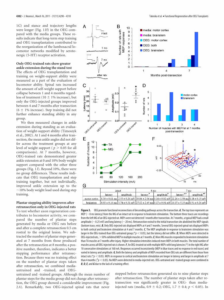
Figure 3. OEG promote functional reconnections of descending pathways across the transection. A, The top trace represents anMEP (�8 ms latency) from the MG of an intact rat in response to brainstem stimulation. The bottom three traces are recordingsfrom the left MG of an OEG-injected rat. MEPs were not detected 1 month after transection. At 7 months, a typical MEP had a smallamplitude (�0.25 mV) and long latency (�20 ms). Retransection rostral to the initial transection site abolished the MEP signals(bottom trace, retx). B, Only OEG-injected rats displayed MEPs at 4 and 7 months. Several OEG-injected spinal rats displayed MEPsto both cortical and brainstem stimulation at 4 and 7 months. C, The MEP amplitude in response to brainstem stimulation waslarger in the OEG-trained than OEG-untrained group (*p � 0.05), but the latency did not differ. D, When MEPs were detected inOEG-injected rats,�50% exhibited MEP in multiple muscles at 7 months. E, More MG muscles responded to brainstem stimulationthan TA muscles at 7 months after injury. Higher stimulation intensities induced more MEPs in both muscles. The total number ofmuscles across all OEG-injected rats is shown. F, An OEG-treated rat with multiple MEPs with long latencies (*) in the right MG after10 consecutive stimulations at 160 mA. Responses occurred inconsistently (MEP in blue traces and no response in red traces) andvaried in latency and amplitude. G, Both the latency and amplitude of MEPs recorded from OEG rats are different from those fromintact rats (*p � 0.05). MEPs in response to cortical and brainstem stimulation are longer in latency and larger in amplitude at 7than 4 months ( #p � 0.05). No MEPs were detected in media-injected rats. OEG-untrained and -trained groups were combined inB, D, E, and G due to the lack of a training effect.
4302 • J. Neurosci., March 16, 2011 • 31(11):4298 – 4310 Takeoka et al. • Functional Regeneration after OEG Transplants
addition, OEG groups demonstrated more consistent step trajec-tories after retransection than before, whereas the media groupsdid not differ, as determined by PCA (Fig. 2B). There was notraining effect on stepping consistency in either the media orOEG groups after retransection (data not shown). Stance andtrajectory lengths were longer in OEG-trained than OEG-untrained rats after retransection, whereas no difference was ob-served between the media-trained and media-untrained rats (Fig.2C). The results from the retransection experiments suggest thatOEG implantation promoted axon regeneration across the orig-inal transection site, and the reestablished connections predom-inantly acted to suppress locomotor performance.
After the combination of retransection and quipazine injec-tion, stepping parameters were compared with those beforeretransection (5 months) (Fig. 1F). Retransection did not alterthe number of plantar steps in any group (data not shown).Moreover, consistency of step trajectories (Fig. 2D) or stepheights evaluated with PCA did not differ after retransectionamong groups. Only the OEG-trained rats improved their stanceand trajectory lengths after retransection (Fig. 2E). These find-
ings imply that the improvement in step-ping performance with quipazine reflectssome intraspinal reorganization not di-rectly dependent on axon regeneration.
OEG transplantation decreases thewithdrawal reflex thresholdThe hindlimb flexion response to me-chanical stimulation primarily representsa spinally organized sensorimotor re-sponse (Zemlan et al., 1984). Becauselesion-induced hyper-reflexia is com-monly associated with SCI (Magladery etal., 1952; Reese et al., 2006), we used thevon Frey test (Chaplan et al., 1994) to ex-amine the effects of OEG transplantationand long-term step training on thehindlimb withdrawal response to me-chanical stimulation. Adult intact rats hada withdrawal threshold of 7.9 � 1.0 g (n �8). One month after transection, all fourgroups of spinal rats were hypersensitiveto stimulation (Fig. 2 F) (overall mean,2.1 � 0.2 g). Although we did not detecta training effect at any time point, theOEG groups were less sensitive to me-chanical stimulation than the mediagroups at 7 months (Fig. 2 F). We thencompared the withdrawal threshold be-fore and after retransection. Retransec-tion did not alter this response inmedia-injected rats but abolished theimprovement in OEG-injected rats.These findings indicate that functionalconnections across the transection me-diated the reduced hypersensitivity ob-served in rats with OEG transplantation.
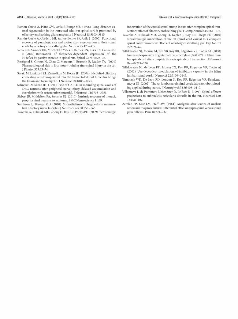
OEG transplantation promotes axonregeneration across the transection:electrophysiological evidenceWe used transcranial electric stimulationto evaluate whether OEG promote func-
tional connectivity across the lesion site after a complete spinalcord transection. Because the stimulation intensity required toelicit MEPs depends on the type of anesthetic, level of anesthesia,location of stimulation and recordings, and diameter of stimula-tion/recording electrodes, we initially performed control experi-ments on intact rats. Stimulation of both the motor cortex andbrainstem of intact rats (n � 4) consistently elicited MEP bilat-erally in the MG and TA muscles that disappeared after acutespinal cord transection, indicating that MEPs detected in MG andTA are transmitted through the spinal cord. Additionally, MEPshad longer latencies and/or were eliminated by pharmacologicalblockage of excitatory neurotransmitters in intact controls (Pro-tas et al., 2008), suggesting that evoked potentials with latencieslonger than 1 ms are very likely attributable to neural transmis-sion. Typical MEPs from intact rats were 2–10 mV with a latencyof �8 ms (Fig. 3A,G). With stronger stimulation intensities,MEP amplitudes increased, whereas the latencies were unaffected(data not shown).
The recordings at 1 month after lesion confirmed electro-physiologically that the spinal cord transections were complete
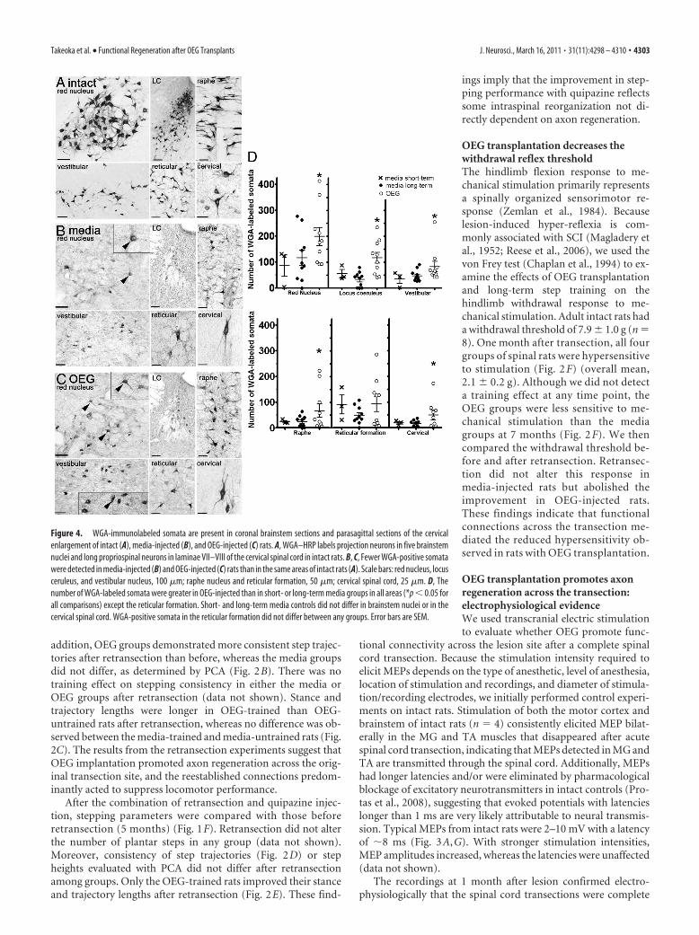
Figure 4. WGA-immunolabeled somata are present in coronal brainstem sections and parasagittal sections of the cervicalenlargement of intact (A), media-injected (B), and OEG-injected (C) rats. A, WGA–HRP labels projection neurons in five brainstemnuclei and long propriospinal neurons in laminae VII–VIII of the cervical spinal cord in intact rats. B, C, Fewer WGA-positive somatawere detected in media-injected (B) and OEG-injected (C) rats than in the same areas of intact rats (A). Scale bars: red nucleus, locusceruleus, and vestibular nucleus, 100 �m; raphe nucleus and reticular formation, 50 �m; cervical spinal cord, 25 �m. D, Thenumber of WGA-labeled somata were greater in OEG-injected than in short- or long-term media groups in all areas (*p � 0.05 forall comparisons) except the reticular formation. Short- and long-term media controls did not differ in brainstem nuclei or in thecervical spinal cord. WGA-positive somata in the reticular formation did not differ between any groups. Error bars are SEM.
Takeoka et al. • Functional Regeneration after OEG Transplants J. Neurosci., March 16, 2011 • 31(11):4298 – 4310 • 4303
(Fig. 3A). No media-injected rats responded to stimulation ateither site at any time point. Four months after transection, wedetected MEPs with long latencies and small amplitudes in 15%(motor cortex, n � 1 of 22; brainstem, n � 3 of 22) of OEG-injected rats (Fig. 3B,G). More OEG-injected rats exhibitedMEPs, the mean latency was shorter, and the amplitude waslarger at 7 than 4 months after transection (Fig. 3B,G). Whencombined, a total of 70% (motor cortex, 5 of 20; brainstem, 12 of20) of the OEG-injected rats responded to cortical and/or brain-stem stimulation at 7 months (Fig. 3B). Brainstem stimulationelicited relatively stronger MEPs, i.e., shorter latencies and largeramplitudes, than cortical stimulation (Fig. 3G). In contrast tointact rats, the MEP amplitudes recorded from OEG-injected ratsdid not increase consistently with increased stimulation intensity(data not shown). In addition, MEP latencies and amplitudeswere longer and smaller, respectively, in OEG than intact rats(Fig. 3G). Interestingly, 15% (3 of 20) of OEG-injected rats dis-played MEPs with latencies indistinguishable from those in intactrats, i.e., �8 ms, in response to either motor cortex or brainstemstimulation at 7 months after transection. Long-term step train-ing did not influence MEP occurrence because 50% (OEG-untrained, 7 of 14; OEG-trained, 7 of 14) of the rats with MEPswere in each OEG group. The mean MEP amplitude, however,was larger in the OEG-trained than -untrained group despiteno differences in the mean latency between groups (Fig. 3C).Because fresh muscle weight of bilateral medial gastrocne-mius, tibialis anterior, soleus, and extensor digitorum longusexamined postmortem did not differ between OEG-untrainedand trained groups (data not shown), the higher MEP ampli-tude in OEG-trained rats is likely attributable to changes in thespinal cord reorganization rather than increased muscle massfrom training.
The number of muscles with MEPs in response to either cor-tical or brainstem stimulation varied in each rat at 7 months (Fig.3D). Seventy-percent (11 of 14) of OEG-injected spinal rats hadresponses in two or more muscles, and 21% (3 of 14) of rats hadresponses in all four muscles tested. More MEPs were elicited inthe MG than TA at all stimulation intensities in response to bothbrainstem and cortical stimulation (Fig. 3E). Although stimula-tion intensity did not correlate with MEP amplitude, more mus-cles were recruited with higher stimulation intensities, i.e., moreTA and MG displayed MEPs at the higher stimulation intensities(Fig. 3E). Many of these muscles exhibited inconsistent responsesto the same stimulation intensity, in that not all stimulationpulses in a series elicited an MEP (incidence of response variedfrom 10 to 100%) and the stimulation intensity did not appear toinfluence the incidence of response (data not shown). Moreover,consecutive stimulations at a single intensity could elicit evokedpotentials with different latencies and amplitudes in 30% of therats (6 of 14) (Fig. 3F), a phenomenon not observed in intact rats.This variation suggests that different synaptic reconnections wereactivated in response to a specific stimulus and that the functionalreconnections of descending pathways were not robust enough torespond to every stimulation.
Eight months after injury, we performed transcranial stimu-lation on 7 media-injected and 10 OEG-injected rats immediatelybefore, immediately after, and 3 and 5 d after retransection todetermine whether these MEP signals represent physiological re-connections across the original transection site. The MEPs wereabolished after retransection in every rat with detectable evokedpotentials before retransection (Fig. 3A, bottom trace, retx), pro-viding additional evidence that OEG promoted electrophysi-ologically viable reconnections across the original lesion.
OEG transplantation may promote axon regeneration acrossthe transection: anatomical evidenceRegeneration of descending brainstem and cervical spinalcord axonsTo determine the extent of axon regeneration across the transec-tion, we injected WGA–HRP into the lower thoracic cords of 3intact and 22 spinal rats 8 months after transection (media-un-trained; n � 5, media-trained; n � 7, OEG-untrained; n � 5,OEG-trained; n � 5). None of the spinal rats included in addi-tional analyses had leakage of WGA–HRP into the lesion core orthe rostral stump (supplemental Fig. 1C, available at www.jneurosci.org as supplemental material). In intact rats, we de-tected WGA-positive somata in the motor cortex (supplementalFig. 1A, available at www.jneurosci.org as supplemental mate-rial), red nucleus, locus coeruleus, vestibular nucleus, reticularformation, raphe nucleus, and cervical enlargement (Fig. 4A).Based on an objective densitometry method, the number ofWGA-positive somata in the brainstem and rostral spinal cord ofboth media and OEG groups was quantified without knowledgeof the experimental status of each rat (Fig. 4B,C). Immunoreac-tive cells were considered as WGA-positive only when theiroptical density was at least 5% greater than the maximum back-ground level determined in each section (supplemental Fig.1D–G, available at www.jneurosci.org as supplemental material).The WGA-labeled neurons in spinal rats were similar in location,size, and shape to those found in intact rats (Fig. 4A–C). Wefound more WGA-positive somata in the red nucleus, locus coer-uleus, vestibular nucleus, and raphe nucleus but not in the retic-ular formation of OEG- than media-injected rats (Table 1, Fig.4D). There were no WGA-positive somata in the primary motorcortex of any spinal rat (supplemental Fig. 1B, available at www.jneurosci.org as supplemental material). We detected moreWGA-positive somata in laminae VII, VIII, and X of the cervicalenlargement in OEG- than media-injected rats (Fig. 4D). Theseretrogradely labeled cell bodies were round or oval in shape and20 –30 �m in diameter with multipolar processes, features typicalof long propriospinal neurons that contact lumbar somatic mo-tor neurons (Menetrey et al., 1985).
Next we asked whether the number of retrogradely labeledsomata in media-injected controls was consistent with spontane-ous axon regeneration or nonspecific uptake of the tracer. Toaddress this issue, we injected WGA–HRP into three media-injected spinal rats 1 month after transection as an additionalcontrol. We detected WGA-labeled somata in all three media rats
Table 1. Quantification of WGA-labeled cell bodies from the retrograde tracing study
Treatment Red nucleus* Locus coeruleus*Vestibularnucleus* Raphe nucleus* Reticular formation
Cervicalenlargement*
Sham rats (n � 3) 2253 � 509 789 � 150 972 � 266 499 � 9 1468 � 138 1361 � 127Short-term media-injected rats (n � 3) 87 � 51 56 � 20 34 � 20 25 � 6 91 � 45 20 � 5Long-term media-injected rats (n � 10) 116 � 30 33 � 9 47 � 7 24 � 7 49 � 11 19 � 4OEG-injected rats (n � 10) 199 � 34 117 � 21 83 � 20 67 � 26 90 � 30 50 � 18
More WGA-positive cell bodies were detected in OEG-injected than short- or long-term media-injected rats, i.e., controls. *p � 0.05, statistical significance between both short- and long-term media-injected versus OEG-injected rats.
4304 • J. Neurosci., March 16, 2011 • 31(11):4298 – 4310 Takeoka et al. • Functional Regeneration after OEG Transplants
maintained for 1 month that presumably would have no axon regen-eration (Table 1, Fig. 4D). Moreover, the average number of labeledcells in the brainstem or cervical spinal cord did not differ signifi-cantly between media rats after 1 or 8 months post-injury. Theseresults suggest a consistent level of spurious WGA-labeled somata inspinal rats rather than spontaneous regeneration (Fig. 4D). Over-all, detection of more retrogradely labeled cells in the brainstem(excluding the reticular formation) and spinal cord of OEG- thanmedia-injected long-term spinal rats is consistent with our be-havioral and electrophysiological evidence that OEG transplan-tation promoted axon regeneration across the transection.
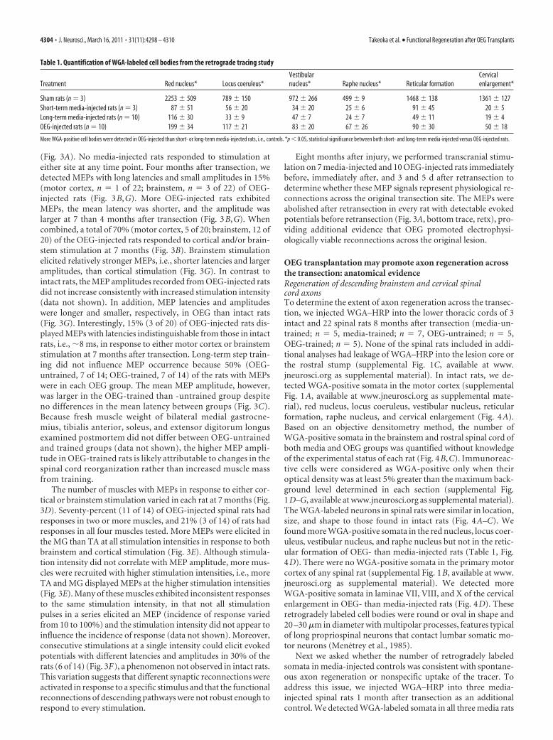
Raphespinal regenerationTo test whether OEG transplantation promotes raphespinal re-generation, we examined the presence of serotonergic axonsspanning the lesion. We found examples of axons that projectedacross both GFAP borders and the GFAP-negative lesion coreonly in OEG-injected rats (Fig. 5A). We also observed 5-HTaxons that sprouted within the lesion core in both media- andOEG-injected rats (Fig. 5B,C), with more found in OEG- thanmedia-injected rats (Fig. 5D). To determine whether these 5-HTaxons in the lesion core originated from the raphe nucleus orspinal cord interneurons (Newton and Hamill, 1988; Takeoka etal., 2009), we examined the original lesion core from retransectedspinal rats and found few 5-HT axons. This finding suggests that
the 5-HT axons that sprouted into the lesion core originatedprimarily from raphespinal neurons rather than intraspinal 5-HTneurons. We did not examine the 5-HT axons in the caudalstump because they could originate from the rare serotonergicspinal neurons and do not necessarily represent raphespinal re-generation (Takeoka et al., 2009).
Ascending axon regenerationGAP43 is transiently expressed in somata and growing axons ofmost neurons during development (Jacobson et al., 1986; Goslinet al., 1990). After axotomy, adult neurons, such as retinal gan-glion cells, dorsal root ganglion, and spinal neurons, transientlyexpress GAP43 in their somata (Doster et al., 1991; Schreyer andSkene, 1991; Siebert et al., 2010). Because GAP43 is a marker ofrecent axotomy, we hypothesized that any ascending axons thatregenerated across the original transection would transiently up-regulate GAP43 in their cell bodies after retransection 0.5 cmrostral to the initial site (diagram in Fig. 5E). First, we examinedspinal rats that did not receive a retransection and found nolabeled somata but numerous GAP43-positive axons within thelesion core, fibers associated with the meninges and penetratingblood vessels, and labeled axons within the dorsal and ventralroots (data not shown), in a pattern similar to peripherally de-rived noradrenergic axons (Takeoka et al., 2010). After retransec-tion, axotomized GAP43-positive somata filled the rostral stump
Figure 5. Axons regenerate in OEG-injected rats. A, A 5-HT-labeled axon (black, arrowheads) projects into and spans the transection site in an OEG-injected rat. Rostral and caudalGFAP-positive borders (brown) are at the top and bottom of the image. B, C, Many 5-HT axons (black) cross the rostral GFAP-positive border (brown, at the top) in both media-injected(B) and OEG-injected (C) rats. D, More 5-HT axons are found within the lesion core of OEG- than media-injected rats (*p � 0.02). E, Diagram of the injury site of the retransected spinalcord. Only GAP43 expression in somata within the caudal stump (*) after retransection suggests axon regeneration of ascending axons. F, G, GAP43-positive cell bodies (black) in media(F ) and OEG (G) GFAP-positive caudal stumps (brown). H, More GAP43-positive somata are detected in the caudal stump of OEG- than media-injected rats (*p � 0.01). Scale bars, 50 �m.Data are mean � SEM.
Takeoka et al. • Functional Regeneration after OEG Transplants J. Neurosci., March 16, 2011 • 31(11):4298 – 4310 • 4305
between the initial and retransection sitesin both media and OEG groups (diagramin Fig. 5E). In contrast, sparse GAP43-labeled somata occupied the caudal stumpof the retransected cords, primarily inlamina V with a few cells in lamina X (Fig.5F,G). These GAP43-labeled somata cau-dal to the initial transection likely repre-sent neurons with axons that hadregenerated past the original lesion andfar enough rostrally to be axotomized bythe retransection. This interpretation ofupregulated somatic GAP43 is only validin the caudal stump of spinal cord injurystudies that use an initial complete tran-section, followed by a lengthy period foraxon regeneration to occur, a completespinal cord retransection rostral to theoriginal injury, and then perfusions per-formed within 14 d after retransection. Wequantified GAP43-labeled cell bodies in thecaudal stump and found more GAP43-immunopositive somata in OEG than me-dia groups (Fig. 5H ). These resultssuggest that OEG implantation pro-motes regeneration of ascending axonsand that a limited amount of spontane-ous regeneration may occur in long-term, media-injected rats.
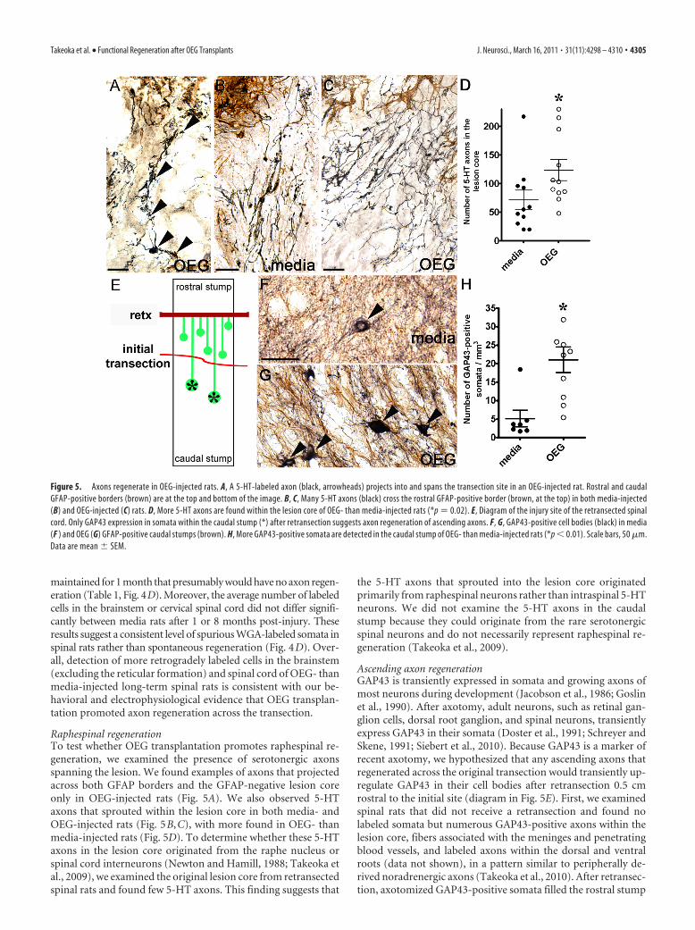
OEG preserve tissue and neuronssurrounding the lesionWe measured the volume of the GFAP-negative lesion core and associated cavitations to determinewhether OEG attenuate tissue degeneration after the injury. Theaverage volume of the injury site was larger in media- than OEG-injected rats (Fig. 6A,B), results consistent with our previousstudies (Kubasak et al., 2008; Munoz-Quiles et al., 2009). Weidentified the rostral and caudal stumps with anti-GFAP andspinal cord neurons with anti-NeuN to evaluate the extent ofinjury-induced neuronal degeneration (Fig. 6C). We foundNeuN-labeled cell bodies immediately adjacent to the GFAP-negative lesion core more often in OEG-injected (Fig. 6D) thanmedia-injected (Fig. 6C) spinal cords. The distance between themost proximal NeuN-labeled neuron and the GFAP-negative le-sion core was shorter in the rostral than in the caudal stump of allspinal rats (media: rostral, 0.67 � 0.06 cm and caudal, 1.31 � 0.11cm; OEG: rostral, 0.25 � 0.07 cm and caudal, 0.48 � 0.06 cm; p �0.05), a result that indicates that more neurons in the rostralstump were preserved than in the caudal stump. Furthermore,both the average GFAP-positive non-neuronal zone (Fig. 6E)and the total distance between neurons located in the rostral andcaudal stumps (Fig. 6F) were shorter in OEG than media groups.These results suggest that OEG transplantation promotes neuronalsurvival after a complete transection, and, consequently, in combi-nation with a smaller lesion volume, regenerating axons would havea shorter distance to project to a neuronal target on the opposite sideof the transection in OEG- than media-injected rats.
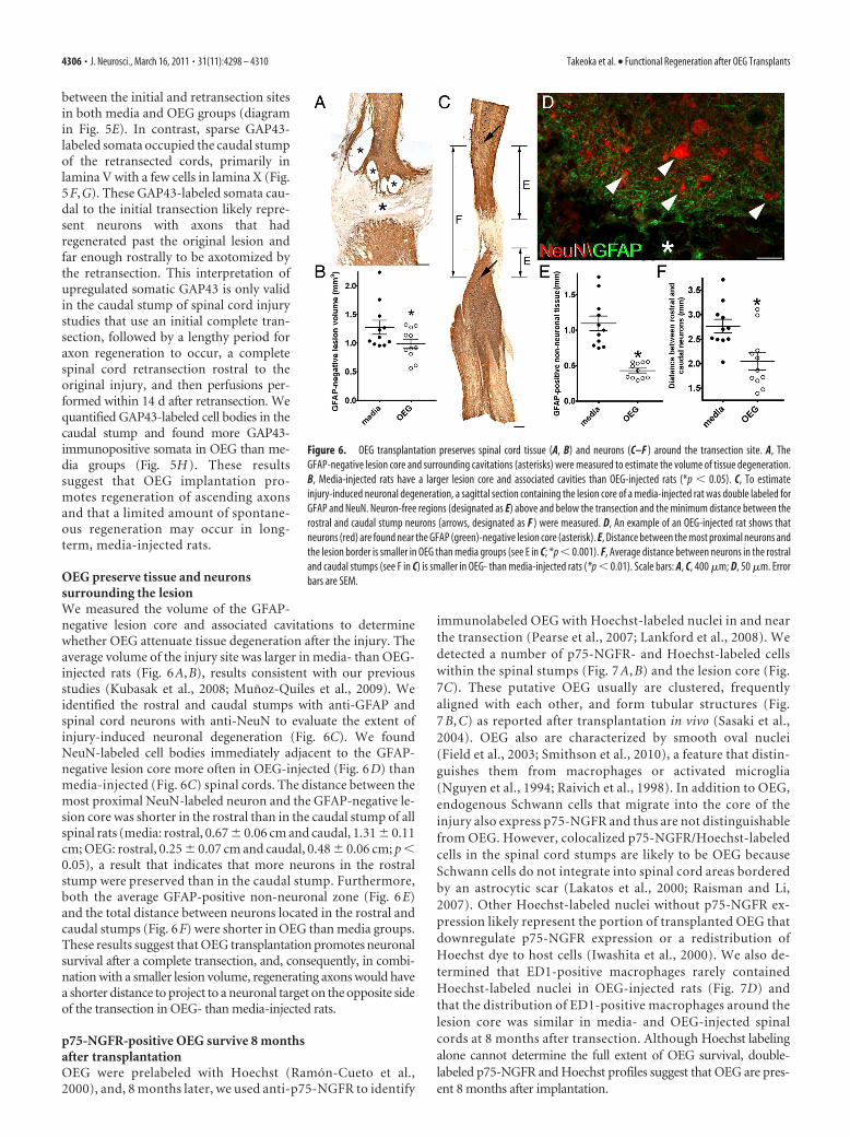
p75-NGFR-positive OEG survive 8 monthsafter transplantationOEG were prelabeled with Hoechst (Ramon-Cueto et al.,2000), and, 8 months later, we used anti-p75-NGFR to identify
immunolabeled OEG with Hoechst-labeled nuclei in and nearthe transection (Pearse et al., 2007; Lankford et al., 2008). Wedetected a number of p75-NGFR- and Hoechst-labeled cellswithin the spinal stumps (Fig. 7 A, B) and the lesion core (Fig.7C). These putative OEG usually are clustered, frequentlyaligned with each other, and form tubular structures (Fig.7 B, C) as reported after transplantation in vivo (Sasaki et al.,2004). OEG also are characterized by smooth oval nuclei(Field et al., 2003; Smithson et al., 2010), a feature that distin-guishes them from macrophages or activated microglia(Nguyen et al., 1994; Raivich et al., 1998). In addition to OEG,endogenous Schwann cells that migrate into the core of theinjury also express p75-NGFR and thus are not distinguishablefrom OEG. However, colocalized p75-NGFR/Hoechst-labeledcells in the spinal cord stumps are likely to be OEG becauseSchwann cells do not integrate into spinal cord areas borderedby an astrocytic scar (Lakatos et al., 2000; Raisman and Li,2007). Other Hoechst-labeled nuclei without p75-NGFR ex-pression likely represent the portion of transplanted OEG thatdownregulate p75-NGFR expression or a redistribution ofHoechst dye to host cells (Iwashita et al., 2000). We also de-termined that ED1-positive macrophages rarely containedHoechst-labeled nuclei in OEG-injected rats (Fig. 7D) andthat the distribution of ED1-positive macrophages around thelesion core was similar in media- and OEG-injected spinalcords at 8 months after transection. Although Hoechst labelingalone cannot determine the full extent of OEG survival, double-labeled p75-NGFR and Hoechst profiles suggest that OEG are pres-ent 8 months after implantation.
Figure 6. OEG transplantation preserves spinal cord tissue (A, B) and neurons (C–F ) around the transection site. A, TheGFAP-negative lesion core and surrounding cavitations (asterisks) were measured to estimate the volume of tissue degeneration.B, Media-injected rats have a larger lesion core and associated cavities than OEG-injected rats (*p � 0.05). C, To estimateinjury-induced neuronal degeneration, a sagittal section containing the lesion core of a media-injected rat was double labeled forGFAP and NeuN. Neuron-free regions (designated as E) above and below the transection and the minimum distance between therostral and caudal stump neurons (arrows, designated as F ) were measured. D, An example of an OEG-injected rat shows thatneurons (red) are found near the GFAP (green)-negative lesion core (asterisk). E, Distance between the most proximal neurons andthe lesion border is smaller in OEG than media groups (see E in C; *p � 0.001). F, Average distance between neurons in the rostraland caudal stumps (see F in C) is smaller in OEG- than media-injected rats (*p � 0.01). Scale bars: A, C, 400 �m; D, 50 �m. Errorbars are SEM.
4306 • J. Neurosci., March 16, 2011 • 31(11):4298 – 4310 Takeoka et al. • Functional Regeneration after OEG Transplants
DiscussionOEG facilitated regeneration across the lesion and reorganizationof neural circuits in the caudal stump. Behavioral and electro-
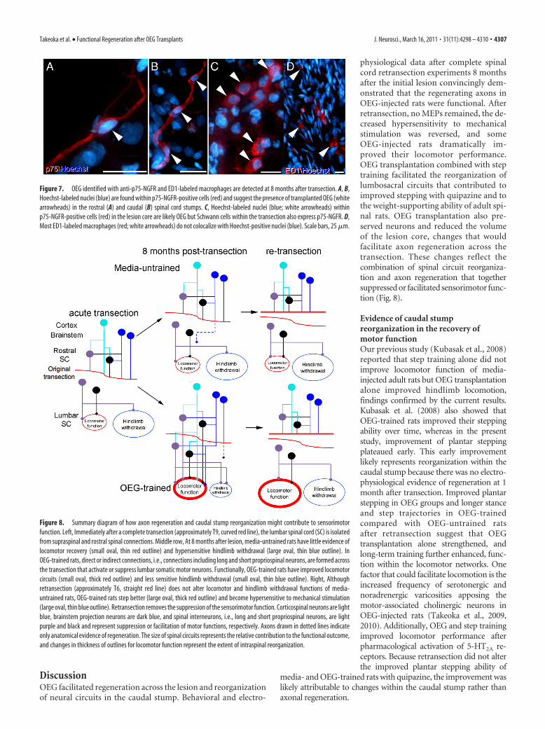
physiological data after complete spinalcord retransection experiments 8 monthsafter the initial lesion convincingly dem-onstrated that the regenerating axons inOEG-injected rats were functional. Afterretransection, no MEPs remained, the de-creased hypersensitivity to mechanicalstimulation was reversed, and someOEG-injected rats dramatically im-proved their locomotor performance.OEG transplantation combined with steptraining facilitated the reorganization oflumbosacral circuits that contributed toimproved stepping with quipazine and tothe weight-supporting ability of adult spi-nal rats. OEG transplantation also pre-served neurons and reduced the volumeof the lesion core, changes that wouldfacilitate axon regeneration across thetransection. These changes reflect thecombination of spinal circuit reorganiza-tion and axon regeneration that togethersuppressed or facilitated sensorimotor func-tion (Fig. 8).
Evidence of caudal stumpreorganization in the recovery ofmotor functionOur previous study (Kubasak et al., 2008)reported that step training alone did notimprove locomotor function of media-injected adult rats but OEG transplantationalone improved hindlimb locomotion,findings confirmed by the current results.Kubasak et al. (2008) also showed thatOEG-trained rats improved their steppingability over time, whereas in the presentstudy, improvement of plantar steppingplateaued early. This early improvementlikely represents reorganization within thecaudal stump because there was no electro-physiological evidence of regeneration at 1month after transection. Improved plantarstepping in OEG groups and longer stanceand step trajectories in OEG-trainedcompared with OEG-untrained ratsafter retransection suggest that OEGtransplantation alone strengthened, andlong-term training further enhanced, func-tion within the locomotor networks. Onefactor that could facilitate locomotion is theincreased frequency of serotonergic andnoradrenergic varicosities apposing themotor-associated cholinergic neurons inOEG-injected rats (Takeoka et al., 2009,2010). Additionally, OEG and step trainingimproved locomotor performance afterpharmacological activation of 5-HT2A re-ceptors. Because retransection did not alterthe improved plantar stepping ability of
media- and OEG-trained rats with quipazine, the improvement waslikely attributable to changes within the caudal stump rather thanaxonal regeneration.
Figure 7. OEG identified with anti-p75-NGFR and ED1-labeled macrophages are detected at 8 months after transection. A, B,Hoechst-labeled nuclei (blue) are found within p75-NGFR-positive cells (red) and suggest the presence of transplanted OEG (whitearrowheads) in the rostral (A) and caudal (B) spinal cord stumps. C, Hoechst-labeled nuclei (blue; white arrowheads) withinp75-NGFR-positive cells (red) in the lesion core are likely OEG but Schwann cells within the transection also express p75-NGFR. D,Most ED1-labeled macrophages (red; white arrowheads) do not colocalize with Hoechst-positive nuclei (blue). Scale bars, 25 �m.
Figure 8. Summary diagram of how axon regeneration and caudal stump reorganization might contribute to sensorimotorfunction. Left, Immediately after a complete transection (approximately T9, curved red line), the lumbar spinal cord (SC) is isolatedfrom supraspinal and rostral spinal connections. Middle row, At 8 months after lesion, media-untrained rats have little evidence oflocomotor recovery (small oval, thin red outline) and hypersensitive hindlimb withdrawal (large oval, thin blue outline). InOEG-trained rats, direct or indirect connections, i.e., connections including long and short propriospinal neurons, are formed acrossthe transection that activate or suppress lumbar somatic motor neurons. Functionally, OEG-trained rats have improved locomotorcircuits (small oval, thick red outline) and less sensitive hindlimb withdrawal (small oval, thin blue outline). Right, Althoughretransection (approximately T6, straight red line) does not alter locomotor and hindlimb withdrawal functions of media-untrained rats, OEG-trained rats step better (large oval, thick red outline) and become hypersensitive to mechanical stimulation(large oval, thin blue outline). Retransection removes the suppression of the sensorimotor function. Corticospinal neurons are lightblue, brainstem projection neurons are dark blue, and spinal interneurons, i.e., long and short propriospinal neurons, are lightpurple and black and represent suppression or facilitation of motor functions, respectively. Axons drawn in dotted lines indicateonly anatomical evidence of regeneration. The size of spinal circuits represents the relative contribution to the functional outcome,and changes in thickness of outlines for locomotor function represent the extent of intraspinal reorganization.
Takeoka et al. • Functional Regeneration after OEG Transplants J. Neurosci., March 16, 2011 • 31(11):4298 – 4310 • 4307
Evidence of axon regeneration across the transectionSpinal cord retransections provided critical evidence that OEGtransplantation facilitated functionally relevant axon regenera-tion. Surprisingly, several OEG-injected rats that never plantarstepped during the 7 monthly kinematics tests dramatically im-proved their stepping performance after retransection, a resultthat suggests that regenerated axons suppressed stepping (Fig. 8).Both facilitation and suppression of the spinal circuitry are im-portant in the control of intact locomotion (Bonnot et al., 1998;Iglesias et al., 2008). It is unclear the extent to which theregenerated axons are functional, i.e., the net effect is that theyare “inhibitory” to locomotor circuits in the classical neuro-physiological definition, or dysfunctional in the sense thatthey interfere with hindlimb stepping functions. Nonetheless,axon regeneration appears to have a net suppressive effect onthe locomotor networks and the injury-induced hypersensitivehindlimb withdrawal in this study. These results emphasize that,even when axon regeneration is identified electrophysiologi-cally and anatomically, behavioral consequences may beunpredictable.
Our electrophysiological results indicate that axons regener-ate across the transection and form functional connections inOEG-injected rats, but the process occurred slowly and connec-tions were less stable than those in intact rats. By 4 months aftertransection, we found reconnections with long latencies, featuresconsistent with polysynaptic and/or slow electrical conduction.We rarely, and only at 7 months, detected MEPs with latenciessimilar to those of intact rats. The small amplitudes of mostMEPs imply that relatively few connections were reestab-lished. Although intact rats had a 100% frequency of responseto transcranial stimulation, we saw inconsistent responses inOEG-injected rats, reflecting weak connections established byregenerating axons.
Combined with electrophysiological data, retrograde trac-ing results suggest that the re-established projections to so-matic motor neurons were mainly polysynaptic. For example,two OEG-injected rats that responded to cortical stimulationwere injected with the retrograde tracer, but neither hadWGA-labeled somata in the motor cortex. Interpreta-tions consistent with these data are that corticospinal axonssprouted in the rostral stump and contacted interneurons thatthen crossed the transection or that corticospinal axonscrossed the transection site and contacted an interneuron thateventually projected to somatic motor neurons (Fig. 8). Sim-ilarly, the brainstem nuclei re-established connections thatwere predominantly polysynaptic, and this may explain whysome OEG-injected rats had relatively few retrogradely labeledcells in the brainstem although MEPs were recorded in theirhindlimb muscles. Alternatively, some regenerated axonscould be electrically silent but able to transport the tracer.Perhaps some regenerating axons establish nonfunctional oraberrant connections that then are later eliminated (Bareyre etal., 2004; Calancie et al., 2005), recapitulating the process ofsynapse elimination during development. Additional evidenceconsistent with axon regeneration in OEG-injected rats in-cludes raphespinal axons that spanned the transection andupregulation of GAP43 in somata that likely represents a re-sponse to axotomy after retransection. GAP43-positive so-mata in the caudal stump of OEG-injected rats were locatedprimarily in lamina V, which contains many ascending sen-sory neurons (Baker and Giesler, 1984; Menetrey and Bas-baum, 1987; Villanueva et al., 1991; Ramon-Cueto et al.,1998). Detailed sensory testing was not performed in this
study, and therefore it is unclear whether these axons havefunctional consequences.
Finally, there is an inconsistency between the electrophysio-logical and anatomical results in long-term media-injected rats.Although some spurious labeling occurred after WGA–HRP in-jections, we detected extensive WGA-labeled somata in the rednucleus of two media rats maintained for 8 months after transec-tion but not in 1 month media controls. Chronic media rats alsohad several GAP43-positive somata in the caudal stump afterretransection. Because we electrophysiologically and anatomi-cally confirmed that transections were complete, these resultsleave open the possibility that limited spontaneous axon regen-eration may occur in long-term spinal rats as suggested byMunoz-Quiles et al. (2009). In the present study, however, therewas no evidence that spontaneous regeneration contributed tofunctional changes. Importantly, the combination of retro-grade tracer and retransection data caution against the over-interpretation of anatomical evidence of regeneration anddemonstrate the value of a retransection experiment to deter-mine whether regenerating axons are functional.
Promotion of axon regeneration and beyondThe current study raises an important question of how OEGpromote axon regeneration after injury. Olfactory bulb-derivedOEG can reduce inhibitory molecules (García-Alías et al., 2004),alter immune and inflammatory responses (Lopez-Vales et al.,2004; Chuah et al., 2010), and integrate into the CNS environ-ment as they intermingle with astrocytes both in vitro and in vivo(Franklin et al., 1996; Imaizumi et al., 1998; Lakatos et al., 2000,2003; Raisman and Li, 2007). The present results do not provideadditional insight into the possible mechanisms.
How axon regeneration and the reorganization of spinal neu-ral circuitry might contribute to the improvement in motor func-tion is complex (Fig. 8). Having now established that matureregenerating axons can form functionally relevant connectionsacross a complete spinal cord transection, an important futurechallenge is to optimize the extent and balance of axon regener-ation, maximize functionally useful axon–target reconnections,and identify strategies to further facilitate functional efficacy.
ReferencesBaker ML, Giesler GJ Jr (1984) Anatomical studies of the spinocervical tract
of the rat. Somatosens Res 2:1–18.Bareyre FM, Kerschensteiner M, Raineteau O, Mettenleiter TC, Weinmann
O, Schwab ME (2004) The injured spinal cord spontaneously forms anew intraspinal circuit in adult rats. Nat Neurosci 7:269 –277.
Bonnot A, Corio M, Bouc AM, Viala D (1998) Involvement of AMPA re-ceptors in posterior locomotor activity in the rabbit: an in vivo study.J Physiol Paris 92:5–15.
Cai LL, Fong AJ, Otoshi CK, Liang Y, Burdick JW, Roy RR, Edgerton VR(2006) Implications of assist-as-needed robotic step training after a com-plete spinal cord injury on intrinsic strategies of motor learning. J Neu-rosci 26:10564 –10568.
Calancie B, Alexeeva N, Broton JG, Molano MR (2005) Interlimb reflexactivity after spinal cord injury in man: strengthening response patternsare consistent with ongoing synaptic plasticity. Clin Neurophysiol116:75– 86.
Chandler CE, Parsons LM, Hosang M, Shooter EM (1984) A monoclonalantibody modulates the interaction of nerve growth factor with PC12cells. J Biol Chem 259:6882– 6889.
Chaplan SR, Bach FW, Pogrel JW, Chung JM, Yaksh TL (1994) Quanti-tative assessment of tactile allodynia in the rat paw. J Neurosci Meth-ods 53:55– 63.
Chuah MI, Hale DM, West AK (2010) Interaction of olfactory ensheathingcells with other cell types in vitro and after transplantation: glial scars andinflammation. Exp Neurol. Advance online publication. Retrieved Janu-ary 24, 2011. doi:10.1016/j.expneurol.2010.08.012.
4308 • J. Neurosci., March 16, 2011 • 31(11):4298 – 4310 Takeoka et al. • Functional Regeneration after OEG Transplants
Cote MP, Gossard JP (2003) Task-dependent presynaptic inhibition. J Neu-rosci 23:1886 –1893.
Cote MP, Menard A, Gossard JP (2003) Spinal cats on the treadmill:changes in load pathways. J Neurosci 23:2789 –2796.
Courtine G, Song B, Roy RR, Zhong H, Herrmann JE, Ao Y, Qi J, EdgertonVR, Sofroniew MV (2008) Recovery of supraspinal control of steppingvia indirect propriospinal relay connections after spinal cord injury. NatMed 14:69 –74.
Courtine G, Gerasimenko Y, van den Brand R, Yew A, Musienko P, Zhong H,Song B, Ao Y, Ichiyama RM, Lavrov I, Roy RR, Sofroniew MV, EdgertonVR (2009) Transformation of nonfunctional spinal circuits into func-tional states after the loss of brain input. Nat Neurosci 12:1333–1342.
de Leon RD, Tamaki H, Hodgson JA, Roy RR, Edgerton VR (1999)Hindlimb locomotor and postural training modulates glycinergic inhibi-tion in the spinal cord of the adult spinal cat. J Neurophysiol 82:359 –369.
Doster SK, Lozano AM, Aguayo AJ, Willard MB (1991) Expression of thegrowth-associated protein GAP-43 in adult rat retinal ganglion cells fol-lowing axon injury. Neuron 6:635– 647.
Edgerton VR, de Leon RD, Tillakaratne N, Recktenwald MR, Hodgson JA,Roy RR (1997) Use-dependent plasticity in spinal stepping and stand-ing. Adv Neurol 72:233–247.
Edgerton VR, Tillakaratne NJ, Bigbee AJ, de Leon RD, Roy RR (2004) Plas-ticity of the spinal neural circuitry after injury. Annu Rev Neurosci27:145–167.
Efron B, Tibshirani R (1991) Statistical data analysis in the computer age.Science 253:390 –395.
Field P, Li Y, Raisman F (2003) Ensheathment of the olfactory nerves in theadult rat. J Neurocytol 32:317–324.
Fong AJ, Cai LL, Otoshi CK, Reinkensmeyer DJ, Burdick JW, Roy RR, Edg-erton VR (2005) Spinal cord-transected mice learn to step in response toquipazine treatment and robotic training. J Neurosci 25:11738 –11747.
Fouad K, Schnell L, Bunge MB, Schwab ME, Liebscher T, Pearse DD (2005)Combining Schwann cell bridges and olfactory-ensheathing glia graftswith chondroitinase promotes locomotor recovery after complete tran-section of the spinal cord. J Neurosci 25:1169 –1178.
Franklin RJ, Gilson JM, Franceschini IA, Barnett SC (1996) Schwann cell-like myelination following transplantation of an olfactory bulb-ensheathing cell line into areas of demyelination in the adult CNS. Glia17:217–224.
Franssen EH, de Bree FM, Verhaagen J (2007) Olfactory ensheathing glia:their contribution to primary olfactory nervous system regeneration andtheir regenerative potential following transplantation into the injuredspinal cord. Brain Res Rev 56:236 –258.
García-Alías G, Verdu E, Fores J, Lopez-Vales R, Navarro X (2003) Func-tional and electrophysiological characterization of photochemical gradedspinal cord injury in the rat. J Neurotrauma 20:501–510.
García-Alías G, Lopez-Vales R, Fores J, Navarro X, Verdu E (2004) Acutetransplantation of olfactory ensheathing cells or Schwann cells promotesrecovery after spinal cord injury in the rat. J Neurosci Res 75:632– 641.
Gerasimenko YP, Ichiyama RM, Lavrov IA, Courtine G, Cai L, Zhong H, RoyRR, Edgerton VR (2007) Epidural spinal cord stimulation plus quipa-zine administration enable stepping in complete spinal adult rats. J Neu-rophysiol 98:2525–2536.
Goslin K, Schreyer DJ, Skene JH, Banker G (1990) Changes in the distribu-tion of GAP-43 during the development of neuronal polarity. J Neurosci10:588 – 602.
Gruner JA, Wade CK, Menna G, Stokes BT (1993) Myoelectric evoked po-tentials versus locomotor recovery in chronic spinal cord injured rats.J Neurotrauma 10:327–347.
Ichiyama RM, Gerasimenko YP, Zhong H, Roy RR, Edgerton VR (2005)Hindlimb stepping movements in complete spinal rats induced by epidu-ral spinal cord stimulation. Neurosci Lett 383:339 –344.
Ichiyama RM, Courtine G, Gerasimenko YP, Yang GJ, van den Brand R,Lavrov IA, Zhong H, Roy RR, Edgerton VR (2008) Step training rein-forces specific spinal locomotor circuitry in adult spinal rats. J Neurosci28:7370 –7375.
Ichiyama R, Potuzak M, Balak M, Kalderon N, Edgerton VR (2009) En-hanced motor function by training in spinal cord contused rats followingradiation therapy. PLoS One 4:e6862.
Iglesias C, Nielsen JB, Marchand-Pauvert V (2008) Corticospinal inhibitionof transmission in propriospinal-like neurones during human walking.Eur J Neurosci 28:1351–1361.
Imaizumi T, Lankford KL, Waxman SG, Greer CA, Kocsis JD (1998) Trans-planted olfactory ensheathing cells remyelinate and enhance axonal con-duction in the demyelinated dorsal columns of the rat spinal cord.J Neurosci 18:6176 – 6185.
Iwashita Y, Crang AJ, Blakemore WF (2000) Redistribution of bisbenzimideHoechst 33342 from transplanted cells to host cells. Neuroreport11:1013–1016.
Jacobson RD, Virag I, Skene JH (1986) A protein associated with axongrowth, GAP-43, is widely distributed and developmentally regulated inrat CNS. J Neurosci 6:1843–1855.
Kubasak MD, Jindrich DL, Zhong H, Takeoka A, McFarland KC, Munoz-Quiles C, Roy RR, Edgerton VR, Ramon-Cueto A, Phelps PE (2008)OEG implantation and step training enhance hindlimb-stepping abilityin adult spinal transected rats. Brain 131:264 –276.
Lakatos A, Franklin RJ, Barnett SC (2000) Olfactory ensheathing cells andSchwann cells differ in their in vitro interactions with astrocytes. Glia32:214 –225.
Lakatos A, Barnett SC, Franklin RJ (2003) Olfactory ensheathing cells in-duce less host astrocyte response and chondroitin sulphate proteoglycanexpression than Schwann cells following transplantation into adult CNSwhite matter. Exp Neurol 184:237–246.
Lankford KL, Sasaki M, Radtke C, Kocsis JD (2008) Olfactory ensheathingcells exhibit unique migratory, phagocytic, and myelinating properties inthe X-irradiated spinal cord not shared by Schwann cells. Glia56:1664 –1678.
Li Y, Sauve Y, Li D, Lund RD, Raisman G (2003) Transplanted olfactoryensheathing cells promote regeneration of cut adult rat optic nerve axons.J Neurosci 23:7783–7788.
Li Y, Yamamoto M, Raisman G, Choi D, Carlstedt T (2007) An experimen-tal model of ventral root repair showing the beneficial effect of transplant-ing olfactory ensheathing cells. Neurosurgery 60:734 –740; discussion740 –741.
Lopez-Vales R, García-Alías G, Fores J, Vela JM, Navarro X, Verdu E (2004)Transplanted olfactory ensheathing cells modulate the inflammatory re-sponse in the injured spinal cord. Neuron Glia Biol 1:201–209.
Lopez-Vales R, Fores J, Verdu E, Navarro X (2006) Acute and delayed trans-plantation of olfactory ensheathing cells promote partial recovery aftercomplete transection of the spinal cord. Neurobiol Dis 21:57– 68.
Magladery JW, Teasdall RD, Park AM, Languth HW (1952) Electrophysio-logical studies of reflex activity in patients with lesions of the nervoussystem. I. A comparison of spinal motoneurone excitability followingafferent nerve volleys in normal persons and patients with upper motorneurone lesions. Bull Johns Hopkins Hosp 91:219 –244; passim.
Menetrey D, Basbaum AI (1987) Spinal and trigeminal projections to thenucleus of the solitary tract: a possible substrate for somatovisceral andviscerovisceral reflex activation. J Comp Neurol 255:439 – 450.
Menetrey D, de Pommery J, Roudier F (1985) Propriospinal fibers reachingthe lumbar enlargement in the rat. Neurosci Lett 58:257–261.
Munoz-Quiles C, Santos-Benito FF, Llamusí MB, Ramon-Cueto A (2009)Chronic spinal injury repair by olfactory bulb ensheathing glia and feasi-bility for autologous therapy. J Neuropathol Exp Neurol 68:1294 –1308.
Newton BW, Hamill RW (1988) The morphology and distribution of ratserotonergic intraspinal neurons: an immunohistochemical study. BrainRes Bull 20:349 –360.
Nguyen KB, McCombe PA, Pender MP (1994) Macrophage apoptosis in thecentral nervous system in experimental autoimmune encephalomyelitis.J Autoimmum 7:145–152.
Pearse DD, Sanchez AR, Pereira FC, Andrade CM, Puzis R, Pressman Y,Golden K, Kifay BM, Blits B, Wood PM, Bunge MB (2007) Transplan-tation of Schwann cells and/or olfactory ensheathing glia into the con-tused spinal cord: survival, migration, axon association, and fundamentalrecovery. Glia 55:976 –1000.
Protas DT, Brown BG, Jung R, Jindrich DL (2008) Selective neurotransmit-ter blockers affect motor evoked potentials in anesthetized rats. Soc Neu-rosci Abstr 34:74.7.
Raisman G, Li Y (2007) Repair of neural pathways by olfactory ensheathingcells. Nat Rev Neurosci 8:312–319.
Raivich G, Jones LL, Kloss CU, Werner A, Neumann H, Kreutzberg GW(1998) Immune surveillance in the injured nervous system:T-lymphocytes invade the axotomized mouse facial motor nucleusand aggregate around sites of neuronal degeneration. J Neurosci18:5804 –5816.
Takeoka et al. • Functional Regeneration after OEG Transplants J. Neurosci., March 16, 2011 • 31(11):4298 – 4310 • 4309
Ramon-Cueto A, Plant GW, Avila J, Bunge MB (1998) Long-distance ax-onal regeneration in the transected adult rat spinal cord is promoted byolfactory ensheathing glia transplants. J Neurosci 18:3803–3815.
Ramon-Cueto A, Cordero MI, Santos-Benito FF, Avila J (2000) Functionalrecovery of paraplegic rats and motor axon regeneration in their spinalcords by olfactory ensheathing glia. Neuron 25:425– 435.
Reese NB, Skinner RD, Mitchell D, Yates C, Barnes CN, Kiser TS, Garcia-RillE (2006) Restoration of frequency-dependent depression of theH-reflex by passive exercise in spinal rats. Spinal Cord 44:28 –34.
Rossignol S, Giroux N, Chau C, Marcoux J, Brustein E, Reader TA (2001)Pharmacological aids to locomotor training after spinal injury in the cat.J Physiol 533:65–74.
Sasaki M, Lankford KL, Zemedkun M, Kocsis JD (2004) Identified olfactoryensheating cells transplanted into the transected dorsal funiculus bridgethe lesion and form myelin. J Neurosci 24:8485– 8493.
Schreyer DJ, Skene JH (1991) Fate of GAP-43 in ascending spinal axons ofDRG neurons after peripheral nerve injury: delayed accumulation andcorrelation with regenerative potential. J Neurosci 11:3738 –3751.
Siebert JR, Middelton FA, Stelzner DJ (2010) Intrinsic response of thoracicpropriospinal neurons to axotomy. BMC Neuroscience 11:69.
Smithson LJ, Kawaja MD (2010) Microglial/macrophage cells in mamma-lian olfactory nerve fascicles. J Neurosci Res 88:858 – 865.
Takeoka A, Kubasak MD, Zhong H, Roy RR, Phelps PE (2009) Serotonergic
innervation of the caudal spinal stump in rats after complete spinal tran-section: effect of olfactory ensheathing glia. J Comp Neurol 515:664 – 676.
Takeoka A, Kubasak MD, Zhong H, Kaplan J, Roy RR, Phelps PE (2010)Noradrenergic innervation of the rat spinal cord caudal to a completespinal cord transection: effects of olfactory ensheathing glia. Exp Neurol222:59 – 69.
Tillakaratne NJ, Mouria M, Ziv NB, Roy RR, Edgerton VR, Tobin AJ (2000)Increased expression of glutamate decarboxylase (GAD67) in feline lum-bar spinal cord after complete thoracic spinal cord transection. J NeurosciRes 60:219 –230.
Tillakaratne NJ, de Leon RD, Hoang TX, Roy RR, Edgerton VR, Tobin AJ(2002) Use-dependent modulation of inhibitory capacity in the felinelumbar spinal cord. J Neurosci 22:3130 –3143.
Timoszyk WK, De Leon RD, London N, Roy RR, Edgerton VR, Reinkens-meyer DJ (2002) The rat lumbosacral spinal cord adapts to robotic load-ing applied during stance. J Neurophysiol 88:3108 –3117.
Villanueva L, de Pommery J, Menetrey D, Le Bars D (1991) Spinal afferentprojections to subnucleus reticularis dorsalis in the rat. Neurosci Lett134:98 –102.
Zemlan FP, Kow LM, Pfaff DW (1984) Analgesia after lesions of nucleusreticularis magnocellularis: differential effect on supraspinal versus spinalpain reflexes. Pain 18:221–237.
4310 • J. Neurosci., March 16, 2011 • 31(11):4298 – 4310 Takeoka et al. • Functional Regeneration after OEG Transplants