Embed Size (px)
Citation preview

Cell, Vol. 98, 437–451, August 20, 1999, Copyright 1999 by Cell Press
Narcolepsy in orexin Knockout Mice:Molecular Genetics of Sleep Regulation
or slow-wave sleep is behaviorally quiet with progres-sively reduced responsiveness to environmental stimuliassociated with high-voltage slow activity on EEG (in-
Richard M. Chemelli,1,2,3 Jon T. Willie,1,2
Christopher M. Sinton,4 Joel K. Elmquist,7
Thomas Scammell,7 Charlotte Lee,7
cluding delta waves of 1–4 Hz and sleep spindles ofJames A. Richardson,5
12–14 Hz), generally absent eye movements, reducedS. Clay Williams,1,2 Yumei Xiong,1,2
muscle tone, and phasic motor activity. REM sleep isYaz Kisanuki,1,2 Thomas E. Fitch,4
associated with rapid but nonpurposeful eye movementsMasamitsu Nakazato,8 Robert E. Hammer,1,6
and frequent muscle twitches or small movements of theClifford B. Saper,7 and Masashi Yanagisawa1,2,9
extremities. The EEG is similar to that recorded during1Howard Hughes Medical Institutewakefulness with low-voltage fast activity. However,2Department of Molecular Geneticstonic muscle tension is absent (REM atonia) and respon-3Department of Pediatricssiveness to environmental stimuli is even further re-4Department of Psychiatryduced as compared with non-REM sleep. Normal sleep5Department of Pathologyis characterized by an orderly progression from wake-6Department of Biochemistryfulness to non-REM sleep and then to REM sleep.University of Texas Southwestern Medical Center
Idiopathic narcolepsy is a debilitating, lifelong neuro-at Dallaslogic disease characterized by excessive daytime sleep-Dallas, Texas 75235-9050iness, cataplexy, and other pathologic manifestations of7Department of NeurologyREM sleep. Narcoleptic patients experience sleepinessBeth Israel Deaconess Medical Centerthat is constant and severe, often complaining of invol-Boston, Massachusetts 02115untary or irresistible daytime “sleep attacks” that can8Third Department of Internal Medicineoccur while talking, standing, walking, eating, or driving.Miyazaki Medical CollegeCataplexy consists of attacks of sudden bilateral skele-Miyazaki 889-1692tal muscle weakness, often provoked by strong emotion,Japanwithout impairment of consciousness or memory, thatlast no more than a few minutes (Honda, 1988). Sleepparalysis and sleep-associated hallucinations are also
Summary experienced by some. The full tetrad of daytime somno-lence, cataplexy, sleep paralysis, and sleep-associated
Neurons containing the neuropeptide orexin (hypocre- hallucinations is present in only about 15% of patientstin) are located exclusively in the lateral hypothalamus (Aldrich, 1998). Clinically, the diagnosis is confirmed byand send axons to numerous regions throughout the polysomnography, a technique that employs simulta-central nervous system, including the major nuclei im- neous recording of EEG and the electromyogram (EMG).plicated in sleep regulation. Here, we report that, by This procedure reveals “sleep-onset REM periods,”behavioral and electroencephalographic criteria, or- REM sleep occurring at sleep onset or within 15 minexin knockout mice exhibit a phenotype strikingly sim- of sleep onset (Zorick et al., 1982; Baker et al., 1986;ilar to human narcolepsy patients, as well as canarc-1 Diagnostic Classification Steering Committee, 1990).mutant dogs, the only known monogenic model of Pathological intrusion of the REM state into wakefulnessnarcolepsy. Moreover, modafinil, an anti-narcoleptic is thought to form the physiologic basis of the symptomdrug with ill-defined mechanisms of action, activates tetrad (Bassetti and Aldrich, 1996).orexin-containing neurons. We propose that orexin Narcolepsy affects males and females equally withregulates sleep/wakefulness states, and that orexin an estimated prevalence of 0.02%–0.18% within whiteknockout mice are a model of human narcolepsy, a populations (Mignot, 1998). Familial narcolepsy, al-disorder characterized primarily by rapid eye move- though rare, is reported in the literature, with risk to firstment (REM) sleep dysregulation. degree relatives estimated at 1%–2% (Mignot, 1998).
Studies of monozygotic twins using strict diagnosticcriteria have found concordance rates of only 25%–Introduction31%, leading most authors to conclude that undefinedenvironmental factors act on a susceptible geneticIn mammals, the sleep–wake cycle is traditionally di-background to produce the disease. Several studiesvided into three states based on distinct behavioral andhave reported a strong association between certainneurophysiologic characteristics (Jones, 1998). Wake-class II HLA haplotypes on human chromosome 6 andfulness is characterized as a behaviorally interactivenarcolepsy. HLA DQB1*0602 and DQA1*0102 are foundstate, with full consciousness, that is associated within up to 90% of affected populations, compared withlow-voltage, mixed frequency, fast activity on electroen-12%–38% in the general population, suggesting that
cephalography (EEG), purposeful eye movements, andautoimmunity plays a role in the disorder (Kadotani et
high muscle tone with phasic motor activity. Non-REMal., 1998). However, extensive studies over the past 10years have failed to find convincing evidence of a sys-temic autoimmune process or one restricted to the cen-9 To whom correspondence should be addressed (e-mail: afcsushi@
aol.com). tral nervous system in narcoleptic patients. Moreover,

Cell438
Figure 1. Targeted Disruption of Mouse orexin Gene
(A) Strategy for the orexin knockout. A partial map of a mouse genomic DNA fragment containing the complete orexin gene with exons 1 and2 shown. Restriction enzymes: H, HindIII; K, KpnI; P, PstI; S, SacI; X, XhoI. Southern blot probes used in (B) as well as locations of PCRprimers for ES cell screening and mouse genotyping used in (C) are shown below the map of the targeted allele. The lengths of the KpnIrestriction fragments hybridized to probes A and B are shown above the maps of the wild-type and targeted alleles.(B) Genomic Southern blots of ES cell DNA. DNA was extracted from untransfected and targeted ES cells and digested completely with KpnI.DNA was separated in a 0.8% agarose gel, blotted to a nylon membrane, and hybridized with the random-primed 32P-labeled probes shownin (A).(C) PCR genotyping of mouse tail DNA. Tail DNA was extracted from F2 littermates of a heterozygote cross and subjected to 37 cycles ofPCR using 59 primers specific for the wild-type (1.5 kb) or knockout (2.1 kb) alleles and a common 39 primer as shown in (A). Integrity of PCRgenotyping was confirmed by Southern blots during initial colony genotyping.

Narcolepsy in orexin Knockout Mice439
patients with familial narcolepsy usually lack these HLA in that, acting directly at axon terminals, they can in-crease the release of the major inhibitory transmitter,risk factors (Mignot, 1998). Extensive gross and micro-GABA, as well as the major excitatory transmitter, gluta-scopic examination of narcoleptic brains as well as so-mate, which are together responsible for almost all fastphisticated in vivo proton spectroscopy studies havesynaptic activity in the hypothalamus (van den Pol etfailed to identify evidence of a structural or biochemicalal., 1998).brainstem lesion in idiopathic narcolepsy (Ellis et al.,
Immunohistochemical studies (Elias et al., 1998a;1998).Peyron et al., 1998; Date et al., 1999; Nambu et al., 1999)Investigations into the neurologic basis of narcolepsy/have demonstrated diffuse, widespread projections ofcataplexy have been greatly facilitated by the discoveryorexin neurons to the olfactory bulb, cerebral cortex,of canine narcolepsy. A colony of narcoleptic Dobermanthalamus, hypothalamus, and brainstem. Notably, somePinschers was established to transmit a candidate nar-of the densest orexin projections were found in the locuscolepsy gene, designated canarc-1, as an autosomalcoeruleus, raphe nuclei, medullary reticular formation,recessive trait with full penetrance (Baker et al., 1982).paraventricular thalamic nucleus, and septal nuclei. Re-Striking similarities between human and canine narco-cently, dense orexin projections were also described inlepsy include emotionally triggered cataplexy, increasedspecific lamina of the dorsal horn of the spinal cord (vandaytime sleepiness, pathological manifestations of REMden Pol, 1999), suggesting a possible role in modulatingsleep, sleep-onset REM periods on polysomnography,sensory input and autonomic tone. In situ hybridizationan early age of onset, and a familial tendency. Extensivestudies of orexin receptor mRNAs (Trivedi et al., 1998;studies of the neuropharmacology and neurochemistryour unpublished data) showed a diffuse pattern, consis-of affected dogs and humans have found a subtle distur-tent with the widespread nature of orexin projectionbance involving hyperactivity of cholinergic, and hypo-sites, although there was a marked differential distribu-activity of monoaminergic, neurotransmitter systemstion of the OX1R and OX2R mRNAs. Investigations into(Guilleminault et al., 1998). These studies have collec-the physiologic role of orexin have focused primarilytively led to a hypothesis that “the pathophysiology ofon energy homeostasis chiefly because of the historicnarcolepsy involves minute abnormalities of the neuro-importance of the lateral hypothalamus in this area (Ber-
chemical mechanisms regulating sleep rather than anardis and Bellinger, 1996). However, based on neuroan-
localized lesion or an obvious developmental abnormal-atomical data, several investigators have suggested a
ity in the CNS” (Nishino et al., 1995). possible role of these neuropeptides in sleep–wake reg-We recently identified and cloned orexins, a pair of ulation (Peyron et al., 1998; Date et al., 1999; Risold et
neuropeptides that are encoded in a single precursor al., 1999).protein, as the endogenous ligands for two G protein– Here, we report that a null mutation induced by tar-coupled receptors that are expressed in the brain and geted disruption of the mouse orexin gene results inwere previously considered to be orphan receptors (Sa- an autosomal recessive phenotype with characteristicskurai et al., 1998). An mRNA encoding the same neuro- remarkably similar to narcolepsy. Our initial studies onpeptide precursor was independently identified by a dif- these knockout mice focused on the putative involve-ferential cloning approach, and the putative encoded ment of orexins in energy homeostasis. While studyingpeptides were named hypocretins (de Lecea et al., the 24 hr open field activity of these mice, as an index of1998). Orexin-A and orexin-B are neuropeptides of 33 energy expenditure, we found that the orexin knockoutand 28 amino acids, respectively, that are produced mice showed periods of reduced activity during the darkexclusively by a well-defined group of neurons in the phase when mice are normally most active. We thenlateral hypothalamus. The active peptides are proteolyti- used infrared videotaping to investigate possible rea-cally processed from a single prepro-orexin precursor sons for this abnormal behavior. Close study of theseand then posttranslationally modified by N-terminal py- videotapes unexpectedly revealed brief periods of be-roglutamyl cyclization, C-terminal amidation, and forma- havioral arrest that occurred exclusively in homozygoustion of intrachain disulfide bridges. Orexin-A is a high- knockout mice. Twenty-four-hour EEG/EMG recording,affinity ligand for the orexin receptor type 1 (OX1R), undertaken to determine whether these episodes mightwhose affinity for orexin-B is 1–2 orders of magnitude be seizure related, demonstrated a complete absencelower. The orexin receptor type 2 (OX2R) exhibits equally of seizures but revealed an unanticipated disruption ofhigh affinity for both peptides. The orexin peptides are REM sleep regulation. We also report, and extend previ-
ous findings, that orexin neurons project directly ontounique among hypothalamic peptide neuromodulators
(D) In situ hybridization in orexin knockout mice. Dark-field images show matched coronal brain sections of the lateral hypothalamus hybridizedwith a 33P-labeled antisense riboprobe for orexin exon 2. Note the symmetric distribution of labeled neurons in the wild-type and heterozygotemice and the absence of signal in the homozygote. No detectable signal beyond background was generated by the sense riboprobe.(E) Orexin-A immunohistochemistry in knockout mice. Contiguous brain sections to those shown in (D) were treated with an orexin-A-specificantibody that revealed immunoreactive neurons in the perifornical nucleus of the wild-type and heterozygote mice. Fx, Fornix. Note that thenuclei are not stained, indicating that orexin is cytoplasmic. The peptide is also found in beaded neuronal processes in these sections. Alsonote the absence of staining in the homozygote. The staining of orexin-A-positive cells was abolished in the presence of excess (100 nM)synthetic orexin-A (data not shown).(F) Radioimmunoassay for orexin peptides of extracts of knockout brains. Total brain, excluding cerebellum, was acid extracted, lyophilized,and the peptides were dissolved in buffers suitable for orexin-A- or orexin-B-specific radioimmunoassays. Graphs show the means andstandard errors of total brain orexin peptide per gram of wet brain weight from four mice with each genotype. *, one-way ANOVA; **, Student-Newman-Keuls test; n.d., not detected; NS, not significant.
Cell440
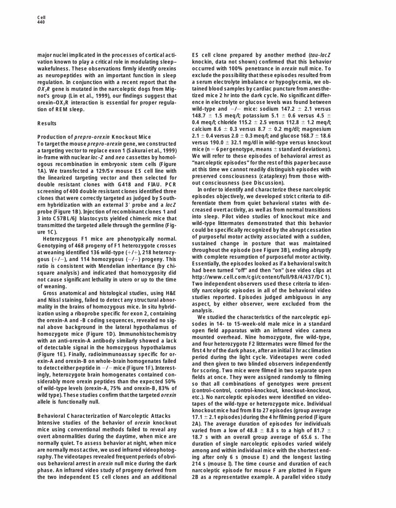
major nuclei implicated in the processes of cortical acti- ES cell clone prepared by another method (tau-lacZknockin, data not shown) confirmed that this behaviorvation known to play a critical role in modulating sleep–occurred with 100% penetrance in orexin null mice. Towakefulness. These observations firmly identify orexinsexclude the possibility that these episodes resulted fromas neuropeptides with an important function in sleepa serum electrolyte imbalance or hypoglycemia, we ob-regulation. In conjunction with a recent report that thetained blood samples by cardiac puncture from anesthe-OX2R gene is mutated in the narcoleptic dogs from Mig-tized mice 2 hr into the dark cycle. No significant differ-not’s group (Lin et al., 1999), our findings suggest thatence in electrolyte or glucose levels was found betweenorexin–OX2R interaction is essential for proper regula-wild-type and 2/2 mice: sodium 147.2 6 2.1 versustion of REM sleep.148.7 6 1.5 meq/l; potassium 5.1 6 0.6 versus 4.5 60.4 meq/l; chloride 115.2 6 2.5 versus 112.8 6 1.2 meq/l;Resultscalcium 8.6 6 0.3 versus 8.7 6 0.2 mg/dl; magnesium2.1 6 0.4 versus 2.0 6 0.3 meq/l; and glucose 168.7 6 18.6Production of prepro-orexin Knockout Miceversus 190.0 6 32.1 mg/dl in wild-type versus knockoutTo target the mouse prepro-orexin gene, we constructedmice (n 5 6 per genotype, means 6 standard deviations).a targeting vector to replace exon 1 (Sakurai et al., 1999)We will refer to these episodes of behavioral arrest asin-frame with nuclear lac-Z and neo cassettes by homol-“narcoleptic episodes” for the rest of this paper becauseogous recombination in embryonic stem cells (Figureat this time we cannot readily distinguish episodes with1A). We transfected a 129/Sv mouse ES cell line withpreserved consciousness (cataplexy) from those with-the linearized targeting vector and then selected forout consciousness (see Discussion).double resistant clones with G418 and FIAU. PCR
In order to identify and characterize these narcolepticscreening of 400 double resistant clones identified threeepisodes objectively, we developed strict criteria to dif-clones that were correctly targeted as judged by South-ferentiate them from quiet behavioral states with de-ern hybridization with an external 39 probe and a lacZcreased overt activity, as well as from normal transitionsprobe (Figure 1B). Injection of recombinant clones 1 andinto sleep. Pilot video studies of knockout mice and3 into C57BL/6J blastocysts yielded chimeric mice thatwild-type littermates demonstrated that this behaviortransmitted the targeted allele through the germline (Fig-could be specifically recognized by the abrupt cessationure 1C).of purposeful motor activity associated with a sudden,Heterozygous F1 mice are phenotypically normal.sustained change in posture that was maintainedGenotyping of 468 progeny of F1 heterozygote crossesthroughout the episode (see Figure 3B), ending abruptlyat weaning identified 136 wild-type (1/1), 218 heterozy-with complete resumption of purposeful motor activity.gous (1/2), and 114 homozygous (2/2) progeny. ThisEssentially, the episodes looked as if a behavioral switchratio is consistent with Mendelian inheritance (by chi-had been turned “off” and then “on” (see video clips atsquare analysis) and indicated that homozygosity didhttp://www.cell.com/cgi/content/full/98/4/437/DC1).not cause significant lethality in utero or up to the timeTwo independent observers used these criteria to iden-of weaning.tify narcoleptic episodes in all of the behavioral videoGross anatomical and histological studies, using H&Estudies reported. Episodes judged ambiguous in anyand Nissl staining, failed to detect any structural abnor-aspect, by either observer, were excluded from themality in the brains of homozygous mice. In situ hybrid-analysis.ization using a riboprobe specific for exon 2, containing
We studied the characteristics of the narcoleptic epi-the orexin-A and -B coding sequences, revealed no sig-sodes in 14- to 15-week-old male mice in a standard
nal above background in the lateral hypothalamus ofopen field apparatus with an infrared video camera
homozygote mice (Figure 1D). Immunohistochemistrymounted overhead. Nine homozygote, five wild-type,
with an anti-orexin-A antibody similarly showed a lack and four heterozygote F2 littermates were filmed for theof detectable signal in the homozygous hypothalamus first 4 hr of the dark phase, after an initial 3 hr acclimation(Figure 1E). Finally, radioimmunoassay specific for or- period during the light cycle. Videotapes were codedexin-A and orexin-B on whole-brain homogenates failed and then given to two blinded observers independentlyto detect either peptide in 2/2 mice (Figure 1F). Interest- for scoring. Two mice were filmed in two separate openingly, heterozygote brain homogenates contained con- fields at once. They were assigned randomly to filmingsiderably more orexin peptides than the expected 50% so that all combinations of genotypes were presentof wild-type levels (orexin-A, 75% and orexin-B, 83% of (control–control, control–knockout, knockout–knockout,wild type). These studies confirm that the targeted orexin etc.). No narcoleptic episodes were identified on video-allele is functionally null. tapes of the wild-type or heterozygote mice. Individual
knockout mice had from 8 to 27 episodes (group averageBehavioral Characterization of Narcoleptic Attacks 17.1 6 2.1 episodes) during the 4 hr filming period (FigureIntensive studies of the behavior of orexin knockout 2A). The average duration of episodes for individualsmice using conventional methods failed to reveal any varied from a low of 48.8 6 8.8 s to a high of 81.7 6overt abnormalities during the daytime, when mice are 18.7 s with an overall group average of 65.6 s. Thenormally quiet. To assess behavior at night, when mice duration of single narcoleptic episodes varied widelyare normally most active, we used infrared videophotog- among and within individual mice with the shortest end-raphy. The videotapes revealed frequent periods of obvi- ing after only 6 s (mouse E) and the longest lastingous behavioral arrest in orexin null mice during the dark 214 s (mouse I). The time course and duration of eachphase. An infrared video study of progeny derived from narcoleptic episode for mouse F are plotted in Figure
2B as a representative example. A parallel video studythe two independent ES cell clones and an additional

Narcolepsy in orexin Knockout Mice441
of female homozygotes and controls revealed similarepisode frequency and duration (data not shown).
The predominant behaviors for the 5 s immediatelypreceding and the 10 s immediately after each episodewere categorized as feeding, drinking, ambulating,grooming, burrowing, climbing, or other (group meanspresented in Figure 2C). Interestingly, while burrowingand climbing were often observed before an episode,they never occurred afterward. Conversely, while feed-ing and drinking rarely preceded an attack they fre-quently occurred afterward. Individual mice appearedto have characteristic triggering behaviors, with mouseA exhibiting ambulatory behavior in 22 of 27 episodes,mouse F burrowing before 8 of 17 episodes, and mouseG grooming in 6 of 8 episodes. Clearly observed gaitataxia lasting 1–3 s immediately preceded 26.5% 68.7% of all observed episodes with a range of 0%–77.8%in individual homozygotes. Grossly observable motoractivity causing side-to-side rocking, without change inoverall posture, frequently occurred several secondsafter the start of the attack. This lasted anywhere from2–10 s and was observed during 75.5% 6 7.0% of allepisodes with a range of 46.2%–100% of all attacks inindividual knockouts. Often these bouts of motor activityintensified, and they appeared to abruptly terminatesome narcoleptic attacks.
We performed close-up video studies of individualknockout mice filmed from various angles to further ex-plore the nature of the postural changes and motor activ-ity noted during the narcoleptic attacks. The posturalchanges characteristically involved sudden collapse ofthe head and neck with simultaneous buckling of thelimb, medially and/or laterally from the body, causingthe ventral surface to fall to the cage floor at angles of458 to perpendicular to the sagittal plane. Occasionally,the mouse fell completely onto its side. Gross motoractivity during attacks always resulted from episodiclimb twitching that rocked the mouse about its central
Figure 2. Infrared Video Characterization of Narcoleptic Episodesaxis. Bulbar motor activity was often noted in the close-
(A) Episode number and duration. Columns represent total numberup videos during attacks, with ear twitching, eye blink-of episodes recorded in the first 4 hr after onset of dark phase.ing, eye movements, and chewing movements some-Filled circles represent the mean duration of all recorded episodes.times observed.T bars indicate the minimum and maximum duration observed. Data
for individual knockout mice are designated A–I. Group is the aver-age count and duration for all mice (A–I), with the T bars indicativeof minimum and maximum individual averages. No narcoleptic epi- Developmental Aspects of Narcoleptic Attackssodes were observed for any of the wild-type (5) or heterozygote
We studied the onset and development of narcoleptic(4) control mice by blinded observers.episodes in two sets of male homozygote littermates(B) Typical behavior during the first 4 hr of the dark phase in knockoutfrom F2 homozygote crosses. Both cohorts were housedmouse. Duration of individual narcoleptic episodes and the time
they occurred from dark phase onset are plotted as vertical lines in groups of 4 or 5 per cage. Mice from one set werefor mouse F. Periods designated as awake and sleeping were judged placed into individual cages for videotaping. The otherby gross behavioral observation and are indicated by the white and set was videotaped in the group cage, allowing socialblack bars above the timeline, respectively.
interaction. Videotaping was conducted at weekly inter-(C) Behavior of mice before and after each narcoleptic episode. Thevals from 3–6 weeks of age. Figure 3A shows the num-predominant behavior observed for the 5 s preceding each episodebers of episodes recorded in the first 4 hr of the darkwas categorized as feeding, drinking, ambulating, grooming, bur-
rowing, climbing, or other (Before). Most behaviors classified as phase for five individually filmed knockout mice, desig-“other” were combinations of behavioral categories where a pre- nated M–Q. The right panel of Figure 3A compares thedominant behavior was not manifest. The predominant behavior for total number of episodes in the individually videotapedthe 10 s after each episode was categorized similarly (After). Number
mice versus the total number in the group filmed mice.of observations and percentage of total observations are reportedThe grouped mice exhibited more episodes; 55 versusfor all knockouts.8 at 4 weeks of age and 64 versus 26 at 6 weeks of age (4mice in group versus 5 mice in individual). Interestingly,while the entire group-filmed cohort had attacks at 4weeks of age, this did not occur until 6 weeks in the

Cell442
collapsed onto their ventral surface without gross motoractivity (Figure 3C), and only in 16/90 (17.7%) of epi-sodes they exhibited adult-like “rocking” motor activity.
EEG/EMG StudiesBodily collapse associated with episodic rocking motoractivity initially suggested the possibility of a seizuredisorder in the orexin knockout mice. To evaluate thishypothesis, we chronically implanted EEG electrodes inthese mice and recorded EEG continuously while themice were allowed ad lib movement in their acclimatedcages. After analyzing more than 300 hr of EEG re-cordings from six knockout mice, we were unable tofind any evidence of seizure activity. Rather, these EEGrecordings revealed that REM sleep was affected in theknockout mice.
Sleep state patterns revealed by simultaneous EEG/EMG recording showed little variability from one 24 hrrecording period to the next. In addition, results ob-tained from the wild-type littermates are identical topreviously published data from the C57BL/6 mousestrain (e.g., Tafti et al., 1997). Taken in conjunction withthe lack of sleep state fragmentation exhibited by wild-type controls (see Figure 4A) and the normal behavioralrepertoire as shown by concurrent infrared videopho-tography, these sleep data indicate a high degree ofadaptation to the recording conditions. Typical hypno-grams for a homozygous knockout and wild-type lit-termate for the 12 hr dark period are displayed in Figures4B and 4A, respectively. The hypnogram for the knock-Figure 3. Development of Narcoleptic Episodesout mouse is characterized by the appearance of sleep-(A) Episode count in first 4 hr dark phase for individually filmed
and group-filmed F3 homozygote littermates. First graph reports onset REM sleep episodes (arrowheads in Figure 4B,episode counts for individual orexin knockout mice (M–Q). Note that also see Figure 4C) and generally more fragmented non-mouse N did not record an episode until 6 weeks of age, and mouse REM sleep episodes, particularly notable toward theQ did not have one at week 5. n.d., none detected. The right panel end of the dark period, typically 04:00 to 06:00. Hypno-contrasts total episodes of individually filmed versus group-filmed
grams for the 12 hr light period, the normal rodent sleeplittermates at 4 and 6 weeks of age.period, were not distinguishable between the genotypes(B) Digitally captured infrared video image of group-filmed knockout
mice at 4 weeks of age. Note that one mouse (arrow) has completely (data not shown; see Table 1).fallen onto his side. The film shows the fuzziness (motion artifact) Sleep state parameters for wild-type and knockoutassociated with body movement in normally acting littermates des- mice are presented in Table 1. Overall, there is a highignated 1 to 3. Dark phase onset at 17:30 (see video of complete degree of similarity between these mice; in particular,episode at http://www.cell.com/cgi/content/full/98/4/437/DC1).
they are essentially identical during the light period ex-(C) Digitally captured infrared video image of group filmed knockoutcept for a shortened REM sleep latency in the orexinmice at 6 weeks of age. Note that one mouse has fallen completely
onto his side (arrow), while the another is collapsed onto his ventral knockout mice (p 5 0.03 by repeated measures ANOVA).surface (star). Littermates designated 1 and 2 are quietly sleeping Differences are apparent, however, during the dark pe-in their usual corner of the cage. Dark phase onset at 17:30, episodes riod (19:00 to 07:00) both in terms of time spent in sleeprecorded at 20:26 (see video of complete episode at http://www.cell. states and in the duration of sleep cycles. Thus, REMcom/cgi/content/full/98/4/437/DC1).
sleep time (p 5 0.0005) and REM episode duration (p 50.02) were greater in the 2/2 mice during the darkperiod. These changes occurred together with a de-individually filmed mice. In the group-filmed mice, chas-
ing, tail biting, and social grooming were observed im- crease in the interval between successive REM sleepperiods (p 5 0.04), highlighting the increased pressuremediately prior to narcoleptic attacks on some occa-
sions. Narcoleptic attacks in the group-filmed setting for REM sleep in these mice. There was also an increasein non-REM sleep time (p 5 0.06) and decrease in awakealso appeared to be cut short by stimulation from lit-
termates. Average episode duration was 45.1 6 4.1 s time (p 5 0.03) of knockout mice during the dark period.The change in awake time was associated with a de-versus 56.9 6 11.7 s (p 5 0.026 by Welch t test) at 4
weeks and 44.4 6 4.1 s versus 89.8 6 11.7 s (p , crease in awake episode duration (p 5 0.02), reflectingincreased fragmentation of the sleep–wake cycle during0.0001) at 6 weeks in the group versus individually filmed
littermates, respectively. Categorization of narcoleptic the dark period in the knockout mice. Thus, in additionto decreased REM sleep latency throughout the darkposture at 6 weeks revealed that, in 37/90 (41.1%) of
episodes, the young mice completely fell over to their and light periods, the orexin null mice show sleep-onsetREM episodes, hypersomnia, and more rapid cyclingside either in group or individually filmed settings (Figure
3B). In another 37/90 (41.1%) of episodes, the mice between sleep and wakefulness during the dark phase.

Narcolepsy in orexin Knockout Mice443
Figure 4. EEG/EMG Characterization of or-exin Knockout Mice
(A and B) Representative 12 hr dark period(19:00 to 07:00) hypnogram for a wild-typelittermate control (A) compared with a typicalhomozygous knockout mouse (B), obtainedby concatenating 20 s epoch stage scores.The height of the horizontal line above base-line indicates the vigilance state of the mouseat the time (min) from the beginning of therecording period. Baseline, W, represents aperiod of wakefulness; S, non-REM sleep;and R, REM sleep. Note episodes of sleep-onset REM, marked by arrowheads, greaternon-REM sleep episode fragmentation, andreduced wakefulness periods in the hypno-gram of the orexin knockout mouse duringthis period.(C and D) Initiation of a sleep-onset REM epi-sode (C) compared with a normal transitionfrom non-REM to REM sleep (D) in an orexinknockout mouse. These examples are ex-tracted from a continuous record, epochs844/845 occurring at 22:08, and epochs 862/863 6 min later at 22:14. Each epoch repre-sents 20 s of recording. Epoch 844 is charac-terized by rhythmic grooming activity withhigh-amplitude EMG and low-amplitude,mixed frequency EEG, typical of an awakeperiod. But at the beginning of epoch 845 thisis rapidly replaced, over a few seconds, bymuscle atonia, leaving only the heart beatregistering on the EMG leads combined withlow-amplitude EEG dominated by theta fre-quencies, characteristic of REM sleep in thisspecies. The star marks the onset of the ob-servable concurrent behavioral collapse. Incontrast, epoch 862 is a typical non-REMsleep period with minimal EMG activity andhigh-amplitude, low-frequency EEG that isreplaced during the first half of epoch 863by low-amplitude EEG dominated by theta,marking a normal transition to REM sleep.Wild-type mice demonstrated only normalnon-REM sleep to REM sleep transitions, in-distinguishable from tracing (D).(E) EEG/EMG recording during a typical nar-coleptic episode identified from concurrentinfrared video photography as sudden immo-bility associated with muscular twitches im-mediately following a period of normal activ-ity. The EEG shows that the start of thisepisode corresponds to two high-amplitudespindling epochs, marked with arrows, asso-ciated with phasic EMG activity as muscletone declines at the onset of attack. The starmarks the onset of observable immobility.These spindles in the EEG are normally ob-served only during the transition phase imme-diately prior to REM sleep.

Cell444
Table 1. Vigilance State Parameters Recorded from orexin Knockout (2/2) and Wild-Type Control (1/1) Mice
REM Sleep Non-REM Sleep Awake
2/2 1/1 2/2 1/1 2/2 1/1
24 hrTotal time (min) 88.3 6 5.2* 72.8 6 4.6 761.5 6 18.6 703.3 6 22.3 587.6 6 18.5* 661.2 6 24.6Episode duration (sec) 80.0 6 2.5 77.8 6 1.3 274.8 6 29.1 329.0 6 15.2 261.6 6 46.4 383.6 6 44.0REM latency (min) 6.7 6 0.7* 9.3 6 0.6Inter-REM interval (min) 20.6 6 1.3 24.7 6 1.7
Light periodTotal time 47.3 6 4.2 50.6 6 3.2 433.8 6 8.1 440.0 6 9.5 237.6 6 6.9 228.1 6 10.3Episode duration 76.2 6 3.5 79.8 6 2.0 322.8 6 21.0 352.8 6 18.6 241.5 6 32.3 248.6 6 39.3REM latency 7.4 6 0.6* 9.9 6 0.8Inter-REM interval 19.5 6 1.5 18.9 6 1.6
Dark periodTotal time 41.0 6 2.3* 22.2 6 3.0 327.2 6 19.8* 263.3 6 24.2 350.0 6 20.2* 433.1 6 27.0Episode duration 83.8 6 3.0* 75.1 6 1.4 235.9 6 39.0 298.8 6 14.1 284.0 6 59.9* 617.7 6 103.1REM latency 6.0 6 0.9* 8.5 6 0.4Inter-REM interval 22.8 6 1.9* 42.1 6 7.8
Total time spent in each state (minutes, mean 6 SEM), episode duration (seconds, 6 SEM), REM latency, and interval between successiveREM sleep episodes (minutes, mean 6 SEM) over 24 hr and itemized separately for light and dark periods. Significant differences between(2/2) and (1/1) mice are indicated with an asterisk. See text for details.
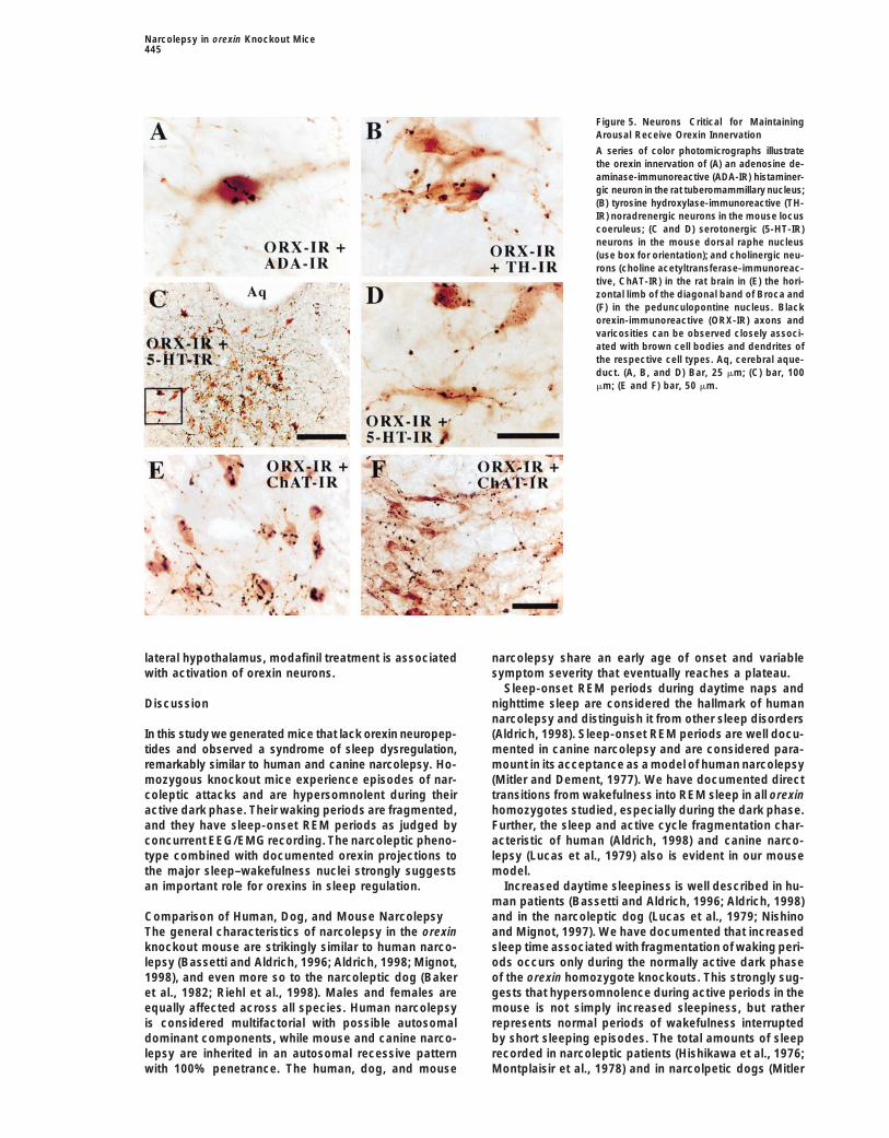
We correlated the infrared videotaped narcoleptic at- sites in both rat and mouse brains. We found that hista-minergic neurons in the tuberomammillary nucleustacks in the first 4 hr of the dark phase against concur-
rent EEG/EMG records. These periods corresponded to (adenosine deaminase-immunoreactive) received verydense orexin innervation on cell bodies and on proximalone of two distinct EEG/EMG patterns: they occurred
either during direct transitions from waking to REM sleep dendrites (Figure 5A). Noradrenergic neurons in the lo-cus coeruleus received a similar dense innervation by(similar to Figure 4C) or during high-amplitude spindle
oscillations in the EEG, superimposed on a slow wave orexin immunoreactive fibers. Somatic and dendritic ap-positions on tyrosine hydroxylase-immunoreactive cellsor non-REM sleep background. These spindles are char-
acteristic of the normal pre-REM phase in the mouse were best observed on solitary neurons on the edgesof the locus coeruleus (Figure 5B). Serotonergic neurons(Glin et al., 1991). In contrast to wild-type mice, however,
in the knockout mice these spindle oscillations occurred in the dorsal and median raphe nucleus also weredensely innervated. As can be seen in Figures 5C andimmediately after a waking period. An example of this
oscillatory EEG activity is displayed in Figure 4E. These 5D, serotonergic neurons in the dorsal raphe are specifi-cally targeted by orexin terminals. In the mouse brain,oscillations can be distinguished from spindles that are
recorded during sleep onset because pre-REM spindles cholinergic neurons in the pedunculopontine nuclei, lat-eral dorsal tegmental nucleus, diagonal band, and me-are of higher amplitude, longer duration, and frequently
show a pointed aspect (Gottesmann, 1996). The exis- dial septal nuclei received orexin innervation. The in-nervation of cholinergic cells was particularly dense intence of pre-REM spindles at sleep onset is a further
indication of abnormalities in the processes of REM the rat brain (Figures 5E and 5F). In all sites, apparentsomatic and dendritic appositions were observed in thesleep initiation and control in the orexin knockout
mouse. chemically characterized neurons.
Activation of Orexin Neurons by ModafinilOrexin Neurons Innervate CriticalSleep–Wakefulness Nuclei To investigate whether the anti-narcoleptic drug Modafi-
nil, whose mechanism of action is unknown, might actWe used double immunostaining techniques to test thehypothesis that orexin neurons projected directly onto through orexin neurons, we injected wild-type mice at
noon with modafinil (150 mg/kg i.p.), or vehicle, andthe central nervous system nuclei known to be importantin sleep–wakefulness regulation. In these micrographs sacrificed them 2 hr later. Brains were removed, double
immunostained for orexin and Fos (an indicator of neu-the orexin-containing axonal endings appear black (Fig-ure 5). The distribution of orexin-immunoreactive termi- ronal activity), and cells were counted in the perifornical
region. The number of orexin-immunoreactive neuronsnals was similar to that previously reported, includingparticularly dense innervation of the locus coeruleus, was the same in both groups (44–47 cells/section), but
the modafinil-treated group had over three times asdorsal and median raphe nuclei, and tuberomammillarynucleus (Elias et al., 1998a; Peyron et al., 1998; Date et many Fos-immunoreactive neuronal nuclei (38 in the
modafinil-treated mice versus 11 in the vehicle controls;al., 1999). We also observed innervation in the peduncu-lopontine nucleus, the lateral dorsal tegmental nucleus, p 5 0.01). Within the population of orexin-immunoreac-
tive neurons, modafinil induced a 9-fold increase in thethe horizontal and vertical limbs of the diagonal bandof Broca, and the medial septal nucleus, as previously number of Fos-immunoreactive cells (64% double-
labeled neurons in the modafinil group versus 7% in thereported (Peyron et al., 1998; Nambu et al., 1999). Weperformed double-label immunohistochemistry in these vehicle group, p 5 0.01) (Figures 6A–6C). Thus, in the
Narcolepsy in orexin Knockout Mice445
Figure 5. Neurons Critical for MaintainingArousal Receive Orexin Innervation
A series of color photomicrographs illustratethe orexin innervation of (A) an adenosine de-aminase-immunoreactive (ADA-IR) histaminer-gic neuron in the rat tuberomammillary nucleus;(B) tyrosine hydroxylase-immunoreactive (TH-IR) noradrenergic neurons in the mouse locuscoeruleus; (C and D) serotonergic (5-HT-IR)neurons in the mouse dorsal raphe nucleus(use box for orientation); and cholinergic neu-rons (choline acetyltransferase-immunoreac-tive, ChAT-IR) in the rat brain in (E) the hori-zontal limb of the diagonal band of Broca and(F) in the pedunculopontine nucleus. Blackorexin-immunoreactive (ORX-IR) axons andvaricosities can be observed closely associ-ated with brown cell bodies and dendrites ofthe respective cell types. Aq, cerebral aque-duct. (A, B, and D) Bar, 25 mm; (C) bar, 100mm; (E and F) bar, 50 mm.
lateral hypothalamus, modafinil treatment is associated narcolepsy share an early age of onset and variablesymptom severity that eventually reaches a plateau.with activation of orexin neurons.
Sleep-onset REM periods during daytime naps andnighttime sleep are considered the hallmark of humanDiscussionnarcolepsy and distinguish it from other sleep disorders(Aldrich, 1998). Sleep-onset REM periods are well docu-In this study we generated mice that lack orexin neuropep-
tides and observed a syndrome of sleep dysregulation, mented in canine narcolepsy and are considered para-mount in its acceptance as a model of human narcolepsyremarkably similar to human and canine narcolepsy. Ho-
mozygous knockout mice experience episodes of nar- (Mitler and Dement, 1977). We have documented directtransitions from wakefulness into REM sleep in all orexincoleptic attacks and are hypersomnolent during their
active dark phase. Their waking periods are fragmented, homozygotes studied, especially during the dark phase.Further, the sleep and active cycle fragmentation char-and they have sleep-onset REM periods as judged by
concurrent EEG/EMG recording. The narcoleptic pheno- acteristic of human (Aldrich, 1998) and canine narco-lepsy (Lucas et al., 1979) also is evident in our mousetype combined with documented orexin projections to
the major sleep–wakefulness nuclei strongly suggests model.Increased daytime sleepiness is well described in hu-an important role for orexins in sleep regulation.
man patients (Bassetti and Aldrich, 1996; Aldrich, 1998)and in the narcoleptic dog (Lucas et al., 1979; NishinoComparison of Human, Dog, and Mouse Narcolepsy
The general characteristics of narcolepsy in the orexin and Mignot, 1997). We have documented that increasedsleep time associated with fragmentation of waking peri-knockout mouse are strikingly similar to human narco-
lepsy (Bassetti and Aldrich, 1996; Aldrich, 1998; Mignot, ods occurs only during the normally active dark phaseof the orexin homozygote knockouts. This strongly sug-1998), and even more so to the narcoleptic dog (Baker
et al., 1982; Riehl et al., 1998). Males and females are gests that hypersomnolence during active periods in themouse is not simply increased sleepiness, but ratherequally affected across all species. Human narcolepsy
is considered multifactorial with possible autosomal represents normal periods of wakefulness interruptedby short sleeping episodes. The total amounts of sleepdominant components, while mouse and canine narco-
lepsy are inherited in an autosomal recessive pattern recorded in narcoleptic patients (Hishikawa et al., 1976;Montplaisir et al., 1978) and in narcolpetic dogs (Mitlerwith 100% penetrance. The human, dog, and mouse

Cell446
polysomnographic findings: (1) direct transitions fromwaking to REM sleep with marked EMG attenuation; and(2) high-amplitude spindle oscillations superimposed ona non-REM sleep background, characteristic of the pre-REM phase in the mouse (Glin et al., 1991). It is temptingto conclude that the direct wakefulness-to-REM transi-tion is identical to that observed in human and dogcataplexy. However, without subtle visual clues to thelevel of consciousness of these mice, possibly by high-resolution close-up video photography in the future, wecannot clearly differentiate narcoleptic from cataplecticepisodes. In addition, we will need to perform powerspectral analysis on the waking and REM periods duringthese behaviorally defined episodes to formally provethat they are identical to normal waking and REM EEGspectra (Kushida et al., 1985; Guilleminault et al., 1998).The high-amplitude spindle oscillations superimposedon a non-REM sleep EEG background are unprece-dented in reported human and dog polysomnographicrecordings of cataplexy. Their presence at the transitionfrom waking is markedly abnormal and provides furtherevidence of REM state disturbance in orexin knockoutmice.
Cataplectic attacks are characteristically less severein humans than those observed in dogs or orexin knock-out mice. While only 35%–75% of patients report dailycataplectic episodes (Bassetti and Aldrich, 1996), narco-leptic dogs often have several attacks during a singleexperimental session (Riehl et al., 1998). We found thatnarcoleptic episodes occur from 8 to 27 times over a 4hr dark period in adult knockout mice. The duration ofcataplectic attacks in all species is remarkably similarFigure 6. In Vivo Activation of Orexin Neurons by Modafinilwith most episodes lasting less than 1 min, about 2 min,(A) Lateral hypothalamus of vehicle treated wild-type mouse show-and about 1 min in humans, dogs, and mice, respec-ing orexin neurons (brown cytoplasmic staining) are rarely stained
for Fos immunoreactivity (black nuclear staining). tively. These episodes can be prematurely terminated(B) Lateral hypothalamus of modafinil-treated wild-type mouse by adequately strong external stimuli in humans andshowing most orexin neurons are positive for Fos immunoreactivity. dogs. We believe that the consistently shorter episodeBar, 50 mm. duration observed in the group-filmed mice, as com-(C) Modafinil induces a 9-fold increase in Fos immunoreactivity in
pared with the individually filmed mice, was due to coin-orexin-immunoreactive neurons when treated with modafinil com-cidental stimulation by littermates. This is consistentpared with vehicle treated controls. *, p 5 0.01.with many videotaped attacks that immediately endedafter stimulation. Only approximately one-third of human
and Dement, 1977) are similar to that found in control patients experience full loss of muscle tone causingsubjects. We also found no significant difference in the collapse to the floor with the majority having partialamount of wakefulness, non-REM sleep, or REM sleep cataplexy evidenced by jaw sagging, head bobbing, armover the entire 24 hr period or during the 12 hr light dropping, ptosis, or dysarthria (Honda, 1988). Partialphase when mice normally sleep. cataplexy in the dog is often evidenced by hindlimb
We have termed the episodes of behavioral arrest in buckling. Unambiguous full postural collapse was fre-the orexin knockout mouse as “narcoleptic” episodes quently observed in young orexin homozygotes, whilefor several reasons. The major distinguishing feature adults tended to collapse onto their ventral surface atbetween a narcoleptic episode in a narrow sense and odd angles, suggesting some residual muscle tone. Cat-a cataplectic episode is that during cataplexy con- aplexy is not always instantaneous in human patientssciousness is preserved (Aldrich, 1998). Muscle atonia but can progress over several seconds with some pa-is characteristic of both types of attack. The electroen- tients experiencing gait ataxia known as “zig-zag walk-cephalographic patterns during cataplexy in human nar- ing” (Honda, 1988). The finding of gait ataxia immedi-coleptics (Guilleminault et al., 1998) and in the canine ately preceding 27% of narcoleptic episodes in adultmodel are very similar. Postural collapse occurs during knockout mice is intriguing.an initial stage resembling wakefulness with marked The most common emotional expression provokingsuppression of EMG activity, followed by a period vir- cataplexy in humans is laughing. However, anger, fear,tually indistinguishable from REM sleep, and then a surprise, and excitement are also common triggerstransitional stage to non-REM sleep or back to wakeful- (Honda, 1988; Bassetti and Aldrich, 1996). Food andness. Using concurrent infrared videotape/EEG/EMG re- play with other dogs are the well-documented para-cordings, we have determined that narcoleptic attacks digms used to trigger cataplexy in the dog (Riehl et
al., 1998). We found that excited ambulation, grooming,in the orexin knockout mouse correlate with two distinct

Narcolepsy in orexin Knockout Mice447
burrowing, and climbing were most frequently associ- potential synapses, and further studies using immuno-ated with narcoleptic episodes in the mouse. The dra- cytochemistry coupled with electron microscopy will bematic increase in the number of episodes noted in the needed to verify actual synaptic contacts.group-filmed young mice as compared with the individu- The brainstem cholinergic neurons in the pedunculo-ally filmed littermates suggests that social interaction pontine nuclei are critical to the central control of REMmay significantly enhance this phenotype. Chasing, tail sleep (Rye, 1997; Jones, 1998). Ascending projectionsbiting, and social grooming were often observed to im- from this nucleus to the thalamus mediate the corticalmediately precede narcoleptic attacks in the group- excitation characteristic of REM, while descending fi-filmed setting. bers to the pontine and medullary reticular formation
are important in mediating REM atonia. PharmacologicNeuroanatomical Considerations studies in the narcoleptic dog, using local perfusion ofSeveral studies have hinted at a possible role of the cholinergic agonists into specific areas of the brain, havelateral hypothalamus in sleep state regulation. Cells in identified the pontine reticular formation and the basalthe lateral hypothalamus display circadian activity forebrain as the likely sites of cholinergic hypersensitiv-rhythms (Koizumi and Nishino, 1976). The ventral portion ity implicated in triggering muscle atonia during REMof the posterior-lateral hypothalamus is innervated by sleep and cataplexy (Nishino et al., 1995). Basal fore-collaterals of the retino-geniculo-hypothalamic tract brain cholinergic hypersensitivity, specifically in the di-(Mikkelsen, 1990), one source of photic information for agonal band of Broca and the magnocellular preopticthe suprachiasmatic nucleus. Thus, orexin neurons are area, is particularly interesting because this area is ana-likely influenced by exogenous and endogenous circa- tomically connected with the limbic system, suggestingdian signals allowing appropriate activation of orexin- an important link with the emotional triggers of cata-mediated vigilance functions. A group of sleep-activated plexy. We have demonstrated using double-labeled im-neurons in the ventrolateral preoptic area (VLPO) have munohistochemical techniques that orexin neuronsbeen identified (Sherin et al., 1996) that send inhibitory project to the cholinergic neurons of the pedunculopon-projections to the tuberomammillary nucleus, an impor- tine nuclei and the diagonal band of Broca. Orexin pro-tant component of the ascending arousal system. These jections to the pontine and medullary reticular formationneurons appear to play an important role in the regula- are well described in the literature (Peyron et al., 1998).tion of non-REM sleep (Sherin et al., 1998) and send With the limitations of these techniques in mind, it isprojections to neurons in the lateral hypothalamus (Ba- clear that orexin neurons are anatomically placed torone et al., 1981) that are not melanin concentrating modulate the critical cholinergic areas implicated in hu-hormone (MCH)-containing neurons (Sherin et al., 1998; man and canine narcolepsy that are responsible for me-J. K. E., unpublished observation), suggesting the possi- diating REM cortical activation, REM sleep atonia, andbility that orexin neurons are the target cells. Interest- pathologic cataplexy.ingly, orexin neurons send projections to the VLPO We have also found that the anti-narcoleptic drug(Peyron et al., 1998). Anatomically, therefore, the orexin modafinil strongly activates orexin neurons in the lateralneurons are ideally placed to be modulated by, and to hypothalamus. However, it is difficult to conclude thatmodulate, the activity of VLPO neurons and thereby modafinil promotes wakefulness solely through orexincoordinate appropriate transitions between non-REM neurons, because it also induces neuronal activationand REM sleep states. in other brain regions implicated in sleep–wakefulness
The ascending cortical activating system includes the regulation, such as the suprachiasmatic nucleus, ante-histaminergic neurons of the tuberomammillary nucleus, rior hypothalamic area (Lin et al., 1996), tuberomammil-the noradrenergic neurons of the locus coeruleus, the lary nucleus, and locus coeruleus (T. S., unpublishedserotonergic neurons of the dorsal and median raphe, data). Since orexin neurons heavily innervate the tubero-and the cholinergic neurons of the pedunculopontine
mammillary nucleus and locus coeruleus (Peyron et al.,nuclei (Rye, 1997; Jones, 1998). At normal sleep onset
1998; data shown here), it is possible that modafinil maythere is a gradual reduction in firing of the cells in these
activate the orexin system, which then recruits othernuclei as deeper levels of non-REM sleep are attainedarousal regions.(McGinty and Harper, 1976; Aston-Jones and Bloom,
1981; Vanni-Mercier et al., 1984; Steriade et al., 1990).Concluding RemarksDuring REM sleep, the histaminergic, noradrenergic,Here, we have shown that lack of orexin neuropeptidesand serotonergic cells essentially cease firing, whileresults in a syndrome of sleep state dysregulation re-cholinergic cells concurrently increase their firing rates.markably similar to human and canine narcolepsy. In-Our double-label immunohistochemical results, in mousedeed, during the submission of this paper, we learnedand rat brain, indicate that orexin neurons innervatethat Mignot’s group demonstrated that the canarc-1all of these critical neurons controlling arousal. Orexingene encodes OX2R, one of the two known orexin recep-neurons appear to be strategically placed to modulatetors (Lin et al., 1999). Thus, the interaction of orexinsthe activity of all the components of the ascending corti-with OX2R appears to be a key signaling pathway incal activating system in a highly coordinated manner.REM sleep regulation. We would like to emphasize theAlthough this hypothesis is attractive, this interpretationimportance of conducting behavioral studies of geneti-should be tempered because of limitations in the tech-cally manipulated mouse strains during their normallyniques employed. Without the use of electron micros-active dark phase using infrared videophotography.copy, it is not possible to demonstrate synaptic contactsWithout this important observational tool, the overt phe-between different immunohistochemically stained neu-
rons. Therefore, the results from this study only identify notype of the orexin knockout mice could easily have

Cell448
Orexin Radioimmunoassaybeen missed. Our study also identifies the lateral hypo-Four 11- to 12-week-old male mice of each genotype were eutha-thalamus as an important player in sleep regulation,nized with pentobarbital, decapitated at the foramen magnum, andwith an anatomically apparent role in coordinating thebrains were quickly removed 1 to 2 hr before the start of the dark
widespread nuclei previously implicated in sleep state cycle. After removal of the cerebellum, brains were weighed andcontrol. Further genetic and neurochemical studies of then boiled for 3 min in 7 ml of Milli-Q water. After cooling on ice,
acetic acid and HCl were added to final concentrations of 1 M andthe orexin system in affected humans, dogs, and mice20 mM, respectively, with enough Milli-Q water for a final volumemay provide important clues to the etiology and treat-of 10 ml. Polytron homogenization at 10,000 rpm for 3 min wasment of this debilitating disorder.followed by two centrifugation steps: 2,500 rpm for 15 min and then28,000 g for 40 min. Five milliliters of each sample was lyophilized
Experimental Procedures and then subjected to RIA for orexin-A and -B. The RIA procedurewas performed as reported previously (Mondal et al., 1999) using
Targeting Vector Construction anti-orexin antisera specific for orexin-A and B with no detectableMurine orexin gene sequences were isolated from a mouse strain cross-reactivity to each other. One-way ANOVA followed by a Stu-129/Sv genomic phage library (Stratagene) using a partial rat cDNA dent-Newman-Keulls multiple comparisons test were used for sta-probe as described previously (Sakurai et al., 1998). Restriction tistical comparisons across genotypes (see Figure 1E).mapping, oligonucleotide hybridization, and sequencing confirmedthat five overlapping phage clones contained the entire orexin gene.
Infrared Videotaping and Scoring of Narcoleptic EpisodesTargeting vector construction was based on a universal lacZ-neo-An 8 mm CCD videocamera with infrared and digital time recordingtk (pN-Z-TK2) template plasmid vector that contained nuclear lacZ,capabilities (Sony TRV-CCD66) was used for documentation andneo, and flanking tk cassettes (gift from R. Palmitier). orexin exonscoring of dark cycle behavior. Characterization of dark phase be-1 was in-frame replaced with the nlacZ cassette using proximal 9.0havior in 14- to 15-week-old mice (see Figure 2) was performed inkb and distal 1.2 kb flanking genomic sequences (see Figure 1A).a standard open field apparatus (Opto-Varimex-3, Columbus Instru-Sequencing confirmed correct insertion of the long and short arms,ments, Columbus, OH) within a plexiglass arena covered with bed-as well as in-frame insertion of the nlacZ cassette. The targetingding material and modified for ad lib food and water delivery. Group-vector was linearized at the unique SacII site between the two tkhoused mice were brought to the behavior room 4 to 5 hr prior tocassettes for ES cell transfection.the their usual dark phase, acclimated to individual arenas for 3 hr,and then remained there for the 12 hr dark phase. InvestigationsProduction of orexin Knockout Miceinto the developmental aspects of mouse narcoleptic behavior (seeThe SM-1 mouse ES cell line (Yanagiawa et al., 1998) was culturedFigure 3) were conducted in knockout littermates videotaped weeklyon irradiated LIF-producing STO feeder layers as described. ESfrom 3 to 6 weeks of age. These mice were videotaped group housedcells were electroporated with the linearized targeting vector andor individually housed in shoebox cages modified for side deliveryselected for double resistance to G418 and FIAU as described (Ya-of ad lib food and water. The first 4 hr of the dark phase wasnagiawa et al., 1998). Double-resistant ES cell clones were screenedvideotaped from overhead for all behavioral paradigms described.by PCR with a 59 neo primer, GTGCCCTGAATGAACTGCAGGACGClose-up videotaping of mouse behavior was performed in homeand a 39 primer external to the targeting vector, TGCTGATCTTTCcages photographed from various acute angles. Still images (seeCAGGGCAACCGA (see Figure 1A). Three ES cell clones were foundFigure 3) and illustrative video segments were captured and editedwhere correct targeting was confirmed by Southern blotting usingwith a video capture board (Dazzle Multimedia).flanking 39 genomic, external to the targeting vector, and nlacZ
Open field narcoleptic episodes were scored with observersprobes (see Figure 1B). Two of these ES cell clones were microin-blinded to genotype for nine homozygote, five wild-type, and fourjected into blastocysts with production of germline transmitting chi-heterozygote F2 littermates from coded videotapes. Narcoleptic epi-meric mice as described (Yanagiawa et al., 1998). PCR genotypingsodes for all experimental paradigms were strictly defined by theof orexin knockout progeny utilized the primers specified above forfollowing features: (1) abrupt transition from obvious purposefulthe mutant allele (2.1 kb) and a genomic 59 primer, AGAGATCATCTmotor activity; (2) a sustained change in posture maintainedCTCCAGATTA, with the same 39 primer noted above, to identifythroughout the episode; and (3) an abrupt end to the episode withthe wild-type allele (1.5 kb, see Figure 1C). Integrity of the PCRresumption of obvious purposeful motor activity (essentially as if agenotyping protocol was confirmed by Southern blotting using theswitch had been turned “off” and then “on”). This strict scoringprobes noted above.methodology likely reduced the sensitivity of episode detection,All experiments using wild-type and heterozygote controls wereespecially during quiet awake behavior, but enhanced specificityperformed on F2 C57Bl/6J-129/SvEv mixed background littermatesto enable unambiguous identification of narcoleptic episodes. Thefrom F1 heterozygote crosses. Experiments performed exclusivelyexact time recorded on the video for the start and end of eachon homozygotes used F3 mixed background littermates from F2episode was recorded along with the following additional observa-homozygote crosses. All mice were provided food and water ad lib,tions: the predominant activity for the 5 s preceding and the 10 smaintained on a 12 hr light:dark cycle at all times, and were housedfollowing an episode were categorized as feeding, drinking, ambu-under conditions that controlled for temperature and humidity.lating, grooming, burrowing, climbing, or other; obvious gait ataxiaAll mouse procedures used in this study were reviewed and preap-preceding and nonpurposeful motor activity (twitching) during theproved by the Institutional Review Board for Animal Research ofepisode were recorded as present or absent. Developmental co-the University of Texas Southwestern Medical Center at Dallas, orhorts were scored similarly with the exception that observers werethe Harvard Medical School and Beth Israel Deaconess Institutionalnot blinded, since all mice were homozygote littermates. All video-Animal Care and Use Committees as appropriate.tapes were scored by two independent observers with disputessettled by quorum.
In Situ HybridizationA 0.29 kb segment of the mouse orexin gene encoding Gln-33 toVal-130 of exon 2 was PCR amplified using CAGCCTCTGCCCGACT EEG/EMG Recording
Male mice (n 5 12, 14–15 weeks old, 30–35 g at the time of surgery)GCTGTCGCCAGAAG and GACTCCGGAGCCTCCCCGGGGTGCTAAAGC as primers and cloned into pCR II (Invitrogen). Sense and were prepared for chronic monitoring of EEG/EMG signals using a
recently developed lightweight implant and cabling procedure. Fullantisense riboprobes were generated with SP6 and T7 polymerases,respectively, using the Maxiscript kit (Ambion) in the presence of details of this technique will be published elsewhere. Briefly, the
EEG/EMG implant was based on a six-pin double inline microcom-33P-CTP (Amersham). In situ hybridization (see Figure 1D) to matchedadult male mouse brain sections containing the lateral hypothala- puter connector, modified to form four EEG electrodes, each 1.3 mm 3
0.3 mm (h 3 w) positioned 4.6 mm 3 2.9 mm (l 3 w) apart with twomus of all genotypes was performed as described (Sakurai et al.,1998). EMG electrodes soldered to the center pins. Mice were anesthetized

Narcolepsy in orexin Knockout Mice449
with sodium pentobarbital (Nembutal, 50–60 mg/kg i.p.), and stan- incubation of the tissue in antisera that had been preadsorbed withthe respective antigens (ChAT and 5-HT). For double-label controlsdard sterile surgical and stereotaxic procedures were employed for
implant placement. The cranium was exposed and four burr holes using the TH antisera coupled with the orexin antisera (becauseboth antisera were made in rabbits), sections were stained sequen-were drilled, anterior and posterior to bregma, bilaterally (AP 1.1,
ML 61.45 and AP 23.5, ML 61.45) according to the atlas of Franklin tially, but the orexin antisera was replaced with normal rabbit serum.These procedures resulted in specific staining only for the orexinand Paxinos (1997). The implant was then inserted into these holes,
cemented to the skull with dental acrylic, and the EMG electrodes antiserum. This was not an issue for the other double-label experi-ments, as the ADA antisera was raised in sheep and the 5-HT andsecured to the nuchal musculature. The design of this EEG/EMG
implant allowed precise insertion of electrodes in a reproducible ChAT antisera were made in goat.Photomicrographs were produced by capturing images with amanner, targeting bilateral stereotaxic coordinates over the frontal
and occipital cortices at a consistent depth, just touching the dura, digital camera (Kodak DCS) mounted directly on the microscope(Zeiss Axioplan) and an Apple Macintosh Power PC computer. Im-while minimizing surgical trauma.
Immediately after recovery from anesthesia, the mouse was age editing software (Adobe Photoshop) was used to combine pho-tomicrographs into plates. Only the sharpness, contrast, and bright-housed individually and the head-mounted connector was coupled
via a 15 cm light weight cable to a slip ring commutator, which was ness were adjusted. All figures were printed on a dye sublimationprinter (Kodak 8600).suspended from a counter-balanced arm mounted to a standard
shoebox cage (19 cm 3 30 cm, Allentown Caging, Allentown, NJ).This allowed full freedom of movement, and the cage was modified Modafinil Treatmentto provide side delivery of food and water that were available ad Five mice (C57BL/6J, Jackson Laboratory, Bar Harbor, ME) werelib throughout the experiment. All mice recovered from surgery and injected with modafinil (150 mg/kg i.p., Cephalon, Inc., West Ches-were habituated to these conditions for a minimum of 14 days before ter, PA) at noon to determine whether a stimulant could activaterecording began. Each mouse was then recorded for three consecu- orexin neurons. Modafinil was suspended in a solution of 0.25%tive 24 hr periods, beginning at lights-on at 07:00. Food and water methylcellulose (pH 7.5, Dow Chemical, Midland, MI) in 0.9% pyro-were replenished at 07:00, and mice were not otherwise disturbed gen-free saline at a concentration of 32.5 mg/ml. Four control micein any way except the minimal perturbation caused by initiation of received an equal volume of vehicle. Two hours after the injections,infrared video recording at 19:00. Four mice were recorded concur- mice were sacrificed with chloral hydrate (700 mg/kg i.p.) and per-rently in matched littermate pairs of homozygotes and wild-type fused transcardially with 50 ml 0.9% saline and 100 ml of 10%controls. EEG/EMG signals were amplified using a Grass Model 78 formalin (pH 7.0) (Sigma). Brains were removed, postfixed, and equil-(Grass Instruments, West Warwick, RI) and filtered (EEG:0.3–100 Hz, ibrated in 20% sucrose/PBS with 0.02% sodium azide. Brains wereEMG:30–300 Hz) before being digitized at a sampling rate of 250 then sectioned (1:4 series, 40 mm) on a freezing microtome, andHz, displayed on a paperless polygraph system, and archived to one series from each brain was double stained for Fos and orexinCD-R for off-line sleep staging and analysis. EEG/EMG records were as described above (Elmquist et al., 1998), using anti-Fos polyclonalvisually scored into 20 s epochs of Awake, REM, and non-REM antiserum (Ab5, Oncogene Science, 1:100,000 dilution).sleep according to standard criteria of rodent sleep (Radulovacki Fos-immunoreactive nuclei and orexin neurons were counted inet al., 1984). In particular, non-REM sleep was scored from the onset three sections bilaterally using a 400 mm box placed just dorsal toof drowsiness, characterized by a reduction of muscle tone and the fornix in the lateral hypothalamus. A Mann-Whitney test wasincreasing EEG amplitude. Differences between vigilance state data used to determine significant differences between the modafinilfor homozygous and wild-type mice were analyzed by repeated and vehicle groups. Counts of Fos-immunoreactive nuclei were notmeasurements ANOVA (JMP statistical software v. 3.2, SAS Insti- corrected for double-counting errors (Konigsmark, 1970), becausetute, Cary, NC) and grouped means calculated across the three there was no change in sizes of labeled structures and only relative,recording periods for display. not absolute, values were sought.
AcknowledgmentsDouble-Staining ImmunohistochemistryAdult male pathogen-free Sprague Dawley rats (250–350 g; Taconic,
We thank Mike Brown and Joe Goldstein for critical reading of then 5 3) and C57BL/6 mice (20–30 g; Jackson Laboratories, n 5 3)manuscript; Howard Gershenfeld and David Clouthier for helpfulwere housed in a light- and temperature-controlled environment (12suggestions; and Shelley Dixon, Sahar Seyedkalal, Ivy Estabrooke,hr on/12 hr off) with food and water available ad lib. Animals wereand Shan Maika for technical assistance. M. Y. is an Investigatordeeply anesthetized with i.p. chloral hydrate (7%; 350 mg/kg) andand Y. K. is an Associate of the Howard Hughes Medical Institute.perfused with 10% neutral buffered formalin (Sigma) and frozenR. M. C. is an NIH fellow of the Pediatric Scientist Developmentcoronal sections were cut at 25 mm (1:6 series for rat and 1:5 forProgram. J. T. W. is an MSTP Fellow of the University of Texasmouse). Sections were processed for double-label immunohisto-Southwestern Medical Center. This work was supported in part bychemistry as reported previously from our laboratory (Elias et al.,research grants from NIH (No. DK53301; J. K. E.), the Perot Family1998a, 1998b; Sherin et al., 1998). Briefly, tissue sections were incu-Foundation (M. Y.), the W. M. Keck Foundation (M. Y.), and Tanabebated in previously characterized Fos (Ab-5, Oncogene Sciences;Medical Frontier Conference (M. Y.).1:100,000) or orexin (1:10,000) rabbit primary antiserum overnight
followed by biotinylated donkey anti-rabbit IgG (Vector; 1:1,000) forReceived July 27, 1999; revised August 2, 1999.2 hr. Sections were then incubated in the avidin–biotin complex
(ABC; Vector Elite Kit; 1:500) and incubated in 0.04% DAB and 0.01%nickel sulfate and 0.01% cobalt chloride (Fisher Scientific), and References0.01% hydrogen peroxide. Tissue sections were next exposed toorexin (for Fos double-label experiments) or one of the following Aldrich, M.S. (1998). Diagnostic aspects of narcolepsy. Neurologyantisera overnight: ChAT (Chemicon; raised in goat; 1:2500), 5-HT 50, S2–S7.(Chemicon; raised in goat; 1:10,000), ADA (kind gift of Dr. Rodney Aston-Jones, G., and Bloom, F.E. (1981). Activity of norepinephrineE. Kellems; 1:50,000 for rat and 1:25,000 in mice; raised in sheep), containing locus coeruleus neurons in behaving rats anticipatesor TH (Eugene Tech; 1:10,000). The sections were reincubated in fluctuations in the sleep-waking cycle. J. Neurosci. 1, 876–886.the appropriate biotinylated secondary antisera made in donkey
Baker, T.L., Foutz, A.S., McNerney, V., Mitler, M.M., and Dement,(1:1,000; Jackson Laboratories) and in ABC and DAB solutions. TheW.C. (1982). Canine model of narcolepsy: genetic and develop-tissue sections were mounted onto subbed slides, dehydrated inmental determinants. Exp. Neurol. 75, 729–742.alcohol, cleared in xylene, coverslipped with Permaslip, and ana-Baker, T.L., Guilleminault, C., Nino-Murcia, G., and Dement, W.C.lyzed with a Zeiss Axioplan light microscope. The orexin, ADA, Fos,(1986). Comparative polysomnographic study of narcolepsy and id-and TH antisera have been previously characterized in our laboratoryiopathic central nervous system hypersomnia. Sleep 9, 232–242.(Elias et al., 1998a, 1998b; Elmquist et al., 1998; Sherin et al., 1998).
Control experiments resulted in no specific staining, which included Barone, F.C., Wayner, M.J., Scharoun, S.L., Guevara-Aguilar, R.,

Cell450
and Aguilar-Baturoni, H.U. (1981). Afferent connections to the lateral targets for amphetamine-, methylphenidate-, and modafinil-inducedwakefulness, evidenced by c-fos immunocytochemistry in the cat.hypothalamus: a horseradish peroxidase study in the rat. Brain Res.
Bull. 7, 75–88. Proc. Natl. Acad. Sci. USA 93, 14128–14133.
Lin, L., Faraco, J., Li, R., Kadotani, H., Rogers, W., Lin, X., Qiu, X.,Bassetti, C., and Aldrich, M.S. (1996). Narcolepsy. Neurol. Clinics14, 545–571. deJong, P.J., Nishino, S., and Mignot, E. (1999). The sleep disorder
canine narcolepsy is caused by a mutation in the hypocretin (orexin)Bernardis, L.L., and Bellinger, L.L. (1996). The lateral hypothalamicreceptor 2 gene. Cell 98, 365–376.area revisited: neuroanatomy, body weight regulation, neuroendo-
crinology, and metabolism. Neurosci. Biobehav. Rev. 20, 189–287. Lucas, E.A., Foutz, A.S., Dement, W.C., and Mitler, M.M. (1979).Sleep cycle organization in narcoleptic and normal dogs. Brain Res.Date, Y., Ueta, Y., Yamashita, H., Yamaguchi, H., Matsukura, S.,23, 737–743.Kangawa, K., Sakurai, T., Yanagisawa, M., and Nakazato, M. (1999).
Orexins, orexigenic hypothalamic peptides, interact with autonomic, McGinty, D.J., and Harper, R.M. (1976). Dorsal raphe neurons: de-pression of firing during sleep in cats. Brain Res. 101, 569–575.neuroendocrine and neuroregulatory systems. Proc. Natl. Acad. Sci.
USA 96, 748–753. Mignot, E. (1998). Genetic and familial aspects of narcolepsy. Neu-rology 50, S16–S22.de Lecea, L., Kilduff, T.S., Peyron, C., Gao, X.B., Foye, P.E., Dan-
ielson, P.E., Fukuhara, C., Battenburg, E.L.F., Gautvik, V.T., Bartlett, Mikkelsen, J.D. (1990). A neuronal projection from the lateral genicu-F.S., II, et al. (1998). The hypocretins: hypothalamic-specific pep- late nucleus to the lateral hypothalamus of the rat demonstratedtides with neuroexcitatory activity. Proc. Natl. Acad. Sci. USA 95, with Phaseolus vulgaris leucoagglutinin tracing. Neurosci. Lett. 116,322–327. 58–63.Diagnostic Classification Steering Committee, M.J. Thorpy, Chair- Mitler, M.M., and Dement, W.C. (1977). Sleep studies on canineman. (1990). The international classification of sleep disorders: diag- narcolepsy: pattern and cycle comparisons between affected andnostic and coding manual (Rochester, MN: American Sleep Disor- normal dogs. Electroencephalogr. Clin. Neurophysiol. 43, 691–699.ders Association). Mondal, M.S., Nakazato, M., Date, Y., Murakami, N., Yanagisawa,Elias, C.F., Saper, C.B., Maratos-Flier, E., Tritos, N.A., Lee, C., Kelly, M., and Matsukura, S. (1999). Widespread distribution of orexin inJ., Tatro, J.B., Hoffman, G.E., Ollmann, M.M., Barsh, G.S., et al. rat brain and its regulation upon fasting. Biochem. Biophys. Res.(1998a). Chemically defined projections linking the mediobasal hy- Commun. 256, 495–499.pothalamus and the lateral hypothalamic area. J. Comp. Neurol. Montplaisir, J., Billiard, S., Takahashi, S., Bell, I.R., Guilleminault,402, 442–459. C., and Dement, W.C. (1978). Twenty-four-hour recording in REM-Elias, C.F., Lee, C., Kelly, J., Aschkenasi, C., Ahima, R.S., Couceyro, narcoleptics with special reference to nocturnal sleep disruption.P.R., Kuhar, M.J., Saper, C.B., and Elmquist, J.K. (1998b). Leptin Biol. Psychiatr. 13, 73–89.activates hypothalamic CART neurons projecting to the spinal cord. Nambu, T., Sakurai, T., Mizukami, K., Hosoya, Y., Yanagisawa, M.,Neuron 21, 1375–1385. and Goto, K. (1999). Distribution of orexin neurons in the adult ratEllis, C.M., Simmons, A., Lemmens, G., Williams, S.C.R., and Parkes, Brain. Brain Res. 827, 243–260.J.D. (1998). Proton spectroscopy in the narcoleptic syndrome. Neu- Nishino, S., and Mignot, E. (1997). Pharmacological aspects of hu-rology 50, S23–S26. man and canine narcolepsy. Prog. Neurobiol. 52, 27–78.Elmquist, J.K., Ahima, R.S., Elias, C.F., Flier, J.S., and Saper, C.B. Nishino, S., Tafti, M., Shelton, J., Seigel, J.M., Dement, W.C., and(1998). Leptin activates distinct projections from the dorsomedial Mignot, E. (1995). Muscle atonia is triggered by cholinergic stimula-and ventromedial hypothalamic nuclei. Proc. Natl. Acad. Sci. USA tion of the basal forebrain: implication for the pathophysiology of95, 741–746. canine narcolepsy. J. Neurosci. 15, 4806–4814.Franklin, K.B.J., and Paxinos, G. (1997). The Mouse Brain in Stereo- Peyron, C., Tighe, D.K., van den Pol, A.N., de Lecea, L., Heller,taxic Coordinates (San Diego: Academic Press). H.C., Sutcliffe, J.G., and Kilduff, T.S. (1998). Neurons containingGlin, L., Arnaud, C., Berracochea, D., Galey, D., Jaffard, R., and hypocretin (orexin) project to multiple neuronal systems. J. Neurosci.Gottesman, C. (1991). The intermediate stage of sleep in mice. Phys- 18, 9996–10015.iol. Behav. 50, 951–953. Radulovacki, M., Virus, R.M., Djuricic-Nedelson, M., and Green, R.D.Gottesmann, C. (1996). The transition from slow-wave sleep to para- (1984). Adenosine analogs and sleep in rats. J. Pharmacol. Exp.doxical sleep: evolving facts and concepts of the neurophysiological Ther. 228, 268–274.processes underlying the intermediate stage of sleep. Neurosci. Riehl, J., Nishino, S., Cederberg, R., Dement, W.C., and Mignot,Biobehav. Rev. 20, 367–387. W.C. (1998). Development of cataplexy in genetically narcolepticGuilleminault, C., Heinzer, R., Mignot, E., and Black, J. (1998). Inves- dobermans. Exp. Neurol. 152, 292–302.tigations into the neurologic basis of narcolepsy. Neurology 50, Risold, P.Y., Griffond, B., Kilduff, T.S., Sutcliffe, J.G., and Fellmann,S8–S15. D. (1999). Preprohypocretin (orexin) and prolactin-like immunoreac-Hishikawa, Y., Wakomatsu, H., Furuya, E., Sugita, Y., and Masaoka, tivity are coexpressed by neurons of the rat lateral hypothalamicS. (1976). Sleep satiation in narcoleptic patients. Electroencepha- area. Neurosci. Lett. 259, 153–156.logr. Clin. Neurophysiol. 41, 1–18. Rye, D.B. (1997). Contributions of the pedunculopontine region toHonda, Y. (1988). Clinical features of narcolepsy: Japanese experi- normal and altered REM sleep. Sleep 20, 757–788.ences. In HLA in Narcolepsy, T. Honda and T. Juji, eds. (Berlin: Sakurai, T., Amemiya, A., Ishii, M., Matsuzaki, I., Chemelli, R.M.,Springer-Verlag), pp. 24–57. Tanaka, H., Williams, S.C., Richardson, J.A., Kozlowski, G.P., Wilson,Jones, B.E. (1998). The neural basis of consciousness across the S., et al. (1998). Orexins and orexin receptors: a family of hypotha-sleep-waking cycle. Adv. Neurol. 77, 75–94. lamic neuropeptides and G protein–coupled receptors that regulate
feeding behavior. Cell 92, 573–585.Kadotani, H., Faraco, J., and Mignot, E. (1998). Genetic studies inthe sleep disorder narcolepsy. Genome Res. 8, 427–434. Sakurai, T., Moriguchi, T., Furuya, K., Kajiwara, N., Nakamura, T.,
Yanagisawa, M., and Goto, K. (1999). Structure and function of hu-Koizumi, K., and Nishino, H. (1976). Circadian and other rhythmicactivity of neurons in the ventromedial nuclei and lateral hypothala- man prepro-orexin gene. J. Biol. Chem. 274, 17771–17776.mus area. J. Physiol. 263, 331–356. Sherin, J.E., Shiromani, P.J., McCarley, R.W., and Saper, C.B. (1996).
Activation of ventrolateral proptic neurons during sleep. ScienceKonigsmark, B.W. (1970). Methods for the counting of neurons. InContemporary Research Methods in Neuroanatomy, W.J.H. Nauta 271, 216–219.and S.O.E. Ebbesson, eds. (Heidelberg: Springer), pp. 315–338. Sherin, J.E., Elmquist, J.K., Torrealba, F., and Saper, C.B. (1998).
Innervation of histaminergic tuberomammillary neurons by GABAer-Kushida, C.A., Baker, T.E., and Dement, W.C. (1985). Electroenceph-alographic correlates of cataplectic attacks in narcoleptic canines. gic and galaninergic neurons in the ventrolateral preoptic nucleus
of the rat. J. Neurosci. 18, 4705–4721.Electroencephalogr. Clin. Neurophysiol. 61, 61–70.
Lin, J.S., Hou, Y., and Jouvet, M. (1996). Potential brain neuronal Steriade, M., Datta, S., Pare, D., Oakson, G., and Curro Dossi, R.
Narcolepsy in orexin Knockout Mice451
(1990). Neuronal activities in brain-stem cholinergic nuclei relatedto tonic activation processes in thalamocortical systems. J. Neu-rosci. 10, 2541–2559.
Tafti, M., Franken, P., Kitahama, K., Malafosse, A., Jouvet, M., andValatx, J.L. (1997). Localization of candidate genomic regions influ-encing paradoxical sleep in mice. Neuroreport 8, 3755–3758.
Trivedi, P., Yu, H., MacNeil, D.J., Van der Ploeg, L.H.T., and Guan,X.M. (1999). Distribution of orexin receptor mRNA in the rat brain.FEBS Lett. 438, 71–75.
van den Pol, A.N. (1999). Hypothalamic hypocretin (orexin): robustinnervation of the spinal cord. J. Neurosci. 19, 3171–3182.
van den Pol, A.N., Gao, X.B., Kilduff, T.S., and Belousev, A.B. (1998).Presynaptic and postsynaptic actions and modulation of neuroen-docrine neurons by a new hypothalamic peptide, Hypocretin/Orexin.J. Neurosci. 18, 7962–7971.
Vanni-Mercier, G., Sakai, K., and Jouvet, M. (1984). “Waking-statespecific” neurons in the caudal hypothalamus of the cat. C. R. Acad.Sci. III 298, 195–200.
Yanagiawa, H., Yanagisawa, M., Kapur, R.P., Richardson, J.A., Wil-liams, S.C., Clouthier, D.E., de Wit, D., Emoto, N., and Hammer, R.E.(1998). Dual genetic pathways of endothelin-mediated intercellularsignaling revealed by targeted disruption of endothelin convertingenzyme-1 gene. Development 125, 825–836.
Zorick, F., Roehrs, T., and Koshorek, G. (1982). Patterns of sleepi-ness in various disorders of excessive daytime somnolence. Sleep5, S165–S174.