Embed Size (px)
Citation preview
DEVELOPMENTAL DISTURBANCES OF TEETH
DR.RAJASHEKHARA .B.S.
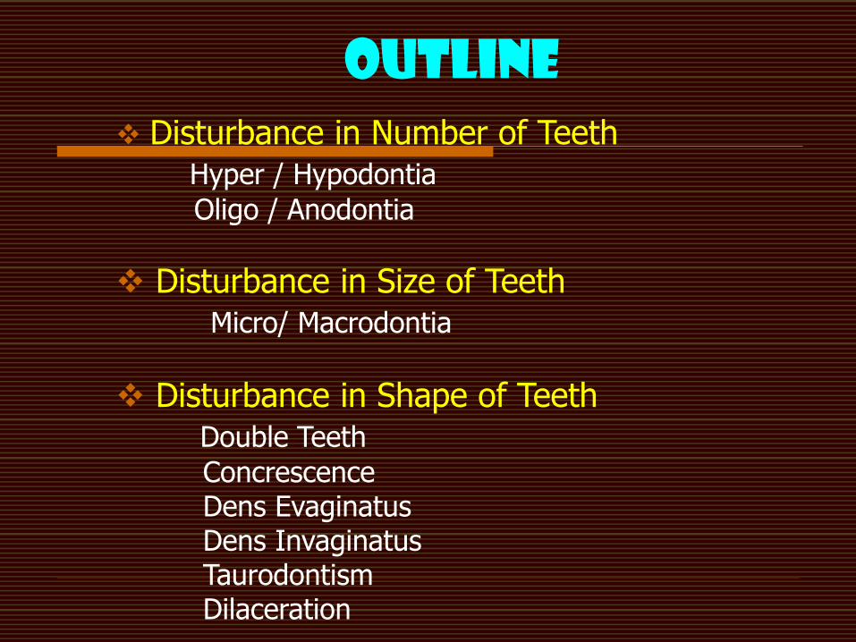
Disturbance in Number of Teeth Hyper / Hypodontia
Oligo / Anodontia
Disturbance in Size of Teeth Micro/ Macrodontia
Disturbance in Shape of Teeth Double Teeth
Concrescence Dens Evaginatus Dens Invaginatus Taurodontism Dilaceration
Outline
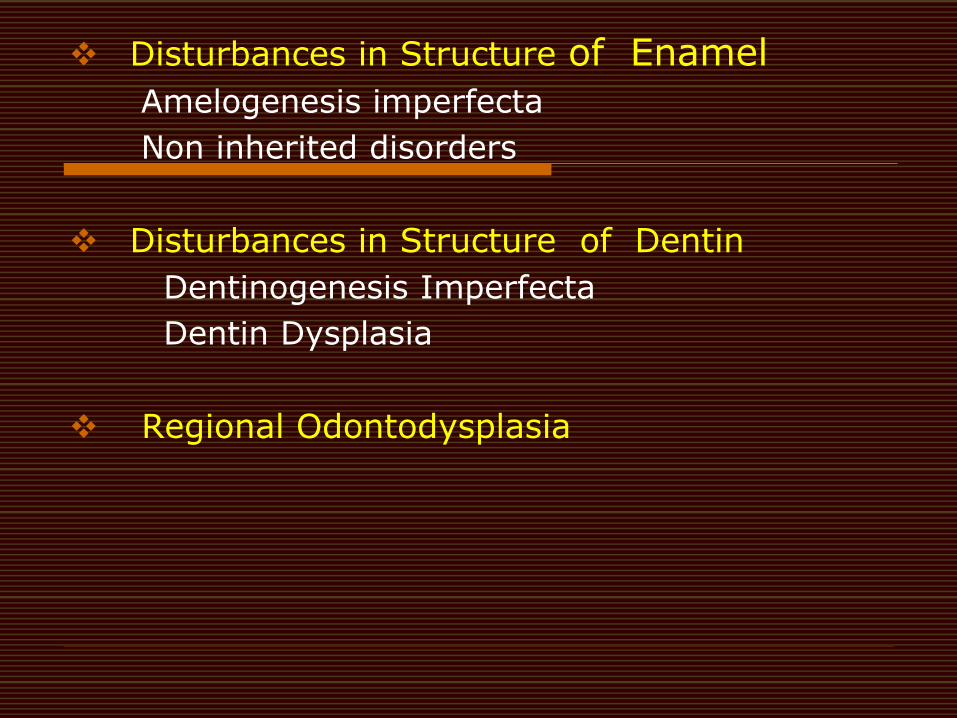
Disturbances in Structure of Enamel
Amelogenesis imperfecta
Non inherited disorders
Disturbances in Structure of Dentin
Dentinogenesis Imperfecta
Dentin Dysplasia
Regional Odontodysplasia
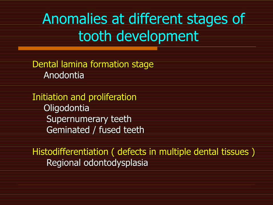
Anomalies at different stages of tooth development
Dental lamina formation stage Anodontia Initiation and proliferation Oligodontia Supernumerary teeth Geminated / fused teeth Histodifferentiation ( defects in multiple dental tissues ) Regional odontodysplasia
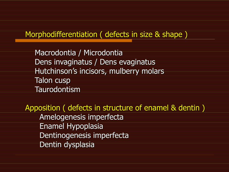
Morphodifferentiation ( defects in size & shape ) Macrodontia / Microdontia Dens invaginatus / Dens evaginatus Hutchinson‟s incisors, mulberry molars Talon cusp Taurodontism Apposition ( defects in structure of enamel & dentin ) Amelogenesis imperfecta Enamel Hypoplasia Dentinogenesis imperfecta Dentin dysplasia
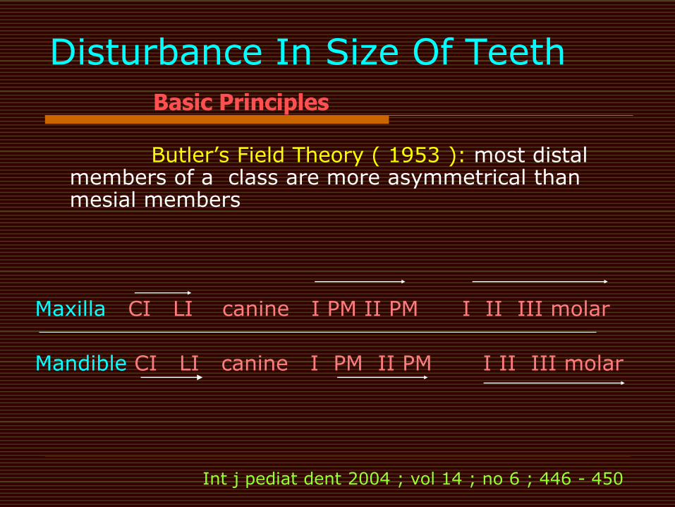
Disturbance In Size Of Teeth
Butler‟s Field Theory ( 1953 ): most distal members of a class are more asymmetrical than mesial members
Maxilla CI LI canine I PM II PM I II III molar
Mandible CI LI canine I PM II PM I II III molar
Basic Principles
Int j pediat dent 2004 ; vol 14 ; no 6 ; 446 - 450
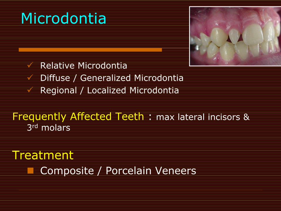
Microdontia
Relative Microdontia
Diffuse / Generalized Microdontia
Regional / Localized Microdontia
Frequently Affected Teeth : max lateral incisors &
3rd molars
Treatment
Composite / Porcelain Veneers
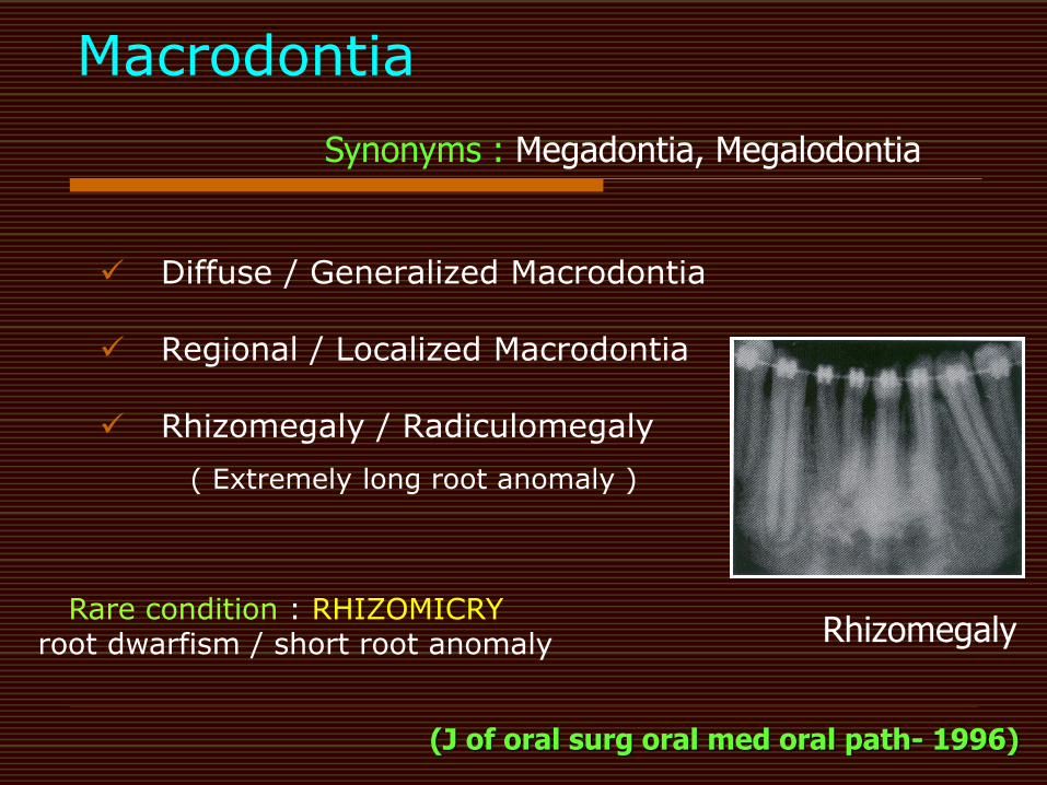
Macrodontia
Diffuse / Generalized Macrodontia
Regional / Localized Macrodontia
Rhizomegaly / Radiculomegaly
( Extremely long root anomaly )
Rhizomegaly
Synonyms : Megadontia, Megalodontia
(J of oral surg oral med oral path- 1996)
Rare condition : RHIZOMICRY root dwarfism / short root anomaly
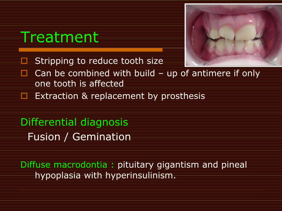
Treatment
Stripping to reduce tooth size
Can be combined with build – up of antimere if only one tooth is affected
Extraction & replacement by prosthesis
Differential diagnosis
Fusion / Gemination
Diffuse macrodontia : pituitary gigantism and pineal
hypoplasia with hyperinsulinism.
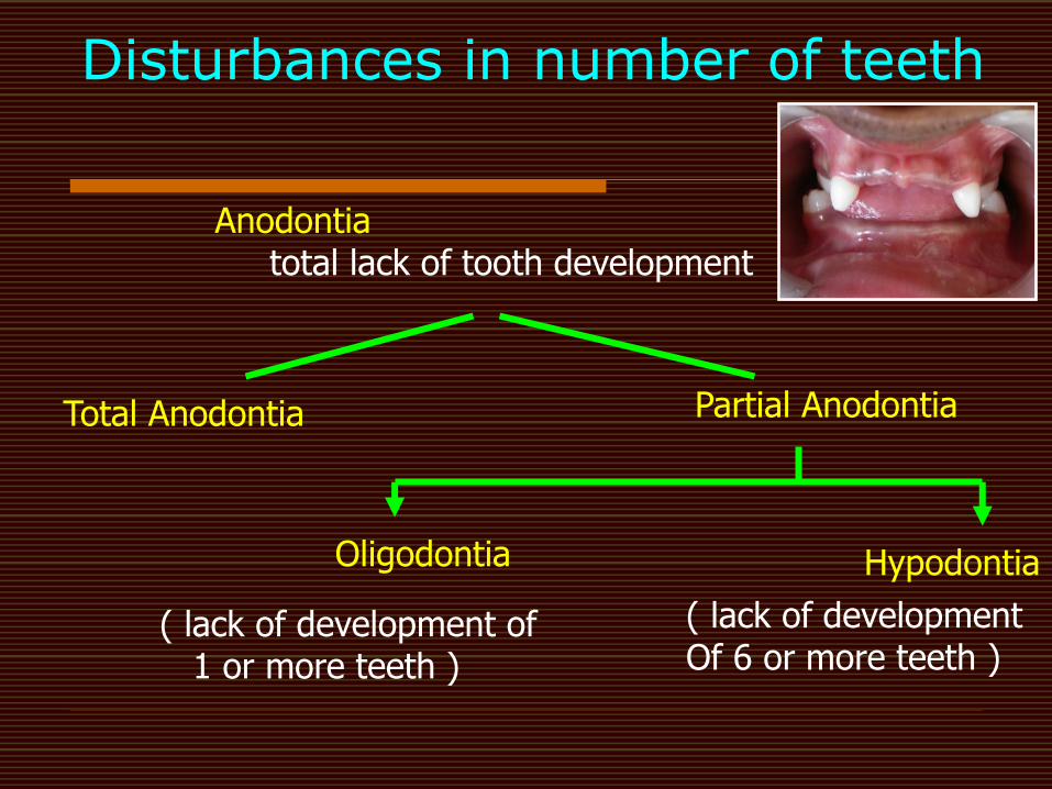
Disturbances in number of teeth
Anodontia total lack of tooth development
Total Anodontia Partial Anodontia
Oligodontia Hypodontia
( lack of development of 1 or more teeth )
( lack of development Of 6 or more teeth )
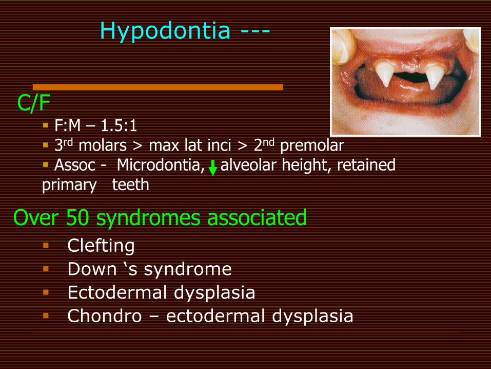
Hypodontia ---
Clefting
Down „s syndrome
Ectodermal dysplasia
Chondro – ectodermal dysplasia
Over 50 syndromes associated
C/F F:M – 1.5:1 3rd molars > max lat inci > 2nd premolar Assoc - Microdontia, alveolar height, retained primary teeth
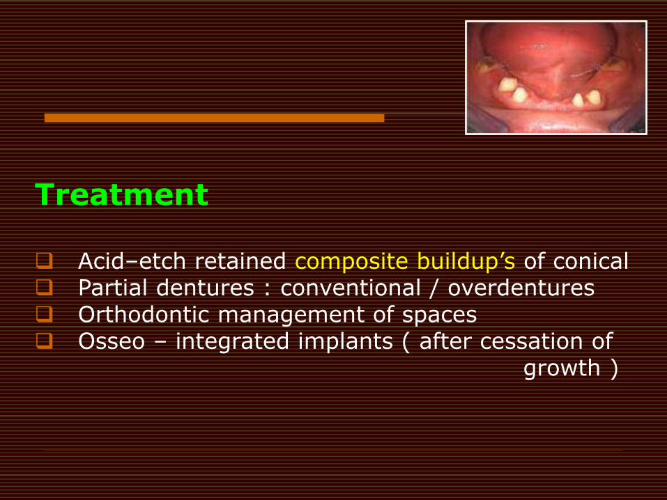
Treatment Acid–etch retained composite buildup‟s of conical Partial dentures : conventional / overdentures Orthodontic management of spaces Osseo – integrated implants ( after cessation of growth )
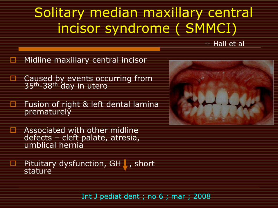
Solitary median maxillary central incisor syndrome ( SMMCI)
Midline maxillary central incisor Caused by events occurring from
35th-38th day in utero Fusion of right & left dental lamina
prematurely Associated with other midline
defects – cleft palate, atresia, umblical hernia
Pituitary dysfunction, GH , short
stature
Int J pediat dent ; no 6 ; mar ; 2008
-- Hall et al
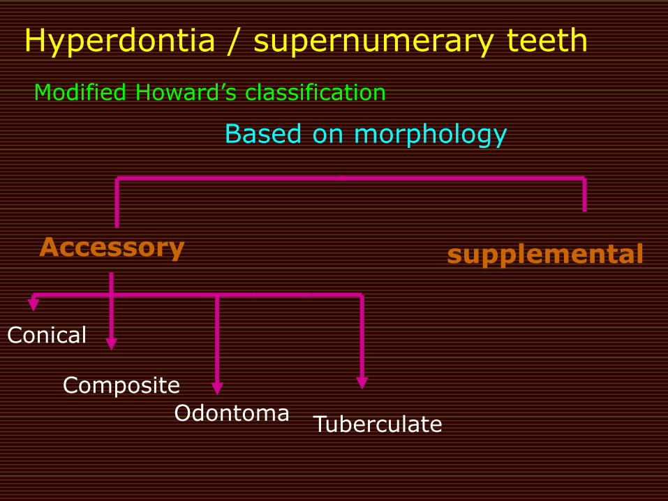
Hyperdontia / supernumerary teeth
Modified Howard‟s classification
Based on morphology
supplemental
Conical
Composite
Odontoma Tuberculate
Accessory
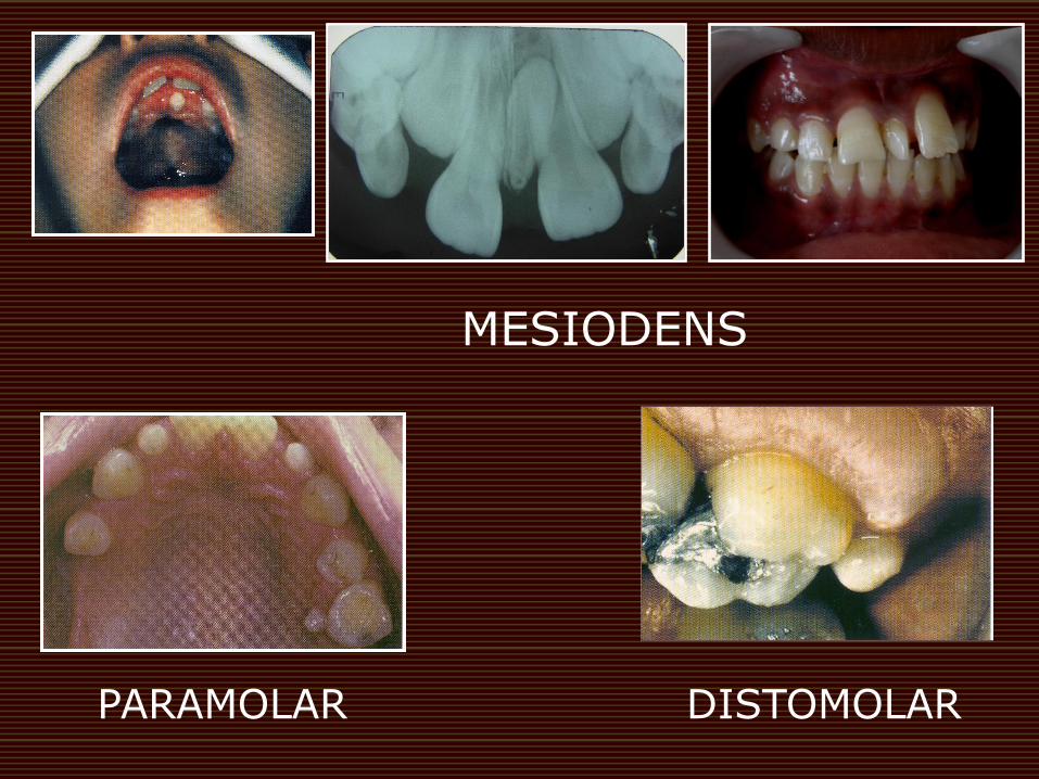
MESIODENS
DISTOMOLAR PARAMOLAR
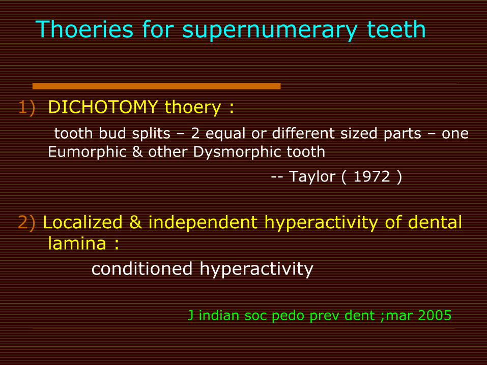
Thoeries for supernumerary teeth
1) DICHOTOMY thoery :
tooth bud splits – 2 equal or different sized parts – one
Eumorphic & other Dysmorphic tooth
-- Taylor ( 1972 )
2) Localized & independent hyperactivity of dental lamina :
conditioned hyperactivity
J indian soc pedo prev dent ;mar 2005
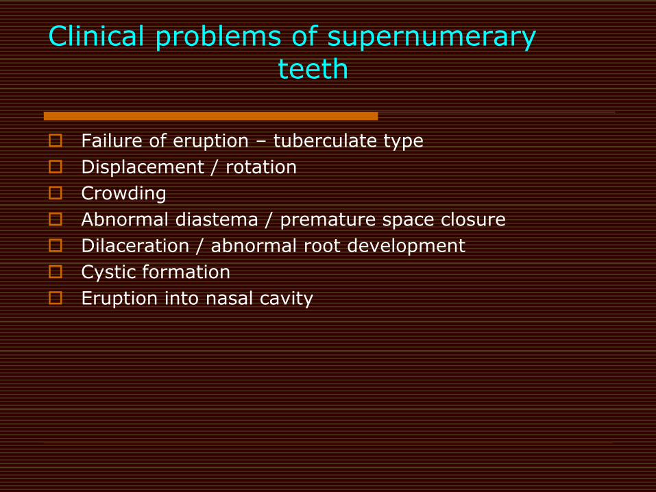
Clinical problems of supernumerary teeth
Failure of eruption – tuberculate type
Displacement / rotation
Crowding
Abnormal diastema / premature space closure
Dilaceration / abnormal root development
Cystic formation
Eruption into nasal cavity
Treatment
Conical often erupt & easily extracted Tuberculate &/ inverted conical teeth require surgical
removal to allow uninhibited eruption of permanent teeth Vertex occlusal radiographs gives clearer view / tube –
shift technique Before 10 yrs : If unerupted central incisor is correctly aligned –
surgical removal of supernumerary tooth After 10 yrs : If central incisor malaligned – surgical exposure
with/without bonding of brackets or chains & subsequent Orthodontic traction may be required
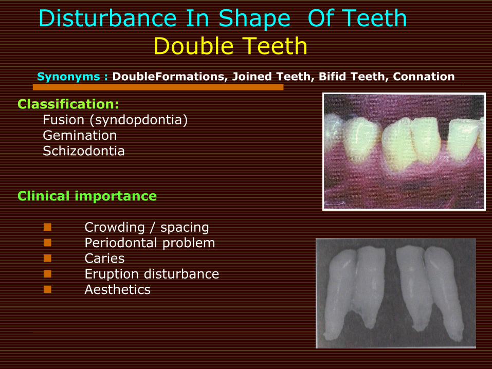
Disturbance In Shape Of Teeth Double Teeth Synonyms : DoubleFormations, Joined Teeth, Bifid Teeth, Connation
Classification: Fusion (syndopdontia) Gemination Schizodontia
Clinical importance
Crowding / spacing Periodontal problem Caries Eruption disturbance Aesthetics
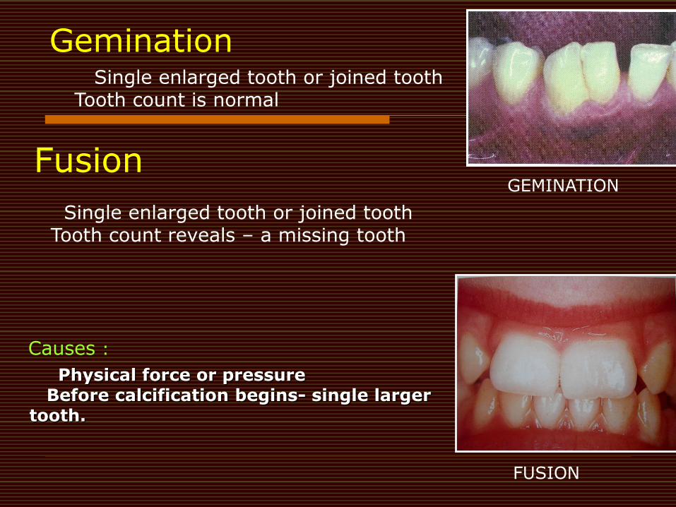
Gemination Single enlarged tooth or joined tooth Tooth count is normal
Fusion
Single enlarged tooth or joined tooth Tooth count reveals – a missing tooth
FUSION
GEMINATION
Physical force or pressure Before calcification begins- single larger tooth.
Causes :
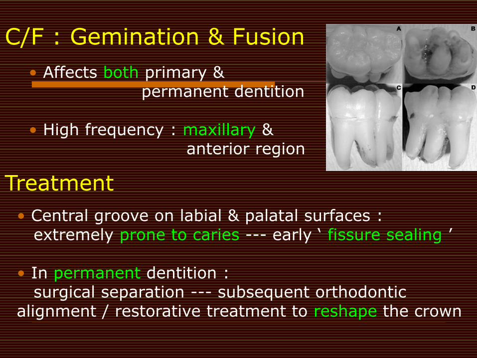
C/F : Gemination & Fusion
• Affects both primary & permanent dentition • High frequency : maxillary & anterior region
Treatment Fusion
• Central groove on labial & palatal surfaces : extremely prone to caries --- early „ fissure sealing ‟ • In permanent dentition : surgical separation --- subsequent orthodontic alignment / restorative treatment to reshape the crown
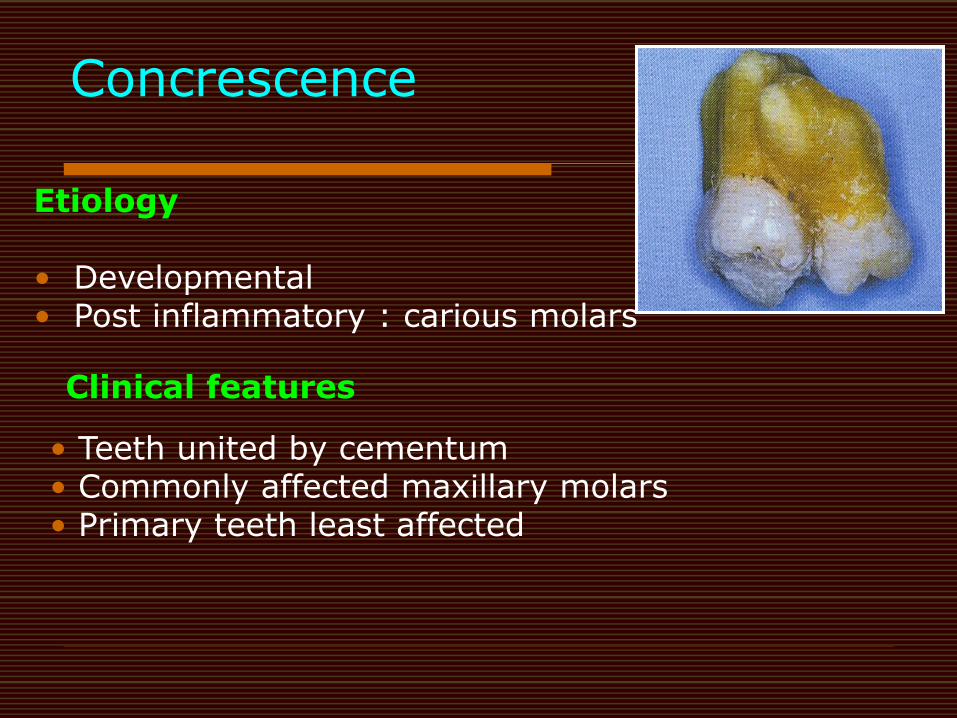
Concrescence
Etiology • Developmental • Post inflammatory : carious molars
Clinical features
• Teeth united by cementum • Commonly affected maxillary molars • Primary teeth least affected
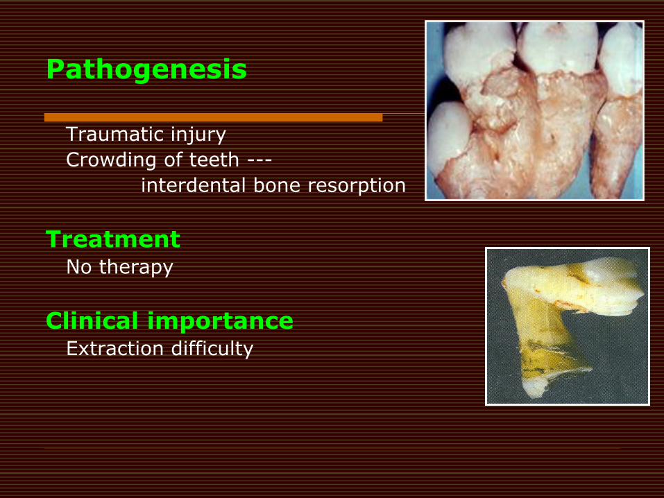
Pathogenesis
Traumatic injury
Crowding of teeth ---
interdental bone resorption
Treatment No therapy
Clinical importance Extraction difficulty
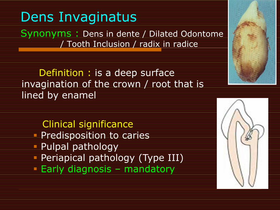
Dens Invaginatus Synonyms : Dens in dente / Dilated Odontome
/ Tooth Inclusion / radix in radice
Definition : is a deep surface invagination of the crown / root that is lined by enamel
Clinical significance Predisposition to caries Pulpal pathology Periapical pathology (Type III) Early diagnosis – mandatory
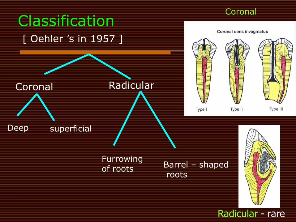
Radicular - rare
Coronal
Classification [ Oehler ‟s in 1957 ]
Coronal Radicular
superficial Deep
Furrowing of roots
Barrel – shaped roots
Treatment
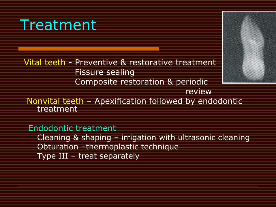
Vital teeth - Preventive & restorative treatment Fissure sealing Composite restoration & periodic review Nonvital teeth – Apexification followed by endodontic
treatment
Endodontic treatment Cleaning & shaping – irrigation with ultrasonic cleaning Obturation –thermoplastic technique Type III – treat separately
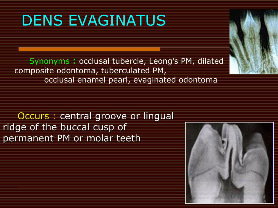
DENS EVAGINATUS
Occurs : central groove or lingual ridge of the buccal cusp of permanent PM or molar teeth
Synonyms : occlusal tubercle, Leong‟s PM, dilated
composite odontoma, tuberculated PM, occlusal enamel pearl, evaginated odontoma
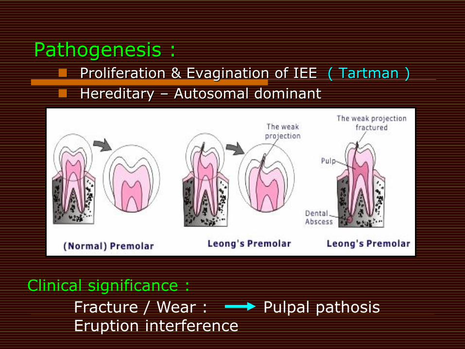
Pathogenesis : Proliferation & Evagination of IEE ( Tartman )
Hereditary – Autosomal dominant
Clinical significance :
Fracture / Wear : Pulpal pathosis Eruption interference
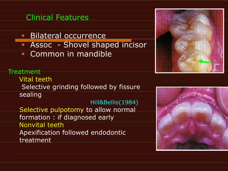
Clinical Features Bilateral occurrence Assoc - Shovel shaped incisor Common in mandible
Treatment Vital teeth
Selective grinding followed by fissure sealing Hill&Bellis(1984)
Selective pulpotomy to allow normal formation : if diagnosed early
Nonvital teeth Apexification followed endodontic treatment
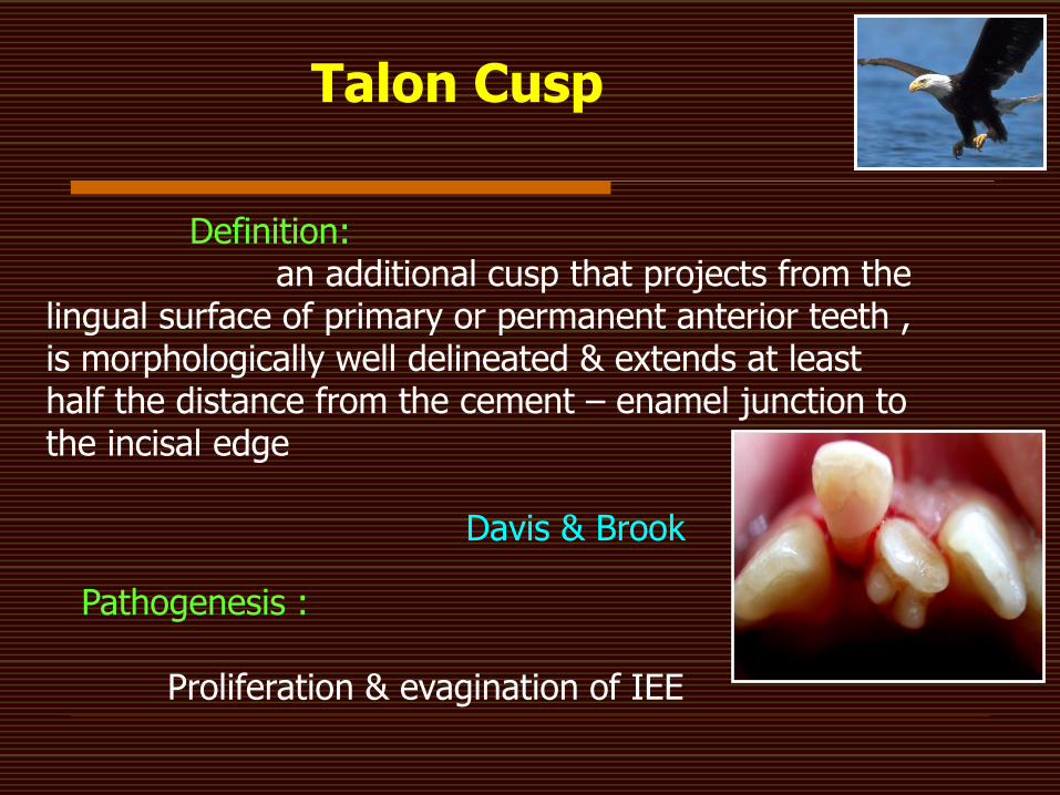
Talon Cusp
Definition: an additional cusp that projects from the lingual surface of primary or permanent anterior teeth , is morphologically well delineated & extends at least half the distance from the cement – enamel junction to the incisal edge Davis & Brook
Pathogenesis :
Proliferation & evagination of IEE
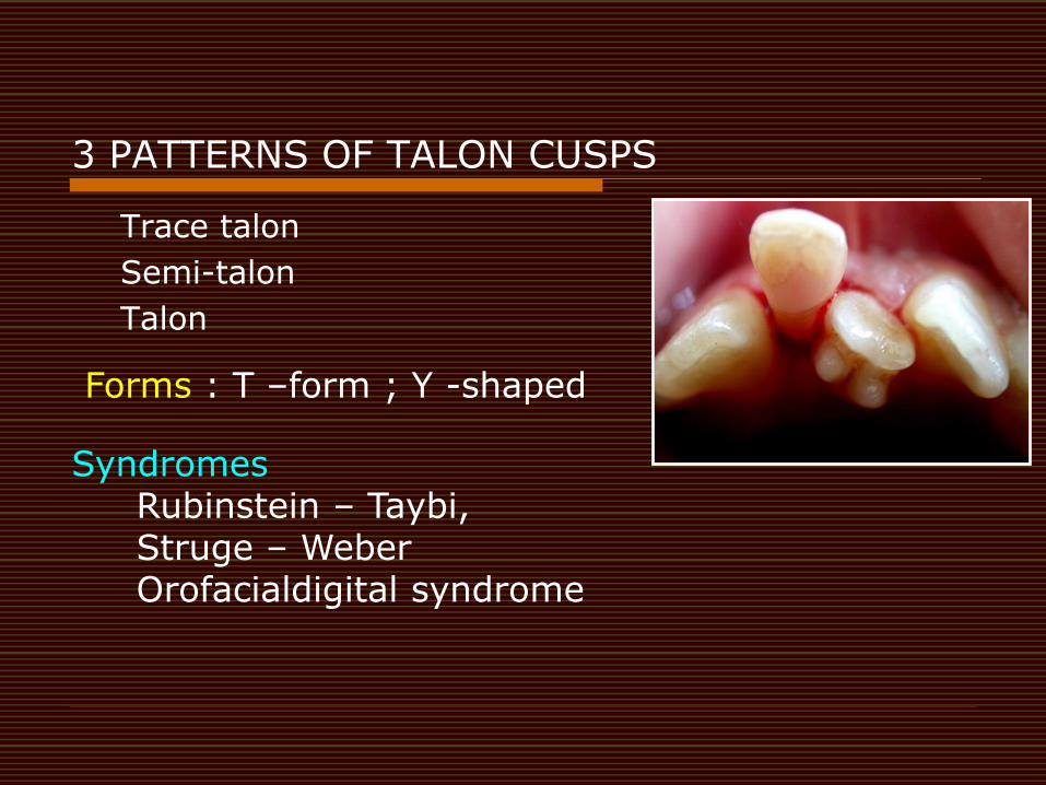
3 PATTERNS OF TALON CUSPS Trace talon
Semi-talon
Talon
Forms : T –form ; Y -shaped
Syndromes Rubinstein – Taybi, Struge – Weber Orofacialdigital syndrome
.
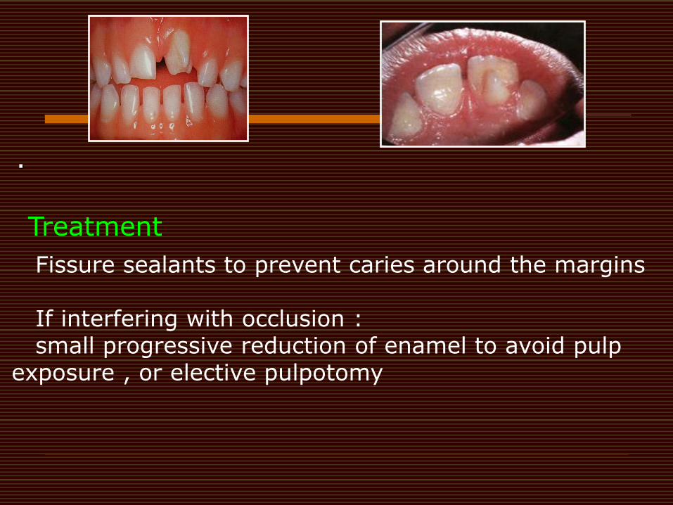
Treatment
Fissure sealants to prevent caries around the margins If interfering with occlusion : small progressive reduction of enamel to avoid pulp exposure , or elective pulpotomy
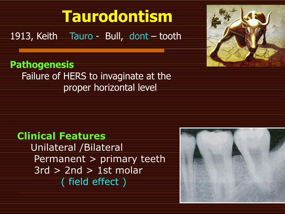
Taurodontism 1913, Keith Tauro - Bull, dont – tooth
Pathogenesis Failure of HERS to invaginate at the proper horizontal level
Clinical Features Unilateral /Bilateral Permanent > primary teeth 3rd > 2nd > 1st molar ( field effect )
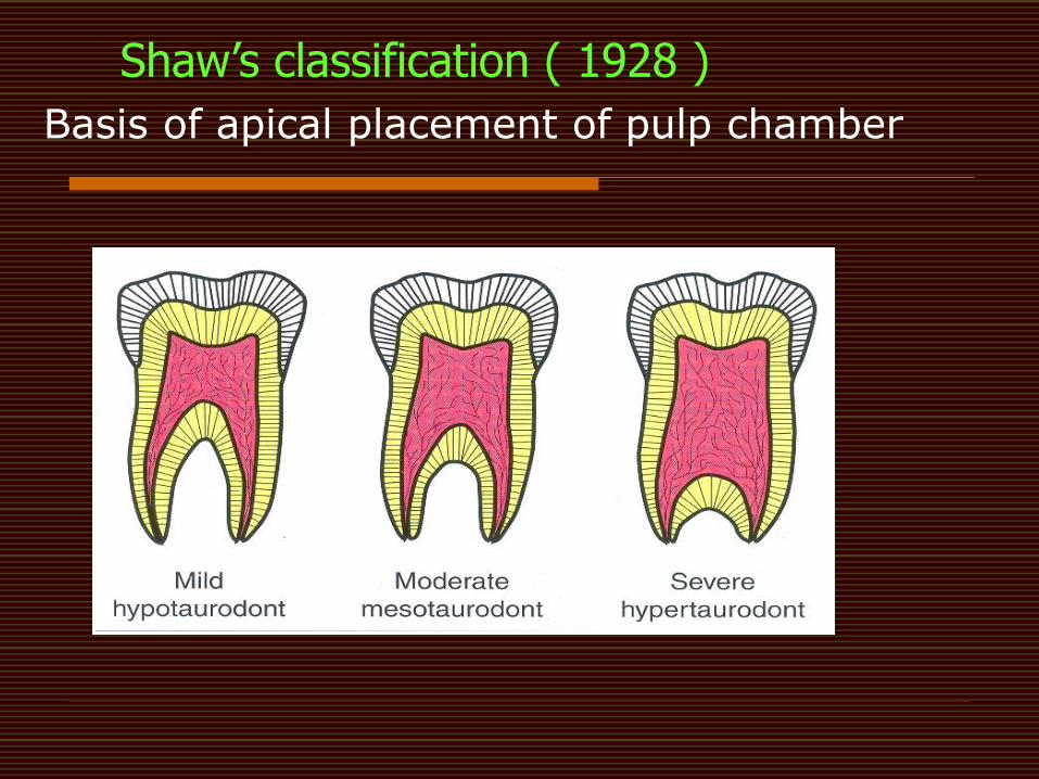
Shaw‟s classification ( 1928 )
Basis of apical placement of pulp chamber
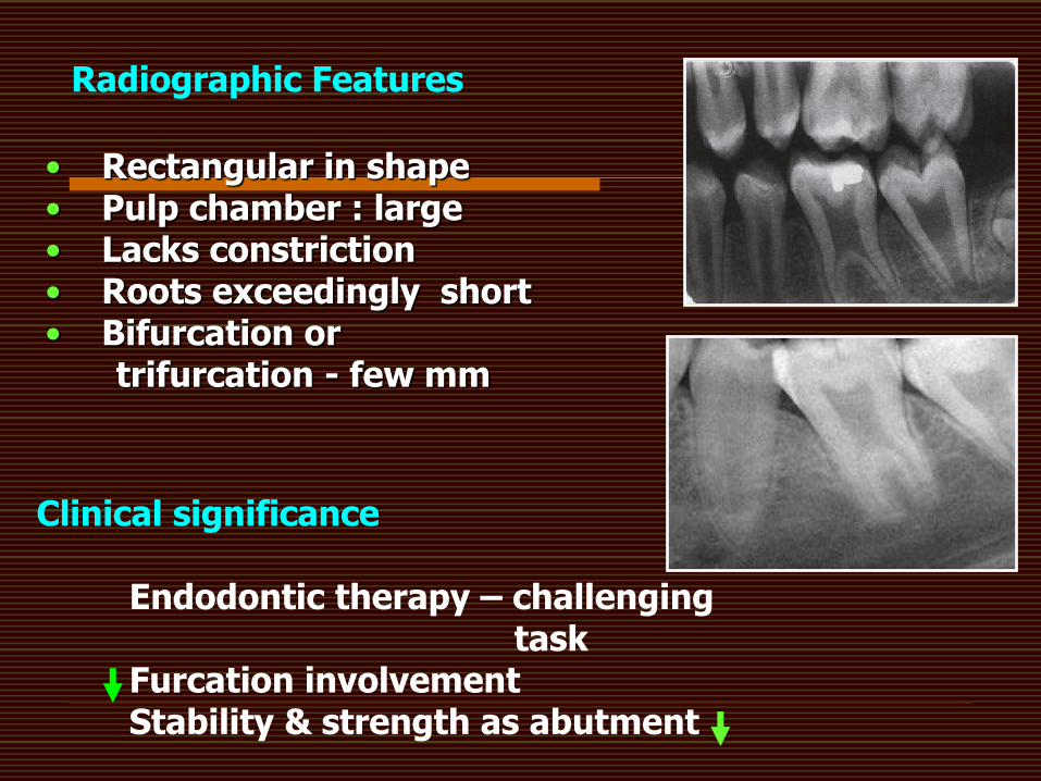
Radiographic Features
• Rectangular in shape • Pulp chamber : large • Lacks constriction • Roots exceedingly short • Bifurcation or trifurcation - few mm
Clinical significance
Endodontic therapy – challenging task Furcation involvement Stability & strength as abutment
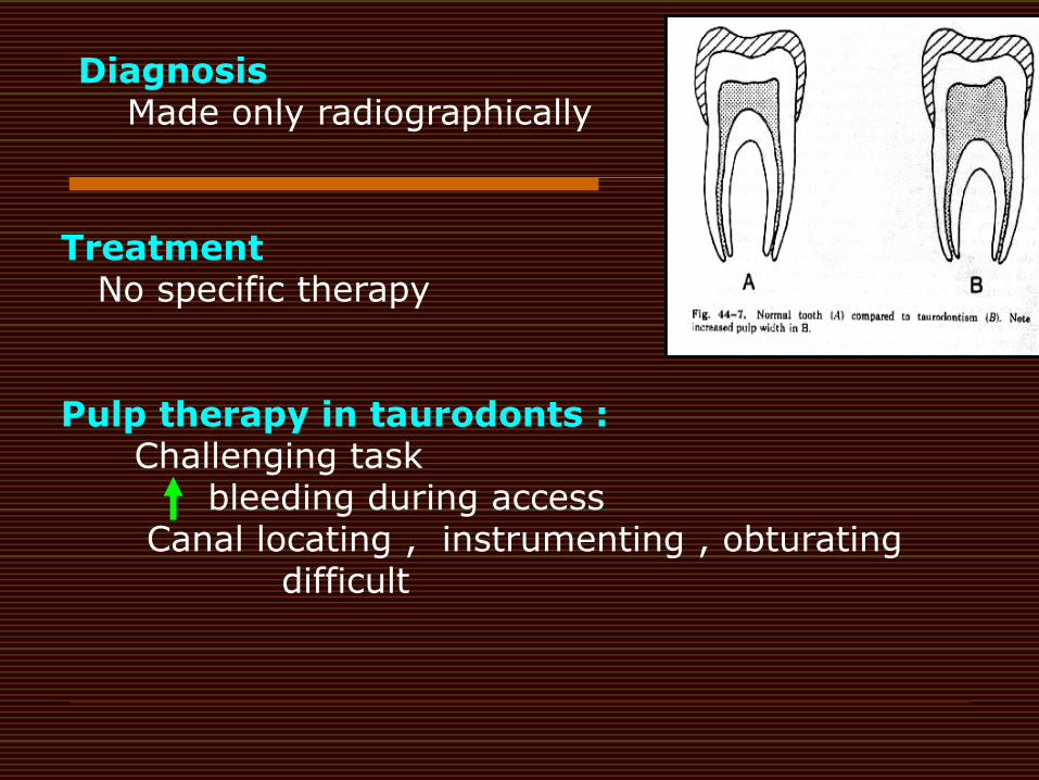
Diagnosis Made only radiographically
Treatment No specific therapy Pulp therapy in taurodonts : Challenging task bleeding during access Canal locating , instrumenting , obturating difficult
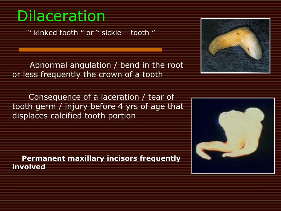
Dilaceration
Abnormal angulation / bend in the root or less frequently the crown of a tooth
Consequence of a laceration / tear of tooth germ / injury before 4 yrs of age that displaces calcified tooth portion
“ kinked tooth ” or “ sickle – tooth ”
Permanent maxillary incisors frequently involved
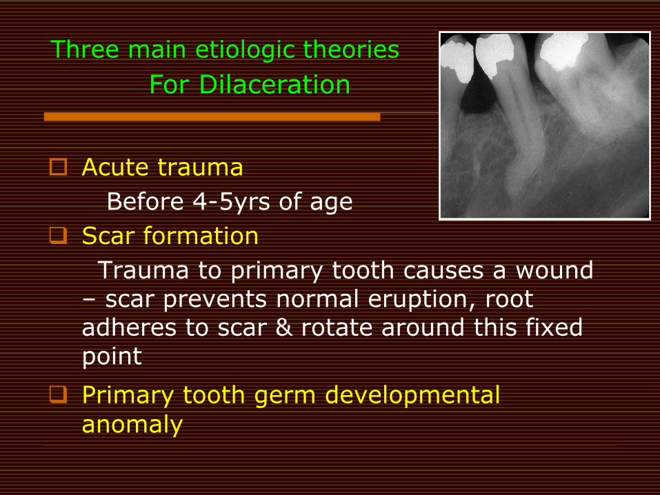
Three main etiologic theories
Acute trauma
Before 4-5yrs of age
Scar formation
Trauma to primary tooth causes a wound – scar prevents normal eruption, root adheres to scar & rotate around this fixed point
Primary tooth germ developmental anomaly
For Dilaceration
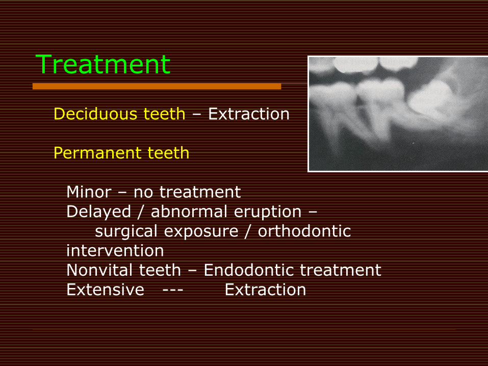
Treatment Deciduous teeth – Extraction Permanent teeth
Minor – no treatment Delayed / abnormal eruption – surgical exposure / orthodontic intervention Nonvital teeth – Endodontic treatment Extensive --- Extraction
Developmental Alteration In The Structure Of Teeth
Hereditary type
Amelogenesis Imperfecta
Inherited systemic conditions with
enamel defects
Acquired Enamel Defect
Non – inherited enamel defects
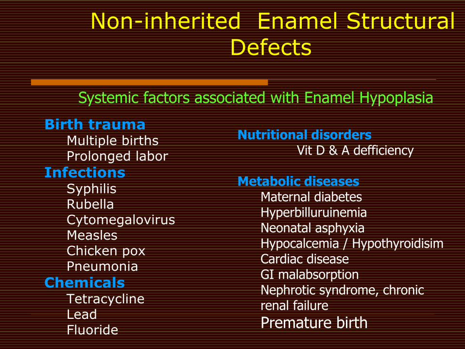
Non-inherited Enamel Structural Defects
Birth trauma Multiple births Prolonged labor
Infections Syphilis Rubella Cytomegalovirus Measles Chicken pox Pneumonia
Chemicals Tetracycline Lead Fluoride
Nutritional disorders Vit D & A defficiency
Metabolic diseases Maternal diabetes Hyperbilluruinemia Neonatal asphyxia Hypocalcemia / Hypothyroidisim Cardiac disease GI malabsorption Nephrotic syndrome, chronic renal failure
Premature birth
Systemic factors associated with Enamel Hypoplasia
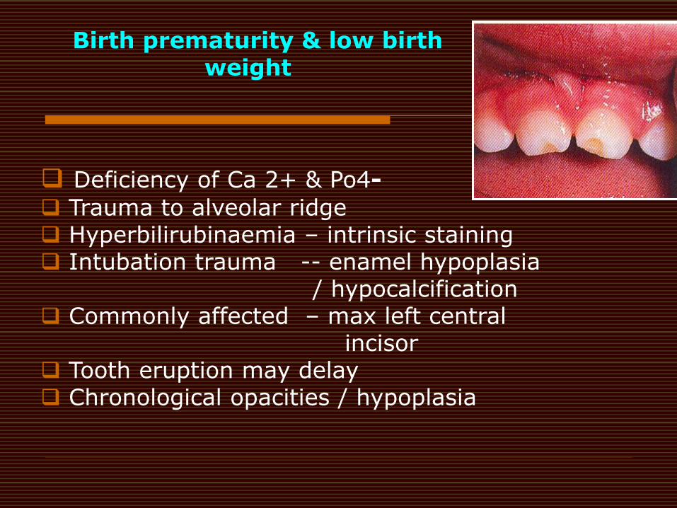
Birth prematurity & low birth weight
Deficiency of Ca 2+ & Po4-
Trauma to alveolar ridge Hyperbilirubinaemia – intrinsic staining Intubation trauma -- enamel hypoplasia / hypocalcification Commonly affected – max left central incisor Tooth eruption may delay Chronological opacities / hypoplasia
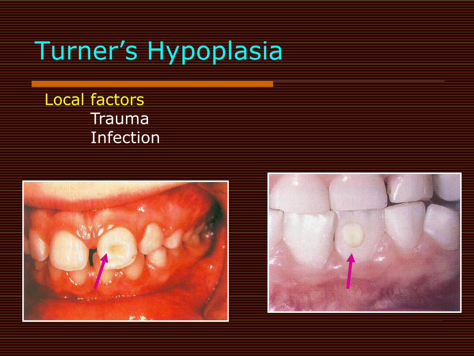
Turner‟s Hypoplasia
Local factors Trauma Infection
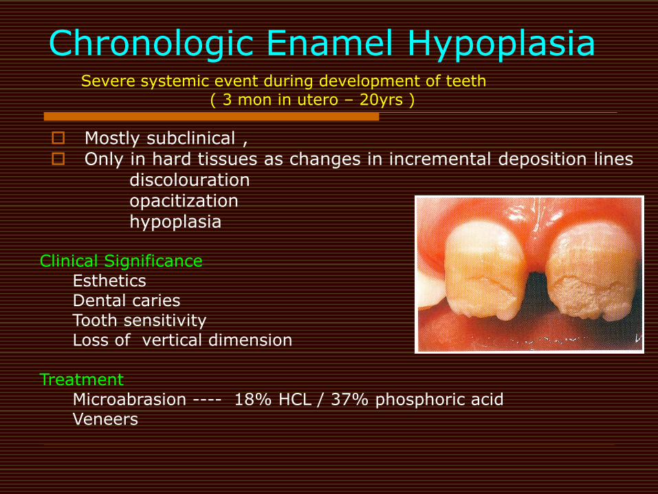
Chronologic Enamel Hypoplasia Severe systemic event during development of teeth ( 3 mon in utero – 20yrs )
Mostly subclinical , Only in hard tissues as changes in incremental deposition lines discolouration opacitization hypoplasia
Clinical Significance Esthetics Dental caries Tooth sensitivity Loss of vertical dimension
Treatment Microabrasion ---- 18% HCL / 37% phosphoric acid Veneers

Flourosis
Pathogenesis
Presecretory stage – no effect at physiologically relevant Fl dose
Secretory stage – inhibit protien synthesis at high
dose
Maturation stage – delayed withdrawl of amelogenin, serine protein-ases activity
-- Enamel matrix protein tightly bound to flourapatite
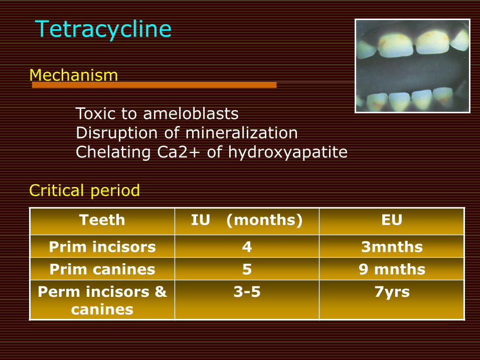
Tetracycline
Teeth IU (months) EU
Prim incisors 4 3mnths
Prim canines 5 9 mnths
Perm incisors & canines
3-5 7yrs
Mechanism
Toxic to ameloblasts Disruption of mineralization Chelating Ca2+ of hydroxyapatite
Critical period
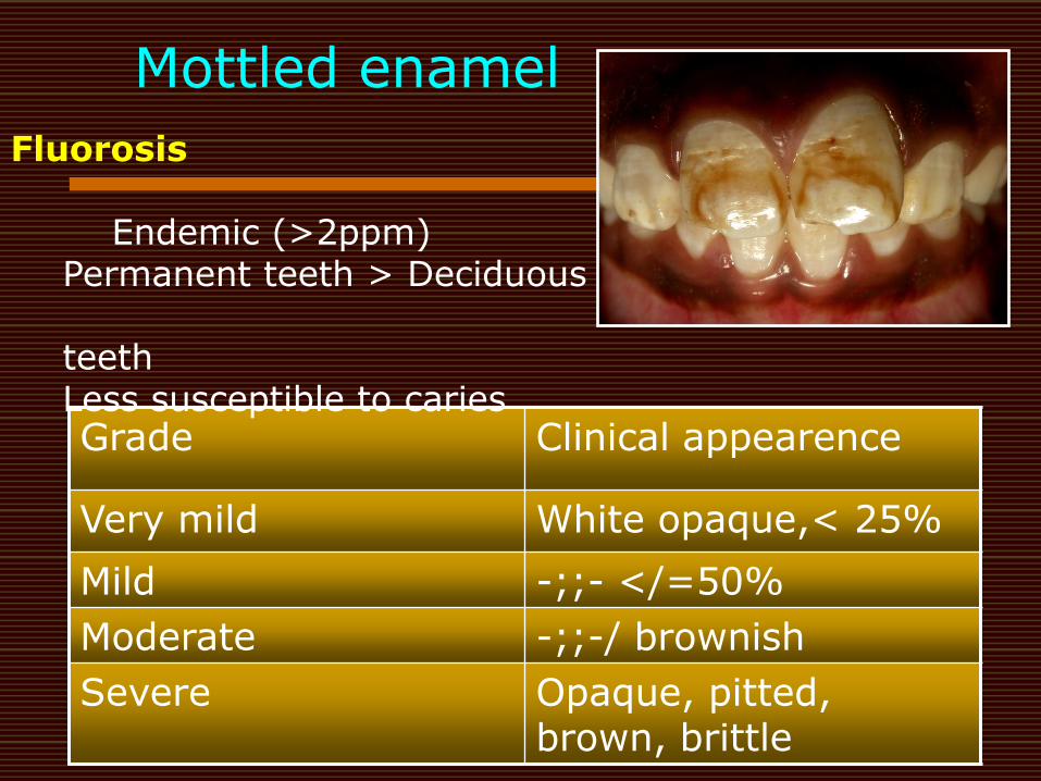
Mottled enamel
Grade Clinical appearence
Very mild White opaque,< 25%
Mild -;;- </=50%
Moderate -;;-/ brownish
Severe Opaque, pitted, brown, brittle
Fluorosis
Endemic (>2ppm) Permanent teeth > Deciduous teeth Less susceptible to caries
Amelogenesis Imperfecta
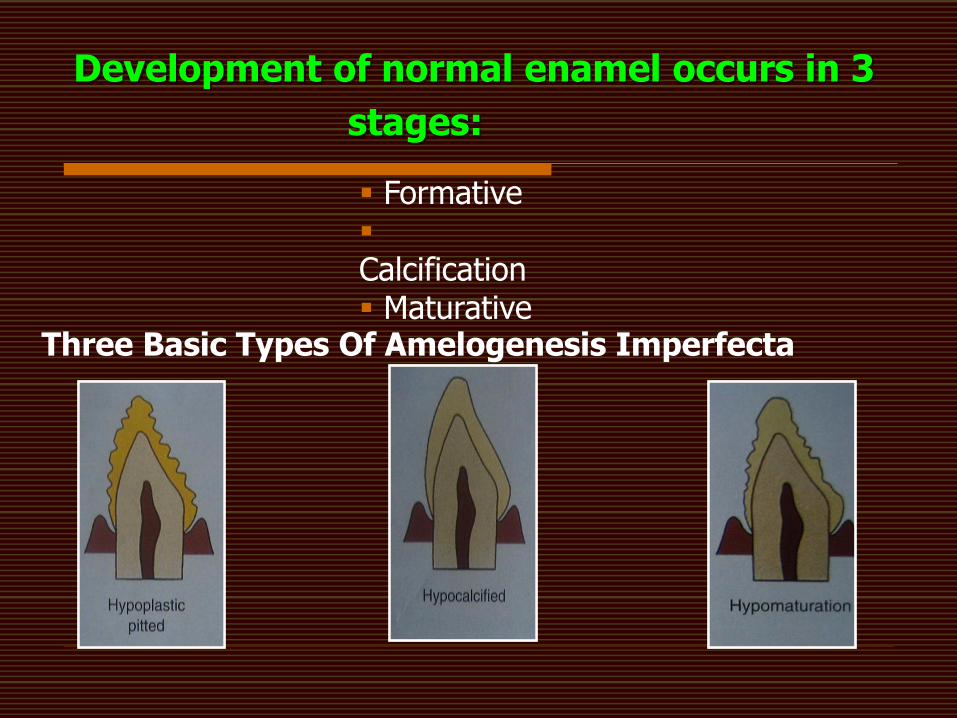
Three Basic Types Of Amelogenesis Imperfecta
Development of normal enamel occurs in 3
stages:
Formative Calcification Maturative
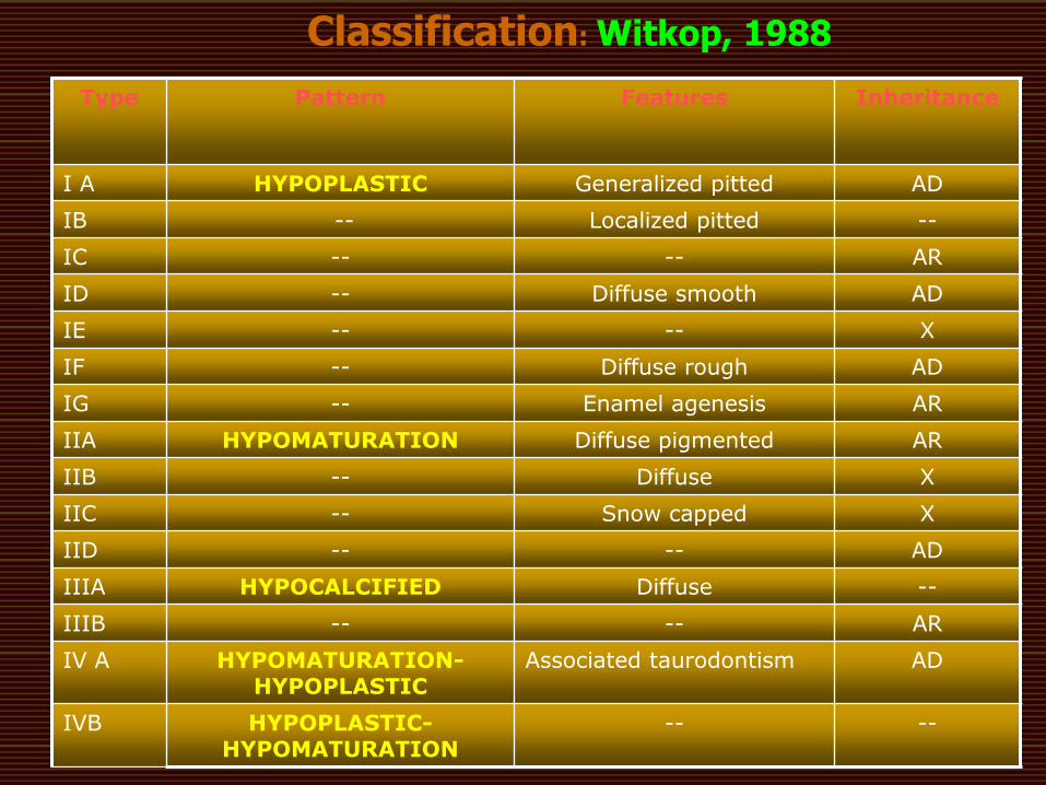
Type Pattern Features Inheritance
I A HYPOPLASTIC Generalized pitted AD
IB -- Localized pitted --
IC -- -- AR
ID -- Diffuse smooth AD
IE -- -- X
IF -- Diffuse rough AD
IG -- Enamel agenesis AR
IIA HYPOMATURATION Diffuse pigmented AR
IIB -- Diffuse X
IIC -- Snow capped X
IID -- -- AD
IIIA HYPOCALCIFIED Diffuse --
IIIB -- -- AR
IV A HYPOMATURATION-HYPOPLASTIC
Associated taurodontism AD
IVB HYPOPLASTIC-HYPOMATURATION
-- --
Classification: Witkop, 1988
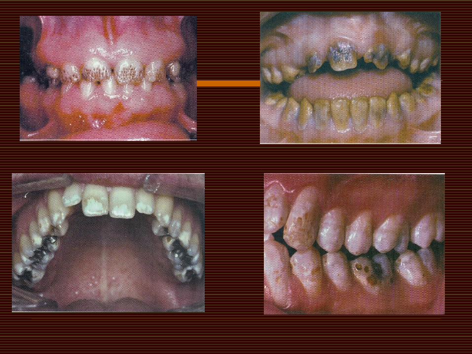
Clinical significance
• Aesthetics
• Vertical dimension
• Caries
Treatment
• Appropriate diagnosis
• Genetic counseling & continued commitment to support both parent & child
• Early orthodontic assessment
• Preservation of VD by full coverage stainless steel crowns
• Aesthetics by composite resin veneers for anterior
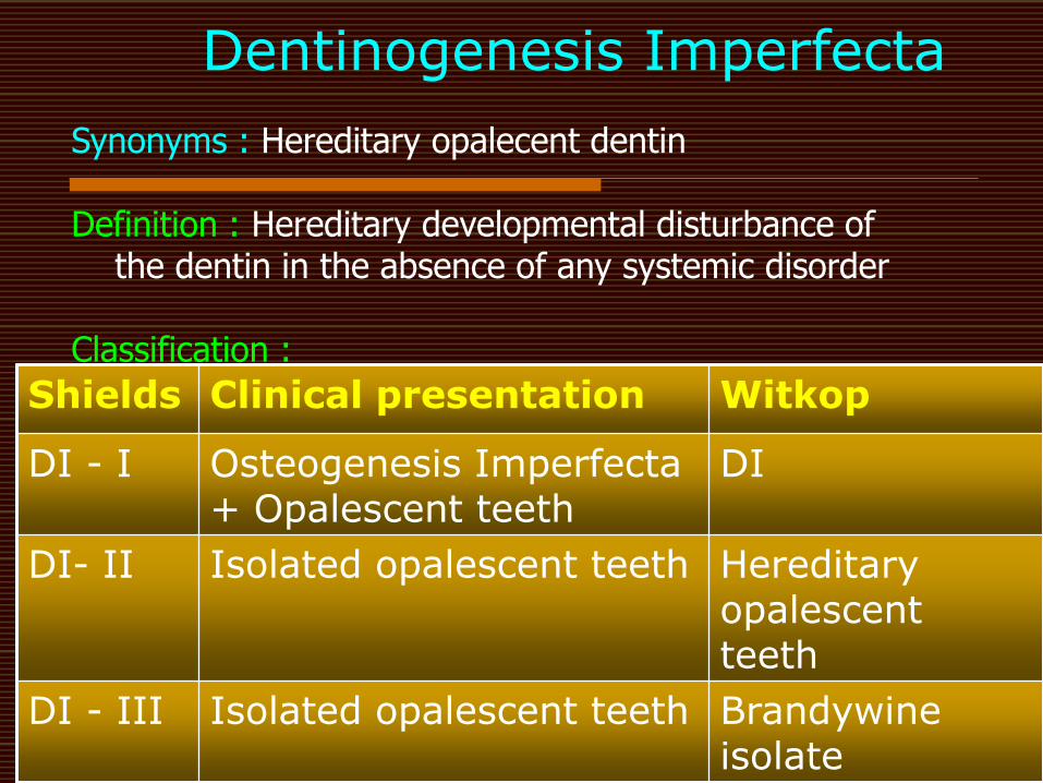
Dentinogenesis Imperfecta
Shields Clinical presentation Witkop
DI - I Osteogenesis Imperfecta + Opalescent teeth
DI
DI- II Isolated opalescent teeth Hereditary opalescent teeth
DI - III Isolated opalescent teeth Brandywine isolate
Synonyms : Hereditary opalecent dentin Definition : Hereditary developmental disturbance of the dentin in the absence of any systemic disorder Classification :
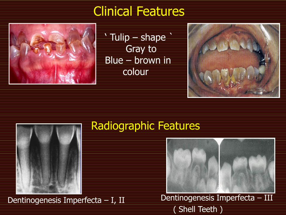
Dentinogenesis Imperfecta – I, II
Dentinogenesis Imperfecta – III
( Shell Teeth )
Radiographic Features
Clinical Features
„ Tulip – shape ` Gray to Blue – brown in colour
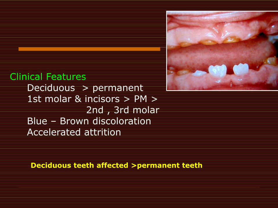
Deciduous teeth affected >permanent teeth
Clinical Features Deciduous > permanent 1st molar & incisors > PM > 2nd , 3rd molar Blue – Brown discoloration Accelerated attrition
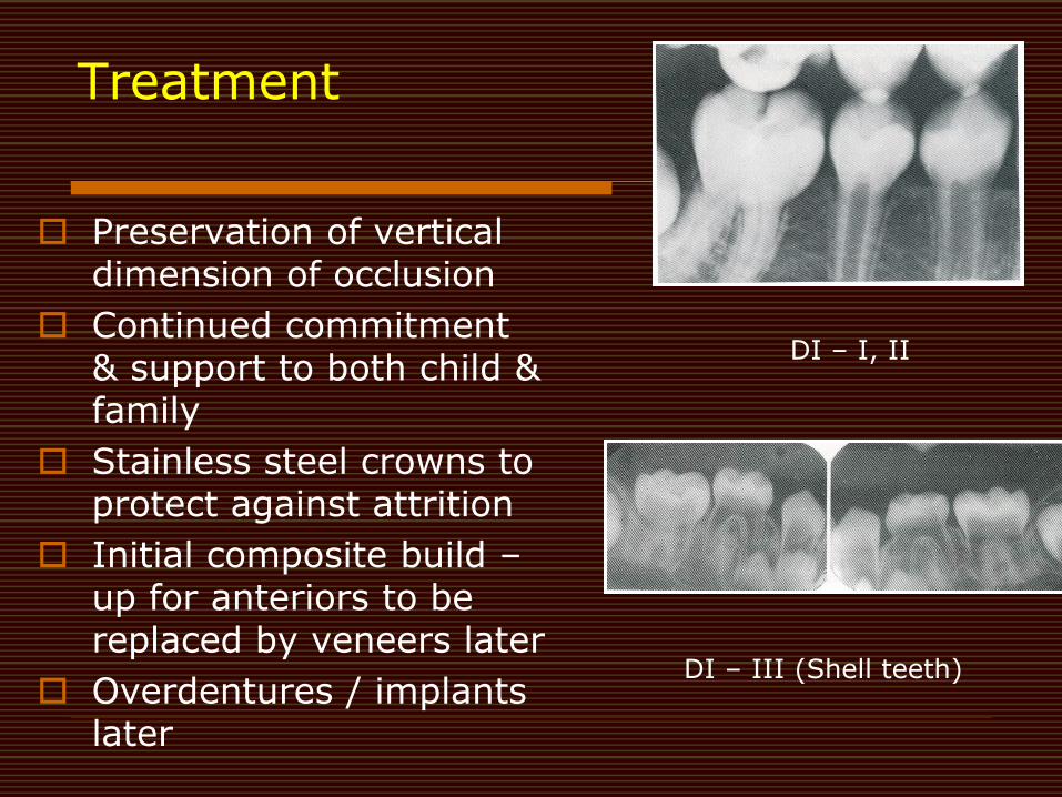
Treatment
Preservation of vertical dimension of occlusion
Continued commitment & support to both child & family
Stainless steel crowns to protect against attrition
Initial composite build – up for anteriors to be replaced by veneers later
Overdentures / implants later
DI – I, II
DI – III (Shell teeth)
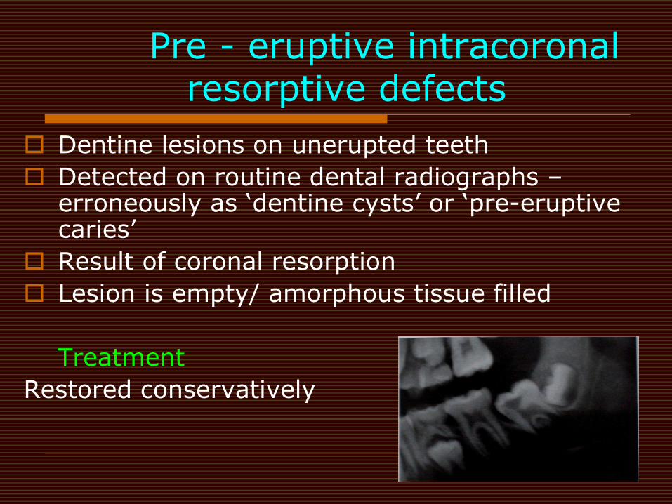
Pre - eruptive intracoronal resorptive defects
Dentine lesions on unerupted teeth
Detected on routine dental radiographs – erroneously as „dentine cysts‟ or „pre-eruptive caries‟
Result of coronal resorption
Lesion is empty/ amorphous tissue filled
Treatment
Restored conservatively
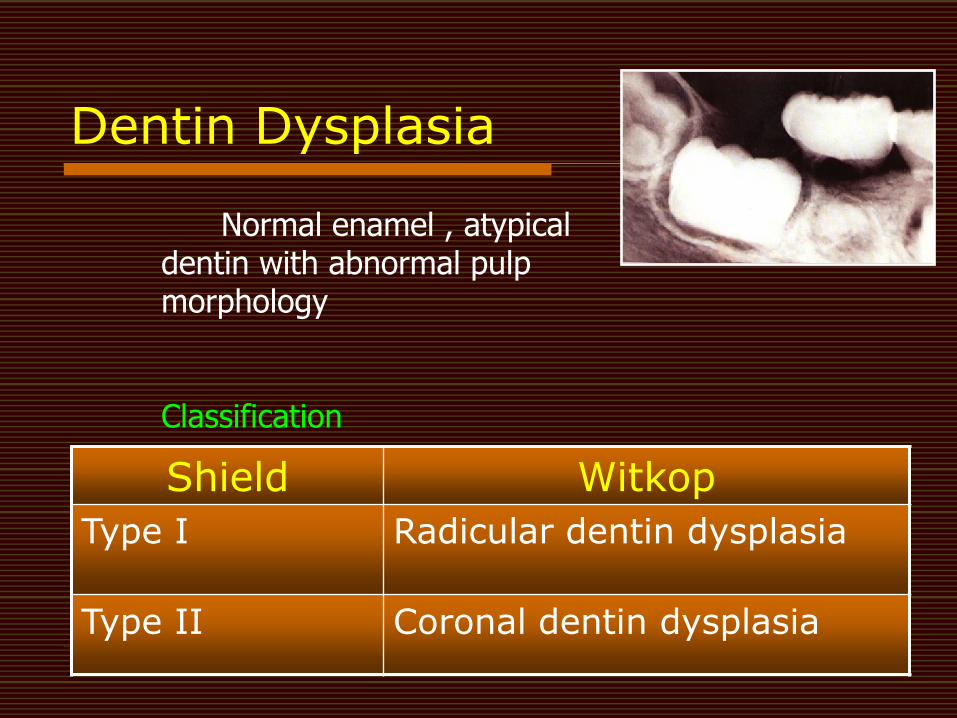
Dentin Dysplasia
Shield Witkop
Type I Radicular dentin dysplasia
Type II Coronal dentin dysplasia
Normal enamel , atypical dentin with abnormal pulp morphology Classification
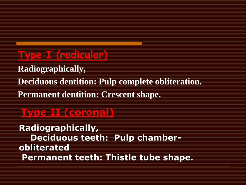
Type I (radicular) Radiographically,
Deciduous dentition: Pulp complete obliteration.
Permanent dentition: Crescent shape.
Radiographically, Deciduous teeth: Pulp chamber- obliterated Permanent teeth: Thistle tube shape.
Type II (coronal)
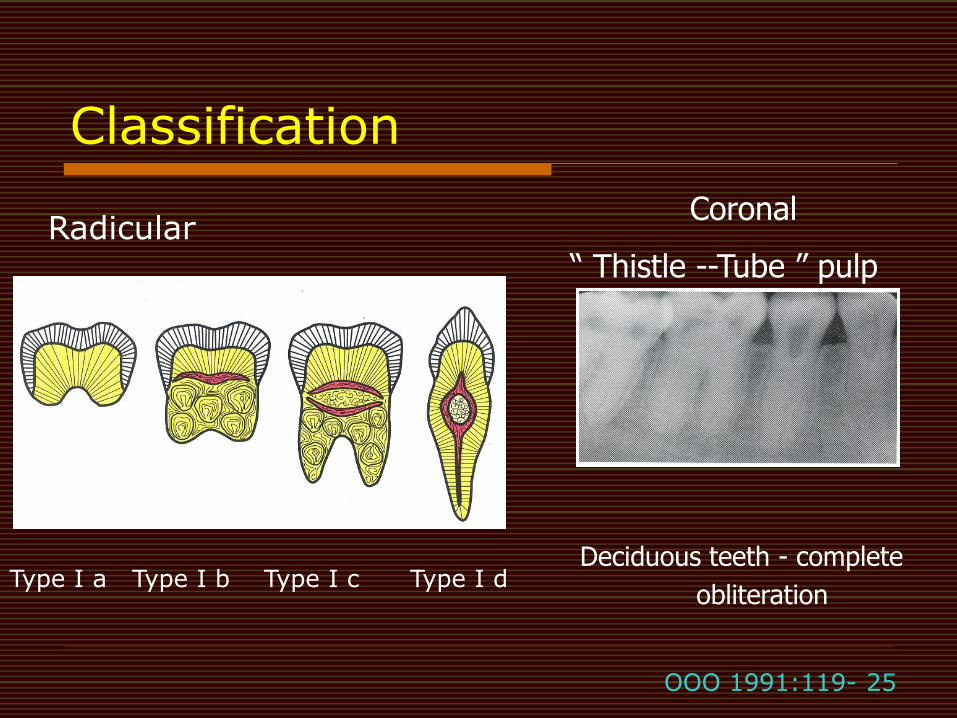
Classification
Coronal
“ Thistle --Tube ” pulp
Deciduous teeth - complete
obliteration Type I a Type I b Type I c Type I d
Radicular
OOO 1991:119- 25
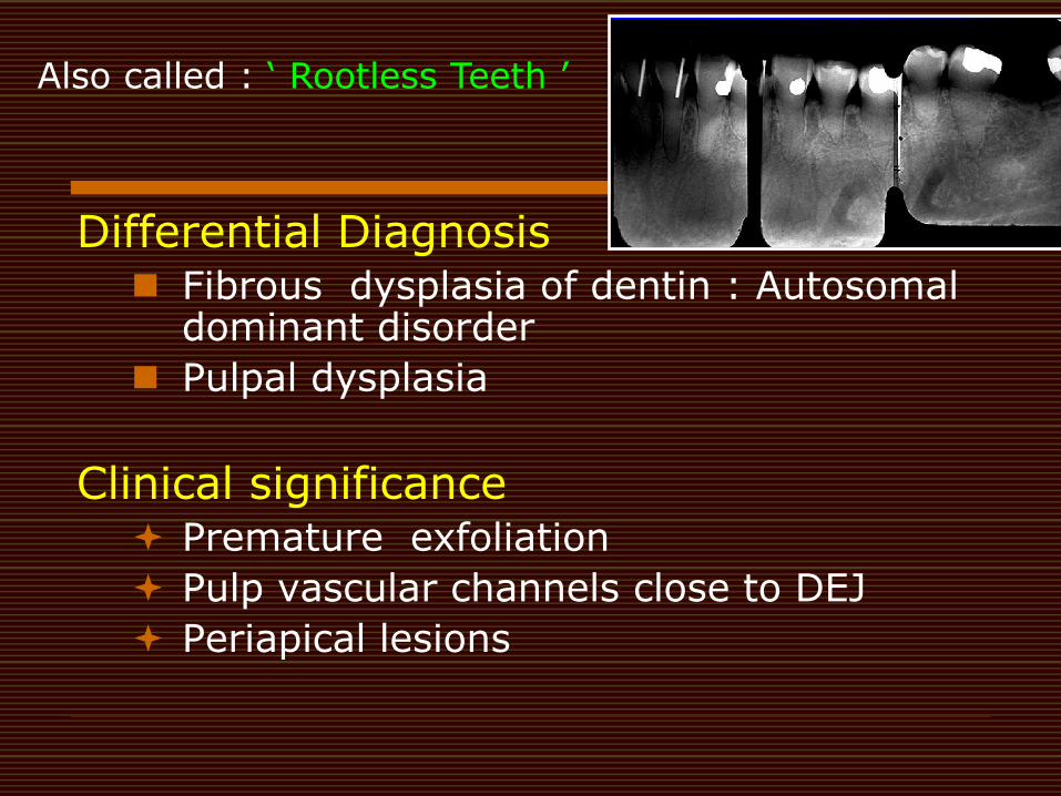
Differential Diagnosis Fibrous dysplasia of dentin : Autosomal
dominant disorder
Pulpal dysplasia
Clinical significance Premature exfoliation
Pulp vascular channels close to DEJ
Periapical lesions
Also called : „ Rootless Teeth ‟
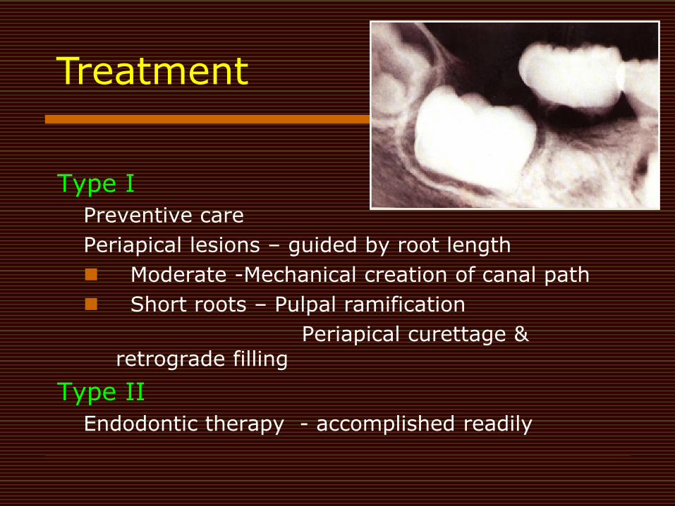
Type I
Preventive care
Periapical lesions – guided by root length
Moderate -Mechanical creation of canal path
Short roots – Pulpal ramification
Periapical curettage & retrograde filling
Type II
Endodontic therapy - accomplished readily
Treatment
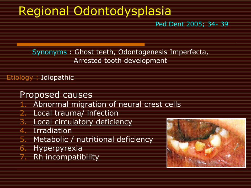
Regional Odontodysplasia
Synonyms : Ghost teeth, Odontogenesis Imperfecta,
Arrested tooth development
Etiology : Idiopathic
Proposed causes 1. Abnormal migration of neural crest cells 2. Local trauma/ infection 3. Local circulatory deficiency 4. Irradiation 5. Metabolic / nutritional deficiency 6. Hyperpyrexia 7. Rh incompatibility
Ped Dent 2005; 34- 39
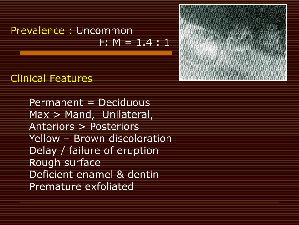
Prevalence : Uncommon F: M = 1.4 : 1 Clinical Features
Permanent = Deciduous Max > Mand, Unilateral, Anteriors > Posteriors Yellow – Brown discoloration Delay / failure of eruption Rough surface Deficient enamel & dentin Premature exfoliated
Overall management of dental
anomalies – pediatric dentists
Informing & supporting child & parent
Establishing a diagnosis
Genetic counselling
Inter – disciplinary formulation of definitive treatment plan
Elimination of pain
Restoration of aesthetics
Provision for adequate function
Maintenance of occlusal vertical dimension
Intermediate restorations through childhood & adolescence
Planning definitive treatment at optimum age

THANK YOU