Embed Size (px)
Citation preview
THE ETIOLOGIC AGENTS OF VARICELLA AND HERPES ZOSTER
SEROLOGIC STUDIES WITH THE VIRUSES AS PROPAGATED IN VITRO*
BY THOMAS H. WELLER, M.D., AND HELEN M. WITTON
(From the Department of Tropical Public Health, Harvard School of Public Health, Boston)
PLATE 62
(Received for publication, July 30, 1958)
The scope of serologic investigations on varicella and herpes zoster has been limited by the lack of an overtly susceptible animal or of a means of propagation of the viruses. The only source of antigen for immunologic studies has been the contents of the vesicular lesions present in the infected individual. In spite of this handicap, vesicle material has been employed as a complement- fixing antigen by a number of workers, among the most recent being Netter and Urbain (1) and Brain (2). Amies (3, 4), in addition, used vesicle fluids as a source of elementary bodies for microagglufination tests.
With the development of procedures for the propagation of the varicella- zoster viruses in vitro (5), it seemed probable that human cells in culture would suffice for the detection of virus in neutralization tests, as well as providing a convenient source of antigen. However, the unusual cytopathic pattern of the agents did not permit direct application of the serologic techniques then in use. As an alternative, the fluorescent antibody method was applied to the examination of infected tissue culture preparations (6). Employing the infected preparations as antigen, fixation of antibody from human sera obtained from cases of varicella and of herpes zoster was detected by the use of a fluorescent antihuman gamma globulin conjugate.
Further experience now has accumulated on the in vitro propagation of vari- cella-zoster viruses; this experience is detailed in an accompanying paper (7). Experiments leading to the development of a complement-fixation test utiliz- ing antigens prepared from infected cultures and of an in vitro neutralization technique are described in the present report. Data, derived from the applica-
* These studies were initiated in the Research Division of Infectious Diseases of the Chil- dren's Medical Center, Boston, with the assistance of a grant from the National Foundation for Infantile Paralysis, Inc. They were continued in the Department of Tropical Public Health with the support of grants from the National Institute of ALlergy and Infectious Diseases, from Lederle Laboratories--American Cyanamid Co., and the United Fruit Company.
869
on April 3, 2019jem.rupress.org Downloaded from http://doi.org/10.1084/jem.108.6.869Published Online: 1 December, 1958 | Supp Info:
870 SEROLOGIC STUDIES O]~ VARICELLA AND HERPES ZOSTER
t ion of these methods , ind ica t ing the t empora l d i s t r ibu t ion of the a n t i b o d y
response and bea t ing on the an t igen ic re la t ionship of the var ice l la (V) and
zoster (Z) agents are also presented .
Materials and Methods
Tissu~ Culture Tevhniques.--The techniques of cultivation of the V and Z agents have been detailed (7). In the immunologic studies human embryonic skin-muscle (SM) tissue was used almost exclusively for the preparation of cultures in view of the profuse growth obtained therewith. Heavily explanted cultures maintained on Enders' bovine amniotic fluid medium (8) usually developed confluent sheets of outgrowth in 10 to 20 days which covered most of the surface of the tube or bottle; provided that active tissue growth was continuing, the cul- tures were then ready for use. A convenient indicator of continuing growth was the appearance in the cultures, 24 to 48 hours after a change of nutrient, of large numbers of "rounded" fibroblast-like cells undergoing mitosis.
Virus Strains.--The several strains of V and Z virus, herein referred to by code letters, i.e., Wd. V or Blu. Z, were isolated from vesicle fluid specimens in tissue culture and main- tained by serial propagation in vitro; pertinent clinical data concerning these strains and their cultivation has been reported (7). In view of the unusual behavior of the agents in vitro, the virus material for neutralization tests consisted of a suspension of infected cells as contained in the freshly harvested cell sheet from cultures showing characteristic cytopathic changes; often the suspension was an aliquot of the inoculum prepared for routine subculture of a strain of virus.
S~ra.--Spedmens of serum were stored in the frozen state at -20°C. Prior to use in serologic studies, the sera were inactivated as routine at 56°C. for 30 minutes. For neutralization tests, specimens were first diluted 1:2 in isotonic phosphate buffered saline (pH 7.1-7.2) and in the CF test inactivation was usually done after a 1:2 dilution had been prepared in veronal buffer. The sera are identified as to clinical entity and dated with reference to the onset of illness which was arbitrarily recorded as the day of appearance of the exanthem.
Complement Fixation; Method and Materials.--The micro method of Fulton and Dumbell (9), as modified by Svedmyr, Enders, and Holloway (10) in a complement-fixation test for poliomyelitis, was used as routine. Glassware in the test was cleaned with H~SO,-diehromate solution and kept separate from the general tissue culture supply. Water was redistilled in glass. The diluent was 0.85 per cent saline containing calcium and magnesium ions and buffered with veronal to a pH of 7.4-7.6 (11); this was prepared as a stock solution, autoclaved, stored at 4°C., and diluted 1:5 for use.
ttemolytic system: Sheep erythrocytes were obtained in Alsever's solution and stored at 4°C. These were washed three times in buffer, centrifuged at 1500 R.r.~. for 10 minutes, and a 0.4 per cent suspension of the packed cells prepared for use. Antisheep hemolysin was ob- tained commercially. The hemolysin was titrated by the drop method on plates and cells were sensitized by incubation of equal volumes of 0.4 per cent red cells and a dilution of hemolysin, containing 1 hemolytic unit per standard drop, at 37°C. for 1 hour.
Complement: Pooled guinea pig sera stored in the CO2 cabinet in sealed ampoules were the source of complement. Two units, (unit is least amount giving complete hemolysis) as de- termined by pretitration, were used, and the exact amount established by concurrent retitra- tion in each test. I t was noted that certain of the lucite plates induced variation per se into the test; therefore a complement titration often was done on each plate utilized in an experi- ment.
Antigens: The preparation of the various antigens is described below. Concentration by ultrafiltration, when indicated, was carried out at 4°C. by the method of Seibert (12) as modi-
T. H. WELLER AND H. M. WITTON 871
fled by Svedmyr and coworkers (10). Complement and heated antigens were titrated as rou- tine in a block titration, and antigens used in a dilution su~den t to eliminate any residual anticomplementary activity.
Controls: The usual controls were set up in each test, including the antigen in the appropriate dilution, and each serum in a 1:4 and 1:16 dilution.
Method o/reading: Absent or minimal hemolysis was recorded as -{-, and hemolysis from this level to an estimated 50 per cent as 4-; hemolysis in excess of an estimated 50 per cent was recorded as negative.
Neulralization Techn~ue.--Ttds is described below.
EXPEI~ra¢~NTAL
The Preparation of a Complement-Fixing Antigen from tke Fluid Phase of Tissue Cultures Infected witk tke VariceUa-Zoster
Viruses
Preliminary Sludies.--A series of complement-fixation (hereafter termed CF) tests were pedormed with antigens consisting of pooled fluids as routinely harvested from cultures infected with the V-Z viruses. The fluids were obtained from cultures showing extensive specific degeneration between the 21st and 35th day after inoculation. The antigens were heated 30 minutes at 60°C. and diluted 1:2 with veronal buffer for use. When paired V or Z sera were tested with these antigens, 4- to 32-fold rises in titer of CF antibody occasionally were demonstable; there was considerable variation in antigenic potency of individual fluid pools, and the level thereof was low. Therefore, attempts were made to prepare concentrates of the routine fluids by ultrafiltration; the ma- terials therein---/.e., serum and embryo extract--provided an anticomplemen- tary product that did not yield to heat treatment. The solution to this problem as utilized by Svedmyr et al. (10) was not applicable, namely, the substitution of a maintenance medium at the time of introduction of poliomyelitis virus. We had noted that the V-Z viruses failed to produce progressive cytopathic changes in cultures maintained on a simple serum-ultrafiltrate balanced salt solution medium (7).
Modification of Cultural Procedures/or Antigen Production.--Bottle cultures of human embryonic SM tissue were then employed. After profuse growth had become established, the bottles were heavily inoculated with suspensions of tissue prepared from infected roller cultures. Regular medium in 20 to 25 ml. amounts was supplied with changes at 3 to 5 day intervals until specific focal cytopathic lesions were well established as observed with a dissecting micro- scope. Thereafter, the serum and embryo extract components of the medium were omitted, and the bottles supplied at the usual intervals with a 100 per cent amniotic fluid medium containing the standard concentration of anti- biotics, soybean trypsin inhibitor, and phenol red. At each change of nutrient fluid, the medium harvested from a group of bottles was pooled and stored at -20°C. In inoculated cultures of SM tissue thus maintained, there was con-
872 SEROLOGIC STUDIES 01~ VARICELLA AND HERPES ZOSTER
tinued slow extension of the specific cytopathic process. Uuinoculated control SM cultures were handled concurrently in the same manner.
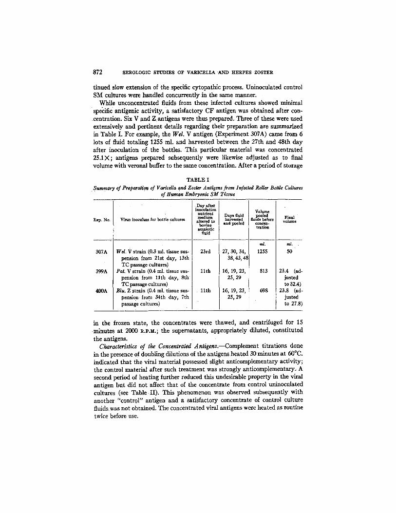
While unconcentrated fluids from these infected cultures showed minimal specific antigenic activity, a satisfactory CF antigen was obtained after con- cent.ration. Six V and Z antigens were thus prepared. Three of these were used extensively and pertinent details regarding their preparation are summarized in Table I. For example, the Wel. V antigen (Experiment 307A) came from 6 tots of fluid totaling 1255 ml. and harvested between the 27th and 48th day after inoculation of the bottles. This particular material was concentrated 25.1X; antigens prepared subsequently were likewise adjusted as to final volume with veronal buffer to the same concentration. After a period of storage
TABLE I
• Summary of PreParation of Varicella and Zoster Antigens from Infected Roller Bottle Cultures of Human Embryonic SM Tissue
Exp. ]?~'o.
307A
399A
400A
Virus inoculum for bottle cultures
Wd. V strain (0.3 ml. tissue sus, pension from 21st day, 13th TC passage cultures)
Pat. V strain (0.4 ml. tissue sus- pension from l l t h day, 8th TC passage cultures)
Blu. Z strain (0.4 ml. tissue sus- pensio n from 34th day, 7th passage cultures)
Day after inoculation nutrient medium
altered to bovine anmiotic
fuid
23rd
l l t h
11th
Days fluid harvested
and pooled
27, 30, 34, 38, 43, 4~
16, 19, 23, 25, 29
16, 19, 23, 25, 29
Volume .I~led
rinds before concen- tration
rnt.
1255
813
698
volume
mL
5O
23.4 (ad- justed to 32.4)
23.8 (ad- justed to 27.8
in the frozen state, the concentrates were thawed, and centrifuged for 15 minutes at 2000 R.P.M.; the supernatants, appropriately diluted, constituted the antigens.
Characteristics of the Concentrated AntQens.--Complement titrations done in the presence of doubling dilutions of the antigens heated 30 minutes at 60°C. indicated that the viral material possessed slight anticomplementary activity; the control material after such treatment was strongly anticomplementary. A second period of heating further reduced this undesirable property in the viral antigen but did not affect that of the concentrate from control uninoculated cultures (see Table II) . This phenomenon was observed subsequently with another "control" antigen and a satisfactory concentrate of control culture fluids was riot obtained. The concentrated viral antigens were heated as routine twice before use.
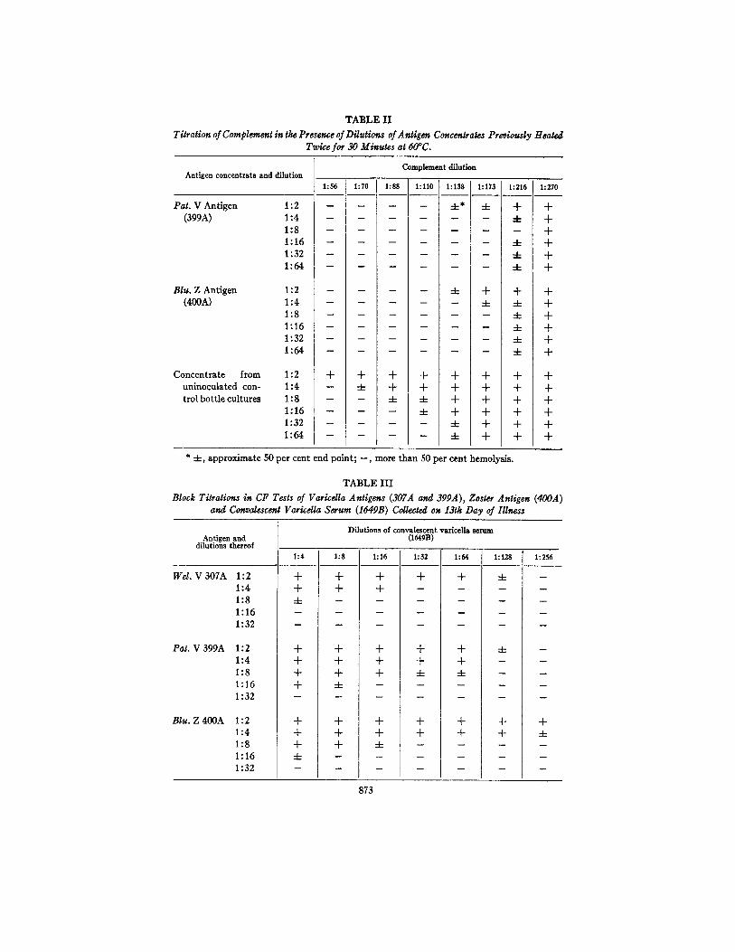
TABLE II
Titration of Complement in the Presence of Dilutions of Antigen Concentrates Previously Heated Twice for 30 Minutes at 60°C.
Antigen concentrate and dilution
Pat . V A n t i g e n
(399A)
Corn dement dilution
1:56 1:70 1:88 1:110 1:138 1:173 1:216 1:270
1:2 . . . . 4-* 4- + + 1 : 4 . . . . . . 4 - +
1:8 . . . . . . . + 1 : 1 6 . . . . . . 4 - + 1 : 3 2 . . . . . . 4 - +
1:64 . . . . . . 4- +
Blu. Z Antigen (400A)
1:2 . . . . 4- + + + 1:4 . . . . . 4- 4- + 1:8 . . . . . . 4- + 1 : 1 6 . . . . . . 4 - +
1 : 3 2 . . . . . . 4 - +
1 : 6 4 . . . . . . 4 - +
Zoncentrate from 1:2 + + + + + + + + uninoculated con- 1:4 - 4- + + + + + + trolbottlecultures 1:8 -- -- 4- 4- + + + +
1:16 -- -- -- 4- + + + + 1 : 3 2 . . . . 4 - + + +
1:64 . . . . .4- + + +
* 4 - , a p p r o x i m a t e 50 p e r c e n t e n d p o i n t ; - - , m o r e t h a n 50 p e r c e n t hemo lys i s .
TABLE ILl
Block Titration, in CF Tests of Varicdla Antigens (307A and 399A), Zoster Antigen (400A) and Convalescenl Varicdla Serum (164gB) Collected on 13th Day of Illness
Antigen and dilutions thereof
Wd. V 3 0 7 A 1 : 2
1 : 4
1 : 8
1 : 1 6
1 : 3 2
P a t . V 3 9 9 A 1 : 2
1 : 4
1 : 8
1 : 1 6
1 : 3 2
Blu. Z 4 0 0 A 1 : 2 1 : 4
1 : 8
1 : 1 6
1 : 3 2
1 : 4
+ +
4-
+ + + +
+ + +
4-
Dilutions of convalescent v&ricelh serum (16OB)
1:8
+ +
+ + + 4-
+ + +
1:16
+ +
+ + + m
+ + ..4-
1 : 3 2
+
+ + .4-
+ +
1 : 6 4
+
+ + -4-
+ +
1 : 1 2 8
.4-
4-
+ +
1:256
m
m
w
I
m
+ -4-
873
874 SEROLOGIC STUDIES OF VARICELLA AND HERPES ZOSTER
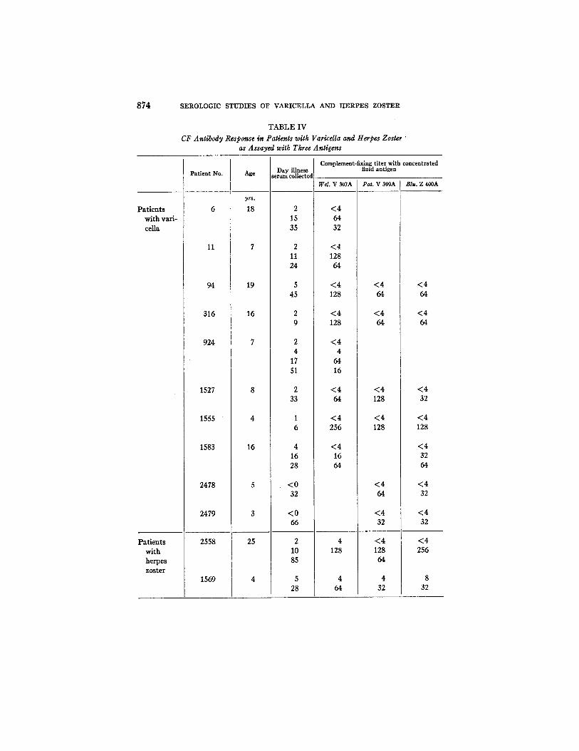
TABLE IV
CF Antibody Response in Patients with Varicella and Herpes Zoster " as Assayed with Three Antigens
Patients with vari- celia
Patient No.
11
94
316
924
1527
1555
1583
2478
2479
Age
yrs.
18
19
16
Day illness serum collected
2 15 35
Patients with herpes zoster
2558
1569
16
2 11 24
5 45
2 4
17 51
2 33
4 16 28
25
<0 32
<0 66
2 10 85
5 28
Complement-fixing titer with concentrated fluid antigen
Wel. V 307A
<4 64 32
<4 128 64
<4 128
<4 128
<4 4
64 16
<4 64
< 4 256
<4 16 64
4 128
4 64
Pat. V 399A
<4 64
<4 64
<4 128
<4 128
< 4 64
<4 32
<4 128 64
4 32
Blu. Z 400A
< 4 64
<4 64
<4 32
<4 128
<4 32 64
< 4 32
< 4 32
<4 256
8 32
T. H. WELLER AND H. M. WITTON
TABLE IV--Condudat
875
Patients with herpes zoster ---Cont.
Patient No. Age
1593
1652
1693
1994
2004
2510
2558
44
67
27
27
85
15
25
Day illness ;erum collected
1 10
33
2 15
17 99
240
3 10
4 22
2 10 85
Complement-fixing titer with concentrated fluid antigen
Wel. V 307A
512 256
32 64
4 64
8
64 128
Pat. V 399A
< 4 512
256 128
16 32
<4 128 64
Btu. Z 400A
<4 512
512 256
16 64
8 64 16
32 64
16 128
( 4 256
Block titrations were performed to determine the potency of the antigens using an arbitrarily selected sample of convalescent-phase varicella serum (Table III) . The three antigens, in view of the findings, were used as routine in a 1:2 dilution.
Complement-Fixing Antibody Response in Varicdla and Herpes Zoster as Assayed with Antigens Prepared from Concentrated Culture Fluids.--Representa- tive results obtained with the three antigens in complement fixation tests on serum specimens collected from individuals with varicella or herpes zoster are tabulated in Table IV. This table summarizes tests done at different times on specific sera. I t is to be noted that consistent and quite similar titers were ob- tained with the three antigens.
An indication of the temporal relationship between CF titer and the appear- ance of the exanthem in varicella and herpes zoster was provided by summariz- ing the titers as determined with the Pat. V antigen on 137 sera collected at various times from 52 individuals with varicella (see Text-fig. 1) and with the same antigen on 53 sera from 29 cases of herpes zoster (Text-fig. 2). Patients with varicella usually developed demonstrable levels of CF antibody about

876 SEROLOGIC STTJ-DIES OF VARICELLA AND t tERPES ZOSTER
Varicella 1:512 •
1:128
qD
1:3Z 4 . -
i : e
1:4
<1:4
O•
• 0 • • eta
• • • O O • • •
• • • •OO
D• • •
Q O O O
O e
4 8 12 16 OATS
70 •
70 •
• e ~ • •
A o•
O• • 900
• Z 0 ' • 4)0
• i t .
• . * j * * / m S S S / m / S / S ~ A , / S S ~
20 3 - 8 2-|2 I - 4 5 ÷ WK8. MOS. YRS. YRS.
Interval serum collected after appearance of exanthem "I~xT-FIG, 1. Complement-fixing titers as determined for 137 sera with Pa~. V antigen;
the specimens were obtained at various intervals after appearance of the eruption from 52 individuals with variceUa.
4 - , . u 4 "
1:512 •
1:128
1:52
1:8
1:4
<1:4
Herpes zoster • aJ~o •
O • O O • 41Q
O• • •
4HI • •
• • • •O • O
O O • e • Q •
0 0 0 • •
JSSS / / / S S / S /
4 8 IZ iS 20 3-S Z-lZ 1o4 DAYS WK8. MO8. YRS.
Interval serum collected after appearance of exanthem
TExT-FIG. 2. Complement-fixing titers as determined for 53 sera with Pat. V antigen; the specimens were obtained at varying intervals from 29 individuals with herpes zoster.
the 4th or 5th day after the exanthem appeared, and during the 2nd week of illness continuing and significant rises in titer were the rule. The order of in- crease in titer observed on the examination of 37 paired sera is indicated in Table V. The initial serum specimen from patients with herpes zoster at times
T. H. WELLER AND H. M. WITTON 877
possessed significant levels of CF antibody and further increases in tiler, while frequently observed, did not always occur (Table V). Otherwise, the antibody pattern, as determined from a limited number of sera, resembled that established for varicella.
The data obtained by means of the multiple examination of individual serum specimens with the different antigens were tabulated to obtain information on relative antigenic activity in the presence of sera from the homologous and heterologous clinical entities. This information is summarized in Table VI,
TABLE V
Change in CF Tiler Observed during Convalescence on Examination of Paired Sera witk Wel. V, Pat. V, or Blu. Z Antigens
Clinical ent i ty
rariceUa terpes zoster Ierpes simplex ~eneralized
vaccinia
I No of pairs sera-tested
37 17 13 4
No change in tlter
0 5
11 4
Rise in t iter
2X [ 4X 8X 15X I s2x ~ x 128X
I 3 3 3 6 10 8 4 2 2 3 2 1 - - 2 1 1 . . . . .
TABLE VI
Comparison of CF Tilers Obtained on Sera Examined Using Antigens Prepared with Varicella Strains and with a Zoster Strain
Clinical entity sera derived No. of sera tested with
both V and Z antigens
Comparison of CF titers obtained with antigen from the two sources
2-fold 4-fold S-fold S a m e d i f fe rence d i f fe rence difference
Varicella 23 16 5 2 0 Herpes zoster 43 23 18 2 0
and confirms the initial impression that the reactive capacity of the two types of antigen was similar in the presence of sera from cases of varicella or herpes zoster. The Blu. Z antigen tended to yield slightly higher titers with sera from either entity; thus, higher levels with Blu. antigen accounted for 15 of the minor discrepancies recorded in Table VI with zoster sera, and for 5 of those with varicella sera.
A small number of CF tests with the V-Z antigens were run on paired sera from patients with two other viral diseases manifested by vesiculopustular lesions (see Table V). The paired herpes simplex sera were known to contain specific complement-fixing antibody and were derived almost equally from primary infections (acute herpetic gingivostomatitis) and from recurrent infec-
878 SEROLOGIC STUDIES OF VARICELLA AND HERPES ZOSTER
tions. The single instance of a 4-fold increase in titer was noted on examination of paired sera from a 23 year old man with a clinical diagnosis of herpes labialis. Using Wel. V. antigen, the CF titer was 1:4 in a specimen collected as lesions developed, and was 1:16 in a specimen collected 13 days later; both sera re- acted to a titer of 1:16 with a herpes simplex antigen. Five of the six pairs of sera from primary herpetic infections in small children and four pairs from infants with generalized vaccinia did not fix complement in the presence of V-Z antigens in dilutions beyond 1:4.
Preparation of a Varicella Complement-Fixing Antigen from Infected Cultures of Monkey Kidney Tissue.--In an experiment performed by Dr. E. J. Bell, bottle cultures of monkey kidney tissue were used for the preparation of antigen. Six 13 day bottle cultures of trypsinized rhesus kidney tissue were inoculated with 1.0 ml. amounts of infected tissue suspension from the 7th passage Ore. V line in FS roller tubes. Five bottle cultures similarly received 1.0 ml. amounts of ground tissue from the comparable Ore. control cultures. Extensive focal lesions involving approximately 20 per cent of the outgrowth developed by the l l th day in the infected bottles; the medium was changed to amniotic fluid without serum or extract on that day, and subsequently, changes of medium and harvests were made on the 14th, 18th, 21st, and 25th days. One control culture showed changes compatib]e with monkey "foamy" virus and these fluids were discarded. The pooled virus and control fluids were concentrated 10 times by filtration.
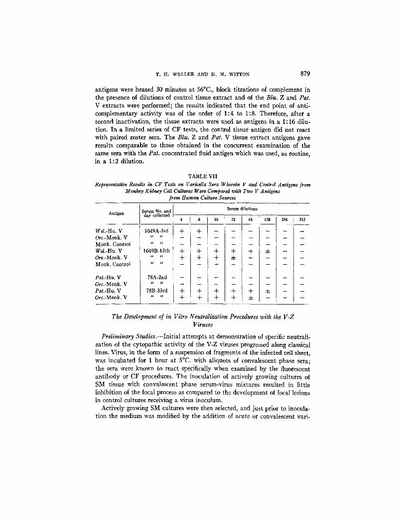
Subsequently, the antigens were heated for 30 minutes at 56°C. and comple- ment titrafions then performed in the presence of doubling dilutions thereof; in this instance neither the viral nor the control antigen was anticomplementary in a 1:2 dilution. The V antigen from monkey cultures was compared with the Wel. and Pat. antigens of human culture origin in three CF tests (see Table VII). The varicella antigen from monkey kidney material appeared slightly less potent but gave equivalent results.
Complement-Fixing Activity of Extracts of Human Tissue Suspensions from Infected Bottle Cultures.--It was determined in one experiment that a satis- factory antigen could be prepared from the tissue phase of infected cultures. The tissue suspensions were obtained from the bottle cultures of SM tissue infected with Pat. or B/u. virus which were used for the production of the fluid antigens. These cultures were terminated on the 29th day after inocula- tion when approximately 90 per cent of the tissue was involved. Following removal of the nutrient fluid, 5 ml. of veronal buffer were added per bottle, the tissue was scraped, and the resulting suspensions were pooled and frozen at -20°C. Tissue was similarly harvested from the control bottles. Nine months later the material was twice quickly thawed and frozen, and thereafter centrifuged at 2000 I~.P.M. for 15 minutes. The supernatant constituted the antigenic material and was stored in the frozen state until used. After the
T. H. WELLER AND H. M. WITTON 879
antigens were heated 30 minutes at 56°C., block fitrafions of complement in the presence of dilutions of control tissue extract and of the B/u. Z and Pat. V extracts were performed; the results indicated that the end point of anti- complementary activity was of the order of 1:4 to 1:8. Therefore, after a second inactivation, the tissue extracts were used as antigens in a 1:16 dilu- tion. In a limited series of CF tests, the control tissue antigen did not react with paired zoster sera. The Blu. Z and Pat. V tissue extract antigens gave results comparable to those obtained in the concurrent examination of the same sera with the Pat. concentrated fluid antigen which was used, as routine, in a 1:2 dilution.
TABLE VII Representative Results in CF Tests on Varicdla Sera Wherein V and Control Antigens from
Monkey Kidney Cell Cultures Were Compared with Two V Antigens from Human Culture Sources
Antigen
Wel.-Hu. V Orc.-Monk. V Monk. Control Wd.-Hu. V Orc.-Monk. V Monk. Control
Pat.-Hu. V Ore.-Monk. V Pat.-Hu. V Ore.-Monk. V
i Serum No. and day collected
1649A-3rd
c a 4 c
1649B-13th
78A-2nd
78B-33rd c l ~c
Serum dilutions
+ + m
1 6 3 2
+ + + -4-
+ + + +
64 128 256
+ 4- -
+ ± --
512
m
n
m
The Development of in Vitro Neutralization Procedures with the V-Z Viruses
Preliminary Studies.--Initial attempts at demonstration of specific neutrali- zation of the cytopathic activity of the V-Z viruses progressed along classical lines. Virus, in the form of a suspension of fragments of the infected cell sheet, was incubated for 1 hour at 5°C. with aliquots of convalescent phase sera; the sera were known to react specifically when examined by the fluorescent antibody or CF procedures. The inoculation of actively growing cultures of SM tissue with convalescent phase serum-virus mixtures resulted in little inhibition of the focal process as compared to the development of focal lesions in control cultures receiving a virus inoculum.
Actively growing SM cultures were then selected, and just prior to inocula- tion the medium was modified by the addition of acute or convalescent vari-

880 SEROLOGIC STUDIES OF VARICELLA AND HERPES ZOSTER
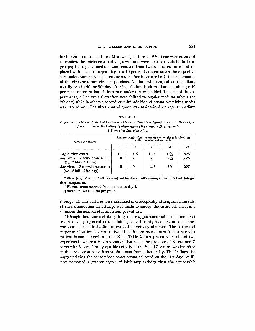
cella serum in a final concentration of 5 per cent. The incubation of virus with serum was omitted. After inoculation, the medium containing human serum was left in the cultures until the first fluid change 3 days later; it was then replaced by the regular nutrient. No significant difference was noted in the number of focal lesions appearing in the different groups of cultures (Table VII I ) although the foci in cultures exposed to the convalescent phase serum appeared to spread more slowly. A definite effect was noted, however, when the serum concentration was increased to 10 per cent. In one experiment, ali- quots of acute and convalescent phase zoster sera were thus incorporated in the medium for a period of 5 days before to 2 days after inoculation of the cultures. As summarized in Table IX, both zoster sera appeared to have defi- nite, but limited inhibitory activity.
TABLE VIII Neutralization Experiment Wherein Acute and Convalescent Var~eUa Sera in a 5 Per Cent
Concentration Were Incorporated as Constituents of the Culture Medium for the 3 Day Postinoculation Period*
Group of cultures
Virus control (Wel. V strain; llth TC passage) Wd. virus + "V" acute phase serum (No. 1006A
--lst day illness) Wel. virus + "V" convalescent serum (No. 1006C
--23rd day)
Average No. of focal lesions per culture or per cent of cell sheet involved on day:$
4 7
2 9.1
5 11
3.1 7
10
12% 25%
8%
* H u m a n serum omit ted from medium on day 3. ~: Based on 3 cultures per group.
Neutralization Tests Wherein Serum Specimens Were Incorporated as a Con- tinuing Constituent of the Culture M e d i u m . - - A s the result of these observations a neutralization technique evolved based on the long term incorporation of the sera under examination in the culture medium. Two series of experiments were conducted; in the first of these, virus was incubated with serum in the custom- ary manner prior to inoculation, while in the second set virus was added with- out prior treatment directly to the serum-containing cultures.
In the first group of experiments, evidence was obtained that cytopathic activity of the V and Z viruses was inhibited by sera deriving from the homol- ogous and the heterologous clinical entities. In these experiments, acute or convalescent phase sera were added in a 10 per cent concentration to freshly prepared suspensions of infected tissue from cultures and the mixtures kept at 4°C. for 1 hour; a third aliquot of tissue was similarly handled after the addition of media in a 10 per cent concentration and constituted the inoculum
T. H . W E L L E R AND H . M. W I T T O N 881
for the virus control cultures. Meanwhile, cultures of SM tissue were examined to confirm the existence of active growth and were usually divided into three groups; the regular medium was removed from two sets of cultures and re- placed with media incorporating in a 10 per cent concentration the respective sera under examination. The cultures were then inoculated with 0.2 ml. amounts of the virus or serum-virus suspensions. At the first change of nutrient fluid, usually on the 4th or 5th day after inoculation, fresh medium containing a 10 per cent concentration of the serum under test was added. In some of the ex- periments, all cultures thereafter were shifted to regular medium (about the 9th day) while in others a second or third addition of serum-containing media was carried out. The virus control group was maintained on regular medium
TABLE IX Experiment Wherein Acute and Convalescent Human Sera Were Incorporated in a I0 Per Cent
Concentration in the Culture Medium during the Period 5 Days before ~o g Days after Inoculation*,
Group of cultures
Bag. Z. virus control Bag. virus + Z acute phase serum
(No, 2510A---4th day) Bag. virus + Z convalescent serum
(No, 2510B--22nd day)
Average number focal lesions or per cent tissue involved per culture as observed on day:§
417 < 1 4 . 5 1 1 . 5 0 1 0 0 2.5
13
s ~ 7%
s%
16
60% 9s%
90%
* Virus (Bag. Z strain, 14th passage) not incubated with serum; added as 0.1 ml. infected tissue suspension.
:~ Human serum removed from medium on day 2. § Based on two cultures per group.
throughout. The cultures were examined microscopically at frequent intervals; at each observation an attempt was made to survey the entire cell sheet and to record the number of focal lesions per culture.
Although there was a striking delay in the appearance and in the number of lesions developing in cultures containing convalescent phase sera, in no instance was complete neutralization of cytopathic activity observed. The pattern of response of varicella virus cultivated in the presence of sera from a varicella patient is summarized in Table X; in Table XI are presented results of two experiments wherein V virus was cultivated in the presence of Z sera and Z virus with V sera. The cytopathic activity of the V and Z viruses was inhibited in the presence of convalescent phase sera from either entity. The findings also suggested that the acute phase zoster serum collected on the " l s t day" of ill- ness possessed a greater degree of inhibitory activity than the comparable
882 SEROLOGIC STUDIES 0]? VARICELLA AND HERPES ZOSTER
varicella serum collected on the 3rd day of illness. That high levels of inhibitory activity could develop in an individual with varicella by the 7th day of illness was shown in another experiment wherein sera (No. 1582) collected on the 7th, 19th, and 34th day of illness were examined with the Rob. V virus; on the 11th day, the average number of loci respectively in the three groups of serum cul- tures was 1.0, 0.3, and 1.6, while the virus control cultures contained an average of 36 lesions per tube.
The inhibition of virus by convalescent phase serum from the heterologous clinical entity was a regular finding. The pair of varicella sera (No. 1550) used with Blu. virus, for example, were also examined with another zoster strain (Bag.). Similar results were obtained; the average number of loci in cultures on the 7th day after inoculation was 44 in the virus control tubes, 36 in cultures
TABLE X Neutralization Test with Human Sera Incorporated as a Constituent of the Medium for 8 Days
after Inoculation. (McEl. V Virus; Varicella, Acute and Convalescent Sera)*
Group of cultures
McEl. virus control McF-J. virus + V acute serum
(334A--4th day) McEI. virus + V convalescent
serum (334B--11th day)
No. focM lesions or per ¢*~t tissue involved per culture as observed on day:$
I l_k_0 14 0.3 1 7.2 13 18 46 35% I 55% 0 0.3 5 7 9 24 25°/0 55°/0
0 [ 0 0 0.3 0.6 1.6 3 7
* Shift from media containing human sera to routine medium on 9th day. Average of 3 cultures per group.
with acute phase serum and 1.2 in the four cultures containing serum collected on the 13th day of illness.
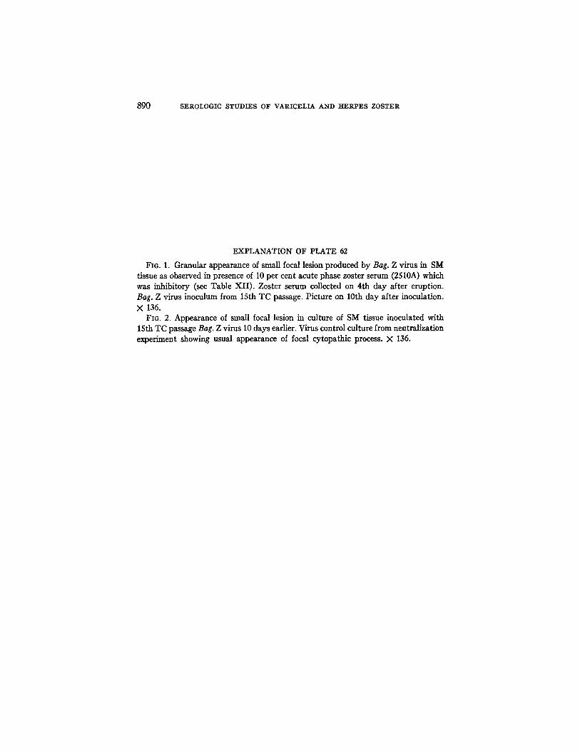
The focal lesions that developed in the presence of convalescent phase sera were smaller in size and progressed less rapidly than did comparable loci in con- trol cultures. In addition, they were often morphologically distinct, appearing relatively opaque and frequently the involved cells were associated with granu- lar debris. This, in part, appeared to be the result of continuing degeneration of infected cells with concurrent partial inhibition of the usual process of in- fection of contiguous cells. I t is possible that some of the observed granula- tion reflected the occurrence of an "in vitro" precipitin reaction, but no defini- tive information was obtained on this point. The phenomenon of granulation of the focal process was also observed with acute phase zoster sera that were inhibitory; the phenomenon is depicted in Fig. 1 and may be compared with the usual lesion in the control cultures shown in Fig. 2.
The repetitive addition of aliquots of human serum to the cultures in the
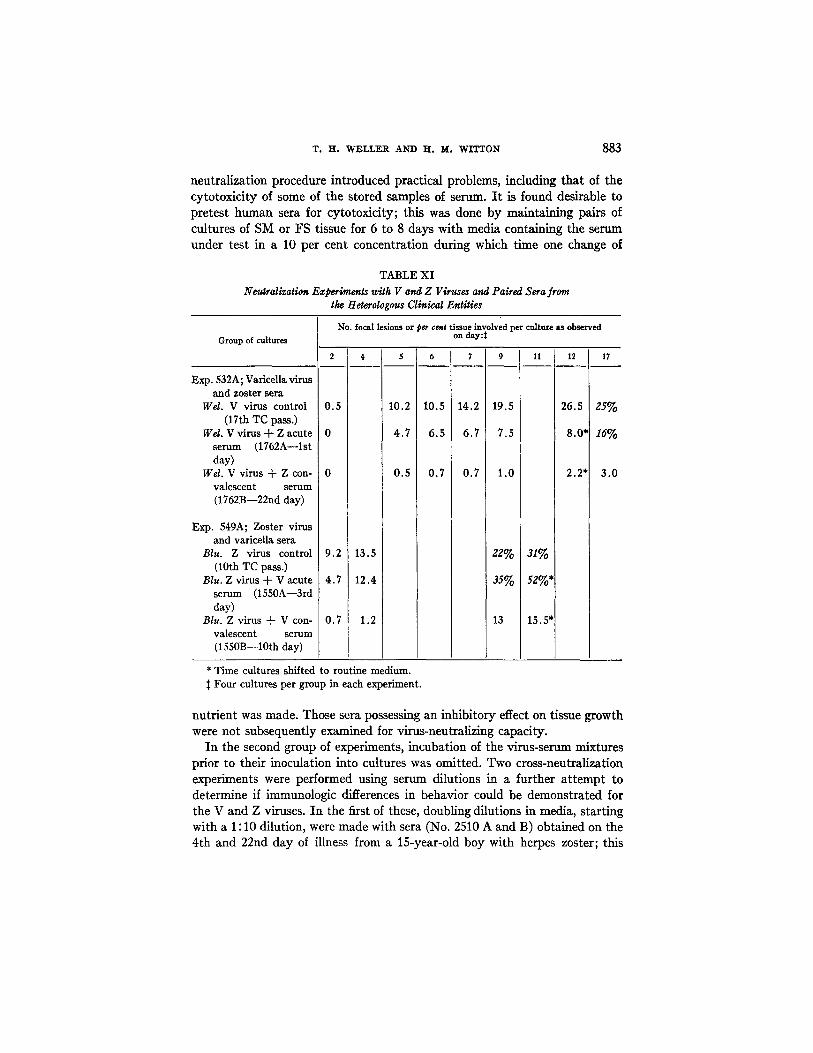
T. H. WELLER AND H. M. WITTON 883
neutralization procedure introduced practical problems, including that of the cytotoxicity of some of the stored samples of serum. I t is found desirable to pretest human sera for cytotoxicity; this was done by maintaining pairs of cultures of SM or FS tissue for 6 to 8 days with media containing the serum under test in a 10 per cent concentration during which time one change of
TABLE XI Neutralization Experiments with V and Z Viruses and Paired Sera from
the Heterologous Clinical Entities
Group of cultures
No. focal lesions or pee cent tissue involved per culture as observed on dsy::~
2 4 5 6
Exp. 532A; Varicella virus and zoster sera
Wd. V virus c on tr o l 0.5 10.2 10.5 (17th TC pass.)
Wd. V virus + Z acute 0 4.7 6.5 serum (1762A--1st day)
Wd. V v i r u s + Z c o n - 0 0.5 0.7 valescent serum (1762B--22nd day)
Exp. 549A; Zoster virus and varicella sera
Blu. Z virus control 9.2 13.5 (10th TC pass.)
Blu. Z virus + V acute 4.7 12.4 serum (1550A--3rd day)
Blu. Zvirus + Vcon- 0.7 1.2 valescent serum (1550B--lOth day)
7 9 1! 12 17
14.2 19.5 2 6 . 5 25%
6.7 7.5 8.0* 16°/o
0.7 1.0 2.2* 3.0
22% s1%
35% 52%*
13 15.5"
* Time cultures shifted to routine medium. Four cultures per group in each experiment.
nutrient was made. Those sera possessing an inhibitory effect on tissue growth were not subsequently examined for virus-neutralizing capacity.
I n the second group of experiments, incubation of the virus-serum mixtures prior to their inoculation into cultures was omitted. Two cross-neutralization experiments were performed using serum dilutions in a further a t tempt to determine if immunologic differences in behavior could be demonstrated f o r
the V and Z viruses. I n the first of these, doubling dilutions in media, starting with a 1 : 10 dilution, were made with sera (No. 2510 A and B) obtained on the 4th and 22nd day of illness from a 15-year-old boy with herpes zoster; this
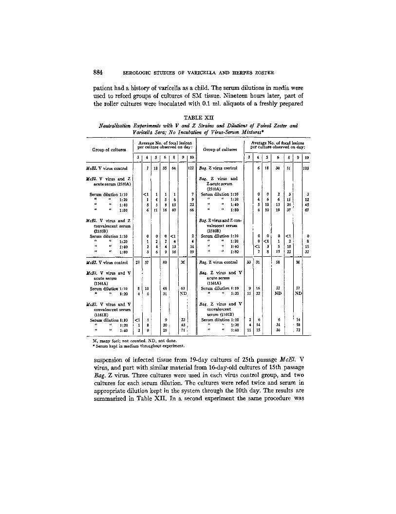
884 S E R O L O G I C S T U D I E S OF V A R I C E L L A AND H E R P E S Z O S T E R
patient had a history of varicella as a child. The serum dilutions in media were used to refeed groups of cultures of SM tissue. Nineteen hours later, part of the roller cultures were inoculated with 0.1 ml. aliquots of a freshly prepared
TABLE XII Neutralization Experiments with V and Z Slrains and Dilutions of Paired Zoster and
Varicella Sera; No Incubation of Virus-Serum Mixtures*
Average No. of focal lesions Gro up of cultures per cu l ture observed on day: Group of cultures
McEl . V virus control
Average No. of focal lesions per culture observed on day:
3 1 4 1 , T 0 1 8 o 10 3 4 s , 8
7 18 35 64 122 Bag. Z virus control 6 18 30 51
M c E L V virus and Z Bag. Z virus and acute serum (2510A) Z acute serum
(2510A) Serum dilution 1:I0 <1 1 1 1 7 Serum dilution 1:10 0 0 2 3 3
" " 1:20 1 4 5 6 9 " " 1:20 4 6 6 11 12 " " 1:40 5 5 8 13 22 '¢ " 1:40 5 15 13 26 i 45 " " 1:80 6 11 16 40 66 " " 1:80 6 10 19 37 I 67
McEI. V virus and Z Bag. Z virus and Z con- ] convalescent serum valescent serum (2510B) (2510B)
Serum dilution 1:10 0 0 0 <1 2 Serum dilution 1:I0 0 0 0 < I 0 " " 1:20 1 2 2 4 4 " " 1:20 0 <1 1 2 i 8 i " " 1:40 2 4 4 i 13 14 " " 1:40 <1 3 $ 10 15 " " 1:80 3 6 9 16 19 " " 1:80 7 8 13 22 37
33 31 McEI. V virus control
McEI. V virus and V a cute serum (l161A)
Serum dilution 1:10 " " 1:20
23 37 80 M
8 10 46 63 4 6 31 ND
<I 3 9 23 1 8 20 43 2 9 29 71
Bag. Z virus control 58 M
Bag. Z virus and V acute serum (l161A)
Sermn dilution 1:10 9 16 32 57 " " 1:20 11 22 ND ND
Bag. Z virus and V convalescent serum (l161B)
Serum dilufion 1:10 2 6 6 14 " " 1:20 4 1 4 34 58 " " 1:40 11 15 36 72
McEl . V virus and V convalescent serum (l161B)
Serum dilution 1:10 4. " 1:20
" " 1:40
M, many focl; not counted. ND, not done. * Serum kept in medium throughout experiment.
suspension of infected tissue from 19-day cultures of 25th passage McEI. V virus, and part with similar material from 16-day-old cultures of 15th passage Bag. Z virus. Three cultures were used in each virus control group, and two cultures for each serum dilution. The cultures were refed twice and serum in appropriate dilution kept in the system through the 10th day. The results are summarized in Table XI I . In a second experiment the same procedure was

T. H . W E L L E R AND H . M. W I T T O N 885
used to examine paired varicella sera collected on the 2nd and 49th day of ill- ness, with the exception that the V and Z viruses were 26th passage McEl. and 16th passage Bag. materials, each harvested on the 13th day of cultivation (see Table XII). The results again indicated that a close serologic relationship existed between the V and the Z strains tested. Both acute phase sera, particu- larly that from the zoster case, had an inhibitory action on the development of the focal lesions. The CF titers of the pair of zoster sera were 1 : 16 and 1 : 128 respectively, and of the varicella pair were 1: 4 and 1: 64.
DISCUSSION
We have previously presented data indicating that the viruses of varicella and herpes zoster have been propagated in cultures of various tissues of human and of monkey origin. The demonstration that the V and Z viruses produce antigenic materials in ~itro capable of binding complement in the presence of convalescent phase sera from patients with varicella or herpes zoster, and the further finding that such sera are capable of specific inhibition of V-Z viral cytopathogenicity, provide additional evidence that we are dealing with the etiologic agents. Indeed, together with the information previously accumulated by the fluorescent antibody method (6), the body of serologic data now appears to afford an incontrovertible documentation in support of this thesis.
The question of the relationship of the viruses of zoster and of vancella has been widely discussed since 1888 when yon Bokay observed that on occasion susceptible individuals developed varicella following contact with patients who had herpes zoster (13). In support of von Bokay's hypothesis, various observations have gradually accumulated indicating the close relationship, and probable identity, of the V-Z viruses. It is to be noted, however, that some experienced clinicians continue to question the monistic hypothesis (14). The data from the laboratory may be summarized as follows. As first described by Tyzzer (15) and Lipschiitz (16), the reaction at the cellular level produced by the V and Z viruses in man is similar. It is evident that this similarity extends to the cytopathic response induced by these viruses in ~itro (7). Electron microscopic examination of vesicle fluid materials from both entities has indi- cated that the fluids contain bodies, interpreted as viral, that appear identical (17-19). Heretofore, immunologic studies have utilized vesicle fluid. In general, the results with vesicle fluid as a CF antigen have indicated a close relation- ship of the two entities (20, 2). Amies (4) recorded frequent cross-reactions in microagglufination studies, although some variation occurred. Our complement fixation studies with the agents propagated in ~itro clearly confirm the exist- ence of common antigenic factors in the V and Z viruses. Further evidence of a close relationship is provided by the findings of the in vitro neutralization experiments. Yet, in view of the present limitations of the neutralization pro-

886 SEROLOGIC STUDIES OF VARICELLA AND HERPES ZOSTER
cedure as dictated by the unusual behavior pattern of the V-Z viruses, the possibility remains that subtle, and now undetectable, strain differences may exist. However, the accumulation of epidemiological and laboratory evidence in support of the hypothesis that a single etiologic agent is responsible for varicella and herpes zoster appears so impressive that the burden of proof must now logically shift to those who desire to refute the monistic concept.
The CF procedure as herein described should, with minor modifications, pro- vide the diagnostic laboratory with a convenient technique for the diagnosis of two additional common clinical entities of viral etiology. In the CF experiments, the difficulties encountered in the preparation of a suitable control antigen from fluid materials and the lack of a reference antiserum were an early source of concern. However, the negative results obtained with the control tissue ex- tract and monkey kidney culture antigens and the uniformity of the CF anti- body response in paired varicella sera gave assurance of the specificity of the reaction. The neutralization technique, as here used, is time-consuming and exacting; it will not find favor as a diagnostic procedure until simplified.
Application of the complement fixation and neutralization reactions should provide a better understanding of the response of the human host to the V-Z viruses. In varicella, specific CF antibody becomes demonstrable around the 4th or 5th day after the appearance of the exanthem; Amies (3) observed that agglutinins appeared after a similar interval, as does antibody reacting by the fluorescent technique (6). As an example of an unusual situation, the failure of CF antibody to appear by the 12th day of illness in a case of varicella with fatal outcome (21) provided serologic evidence of a defect in host response.
Relatively few sera were obtained immediately after the appearance of the eruption in patients with zoster; some of these already contained significant levels of CF antibody. It is tempting to consider that these early antibody responses might be a reflection of previous experience with V virus; on the other hand, they may reflect the existence of a long prodromal stage in an en- tity with an ill defined incubation period. The appearance of a varicelliform eruption in the patient with herpes zoster is a recognized complication. We obtained paired sera from 6 zoster patients who developed disseminated lesions. In four of these the CF antibody response was of the usual order, but in two, titers of 1:4 and 1:8 were recorded for sera collected on the 12th and 17th day respectively. Application of the new serologic techniques should assist in elucidating the pathophysiologic mechanism of herpes zoster and its bizarre clinical manifestations. Almost certainly such an understanding will be based on a concept involving intracellular proliferation and persistence of the V-Z viruses in the presence of humoral antibody. The factors controlling transfer of the virus from cell to contiguous cell, both in vitro and in vivo, form a challenging area for investigation of fundamental importance.
T. H. WELLER AND H. M. WITTOlq 887
SUMMARY
The preparation of antigenic materials capable of specific fixation of comple- ment in the presence of convalescent phase sera from patients with varicella and herpes zoster is described. Satisfactory antigens were obtained by the repetitive harvest and subsequent concentration of the pooled nutrient fluids from bottle cultures of human embryonic skin-muscle tissue or of monkey kid- ney tissue infected with varicella or herpes zoster viruses. Specific fixation of complement was also demonstrated with antigens prepared from extracts of the infected cell sheets harvested from bottle cultures.
In individuals with varicella, complement-fixing antibody usually appeared in the serum 4 or 5 days after the development of the exanthem and further significant increases in titer were characteristically observed during the 2nd week of illness.
The complement-fixing antibody response in herpes zoster tended to follow the same pattern as in varicella, with the exception that in sera from some individuals relatively high titers were present in the acute phase specimen.
Complement-fixing antigens prepared from varicella strains or from a zoster strain reacted to essentially the same degree with convalescent sera from the homologous and the heterologous clinical entities. The varicella-zoster anti- gens did not fix complement in the presence of paired sera obtained from a limited number of individuals with primary infections due to herpes simplex virus or from individuals with generalized vaccinia infection.
Specific inhibition in dtro of the focal cytopathic process produced by the varicella-zoster viruses was demonstrated. This was accomplished by the incorporation of the sera under test as constituents of nutrient media of the cultures, either prior to or at the time of their inoculation with virus. The neutralization of focal cytopathogenicity thus obtained was relative in degree and never absolute; it was therefore assayed by repetitive counts of the number of focal lesions per culture in the various test groups.
Inhibition of varicella-zoster viral cytopathogenicity occurred in the presence of convalescent serum from either clinical entity.
The results of the immunologic studies with the viruses of herpes zoster and varicella as propagated in vitro are considered as providing further evidence in support of the concept of the close relationship and probable identity of the two agents.
The authors are indebted to many physicians who have provided clinical material or have contributed sera; and in particular to Dr. J. J. Finn, Jr., Dr. J. T. Heyl, and Dr. Conrad Wesselhoeft.
BIBLIOGRAPHY
1. Netter, A., and Urbain, A., Le virus varicello-zonateux, Ann. Inst. Pasteur, 1931, 46, 17.
888 SEROLOGIC STUDIES OF VARICELIA AND HERPES ZOSTER
2. Brain, R. T., The relationship between the viruses of zoster and varicella as demonstrated by the complement fixation reaction, Brit. J. Exp. Path., 1933, 14, 67.
3. Amies, C. R., The elementary bodies of variceUa and their agglutination in pure suspension by the serum of chickenpox patients, Lancet, 1933, 1, 1015.
4. Amies, C. R., Elementary bodies of zoster and their serological relationship to those of varicelIa, Brit. J. Exp. Path., 1934, 16, 314.
5. Weller, T. H., Serial propagation in vitro of agents producing inclusion bodies derived from varicella and herpes zoster, Proc. Soc. Exp. Biol. and Me.d., 1953, 83, 340.
6. WeUer, T. H., and Coons, A. It., Fluorescent antibody studies with agents of varicella and herpes zoster propagated in dtro, Proc. Soc. Exp. Biol. and Med. 1934, 86, 789.
7. Weller, T. H., Witton, H. M., and Bell, E. :[., The etiologic agents of varieella and herpes zoster: isolation, propagation, and cultural characteristics in vitro, J. Exp. Med., 1958, 1081 843.
8. Enders, J. F., Bovine amniotie fluid as tissue culture medium in cultivation of poliomyelitis and other viruses, Proc. Soc. Exp. Biol. and Med., 1953, 89., 100.
9. Fulton, F., and DumbeU, K. R., Serological comparison of strains of influenza virus, J. Gen. Microbiol., 1949, 3, 97.
10. Svedmyr, A., Enders, J. F., and HoUoway, A., Complement fixation with Brun- hilde and Lansing poliomyelitis virus propagated in tissue culture, Proc. Soc. Exp. Biol. and Meal., 1952, 79, 296.
11. Mayer, M. M., Osier, A. G., Bier, O. G., and Heidelburger, M., Activating effect of magnesium and other cations on hemolytic function of complement, J. Exp. Med., 1946, 84, 535.
12. Seibert, F. B., Chemical composition of active principle of tuberculin. XI. Im- proved and simplified method for making standard undenatured tuberculin of any desired strength and method of chemical assay, J. Biol. Chem., 1928, 78, 345.
13. yon Bokay, J., l~ber den aetiologischen Zusammenhang der Varizellen mit gewis- sen F~llen yon Herpes zoster, Wein. klin. woch., 1909, 9.9. 1323.
14. Wesselhoeft, C., Chickenpox and herpes zoster, Rhode Island Med. J., 1957, 40, 387.
15. Tyzzer, E. E., The histology of the skin lesions in varicella, g. Med. Research, 1906, 14, 361.
16. Lipschfitz, B., Untersuchungen fiber die )~tiologie der Krankheiten der Herpes- gruppe (Herpes zoster, Herpes genitalis, Herpes febrilis), Arck. Dermatol. u. Sypk., 1921, 136, 428.
17. Ruska, H., l~ber das virus der varicellen nnd des zoster, Klin. Woch., 1943, 22, 703.
18. Rake, G., Blank, H., Coriell, L. L., Nagler, F. P. 0., and Scott, T. F. M., The relationship of varicella and herpes zoster: Electron microscope studies, J. Bact., 1948, §6, 293.
19. Farrant, J. L., and O'Connor, J. L., Elementary bodies of varicel!a and herpes zoster, NaZure, 1949, 163, 260.

T. H° W E L L E R AND H . M. W I T T O N 889
20. Netter, A., and Urbain, A., Les relations du zona et de la variceUe: 6tude serolo- gique de 100 cas de zona, Compt. rend. Soc. biol., 1926, 94, 98.
21. Cheatham, W. J., Weller, T. H., Dolan, T. F., Jr., and Dower, J. C., VariceUa: Report of two fatal cases with necropsy, virus isolation, and serologic studies, Am. J. Path., 1956, 32, 1015.
890 SEROLOGIC STUDIES OF VARICELIA AND HERPES ZOSTER
EXPLANATION OF PLATE 62
FIG. 1. Granular appearance of small focal lesion produced by Bag. Z virus in SM tissue as observed in presence of l0 per cent acute phase zoster serum (2510A) which was inhibitory (see Table XII) . Zoster serum collected on 4th day after eruption. Bag. Z virus inoculum from 15th TC passage. Picture on 10th day after inoculation. X 136.
FIG. 2. Appearance of small focal lesion in culture of SM tissue inoculated with 15th TC passage Bag. Z virus l0 days earlier. Virus control culture from neutralization experiment showing usual appearance of focal cytopathic process. × 136.

THE JOURNAL OF EXPERIMENTAL MEDICINE VOL. 108 PLATE 62
(Weller and Witton: Serologic studies of varicella and herpes zoster)