Embed Size (px)
Citation preview
Development of Viral Tools for CNS Gene Transfer:
Adeno-Associated Viral Vectors in Gene Therapy of Parkinson’s Disease
PhD Thesis
in partial fulfilment of the requirements for the degree
“Doctor of Philosophy (PhD)/Dr. rer. nat.”
in the Neuroscience Program
at the Georg August University Göttingen,
Faculty of Biology
submitted by
Zinayida Shevtsova
born in
Dnipropetrovsk
2006

Declaration
I hereby declare that the thesis: “Development of Viral Tools for CNS Gene Transfer: Adeno-
Associated Viral Vectors in Gene Therapy of Parkinson’s Disease” has been written independently
and with no other sources and aids than quoted.
Zinayida Shevtsova
Göttingen, March 2006
2
CONTENTS
Abbreviations 7
1. Introduction 10 1.1. Neurodegeneration 10
1.2. Parkinson’s disease (PD) 10
1.2.1. Prevalence and symptomatology of PD 10
1.2.2. Morpho-pathological background of PD 11
1.2.3. Genetic clues to the etiology of PD 12
1.2.4. Environmental contribution to the etiopathogenesis of PD 13
1.2.5. Mechanisms of cell death in PD 13
1.2.6. Oxidative stress and mitochondrial dysfunction in PD 14
1.2.7. Basal ganglia physiology 17
1.2.8. Animal models of PD: complete unilateral 6-OHDA rat model 19
1.3. Therapeutic approaches for PD treatment 21
1.3.1. Current therapeutical strategies and limitations 21
1.3.2. Neuroprotective gene therapy: achievements and perspectives 22
1.3.3. Glial cell line-derived neurotrophic factor (GDNF) 22
1.3.4. BclXL, an antiapoptotic member of the bcl-2 family proteins 24
1.4. AAV vectors as tools for gene therapy 25
1.5. Semliki Forest virus (SFV) vectors 27
1.6. Epitope-tagging 28
2. Materials and Methods 29 2.1. Materials 29
2.1.1. Chemicals 29
2.1.2. Antibodies 29
2.1.3. Plasmids 30
2.1.4. Oligonucleotides (Sigma-Aldrich) 30
2.1.5. Cell lines and electrocompetent cells 32
2.1.6. Buffers and Solutions 32
2.2. Methods 33
2.2.1. Cloning procedures 33
3
2.2.1.1. PCR-amplification 33
2.2.1.2. In vitro transcription 34
2.2.1.3. Sequencing of PCR-amplified DNA 34
2.2.1.4. DNA precipitation 34
2.2.1.5. DNA restriction, electrophoresis, gel extraction,
concentration determination 35
2.2.1.6. DNA ligation and transformation in E. coli 35
2.2.1.7. Plasmid Mini- and Maxi - Preps 36
2.2.1.8. Preparation of electrocompetent E. coli 36
2.2.1.9. Cloning into pSFV-plasmid 36
2.2.1.10. Cloning into pAAV-2 plasmid 38
2.2.2. Viral vectors production and purification 41
2.2.2.1. SFV vectors 41
2.2.2.2. AAV vectors 41
2.2.3. Cell culturing 44
2.2.3.1. Continuous cell culture 44
2.2.3.2. Primary culture of hippocampal neurons 44
2.2.3.3. SFV-transduction and cell lysis for western blotting 45
2.2.3.4. Indirect immunofluorescence on primary neurons 45
2.2.4. Animal procedures 45
2.2.4.1. Stereotaxic injection into the rat brain 46
2.2.4.2. Intravitreal injections and optic nerve axotomy 46
2.2.4.3. Transcardial perfusion and brain tissue processing 47
2.2.4.4. Preparation of the brain tissue lysates for WB 48
2.2.5. Indirect immunofluorescence on brain slices 48
2.2.6. Protein handling procedures 48
2.2.6.1. Protein concentration determination 49
2.2.6.2. SDS-polyacrylamide gel electrophoresis (SDS-PAGE) 49
2.2.6.3. Immune blotting 49
2.2.7. GDNF-ELISA 50
2.2.8. Microscopy and image analysis 50
2.2.9. Quantification of neuroprotection in 6-OHDA rat model of PD 51
2.2.10. Rotational behaviour test 51
2.2.11. Statistics 52
4
3. Results 53 3.1. Development of AAV vectors for targeting different cell types of CNS 53
3.1.1. Construction of AAV vectors 53
3.1.2. In vitro transduction of brain cells by AAV vectors 54
3.1.3. In vivo application of AAV vectors 55
3.2. Evaluation of epitope tags suitability for CNS gene transfer studies 57
3.2.1. Expression constructs of epitope tags used for in vivo gene transfer 57
3.2.2. Structural influence of the epitope tags 58
3.2.3. Evaluation of epitope tags in the central nervous system 59
3.2.4. Evaluation of "established" epitope tags (HA, cMyc, FLAG) 60
3.2.5. Evaluation of "new" epitope tags (AU1, EE, IRS) 60
3.2.5. Proof -of-principle: discrimination of ectopically from
endogenously expressed protein 63
3.3. Long-term neuroprotection mediated by AAV gene transfer
in the complete 6-OHDA rat model of PD 65
3.3.1. Targeted transduction of SNpc by stereotaxic injection of AAV-2 65
3.3.2. Establishment of the complete 6-OHDA rat model of PD 66
3.3.3. BclXL mediated protection of DA cell bodies in 6-OHDA
rat model of PD 67
3.3.4. GDNF protects DA cell bodies and dendritic protrusions
from 6-OHDA toxicity 68
3.3.5. BclXL and GDNF co-expression has additive neuroprotective
effect at 2 weeks after 6-OHDA lesion 68
3.3.6. Co-expression of BclXL with GDNF has no beneficial effect on
dopaminergic neuron survival at 6 weeks after 6-OHDA lesion 70
3.3.7. Effects of delayed AAV-GDNF administration in the rat
striatum after the complete 6-OHDA lesion of the MFB 70
4. Discussion 73 4.1. AAV vector targeting 73
4.2. Epitope-tagging for in vivo transgene detection 74
4.3. Neuroprotective therapy of PD as evaluated in the complete
6-OHDA lesion model 77
4.4. BclXL mediated neuroprotection 78
5
4.5. Combination of BclXL and GDNF for neuroprotection
of DA neurons 79
4.6. GDNF mediated neurorestorative therapy 80
5. Summary 83
6. Acknowledgements 84
7. References 85
Curriculum vitae 101
Publications 102
6
Abbreviations
6-OHDA - 6-hydroxydopamine
AAV - adeno-associated virus
ANT - adenine nucleotide transporter
AD - Alzheimer’s disease
ADP - adenosine diphosphate
ATP - adenosine triphosphate
BAX - bcl-2-associated X protein
bGH - bovine growth hormone derived polyadenylation site
bp - base pairs
BPB - bromphenol blue sodium salt
BSA - bovine serum albumine
cAMP - cyclic adenosine monophosphate
CNS - central nervous system
COMT - catechol-O-methyltransferase
CPu - caudate putamen (striatum)
Cx - cortex
DA - dopaminergic
DAPI - 4',6-diamidino-2-phenylindole
DIV - day in vitro
DMEM - Dulbecco’s modified Eagle’s medium
DNA - desoxyribonucleic acid
DsRed - red fluorescent protein
DTT - dithiothreitol
EDTA - ethylenediaminetetraacetic acid
EGFP - enhanced green fluorescent protein
ELISA - enzyme-linked immunosorbent assay
ERK - same as MAPK
FA - formaldehyde
FCS - fetal calf serum
FG - fluorogold
FPLC - fast protein liquid chromatography
GABA - γ-aminobutyric acid
7
GDNF - glial cell line-derived neurotrophic factor
GFAP - glial fibrialy acidic protein
GFL - GDNF family ligands
GFR - GDNF family receptor
GP - globus pallidus
GPe - globus pallidus externum
GPi - globus pallidus internum
GPI - glycosyl phosphatidylinositol
GSH - glutathione
HNC - hippocampal neuron cultureshSyn1 – human synapsin 1 gene promoter
HRP - horse reddish peroxidase
HSPG - heparan sulfate proteoglycan
Int - intron
LB - Luria broth
L-DOPA - 1-3,4-dihydroxyphenylalanine
MAPK- mitogen-activated protein kinase, same as ERK
MAO-A - monoamine oxidase A
MAO-B - monoamine oxidase B
mCMV - murine cytomegalovirus immediate early promoter
MFB - medial forebrain bundle
MPTP - 1-methyl-4-phenyl-1,2,3,6-tetrahydropyridin
MWCO - molecular weight cut off
NBM - neurobasal medium
NCAM - neural cell adhesion molecule
NDD - neurodegenerative disorders
NIA - National Institute on Aging (United States of America)
NGS - newborn goat serum
NS - nervous system
nsP - non-structural protein
ORF - open reading frame
PAGE - polyacrylamide gel electrophoresis
PBS - phosphate buffered saline
PCR - polymerase chain reaction
PD - Parkinson’s disease
PDGFR - platelet-derived growth factor receptor
8
PFA - paraformaldehyde
PI3K- phosphatidylinositol 3-kinase
PLCγ - phospholypase-Cγ
PNS - peripheral nervous system
PS (-N) - penicillin-streptomycin (-neomycin)
PTP - permeability transition pore
RET - receptor tyrosine kinase
RGC - retinal ganglion cell
ROS - reactive oxygen species
RT - room temperature
RT-PCR - real time PCR
SDS - sodium dodecylsulfate
SFV - Semliki-Forest virus
SN - substantia nigra of the midbrain
SNpc - substantia nigra pars compacta
STN - subthalamic nucleus
SV40 - simian virus 40 polyadenylation site
TB - synthetic transcription blocker
TBS - tris-buffered saline
TBS-T - TBS-tween
TCA - tricyclic aminoacids
TEMED - tetraeminethylendiamine
TH - tyrosine hydroxylase
TH-IR - TH immunoreactive
TNF - tumour necrosis factor
TRAIL - tumour necrosis factor-related apoptosis-inducing ligand
VTA - ventral tegmental area of the midbrain
WPRE - woodchuck hepatitis virus posttranscriptional regulatory element
WB - western blotting
XIAP - X-linked inhibitor of apoptosis protein
9
1. Introduction
1.1. Neurodegeneration
Nervous system disorders are a major cause of morbidity in the western society,
which impair quality of life to a degree rarely affected by other diseases. More than 1% of the
population suffer from Parkinson’s disease after reaching 55 years of age (de Rijk, M. C. et al.,
1997) and nearly half of those who reach the age of 85 years and older suffer from Alzheimer’s
disease (excerpt from NIA's Progress Report on Alzheimer's Disease, 1998: NIA). Among the
factors contributing to the burdensome cost of managing neurodegenerative diseases is the fact
that only few symptomatic and no causative therapies are currently available. Despite
considerable efforts in basic research, novel diagnostic approaches and effective therapies have
yet to be developed.
The majority of human neurodegenerative diseases is not related to inherited mutations
of specific proteins but develops with aging as a multifactorial pathology. It is often
characterized by features of oxidative stress (Tretter, L. et al., 2004) and axonal degeneration
preceding neuronal cell loss, as demonstrated for Alzheimer’s disease (Stokin, G. B. et al.,
2005), Parkinson’s disease (Dauer, W. et al., 2003), Huntington’s and other polyglutamine
diseases (Gunawardena, S. et al., 2005;Li, H. et al., 2001). Various experimental animal
models of neurodegenerative diseases have been developed to provide a better understanding
of the complex mechanisms involved in neurodegeneration and to serve for the preclinical
evaluation of prospective diagnostic tools and therapies. However, every model can reflect
only certain aspects of the complex aetiology of respective diseases in humans. Thus, the
evaluation of neuroprotective strategies aiming at generalized inhibition of neurodegeneration
should be performed in more than one particular model system. Furthermore, considering the
diversity of mechanisms leading to neurodegeneration, effective human therapy may
necessitate targeting of more than one neurodegenerative pathway.
1.2. Parkinson’s disease (PD) 1.2.1. Prevalence and symptomatology of PD
Parkinson’s disease is the second most common neurodegenerative disorder, which
affects more than 1% of 55-year-old individuals and more than 3% of those over 75 years of
age (de Rijk, M. C. et al., 1997). The overall age- and gender-adjusted incidence rate is 13.4
per 100,000, with a higher prevalence among males (19.0 per 100,000) than females (9.9 per
100,000)(Van Den Eeden, S. K. et al., 2003). It was initially described by James Parkinson in
1817 and characterized by a typical triad of symptoms: 1) poverty and slowness of movement
10
without paralysis (bradykinesia), 2) changes in posture and muscle tone towards rigidity and 3)
resting tremor. In most cases these symptoms are sufficient for the diagnosis of the disease
(Hughes, A. J. et al., 2001). Psychological symptoms and autonomic nervous system
dysfunction typically develop as the disease progresses and often become a major cause of
disability.
1.2.2. Morpho-pathological background of PD
The pathological basis of the disease remained unknown for over 100 years, although
the clinical features have been well-described. The importance of the substantia nigra (SN) was
emphasized by Tretiakoff in 1919, who studied the substantia nigra in thirteen cases of
parkinsonism and found lesions in this nucleus in all cases. The SN, named so because of the
native content of the neuromelanin pigment, was noted to show depigmentation, loss of nerve
cells, and gliosis. Tretiakoff also confirmed the earlier observation of Lewy (1914), who had
found the presence of lipophylic cytoplasmic inclusions in Parkinson’s disease, now referred to
as Lewy bodies and recognized as the pathologic hallmark of the disorder. In 1959, at the
International Catecholamine Symposium, Carlsson suggested that Parkinson’s disease was
related to brain dopamine. In 1960, Ehringer and Hornykiewicz, using Carlsson’s
methodology, measured greatly reduced DA concentrations, to about one-tenth of normal, in
the caudate, putamen and substantia nigra in brains from parkinsonian patients. The underlying
key morphopathological feature is the progressive loss of dopaminergic (DA) neurons in the
substantia nigra pars compacta (SNpc) of the midbrain, with intracytoplasmic inclusions (Lewy
bodies) in the remaining intact nigral neurons (Braak, H. et al., 2000;Forno, L. S., 1996). It is
believed that the disease becomes symptomatic when 50-60% of DA neurons in SNpc and
more than 70-80% of their projections in the striatum are lost (Deumens, R. et al., 2002).
Moreover, the severity and the stage of the disease are correlated with the extent of
neurodegeneration (Riederer, P. et al., 1976;Fearnley, J. M. et al., 1991;Foley, P. et al., 1999).
DA neurons of SNpc also have a tendency to degenerate with aging at a rate of approximately
5% per decade (Fearnley, J. M. et al., 1991). In PD patients, however, both the rate and the
pattern of DA neurodegeneration significantly differ from that observed during normal aging.
Thus, the DA neuronal loss in PD in contrast to normal aging is uneven and occurs mainly in
the ventro-lateral part of SNpc. While during normal aging the rate of neurodegeneration is
about 5 % per decade, it is about 10 fold faster in PD patients.
11
1.2.3. Genetic clues to the etiology of PD
Despite the early descriptions of clinical and pathomorphological features of PD the
etiology of the disease remains unclear. Several genes which are implicated in rare familial
forms of PD have been identified over the last decade and revealed novel proteins and
pathways that may induce parkinsonism as a result of nigral neurodegeneration. The genetic
load of known mutations in PD is small, accounting for less than 5% of the overall PD
population. However, the typical and extremely consistent phenotype of both idiopathic and
familial PD suggests that one common molecular mechanism may underlie PD. Similar
pathways underlying familial forms of the disease might be strongly implicated in the
pathogenesis of the sporadic forms. Understanding the pathogenesis of the sporadic form of PD
will have the greatest impact on advancing novel therapies for this common incurable
neurodegenerative disorder. Five disease genes (SNCA, PARK2, PINK1, DJ-1, LRRK2) have
been conclusively implicated in PD (Abou-Sleiman, P. M. et al., 2006), out of which the most
studied are the mutations in the gene encoding for α-synuclein. This protein has also been
detected as a major component of the Lewy bodies suggesting its unequivocal role in DA
neurons degeneration. The only genetic evidence associating a single mutation in UCHL1 gene
with PD was not sufficient for the conclusive linkage (Abou-Sleiman, P. M. et al., 2006;Leroy,
E. et al., 1998).
Data obtained from studies on DJ-1 and PINK1 mutations may serve as an indirect
evidence of the seminal role of mitochondrial dysfunction and oxidative stress in PD
pathogenesis. Endogenous DJ-1 is localised to the mitochondrial matrix and the mitochondrial
intermembrane space in addition to its cytoplasmic pool (Zhang, L. et al., 2005), and may act
as an antioxidant since it can be oxidized at the cysteine residue C106. Furthermore, a
quantitative proteomic study of the SN of mice treated with MPTP revealed a significant
increase in the protein DJ-1 in mitochondrial fraction of the SN (Jin, J. et al., 2005). These data
suggest that DJ-1 may be involved in neuroprotection of DA neurons from oxidative damage.
Another protein implicated in PD, PINK1, consists of a highly conserved kinase domain
(Unoki, M. et al., 2001;Nakajima, A. et al., 2003) and a mitochondrial targeting motif
(Valente, E. M. et al., 2004). Together with the study showing localization of PINK1 to
mitochondria in transfected cells (Beilina, A. et al., 2005), this data may suggest that PINK1
may also act as a protein protecting from oxidative stress (Valente, E. M. et al., 2004).
Mutation and polymorphism in another gene, encoding the mitochondrial protease HtrA2/ Omi
have been recently associated with PD (Strauss, K. M. et al., 2005).
In addition, possible susceptibility genes (e.g. NR4A2, SNCAIP) that may increase, or
decrease the risk of an individual to develop PD have been identified. However, since only a
12
minor group of PD patients reveal a genetic predisposition, external factors influencing the
initiation and progression of the disease must be examined.
1.2.4. Environmental contribution to the etiopathogenesis of PD
One of the earliest hypotheses of PD pathogenesis was based on the finding that three
inhibitors of mitochondrial complex 1, namely MPTP, rotenone and paraquat, were able to
reproduce parkinsonism with selective DA neuronal loss in mice and primate models (Seniuk,
N. A. et al., 1990). The first toxic models of PD did not fully reproduce the features of the
disease, mainly because of the absence of one of the major pathological hallmark of PD, the
Lewy body. Later, a chronic infusion of either rotenone (Betarbet, R. et al., 2000) or MPTP
(Fornai, F. et al., 2005) in rodents was shown to induce the formation of α-synuclein positive
aggregates. These data support the theory that sporadic PD may be caused by a combination of
genetic predisposition and environmental toxins, which act via inhibition of the mitochondrial
complex I to produce selective DA cell loss. Inhibition of complex I may result in the depletion
of ATP leading to impairment of all ATP dependent cellular processes, and in generation of
free radicals responsible for oxidative stress damage. Clear evidence of increased oxidative
stress was found in postmortem PD brains, as compared to the age-matching controls
(Andersen, J. K., 2004;Sian, J. et al., 1994). Furthermore, a reduced activity of complex I was
found in the brains of idiopathic PD patients (Schapira, A. H. et al., 1990;Parker, W. D., Jr. et
al., 1989).
1.2.5. Mechanisms of cell death in PD
The loss of dopaminergic neurons in SNpc is a characteristic feature of PD. However,
mechanisms underlying this neurodegeneration are not very well understood. Different modes
of cell death, apoptotic, necrotic and autophagic have been described to contribute to the
neuronal loss occurring in PD (von Bohlen und Halbach O. et al., 2004;Blum, D. et al., 2001).
Several pathogenetic mechanisms have been proposed to be implicated in cell death observed
in PD: mitochondrial dysfunction and oxidative stress, defective proteolysis and proteotoxic
stress, excitoxicity, and inflammation (Gandhi, S. et al., 2005;von Bohlen und Halbach O. et
al., 2004;Blum, D. et al., 2001). It is likely that PD pathogenesis is multifactorial and all
mechanisms listed above to a certain extent contribute to the PD associated degeneration of
nigrostriatal neurons. However, one of the single mutations described above, being present in
all cells of the body, affects specifically DA cells in SNpc and results in clinico-pathological
characteristics of PD. Furthermore, the fact that mutations described in PD affect the proteins
remote from each other structurally and functionally suggests the native susceptibility of DA
13
neurons to various types of cellular stress. In other words, independently on the original
trigger, all pathogenetic pathways mentioned above may sequentially be involved in the
process of neurodegeneration of SN neurons. Conversely, if various pathogenetic mechanisms
may ultimately lead to the same PD phenotype, the evidence of molecular convergence of these
pathways must exist. The cellular “powerhouse organelle” mitochondria is a good candidate
for a subcellular localization of such a convergence (Gandhi, S. et al., 2005;Beal, M. F., 2005).
1.2.6. Oxidative stress and mitochondrial dysfunction in PD
Mitochondria are important intracellular organelles that play a crucial role in various
cellular processes including energy production via pyrimidine biosynthesis, fatty acid
oxidation, calcium homeostasis and cell survival (Wang, H. Y. et al., 1991;Elston, T. et al.,
1998;Thress, K. et al., 1999;Newmeyer, D. D. et al., 2003;Melov, S., 2004).
Oxydative phosphorylation, occuring in the mitochondria through tricyclic aminoacid
(TCA) cycle and proton gradient, is the main source of high energy compounds (e.g. ATP) in
the cell. Electrons derived from metabolic utilization of glucose in TCA enter the respiratory
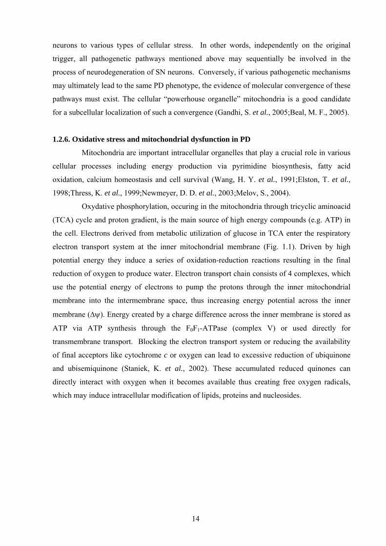
electron transport system at the inner mitochondrial membrane (Fig. 1.1). Driven by high
potential energy they induce a series of oxidation-reduction reactions resulting in the final
reduction of oxygen to produce water. Electron transport chain consists of 4 complexes, which
use the potential energy of electrons to pump the protons through the inner mitochondrial
membrane into the intermembrane space, thus increasing energy potential across the inner
membrane (Δψ). Energy created by a charge difference across the inner membrane is stored as
ATP via ATP synthesis through the F0F1-ATPase (complex V) or used directly for
transmembrane transport. Blocking the electron transport system or reducing the availability
of final acceptors like cytochrome c or oxygen can lead to excessive reduction of ubiquinone
and ubisemiquinone (Staniek, K. et al., 2002). These accumulated reduced quinones can
directly interact with oxygen when it becomes available thus creating free oxygen radicals,
which may induce intracellular modification of lipids, proteins and nucleosides.
14
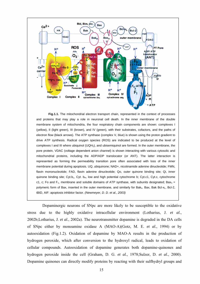
Fig.1.1. The mitochondrial electron transport chain, represented in the context of processes
and proteins that may play a role in neuronal cell death. In the inner membrane of the double
membrane system of mitochondria, the four respiratory chain components are shown: complexes I
(yellow), II (light green), III (brown), and IV (green), with their substrates, cofactors, and the paths of
electron flow (black arrows). The ATP synthase (complex V, blue) is shown using the proton gradient to
drive ATP synthesis. Radical oxygen species (ROS) are indicated to be produced at the level of
complexes I and III where ubiquinol (UQH2), and ubisemiquinol are formed. In the outer membrane, the
pore protein, VDAC (voltage dependent anion channel) is shown interacting with various cytosolic and
mitochondrial proteins, including the ADP/ADP translocator (or ANT). The latter interaction is
represented as forming the permeability transition pore often associated with loss of the inner
membrane potential during apoptosis. UQ, ubiquinone; NAD+, nicotinamide adenine dinucleotide; FMN,
flavin mononucleotide; FAD, flavin adenine dinucleotide; Qo, outer quinone binding site; Qi, inner
quinone binding site; Cyt.bL, Cyt. bH, low and high potential cytochrome b; Cyt.c1, Cyt.c, cytochrome
c1, c; Fo and F1, membrane and soluble domains of ATP synthase, with subunits designated; Baxn =
polymeric form of Bax, inserted in the outer membrane, and similarly for Bakn. Bax, Bak Bcl-xL, Bcl-2,
tBID, AIF: apoptosis inhibitor factor. (Newmeyer, D. D. et al., 2003)
Dopaminergic neurons of SNpc are more likely to be susceptible to the oxidative
stress due to the highly oxidative intracellular environment (Lotharius, J. et al.,
2002b;Lotharius, J. et al., 2002a). The neurotransmitter dopamine is degraded in the DA cells
of SNpc either by monoamine oxidase A (MAO-A)(Gotz, M. E. et al., 1994) or by
autooxidation (Fig.1.2). Oxidation of dopamine by MAO-A results in the production of
hydrogen peroxide, which after conversion to the hydroxyl radical, leads to oxidation of
cellular compounds. Autooxidation of dopamine generates both dopamine-quinones and
hydrogen peroxide inside the cell (Graham, D. G. et al., 1978;Sulzer, D. et al., 2000).
Dopamine quinones can directly modify proteins by reacting with their sulfhydryl groups and
15
reduce the level of intracellular antioxidant glutathione (Graham, D. G. et al., 1978;Stokes, A.
H. et al., 1999). The conversion of hydrogen peroxide to highly reactive hydroxyl radical
(Fenton reaction) requires Fe2+ ions (Fig. 1.2, 1.3). Interestingly, the Fe2+ level in the SNpc is
natively higher than in the other areas of the brain and, moreover, was found to be increased in
PD (Sofic, E. et al., 1988;Dexter, D. T. et al., 1989b).
Fig.1.2. Oxidation of dopamine. Oxidation of dopamine by MAO or by autooxidation leads to
the production of H2O2, which when converted to OH radicals can lead to the oxidization of
proteins, lipids, and nucleosides. Autooxidation of dopamine also leads to the generation of
dopamine quinone, which may covalently bind to proteins or further be converted to neuromelanin.
The modification of biomolecules by OH or dopamine quinone may exert toxic effects on
dopaminergic neurons. (Hald, A. et al., 2005)
Excess of dopamine was reported to inhibit the complex I function in the brain (Ben
Shachar, D. et al., 1995). The reduced activity of the complex I of the respiratory chain was
also found in PD (Schapira, A. H., 1994). Selective vulnerability of DA neurons of SNpc to the
toxins (MPTP, rotenone, 6-OHDA) acting through the inhibition of the complex I activity,
even when systemically applied (MPTP), suggests that these neurons are intrinsically more
sensitive to the oxidative damage and mitochondrial dysfunction.
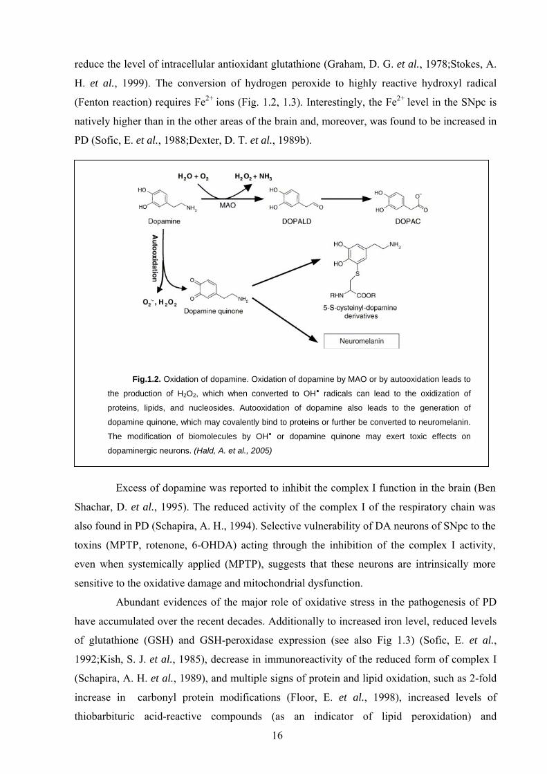
Abundant evidences of the major role of oxidative stress in the pathogenesis of PD
have accumulated over the recent decades. Additionally to increased iron level, reduced levels
of glutathione (GSH) and GSH-peroxidase expression (see also Fig 1.3) (Sofic, E. et al.,
1992;Kish, S. J. et al., 1985), decrease in immunoreactivity of the reduced form of complex I
(Schapira, A. H. et al., 1989), and multiple signs of protein and lipid oxidation, such as 2-fold
increase in carbonyl protein modifications (Floor, E. et al., 1998), increased levels of
thiobarbituric acid-reactive compounds (as an indicator of lipid peroxidation) and
16
malondialdehyde, and 8-hydroxy-2’deoxyguanosine (a nucleoside oxidation product)(Zhang, J.
et al., 1999;Dexter, D. T. et al., 1989a) were found in SN of PD-affected brains in comparison
to unaffected age-matching controls.
Fig.1.3. Factors leading to oxidative stress in the SNpc: (A) a deficiency in glutathione (GSH),
thereby diminishing the capability to clear H2O2; (B) an increase in reactive iron, which can promote
OH* formation; (C) auto-oxidation of DA into toxic dopamine-quinone species. GSSG: oxidized
gluthation; SO: dopamine-quinone species.(von Bohlen und Halbach O. et al., 2004)
Beside ROS generation, mitochondria are also important regulators of Ca2+
signalling. They actively and sensitively respond to the local increase in Ca2+ concentration by
transient but massive uptake of the ion into the organelle (Rizzuto, R. et al., 1999). Ultimately,
this may lead to the loss of mitochondrial membrane potential, energy deprivation and cell
death. The fluctuations of intracellular Ca2+ are observed in glutamate excitotoxic damage,
which was suggested as one of the factors involved in DA cell death in PD, and may serve as
an additional link between mitochondrial dysfunction and SNpc pathology.
1.2.7. Basal ganglia physiology
Development of new treatment strategies as well as appropriate model systems for the
evaluation of their therapeutic value require a better understanding of the basal ganglia
physiology, since a dysbalance in their functional circuits results in a typical for PD
symptomatology. The basal ganglia consist of several interconnected nuclei which send their
projections to different cortical motor areas, thalamus, certain brainstem nuclei, and indirectly
to the cerebellum. The principal four nuclei of basal ganglia are the striatum, the globus
pallidus (GP), subdivided into the external (GPe) and internal (GPi) segments, the substantia
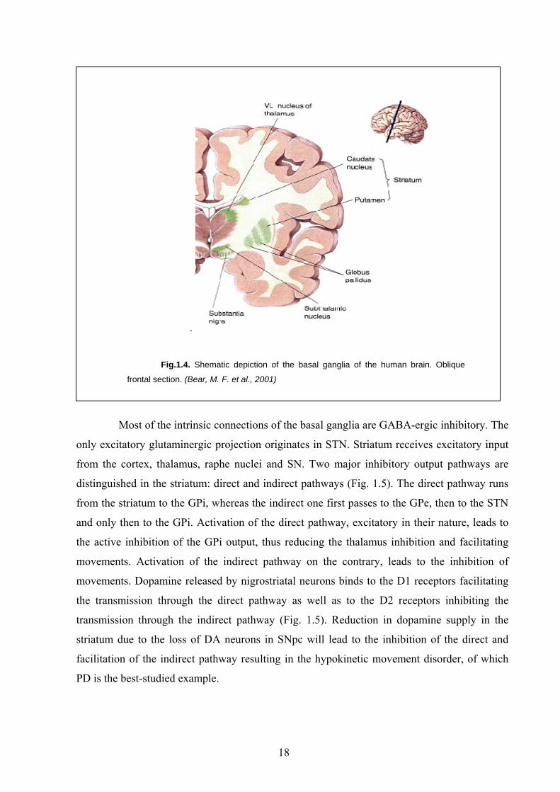
nigra (SN) and the subthalamic nucleus (STN) (Fig. 1.4).
17
.
Fig.1.4. Shematic depiction of the basal ganglia of the human brain. Oblique
frontal section. (Bear, M. F. et al., 2001)
Most of the intrinsic connections of the basal ganglia are GABA-ergic inhibitory. The
only excitatory glutaminergic projection originates in STN. Striatum receives excitatory input
from the cortex, thalamus, raphe nuclei and SN. Two major inhibitory output pathways are
distinguished in the striatum: direct and indirect pathways (Fig. 1.5). The direct pathway runs
from the striatum to the GPi, whereas the indirect one first passes to the GPe, then to the STN
and only then to the GPi. Activation of the direct pathway, excitatory in their nature, leads to
the active inhibition of the GPi output, thus reducing the thalamus inhibition and facilitating
movements. Activation of the indirect pathway on the contrary, leads to the inhibition of
movements. Dopamine released by nigrostriatal neurons binds to the D1 receptors facilitating
the transmission through the direct pathway as well as to the D2 receptors inhibiting the
transmission through the indirect pathway (Fig. 1.5). Reduction in dopamine supply in the
striatum due to the loss of DA neurons in SNpc will lead to the inhibition of the direct and
facilitation of the indirect pathway resulting in the hypokinetic movement disorder, of which
PD is the best-studied example.
18
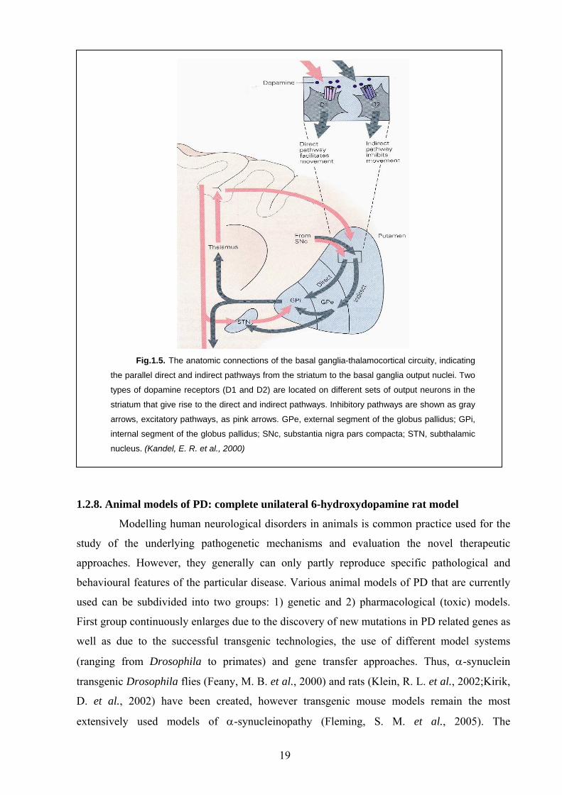
Fig.1.5. The anatomic connections of the basal ganglia-thalamocortical circuity, indicating
the parallel direct and indirect pathways from the striatum to the basal ganglia output nuclei. Two
types of dopamine receptors (D1 and D2) are located on different sets of output neurons in the
striatum that give rise to the direct and indirect pathways. Inhibitory pathways are shown as gray
arrows, excitatory pathways, as pink arrows. GPe, external segment of the globus pallidus; GPi,
internal segment of the globus pallidus; SNc, substantia nigra pars compacta; STN, subthalamic
nucleus. (Kandel, E. R. et al., 2000)
1.2.8. Animal models of PD: complete unilateral 6-hydroxydopamine rat model
Modelling human neurological disorders in animals is common practice used for the
study of the underlying pathogenetic mechanisms and evaluation the novel therapeutic
approaches. However, they generally can only partly reproduce specific pathological and
behavioural features of the particular disease. Various animal models of PD that are currently
used can be subdivided into two groups: 1) genetic and 2) pharmacological (toxic) models.
First group continuously enlarges due to the discovery of new mutations in PD related genes as
well as due to the successful transgenic technologies, the use of different model systems
(ranging from Drosophila to primates) and gene transfer approaches. Thus, α-synuclein
transgenic Drosophila flies (Feany, M. B. et al., 2000) and rats (Klein, R. L. et al., 2002;Kirik,
D. et al., 2002) have been created, however transgenic mouse models remain the most
extensively used models of α-synucleinopathy (Fleming, S. M. et al., 2005). The
19
pharmacological models of PD remain a widely employed alternative, since they are easy to
create and are able to reproduce the major etiopathologic features of PD induced by oxidative
stress and inhibition of complex I. Several different neurotoxins, 6-hydroxydopamine (6-
OHDA), MPTP, and rotenone are commonly used to this purpose (Blum, D. et al., 2001;von
Bohlen und Halbach O. et al., 2004).
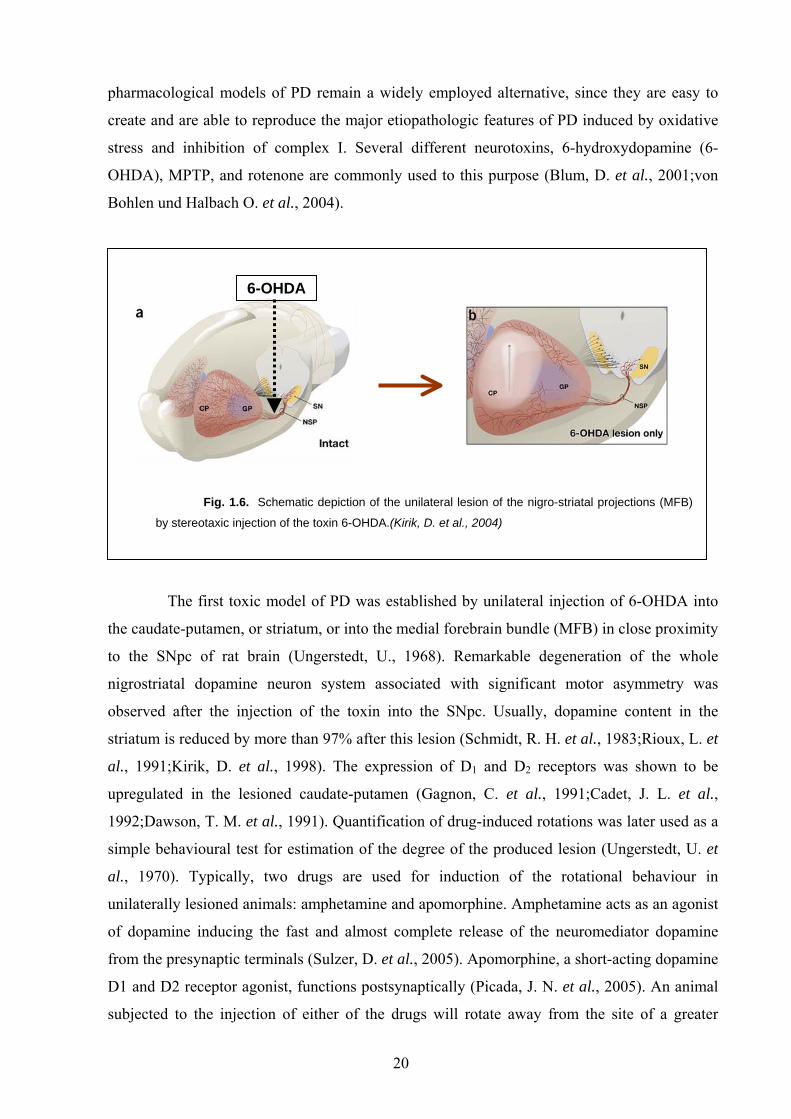
Fig. 1.6. Schematic depiction of the unilateral lesion of the nigro-striatal projections (MFB)
by stereotaxic injection of the toxin 6-OHDA.(Kirik, D. et al., 2004)
6-OHDA
The first toxic model of PD was established by unilateral injection of 6-OHDA into
the caudate-putamen, or striatum, or into the medial forebrain bundle (MFB) in close proximity
to the SNpc of rat brain (Ungerstedt, U., 1968). Remarkable degeneration of the whole
nigrostriatal dopamine neuron system associated with significant motor asymmetry was
observed after the injection of the toxin into the SNpc. Usually, dopamine content in the
striatum is reduced by more than 97% after this lesion (Schmidt, R. H. et al., 1983;Rioux, L. et
al., 1991;Kirik, D. et al., 1998). The expression of D1 and D2 receptors was shown to be
upregulated in the lesioned caudate-putamen (Gagnon, C. et al., 1991;Cadet, J. L. et al.,
1992;Dawson, T. M. et al., 1991). Quantification of drug-induced rotations was later used as a
simple behavioural test for estimation of the degree of the produced lesion (Ungerstedt, U. et
al., 1970). Typically, two drugs are used for induction of the rotational behaviour in
unilaterally lesioned animals: amphetamine and apomorphine. Amphetamine acts as an agonist
of dopamine inducing the fast and almost complete release of the neuromediator dopamine
from the presynaptic terminals (Sulzer, D. et al., 2005). Apomorphine, a short-acting dopamine
D1 and D2 receptor agonist, functions postsynaptically (Picada, J. N. et al., 2005). An animal
subjected to the injection of either of the drugs will rotate away from the site of a greater
20
activity. This can be explained by the anatomical structure of the mammalian CNS, particularly
the fact of crossing of the afferent tracts to the contralateral side at the level of either brainstem
or spinal cord. However, due to the difference in mechanisms of action of the drugs, the rat
exposed to amphetamine will rotate ipsilaterally, while the apomorphine-injected rat will
exhibit contralateral to the lesion turns. Drug-induced rotational behaviour is often used to
select the animals with complete lesions, since only those rats with complete degeneration of
the nigrostriatal projections will exhibit robust turning behaviour induced by the drug.
1.3. Therapeutic approaches for PD treatment 1.3.1. Current therapeutical strategies and limitations
The pharmacological standard for the treatment of PD remains a replacement of
dopamine. This is typically accomplished with the precursor of dopamine, 1-3,4-
dihydroxyphenylalanine (L-DOPA). The clinical effects of L-DOPA on akinesia in
parkinsonism were first presented in 1967 by Birkmayer and Hornykiewicz. L-DOPA is
typically administered in a combination with carbi-dopa, an inhibitor of DOPA-decarboxylase
which reduces the peripheral conversion of L-DOPA to dopamine thus allowing for
considerable amount of the drug to cross the blood brain barrier and undergo decarboxylation
to DA in the brain. Although the initial therapeutic effects of L-DOPA are excellent, patients
develop drug-related side effects over the course of the disease, which include motor
fluctuations (the so-called "wearing-off" and "on–off" phenomena) and dyskinesias (Lang, A.
E. et al., 1998). Other medications including anticholinergic agents, inhibitors of catechol-O-
methyltransferase (COMT) or monoamine oxidase-B (MAO-B) provide only mild-to-moderate
benefit (Lang, A. E. et al., 1998;Hristova, A. H. et al., 2000;Olanow, C. W. et al.,
2004;Marjama-Lyons, J. M. et al., 2001). Eventually, L-DOPA or dopamine agonists are
required for management of progressive disability. However, dopamine agonists usually take
longer than L-DOPA to reach effective doses, and also require co-administration of L-DOPA
for supervening disability after varying periods of time.
At the later stages of the disease patients developing resistance or severe side effects
to pharmacological therapy may benefit from neurosurgical procedures such as pallidotomy or
deep brain stimulation of the subthalamic nucleus (STN) (Esselink, R. A. et al., 2004).
Nevertheless, none of the currently available treatments has been proven to slow the
progression of PD.
21
1.3.2. Neuroprotective gene therapy: achievements and perspectives
Different strategies have been employed to inhibit neurodegenerative processes: early
studies aimed at blocking the executioners of apoptotic cell death, cysteine proteases of the
caspase family, however, no sustained neuroprotection could be achieved (Kermer, P. et al.,
1999;Perrelet, D. et al., 2000;Rideout, H. J. et al., 2001). Most pro-apoptotic signals converge
on breakdown of mitochondrial membrane potential, followed by release of pro-apoptotic
factors and subsequent caspase activation (Chang, L. K. et al., 2002). Thus, several studies
focused on the maintenance of mitochondrial integrity by overexpression of anti-apoptotic
members of the bcl-2 family of proteins (Azzouz, M. et al., 2000;Malik, J. M. et al.,
2005;Wong, L. F. et al., 2005). This strategy proved to be significantly more efficient than
caspase inhibition, although in long-term studies substantial neuronal cell loss was still
observed (Malik, J. M. et al., 2005;Kim, R., 2005).
Neurotrophic factors in several paradigms could only shortly postpone neuronal
degeneration (Cheng, H. et al., 2002;van Adel, B. A. et al., 2003). Glial cell line-derived
neurotrophic factor (GDNF) appears to be an exception and remains a promising candidate in
the treatment of Parkinson’s disease.
1.3.3. Glial cell line-derived neurotrophic factor (GDNF)
GDNF was originally identified as a trophic factor promoting the survival of
embryonic midbrain dopaminergic neurons (Lin, L. F. et al., 1993). Subsequently, it was found
to be a potent trophic factor for noradrenergic neurons of the CNS (Arenas, E. et al., 1995) as
well as for the moto- and sensory neurons of the PNS (Henderson, C. E. et al., 1994;Ramer, M.
S. et al., 2000). GDNF was purified from a glioma cell line supernatant and then found to be
expressed in several glial cell types of the NS (Schaar, D. G. et al., 1993;Strelau, J. et al.,
1999). In toxicity animal models of PD, GDNF was proven to rescue DA neurons of SNpc
from the neurotoxic damage and promote behavioural recovery (Grondin, R. et al., 1998).
However, lentiviral delivery of GNDF to SNpc failed to prevent neurodegeneration in α-
synuclein transgenic rat model of PD (Lo, Bianco C. et al., 2004). Furthermore, recent clinical
trials demonstrated divergent outcomes (Gill, S. S. et al., 2003;Nutt, J. G. et al., 2003;Patel, N.
K. et al., 2005). GDNF has also been shown to induce resprouting of the lesioned nigrostriatal
neuron system and thus may have beneficial effects on deafferentiated neurons in general
(Björklund, A. et al., 1997;Love, S. et al., 2005).
Together with three other neurotrophic factors (neurturin, artemin, persephin) GNDF
comprises a family of proteins, the so-called GDNF family ligands (GFLs). GFLs are distantly
related to the transforming growth factor-beta superfamily, containing seven cysteine residues
22
with the same relative spacing and acquiring similar conformation as the other members of this
superfamily (Ibanez, C. F., 1998). The main signalling pathway of GFLs is mediated by RET-
receptor tyrosine kinase (Sariola, H. et al., 2003), which was initially discovered as a
protooncogen (Takahashi, M., 2001). RET is activated upon binding of GDNF dimer to
GFRα1 receptors linked to the plasma membrane via a glycosyl phosphatidylinositol (GPI)
anchor (Fig. 1.7). Dimerization of RET triggers its autophosphorylation, thus initiating various
intracellular signalling cascades, that regulate cell survival, proliferation, differentiation,
neurite outgrowth, synaptic plasticity and morphogenesis (Airaksinen, M. S. et al., 2002). For
example, MAPK and phosphatidylinositol 3-kinase (PI3K) may be involved in neurite
outgrowth and neuronal survival, while PLCγ regulates intracellular Ca2+ levels (Sariola, H. et
al., 2003).
Fig. 1.7. GDNF interaction with its receptors. A dimer of GDNF brings together two molecules
of GFR 1. This complex dimerizes two molecules of Ret leading to transphophorylation of their
tyrosine kinase domains. GFR proteins are attached to the plasma membrane through a GPI-
anchor and consist of three globular cysteine-rich domains joined together by adapter
sequences.(Sariola, H. et al., 2003)
GDNF may also signal independently of RET but involving the same receptor
GFRα1. In RET-deficient cells GDNF may act through the Src family kinases, inducing
sustained activation of the Ras/ERK and PI3K/Akt pathways, cAMP response element-
binding protein phosphorylation, and increased c-fos expression (Trupp, M. et al., 1999). In the
absence of RET, Src-mediated cellular events may also promote neuronal survival and neurite
outgrowth (Sariola, H. et al., 2003).
23
Fig. 1.8. Non-RET signalling for GDNF through NCAM. NCAM interacts with a GDNF-
GFR dimer leading to activation of Fyn, a Src-like kinase, for instance. It is not known whether
Fyn activates Met in NCAM-mediated GDNF signalling.(Sariola, H. et al., 2003)
NCAM has been proposed as an alternative signalling receptor for GDNF (Paratcha,
G. et al., 2003). When GFRα1 is absent, GFL associates with NCAM with low-affinity. In
turn, in the presence of GFRα1, GDNF may bind to the p140-NCAM and activate cytoplasmic
Src-like Fyn and FAK kinases (Fig. 1.8) (Paratcha, G. et al., 2003). Interestingly, both in vivo
and in vitro effects of GDNF, such as DA neuron survival, neurite outgrowth, DA turnover,
and locomotor activity in rats were inhibited by anti-NCAM antibodies (Chao, C. C. et al.,
2003).
1.3.4. BclXL, an antiapoptotic member of the bcl-2 family proteins
The antiapoptotic member of the bcl-2 protein family, BclXL plays an important role
in the inhibition of mitochondria-dependent cell death pathways. Mouse embryos deficient in
BclXL show massive cell death of immature neurons of the CNS and dorsal root ganglia
(Motoyama, N. et al., 1995). Co-deletion of BAX largely rescued the BclXL knockout
phenotype providing evidence that BclXL serves as a negative regulator of BAX -mediated cell
death in vivo (Shindler, K. S. et al., 1997). Together with bcl-2, BclXL is considered a
prominent native inhibitor of apoptosis and BAX and BAK function. The latter belong to the
pro-apoptotic proteins of the bcl-2 family, which are believed to induce permeabilization of the
mitochondrial membrane in response to both internal and external apoptotic triggers (Kim, R.,
2005).
Several models have been proposed to explain the mechanism for the formation of
such permeability transition pore (Hengartner, M. O., 2000). Truncated Bid, the caspase-
24
activated form of a "BH3-domain-only" bcl-2 family member, triggers the
homooligomerization of "multidomain" conserved proapoptotic family members BAK or
BAX. This interaction may lead to the formation of a channel pore, through which cytochrome
c and other mitochondrial intermembrane proteins can escape. The recruitment of the voltage-
dependent anion channel (VDAC) located at the outer leaflet of the mitochondria and the
adenine nucleotide transporter (ANT) of the inner mitochondrial membrane for the formation
of the permeability transition pore was also suggested (see also Fig. 1.1). Both bcl-2 and BclXL
might prevent the interaction of BAX/BAK via heterodimerization, which is achieved when the
BH3 domain of one molecule binds into a hydrophobic pocket formed by the BH1, BH2 and
BH3 domains of another family member (Sattler, M. et al., 1997;Cheng, E. H. et al., 2001).
The ability of BclXL and bcl-2 to inhibit mitochondrial permeability transition pore
(PTP) formation and cytochrome c release rendering cells more resistant to the oxidative stress
makes these proteins attractive candidates for neuroprotective therapy. In contrast to bcl-2,
BclXL was shown to possess a C-terminal mitochondrial targeting motif which ensures its
more effective localization to mitochondria and may in part explain the higher potential of
BclXL to inhibit the loss of mitochondrial membrane potential as compared to bcl-2
(Kaufmann, T. et al., 2003;Kim, R., 2005). Also, overexpression of BclXL, but not bcl-2
suppressed TNF-induced cell death in tumour cells (Marini, P. et al., 2003).
1.4. AAV vectors as tools for gene therapy In order to express transgenic proteins in the lesioned CNS, effective gene transfer
tools, such as viral vectors, are needed. Adeno-associated virus (AAV) is a small dependovirus
from the Parvoviridae family, which is replication deficient in the absence of adenovirus,
herpesvirus or vaccinia virus (Buller, R. M. et al., 1981). Wild-type AAV is not known to be
associated with any disease in humans or mammals, which makes it an attractive tool for
human gene therapy. AAV harbours a linear single-stranded genome of 4,675 nucleotides in
length and contains inverted terminal repeats (ITRs) of 145 nucleotides, the first 125
nucleotides of which form a palindromic sequence (Srivastava, A. et al., 1983). AAV genome
possesses two large open reading frames (ORF) that do not overlap. One (cap) encodes for
virus coat proteins, the other (rep), for the proteins necessary for virus replication and
transcription of the viral genes. To date, ten different AAV serotypes (AAV-1 – AAV-9, and
AAV-Rh10) have been identified (Cearley, C. N. et al., 2006). While most AAV serotypes
share certain sequence homology in cap genes, AAV-5 appears to be the most distantly related
serotype to the other parvoviruses (Bantel-Schaal, U. et al., 1999).
25
AAV vectors used for gene therapy are typically devoid of 96% of their genome with
the exception of ITRs, the only cis elements which are required for packaging (Samulski, R. J.
et al., 1989). Therefore, recombinant AAV vectors are considered to have one of the highest
biosafety ranking among the viral vectors.
Different transduction properties of the AAV serotypes have been employed using
“pseudotyping”, the generation of hybrid AAV vectors which contain the genome of one
serotype (typically AAV-2) packaged into the capsid of another serotype (Duan, D. et al.,
2001;Auricchio, A. et al., 2001;Hildinger, M. et al., 2001). The other technique exploiting the
different serotype properties of capsid proteins is generation of chimeric rAAV vectors by
cross-dressing of the virion (Hauck, B. et al., 2003). By mixing helper plasmids of different
AAV serotypes at different ratios, the final contribution of the serotype-specific capsid
components to the mature virion can be controlled (Rabinowitz, J. E. et al., 2004). Except for
variations in cellular tropism, the capsid proteins of different serotypes may also influence the
onset and the intensity of gene expression (Auricchio, A. et al., 2001;Chao, H. et al.,
2000;Rabinowitz, J. E. et al., 2004).
Recombinant AAV vectors are lacking the wild-type rep gene, which is responsible
for the site-specific integration in the chromosomal DNA, and thus persist mainly in an
episomal form (Duan, D. et al., 1998). They can, nevertheless, mediate stable transgene
expression for more than one year (Woo, Y. J. et al., 2005;Stieger, K. et al., 2006). Among
other beneficial features of the AAV for gene transfer is the ability to infect both dividing and
non-dividing cells (Flotte, T. R. et al., 1994;Flotte, T. R. et al., 1992). Recombinant AAV
vectors have been reported to be non-toxic, non-inflammatory and inducing only a very limited
immune response without any noticeable decrease in transgene expression after injection into
the brain (Mastakov, M. Y. et al., 2002)
Apart from the fact that neutralizing antibodies to AAV have been found in
circulation of certain individuals (Moskalenko, M. et al., 2000), a relatively small packaging
capacity (less than 5 kb) has limited the application of rAAV for gene therapy (Dong, J. Y. et
al., 1996). However, by exploring a unique feature of AAV biology, the possibility of viral
DNA heterodimerization and formation of head-to-tail concatemers, the expression of genes
larger than 5 kb could be achieved (Duan, D. et al., 2001;Sun, L. et al., 2000).
26
1.5. Semliki Forest virus (SFV) vectors Semliki Forest virus (SFV) is a positive-stranded enveloped RNA virus that belongs
to the alphavirus genus of the Togaviridae family. The single-stranded, around 12 kb long
genome of SFV functions directly as an mRNA encoding its own replicase, being capped at
the 5’ terminus and polyadenylated at the 3’terminus. The SFV genome is divided into two
open reading frames (ORF). The first ORF encodes four non-structural proteins (nsP1-4)
responsible for transcription and replication of viral RNA. The second ORF codes for the
structural proteins required for the encapsidation of the viral genome and the assembly into
enveloped particles under the control of a 26S subgenomic promoter (Lundstrom, K.,
2005;Lundstrom, K. et al., 2003).
Alphavirus gene expression is transient by nature due to the cytoplasmic replication
and strong cytotoxicity leading to the shutdown of endogenous gene expression of the host
cell. This property of alphaviruses can serve as an advantage for application in cancer gene
therapy. However, the same feature makes SFV vectors non-useful for long-term gene therapy
as well as for functional studies.
Alphaviruses possess certain advantages for gene transfer, known to have a very
broad host range and replicating efficiently to high titres in many cells of both vertebrates and
invertebrates (Strauss, J. H. et al., 1994). Principally, three types of SFV vector systems have
been developed so far: 1) replication-competent viral particles, 2) replication-deficient RNA
vectors (replicons), and 3) DNA-based vectors. Most applications of alphavirus vector systems
have been attributed to the replicon vectors, in which the genome for the viral structural
proteins has been replaced by a multiple cloning site (Liljestrom, P. et al., 1991). However,
they retain the entire non-structural region as well as the natural subgenomic promoter.
Considerable efforts have been made over the recent years to generate novel alphavirus vectors
with reduced cytotoxicity and prolonged expression profiles. Thus, the SFV(PD) vector, which
bears two point mutations in nsP2, shows lower host cell toxicity, probably due to the
decreased replication (Lundstrom, K. et al., 2003). On the basis of SFV(PD) vector, a triple
mutant which substantially prolongs transgene expression in cell cultures for more than 20
days was generated (Lundstrom, K. et al., 2003). Typically, SFV vectors infect both neurons
and glial cells in the CNS (Atkins, G. J. et al., 1999). However, generation of novel vector
based on the avirulent A7 strain exhibit a temperature dependent expression pattern in
organotypic hippocampal cultures in that the expression pattern was mainly restricted to glial
cells at 37°C, and was neuron-specific at 31°C (Ehrengruber, M. U. et al., 2003).
27
Taken together, these novel rapid-to-generate mutant SFV vectors with reduced
cytotoxicity are excellent tools for fast evaluation of epitope tags applicability for in vivo
transgene tracing in the brain.
1.6. Epitope-tagging In the post-genomic era it will be a major task to assign cellular functions to proteins.
Recent estimations of the number of protein-coding genes in complex eukaryotic organisms
like mice and humans have been between 25,000 to 35,000 (Ewing, B. et al., 2000) which
would be only one-third more than that of simple organisms like C. elegans with 19,000 genes.
Alternative splice variants of genes and posttranslational modifications of proteins may thus
significantly contribute to the higher complexity of higher eucaryotes (Liu, S. et al., 2003).
Assessing the functional diversity of proteins may require the ability to trace multiple variants
of a protein. This task may be one of the major applications for epitope tagging techniques,
which allow for a specific recognition of short peptides (epitopes), fused to a protein under
investigation, by commercially available antibodies. This will greatly facilitate studies in fields
such as sub-cellular tracing, protein-protein interactions, or the effects of dominant negative
mutants in vitro and in vivo. Another prominent field for using epitope tagging technology is
the evaluation of proteins to which no antibodies for immunodetection exist yet. Furthermore,
gene transfer studies seeking to overexpress a protein, or its dominant-negative mutants, may
encounter an intrinsically expressed equivalent in either the transduced cell type or in cells near
to the site of transduction. Therefore, unequivocal identification of the overexpressed transgene
is required and benefits substantially from an epitope tag.
While epitope tagging is frequently used for in vitro applications (Terpe, K., 2003),
no attempt has been made so far to elucidate the properties of the different available epitope
tag / antibody combinations in tissue sections derived from animals expressing respectively
tagged proteins. In the present study we approached this question by overexpressing several
epitope tags fused to the fluorescent reporter protein EGFP in different areas of the central
nervous system, e. g. striatum, retina and primary hippocampal cultures, by means of viral
vector based gene transfer.
28
2. Materials and Methods
2.1. Materials
2.1.1. Chemicals
Applichem: 2-Propanol, Acetone, Agarose, Ethanol absolute, Boric acid, Glycine, HEPES, LB
medium (powder), LB agar (powder), Magnesium chloride (MgCl2), Methanol,
Paraformaldehyde, PBS (1x Dulbecco’s, powder), Potassium chloride (KCl), Sodium chloride
(NaCl), Sodium dodecylsulfate (SDS), Sodium hydroxide pellets, D(+)-Sucrose, Tris.
BIO-RAD: Avidin-HRPO.
Calbiochem: Moviol, Sodium citrate.
Gibco: B27 Supplement, DMEM, DMEM: F12 (1:1), Neurobasal medium (NBM), OptiMEM,
PS-N
Fluka: Chloral hydrate, Coumaric acid, Sodium acetate, Tween 20.
Merck: Agar, Ammonium peroxide, Formaldehyde, Hydrogen peroxide (H2O2).
PAA (cell culture company): NGS, FCS, penicillin/streptomycin (PS).
QIAGEN: QIAGEN Plasmid Maxi Kit, QIAprep spin MiniPrep kit, QIAquick Gel Extraction
Kit, RNeasy QIAGEN Kit
Roche: DNAse I
Roth: Glycerol, Milk powder, Rothiphorese.
Riedel-deHaen: Diethylether.
Seromed: L-glutamine
Serva: Bromphenol blue sodium salt (BPB), TEMED.
Sigma: 2-mercaptoethanol, 6-hydroxydopamine hydrochloride, α-chymotrypsin, Ampicillin
(Sodium salt), Bactotryptone, Bezonase, Biotinylated SDS Molecular Weight Standard
Mixture for SDS-PAGE (Molecular Weight Range 14,300 - 97,000 Da), Bradford reagent,
BSA D(+)-glucose, Dithiothreitol (DTT), EDTA, Ethidium bromide, Laminin, L-ascorbic acid,
Luminol, MOPS, Poly-L-Ornithine, Sodium azide, Sodium bicarbonate (Na2CO3),
Streptavidin-Peroxydase, Transferrin, Triton X-100, Trypsin, Yeast extract.
2.1.2. Antibodies
Anti-HA, mouse monoclonal (Covance, # MMS-101R)
Anti-FLAG M2, mouse monoclonal (Sigma, # F3165)
Anti-c-Myc, rabbit polyclonal (Cell Signalling Technologies, # 2272)
Anti-EE (Glu-Glu), rabbit polyclonal (Covance, # PRB-115C)
Anti-AU1, mouse monoclonal (Covance, # MMS-130R)
29
Anti-IRS, mouse monoclonal (Covance, # MMS-166R)
Anti-TH, rabbit polyclonal (Advanced Immunochemicals, RATH 4-6b)
Anti-BclX, rabbit polyclonal (BD Transduction Laboratories, # 610211)
Anti-cabindin D28K, rabbit polyclonal (Swant, # CB38a)
Anti-GFP, rabbit polyclonal (Clontech, # 8367-1)
Anti-GFAP, rabbit polyclonal (DAKO A/S, #Z0334)
Anti-NeuN, mouse monoclonal (Chemicon, # MAB377)
Anti-Tubulin, rabbit polyclonal (Sigma, #T3526)
Secondary antibodies for immunofluorescence were Cy3-coupled anti-mouse or anti-
rabbit IgGs (Dianova), for western blotting - HRP-coupled anti-mouse and anti-rabbit IgGs
(Santa Cruz Biotechnology).
2.1.3. Plasmids
pAAV-6p1-TB, kindly provided by S. Kügler
pAdV-hGDNF, kindly provided by S. Kügler
pBluescript-II-KS (Stratagene)
pDG, kindly provided by Kleinschmidt
pDP5, kindly provided by Kleinschmidt
pDsRed2 (Clontech)
pEGFP (Clontech)
pMH4 (Microbix Biosystems)
pSFV-PD, a generous gift of K. Lundström
pSFV-helper2, a generous gift of K. Lundström
2.1.4. Oligonucleotides (Sigma-Aldrich)
5’-BamHI-HA-EGFP:
AAAAAAGGATCCACCATGGGATACCCATATGACGTACCAGACTACGCAGGCGG
AGTGAGCAAGGGCGAGGAGCTGTTC
3’-XhoI-HA-EGFP:
AAAAAACTCGAGTTATGCGTAGTCTGGTACGTCATATGGGTATCCGCCCTTGTA
CAGCTCGTCCATGCCGAGAG
3’-XhoI-IRS-EGFP:
AAAAAACTCGAGTTATGAGCGTATATATCGGCCTCCCTTGTACAGCTCGTCCAT
GCCGAGAG
30
5’-BamHI-AU1-EGFP:
AAAAAAGGATCCACCATGGGAGACACATATCGATACATAGGCGGAGTGAGCA
AGGGCGAGGAGGTCTTC
3’-XhoI-EE-EGFP:
AAAAAACTCGAGTTATTCCATTGGCATGTATTCTCCGCCCTTGTACAGCTCGTC
CATGCCGAGAG
5’-BamHI-Myc-EGFP:
AAAAAAGGATCCACCATGGAGCAGAAACTCATCTCTGAAGAGGATCTGGGCG
GAGTG AGCAAGGGCGAGGAGCTGTTC
3’-XhoI-FLAG-EGFP:
AAAAAACTCGAGTTACTTATCGTCGTCATCCTTGTAATCTCCGCCCTTGTACAG
CTCGTCCATGC CGAGAG
5’-PstI-Intron-SV40pA-w/oEcoRV:
AAAAAACTGCAGCTGCTGGGCTCACTCTTCAGTCGGAAG
3’-EcoRI-Intron-SV40pA-w/oEcoRV:
AAAAAAGAATTCACGCGACATATCTCGATGCTAGACGAT
pBS-woMCS-oligo:
AAAAAGAGCTCACTAGTAGTGATACTAGGACGCGTGGTACCTTTTT
5’-MfeI-GDNF:
AAAAACAATTGAAGCTGCTAGCAAGGATCCACCGGTCGCCACCATGGGAAAGT
TATGGGATGTCGTGG
3’-NotI-GDNF:
AAAAAGCGGCCGCTCATGCGTAGTCTGGTACGTCATA
RT- calbindin D28K:
GATACAATGTATCACTAGCAAGTGG
5’-calbindin D28K -cDNA:
CTCTAACTAGCCGCTGCACCATGG
3’-calbindin D28K -cDNA:
AGTGGTTGTGGCCACCAACTCTA
5’-hSyn1-MfeI-AU1-calbindin D28K:
AAAAACAATTGAAGCTGCTAGCAAGGATCCACCGGTCGCCACCATGGGAGACA
CATATCGATACATAGGCGGA
3’-NotI-calbindin D28K:
AAAAAGCGGCCGCCTAGTTGTCCCCAGCAGAGAG
31
5’-BamHI-AgeI-DsRed2:
AAAAAAGGATCCACCGGTATGGCCTCCTCCGAGAACGTC
3’- HindIII-BsrgI-DsRed2:
AAAAAAAAGCTTTGTACACTACAGGAACAGGTGGTGGCG
5’-SpeI-mCMV:
AAAAACTAGTCTGACTAGAGATATCTGAGTCATT
3’-NotI-MfeI-mCMV:
AAAAAGCGGCCGCAATTATCCAATTGCGACCGGTGGATCCTTGCTAG
2.1.5. Cell lines and electrocompetent cells
BHK-21 (Sigma)
HEK 293 (ATCC)
AAV-293 (STRATAGENE)
DH5α and SURE E. coli strains (STRATAGENE)
2.1.6. Buffers and Solutions
Blocking solution for IHC: 10% NGS, 0.3% Triton X-100 in PBS.
Blocking solution for WB: 10% Milk in TBS-T (see below).
Citric saline (1 x): 135 mM potassium chloride, 15 mM sodium citrate.
DNA loading buffer (6 x): 2% glycerol, 60 mM Na2EDTA, pH 8; 0.6% SDS, 0.003% BPB
ECL-1: 2.5 mM Luminol, 0.4 mM p-Coumar acid, 0.1 M Tris-HCl, pH 8.5.
ECL-2: 18% H2O2, 0.1 M Tris, pH 8.5.
Electrophoresis buffer: 192 mM Glycine, 0.1% SDS, 25 mM Tris-HCl, pH 8.3.
HBS (2 x): 280 mM NaCl,1.5 mM Na2HPO4, 50 mM HEPES, pH 7.1.
Incubation solution -1 for immunofluorescence (IF): 2% NGS, 0.3% Triton X-100 in PBS
Incubation solution -2 for immunofluorescence (IF): 2% NGS in PBS.
Lysis buffer I (cell culture): 0.5% SDS, 1 mM DTT, 50 mM Tris-HCl, pH 8.0.
Lysis buffer II (tissue): 150 mM NaCl, 1% Triton X-100, 50 mM Tris-HCl, pH 8.0.
MOPS (10x): 0.2 M MOPS, 50 mM Na acetate, 10 mM EDTA, pH 7.0.
PBS: 9.55 g of PBS powder in 1 L millipore H2O, autoclaved.
RNA loading buffer: 62.5% Formamide, 8% Formaldehyde, 12.5% 10xMOPS, 0.025%
BPB, 0.017% Ethidium bromide.
SDS-Sample buffer (2x): 0.125 M Tris-HCl, pH 6.8, 4% SDS, 0.15 M DTT, 20%
Glycerol, 0.01% Bromphenolblue.
TE: 0.01 M Tris-HCl, pH 7.4, 1 mM EDTA, pH 8.0.
32
TBE: 42 mM Boric Acid, 10 mM EDTA, 50 mM Tris-HCl, pH 8.0.
TBS: 150 mM NaCl, 10 mM Tris-HCl, pH 9.0 (for antigen retrieval).
TBS-T: 0.1% Tween in TBS, pH 7.6 (for WB).
Transfer buffer: 192 mM Glycine, 20% Methanol, 25 mM Tris-HCl, pH 8.3.
Trypsin solution (0.25%) for primary culture: 25 mg Trypsin, 10 ml CMF.
2.2. Methods 2.2.1. Cloning procedures
Primer design and all major cloning steps were first simulated using SECentral
software. Restriction sites, necessary for cloning of the DNA fragments into respective
plasmids, as well as epitope sequences were added to the cDNA by using corresponding
primers via PCR. All basic DNA procedures were essentially performed according to the
protocols described in Molecular Cloning Laboratory Manual, 2nd edition (Sambrook, J. et al.,
1989).
2.2.1.1. PCR-amplification
Before amplification of the cDNA sequences of interest, the most suitable PCR
conditions were checked. Thus, different concentrations (2 mM, 4 mM and 6 mM) of
magnesium sulfate salt (MgSO4) in the buffer and annealing temperatures (usually 50°C and
60°C) were used for initial amplification. Those conditions that resulted in the best yield of
PCR-product were chosen for further amplification. PCR reaction mix typically contained: the
ThermoPol Reaction Buffer (20 mM Tris-HCl, pH 8.8, 10 mM (NH ) SO , 10 mM KCl,
2 mM MgSO , 0.1 % Triton X-100; BioLabs4 2 4
4 ), 2-6 mM of MgSO4, 400 nM of sense and
antisense primers, 200 µM of dATP, dCTP, dGTP, dTTP (BioLabs), 5-10ng of the template
DNA, 0.5 unit of the Vent DNA Polymerase (2,000 units/ml; BioLabs), and millipore H20 to
achieve a total volume of 50 µl. The amplification was performed on PCR machine from MJ
Research Biozym (Hessisch Oldendorf, Germany). The amplification was started with 1 min
incubation at 95°C followed by 30 cycles of amplification (annealing – 30 sec at 50°C or 60°C,
elongation – 45 sec at 72°C, separation of DNA strands – 30 sec at 95°C). The amplified
fragments were purified from the 1% agarose gel by DNA electrophoresis using the Gel
Extraction Kit (QIAGEN).
2.2.1.2. In vitro transcription
Calbindin D28K cDNA was obtained through reverse transcription amplification from
total RNA extract from a rat brain. RNA was extracted using RNA Extraction Kit (QIAGEN) 33
according to the protocol of the manufacturer. 500 ng of total RNA, 2 pmol of RT-calbindin
D28K primer, 1 µl of dNTP mix (10 mM each; BioLabs) and RNAse-free sterile H2O were
mixed in a total volume of 12 µl, heated to 65°C for 5 min and quickly chilled on ice. After
collecting the content of the tube by a brief centrifugation 4 µl of 5 x First-Strand Buffer (250
mM Tris-HCl, pH 8.3, 375 mM KCl, 15 mM MgCl2, Invitrogen) and 2 µl of 0.1 M DTT were
added, and the reaction mix was incubated at 42°C for 2 min. SuperScript II reverse
transcriptase (1 µl or 200 units; Invitrogen) was added and mixed with the content of the tube
by gentle pipetting up and down. The final reaction mix was incubated for 50 min at 42°C. The
transcription reaction was stopped by heating at 72°C for 15 min. To remove RNA
complementary to the cDNA 1µl of E. coli RNase H (Invitrogen) was added and the mix was
incubated at 37°C for 20 min.
2.2.1.3. Sequencing of PCR-amplified DNA
The sequencing reactions were carried out using the ‘ABI PRISM® BigDyeTM
Terminator Cycle Sequencing Ready Reaction Kit’ (Perkin Elmer Applied Biosystems
Division, Foster City, CA) according to the manufacturer’s protocol. The Terminator Ready
Reaction Mix contained the four dNTPs with different fluorescence labels (BigDyeTM
Terminators), unlabeled dNTPs, AmpliTaq DNA Polymerase FS, MgCl2, Tris-HCl buffer (pH
9.0). Template DNA (10 ng) and 3.2 pmol of the sequencing primer (universal T7 or T3
promoter primers for sequencing of cDNA inserted in pBluescript vector) and 8 µl of the
Terminator Ready Reaction Mix were used in a 20 μl sequencing reaction. The sequencing
reaction mixes were subjected to 25 linear amplification cycles. Each cycle consisted of 30 sec
at 95°C, 45 sec at 50°C and 1 min at 60°C. The amplified probes were then precipitated as
described in DNA precipitation section (3.2.1.4.), resuspended in 20 µl of template suppression
reagent (Applied Biosystems) and subjected to DNA capillary electrophoresis on the ABI
PRISMTM 310 Genetic Analyzer. Sequence analysis and alignment with reference cDNA were
performed using ABI PRISMTM (Applied Biosystems) 3100 and SECentral softwares.
2.2.1.4. DNA precipitation
One tenth volume of 3 M Sodium Acetate buffer (pH = 5.0 -5.3) was added to the
DNA solution to equalize ion concentrations. Two volumes of ice-cold 100 % ethanol were
added to the mix, then the mix was briefly vortexed and left on dry ice for 5 min. The sample
mix was then centrifuged for 30 min at highest speed in a 4°C microcentrifuge. The
supernatant was removed and 200 µl of ice-cold 70 % ethanol were added to the precipitate.
The mix was centrifuged again for 10 minutes in a 4°C centrifuge. The supernatant was
34
carefully removed and the precipitate left for drying at room temperature for 5-10 min. The
DNA pellet was resuspended in desired volume of water or TE buffer.
2.2.1.5. DNA restriction, electrophoresis, gel extraction, concentration determination
For restriction digest 5-10 µg of plasmid DNA and 0.5-3 µg of PCR product were
used. Appropriate endonucleases in corresponding buffers (BioLabs) were mixed with DNA
and left for (us. 1 - 1.5 h) restriction digest at the temperature specified for each enzyme in the
instruction manual of BioLabs. Analysis of the DNA size was performed by agarose gel
electrophoresis. To prepare the gel 1% agarose was dissolved in TBE buffer in a microwave
oven and 0.3µg/ml ethidium bromide solution was added then. DNA samples were mixed with
DNA loading buffer and millipore sterile water to reach a final volume of 12.5 µl for loading
the gel. The gel was run in 1 x TBE buffer. The DNA bands were visualized by UV-light of
302 nm at Gel Documentation 2000TM UV-transilluminator (Bio-Rad) using the Quantity One
software (version 4.2.1). DNA extraction after gel electrophoresis was performed in
accordance with QIAquick Gel Extraction Kit (QIAGEN) protocol. To increase the purity and
the concentration of DNA in the final solution DNA precipitation step typically followed the
extraction procedure. The concentration of DNA in the final solution was measured at the
Biophotometer (Eppendorf) at 260 nm.
2.2.1.6. DNA ligation and transformation in E. coli
For ligation vector DNA and cDNA fragment were mixed in a molar ratio of 1:1 and
1:3 with ligation buffer (10 mM MgCl , 1 mM ATP, 10 mM dithiothreitol, 25 µg/ml BSA,
50 mM Tris-HCl, pH 7.5 @ 25°C2
), millipore sterile water and T4 DNA ligase (1µl = 2 000
units, BioLabs) in a total volume of 10 µl. The ligation reaction was performed for 1 h at room
temperature. The DNA ligation product (3 - 5 ng DNA) was added to 50 µl of de-frozen and
kept on ice electrocompetent E.coli cells. The mixture was transferred to a prechilled on ice
cuvette and subjected to the electroporation pulse procedure at Bio-Rad Gene Pulser II
(Voltage = 1.8 kV, pulse controller- low resistance = 200 Ohm, capacitance = 25 µF).
Immediately after the pulse 1 ml SOC medium (2% bacto-tryptone, 0.5% yeast extract, 10 mM
NaCl, 10 mM KCl, 20 mM MgCl2 and 2 mM glucose) was added and the cells transferred to a
sterile culture tube. The transformed cells were incubated for 40 min with moderate shaking at
37°C and then plated on LB agar plates containing ampicillin (100µg/ml) for the selection of
the clones.
35
2.2.1.7. Plasmid Mini- and Maxi - Preps
Small- and large-scale DNA plasmid extractions were performed using the QIAGEN
Plasmid Mini and Maxi kits according to the protocol of the manufacturer. Briefly, the
procedure consists of alkaline lysis of the bacterial cell wall, removing the cell debris while
keeping the supernatant containing the nucleic acids, degradation of RNA by RNase, binding
of plasmid DNA to a silica-gel matrix and washing with high-salt solution (to remove the
chromosomal DNA and proteins) and elution of plasmid DNA.
2.2.1.8. Preparation of electrocompetent E. coli
One liter of LB medium was inoculated with 10 ml of a fresh overnight culture of E.
coli (DH5α or SURE strains). The cultures were grown at 37 °C with shaking to an O.D. 600 =
0.5 - 0.8 and chilled on ice for 1 h. The cells were transferred to a prechilled centrifuge bottles
and harvested by centrifugation (10 minutes, 5,000 x g, 4°C). The pellets were resuspended in
the original culture volume of ice-cold water. After a second centrifugation step (15 minutes,
5,000 x g, 4°C) the pellets were resuspended again in a 10 ml of ice-cold water and washed
with an original culture half-volume of ice-cold water. The cells were centrifuged again (15
minutes, 5,000 x g, 4°C). Bacterial pellets now were re-suspended in 30 ml ice-cold 10%
Glycerol in water and transferred to 30 ml centrifugation tubes (Beckmann). Following the last
centrifugation (15 minutes, 5,000 x g, 4°C), the pellets were now re-suspended in 1.5 ml of ice-
cold 10% Glycerol in water, aliquoted and quickly frozen in dry ice-ethanol bath and stored at
–80 °C. The cells were kept ice-cold during the entire procedure.
2.2.1.9. Cloning into pSFV-plasmid
Epitope tags were fused to EGFP by PCR based cloning. The following amino acid
sequences were used for the tags: HA = YPYDVPDYA; c-Myc = EQKLISEEDL; FLAG =
DYKDDDDK; AU1 = DTYRYI; EE (also called Glu-Glu) = EYMPME; IRS = RYIRS. N-
terminal tags were separated by one glycine residue from the initiator methionine and by two
glycine residues from EGFP. C-terminal tags were separated by two glycine residues from
EGFP (Fig. 2.1). BamHI and XhoI restriction sites were inserted in the primers at the N- and C-
terminus respectively to allow subcloning into pSFV-PD plasmid (Fig. 2.2) as well as into
pBluescript-II-KS vector (Fig. 2.3) for sequencing of the subcloned PCR product. All primers
are listed in the materials section.
36
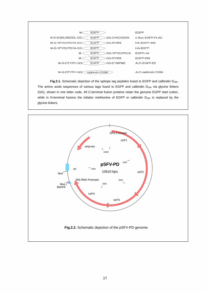
Fig.2.1. Schematic depiction of the epitope tag peptides fused to EGFP and calbindin D28K.
The amino acids sequences of various tags fused to EGFP and calbindin D28K via glycine linkers
(GG), shown in one letter code. All C-terminal fusion proteins retain the genuine EGFP start codon,
while in N-terminal fusions the initiator methionine of EGFP or calbindin D28K is replaced by the
glycine linkers.
pSFV-PD10610 bps
2000
4000
6000
8000
10000
BamHIXhoI
NruI
nsP1
nsP2
nsP3
nsP4
26S-RNA Promoter
ori
amp-res
SP6 Promoter
Fig.2.2. Schematic depiction of the pSFV-PD genome.
37
pBluescript II KS2961 bps
500
1000
1500
2000
2500
PsiIDraIIIBsaAI
NaeINgoMIV
BssHII
Ecl136IISacIBstXIBtgISacIINotIEco52IXbaISpeIBamHISmaIXmaIPstIEcoRIEcoRVHindIIIClaIAccIHincIISalIXhoIEcoO109IApaIPspOMIAcc65IKpnI
BssHII
SapI
AflIIINspIPciI
AlwNI
AhdI
BsaIBpmI
ScaITatIBsaHI
XmnI
F1-Ori
T7
T3
pUC Ori
Amp
pBluescript-modified2889 bps
500
1000
1500
2000
2500
BstUIPsiI
DraIIIBsaAI
BanIINaeINgoMIV
BstUIBstUIBstUIHgaI
BstUIBstUI
BanIISacISpeIHgaIAflIIIMluIBstUIKpnI
BstUI
BstUIBstUI
SapI
AflIIIBspLU11INspI
BstUIHgaI
AlwNI
BstUIHgaI
AhdIBstUIBsaI
BpmI
ScaITatIBsaHI
HgaIBstUI
XmnI
BstUI
F1-Ori
T7
T3
pUC Ori
Amp
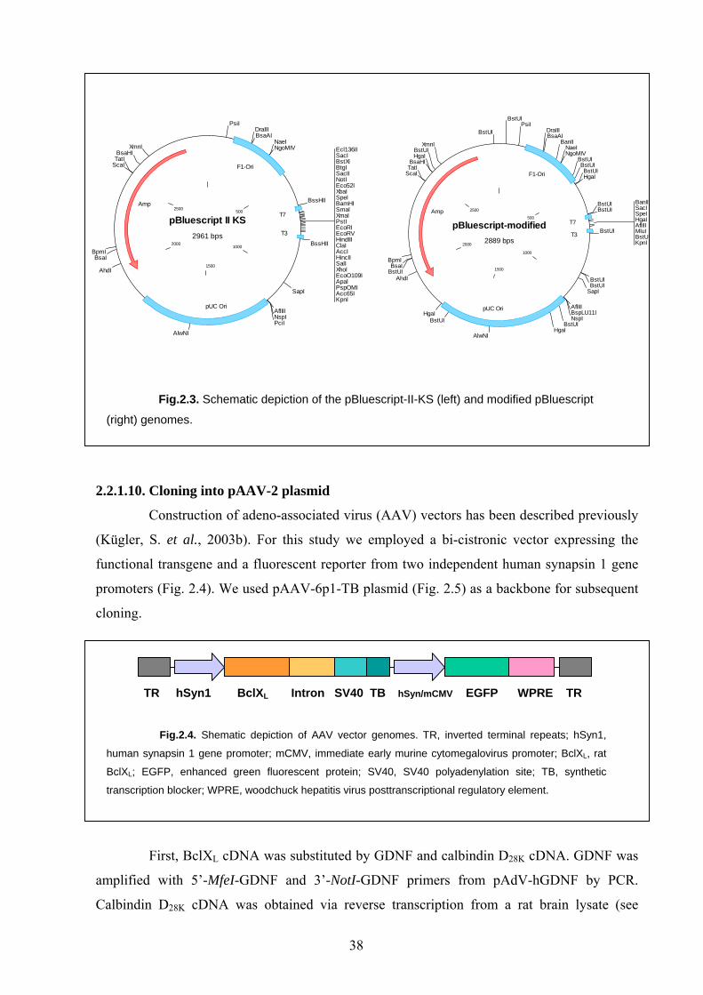
Fig.2.3. Schematic depiction of the pBluescript-II-KS (left) and modified pBluescript
(right) genomes.
2.2.1.10. Cloning into pAAV-2 plasmid
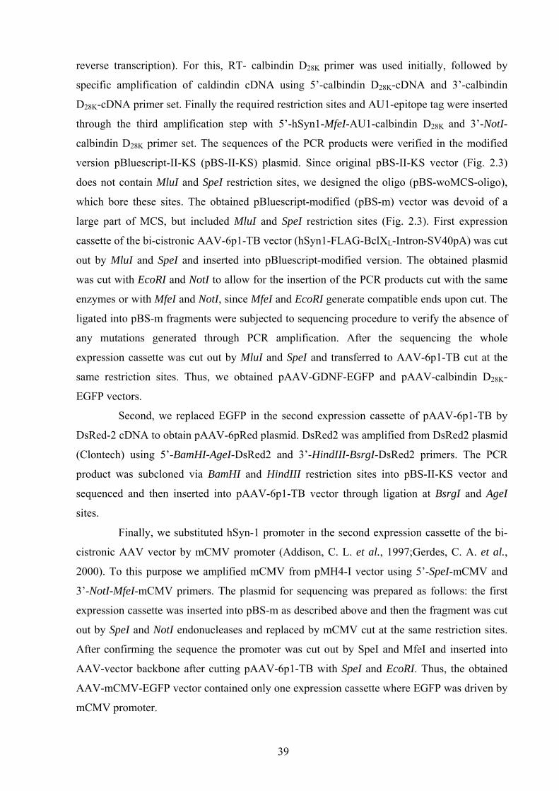
Construction of adeno-associated virus (AAV) vectors has been described previously
(Kügler, S. et al., 2003b). For this study we employed a bi-cistronic vector expressing the
functional transgene and a fluorescent reporter from two independent human synapsin 1 gene
promoters (Fig. 2.4). We used pAAV-6p1-TB plasmid (Fig. 2.5) as a backbone for subsequent
cloning.
TR hSyn1 BclXL Intron SV40 TB hSyn/mCMV EGFP WPRE TR
Fig.2.4. Shematic depiction of AAV vector genomes. TR, inverted terminal repeats; hSyn1,
human synapsin 1 gene promoter; mCMV, immediate early murine cytomegalovirus promoter; BclXL, rat
BclXL; EGFP, enhanced green fluorescent protein; SV40, SV40 polyadenylation site; TB, synthetic
transcription blocker; WPRE, woodchuck hepatitis virus posttranscriptional regulatory element.
First, BclXL cDNA was substituted by GDNF and calbindin D28K cDNA. GDNF was
amplified with 5’-MfeI-GDNF and 3’-NotI-GDNF primers from pAdV-hGDNF by PCR.
Calbindin D28K cDNA was obtained via reverse transcription from a rat brain lysate (see
38
reverse transcription). For this, RT- calbindin D28K primer was used initially, followed by
specific amplification of caldindin cDNA using 5’-calbindin D28K-cDNA and 3’-calbindin
D28K-cDNA primer set. Finally the required restriction sites and AU1-epitope tag were inserted
through the third amplification step with 5’-hSyn1-MfeI-AU1-calbindin D28K and 3’-NotI-
calbindin D28K primer set. The sequences of the PCR products were verified in the modified
version pBluescript-II-KS (pBS-II-KS) plasmid. Since original pBS-II-KS vector (Fig. 2.3)
does not contain MluI and SpeI restriction sites, we designed the oligo (pBS-woMCS-oligo),
which bore these sites. The obtained pBluescript-modified (pBS-m) vector was devoid of a
large part of MCS, but included MluI and SpeI restriction sites (Fig. 2.3). First expression
cassette of the bi-cistronic AAV-6p1-TB vector (hSyn1-FLAG-BclXL-Intron-SV40pA) was cut
out by MluI and SpeI and inserted into pBluescript-modified version. The obtained plasmid
was cut with EcoRI and NotI to allow for the insertion of the PCR products cut with the same
enzymes or with MfeI and NotI, since MfeI and EcoRI generate compatible ends upon cut. The
ligated into pBS-m fragments were subjected to sequencing procedure to verify the absence of
any mutations generated through PCR amplification. After the sequencing the whole
expression cassette was cut out by MluI and SpeI and transferred to AAV-6p1-TB cut at the
same restriction sites. Thus, we obtained pAAV-GDNF-EGFP and pAAV-calbindin D28K-
EGFP vectors.
Second, we replaced EGFP in the second expression cassette of pAAV-6p1-TB by
DsRed-2 cDNA to obtain pAAV-6pRed plasmid. DsRed2 was amplified from DsRed2 plasmid
(Clontech) using 5’-BamHI-AgeI-DsRed2 and 3’-HindIII-BsrgI-DsRed2 primers. The PCR
product was subcloned via BamHI and HindIII restriction sites into pBS-II-KS vector and
sequenced and then inserted into pAAV-6p1-TB vector through ligation at BsrgI and AgeI
sites.
Finally, we substituted hSyn-1 promoter in the second expression cassette of the bi-
cistronic AAV vector by mCMV promoter (Addison, C. L. et al., 1997;Gerdes, C. A. et al.,
2000). To this purpose we amplified mCMV from pMH4-I vector using 5’-SpeI-mCMV and
3’-NotI-MfeI-mCMV primers. The plasmid for sequencing was prepared as follows: the first
expression cassette was inserted into pBS-m as described above and then the fragment was cut
out by SpeI and NotI endonucleases and replaced by mCMV cut at the same restriction sites.
After confirming the sequence the promoter was cut out by SpeI and MfeI and inserted into
AAV-vector backbone after cutting pAAV-6p1-TB with SpeI and EcoRI. Thus, the obtained
AAV-mCMV-EGFP vector contained only one expression cassette where EGFP was driven by
mCMV promoter.
39
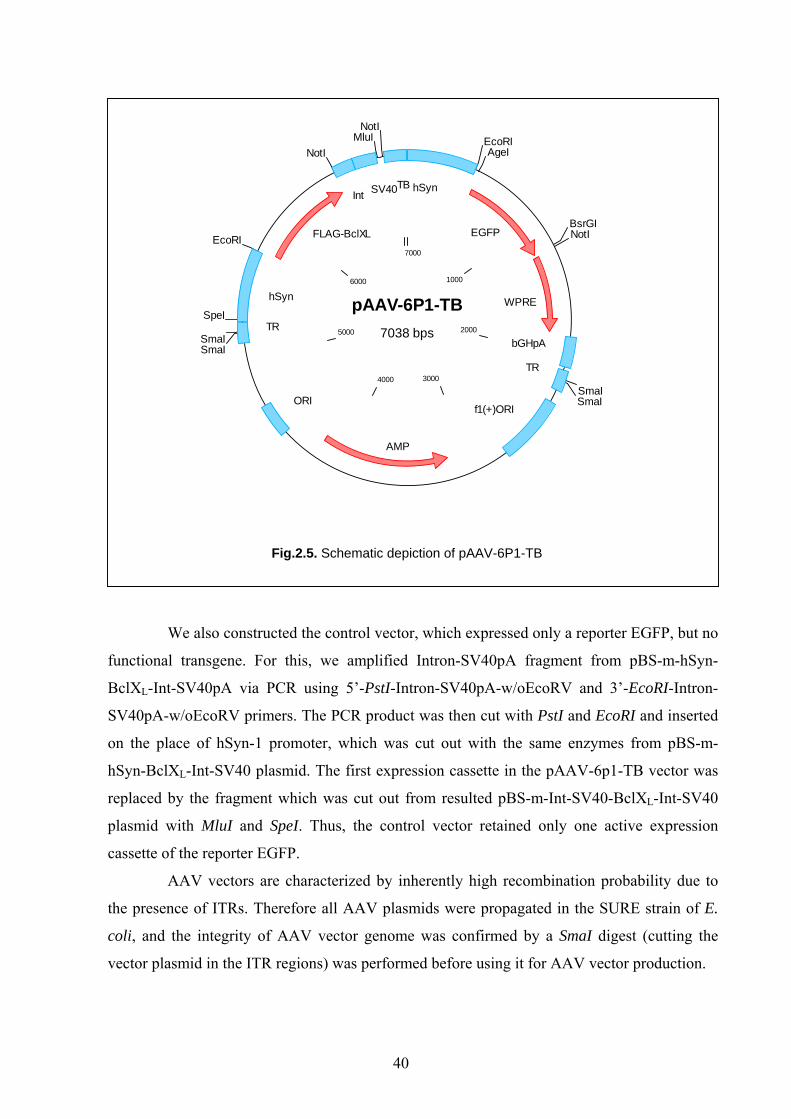
pAAV-6P1-TB7038 bps
1000
2000
30004000
5000
6000
7000
EcoRIAgeI
BsrGINotI
SmaISmaI
SmaISmaI
SpeI
EcoRI
NotIMluI
NotI
hSyn
EGFP
WPRE
bGHpA
TR
f1(+)ORI
AMP
ORI
TR
hSyn
FLAG-BclXL
Int SV40TB
Fig.2.5. Schematic depiction of pAAV-6P1-TB
We also constructed the control vector, which expressed only a reporter EGFP, but no
functional transgene. For this, we amplified Intron-SV40pA fragment from pBS-m-hSyn-
BclXL-Int-SV40pA via PCR using 5’-PstI-Intron-SV40pA-w/oEcoRV and 3’-EcoRI-Intron-
SV40pA-w/oEcoRV primers. The PCR product was then cut with PstI and EcoRI and inserted
on the place of hSyn-1 promoter, which was cut out with the same enzymes from pBS-m-
hSyn-BclXL-Int-SV40 plasmid. The first expression cassette in the pAAV-6p1-TB vector was
replaced by the fragment which was cut out from resulted pBS-m-Int-SV40-BclXL-Int-SV40
plasmid with MluI and SpeI. Thus, the control vector retained only one active expression
cassette of the reporter EGFP.
AAV vectors are characterized by inherently high recombination probability due to
the presence of ITRs. Therefore all AAV plasmids were propagated in the SURE strain of E.
coli, and the integrity of AAV vector genome was confirmed by a SmaI digest (cutting the
vector plasmid in the ITR regions) was performed before using it for AAV vector production.
40
2.2.2. Viral vectors production and purification
2.2.2.1. SFV vectors
Recombinant Semliki-Forest virus (SFV) vectors with reduced cytotoxicity and
conditionally infective particles were generated as described (Lundstrom, K. et al., 2003).
pSFV-PD vector, containing epitope-tagged EGFP was linearized with NruI, while pSFV-
helper2 plasmid was lineraized with SpeI endonuclease. Linearization of the plasmids was
necessary for in vitro transcription step that followed. The reaction conditions were following:
3-5 µg DNA was mixed with 5 µl of rNTPs (10 mM each), 5 µl of Cap structure (Ribo m7G
Cap Analog, Promega), 5 µl of 10 x SP6-transcription buffer, 1.5 µl of RNasin, 3.5 µl of SP6
polymerase (Promega) and millipore sterile water to achieve a volume of 50 µl, and the
reaction mix was incubated for 1 h at 37°C. RNA was purified using RNeasy QIAGEN Kit,
precipitated as described above for DNA, and run in the denaturing RNA agarose gel (1%
agarose, 20 mM MOPS , 5 mM Na acetate, 1 mM EDTA, pH 7.0; 2% Formaldehyde) to check
for the purity. RNA concentration was determined at a photometer as described above for
DNA. Each vector production run required 30 µg of SFV-RNA , 15 µg of SFV-helper2 RNA
and only 2 x 10⁷ BHK-21 cells, which after electroporation (Bio-Rad Gene Pulser II: voltage =
0.36 kV, pulse controller- high resistance = 500 Ohm, capacitance = 75 µF) were seeded in one
6-well plate (Sarstedt) and cultured for 24 h at 31°C. Electroporation medium contained 1:1
DMEM/F12, 10% FCS, 4 mM glutamate, 1% PS. The supernatants containing the virus
particles were collected twice at 24 (2 ml/well) and 48 h (1 ml/well) after the transfection,
sterile filtered to remove the cell debris and stored at -20°C until processed further. Viral
particles were precipitated by ultracentrifugation of the activated by addition of α-
chymotrypsin treatment supernatants in a Beckman SW41 rotor (39,000 rpm, 1 h, 4°C) and
resuspended in a small volume of phosphate buffered saline (PBS). Titration of transducing
units was performed on BHK-21 cells by counting EGFP positive cells.
2.2.2.2. AAV vectors
AAV-293 cells were harvested after they reached ca. 50% confluency in 175 cm2
culture flasks and then seeded in cell factories (Nunc) at 1.1 x 10⁸ cells per factory in 700 to
800 ml of medium (see cell culturing). Typically 6 culture flasks are required for seeding of
one cell factory. When the cells reached 50-60% confluency (ca. 24 h), they were subjected to
calcium-phosphate transfection. Cells were first washed with ca. 400 ml of DMEM. The
transfection mix contained 265 µg of AAV-plasmid, 1 mg of helper plasmid (pDG for AAV-2
and pDP5 for AAV-5), 0.25M CaCl2 in 16.5 ml of deionized sterile water. 16.5 ml of 2 x HBS
(pH 7.05) were added to 16.5 ml of transfection mix and incubated for 1 min at 25°C. The
41
mixture was added to 315 ml of DMEM/ 2%FCS and the obtained transfection medium was
equally distributed through the layers of the cell factory. For the transfection the cell factories
were incubated overnight (12-14 h) at 37°C, 5% CO2, 95% humidity. The medium was
changed then to 10% FCS/ 1% PS/ DMEM (ca. 750 ml/ cell factory). At ca. 36-40 h after
transfection the cells were checked for reporter fluorescence at the microscope and then
harvested. After removing the medium from the cell factory, the cells were briefly washed 300
ml with citric saline (CS). Then 200 ml of CS were added and the cells incubated at 37°C for
several min until detached and then collected to a sterile bottle. The remaining cells were
collected by a second rinse with ca. 100 ml of CS. Collected cells were centrifuged then for 5-
10 min at 1,300 rpm at RT. The cell pellets (the volume of the pellet was noted) were
resuspended in total volume of 20 ml of Tris-buffered saline (150 mM NaCl, 50 mM Tris, pH
8.5) and stored at -80°C until further processing. For virus gradient centrifugation the virus-
containing cells were first thawed and frozen 3 times in an ethanol/ dry ice bath. 1.6 µl of
Bezonase (300 U/ml) was added to the cells and incubated for 30 min at 37°C water bath. The
cell mix was slightly shaken every 5-10 min during incubation. Following incubation, the cell
mix was centrifuged at 4000 rpm 18°C for 30 min. The supernatants were carefully transferred
to new 50 ml falcon tube and adjusted to a 30 ml volume by addition of ca. 10 ml of Tris-
buffered saline. After washing the pump catheter connected to a 20G needle placed into a 50
ml falcon tube pump with millipore sterile water, the iodixanol (Opti PrepTM Fresenius Kabi,
Norge, Axis-Shield, Oslo, Norway) step gradient was performed. For this, the following
solutions were prepared first:
B = 10 x PBS-MK (80g NaCl + 2 g KCl + 14.4 g Na2HPO4 + 2.4 g KH2PO4 dissolved in 990
ml millipore sterile water, pH 7.4; and then 5 ml 2,5 M KCl and 5 ml 1 M MgCl2 were added
and the solution sterile filtered).
C = 1x PBS-MK (500 µl 2.5 M KCl; 500 µl 1M MgCl2 in 499 ml PBS)
D = 2 M NaCl in 1x PBS-MK
E = 54 % working solution: (45 ml iodixanol + 5 ml B).
For obtaining different iodixanol gradient solutions the solutions C, D, E, iodixanol and phenol
red were intermixed as it is shown in the following table:
42
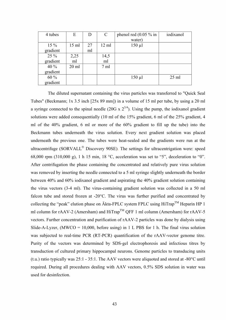
4 tubes E D C phenol red (0.05 % in water)
iodixanol
15 % gradient
15 ml 27 ml
12 ml 150 µl
25 % gradient
2,25 ml
14,5 ml
40 % gradient
20 ml 7 ml
60 % gradient
150 µl 25 ml
The diluted supernatant containing the virus particles was transferred to "Quick Seal
Tubes" (Beckmann; 1x 3.5 inch [25x 89 mm]) in a volume of 15 ml per tube, by using a 20 ml
a syringe connected to the spinal needle (20G x 23/4). Using the pump, the iodixanol gradient
solutions were added consequentially (10 ml of the 15% gradient, 6 ml of the 25% gradient, 4
ml of the 40% gradient, 6 ml or more of the 60% gradient to fill up the tube) into the
Beckmann tubes underneath the virus solution. Every next gradient solution was placed
underneath the previous one. The tubes were heat-sealed and the gradients were run at the
ultracentrifuge (SORVALL® Discovery 90SE). The settings for ultracentrigation were: speed
68,000 rpm (310,000 g), 1 h 15 min, 18 °C, acceleration was set to “5”, deceleration to “0”.
After centrifugation the phase containing the concentrated and relatively pure virus solution
was removed by inserting the needle connected to a 5 ml syringe slightly underneath the border
between 40% and 60% iodixanol gradient and aspirating the 40% gradient solution containing
the virus vectors (3-4 ml). The virus-containing gradient solution was collected in a 50 ml
falcon tube and stored frozen at -20°C. The virus was further purified and concentrated by
collecting the “peak” elution phase on Äkta-FPLC system FPLC using HiTrapTM Heparin HP 1
ml column for rAAV-2 (Amersham) and HiTrapTM QFF 1 ml column (Amersham) for rAAV-5
vectors. Further concentration and purification of rAAV-2 particles was done by dialysis using
Slide-A-Lyzer, (MWCO = 10,000, before using) in 1 L PBS for 1 h. The final virus solution
was subjected to real-time PCR (RT-PCR) quantification of the rAAV-vector genome titre.
Purity of the vectors was determined by SDS-gel electrophoresis and infectious titres by
transduction of cultured primary hippocampal neurons. Genome particles to transducing units
(t.u.) ratio typically was 25:1 - 35:1. The AAV vectors were aliquoted and stored at -80°C until
required. During all procedures dealing with AAV vectors, 0.5% SDS solution in water was
used for desinfection.
43
2.2.3. Cell culturing
2.2.3.1. Continuous cell culture
Working with continuous cell culture was always performed under sterile conditions.
Mammalian AAV-293 were cultured in DMEM supplemented with 10% FCS and 1% PS and
BHK-21 cells in DMEM/F12 1:1 supplemented with 10% FCS, 4 mM glutamine, 1% PS in
175 cm2 culture flasks (Sarstedt) at 37°C, 5% CO2 supply, 95 % humidity. After reaching
approximately 70% confluency, the cells were split 1:4 (every 2-4 days). For this, the medium
was removed, 4 ml of pre-warmed to 37°C 0.05% trypsin/ 0.02% EDTA solution were added
to the culture and left in the incubator at 37% for 5-12 min until detached from the flask.
Trypsin activity was stopped by addition of 7 ml of the cell culture medium to the flask. The
mix containing detached cells was carefully transferred to a 15 ml Falcon tube and spun down
at 4,000 g, 4°C, 5 min. After removing the supernatant a cell pellet was resuspended in 100 ml
of the fresh medium and seeded in 4 (175cm2) flasks.
2.2.3.2. Primary culture of hippocampal neurons
Coating of 24-well plates (Nunc) or coverslips with polyornithine/ laminin is
particularly useful for culturing CNS neurons. For this, ethanol-soaked and flamed coverslips
(∅10 mm) incubated with polyornithine solution (500 µl/well, 100 µg/ml in millipore sterile
water) for 12-24 h at RT, then washed twice with 1 ml/well of millipore sterile water and
coated with laminin (500 µl/well, 1µg/ml in NBM) by incubation at 37°C, 5% CO2, 95 %
humidity overnight. Before the preparation of the hippocampi, the coverslips were washed
twice with NBM and then 500 µl of hippocampus medium (1% PS-N, 0.5 mM L-glutamine,
0.05% Transferrin, 2% B27 Supplement in NBM) was added and the plates kept at 37°C. To
obtain primary hippocampal neurons, hippocampi were dissected from Wistar rat embryos
(embryonic day 18) and further processed for establishing dissociated cell cultures. All surgical
procedures were performed on ice. In brief, hippocampal tissue pieces were collected in ice-
cold hippocampus medium and centrifuged at 1,000 rpm for 4 min. The medium was removed
and the tissue pellet was incubated in 750 µl trypsin (0.25%, 15 min, 37°C). Trypsin was
inactivated by addition of 750 µl ice-cold FCS. The pellet was then dissociated by gentle
trituration using a fire-polished Pasteur pipette. After centrifugation at 1,000 rpm for 4 min, the
pellet was resuspended in warm hippocampus culture medium. Cells were seeded in poly-L-
ornithine/laminin 24-well culture plates (Nunc) at a density 250,000 cells/well. Cultures were
maintained at 37°C in a humidified atmosphere and 5% CO2 in hippocampus medium.
44
2.2.3.3. SFV-transduction and cell lysis for western blotting
After reaching 70% confluency HEK-293 cells seeded in a 6-well plate were
transduced by addition of 5 x 10⁵ SFV t.u. per well in a 500µl volume of OptiMEM and kept at
31°C, 5% CO2, 95% humidity for 1 h. Then 1.5 ml of OptiMEM was added to each well and
the plates were transferred to a 35°C, 5% CO2, 95% humidity for overnight incubation (12-13
h). After removing the medium the cells were washed twice with PBS, the lysis buffer for cells
was added (250µl/well) and the cells scraped from the plate and transferred to a 1.5 ml
eppendorf tube. The lysates were boiled for 5 min and chilled on ice. After addition of protease
inhibitor mix the lysates were centrifuged at 4°C at maximum speed for 20 min. The
supernatant was collected, aliquoted and frozen at -20°C.
2.2.3.4. Indirect immunofluorescence on primary neurons
For transduction of primary hippocampal neurons seeded in 24-well plates 1 x 10⁸ t.u.
of AAV-2 vector in 500 µl of hippocampal medium were used per well. After the reporter gene
fluorescence (either EGFP or DsRed2) could be clearly detected by microscopy (us. 4-5 days
after AAV-2 transduction), the cultured neurons were subjected to indirect
immunofluorescence assay. The coverslips with primary hippocampal neurons were rinsed
with PBS and fixed by incubation in 4% formaldehyde for 10 min at room temperature. After
washing 2 times for 5 min with PBS the cells were subjected to permeabilization procedure by
incubation with ice-cold acetone at -20°C for 5 min. Following a second washing with PBS (3
x 5 min), blocking solution (10% NGS in PBS) was applied for 30 min at room temperature to
block unspecific binding of the antibodies. Incubation with a primary antibody (all anti-tag
antibodies were used in a dilution of 1:500) was performed for 1 h at 37°C. The cells were
rinsed with PBS 3 x 5 min and then incubated for 1 h at room temperature with a secondary
antibody coupled to a fluorescent reporter cy3 (1:200). The cells were washed again 3 x 5 min
with PBS and mounted on the glass slides embedded in Moviol mounting medium (16 %
Moviol in 30 % Glycerol/ PBS, 0.02 % sodium azide) to protect immunofluorescence from
bleaching.
2.2.4. Animal procedures
Adult female Wistar rats (University Hospital Animal Facility, Göttingen) of 250-
280g of weight were housed at 12h/12h of light/dark cycle, provided with food and water ad
libitum. Animals were deeply anaesthetised by 7% chloral hydrate intraperitoneally
(340mg/kg) before the respective surgery. All animal procedures were performed in
45
accordance to German guidelines for care and use of laboratory animals and to the respective
European Community Council Directive 86/609/EEC.
2.2.4.1. Stereotaxic injection into the rat brain
The head of the rat was fixed by ear bars and a jaw holder in a stereotaxic frame
(“Kopf” Instruments) after the appropriate anaesthesia. A skin covering the skull was
longitudinally cut from the imaginary line connecting the eyes in front till the imaginary line
connecting the auditory channels in the back. Remaining connective tissue was carefully
removed aside by scalpel in order to achieve appropriate visualisation of the skull sutures and
to define the bregma (a crossing point of parasaggital with coronal sutures) on the surface of
the skull as “zero” or “start” point. After achieving flat positioning of the skull, the coordinates
for the injection were calculated related to bregma. For the injection into the SNpc, the
following coordinates were used according to Paxinos and Watson atlas of a rat brain: antero-
posterior (AP): -0.53; medio-lateral (ML): +0.22; dorso-ventral (DV): -0.78. The coordinates
for the injection into the LMFB were the following: AP: -0.2; ML: +0.2; DV: -0.85; into the
striatum: AP: +0.2 mm; ML: -2.8 mm; DV: -5.6 mm; and into hippocampus: AP: -4.3; ML:
+3.0; DV: -3.4 mm. Glass capillaries (World Precision Instruments, WPI), filled with mineral
oil and the solution for the injection were attached to the nanoliter injector (WPI). The injector
was connected to a microprocessor-based controller, Micro4 smart controller (WPI), which
allows for setting the operating parameters, such as the volume and the speed (nl/min) of the
injection. AAV-vectors were injected into SNpc in a volume of 2 µl and in the striatum in a
volume of 2-4 µl depending on the original infectious titre of the virus. The volume 3 µl of
5mg/ml of 6-hydroxydopamine hydrochloride in 0.2% ascorbic acid/PBS was injected in
LMFB to produce the unilateral PD lesion model. The speed for all injections was set to
500nl/min.
2.2.4.2. Intravitreal injections and optic nerve axotomy
Wistar rats of 200-270g were anesthetized with 7% chloral hydrate (340mg/kg body
weight). Pupillary dilatation for better visualisation of the lens was achieved with subsequent
surface application of 1% tropicamide and 2% mepivacaine. Injections into the eye were
performed by a fine bore glass capillary with a tip diameter of 30 µm attached to a catheter on
a Hamilton syringe. 2 µl (1 x 10⁵ transducing units of SFV, 1 x 10⁷ transducing units of AAV)
of viral suspension were injected intravitreally by puncturing the eyeball below the cornea-
sclera junction. Due to pupillary dilatation, the tip could be observed during the injection and
care was taken to avoid contact with the lens. Vector injections were performed 3 weeks before
46
the optic nerve axotomy. After skin incision close to the superior orbital rim, the orbita was
opened and lacrimal gland was partially removed to achieve a better visualisation of the optical
nerve and surrounding vessels. Following the spreading of superior extraocular muscle by a
small retractor, the optical nerve was exposed by longitudinal incision of the perineurium.
Optic nerve was transected at a distance of ca. 2 mm from the posterior pole of the eye leaving
the retinal blood supply intact. Retrograde fluorogold (FG) labelling of RGCs was performed
by putting a small sponge soaked with FG tracer onto the proximal to the eyeball optic nerve
stump. Animals with retinal ischemia, as verified after the surgery fundoscopically, have been
excluded from the study.
2.2.4.3. Transcardial perfusion and brain tissue processing
Transcardial perfusion under terminal anaesthesia is a commonly used method for
tissue fixation in immunohistochemical protocols. This method takes advantage of the animal’s
circulatory system to deliver the fixative solution evenly throughout the body tissues, with
optimal penetration of the brain. Fixation preserves the ultrastructure, stabilizes protein and
peptide conformation so that antibodies can bind to antigen sites. We used 4%
paraformaldehyde (PFA) in phosphate buffer solution (PBS) as a fixative since it is optimal for
visualization using fluorescent dyes.
After a deep anaesthesia with 7% chloral hydrate (800 mg/ml) the rat was fixed on
rack, the abdominal cavity was opened with sharp scissors, and the diaphragm was cut to allow
access to the thorax. With large scissors, blunt side down, ribs were bilaterally cut and the rib
cage was open to allow access to the heart. While holding heart steady, the left ventricle was
cut and the catheter blunt-ended needle inserted through the ventricle and atrium into aorta.
Needle position was secured by clamping in place. The liver was cut then to allow a large part
of blood volume to leave the body. The abdominal aorta was clamped to restrict perfusion to
the upper par of the body. The nose was cut for the control of procedure. Thus, if fluid was
flowing freely from the cut nose the needle was considered to be appropriately positioned. The
syringe containing ice-cold PBS and connected to the catheter was operated with a speed of
15–20 ml/min. When blood has been cleared from body (ca. 200 ml of PBS is usually
required), PBS was replaced with ice-cold 4% PFA solution (200 ml). The rat was decapitated
and the brain carefully removed from the skull and placed into 4% PFA overnight at 4°C for
post-fixation. The brain was cryoprotected by dehydration in 30% sucrose solution in PBS,
frozen on dry ice and cut on a cryostat (Leica CM 3050 S) to 18 µm coronary sections.
47
2.2.4.4. Preparation of the brain tissue lysates for Western Blotting
Rats were sacrificed by an overdose of chloral hydrate (1mg/kg) and transcardially
perfused with 150 ml ice-cold PBS. The brain was removed and quickly frozen on dry ice. 3
mm thick coronal section containing the substantia nigra was cut out and subjected to biopsy
in the SNpc region by using a small biopsy needle with 2 mm inner diameter. The excised
tissue was weighted and homogenized in a tissue lysis buffer by sonication and incubated on
ice for 15 min. The lysates were then centrifuged twice at 4°C with maximum speed for 15 min
to remove the debris. The supernatants were aliquoted and kept frozen at -20°C.
2.2.5. Indirect immunofluorescence on brain slices
Brain slices were dried for 1 h at 37°C, then rehydrated in PBS for 45 min and
exposed to antigen retrieval in TBS (pH 9.0) for 5 h in a water bath at 60°C. While regular
antigen retrieval destroys EGFP fluorescence, this procedure allows for keeping it intact. The
slides were shortly washed with PBS and incubated in a blocking solution for 30 min at room
temperature to avoid unspecific binding of the antibodies. Incubation with primary antibody
(anti-TH, 1:600; anti-calbindin 1:1000, anti-GFAP, 1:500; anti-NeuN 1:200; all anti-tag
antibodies were used in 1:500 dilution after testing different dilutions, ranging from 1:200 to
1:5,000) was performed overnight at 4°C in incubation solution -1 for IF. After washing 3 x 5
min with PBS the secondary cy3-coupled antibody (1:250 in incubation solution -2 for IF) was
applied for 1 h at room temperature. The secondary antibody was washed out with PBS (3 x 5
min). Slides were embedded in Moviol, coated by coverslips and kept at 4°C until microscopy.
2.2.6. Protein handling procedures
Western Blotting or immunoblotting allows for determination, with a specific primary
antibody, the relative amounts of the protein present in different samples. Briefly, 1) samples
are prepared from tissues or cells that are homogenized in a buffer that protects the protein of
interest from degradation; 2) the sample is separated using SDS-PAGE and then transferred to
a membrane for detection; 3) the membrane is incubated with a generic protein (such as milk
proteins) to bind to any remaining sticky places on the membrane. A primary antibody is then
added to the solution which is able to bind to its specific protein; 4) a secondary antibody-
enzyme conjugate, which recognizes the primary antibody, is added to find locations where the
primary antibody bound.
48
2.2.6.1. Protein concentration determination
Protein concentration was determined by Bradford assay. The assay is based on the
observation that the absorbance maximum for an acidic solution of Coomassie Brilliant Blue
G-250 shifts from 465 nm to 595 nm when binding to protein occurs. Both hydrophobic and
ionic interactions stabilize the anionic form of the dye, causing a visible colour change. The
reaction was performed in a 96-well plate at room temperature. Protein standards were
prepared in PBS in a concentration range between 0.1-1.4 mg/ml. Unknown protein samples
were diluted to 1:2, 1:10 and 1:100. 5 µl of protein standards or diluted unknowns were mixed
with 250 µl of Bradford reagent in each well and incubated for 15 min. The absorbance of the
samples was measured at 595 nm on a TECAN Rainbow plate reader using the easyWINbasic
software (SLT Company). Plotting of the absorbance vs. the protein concentration for protein
standards to obtain a standard curve and calculation of the unknown samples protein
concentration was done using the Microsoft Office 2003 Excel software.
2.2.6.2. SDS-polyacrylamide gel electrophoresis (SDS-PAGE)
SDS-PAGE is based on separation of a large range of proteins of varying molecular
weights and charges under the influence of the electrical field within the continuous cross-
linked polymer matrix polyacrylamide/ bis-acrylamide (Rothiphorese). Cross-linking is
catalysed by free radicals produced upon addition of ammonium peroxide and TEMED. Two-
phase gels were used for collection and separation of the proteins according to their molecular
weight. Usually, 12% resolving gel and 5% stacking gel were used to separate the proteins of
interest. To define a molecular weight of loaded proteins the molecular weight marker was
loaded and separated in parallel. The protein concentration in the samples was adjusted by
dilution of more concentrated samples in PBS. Equal volumes of protein samples and 2 x SDS-
loading buffer were mixed, subjected to heating at 95°C for 5 min, chilled on ice and loaded in
the gel combs. SDS-PAGE was run at 4°C in ice-cold electrophoresis buffer. All equipment
used for SDS-PAGE and WB was purchased from Bio-Rad. Electric field of 70 V was applied
for 15 min to allow samples to enter the gel and collect without a smearing and then increased
to 100 V and kept constant until the bromophenol blue reached the bottom of the resolving gel.
2.2.6.3. Immune blotting
For the western blot procedure a Mini Trans-Blot Cell setup (Bio-Rad) was
employed. After electrophoresis completion the polyacrylamide gel was placed between two
sheets of Whatman filter paper and a nitrocellulose membrane (Protran), all preliminary soaked
in transfer buffer. Two additional Whatman filter papers were placed on the top on the
49
membrane and the “sandwich” was placed in the Mini Tans-Blot Cell. A voltage of 100 V was
applied for 1 h at 4°C.
After the completion of the transfer the membrane was treated with blocking solution
for 1 h at room temperature to avoid unspecific binding of the antibody. Incubation with both
primary and secondary antibodies was performed in 2% Milk/TBS-T solution for 1 h at room
temperature. The membrane was washed 3 x 10 min with TBS-T between primary and
secondary antibody application and 3 x 15 min with TBS-T after the incubation with secondary
antibody. The following dilutions of the primary antibodies were used: anti-EGFP, 1:1,000;
anti-FLAG 1:500; anti BclX, 1:2,000. All secondary antibodies coupled to HRP were used in
dilution 1:3000. Together with a secondary antibody streptavidin peroxidase was applied in
dilution 1:3000 to allow for later visualization of biotinylated molecular weight protein marker
(Sigma). For developing the membrane equal volumes of ECL-1 and ECL-2 reagents were
mixed, applied for 1 min in the dark onto the membrane and imaged at Fluor-S-Max Gel
Imager (Bio-Rad) and Quantity One software (version 4.2.1).
2.2.7. GDNF-ELISA
Quantification of GDNF tissue levels in transduced and contralateral SNpc was
determined by ELISA (GDNF Emax® ImmunoAssay System, Promega) according to the
instructions of the manufacturer. Tissue samples of SNpc were obtained from 3 mm thick
tissue slices by means of small biopsy needles with inner diameter of 2 mm and lysed
according to the procedure described in section 2.2.4.4. After an overnight coating of a 96 well
plate, the specific protein is detected using an antibody sandwich format. To this purpose 96
well plates are coated with anti-GDNF monoclonal antibody, which binds soluble GDNF. The
captured GDNF is then bound by a second, specific polyclonal antibody. After several washing
steps, the amount of specifically bound polyclonal antibody is detected using a species-specific
antibody conjugated to horseradish peroxidase as a tertiary reactant. The unbound conjugate is
removed by washing, and after incubation with a chromogenic substrate, the colour change is
detected. The amount of GDNF in the test solutions corresponds to the colour generated in the
oxidation-reduction reaction. Using this system, GDNF in tissue culture supernatants or tissue
homogenates can be quantitatively measured.
2.2.8. Microscopy and image analysis
Fluorescence from tissue sections and fixed cultured cells was recorded on a Zeiss
Axioplan-2 microscope equipped with a 16 bit greyscale CCD camera (Zeiss). Narrow band
filter sets specific for EGFP and cy3 fluorescence were used. For all recorded EGFP images
50
exposure time was 3 000 ms, except for pictures taken from HA-tag transduced sections which
are labelled EGFP x 5. These images were recorded with an exposure time of 15,000ms. cy3
fluorescence was recorded with an exposure time of 500 ms. Images from individual
fluorescences were recorded separately as greyscales. Pseudo-colouring of images, generation
of overlays and quantification of EGFP / cy3 co-localization was performed with AxioVision 3
software package (Zeiss).
2.2.9. Quantification of neuroprotection in 6-OHDA rat model of PD
At the respective time points after 6-OHDA injection into medial forebrain bundle
(MFB), rats (n = 5 per condition) were sacrificed by an overdose of chloral hydrate and
transcardially perfused with 4% PFA. Brains were processed to 18 µm slices, and
immunofluorescent visualization of dopaminergic neurons in SNpc was performed with
primary rabbit antibody against tyrosine hydroxylase (anti-TH, 1:500, Advanced
ImmunoChemicals Inc., USA; overnight incubation at 4°C) followed by 1h incubation at RT
with secondary cy3-conjugated goat anti-rabbit antibody (Dianova). Images of brain sections
were recorded as described above. TH-immunoreactive (TH-IR) cells were counted in every 8th
brain section in the region anatomically correspondent to SNpc. TH-IR cells of the ventral
tegmental area (VTA) area were excluded from counting. To obtain the corresponding total
DA cell numbers the counted values were multiplied by 8, assuming that the average diameter
of the adult rat DA-neuron is 16-18µm (Juraska, J. M. et al., 1977).
2.2.10. Rotational behaviour test
One of the most simple to perform and widely used test for evaluation of the
unilateral 6-hydroxydopamine lesion is drug-induced rotational behaviour test (Ungerstedt, U.
et al., 1970). Disbalance in dopamine secretion in the brain results in preferable turning of the
animal to the side of the lesion. When the rat is challenged with certain drugs (apomorphine,
amphetamine), this rotation tendency is greatly increased and leads to a typical “rotational
behaviour” when an animal “tries to catch its own tail”. The more complete the lesion is- the
more complete rotations to one side per min will be counted. To evaluate the degree of the
lesion of dopaminergic system each rat was subjected to intraperitoneal apomorphine
hydrochloride injection (0.2 mg/kg, Teclafarm) at 2 weeks after the lesion. If a rat exhibited 5
or more rotations per min toward the contralateral to the lesion side during one hour after
apomorphine injection, the lesion was considered to be complete. This typically corresponded
to the ≥ 80 % loss of dopaminergic neurons in SNpc and almost absolute loss of dopaminergic
51
projections of these neurons to the striatum of a rat brain as revealed by indirect TH-
immunofluorescence.
2.2.11. Statistics
RGC and TH-IR cell counts are presented as means ± standard deviations. For each
time point and experimental condition n = 5 were used. For retinas, 3 independent fields per
retina were counted, for TH-IR cells of SNpc region at least 13 sections were counted per
animal. Statistical significance was assessed by split-plot analysis of variance and alpha-
adjusting for multiplicity (Bonferroni-Holm sequentially rejective procedure) and, at the 1%
level, revealed p < 0.001 for either BclXL or GDNF transduced areas vs. untransduced and
versus the control AAV-EGFP-transduced SNpc at all time points. Analysis of variance (factor
1 - BclXL, factor 2 - GDNF, interaction 1 / 2 = BclXL + GDNF) revealed that interaction was
not additive (p<0.001), but more than additive (= synergistic) for retina, but not for the brain,
where it is additive at 2 weeks, but not significant at 6 weeks after lesion when compared to the
GDNF-treated group. Pairwise comparisons of values were performed by paired, one-tailed
Student’s t-test.
52
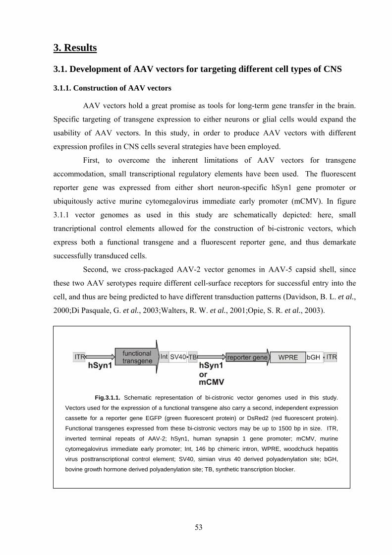
3. Results
3.1. Development of AAV vectors for targeting different cell types of CNS
3.1.1. Construction of AAV vectors
AAV vectors hold a great promise as tools for long-term gene transfer in the brain.
Specific targeting of transgene expression to either neurons or glial cells would expand the
usability of AAV vectors. In this study, in order to produce AAV vectors with different
expression profiles in CNS cells several strategies have been employed.
First, to overcome the inherent limitations of AAV vectors for transgene
accommodation, small transcriptional regulatory elements have been used. The fluorescent
reporter gene was expressed from either short neuron-specific hSyn1 gene promoter or
ubiquitously active murine cytomegalovirus immediate early promoter (mCMV). In figure
3.1.1 vector genomes as used in this study are schematically depicted: here, small
trancriptional control elements allowed for the construction of bi-cistronic vectors, which
express both a functional transgene and a fluorescent reporter gene, and thus demarkate
successfully transduced cells.
Second, we cross-packaged AAV-2 vector genomes in AAV-5 capsid shell, since
these two AAV serotypes require different cell-surface receptors for successful entry into the
cell, and thus are being predicted to have different transduction patterns (Davidson, B. L. et al.,
2000;Di Pasquale, G. et al., 2003;Walters, R. W. et al., 2001;Opie, S. R. et al., 2003).
Fig.3.1.1. Schematic representation of bi-cistronic vector genomes used in this study.
Vectors used for the expression of a functional transgene also carry a second, independent expression
cassette for a reporter gene EGFP (green fluorescent protein) or DsRed2 (red fluorescent protein).
Functional transgenes expressed from these bi-cistronic vectors may be up to 1500 bp in size. ITR,
inverted terminal repeats of AAV-2; hSyn1, human synapsin 1 gene promoter; mCMV, murine
cytomegalovirus immediate early promoter; Int, 146 bp chimeric intron, WPRE, woodchuck hepatitis
virus posttranscriptional control element; SV40, simian virus 40 derived polyadenylation site; bGH,
bovine growth hormone derived polyadenylation site; TB, synthetic transcription blocker.
53
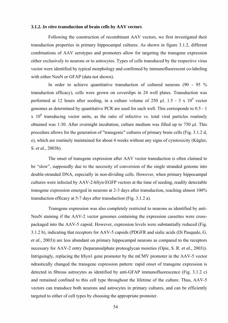
3.1.2. In vitro transduction of brain cells by AAV vectors
Following the construction of recombinant AAV vectors, we first investigated their
transduction properties in primary hippocampal cultures. As shown in figure 3.1.2, different
combinations of AAV serotypes and promoters allow for targeting the transgene expression
either exclusively to neurons or to astrocytes. Types of cells transduced by the respective virus
vector were identified by typical morphology and confirmed by immunofluorescent co-labeling
with either NeuN or GFAP (data not shown).
In order to achieve quantitative transduction of cultured neurons (90 - 95 %
transduction efficacy), cells were grown on coverslips in 24 well plates. Transduction was
performed at 12 hours after seeding, in a culture volume of 250 µl. 1.5 - 3 x 10⁹ vector
genomes as determined by quantitative PCR are used for each well. This corresponds to 0.5 - 1
x 108 transducing vector units, as the ratio of infective vs. total viral particles routinely
obtained was 1:30. After overnight incubation, culture medium was filled up to 750 µl. This
procedure allows for the generation of "transgenic" cultures of primary brain cells (Fig. 3.1.2 d,
e), which are routinely maintained for about 4 weeks without any signs of cytotoxicity (Kügler,
S. et al., 2003b).
The onset of transgene expression after AAV vector transduction is often claimed to
be “slow”, supposedly due to the necessity of conversion of the single stranded genome into
double-stranded DNA, especially in non-dividing cells. However, when primary hippocampal
cultures were infected by AAV-2-hSyn-EGFP vectors at the time of seeding, readily detectable
transgene expression emerged in neurons at 2-3 days after transduction, reaching almost 100%
transduction efficacy at 5-7 days after transduction (Fig. 3.1.2 a).
Transgene expression was also completely restricted to neurons as identified by anti-
NeuN staining if the AAV-2 vector genomes containing the expression cassettes were cross-
packaged into the AAV-5 capsid. However, expression levels were substantially reduced (Fig.
3.1.2 b), indicating that receptors for AAV-5 capsids (PDGFR and sialic acids (Di Pasquale, G.
et al., 2003)) are less abundant on primary hippocampal neurons as compared to the receptors
necessary for AAV-2 entry (heparansulphate proteoglycan moieties (Opie, S. R. et al., 2003)).
Intriguingly, replacing the hSyn1 gene promoter by the mCMV promoter in the AAV-5 vector
ndrastically changed the transgene expression pattern: rapid onset of transgene expression is
detected in fibrous astrocytes as identified by anti-GFAP immunofluorescence (Fig. 3.1.2 c)
and remained confined to this cell type throughout the lifetime of the culture. Thus, AAV-5
vectors can transduce both neurons and astrocytes in primary cultures, and can be efficiently
targeted to either of cell types by choosing the appropriate promoter.
54
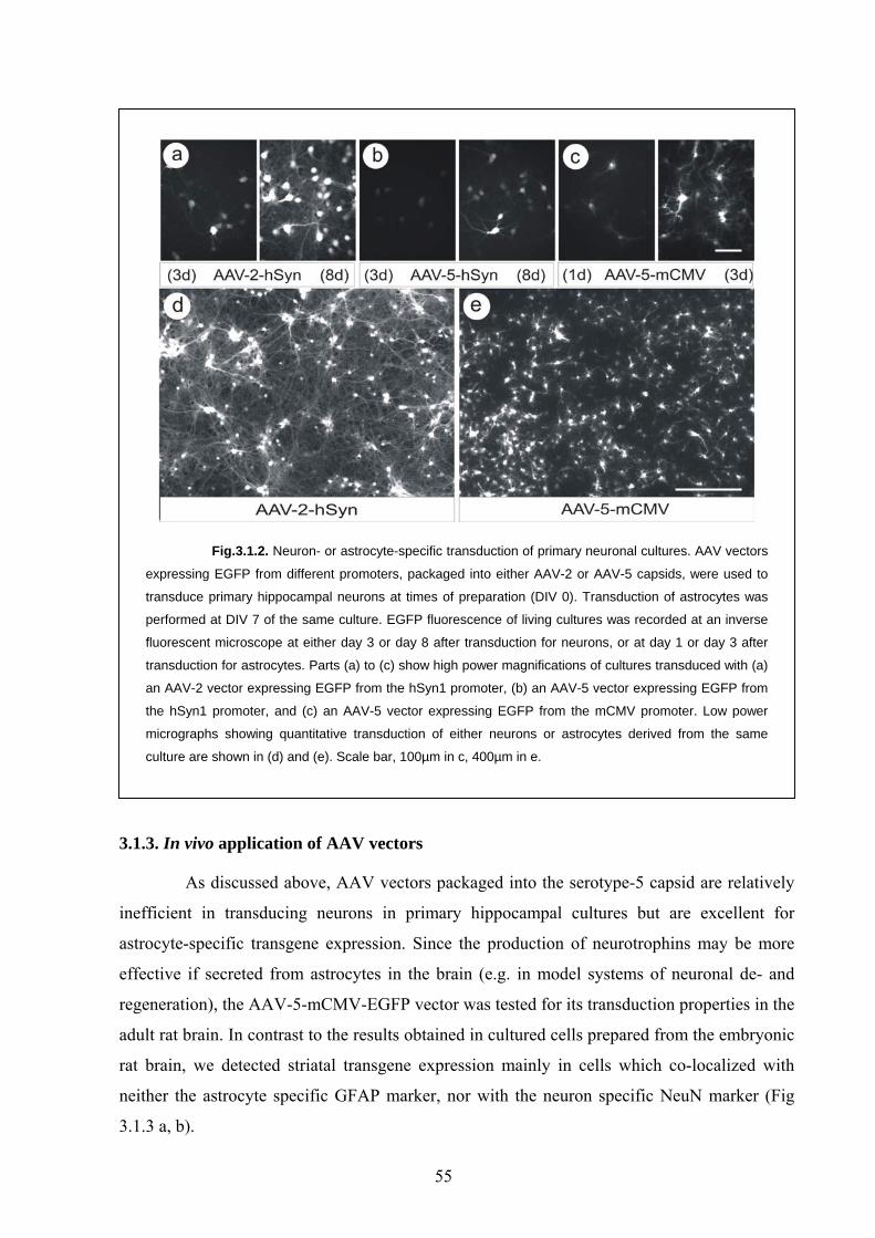
Fig.3.1.2. Neuron- or astrocyte-specific transduction of primary neuronal cultures. AAV vectors
expressing EGFP from different promoters, packaged into either AAV-2 or AAV-5 capsids, were used to
transduce primary hippocampal neurons at times of preparation (DIV 0). Transduction of astrocytes was
performed at DIV 7 of the same culture. EGFP fluorescence of living cultures was recorded at an inverse
fluorescent microscope at either day 3 or day 8 after transduction for neurons, or at day 1 or day 3 after
transduction for astrocytes. Parts (a) to (c) show high power magnifications of cultures transduced with (a)
an AAV-2 vector expressing EGFP from the hSyn1 promoter, (b) an AAV-5 vector expressing EGFP from
the hSyn1 promoter, and (c) an AAV-5 vector expressing EGFP from the mCMV promoter. Low power
micrographs showing quantitative transduction of either neurons or astrocytes derived from the same
culture are shown in (d) and (e). Scale bar, 100µm in c, 400µm in e.
3.1.3. In vivo application of AAV vectors
As discussed above, AAV vectors packaged into the serotype-5 capsid are relatively
inefficient in transducing neurons in primary hippocampal cultures but are excellent for
astrocyte-specific transgene expression. Since the production of neurotrophins may be more
effective if secreted from astrocytes in the brain (e.g. in model systems of neuronal de- and
regeneration), the AAV-5-mCMV-EGFP vector was tested for its transduction properties in the
adult rat brain. In contrast to the results obtained in cultured cells prepared from the embryonic
rat brain, we detected striatal transgene expression mainly in cells which co-localized with
neither the astrocyte specific GFAP marker, nor with the neuron specific NeuN marker (Fig
3.1.3 a, b).
55
Fig.3.1.3. AAV-5 vector targeting in the brain. An attempt to target transgene
expression from an AAV-5 vector to astrocytes in the rat brain is shown in (a) and (b). EGFP
expression driven by the mCMV promoter is shown as an overlay with the astrocyte-specific anti-
GFAP stain (red (a)) or the neuron-specific anti-NeuN stain (red (b)). Note that EGFP expression
(green) was only rarely co-localized (yellow) with these markers. However, AAV-5 vectors
expressing EGFP from the hSyn1 promoter very efficiently expressed EGFP in neurons of the
striatum (c). This expression was neuron specific as demonstrated by the co-localization of
EGFP expression with the neuronal marker NeuN (e - g). An AAV-2 vector used at the same titre
showed transgene expression closely confined to the injection tract (arrows). V = ventricle, CC =
corpus callosum, Cx = cortex, CPu = caudate putamen. Scale bar, 200µm in b, 500µm in d,
300µm in g.
56
Although the combination of mCMV promoter and serotype-5 capsid failed to do the
intended task in the brain, AAV-5 vector in combination with hSyn1 promoter offered very
promising features. While AAV vectors packaged into the most widely used serotype-2 capsid
show transduction of brain tissue fairly restricted to areas near the injection site (Fig. 3.1.3 d), a
much larger transduction area was obtained when the serotype-5 capsid was used (Fig. 3.1.3 c).
In combination with the neuron specific hSyn1 promoter, transgene expression was found
exclusively in neurons (Fig. 3.1.3 e-g), over large areas in the striatum. Quantification of
transduced neurons versus total neuron number (as revealed by NeuN staining) showed that in
a volume of ca. 15 mm3 about 40% of the striatal neurons expressed the transgene EGFP after
a single injection of 2 µl containing 1 x 10⁸ transducing vector units (3 x 10⁹ genomes). While
the AAV-5-mCMV-EGFP vector demonstrated that AAV-5 is not a neuron specific vector per
se, targeting the vector to neurons by the hSyn promoter allowed for efficient and wide spread
neuronal transduction.
Since co-expression of several interacting proteins may be desirable, AAV genome
size may constitute restrictions even if small transcriptional control units are used. Therefore,
we demonstrate that this problem may be readily overcome by the use of a double transduction
approach: after stereotaxic injection of two different AAV-2 vectors, one expressing EGFP, the
other DsRed (1 x 10⁸ transducing units / 3 x 10⁹ genomes each) the majority of neurons
(>90%) was found to express both transgenes (more details are given in section 3.3, Fig. 3.3.2)
3.2. Evaluation of epitope tags suitability for CNS gene transfer studies
3.2.1. Expression constructs of epitope tags used for in vivo gene transfer
Although the vector genomes used in this study contained the fluorescent reporter
gene to demarcate the transduced cells, unambiguous detection of transgenic protein
expression may be required. This may become necessary when reporter gene has to be
removed to increase the capacity for accomodation of relatively large transgene, or when the
intracellular localization of the transgenic protein has to be determined.
Since in this study we aimed to overexpress proteins that are endogenously present in
the brain, discrimination of the transgenic protein from the endogenously expressed is
necessary and can be successfully accomplished by the epitope-tagging. Although extensively
used in vitro, little information is currently available on the effective detection of the
established epitopes by available antibodies. In order to investigate epitope tags and respective
antibodies in tissue sections, the short peptide sequences were fused to the fluorescent protein
57
EGFP for demarcation of the transduced cells. Gene transfer of the respective constructs was
achieved by viral vectors based on either Semliki-Forest virus (SFV) or adeno-associated virus
(AAV). If EGFP fluorescence was detected in cells not recognized by the epitope-specific
antibody, then sensitivity of tag detection was considered to be insufficient. If
immunoreactivity was detected in cells not transduced by the vector (EGFP negative) then
specificity of the tag detection was considered to be inappropriate.
3.2.2. Structural influence of the epitope tags
Denaturing SDS-PAGE revealed that epitopes might have considerable influence on
the tagged protein (Fig. 3.2.1). First, fusion of the cMyc- and the FLAG-tags to EGFP resulted
in more pronounced gel mobility retardation than could be expected from addition of the
respective amino acids alone (EGFP = 26.9 kDa; expected for cMyc-EGFP-FLAG = 29.4 kDa,
observed for cMyc-EGFP-FLAG = 34 kDa). Analysis of the amino acid sequences of the cMyc
and the FLAG-tags by the neural network trained NetPhos 2.0 server (Blom, N. et al., 2004)
revealed that (cMyc : EQKLISEEDL) or a tyrosine (FLAG : DYKDDDDK) residue, which
both have a high probability to be phosphorylated in the context of the EGFP fusion protein
(scores = 0.997 for serine in cMyc and 0.937 for tyrosine in FLAG, with 1.0 being the highest
probability score possible). Although tyrosine residues are present in the HA-, AU1-, IRS- and
EE-tags as well, their scores were close to or significantly lower than a threshold value of 0.5
and thus, phosphorylation of these residues is unlikely to occur (first and second tyrosine in
HA below 0.1, third tyrosine in HA = 0.515; tyrosine in AU1 = 0.671; tyrosine in IRS = below
0.1; tyrosine in EE = 0.564).
Second, fusion of the HA-tag to EGFP resulted in distinctly different migration
patterns, depending on whether the tag was fused to either the amino- or the carboxy-terminus.
Two distinct protein bands were detected with the amino-terminal HA-tag, by using two
independent constructs (HA-EGFP and HA-EGFP-IRS). Conversely, only one protein band
was detected after the expression of the carboxy-terminally fused HA-tag. Intriguingly, all HA-
tagged EGFP constructs also showed "functional" impairment: regardless of the position of the
HA-tag at either the amino- or the carboxy-terminus of EGFP, fluorescence of EGFP was
reduced considerably as compared to EGFP alone or to EGFP fused to the other epitope tags.
Using the same exposure time for recording EGFP fluorescence from all tagged constructs, we
found that the HA-tagged EGFP yielded only 20 - 25 % of the brightness that was obtained
from EGFP tagged with the other epitopes. Only when we used 5 times longer exposure time
for recording EGFP fluorescence (i.e. 15,000 vs. 3,000 ms), the greyscale fluorescence level of
HA - tagged EGFP reached that of the other constructs, suggesting a roughly 4 to 5 fold
58
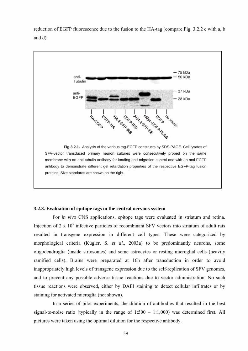
reduction of EGFP fluorescence due to the fusion to the HA-tag (compare Fig. 3.2.2 c with a, b
and d).
Fig.3.2.1. Analysis of the various tag-EGFP constructs by SDS-PAGE. Cell lysates of
SFV-vector transduced primary neuron cultures were consecutively probed on the same
membrane with an anti-tubulin antibody for loading and migration control and with an anti-EGFP
antibody to demonstrate different gel retardation properties of the respective EGFP-tag fusion
proteins. Size standards are shown on the right.
3.2.3. Evaluation of epitope tags in the central nervous system
For in vivo CNS applications, epitope tags were evaluated in striatum and retina.
Injection of 2 x 10⁵ infective particles of recombinant SFV vectors into striatum of adult rats
resulted in transgene expression in different cell types. These were categorized by
morphological criteria (Kügler, S. et al., 2003a) to be predominantly neurons, some
oligodendroglia (inside striosomes) and some astrocytes or resting microglial cells (heavily
ramified cells). Brains were preparated at 16h after transduction in order to avoid
inappropriately high levels of transgene expression due to the self-replication of SFV genomes,
and to prevent any possible adverse tissue reactions due to vector administration. No such
tissue reactions were observed, either by DAPI staining to detect cellular infiltrates or by
staining for activated microglia (not shown).
In a series of pilot experiments, the dilution of antibodies that resulted in the best
signal-to-noise ratio (typically in the range of 1:500 – 1:1,000) was determined first. All
pictures were taken using the optimal dilution for the respective antibody.
59
3.2.4. Evaluation of "established" epitope tags (HA, cMyc, FLAG)
The FLAG-tag is readily detected in SFV-cMyc-EGFP-FLAG transduced cells (Fig.
3.2.2 c). However, in control brain transduced with SFV-EGFP or without any transduction,
the anti-FLAG M2 antibody moderately but reproducibly stained the cytoplasm of cells with
neuronal morphology (Fig. 3.2.2 a). Even more pronounced background staining was observed
using anti-FLAG M2 antibody in the retinal ganglion cell layer (Fig. 3.2.2 b). Further dilution
of the antibody reduced both background staining and specific signal detection, but did not
enhance signal-to-noise ratio. Despite sensitive detection of the FLAG epitope in the brain, we
noticed that a subpopulation (about 10-20%) of EGFP expressing cells was not stained by anti-
FLAG antibody (Fig. 3.2.2 c).
Detection of the cMyc-tag was highly sensitive and specific in the brain, but not in
the retina, where we noticed that about 20-30% of cells, which showed cMyc immunoreactivity
were not EGFP-positive (Fig. 3.2.2 d). This finding suggests that endogenous cMyc protein (of
which 10 amino acids represent the cMyc epitope peptide, corresponding to residues 423 - 432
of human cMyc or residues 424 - 433 of rat cMyc) was detected in these cells rather than the
vector expressed cMyc epitope tagged transgene. Since cMyc is a protein crucial for cell cycle
progression but also is upregulated in several paradigms of neuronal degeneration (Qin, Z. H.
et al., 1999;Di Giovanni, S. et al., 2003), our finding emphasizes that physiological tissue
reactions may readily result in the false-positive detection of the cMyc-tag.
The anti-HA antibody sensitively and specifically detected the HA-tag, however,
EGFP fluorescence of the fusion proteins (HA-EGFP, HA-EGFP-IRS, EGFP-HA) was
considerably reduced in brain sections and in cultured brain cells as compared to other tagged
EGFP constructs (compare Fig. 3.2.2 e with a-d and f). Regardless, the remaining EGFP
fluorescence was still sufficient to detect the fusion protein by using longer exposure times
(approximately 5 times longer than for the other constructs).
Taken together, all "established" epitope tag / antibody combinations allowed for
sensitive detection of the expressed transgene, although we also document either structural
influence of the epitope tag on the fusion partner (HA - tag) or problems with background
staining, depending on either tissue composition (FLAG - tag) or potential physiological tissue
reactions as in case of cMyc - tag.
3.2.5. Evaluation of "new" epitope tags (AU1, EE, IRS)
The very short IRS-tag was detected specifically by its respective antibody. However,
this detection was limited to cells expressing high levels of EGFP. Cells expressing lower
levels of EGFP were not detected by the antibody in brain sections (Fig. 3.2.3 a), or in cultured
60
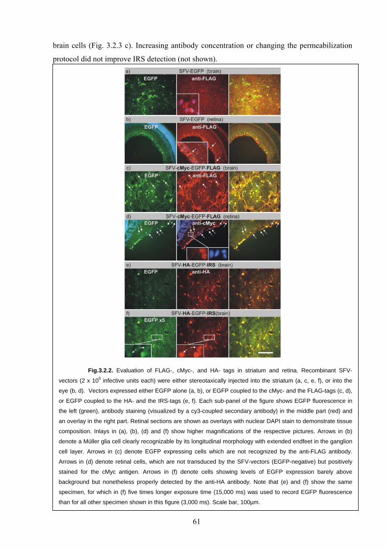
brain cells (Fig. 3.2.3 c). Increasing antibody concentration or changing the permeabilization
protocol did not improve IRS detection (not shown).
Fig.3.2.2. Evaluation of FLAG-, cMyc-, and HA- tags in striatum and retina. Recombinant SFV-
vectors (2 x 10⁵ infective units each) were either stereotaxically injected into the striatum (a, c, e, f), or into the
eye (b, d). Vectors expressed either EGFP alone (a, b), or EGFP coupled to the cMyc- and the FLAG-tags (c, d),
or EGFP coupled to the HA- and the IRS-tags (e, f). Each sub-panel of the figure shows EGFP fluorescence in
the left (green), antibody staining (visualized by a cy3-coupled secondary antibody) in the middle part (red) and
an overlay in the right part. Retinal sections are shown as overlays with nuclear DAPI stain to demonstrate tissue
composition. Inlays in (a), (b), (d) and (f) show higher magnifications of the respective pictures. Arrows in (b)
denote a Müller glia cell clearly recognizable by its longitudinal morphology with extended endfeet in the ganglion
cell layer. Arrows in (c) denote EGFP expressing cells which are not recognized by the anti-FLAG antibody.
Arrows in (d) denote retinal cells, which are not transduced by the SFV-vectors (EGFP-negative) but positively
stained for the cMyc antigen. Arrows in (f) denote cells showing levels of EGFP expression barely above
background but nonetheless properly detected by the anti-HA antibody. Note that (e) and (f) show the same
specimen, for which in (f) five times longer exposure time (15,000 ms) was used to record EGFP fluorescence
than for all other specimen shown in this figure (3,000 ms). Scale bar, 100µm.
61
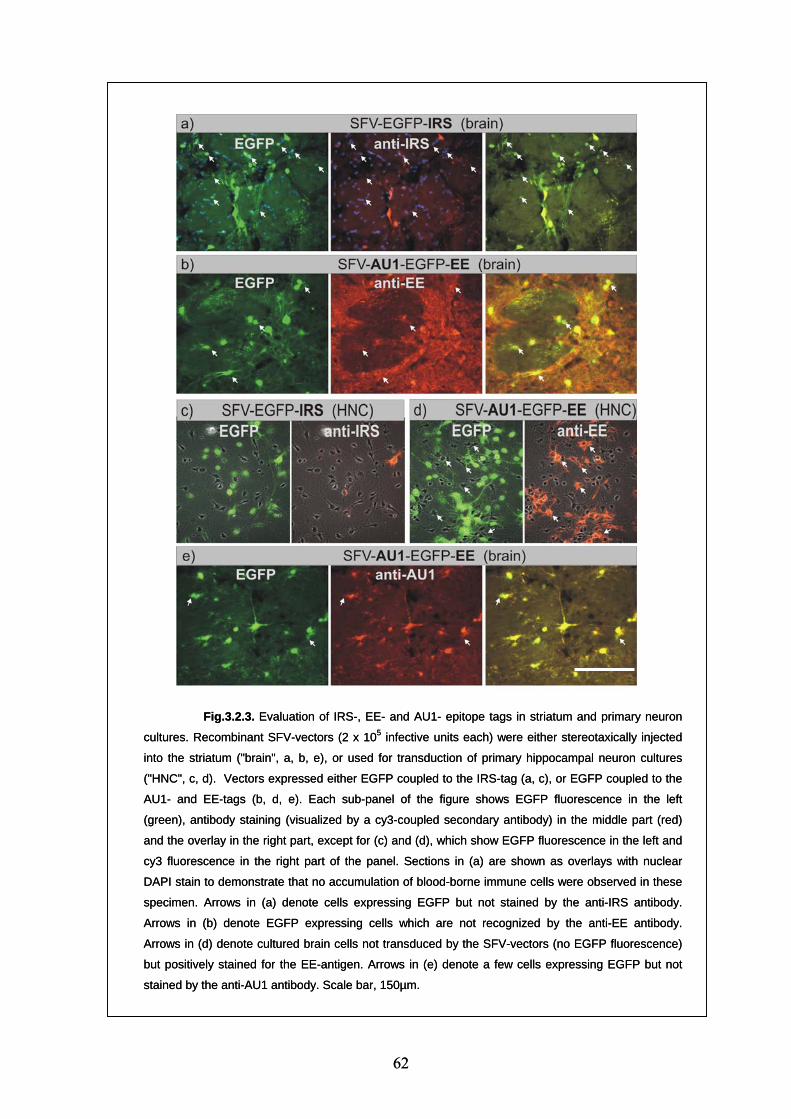
Fig.3.2.3. Evaluation of IRS-, EE- and AU1- epitope tags in striatum and primary neuron
cultures. Recombinant SFV-vectors (2 x 10⁵ infective units each) were either stereotaxically injected
into the striatum ("brain", a, b, e), or used for transduction of primary hippocampal neuron cultures
("HNC", c, d). Vectors expressed either EGFP coupled to the IRS-tag (a, c), or EGFP coupled to the
AU1- and EE-tags (b, d, e). Each sub-panel of the figure shows EGFP fluorescence in the left
(green), antibody staining (visualized by a cy3-coupled secondary antibody) in the middle part (red)
and the overlay in the right part, except for (c) and (d), which show EGFP fluorescence in the left and
cy3 fluorescence in the right part of the panel. Sections in (a) are shown as overlays with nuclear
DAPI stain to demonstrate that no accumulation of blood-borne immune cells were observed in these
specimen. Arrows in (a) denote cells expressing EGFP but not stained by the anti-IRS antibody.
Arrows in (b) denote EGFP expressing cells which are not recognized by the anti-EE antibody.
Arrows in (d) denote cultured brain cells not transduced by the SFV-vectors (no EGFP fluorescence)
but positively stained for the EE-antigen. Arrows in (e) denote a few cells expressing EGFP but not
stained by the anti-AU1 antibody. Scale bar, 150µm.
Fig.3.2.3. Evaluation of IRS-, EE- and AU1- epitope tags in striatum and primary neuron
cultures. Recombinant SFV-vectors (2 x 10
62
5 infective units each) were either stereotaxically injected
into the striatum ("brain", a, b, e), or used for transduction of primary hippocampal neuron cultures
("HNC", c, d). Vectors expressed either EGFP coupled to the IRS-tag (a, c), or EGFP coupled to the
AU1- and EE-tags (b, d, e). Each sub-panel of the figure shows EGFP fluorescence in the left
(green), antibody staining (visualized by a cy3-coupled secondary antibody) in the middle part (red)
and the overlay in the right part, except for (c) and (d), which show EGFP fluorescence in the left and
cy3 fluorescence in the right part of the panel. Sections in (a) are shown as overlays with nuclear
DAPI stain to demonstrate that no accumulation of blood-borne immune cells were observed in these
specimen. Arrows in (a) denote cells expressing EGFP but not stained by the anti-IRS antibody.
Arrows in (b) denote EGFP expressing cells which are not recognized by the anti-EE antibody.
Arrows in (d) denote cultured brain cells not transduced by the SFV-vectors (no EGFP fluorescence)
but positively stained for the EE-antigen. Arrows in (e) denote a few cells expressing EGFP but not
stained by the anti-AU1 antibody. Scale bar, 150µm.
62
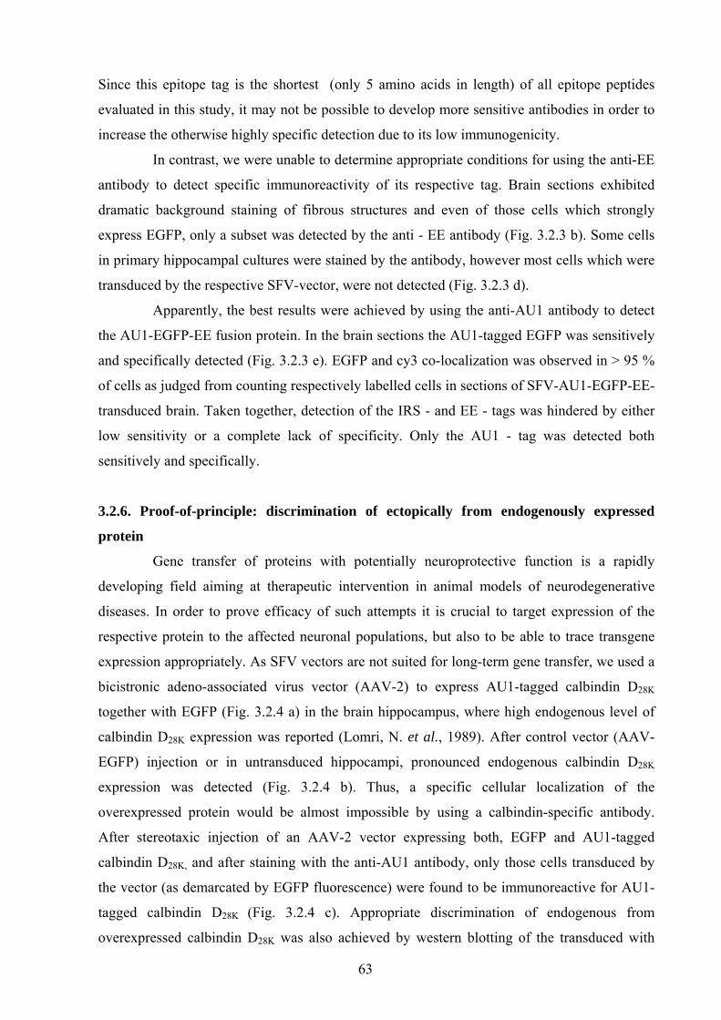
Since this epitope tag is the shortest (only 5 amino acids in length) of all epitope peptides
evaluated in this study, it may not be possible to develop more sensitive antibodies in order to
increase the otherwise highly specific detection due to its low immunogenicity.
In contrast, we were unable to determine appropriate conditions for using the anti-EE
antibody to detect specific immunoreactivity of its respective tag. Brain sections exhibited
dramatic background staining of fibrous structures and even of those cells which strongly
express EGFP, only a subset was detected by the anti - EE antibody (Fig. 3.2.3 b). Some cells
in primary hippocampal cultures were stained by the antibody, however most cells which were
transduced by the respective SFV-vector, were not detected (Fig. 3.2.3 d).
Apparently, the best results were achieved by using the anti-AU1 antibody to detect
the AU1-EGFP-EE fusion protein. In the brain sections the AU1-tagged EGFP was sensitively
and specifically detected (Fig. 3.2.3 e). EGFP and cy3 co-localization was observed in > 95 %
of cells as judged from counting respectively labelled cells in sections of SFV-AU1-EGFP-EE-
transduced brain. Taken together, detection of the IRS - and EE - tags was hindered by either
low sensitivity or a complete lack of specificity. Only the AU1 - tag was detected both
sensitively and specifically.
3.2.6. Proof-of-principle: discrimination of ectopically from endogenously expressed
protein
Gene transfer of proteins with potentially neuroprotective function is a rapidly
developing field aiming at therapeutic intervention in animal models of neurodegenerative
diseases. In order to prove efficacy of such attempts it is crucial to target expression of the
respective protein to the affected neuronal populations, but also to be able to trace transgene
expression appropriately. As SFV vectors are not suited for long-term gene transfer, we used a
bicistronic adeno-associated virus vector (AAV-2) to express AU1-tagged calbindin D28K
together with EGFP (Fig. 3.2.4 a) in the brain hippocampus, where high endogenous level of
calbindin D28K expression was reported (Lomri, N. et al., 1989). After control vector (AAV-
EGFP) injection or in untransduced hippocampi, pronounced endogenous calbindin D28K
expression was detected (Fig. 3.2.4 b). Thus, a specific cellular localization of the
overexpressed protein would be almost impossible by using a calbindin-specific antibody.
After stereotaxic injection of an AAV-2 vector expressing both, EGFP and AU1-tagged
calbindin D28K, and after staining with the anti-AU1 antibody, only those cells transduced by
the vector (as demarcated by EGFP fluorescence) were found to be immunoreactive for AU1-
tagged calbindin D28K (Fig. 3.2.4 c). Appropriate discrimination of endogenous from
overexpressed calbindin D28K was also achieved by western blotting of the transduced with
63
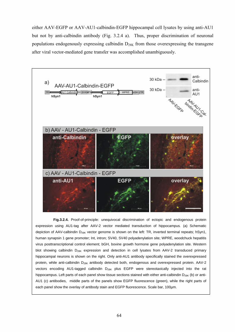
either AAV-EGFP or AAV-AU1-calbindin-EGFP hippocampal cell lysates by using anti-AU1
but not by anti-calbindin antibody (Fig. 3.2.4 a). Thus, proper discrimination of neuronal
populations endogenously expressing calbindin D28K from those overexpressing the transgene
after viral vector-mediated gene transfer was accomplished unambiguously.
Fig.3.2.4. Proof-of-principle: unequivocal discrimination of ectopic and endogenous protein
expression using AU1-tag after AAV-2 vector mediated transduction of hippocampus. (a) Schematic
depiction of AAV-calbindin D28K vector genome is shown on the left: TR, inverted terminal repeats; hSyn1,
human synapsin 1 gene promoter; Int, intron; SV40, SV40 polyadenylation site; WPRE, woodchuck hepatitis
virus posttranscriptional control element; bGH, bovine growth hormone gene polyadenylation site. Western
blot showing calbindin D28K expression and detection in cell lysates from AAV-2 transduced primary
hippocampal neurons is shown on the right. Only anti-AU1 antibody specifically stained the overexpressed
protein, while anti-calbindin D28K antibody detected both, endogenous and overexpressed protein. AAV-2
vectors encoding AU1-tagged calbindin D28K plus EGFP were stereotaxically injected into the rat
hippocampus. Left parts of each panel show tissue sections stained with either anti-calbindin D28K (b) or anti-
AU1 (c) antibodies, middle parts of the panels show EGFP fluorescence (green), while the right parts of
each panel show the overlay of antibody stain and EGFP fluorescence. Scale bar, 100µm.
64
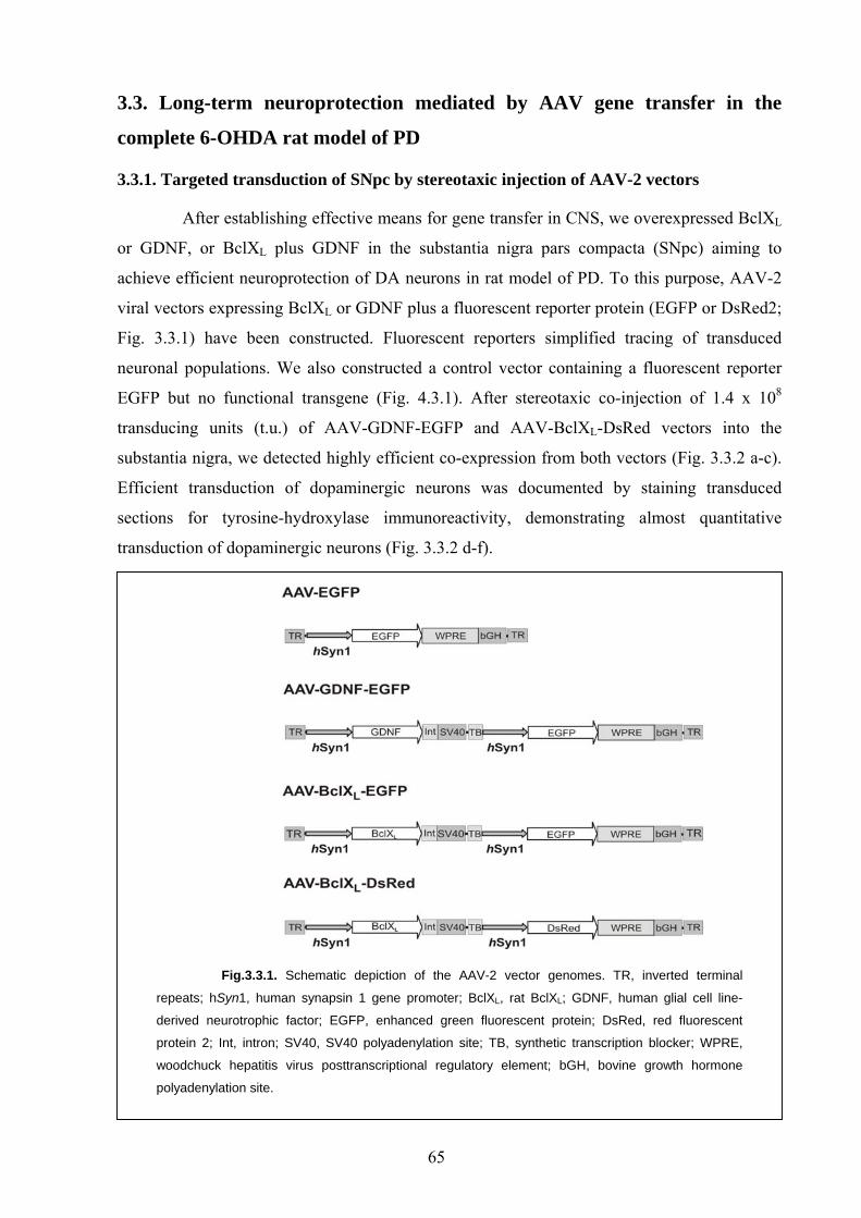
3.3. Long-term neuroprotection mediated by AAV gene transfer in the
complete 6-OHDA rat model of PD
3.3.1. Targeted transduction of SNpc by stereotaxic injection of AAV-2 vectors
After establishing effective means for gene transfer in CNS, we overexpressed BclXL
or GDNF, or BclXL plus GDNF in the substantia nigra pars compacta (SNpc) aiming to
achieve efficient neuroprotection of DA neurons in rat model of PD. To this purpose, AAV-2
viral vectors expressing BclXL or GDNF plus a fluorescent reporter protein (EGFP or DsRed2;
Fig. 3.3.1) have been constructed. Fluorescent reporters simplified tracing of transduced
neuronal populations. We also constructed a control vector containing a fluorescent reporter
EGFP but no functional transgene (Fig. 4.3.1). After stereotaxic co-injection of 1.4 x 10⁸
transducing units (t.u.) of AAV-GDNF-EGFP and AAV-BclXL-DsRed vectors into the
substantia nigra, we detected highly efficient co-expression from both vectors (Fig. 3.3.2 a-c).
Efficient transduction of dopaminergic neurons was documented by staining transduced
sections for tyrosine-hydroxylase immunoreactivity, demonstrating almost quantitative
transduction of dopaminergic neurons (Fig. 3.3.2 d-f).
Fig.3.3.1. Schematic depiction of the AAV-2 vector genomes. TR, inverted terminal
repeats; hSyn1, human synapsin 1 gene promoter; BclXL, rat BclXL; GDNF, human glial cell line-
derived neurotrophic factor; EGFP, enhanced green fluorescent protein; DsRed, red fluorescent
protein 2; Int, intron; SV40, SV40 polyadenylation site; TB, synthetic transcription blocker; WPRE,
woodchuck hepatitis virus posttranscriptional regulatory element; bGH, bovine growth hormone
polyadenylation site.
65
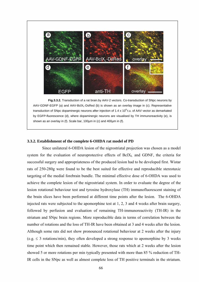
Fig.3.3.2. Transduction of a rat brain.by AAV-2 vectors. Co-transduction of SNpc neurons by
AAV-GDNF-EGFP (a) and AAV-BclXL-DsRed (b) is shown as an overlay image in (c). Representative
transduction of SNpc dopaminergic neurons after injection of 1.4 x 10⁸ t.u. of AAV vector as demarkated
by EGFP-fluorescence (d), where dopaminergic neurons are visualised by TH immunoreactivity (e), is
shown as an overlay in (f). Scale bar, 100µm in (c) and 400µm in (f).
3.3.2. Establishment of the complete 6-OHDA rat model of PD
Since unilateral 6-OHDA lesion of the nigrostriatal projection was chosen as a model
system for the evaluation of neuroprotective effects of BclXL and GDNF, the criteria for
successful surgery and appropriateness of the produced lesion had to be developed first. Wistar
rats of 250-280g were found to be the best suited for effective and reproducible stereotaxic
targeting of the medial forebrain bundle. The minimal effective dose of 6-OHDA was used to
achieve the complete lesion of the nigrostriatal system. In order to evaluate the degree of the
lesion rotational behaviour test and tyrosine hydroxylase (TH) immunofluorescent staining of
the brain slices have been performed at different time points after the lesion. The 6-OHDA
injected rats were subjected to the apomorphine test at 1, 2, 3 and 4 weeks after brain surgery,
followed by perfusion and evaluation of remaining TH-immunoreactivity (TH-IR) in the
striatum and SNpc brain regions. More reproducible data in terms of correlation between the
number of rotations and the loss of TH-IR have been obtained at 3 and 4 weeks after the lesion.
Although some rats did not show pronounced rotational behaviour at 2 weeks after the injury
(e.g. ≤ 3 rotations/min), they often developed a strong response to apomorphine by 3 weeks
time point which then remained stable. However, those rats which at 2 weeks after the lesion
showed 5 or more rotations per min typically presented with more than 85 % reduction of TH-
IR cells in the SNpc as well as almost complete loss of TH positive terminals in the striatum.
66
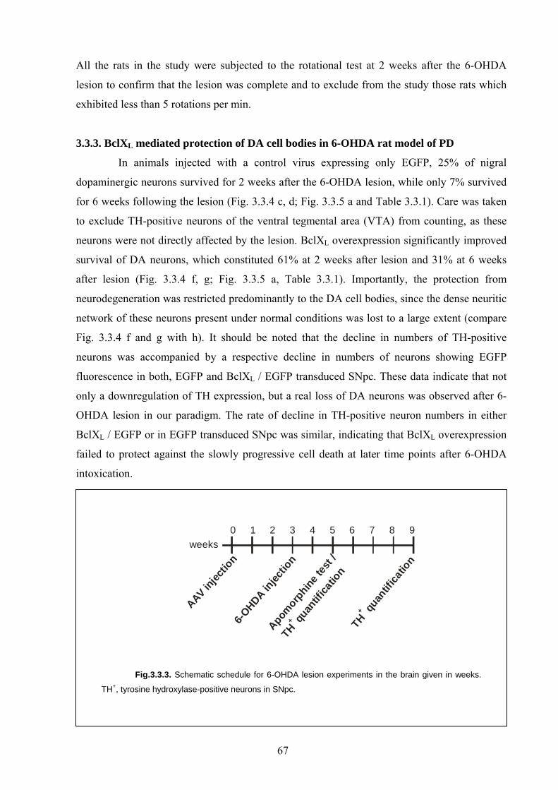
All the rats in the study were subjected to the rotational test at 2 weeks after the 6-OHDA
lesion to confirm that the lesion was complete and to exclude from the study those rats which
exhibited less than 5 rotations per min.
3.3.3. BclXL mediated protection of DA cell bodies in 6-OHDA rat model of PD
In animals injected with a control virus expressing only EGFP, 25% of nigral
dopaminergic neurons survived for 2 weeks after the 6-OHDA lesion, while only 7% survived
for 6 weeks following the lesion (Fig. 3.3.4 c, d; Fig. 3.3.5 a and Table 3.3.1). Care was taken
to exclude TH-positive neurons of the ventral tegmental area (VTA) from counting, as these
neurons were not directly affected by the lesion. BclXL overexpression significantly improved
survival of DA neurons, which constituted 61% at 2 weeks after lesion and 31% at 6 weeks
after lesion (Fig. 3.3.4 f, g; Fig. 3.3.5 a, Table 3.3.1). Importantly, the protection from
neurodegeneration was restricted predominantly to the DA cell bodies, since the dense neuritic
network of these neurons present under normal conditions was lost to a large extent (compare
Fig. 3.3.4 f and g with h). It should be noted that the decline in numbers of TH-positive
neurons was accompanied by a respective decline in numbers of neurons showing EGFP
fluorescence in both, EGFP and BclXL / EGFP transduced SNpc. These data indicate that not
only a downregulation of TH expression, but a real loss of DA neurons was observed after 6-
OHDA lesion in our paradigm. The rate of decline in TH-positive neuron numbers in either
BclXL / EGFP or in EGFP transduced SNpc was similar, indicating that BclXL overexpression
failed to protect against the slowly progressive cell death at later time points after 6-OHDA
intoxication.
weeks
AAV injec
tion
6-OHDA in
jectio
n
Apomorp
hine te
st /
THqu
antif
icatio
n
+
T
H qu
antif
icatio
n
+
0 1 2 3 4 5 6 7 8 9
Fig.3.3.3. Schematic schedule for 6-OHDA lesion experiments in the brain given in weeks.
TH+, tyrosine hydroxylase-positive neurons in SNpc.
67
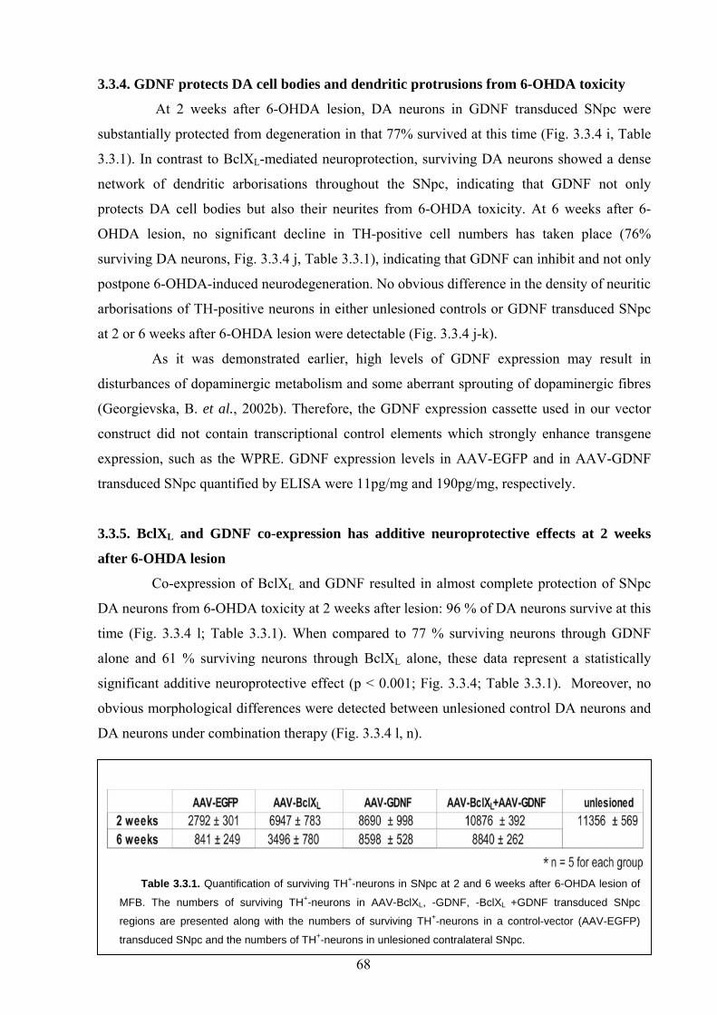
3.3.4. GDNF protects DA cell bodies and dendritic protrusions from 6-OHDA toxicity
At 2 weeks after 6-OHDA lesion, DA neurons in GDNF transduced SNpc were
substantially protected from degeneration in that 77% survived at this time (Fig. 3.3.4 i, Table
3.3.1). In contrast to BclXL-mediated neuroprotection, surviving DA neurons showed a dense
network of dendritic arborisations throughout the SNpc, indicating that GDNF not only
protects DA cell bodies but also their neurites from 6-OHDA toxicity. At 6 weeks after 6-
OHDA lesion, no significant decline in TH-positive cell numbers has taken place (76%
surviving DA neurons, Fig. 3.3.4 j, Table 3.3.1), indicating that GDNF can inhibit and not only
postpone 6-OHDA-induced neurodegeneration. No obvious difference in the density of neuritic
arborisations of TH-positive neurons in either unlesioned controls or GDNF transduced SNpc
at 2 or 6 weeks after 6-OHDA lesion were detectable (Fig. 3.3.4 j-k).
As it was demonstrated earlier, high levels of GDNF expression may result in
disturbances of dopaminergic metabolism and some aberrant sprouting of dopaminergic fibres
(Georgievska, B. et al., 2002b). Therefore, the GDNF expression cassette used in our vector
construct did not contain transcriptional control elements which strongly enhance transgene
expression, such as the WPRE. GDNF expression levels in AAV-EGFP and in AAV-GDNF
transduced SNpc quantified by ELISA were 11pg/mg and 190pg/mg, respectively.
3.3.5. BclXL and GDNF co-expression has additive neuroprotective effects at 2 weeks
after 6-OHDA lesion
Co-expression of BclXL and GDNF resulted in almost complete protection of SNpc
DA neurons from 6-OHDA toxicity at 2 weeks after lesion: 96 % of DA neurons survive at this
time (Fig. 3.3.4 l; Table 3.3.1). When compared to 77 % surviving neurons through GDNF
alone and 61 % surviving neurons through BclXL alone, these data represent a statistically
significant additive neuroprotective effect (p < 0.001; Fig. 3.3.4; Table 3.3.1). Moreover, no
obvious morphological differences were detected between unlesioned control DA neurons and
DA neurons under combination therapy (Fig. 3.3.4 l, n).
Table 3.3.1. Quantification of surviving TH+-neurons in SNpc at 2 and 6 weeks after 6-OHDA lesion of
MFB. The numbers of surviving TH+-neurons in AAV-BclXL, -GDNF, -BclXL +GDNF transduced SNpc
regions are presented along with the numbers of surviving TH+-neurons in a control-vector (AAV-EGFP)
transduced SNpc and the numbers of TH+-neurons in unlesioned contralateral SNpc.
68
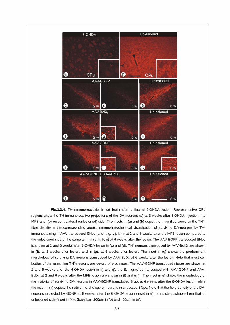
69
Fig.3.3.4. TH-immunoreactivity in rat brain after unilateral 6-OHDA lesion. Representative CPu
regions show the TH-immunoreactive projections of the DA-neurons (a) at 3 weeks after 6-OHDA injection into
MFB and, (b) on contralateral (unlesioned) side. The insets in (a) and (b) depict the magnified views on the TH+-
fibre density in the corresponding areas. Immunohistochemical visualisation of surviving DA-neurons by TH-
immunostaining in AAV-transduced SNpc (c, d, f, g, i, j, l, m) at 2 and 6 weeks after the MFB lesion compared to
the unlesioned side of the same animal (e, h, k, n) at 6 weeks after the lesion. The AAV-EGFP transduced SNpc
is shown at 2 and 6 weeks after 6-OHDA lesion in (c) and (d). TH+ neurons transduced by AAV-BclXL are shown
in (f), at 2 weeks after lesion, and in (g), at 6 weeks after lesion. The inset in (g) shows the predominant
morphology of surviving DA-neurons transduced by AAV-BclXL at 6 weeks after the lesion. Note that most cell
bodies of the remaining TH+-neurons are devoid of processes. The AAV-GDNF transduced nigrae are shown at
2 and 6 weeks after the 6-OHDA lesion in (i) and (j); the S. nigrae co-transduced with AAV-GDNF and AAV-
BclXL at 2 and 6 weeks after the MFB lesion are shown in (l) and (m). The inset in (j) shows the morphology of
the majority of surviving DA-neurons in AAV-GDNF transduced SNpc at 6 weeks after the 6-OHDA lesion, while
the inset in (k) depicts the native morphology of neurons in untreated SNpc. Note that the fibre density of the DA-
neurons protected by GDNF at 6 weeks after the 6-OHDA lesion (inset in (j)) is indistinguishable from that of
unlesioned side (inset in (k)). Scale bar, 200µm in (b) and 400µm in (n).
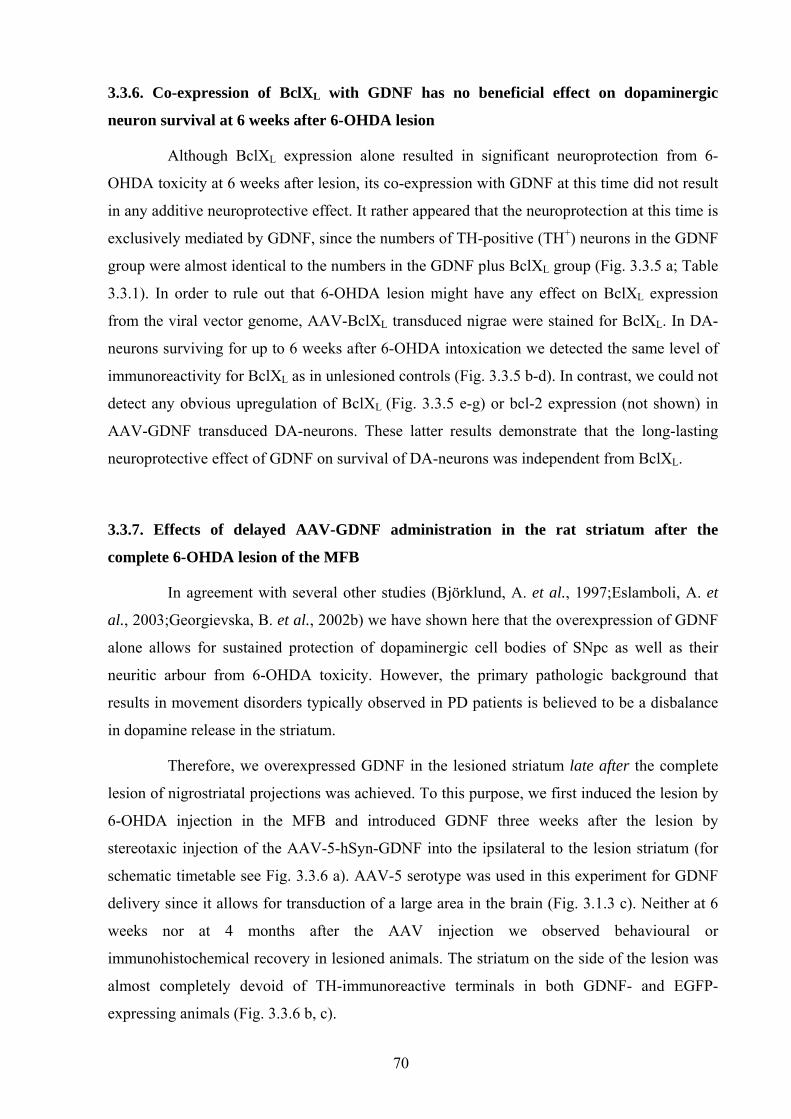
3.3.6. Co-expression of BclXL with GDNF has no beneficial effect on dopaminergic
neuron survival at 6 weeks after 6-OHDA lesion
Although BclXL expression alone resulted in significant neuroprotection from 6-
OHDA toxicity at 6 weeks after lesion, its co-expression with GDNF at this time did not result
in any additive neuroprotective effect. It rather appeared that the neuroprotection at this time is
exclusively mediated by GDNF, since the numbers of TH-positive (TH+) neurons in the GDNF
group were almost identical to the numbers in the GDNF plus BclXL group (Fig. 3.3.5 a; Table
3.3.1). In order to rule out that 6-OHDA lesion might have any effect on BclXL expression
from the viral vector genome, AAV-BclXL transduced nigrae were stained for BclXL. In DA-
neurons surviving for up to 6 weeks after 6-OHDA intoxication we detected the same level of
immunoreactivity for BclXL as in unlesioned controls (Fig. 3.3.5 b-d). In contrast, we could not
detect any obvious upregulation of BclXL (Fig. 3.3.5 e-g) or bcl-2 expression (not shown) in
AAV-GDNF transduced DA-neurons. These latter results demonstrate that the long-lasting
neuroprotective effect of GDNF on survival of DA-neurons was independent from BclXL.
3.3.7. Effects of delayed AAV-GDNF administration in the rat striatum after the
complete 6-OHDA lesion of the MFB
In agreement with several other studies (Björklund, A. et al., 1997;Eslamboli, A. et
al., 2003;Georgievska, B. et al., 2002b) we have shown here that the overexpression of GDNF
alone allows for sustained protection of dopaminergic cell bodies of SNpc as well as their
neuritic arbour from 6-OHDA toxicity. However, the primary pathologic background that
results in movement disorders typically observed in PD patients is believed to be a disbalance
in dopamine release in the striatum.
Therefore, we overexpressed GDNF in the lesioned striatum late after the complete
lesion of nigrostriatal projections was achieved. To this purpose, we first induced the lesion by
6-OHDA injection in the MFB and introduced GDNF three weeks after the lesion by
stereotaxic injection of the AAV-5-hSyn-GDNF into the ipsilateral to the lesion striatum (for
schematic timetable see Fig. 3.3.6 a). AAV-5 serotype was used in this experiment for GDNF
delivery since it allows for transduction of a large area in the brain (Fig. 3.1.3 c). Neither at 6
weeks nor at 4 months after the AAV injection we observed behavioural or
immunohistochemical recovery in lesioned animals. The striatum on the side of the lesion was
almost completely devoid of TH-immunoreactive terminals in both GDNF- and EGFP-
expressing animals (Fig. 3.3.6 b, c).
70
Fig.3.3.5. Quantification of DA-neuron survival in SNpc after 6-OHDA lesion. In (a) the
number of surviving TH+-neurons was determined after the control vector injection (AAV-EGFP) and
after the injection of AAV-BclXL, or AAV-GDNF, or AAV-BclXL plus AAV-GDNF (gray bars). Dark-gray
bars represent the mean values of surviving TH+-neurons in SNpc at 2 weeks after 6-OHDA lesion,
light-gray bars represent mean values of surviving TH+-neurons at 6 weeks after 6-OHDA lesion. The
mean number of TH+-neurons in unlesioned SNpc is shown by black bar. Bars represent mean
values ± standard deviations (***p < 0.001, n.s., non-significant). Immunohistochemical staining for
BclXL in SNpc neurons transduced by AAV-BclXL confirmed that BclXL expression is not
downregulated in the course of the experiment (b - d). Immunohistochemical staining for BclXL was
also used to demonstrate that GDNF expression did not result in BclXL upregulation in SNpc neurons
after AAV-GDNF transduction (e - g). Scale bar, 40µm.
Fig.3.3.5. Quantification of DA-neuron survival in SNpc after 6-OHDA lesion. In (a) the
number of surviving TH
71
+-neurons was determined after the control vector injection (AAV-EGFP) and
after the injection of AAV-BclXL, or AAV-GDNF, or AAV-BclXL plus AAV-GDNF (gray bars). Dark-gray
bars represent the mean values of surviving TH+-neurons in SNpc at 2 weeks after 6-OHDA lesion,
light-gray bars represent mean values of surviving TH+-neurons at 6 weeks after 6-OHDA lesion. The
mean number of TH+-neurons in unlesioned SNpc is shown by black bar. Bars represent mean
values ± standard deviations (***p < 0.001, n.s., non-significant). Immunohistochemical staining for
BclXL in SNpc neurons transduced by AAV-BclXL confirmed that BclXL expression is not
downregulated in the course of the experiment (b - d). Immunohistochemical staining for BclXL was
also used to demonstrate that GDNF expression did not result in BclXL upregulation in SNpc neurons
after AAV-GDNF transduction (e - g). Scale bar, 40µm.
71
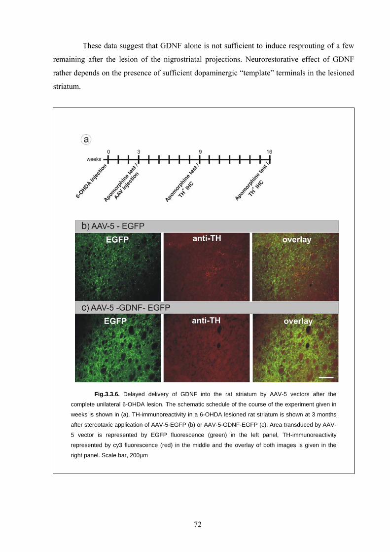
These data suggest that GDNF alone is not sufficient to induce resprouting of a few
remaining after the lesion of the nigrostriatal projections. Neurorestorative effect of GDNF
rather depends on the presence of sufficient dopaminergic “template” terminals in the lesioned
striatum.
Fig.3.3.6. Delayed delivery of GDNF into the rat striatum by AAV-5 vectors after the
complete unilateral 6-OHDA lesion. The schematic schedule of the course of the experiment given in
weeks is shown in (a). TH-immunoreactivity in a 6-OHDA lesioned rat striatum is shown at 3 months
after stereotaxic application of AAV-5-EGFP (b) or AAV-5-GDNF-EGFP (c). Area transduced by AAV-
5 vector is represented by EGFP fluorescence (green) in the left panel, TH-immunoreactivity
represented by cy3 fluorescence (red) in the middle and the overlay of both images is given in the
right panel. Scale bar, 200µm
72
4. Discussion
In the work presented here the following aims have been pursued: 1) to evaluate the
neuroprotective and neurorestorative properties of BclXL and GDNF in the rat model of
Parkinson’s disease; 2) to develop appropriate adeno-associated viral vectors for efficient gene
transfer to the target regions in the brain; 3) to improve detection the transgene for ongoing and
future gene transfer studies by epitope-tagging approach.
4.1. AAV vector targeting
In this study we aimed to overexpress BclXL and GDNF in the substantia nigra and
GDNF in the striatum. The purpose of nigral gene transfer was to achieve efficient and long-
lasting neuroprotection of DA cells. AAV-2 serotype was employed as a gene transfer tool,
since it preferentially transduces neurons in the CNS and has especially high affinity to DA
neurons of SNpc (Paterna, J. C. et al., 2004). This property of AAV-2 vectors can be explained
by high density of HSPG receptors, necessary for virus entry, on nigral neurons (Fuxe, K. et
al., 1994). To further restrict the expression of the transgene to neurons, hSyn1 promoter was
used. However, protecting the DA cell bodies in SNpc may not be sufficient for maintenance
of the basal ganglia circuit in the brain. To achieve reconstruction of functional connections of
DA cell bodies to the striatum, striatal overexpression of GDNF may be required (Kirik, D. et
al., 2004). GDNF was found to be expressed primarily from astrocytes in the brain. Therefore,
we attempted to redirect the AAV-mediated transgene expression from neurons to astrocytes.
Previous studies aiming to restrict the transduction by AAV vectors to either astrocytes or
oligodendroglia using the glial cell type specific promoters GFAP and myelin basic protein
promoter failed (Peel, A. L. et al., 1997;Peel, A. L. et al., 2000). In this study we used
ubiquitously active mCMV promoter which has been shown to target the transgene expression
to astrocytes after adenoviral transduction of the CNS (Kügler, S. et al., 2001). Moreover we
made use of the AAV-5 capsid for packaging since this AAV serotype was shown to equally
transduce neurons and astrocytes (Tenenbaum, L. et al., 2004). With this approach, efficient
astroglia-specific transgene expression was achieved in primary hippocampal culture.
Incorporation of EGFP into AAV vector genome greatly simplified visualization of the
transduced cells. However, when the same promoter/capsid (mCMV/AAV-5) combination was
utilized for AAV-mediated gene transfer into the brain, a different cellular transduction pattern
was observed as compared to in vitro results. Instead of predominant localization of EGFP to
astrocytes as it would be expected after the in vitro experiments, some neurons and cells of
73
unidentified origin, which were negative to both astroglial and microglial immunological
markers have been transduced. These apparently divergent data obtained from in vitro and in
vivo studies on AAV vector targeting may be explained by variable expression of receptors
required for successful entry of the recombinant virus into the cell in embryonic and adult
brain. In neonatal mice intraventricular brain injection of AAV-2 or AAV-1 has been shown to
result in widespread neuronal transduction, whereas AAV-5 injection led to a very limited
transgene expression (Passini, M. A. et al., 2003). In the brain of adult animals, however,
AAV-2 transduction is localized while AAV-5 transgene expression is typically widespread
(Davidson, B. L. et al., 2000;Shevtsova, Z. et al., 2005). Moreover, the extracellular
environment and tissue composition which may contribute to the successful entry of the virus
into the cell are very different in primary cultures and in the living organism. For example, in
addition to glycosamynoglycans AAV-2 serotype requires fibroblast growth factor receptor
and integrins as co-receptors for effective transduction (Qing, K. et al., 1999;Summerford, C.
et al., 1999), which appear to be differentially expressed in neonate and adult brain. On the
contrary, AAV-5 mediated transduction does not require heparan sulfate proteoglycan (HSPG),
but α-2,3- or α-2,6-N-linked sialic acid (Kaludov, N. et al., 2001;Walters, R. W. et al., 2001)
and platelet-derived growth factor receptor (PDGFR) (Di Pasquale, G. et al., 2003). In
addition, mCMV promoter activity in mature and in non-differentiated CNS cells may also
influence the observed difference in expression patterns.
Consistent with the results obtained by others (Burger, C. et al., 2004), the area of
virus transduction in the brain was restricted to the injection site after stereotaxic application of
AAV-2 vector and was spread beyond the striatum borders (ca. 10x larger) after injecting the
same titre of AAV-5 vector. These serotype properties of AAV can define the choice of the
vector for gene transfer: when the transduction of a relatively small-size region is intended, the
use of AAV-2 serotype is appropriate. For effective transduction of a large structure in the brain
(e.g. striatum), AAV-5 would be the vector of choice. Thus, AAV-2 serotype was selected for
gene transfer into the small nucleus SNpc whereas AAV-5 was used to deliver GDNF into the
striatum.
4.2. Epitope-tagging for in vivo transgene detection
Difficulties in detection of transgenic protein expression by respective antibodies and
in its discrimination from the endogenously expressed analogue in a living organism (Weise, J.
et al., 2000;Wagenknecht, B. et al., 1999) necessitate the search for alternative confirmation of
transgene expression. Advanced viral gene transfer systems have made it possible to express
almost every protein of interest not only in rodent but also in the more complex primate brain
74

(Gerdes, C. A. et al., 2000;Kirik, D. et al., 2003). Gene transfer studies directed towards
therapeutic interventions in disease models or towards basic research on protein functions
require the unambiguous verification of the transgene expression. An antibody, specific for the
respective protein may be used, but this antibody might not be able to discriminate intrinsically
expressed proteins from the ectopically or overexpressed one. Additionally, for dominant-
negative or constitutively activated variants of a protein, or for splice variants as well as for
many proteins investigated for the first time specific antibodies may not be available. Thus,
epitope tags, short peptides which can be fused to the protein under investigation and
specifically detected by well-characterized antibodies, appear to be an attractive alternative.
Although proven to be useful for protein tracing in cell-free systems or in cultured cells in vitro
(Terpe, K., 2003), thus far there has been no systematic attempt to characterize different epitope
tags for in vivo gene transfer applications.
The study presented here is the first systematic evaluation of different epitope tags in
the CNS. The data demonstrate that all "established" tags (HA-, cMyc-, FLAG-tag) were
detected by their respective antibodies with high sensitivity, but in all cases significant
drawbacks have also been observed. The anti-FLAG and the anti-cMyc antibodies showed
significant background staining of distinct CNS structures, and at least one of these tags was
responsible for pronounced gel mobility retardation of the tagged EGFP, probably due to the
introduction of an odd phosphorylation site. Fusion of the HA-tag to EGFP resulted in
pronounced reduction of EGFP fluorescence, and the N-terminally tagged EGFP showed altered
mobility in SDS gel electrophoresis while the C-terminally tagged version did not. Among the
"new" tags evaluated, the very short IRS tag (5 amino acids in length) was specifically detected,
but not with sufficient sensitivity for routine use. In contrast, the antibody used to detect the EE-
tag proved to be inoperative in brain sections due to tremendous background staining. Only
AU1-tag detection was accomplished with high specificity and sensitivity, both on brain
sections and in cultured neurons, and did not show any obvious influence on the tagged protein.
Subtle to moderate changes in protein structure and function due to the addition of extra
amino acids may be less relevant in cell-free or cell culture experiments, in which super-
physiological amounts of recombinant or overexpressed proteins are used for experiments (e.g.
co-precipitation assays). However, if functional studies require physiological protein expression
levels, then the influence of an epitope tag may turn out to affect the results of the study.
Despite this important issue, only limited information is currently available with respect to the
influence of epitope tags on the fusion protein. Transgenic mice expressing the wild-type P0-
glycoprotein, which is important for peripheral nerve myelination, and which was N-terminally
tagged by the cMyc epitope, demonstrated clinical symptoms of Charcot-Marie-Tooth disease
75
solely due to the presence of the cMyc-tag (Previtali, S. C. et al., 2000), although
crystallographic studies suggest that the N-terminus of this protein is not involved in cis or trans
interactions (Shapiro, L. et al., 1996). Fusion of the FLAG epitope to the membrane-binding
subunit of influenza virus haemagglutinin caused the protein to localize to the cytoplasm of cells
rather than to the membrane (Chen, J. et al., 1998). Tagging the small GTPase H-Ras by the
FLAG epitope resulted in activity changes as well as in altered mobility in SDS gel
electrophoresis (Johnson, K. Y. et al., 2002). As such, potential influences of epitope tags must
be taken into account especially in studies regarding proteins with as-of-yet unknown functions.
Putative phosphorylation sites as present in the cMyc- and the FLAG-tag amino acids sequences
may only be post-translationally modified depending on a particular cellular environment.
Regardless, such modifications may have unpredictable impacts on the protein under
investigation. While some background staining is to be expected if an epitope derived from a cellular
protein (like the cMyc-tag) is used, epitopes derived from viral proteins should not have
matching cellular counterparts and thus the respective antibodies should be less likely to result
in background staining. In the present study, this assumption was found to be correct for the
influenza hemagglutinin (HA-) tag (Wilson, I. A. et al., 1984) and for the AU1-tag, which is
derived from the bovine papilloma virus capsid protein L1 (Lim, P. S. et al., 1990), but not for
the EE (or Glu-Glu) tag, which is derived from polyoma virus medium T-antigen
(Grussenmeyer, T. et al., 1985). This latter result together with the finding of FLAG - antibody
background staining clearly demonstrates the need to evaluate potential epitope tags and their
respective antibodies in the tissue under study, since it can not be excluded that epitope tag -
specific antibodies bind to unexpected cellular antigenes. SFV vectors appear to be an attractive
tool for such evaluation due to their property of fast and efficient transduction of broad
specificity.
In a proof-of-principle experiment we used an AAV-2 based viral vector for
demonstration of the functionality of the AU1-tag in an in vivo gene transfer approach. The
AAV vector genome was designed in a way that EGFP is co-expressed with calbindin D28K,
clearly demarcating the transduced areas. It should be noted that the approach of co-expressing a
reporter gene with the transgene under investigation is a valuable, but indirect way of proving
transgene expression. It may fail if detailed sub-cellular localisation of the transgene has to be
documented, if the transgene is expressed with different kinetics, or if it is processed or
secreted. Therefore, epitope-tagging may be a more reliable methodological approach in
targeting transgenic protein expression. Despite robust endogenous calbindin D28K expression in
several retinal neuron populations, it was possible to unequivocally trace the expression of the
76
vector derived AU1-tagged calbindin D28K, clearly discriminating endogenous from
overexpressed protein. These results demonstrate the usefulness of this epitope and of epitope-
tagging techniques for in vivo studies in general. However, this data also clearly demonstrated
that a careful pre-evaluation of epitope tags and their respective antibodies is necessary for
different types of tissues and transduction paradigms, in order to avoid problems with
background detection and unexpected influences of the epitope tags on the protein under
investigation.
In this study we have chosen to tag the calcium buffering protein calbindin D28K as its
role in neurodegeneration has yet to be clarified. It remains unclear whether localized rather
than general increase in cytosolic free calcium is critical for cell death (Rizzuto, R. et al.,
1999;Mattson, M. P. et al., 2003). In ongoing experiments we aim to investigate the role of
calcium and calbindin D28K in two models of neurodegeneration: optic nerve axotomy and 6-
OHDA nigrostriatal lesion. Since relatively high expression of calbindin in retina and certain
brain regions was detected in a series of preliminary experiments, appropriate detection of
overexpressed calbindin by using the AU1-tag may prove to be useful.
4.3. Neuroprotective therapy of PD as evaluated in the complete 6-OHDA lesion model
Neurodegenerative diseases are characterized by slow and multifactorial progression,
and potential therapeutic treatment is difficult due to diagnosis at relatively late stages of life.
After diagnosis patients have one or more decades of life expectation, which would necessitate a
long-lasting therapeutical intervention in order to halt, or at least significantly postpone the
neurodegenerative processes. Any potential therapeutic intervention should thus be confirmed to
be effective over longer periods of time. Furthermore, the multifactorial nature of
neurodegeneration in its later stages suggests that therapeutic approaches targeting more than
one neurodegenerative mechanism might be more effective than mono-specific attempts.
In this study we have evaluated such combination gene transfer in a widely used animal
model of neurodegeneration induced by oxidative stress, 6-OHDA lesion of the nigrostriatal
system. The unilateral 6-OHDA lesion model of PD offers certain advantages in that it allows
the immunohistological analysis of the lesioned hemisphere in comparison to the unaffected
brain hemisphere of the same animal as an internal control. Furthermore, an easy-to-perform
apomorphine induced rotational behaviour test reflects the degree of assymetry in striatal
dopamine content and can be used to verify the successful lesioning surgery. In order to produce
the unilateral toxic model of PD, 6-OHDA is typically injected either into the striatum or into the
medial forebrain bundle (MFB) (Blum, D. et al., 2001;Deumens, R. et al., 2002). Multiple
striatal injections are usually required to induce the degeneration of more than 50-60% of the DA
77
neurons in SNpc which increase the time and traumatisation of the surgery (Deumens, R. et al.,
2002). An alternative way to achieve the selective degeneration of the nigrostriatal DA neurons
in SNpc is to deliver the 6-OHDA toxin directly into the nigrostriatal projection, the MFB.
Injection of 6-OHDA into the MFB represents a more severe oxidative stress lesion of DA
neurons than the striatal 6-OHDA injection or systemic MPTP application (Deumens, R. et al.,
2002;Dauer, W. et al., 2003). Therefore, molecules, able to counteract neurodegeneration in this
model may have high potential for therapeutic benefit in PD, in which degeneration of DA
neurons occurs at much slower rates.
4.4. BclXL mediated neuroprotection
The data obtained in this study demonstrate that BclXL overexpression can promote the
survival of 61% of dopaminergic neurons at 2 weeks and 31% at 6 weeks after the 6-OHDA
lesion of the medial forebrain bundle. The numbers of remaining neurons in AAV-2-BclXL
injected group are significantly higher (p<0.001) than those transduced with the AAV-2-EGFP
control virus (25% at 2 weeks and 7% at 6 weeks). However, a decline in DA cell numbers from
2 to 6 weeks post-lesion was observed, and thus, stable neuroprotection could not be achieved
through BclXL overexpression alone.
Several studies have focused on the role of bcl-2, another anti-apoptotic protein of the
same family as BclXL, in the protection of CNS neurons from cell death. In the partial 6-OHDA
toxicity model of PD, herpes virus mediated overexpression of bcl-2 supported the survival of
approximately 50% of nigral neurons at 2 weeks after the lesion (Yamada, M. et al., 1999). Also,
transgenic mice expressing human bcl-2 under the neuron-specific enolase promoter have been
found to be more resistant to MPTP and 6-OHDA toxicity (Offen, D. et al., 1998). However,
both experiments were estimating only the short-term effects of the bcl-2 on DA cell survival.
Moreover, the toxic lesions performed in either of the studies were relatively mild as compared
to the complete unilateral lesion paradigm (Deumens, R. et al., 2002;Dauer, W. et al., 2003).
In this study, overexpression of BclXL but not bcl-2 was chosen mainly for two
reasons. First, BclXL has been shown to be more potent in inhibiting mitochondrial membrane
potential breakdown than bcl-2 (Kim, R., 2005), probably due to its more efficient targeting to
mitochondria (Kaufmann, T. et al., 2003). Second, BclXL but not bcl-2 can inhibit TNFα-
activated TRAIL mediated apoptotic and non-apoptotic cell death (necrosis and autophagy)
(Kim, I. K. et al., 2003;Kim, R., 2005;Keogh, S. A. et al., 2000), which play important roles in
78
several neurodegenerative paradigms (Anglade, P. et al., 1997;Hirsch, E. C. et al.,
2003;Cacquevel, M. et al., 2004).
Taken together, BclXL overexpression failed to finally preserve neurons from
degeneration after severe unilateral lesion of nigrostriatal system, with a 50% decrease of
surviving neuron numbers late after lesion as compared to the early time of analysis. Moreover,
BclXL failed to preserve the DA neurites, protecting only the cell bodies of DA neurons. Thus,
maintenance of mitochondrial integrity may be essential for neuronal survival early after lesion
but appeared to be less important for the long-term survival of deafferentiated neurons and
insufficient for the maintenance of the functional DA connections.
4.5. Combination of BclXL and GDNF for neuroprotection of DA neurons
In order to achieve sustained and complete protection of nigrostriatal neurons, a
combination of mitochondrial integrity protection by BclXL overexpression with neurotrophic
support by GDNF expression was used. To this purpose a mixture of two AAV-2 vectors each
carrying a functional transgene (BlcXL or GDNF) and a fluorescent reporter gene (DsRed or
EGFP) was stereotaxically applied into the SNpc. The presence of the fluorescent transgenes
was required to prove the efficient co-transduction of nigral DA neurons upon co-injection of the
AAV vectors. As it has been shown before for retinal ganglion cells (Michel, U. et al., 2005),
double transduction of the vast majority of DA neurons (>90%) by two different AAV-2 vectors
could be accomplished.
When used in combination, BclXL and GDNF provided almost complete
neuroprotection (96%) in the 6-OHDA lesion paradigm at early time after lesion. The combined
effect of these two molecules was found to be additive indicating the important role for targeting
multiple pathways in neurodegenerative diseases. The results obtained in the short-term
experiments are in coincidence with previous studies demonstrating that a combination of
expression of GDNF with the anti-apoptotic bcl-2 or XIAP proteins had additive neuroprotective
effects in several other lesion models (Natsume, A. et al., 2002;Natsume, A. et al., 2001;Straten,
G. et al., 2002;Eberhardt, O. et al., 2000). Altogether, this strengthens the notion that aiming to
prevent neurodegeneration through different pathways may be a more effective approach to slow
down the progression of neurodegenerative processes than by targeting single cellular pathways.
In the model system evaluated in this study, the BclXL plus GDNF co-expression
resulted in the most effective short-term prevention from neurodegeneration reported so far.
While the mode of action for BclXL is relatively well-defined (Kim, R., 2005), this does not hold
79
true for the neurotrophin GDNF. GDNF may exert its beneficial effects through several receptor
systems, including its prototype receptors GFRα and Ret, but also NCAM and heparansulphate-
proteoglycans like syndecan-3 (Sariola, H. et al., 2003). Dopaminergic neurons of the SNpc
express high levels of GDNF receptors (Trupp, M. et al., 1996), and thus may be responsive to
the neurotrophin without a delay, explaining the stable and efficient protection mediated by
GDNF expression.
Nevertheless, at later time after lesions co-expressed BclXL and GDNF failed to
demonstrate any additive effect of on DA neuronal survival. This outcome may be partially
explained by the fact that GDNF may induce the upregulation of bcl-2 and BclXL expression
(Ghribi, O. et al., 2001;Cheng, H. et al., 2002) and further AAV mediated overexpression of
BclXL may not be beneficial. However, immunofluorescence did not reveal any GDNF-induced
upregulation of BclXL or bcl-2 levels in transduced nigral neurons.
In many neurodegenerative paradigms axonal damage precedes the loss of neuronal
cell bodies (Dauer, W. et al., 2003;Stokin, G. B. et al., 2005;Li, H. et al., 2001;Gunawardena, S.
et al., 2005). This principle is at least partially recapitulated in the model system used in this
study, since DA neurons of SNpc are lesioned primarily by the axonal damage. In this paradigm
protection of mitochondrial integrity was substantially more effective at early time after lesion,
but no stable prevention of neurodegeneration over time was achieved by BclXL expression.
These data suggest that in deafferentiated CNS neurons degeneration can only be postponed by
targeting mitochondria mediated pathways. Further neuroprotective signalling by GDNF may
then be necessary to inhibit neurodegeneration in its later stages. These results should have
important implications for the design of therapeutic neuroprotective approaches.
Taken together, it was demonstrated in this work that BclXL and GDNF act additively
in protecting DA cells of SNpc at early time points after lesion, however loose this combinatorial
effect when investigated at later time points. This data suggest that the actual potency of the
therapeutic approach could only be revealed over long-term experiments suggesting that studies
aiming to define neuroprotective strategies should be generally performed over longer periods of
time.
4.6. GDNF mediated neurorestorative therapy
The highly efficient neuroprotection of DA cell bodies in SNpc reported in this study
does not, however, preserve their striatal projections. Therefore, the ultimate goal to restore the
balance in nigrostriatal DA system could not be achieved due to the model system outlay.
80
GDNF appears to become the molecule of choice for restoration of the dopaminergic
projection network in the striatum. Striatal delivery of GDNF was proven to be effective in
protecting both nigral dopamine (DA) neurons and their axons in the striatum accompanied by
recovery of spontaneous sensorimotor behaviors in various animal models (Chao, C. C. et al.,
2003;Georgievska, B. et al., 2002b;Jollivet, C. et al., 2004;Rosenblad, C. et al., 2000).
Sprouting of the lesioned axons was documented along the nigrostriatal pathway, precisely
corresponding to the areas containing anterogradely transported GDNF (Georgievska, B. et al.,
2002b). First successful phase 1 clinical trials on intraputamenal delivery of GDNF reported to
improve motor symptoms and striatal dopamine content in human patients (Nutt, J. G. et al.,
2003;Gill, S. S. et al., 2003). However, recent results from a double-controlled clinical study
delivering the recombinant GDNF directly into brain did not demonstrate efficacy and safety of
such a treatment (Lang, A. E. et al., 2006). Various methodological reasons could contribute to
this outcome, such as surgical procedure of peptide delivery, the development of immune
response to the recombinant protein, and overall dosage of the GDNF. It may therefore be
premature to conclude the ineffectiveness of GDNF trophic support therapy for PD patients.
Nevertheless, before proceeding with any further clinical trials, the safety issues regarding the
tools for efficient GDNF delivery, the dosages and therapeutic indications have to be carefully
re-evaluated. More preclinical studies in animal models will be required to fulfil these
requirements. Future approaches, such as gene therapy or cells engineered to produce GDNF
(Kishima, H. et al., 2004;Behrstock, S. et al., 2006), or viral vector mediated GDNF expression
(Eslamboli, A. et al., 2003) may prove more effective for providing local and sustained
delivery of GDNF than simple direct brain infusion.
In this work the question was posed on whether GDNF may contribute to the re-
sprouting and behavioural recovery after the complete nigrostriatal lesion which may reflect
the situation at the very late stages of PD, when more than 90% of DA innervation is lost. The
appropriate dose of GDNF may be crucial for beneficial outcome. Long-term striatal
overexpression of GDNF was shown to selectively downregulate tyrosine hydroxylase in intact
nigral neurons (Rosenblad, C. et al., 2003) and may induce aberrant neuritic sprouting
(Georgievska, B. et al., 2002a). To avoid the overdose effect, GDNF was expressed from the
hSyn1 promoter which typically allows for physiological levels of protein expression (Kügler,
S. et al., 2003a).
In contrast to the data showing that delayed delivery of AAV-GDNF promoted
functional recovery in the partial 6-OHDA lesion of the nigrostriatal system (Wang, L. et al.,
2002), it failed to produce such effect in the complete lesion model in the study presented here.
This result favours the hypothesis that GDNF requires the presence of sufficient remaining DA
81

projections in the striatum (as in partial lesion model) in order to exert its neurorestorative
effect and may therefore be inefficient at late stages of PD.
82
5. Summary
Gene transfer to the brain is a promising therapeutic strategy for a variety of
neurodegenerative disorders including Parkinson’s disease (PD). PD is characterized by a
progressive degeneration of the nigrostriatal dopaminergic system. By using adeno-associated
viral (AAV) vector that allows for long-term and stable transgene expression, a single
intervention rather than continuous pharmacological treatment may provide a therapeutic effect.
In this study, BclXL and GDNF have been evaluated separately and in combination as
prospective neuroprotective molecules for gene therapy in the complete 6-hydroxydopamine
lesion model of PD. Efficiency of gene therapy is dependent on the effective transduction of the
selected regions in the brain. AAV-2 serotype is known to preferentially transduce
dopaminergic neurons in substantia nigra (SN) and therefore was chosen for BclXL and/or
GDNF gene transfer into this region. Wide-spread transduction of the rat striatum for GDNF
delivery could be achieved by cross-packaging of the AAV-2 genome into the AAV-5 capsid.
Attempts to restrict GDNF transgene expression to astrocytes, as it is naturally secreted from
glial cells, by using murine cytomegalovirus (mCMV) promoter and AAV-5 serotype, were
successful in vitro but not in vivo. In order to facilitate tracing of the transgenic protein
expression in vivo, epitope-tagging technique has been employed. The first systematic
evaluation of six different epitope/antibody combinations for in vivo application is presented in
this study and reveals the AU1 epitope tag as the best suited for detection of the transgenic
protein in the central nervous system.
AAV-2-mediated overexpression of BclXL in substantia nigra of rat brain mediated
the survival of 61% of dopaminergic neurons at 2 weeks and 31% at 6 weeks after the lesion as
compared to 25% and 7%, respectively, in the control-virus transduced SN. Co-expression of
BclXL and GDNF resulted in the most effective protection of dopaminergic neurons at 2 weeks
after the 6-OHDA lesion reported so far (96% surviving DA neurons). At this time point of
analysis, additive neuroprotective effects of BclXL and GDNF were evident. At 6 weeks after
the lesion, however, no additive effect of BclXL to GDNF-mediated neuroprotection was
observed. Therefore, long-term studies should be generally performed to determine the actual
potency of particular gene transfer.
Aiming for delayed restoration of dopaminergic projections, AAV-5-GDNF
transduction of the striatum was performed after the complete lesion of nigrostriatal system.
Failure to observe any behavioural or histo-morphological improvement at 3 months after the
AAV injection into the lesioned striatum favours the conclusion that sufficient remaining
nigrostriatal projections are required as a template for GDNF to induce the resprouting.
83

6. Acknowledgements I would like to express my gratitude to all people who have supported me during the work on
the PhD thesis, especially to
Dr. Sebastian Kügler, for his excellent supervision, fruitful discussions and guiding me
through the every-day-experiments, for creating nice and friendly atmosphere in the laboratory
Prof. Mathias Bähr, for financial and mental support, for closely following my progress on the
project
My PhD Committee Members, Prof. Reinhard Jahn and Prof. Markus Missler, for the
feedback on the project and supervision during the years of PhD
My colleagues and friends in the laboratory, Dr. Malik Ibrahim, Manuel Garrido, Dr. Uwe
Michel, Ulrike Schöll and Veronique Planchamp, for their helping hands and open hearts, for
making my time in the laboratory and outside full of great memories
My dear friends, Elena Kvachnina, Rocio Finol and Nataliya Korogod, always being there to
share wonderful and sad moments, giving warmth and understanding like only family can give
To all my friends in Göttingen, Ukraine and all-round-the-world for their helping hands and
open minds
Last, but not least, I want to thank my parents, for continuous support and love in every
moment of my life independent on distance and time that parts me from home.
84
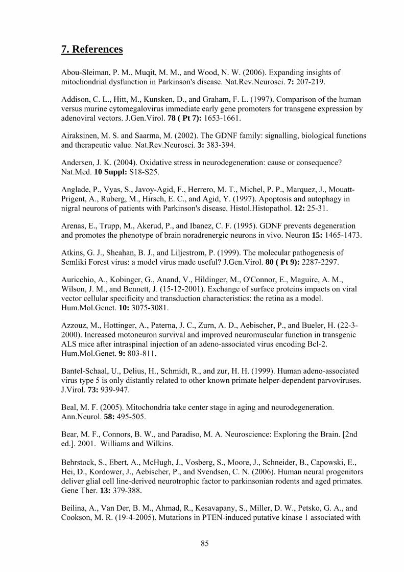
7. References Abou-Sleiman, P. M., Muqit, M. M., and Wood, N. W. (2006). Expanding insights of mitochondrial dysfunction in Parkinson's disease. Nat.Rev.Neurosci. 7: 207-219.
Addison, C. L., Hitt, M., Kunsken, D., and Graham, F. L. (1997). Comparison of the human versus murine cytomegalovirus immediate early gene promoters for transgene expression by adenoviral vectors. J.Gen.Virol. 78 ( Pt 7): 1653-1661.
Airaksinen, M. S. and Saarma, M. (2002). The GDNF family: signalling, biological functions and therapeutic value. Nat.Rev.Neurosci. 3: 383-394.
Andersen, J. K. (2004). Oxidative stress in neurodegeneration: cause or consequence? Nat.Med. 10 Suppl: S18-S25.
Anglade, P., Vyas, S., Javoy-Agid, F., Herrero, M. T., Michel, P. P., Marquez, J., Mouatt-Prigent, A., Ruberg, M., Hirsch, E. C., and Agid, Y. (1997). Apoptosis and autophagy in nigral neurons of patients with Parkinson's disease. Histol.Histopathol. 12: 25-31.
Arenas, E., Trupp, M., Akerud, P., and Ibanez, C. F. (1995). GDNF prevents degeneration and promotes the phenotype of brain noradrenergic neurons in vivo. Neuron 15: 1465-1473.
Atkins, G. J., Sheahan, B. J., and Liljestrom, P. (1999). The molecular pathogenesis of Semliki Forest virus: a model virus made useful? J.Gen.Virol. 80 ( Pt 9): 2287-2297.
Auricchio, A., Kobinger, G., Anand, V., Hildinger, M., O'Connor, E., Maguire, A. M., Wilson, J. M., and Bennett, J. (15-12-2001). Exchange of surface proteins impacts on viral vector cellular specificity and transduction characteristics: the retina as a model. Hum.Mol.Genet. 10: 3075-3081.
Azzouz, M., Hottinger, A., Paterna, J. C., Zurn, A. D., Aebischer, P., and Bueler, H. (22-3-2000). Increased motoneuron survival and improved neuromuscular function in transgenic ALS mice after intraspinal injection of an adeno-associated virus encoding Bcl-2. Hum.Mol.Genet. 9: 803-811.
Bantel-Schaal, U., Delius, H., Schmidt, R., and zur, H. H. (1999). Human adeno-associated virus type 5 is only distantly related to other known primate helper-dependent parvoviruses. J.Virol. 73: 939-947.
Beal, M. F. (2005). Mitochondria take center stage in aging and neurodegeneration. Ann.Neurol. 58: 495-505.
Bear, M. F., Connors, B. W., and Paradiso, M. A. Neuroscience: Exploring the Brain. [2nd ed.]. 2001. Williams and Wilkins. Behrstock, S., Ebert, A., McHugh, J., Vosberg, S., Moore, J., Schneider, B., Capowski, E., Hei, D., Kordower, J., Aebischer, P., and Svendsen, C. N. (2006). Human neural progenitors deliver glial cell line-derived neurotrophic factor to parkinsonian rodents and aged primates. Gene Ther. 13: 379-388.
Beilina, A., Van Der, B. M., Ahmad, R., Kesavapany, S., Miller, D. W., Petsko, G. A., and Cookson, M. R. (19-4-2005). Mutations in PTEN-induced putative kinase 1 associated with
85
recessive parkinsonism have differential effects on protein stability. Proc.Natl.Acad.Sci.U.S.A 102: 5703-5708.
Ben Shachar, D., Zuk, R., and Glinka, Y. (1995). Dopamine neurotoxicity: inhibition of mitochondrial respiration. J.Neurochem. 64: 718-723.
Betarbet, R., Sherer, T. B., MacKenzie, G., Garcia-Osuna, M., Panov, A. V., and Greenamyre, J. T. (2000). Chronic systemic pesticide exposure reproduces features of Parkinson's disease. Nat.Neurosci. 3: 1301-1306.
Björklund, A., Rosenblad, C., Winkler, C., and Kirik, D. (1997). Studies on neuroprotective and regenerative effects of GDNF in a partial lesion model of Parkinson's disease. Neurobiol.Dis. 4: 186-200.
Blom, N., Sicheritz-Ponten, T., Gupta, R., Gammeltoft, S., and Brunak, S. (2004). Prediction of post-translational glycosylation and phosphorylation of proteins from the amino acid sequence. Proteomics. 4: 1633-1649.
Blum, D., Torch, S., Lambeng, N., Nissou, M., Benabid, A. L., Sadoul, R., and Verna, J. M. (2001). Molecular pathways involved in the neurotoxicity of 6-OHDA, dopamine and MPTP: contribution to the apoptotic theory in Parkinson's disease. Prog.Neurobiol. 65: 135-172.
Braak, H. and Braak, E. (2000). Pathoanatomy of Parkinson's disease. J.Neurol. 247 Suppl 2: II3-10.
Buller, R. M., Janik, J. E., Sebring, E. D., and Rose, J. A. (1981). Herpes simplex virus types 1 and 2 completely help adenovirus-associated virus replication. J.Virol. 40: 241-247.
Burger, C., Gorbatyuk, O. S., Velardo, M. J., Peden, C. S., Williams, P., Zolotukhin, S., Reier, P. J., Mandel, R. J., and Muzyczka, N. (2004). Recombinant AAV viral vectors pseudotyped with viral capsids from serotypes 1, 2, and 5 display differential efficiency and cell tropism after delivery to different regions of the central nervous system. Mol.Ther. 10: 302-317.
Cacquevel, M., Lebeurrier, N., Cheenne, S., and Vivien, D. (2004). Cytokines in neuroinflammation and Alzheimer's disease. Curr.Drug Targets. 5: 529-534.
Cadet, J. L., Zhu, S. M., and Angulo, J. A. (1992). Quantitative in situ hybridization evidence for differential regulation of proenkephalin and dopamine D2 receptor mRNA levels in the rat striatum: effects of unilateral intrastriatal injections of 6-hydroxydopamine. Brain Res.Mol.Brain Res. 12: 59-67.
Cearley, C. N. and Wolfe, J. H. (2006). Transduction characteristics of adeno-associated virus vectors expressing cap serotypes 7, 8, 9, and Rh10 in the mouse brain. Mol.Ther. 13: 528-537.
Chang, L. K., Putcha, G. V., Deshmukh, M., and Johnson, E. M., Jr. (2002). Mitochondrial involvement in the point of no return in neuronal apoptosis. Biochimie 84: 223-231.
Chao, C. C., Ma, Y. L., Chu, K. Y., and Lee, E. H. (2003). Integrin alphav and NCAM mediate the effects of GDNF on DA neuron survival, outgrowth, DA turnover and motor activity in rats. Neurobiol.Aging 24: 105-116.
86
Chao, H., Liu, Y., Rabinowitz, J., Li, C., Samulski, R. J., and Walsh, C. E. (2000). Several log increase in therapeutic transgene delivery by distinct adeno-associated viral serotype vectors. Mol.Ther. 2: 619-623.
Chen, J., Skehel, J. J., and Wiley, D. C. (29-9-1998). A polar octapeptide fused to the N-terminal fusion peptide solubilizes the influenza virus HA2 subunit ectodomain. Biochemistry 37: 13643-13649.
Cheng, E. H., Wei, M. C., Weiler, S., Flavell, R. A., Mak, T. W., Lindsten, T., and Korsmeyer, S. J. (2001). BCL-2, BCL-X(L) sequester BH3 domain-only molecules preventing BAX- and BAK-mediated mitochondrial apoptosis. Mol.Cell 8: 705-711.
Cheng, H., Wu, J. P., and Tzeng, S. F. (1-8-2002). Neuroprotection of glial cell line-derived neurotrophic factor in damaged spinal cords following contusive injury. J.Neurosci.Res. 69: 397-405.
Dauer, W. and Przedborski, S. (11-9-2003). Parkinson's disease: mechanisms and models. Neuron 39: 889-909.
Davidson, B. L., Stein, C. S., Heth, J. A., Martins, I., Kotin, R. M., Derksen, T. A., Zabner, J., Ghodsi, A., and Chiorini, J. A. (28-3-2000). Recombinant adeno-associated virus type 2, 4, and 5 vectors: transduction of variant cell types and regions in the mammalian central nervous system. Proc.Natl.Acad.Sci.U.S.A 97: 3428-3432.
Dawson, T. M., Dawson, V. L., Gage, F. H., Fisher, L. J., Hunt, M. A., and Wamsley, J. K. (1991). Functional recovery of supersensitive dopamine receptors after intrastriatal grafts of fetal substantia nigra. Exp.Neurol. 111: 282-292.
de Rijk, M. C., Tzourio, C., Breteler, M. M., Dartigues, J. F., Amaducci, L., Lopez-Pousa, S., Manubens-Bertran, J. M., Alperovitch, A., and Rocca, W. A. (1997). Prevalence of parkinsonism and Parkinson's disease in Europe: the EUROPARKINSON Collaborative Study. European Community Concerted Action on the Epidemiology of Parkinson's disease. J.Neurol.Neurosurg.Psychiatry 62: 10-15.
Deumens, R., Blokland, A., and Prickaerts, J. (2002). Modeling Parkinson's disease in rats: an evaluation of 6-OHDA lesions of the nigrostriatal pathway. Exp.Neurol. 175: 303-317.
Dexter, D. T., Carter, C. J., Wells, F. R., Javoy-Agid, F., Agid, Y., Lees, A., Jenner, P., and Marsden, C. D. (1989a). Basal lipid peroxidation in substantia nigra is increased in Parkinson's disease. J.Neurochem. 52: 381-389.
Dexter, D. T., Wells, F. R., Lees, A. J., Agid, F., Agid, Y., Jenner, P., and Marsden, C. D. (1989b). Increased nigral iron content and alterations in other metal ions occurring in brain in Parkinson's disease. J.Neurochem. 52: 1830-1836.
Di Giovanni, S., Knoblach, S. M., Brandoli, C., Aden, S. A., Hoffman, E. P., and Faden, A. I. (2003). Gene profiling in spinal cord injury shows role of cell cycle in neuronal death. Ann.Neurol. 53: 454-468.
Di Pasquale, G., Davidson, B. L., Stein, C. S., Martins, I., Scudiero, D., Monks, A., and Chiorini, J. A. (2003). Identification of PDGFR as a receptor for AAV-5 transduction. Nat.Med. 9: 1306-1312.
87
Dong, J. Y., Fan, P. D., and Frizzell, R. A. (10-11-1996). Quantitative analysis of the packaging capacity of recombinant adeno-associated virus. Hum.Gene Ther. 7: 2101-2112.
Duan, D., Sharma, P., Yang, J., Yue, Y., Dudus, L., Zhang, Y., Fisher, K. J., and Engelhardt, J. F. (1998). Circular intermediates of recombinant adeno-associated virus have defined structural characteristics responsible for long-term episomal persistence in muscle tissue. J.Virol. 72: 8568-8577.
Duan, D., Yue, Y., and Engelhardt, J. F. (2001). Expanding AAV packaging capacity with trans-splicing or overlapping vectors: a quantitative comparison. Mol.Ther. 4: 383-391.
Eberhardt, O., Coelln, R. V., Kügler, S., Lindenau, J., Rathke-Hartlieb, S., Gerhardt, E., Haid, S., Isenmann, S., Gravel, C., Srinivasan, A., Bähr, M., Weller, M., Dichgans, J., and Schulz, J. B. (15-12-2000). Protection by synergistic effects of adenovirus-mediated X-chromosome-linked inhibitor of apoptosis and glial cell line-derived neurotrophic factor gene transfer in the 1-methyl-4-phenyl-1,2,3,6-tetrahydropyridine model of Parkinson's disease. J.Neurosci. 20: 9126-9134.
Ehrengruber, M. U., Renggli, M., Raineteau, O., Hennou, S., Vähä-Koskela, M. J., Hinkkanen, A. E., and Lundstrom, K. (2003). Semliki Forest virus A7(74) transduces hippocampal neurons and glial cells in a temperature-dependent dual manner. J.Neurovirol. 9: 16-28.
Elston, T., Wang, H., and Oster, G. (29-1-1998). Energy transduction in ATP synthase. Nature 391: 510-513.
Eslamboli, A., Cummings, R. M., Ridley, R. M., Baker, H. F., Muzyczka, N., Burger, C., Mandel, R. J., Kirik, D., and Annett, L. E. (2003). Recombinant adeno-associated viral vector (rAAV) delivery of GDNF provides protection against 6-OHDA lesion in the common marmoset monkey (Callithrix jacchus). Exp.Neurol. 184: 536-548.
Esselink, R. A., de Bie, R. M., de Haan, R. J., Lenders, M. W., Nijssen, P. C., Staal, M. J., Smeding, H. M., Schuurman, P. R., Bosch, D. A., and Speelman, J. D. (27-1-2004). Unilateral pallidotomy versus bilateral subthalamic nucleus stimulation in PD: a randomized trial. Neurology 62: 201-207.
Ewing, B. and Green, P. (2000). Analysis of expressed sequence tags indicates 35,000 human genes. Nat.Genet. 25: 232-234.
Feany, M. B. and Bender, W. W. (23-3-2000). A Drosophila model of Parkinson's disease. Nature 404: 394-398.
Fearnley, J. M. and Lees, A. J. (1991). Ageing and Parkinson's disease: substantia nigra regional selectivity. Brain 114 ( Pt 5): 2283-2301.
Fleming, S. M., Fernagut, P. O., and Chesselet, M. F. (2005). Genetic mouse models of parkinsonism: strengths and limitations. NeuroRx. 2: 495-503.
Floor, E. and Wetzel, M. G. (1998). Increased protein oxidation in human substantia nigra pars compacta in comparison with basal ganglia and prefrontal cortex measured with an improved dinitrophenylhydrazine assay. J.Neurochem. 70: 268-275.
88

Flotte, T. R., Afione, S. A., and Zeitlin, P. L. (1994). Adeno-associated virus vector gene expression occurs in nondividing cells in the absence of vector DNA integration. Am.J.Respir.Cell Mol.Biol. 11: 517-521.
Flotte, T. R., Solow, R., Owens, R. A., Afione, S., Zeitlin, P. L., and Carter, B. J. (1992). Gene expression from adeno-associated virus vectors in airway epithelial cells. Am.J.Respir.Cell Mol.Biol. 7: 349-356.
Foley, P. and Riederer, P. (1999). Pathogenesis and preclinical course of Parkinson's disease. J.Neural Transm.Suppl 56: 31-74.
Fornai, F., Schluter, O. M., Lenzi, P., Gesi, M., Ruffoli, R., Ferrucci, M., Lazzeri, G., Busceti, C. L., Pontarelli, F., Battaglia, G., Pellegrini, A., Nicoletti, F., Ruggieri, S., Paparelli, A., and Sudhof, T. C. (1-3-2005). Parkinson-like syndrome induced by continuous MPTP infusion: convergent roles of the ubiquitin-proteasome system and alpha-synuclein. Proc.Natl.Acad.Sci.U.S.A 102: 3413-3418.
Forno, L. S. (1996). Neuropathology of Parkinson's disease. J.Neuropathol.Exp.Neurol. 55: 259-272.
Fuxe, K., Chadi, G., Tinner, B., Agnati, L. F., Pettersson, R., and David, G. (4-2-1994). On the regional distribution of heparan sulfate proteoglycan immunoreactivity in the rat brain. Brain Res. 636: 131-138.
Gagnon, C., Bedard, P. J., Rioux, L., Gaudin, D., Martinoli, M. G., Pelletier, G., and Di Paolo, T. (6-9-1991). Regional changes of striatal dopamine receptors following denervation by 6-hydroxydopamine and fetal mesencephalic grafts in the rat. Brain Res. 558: 251-263.
Gandhi, S. and Wood, N. W. (15-9-2005). Molecular pathogenesis of Parkinson's disease. Hum.Mol.Genet. 14: 2749-2755.
Georgievska, B., Kirik, D., and Björklund, A. (2002a). Aberrant sprouting and downregulation of tyrosine hydroxylase in lesioned nigrostriatal dopamine neurons induced by long-lasting overexpression of glial cell line derived neurotrophic factor in the striatum by lentiviral gene transfer. Exp.Neurol. 177: 461-474.
Georgievska, B., Kirik, D., Rosenblad, C., Lundberg, C., and Björklund, A. (21-1-2002b). Neuroprotection in the rat Parkinson model by intrastriatal GDNF gene transfer using a lentiviral vector. Neuroreport 13: 75-82.
Gerdes, C. A., Castro, M. G., and Löwenstein, P. R. (2000). Strong promoters are the key to highly efficient, noninflammatory and noncytotoxic adenoviral-mediated transgene delivery into the brain in vivo. Mol.Ther. 2: 330-338.
Ghribi, O., Herman, M. M., Forbes, M. S., DeWitt, D. A., and Savory, J. (2001). GDNF protects against aluminum-induced apoptosis in rabbits by upregulating Bcl-2 and Bcl-XL and inhibiting mitochondrial Bax translocation. Neurobiol.Dis. 8: 764-773.
Gill, S. S., Patel, N. K., Hotton, G. R., O'Sullivan, K., McCarter, R., Bunnage, M., Brooks, D. J., Svendsen, C. N., and Heywood, P. (2003). Direct brain infusion of glial cell line-derived neurotrophic factor in Parkinson disease. Nat.Med. 9: 589-595.
Gotz, M. E., Dirr, A., Burger, R., Janetzky, B., Weinmuller, M., Chan, W. W., Chen, S. C., Reichmann, H., Rausch, W. D., and Riederer, P. (15-2-1994). Effect of lipoic acid on redox
89
state of coenzyme Q in mice treated with 1-methyl-4-phenyl-1,2,3,6-tetrahydropyridine and diethyldithiocarbamate. Eur.J.Pharmacol. 266: 291-300.
Graham, D. G., Tiffany, S. M., Bell, W. R., Jr., and Gutknecht, W. F. (1978). Autoxidation versus covalent binding of quinones as the mechanism of toxicity of dopamine, 6-hydroxydopamine, and related compounds toward C1300 neuroblastoma cells in vitro. Mol.Pharmacol. 14: 644-653.
Grondin, R. and Gash, D. M. (1998). Glial cell line-derived neurotrophic factor (GDNF): a drug candidate for the treatment of Parkinson's disease. J.Neurol. 245: 35-42.
Grussenmeyer, T., Scheidtmann, K. H., Hutchinson, M. A., Eckhart, W., and Walter, G. (1985). Complexes of polyoma virus medium T antigen and cellular proteins. Proc.Natl.Acad.Sci.U.S.A 82: 7952-7954.
Gunawardena, S. and Goldstein, L. S. (2005). Polyglutamine diseases and transport problems: deadly traffic jams on neuronal highways. Arch.Neurol. 62: 46-51.
Hald, A. and Lotharius, J. (2005). Oxidative stress and inflammation in Parkinson's disease: is there a causal link? Exp.Neurol. 193: 279-290.
Hauck, B., Chen, L., and Xiao, W. (2003). Generation and characterization of chimeric recombinant AAV vectors. Mol.Ther. 7: 419-425.
Henderson, C. E., Phillips, H. S., Pollock, R. A., Davies, A. M., Lemeulle, C., Armanini, M., Simmons, L., Moffet, B., Vandlen, R. A., Simpson LC [corrected to Simmons, and . (11-11-1994). GDNF: a potent survival factor for motoneurons present in peripheral nerve and muscle. Science 266: 1062-1064.
Hengartner, M. O. (12-10-2000). The biochemistry of apoptosis. Nature 407: 770-776.
Hildinger, M., Auricchio, A., Gao, G., Wang, L., Chirmule, N., and Wilson, J. M. (2001). Hybrid vectors based on adeno-associated virus serotypes 2 and 5 for muscle-directed gene transfer. J.Virol. 75: 6199-6203.
Hirsch, E. C., Breidert, T., Rousselet, E., Hunot, S., Hartmann, A., and Michel, P. P. (2003). The role of glial reaction and inflammation in Parkinson's disease. Ann.N.Y.Acad.Sci. 991: 214-228.
Hristova, A. H. and Koller, W. C. (2000). Early Parkinson's disease: what is the best approach to treatment. Drugs Aging 17: 165-181.
Hughes, A. J., Ben Shlomo, Y., Daniel, S. E., and Lees, A. J. (2001). What features improve the accuracy of clinical diagnosis in Parkinson's disease: a clinicopathologic study. 1992. Neurology 57: S34-S38.
Ibanez, C. F. (1998). Emerging themes in structural biology of neurotrophic factors. Trends Neurosci. 21: 438-444.
Jin, J., Meredith, G. E., Chen, L., Zhou, Y., Xu, J., Shie, F. S., Lockhart, P., and Zhang, J. (24-3-2005). Quantitative proteomic analysis of mitochondrial proteins: relevance to Lewy body formation and Parkinson's disease. Brain Res.Mol.Brain Res. 134: 119-138.
90
Johnson, K. Y., Liu, L., and Vincent, T. S. (2002). Minimal FLAG sequence useful in the functional epitope tagging of H-Ras. Biotechniques 32: 1270, 1272-6, 1278, 1280.
Jollivet, C., Aubert-Pouessel, A., Clavreul, A., Venier-Julienne, M. C., Remy, S., Montero-Menei, C. N., Benoit, J. P., and Menei, P. (2004). Striatal implantation of GDNF releasing biodegradable microspheres promotes recovery of motor function in a partial model of Parkinson's disease. Biomaterials 25: 933-942.
Juraska, J. M., Wilson, C. J., and Groves, P. M. (15-4-1977). The substantia nigra of the rat: a Golgi study. J.Comp Neurol. 172: 585-600.
Kaludov, N., Brown, K. E., Walters, R. W., Zabner, J., and Chiorini, J. A. (2001). Adeno-associated virus serotype 4 (AAV4) and AAV5 both require sialic acid binding for hemagglutination and efficient transduction but differ in sialic acid linkage specificity. J.Virol. 75: 6884-6893.
Kandel, E. R., Schwartz, J. H., and Jessell, T. M. Principles of Neural Science. [ 4th ed.]. 2000. New York, McGraw-Hill. Kaufmann, T., Schlipf, S., Sanz, J., Neubert, K., Stein, R., and Borner, C. (6-1-2003). Characterization of the signal that directs Bcl-x(L), but not Bcl-2, to the mitochondrial outer membrane. J.Cell Biol. 160: 53-64.
Keogh, S. A., Walczak, H., Bouchier-Hayes, L., and Martin, S. J. (7-4-2000). Failure of Bcl-2 to block cytochrome c redistribution during TRAIL-induced apoptosis. FEBS Lett. 471: 93-98.
Kermer, P., Klöcker, N., and Bähr, M. (1999). Long-term effect of inhibition of ced 3-like caspases on the survival of axotomized retinal ganglion cells in vivo. Exp.Neurol. 158: 202-205.
Kim, I. K., Jung, Y. K., Noh, D. Y., Song, Y. S., Choi, C. H., Oh, B. H., Masuda, E. S., and Jung, Y. K. (24-3-2003). Functional screening of genes suppressing TRAIL-induced apoptosis: distinct inhibitory activities of Bcl-XL and Bcl-2. Br.J.Cancer 88: 910-917.
Kim, R. (29-7-2005). Unknotting the roles of Bcl-2 and Bcl-xL in cell death. Biochem.Biophys.Res.Commun. 333: 336-343.
Kirik, D., Annett, L. E., Burger, C., Muzyczka, N., Mandel, R. J., and Bjorklund, A. (4-3-2003). Nigrostriatal alpha-synucleinopathy induced by viral vector-mediated overexpression of human alpha-synuclein: a new primate model of Parkinson's disease. Proc.Natl.Acad.Sci.U.S.A 100: 2884-2889.
Kirik, D., Georgievska, B., and Björklund, A. (2004). Localized striatal delivery of GDNF as a treatment for Parkinson disease. Nat.Neurosci. 7: 105-110.
Kirik, D., Rosenblad, C., and Björklund, A. (1998). Characterization of behavioral and neurodegenerative changes following partial lesions of the nigrostriatal dopamine system induced by intrastriatal 6-hydroxydopamine in the rat. Exp.Neurol. 152: 259-277.
Kirik, D., Rosenblad, C., Burger, C., Lundberg, C., Johansen, T. E., Muzyczka, N., Mandel, R. J., and Björklund, A. (1-4-2002). Parkinson-like neurodegeneration induced by targeted overexpression of alpha-synuclein in the nigrostriatal system. J.Neurosci. 22: 2780-2791.
91
Kish, S. J., Morito, C., and Hornykiewicz, O. (5-8-1985). Glutathione peroxidase activity in Parkinson's disease brain. Neurosci.Lett. 58: 343-346.
Kishima, H., Poyot, T., Bloch, J., Dauguet, J., Conde, F., Dolle, F., Hinnen, F., Pralong, W., Palfi, S., Deglon, N., Aebischer, P., and Hantraye, P. (2004). Encapsulated GDNF-producing C2C12 cells for Parkinson's disease: a pre-clinical study in chronic MPTP-treated baboons. Neurobiol.Dis. 16: 428-439.
Klein, R. L., King, M. A., Hamby, M. E., and Meyer, E. M. (20-3-2002). Dopaminergic cell loss induced by human A30P alpha-synuclein gene transfer to the rat substantia nigra. Hum.Gene Ther. 13: 605-612.
Kügler, S., Kilic, E., and Bähr, M. (2003a). Human synapsin 1 gene promoter confers highly neuron-specific long-term transgene expression from an adenoviral vector in the adult rat brain depending on the transduced area. Gene Ther. 10: 337-347.
Kügler, S., Lingor, P., Schöll, U., Zolotukhin, S., and Bähr, M. (20-6-2003b). Differential transgene expression in brain cells in vivo and in vitro from AAV-2 vectors with small transcriptional control units. Virology 311: 89-95.
Kügler, S., Meyn, L., Holzmuller, H., Gerhardt, E., Isenmann, S., Schulz, J. B., and Bähr, M. (2001). Neuron-specific expression of therapeutic proteins: evaluation of different cellular promoters in recombinant adenoviral vectors. Mol.Cell Neurosci. 17: 78-96.
Lang, A. E., Gill, S., Patel, N. K., Lozano, A., Nutt, J. G., Penn, R., Brooks, D. J., Hotton, G., Moro, E., Heywood, P., Brodsky, M. A., Burchiel, K., Kelly, P., Dalvi, A., Scott, B., Stacy, M., Turner, D., Wooten, V. G., Elias, W. J., Laws, E. R., Dhawan, V., Stoessl, A. J., Matcham, J., Coffey, R. J., and Traub, M. (2006). Randomized controlled trial of intraputamenal glial cell line-derived neurotrophic factor infusion in Parkinson disease. Ann.Neurol. 59: 459-466.
Lang, A. E. and Lozano, A. M. (15-10-1998). Parkinson's disease. Second of two parts. N.Engl.J.Med. 339: 1130-1143.
Leroy, E., Boyer, R., Auburger, G., Leube, B., Ulm, G., Mezey, E., Harta, G., Brownstein, M. J., Jonnalagada, S., Chernova, T., Dehejia, A., Lavedan, C., Gasser, T., Steinbach, P. J., Wilkinson, K. D., and Polymeropoulos, M. H. (1-10-1998). The ubiquitin pathway in Parkinson's disease. Nature 395: 451-452.
Li, H., Li, S. H., Yu, Z. X., Shelbourne, P., and Li, X. J. (1-11-2001). Huntingtin aggregate-associated axonal degeneration is an early pathological event in Huntington's disease mice. J.Neurosci. 21: 8473-8481.
Liljestrom, P. and Garoff, H. (1991). A new generation of animal cell expression vectors based on the Semliki Forest virus replicon. Biotechnology (N.Y.) 9: 1356-1361.
Lim, P. S., Jenson, A. B., Cowsert, L., Nakai, Y., Lim, L. Y., Jin, X. W., and Sundberg, J. P. (1990). Distribution and specific identification of papillomavirus major capsid protein epitopes by immunocytochemistry and epitope scanning of synthetic peptides. J.Infect.Dis. 162: 1263-1269.
Lin, L. F., Doherty, D. H., Lile, J. D., Bektesh, S., and Collins, F. (21-5-1993). GDNF: a glial cell line-derived neurotrophic factor for midbrain dopaminergic neurons. Science 260: 1130-1132.
92
Liu, S. and Altman, R. B. (15-8-2003). Large scale study of protein domain distribution in the context of alternative splicing. Nucleic Acids Res. 31: 4828-4835.
Lo, B. C., Deglon, N., Pralong, W., and Aebischer, P. (2004). Lentiviral nigral delivery of GDNF does not prevent neurodegeneration in a genetic rat model of Parkinson's disease. Neurobiol.Dis. 17: 283-289.
Lomri, N., Perret, C., Gouhier, N., and Thomasset, M. (1-8-1989). Cloning and analysis of calbindin-D28K cDNA and its expression in the central nervous system. Gene 80: 87-98.
Lotharius, J. and Brundin, P. (1-10-2002a). Impaired dopamine storage resulting from alpha-synuclein mutations may contribute to the pathogenesis of Parkinson's disease. Hum.Mol.Genet. 11: 2395-2407.
Lotharius, J. and Brundin, P. (2002b). Pathogenesis of Parkinson's disease: dopamine, vesicles and alpha-synuclein. Nat.Rev.Neurosci. 3: 932-942.
Love, S., Plaha, P., Patel, N. K., Hotton, G. R., Brooks, D. J., and Gill, S. S. (2005). Glial cell line-derived neurotrophic factor induces neuronal sprouting in human brain. Nat.Med. 11: 703-704.
Lundstrom, K. (2005). Biology and application of alphaviruses in gene therapy. Gene Ther. 12 Suppl 1: S92-S97.
Lundstrom, K., Abenavoli, A., Malgaroli, A., and Ehrengruber, M. U. (2003). Novel Semliki Forest virus vectors with reduced cytotoxicity and temperature sensitivity for long-term enhancement of transgene expression. Mol.Ther. 7: 202-209.
Malik, J. M., Shevtsova, Z., Bähr, M., and Kügler, S. (2005). Long-term in vivo inhibition of CNS neurodegeneration by Bcl-XL gene transfer. Mol.Ther. 11: 373-381.
Marini, P., Jendrossek, V., Durand, E., Gruber, C., Budach, W., and Belka, C. (2003). Molecular requirements for the combined effects of TRAIL and ionising radiation. Radiother.Oncol. 68: 189-198.
Marjama-Lyons, J. M. and Koller, W. C. (2001). Parkinson's disease. Update in diagnosis and symptom management. Geriatrics 56: 24-30, 33.
Mastakov, M. Y., Baer, K., Symes, C. W., Leichtlein, C. B., Kotin, R. M., and During, M. J. (2002). Immunological aspects of recombinant adeno-associated virus delivery to the mammalian brain. J.Virol. 76: 8446-8454.
Mattson, M. P. and Chan, S. L. (2003). Calcium orchestrates apoptosis. Nat.Cell Biol. 5: 1041-1043.
Melov, S. (2004). Modeling mitochondrial function in aging neurons. Trends Neurosci. 27: 601-606.
Michel, U., Malik, I., Ebert, S., Bähr, M., and Kügler, S. (14-1-2005). Long-term in vivo and in vitro AAV-2-mediated RNA interference in rat retinal ganglion cells and cultured primary neurons. Biochem.Biophys.Res.Commun. 326: 307-312.
Moskalenko, M., Chen, L., van Roey, M., Donahue, B. A., Snyder, R. O., McArthur, J. G., and Patel, S. D. (2000). Epitope mapping of human anti-adeno-associated virus type 2
93
neutralizing antibodies: implications for gene therapy and virus structure. J.Virol. 74: 1761-1766.
Motoyama, N., Wang, F., Roth, K. A., Sawa, H., Nakayama, K., Nakayama, K., Negishi, I., Senju, S., Zhang, Q., Fujii, S., and . (10-3-1995). Massive cell death of immature hematopoietic cells and neurons in Bcl-x-deficient mice. Science 267: 1506-1510.
Nakajima, A., Kataoka, K., Hong, M., Sakaguchi, M., and Huh, N. H. (25-11-2003). BRPK, a novel protein kinase showing increased expression in mouse cancer cell lines with higher metastatic potential. Cancer Lett. 201: 195-201.
Natsume, A., Mata, M., Goss, J., Huang, S., Wolfe, D., Oligino, T., Glorioso, J., and Fink, D. J. (2001). Bcl-2 and GDNF delivered by HSV-mediated gene transfer act additively to protect dopaminergic neurons from 6-OHDA-induced degeneration. Exp.Neurol. 169: 231-238.
Natsume, A., Mata, M., Wolfe, D., Oligino, T., Goss, J., Huang, S., Glorioso, J., and Fink, D. J. (2002). Bcl-2 and GDNF delivered by HSV-mediated gene transfer after spinal root avulsion provide a synergistic effect. J.Neurotrauma 19: 61-68.
Newmeyer, D. D. and Ferguson-Miller, S. (21-2-2003). Mitochondria: releasing power for life and unleashing the machineries of death. Cell 112: 481-490.
Nutt, J. G., Burchiel, K. J., Comella, C. L., Jankovic, J., Lang, A. E., Laws, E. R., Jr., Lozano, A. M., Penn, R. D., Simpson, R. K., Jr., Stacy, M., and Wooten, G. F. (14-1-2003). Randomized, double-blind trial of glial cell line-derived neurotrophic factor (GDNF) in PD. Neurology 60: 69-73.
Offen, D., Beart, P. M., Cheung, N. S., Pascoe, C. J., Hochman, A., Gorodin, S., Melamed, E., Bernard, R., and Bernard, O. (12-5-1998). Transgenic mice expressing human Bcl-2 in their neurons are resistant to 6-hydroxydopamine and 1-methyl-4-phenyl-1,2,3,6- tetrahydropyridine neurotoxicity. Proc.Natl.Acad.Sci.U.S.A 95: 5789-5794.
Olanow, C. W. and Stocchi, F. (13-1-2004). COMT inhibitors in Parkinson's disease: can they prevent and/or reverse levodopa-induced motor complications? Neurology 62: S72-S81.
Opie, S. R., Warrington, K. H., Jr., Agbandje-McKenna, M., Zolotukhin, S., and Muzyczka, N. (2003). Identification of amino acid residues in the capsid proteins of adeno-associated virus type 2 that contribute to heparan sulfate proteoglycan binding. J.Virol. 77: 6995-7006.
Paratcha, G., Ledda, F., and Ibanez, C. F. (27-6-2003). The neural cell adhesion molecule NCAM is an alternative signaling receptor for GDNF family ligands. Cell 113: 867-879.
Parker, W. D., Jr., Boyson, S. J., and Parks, J. K. (1989). Abnormalities of the electron transport chain in idiopathic Parkinson's disease. Ann.Neurol. 26: 719-723.
Passini, M. A., Watson, D. J., Vite, C. H., Landsburg, D. J., Feigenbaum, A. L., and Wolfe, J. H. (2003). Intraventricular brain injection of adeno-associated virus type 1 (AAV1) in neonatal mice results in complementary patterns of neuronal transduction to AAV2 and total long-term correction of storage lesions in the brains of beta-glucuronidase-deficient mice. J.Virol. 77: 7034-7040.
94
Patel, N. K., Bunnage, M., Plaha, P., Svendsen, C. N., Heywood, P., and Gill, S. S. (2005). Intraputamenal infusion of glial cell line-derived neurotrophic factor in PD: a two-year outcome study. Ann.Neurol. 57: 298-302.
Paterna, J. C., Feldon, J., and Bueler, H. (2004). Transduction profiles of recombinant adeno-associated virus vectors derived from serotypes 2 and 5 in the nigrostriatal system of rats. J.Virol. 78: 6808-6817.
Peel, A. L. and Klein, R. L. (1-6-2000). Adeno-associated virus vectors: activity and applications in the CNS. J.Neurosci.Methods 98: 95-104.
Peel, A. L., Zolotukhin, S., Schrimsher, G. W., Muzyczka, N., and Reier, P. J. (1997). Efficient transduction of green fluorescent protein in spinal cord neurons using adeno-associated virus vectors containing cell type-specific promoters. Gene Ther. 4: 16-24.
Perrelet, D., Ferri, A., MacKenzie, A. E., Smith, G. M., Korneluk, R. G., Liston, P., Sagot, Y., Terrado, J., Monnier, D., and Kato, A. C. (2000). IAP family proteins delay motoneuron cell death in vivo. Eur.J.Neurosci. 12: 2059-2067.
Picada, J. N., Roesler, R., and Henriques, J. A. (2005). Genotoxic, neurotoxic and neuroprotective activities of apomorphine and its oxidized derivative 8-oxo-apomorphine. Braz.J.Med.Biol.Res. 38: 477-486.
Previtali, S. C., Quattrini, A., Fasolini, M., Panzeri, M. C., Villa, A., Filbin, M. T., Li, W., Chiu, S. Y., Messing, A., Wrabetz, L., and Feltri, M. L. (27-11-2000). Epitope-tagged P(0) glycoprotein causes Charcot-Marie-Tooth-like neuropathy in transgenic mice. J.Cell Biol. 151: 1035-1046.
Qin, Z. H., Chen, R. W., Wang, Y., Nakai, M., Chuang, D. M., and Chase, T. N. (15-5-1999). Nuclear factor kappaB nuclear translocation upregulates c-Myc and p53 expression during NMDA receptor-mediated apoptosis in rat striatum. J.Neurosci. 19: 4023-4033.
Qing, K., Mah, C., Hansen, J., Zhou, S., Dwarki, V., and Srivastava, A. (1999). Human fibroblast growth factor receptor 1 is a co-receptor for infection by adeno-associated virus 2. Nat.Med. 5: 71-77.
Rabinowitz, J. E., Bowles, D. E., Faust, S. M., Ledford, J. G., Cunningham, S. E., and Samulski, R. J. (2004). Cross-dressing the virion: the transcapsidation of adeno-associated virus serotypes functionally defines subgroups. J.Virol. 78: 4421-4432.
Ramer, M. S., Priestley, J. V., and McMahon, S. B. (20-1-2000). Functional regeneration of sensory axons into the adult spinal cord. Nature 403: 312-316.
Rideout, H. J. and Stefanis, L. (2001). Caspase inhibition: a potential therapeutic strategy in neurological diseases. Histol.Histopathol. 16: 895-908.
Riederer, P. and Wuketich, S. (1976). Time course of nigrostriatal degeneration in parkinson's disease. A detailed study of influential factors in human brain amine analysis. J.Neural Transm. 38: 277-301.
Rioux, L., Gaudin, D. P., Gagnon, C., Di Paolo, T., and Bedard, P. J. (1991). Decrease of behavioral and biochemical denervation supersensitivity of rat striatum by nigral transplants. Neuroscience 44: 75-83.
95
Rizzuto, R., Pinton, P., Brini, M., Chiesa, A., Filippin, L., and Pozzan, T. (1999). Mitochondria as biosensors of calcium microdomains. Cell Calcium 26: 193-199.
Rosenblad, C., Georgievska, B., and Kirik, D. (2003). Long-term striatal overexpression of GDNF selectively downregulates tyrosine hydroxylase in the intact nigrostriatal dopamine system. Eur.J.Neurosci. 17: 260-270.
Rosenblad, C., Kirik, D., and Björklund, A. (2000). Sequential administration of GDNF into the substantia nigra and striatum promotes dopamine neuron survival and axonal sprouting but not striatal reinnervation or functional recovery in the partial 6-OHDA lesion model. Exp.Neurol. 161: 503-516.
Sambrook, J., Fritsch, E. F., and Maniatis, T. Molecular Cloning: A Laboratory Manual. [2nd ed.]. 1989. Cold Spring Harbor, Cold Spring Harbor Laboratory Press. Samulski, R. J., Chang, L. S., and Shenk, T. (1989). Helper-free stocks of recombinant adeno-associated viruses: normal integration does not require viral gene expression. J.Virol. 63: 3822-3828.
Sariola, H. and Saarma, M. (1-10-2003). Novel functions and signalling pathways for GDNF. J.Cell Sci. 116: 3855-3862.
Sattler, M., Liang, H., Nettesheim, D., Meadows, R. P., Harlan, J. E., Eberstadt, M., Yoon, H. S., Shuker, S. B., Chang, B. S., Minn, A. J., Thompson, C. B., and Fesik, S. W. (14-2-1997). Structure of Bcl-xL-Bak peptide complex: recognition between regulators of apoptosis. Science 275: 983-986.
Schaar, D. G., Sieber, B. A., Dreyfus, C. F., and Black, I. B. (1993). Regional and cell-specific expression of GDNF in rat brain. Exp.Neurol. 124: 368-371.
Schapira, A. H. (1994). Evidence for mitochondrial dysfunction in Parkinson's disease--a critical appraisal. Mov Disord. 9: 125-138.
Schapira, A. H., Cooper, J. M., Dexter, D., Clark, J. B., Jenner, P., and Marsden, C. D. (1990). Mitochondrial complex I deficiency in Parkinson's disease. J.Neurochem. 54: 823-827.
Schapira, A. H., Cooper, J. M., Dexter, D., Jenner, P., Clark, J. B., and Marsden, C. D. (3-6-1989). Mitochondrial complex I deficiency in Parkinson's disease. Lancet 1: 1269.
Schmidt, R. H., Björklund, A., Stenevi, U., Dunnett, S. B., and Gage, F. H. (1983). Intracerebral grafting of neuronal cell suspensions. III. Activity of intrastriatal nigral suspension implants as assessed by measurements of dopamine synthesis and metabolism. Acta Physiol Scand.Suppl 522: 19-28.
Seniuk, N. A., Tatton, W. G., and Greenwood, C. E. (10-9-1990). Dose-dependent destruction of the coeruleus-cortical and nigral-striatal projections by MPTP. Brain Res. 527: 7-20.
Shapiro, L., Doyle, J. P., Hensley, P., Colman, D. R., and Hendrickson, W. A. (1996). Crystal structure of the extracellular domain from P0, the major structural protein of peripheral nerve myelin. Neuron 17: 435-449.
96
Shevtsova, Z., Malik, J. M., Michel, U., Bähr, M., and Kügler, S. (2005). Promoters and serotypes: targeting of adeno-associated virus vectors for gene transfer in the rat central nervous system in vitro and in vivo. Exp.Physiol 90: 53-59.
Shindler, K. S., Latham, C. B., and Roth, K. A. (1-5-1997). Bax deficiency prevents the increased cell death of immature neurons in bcl-x-deficient mice. J.Neurosci. 17: 3112-3119.
Sian, J., Dexter, D. T., Lees, A. J., Daniel, S., Agid, Y., Javoy-Agid, F., Jenner, P., and Marsden, C. D. (1994). Alterations in glutathione levels in Parkinson's disease and other neurodegenerative disorders affecting basal ganglia. Ann.Neurol. 36: 348-355.
Sofic, E., Lange, K. W., Jellinger, K., and Riederer, P. (17-8-1992). Reduced and oxidized glutathione in the substantia nigra of patients with Parkinson's disease. Neurosci.Lett. 142: 128-130.
Sofic, E., Riederer, P., Heinsen, H., Beckmann, H., Reynolds, G. P., Hebenstreit, G., and Youdim, M. B. (1988). Increased iron (III) and total iron content in post mortem substantia nigra of parkinsonian brain. J.Neural Transm. 74: 199-205.
Srivastava, A., Lusby, E. W., and Berns, K. I. (1983). Nucleotide sequence and organization of the adeno-associated virus 2 genome. J.Virol. 45: 555-564.
Staniek, K., Gille, L., Kozlov, A. V., and Nohl, H. (2002). Mitochondrial superoxide radical formation is controlled by electron bifurcation to the high and low potential pathways. Free Radic.Res. 36: 381-387.
Stieger, K., Le Meur, G., Lasne, F., Weber, M., Deschamps, J. Y., Nivard, D., Mendes-Madeira, A., Provost, N., Martin, L., Moullier, P., and Rolling, F. (24-1-2006). Long-term doxycycline-regulated transgene expression in the retina of nonhuman primates following subretinal injection of recombinant AAV vectors. Mol.Ther.
Stokes, A. H., Hastings, T. G., and Vrana, K. E. (15-3-1999). Cytotoxic and genotoxic potential of dopamine. J.Neurosci.Res. 55: 659-665.
Stokin, G. B., Lillo, C., Falzone, T. L., Brusch, R. G., Rockenstein, E., Mount, S. L., Raman, R., Davies, P., Masliah, E., Williams, D. S., and Goldstein, L. S. (25-2-2005). Axonopathy and transport deficits early in the pathogenesis of Alzheimer's disease. Science 307: 1282-1288.
Straten, G., Schmeer, C., Kretz, A., Gerhardt, E., Kügler, S., Schulz, J. B., Gravel, C., Bähr, M., and Isenmann, S. (2002). Potential synergistic protection of retinal ganglion cells from axotomy-induced apoptosis by adenoviral administration of glial cell line-derived neurotrophic factor and X-chromosome-linked inhibitor of apoptosis. Neurobiol.Dis. 11: 123-133.
Strauss, J. H. and Strauss, E. G. (1994). The alphaviruses: gene expression, replication, and evolution. Microbiol.Rev. 58: 491-562.
Strauss, K. M., Martins, L. M., Plun-Favreau, H., Marx, F. P., Kautzmann, S., Berg, D., Gasser, T., Wszolek, Z., Muller, T., Bornemann, A., Wolburg, H., Downward, J., Riess, O., Schulz, J. B., and Krüger, R. (1-8-2005). Loss of function mutations in the gene encoding Omi/HtrA2 in Parkinson's disease. Hum.Mol.Genet. 14: 2099-2111.
97
Strelau, J. and Unsicker, K. (1999). GDNF family members and their receptors: expression and functions in two oligodendroglial cell lines representing distinct stages of oligodendroglial development. Glia 26: 291-301.
Sulzer, D., Sonders, M. S., Poulsen, N. W., and Galli, A. (2005). Mechanisms of neurotransmitter release by amphetamines: a review. Prog.Neurobiol. 75: 406-433.
Sulzer, D. and Zecca, L. (2000). Intraneuronal dopamine-quinone synthesis: a review. Neurotox.Res. 1: 181-195.
Summerford, C., Bartlett, J. S., and Samulski, R. J. (1999). AlphaVbeta5 integrin: a co-receptor for adeno-associated virus type 2 infection. Nat.Med. 5: 78-82.
Sun, L., Li, J., and Xiao, X. (2000). Overcoming adeno-associated virus vector size limitation through viral DNA heterodimerization. Nat.Med. 6: 599-602.
Takahashi, M. (2001). The GDNF/RET signaling pathway and human diseases. Cytokine Growth Factor Rev. 12: 361-373.
Tenenbaum, L., Chtarto, A., Lehtonen, E., Velu, T., Brotchi, J., and Levivier, M. (2004). Recombinant AAV-mediated gene delivery to the central nervous system. J.Gene Med. 6 Suppl 1: S212-S222.
Terpe, K. (2003). Overview of tag protein fusions: from molecular and biochemical fundamentals to commercial systems. Appl.Microbiol.Biotechnol. 60: 523-533.
Thress, K., Kornbluth, S., and Smith, J. J. (1999). Mitochondria at the crossroad of apoptotic cell death. J.Bioenerg.Biomembr. 31: 321-326.
Tretter, L., Sipos, I., and Adam-Vizi, V. (2004). Initiation of neuronal damage by complex I deficiency and oxidative stress in Parkinson's disease. Neurochem.Res. 29: 569-577.
Trupp, M., Arenas, E., Fainzilber, M., Nilsson, A. S., Sieber, B. A., Grigoriou, M., Kilkenny, C., Salazar-Grueso, E., Pachnis, V., and Arumae, U. (27-6-1996). Functional receptor for GDNF encoded by the c-ret proto-oncogene. Nature 381: 785-789.
Trupp, M., Scott, R., Whittemore, S. R., and Ibanez, C. F. (23-7-1999). Ret-dependent and -independent mechanisms of glial cell line-derived neurotrophic factor signaling in neuronal cells. J.Biol.Chem. 274: 20885-20894.
Ungerstedt, U. (1968). 6-Hydroxy-dopamine induced degeneration of central monoamine neurons. Eur.J.Pharmacol. 5: 107-110.
Ungerstedt, U. and Arbuthnott, G. W. (18-12-1970). Quantitative recording of rotational behavior in rats after 6-hydroxy-dopamine lesions of the nigrostriatal dopamine system. Brain Res. 24: 485-493.
Unoki, M. and Nakamura, Y. (27-7-2001). Growth-suppressive effects of BPOZ and EGR2, two genes involved in the PTEN signaling pathway. Oncogene 20: 4457-4465.
Valente, E. M., Abou-Sleiman, P. M., Caputo, V., Muqit, M. M., Harvey, K., Gispert, S., Ali, Z., Del Turco, D., Bentivoglio, A. R., Healy, D. G., Albanese, A., Nussbaum, R., Gonzalez-Maldonado, R., Deller, T., Salvi, S., Cortelli, P., Gilks, W. P., Latchman, D. S., Harvey, R. J.,
98
Dallapiccola, B., Auburger, G., and Wood, N. W. (21-5-2004). Hereditary early-onset Parkinson's disease caused by mutations in PINK1. Science 304: 1158-1160.
van Adel, B. A., Kostic, C., Deglon, N., Ball, A. K., and Arsenijevic, Y. (20-1-2003). Delivery of ciliary neurotrophic factor via lentiviral-mediated transfer protects axotomized retinal ganglion cells for an extended period of time. Hum.Gene Ther. 14: 103-115.
Van Den Eeden, S. K., Tanner, C. M., Bernstein, A. L., Fross, R. D., Leimpeter, A., Bloch, D. A., and Nelson, L. M. (1-6-2003). Incidence of Parkinson's disease: variation by age, gender, and race/ethnicity. Am.J.Epidemiol. 157: 1015-1022.
von Bohlen und Halbach O., Schober, A., and Krieglstein, K. (2004). Genes, proteins, and neurotoxins involved in Parkinson's disease. Prog.Neurobiol. 73: 151-177.
Wagenknecht, B., Glaser, T., Naumann, U., Kügler, S., Isenmann, S., Bähr, M., Korneluk, R., Liston, P., and Weller, M. (1999). Expression and biological activity of X-linked inhibitor of apoptosis (XIAP) in human malignant glioma. Cell Death.Differ. 6: 370-376.
Walters, R. W., Yi, S. M., Keshavjee, S., Brown, K. E., Welsh, M. J., Chiorini, J. A., and Zabner, J. (8-6-2001). Binding of adeno-associated virus type 5 to 2,3-linked sialic acid is required for gene transfer. J.Biol.Chem. 276: 20610-20616.
Wang, H. Y., Baxter, C. F., Jr., and Schulz, H. (1991). Regulation of fatty acid beta-oxidation in rat heart mitochondria. Arch.Biochem.Biophys. 289: 274-280.
Wang, L., Muramatsu, S., Lu, Y., Ikeguchi, K., Fujimoto, K., Okada, T., Mizukami, H., Hanazono, Y., Kume, A., Urano, F., Ichinose, H., Nagatsu, T., Nakano, I., and Ozawa, K. (2002). Delayed delivery of AAV-GDNF prevents nigral neurodegeneration and promotes functional recovery in a rat model of Parkinson's disease. Gene Ther. 9: 381-389.
Weise, J., Isenmann, S., Klöcker, N., Kügler, S., Hirsch, S., Gravel, C., and Bähr, M. (2000). Adenovirus-mediated expression of ciliary neurotrophic factor (CNTF) rescues axotomized rat retinal ganglion cells but does not support axonal regeneration in vivo. Neurobiol.Dis. 7: 212-223.
Wilson, I. A., Niman, H. L., Houghten, R. A., Cherenson, A. R., Connolly, M. L., and Lerner, R. A. (1984). The structure of an antigenic determinant in a protein. Cell 37: 767-778.
Wong, L. F., Ralph, G. S., Walmsley, L. E., Bienemann, A. S., Parham, S., Kingsman, S. M., Uney, J. B., and Mazarakis, N. D. (2005). Lentiviral-mediated delivery of Bcl-2 or GDNF protects against excitotoxicity in the rat hippocampus. Mol.Ther. 11: 89-95.
Woo, Y. J., Zhang, J. C., Taylor, M. D., Cohen, J. E., Hsu, V. M., and Sweeney, H. L. (28-4-2005). One year transgene expression with adeno-associated virus cardiac gene transfer. Int.J.Cardiol. 100: 421-426.
Yamada, M., Oligino, T., Mata, M., Goss, J. R., Glorioso, J. C., and Fink, D. J. (30-3-1999). Herpes simplex virus vector-mediated expression of Bcl-2 prevents 6-hydroxydopamine-induced degeneration of neurons in the substantia nigra in vivo. Proc.Natl.Acad.Sci.U.S.A 96: 4078-4083.
99

Zhang, J., Perry, G., Smith, M. A., Robertson, D., Olson, S. J., Graham, D. G., and Montine, T. J. (1999). Parkinson's disease is associated with oxidative damage to cytoplasmic DNA and RNA in substantia nigra neurons. Am.J.Pathol. 154: 1423-1429.
Zhang, L., Shimoji, M., Thomas, B., Moore, D. J., Yu, S. W., Marupudi, N. I., Torp, R., Torgner, I. A., Ottersen, O. P., Dawson, T. M., and Dawson, V. L. (15-7-2005). Mitochondrial localization of the Parkinson's disease related protein DJ-1: implications for pathogenesis. Hum.Mol.Genet. 14: 2063-2073.
100
Curriculum vitae Personal details:
Name: Zinayida Shevtsova
Citizenship: Ukrainian
Date of birth: 12 August 1978
Place of birth: Dnipropetrovsk, Ukraine
Marital status: Single
Education:
1993-1995 High school N 23, Dnipropetrovsk, Ukraine
Graduation with honours
1995-2001 Dnipropetrovsk State Medical Academy, Dnipropetrovsk, Ukraine
Diploma in general medicine with honours
1999/ Oct-Dec Clinical clerkship rotation, University of Rochester
School of Medicine and Dentistry, USA
2001-2002 International MSc/MD/PhD program in Neurosciences, Göttingen, Germany
Completion of the 1st MSc year, admission to the PhD
2002-2006 Department of Neurology, University Hospital, Göttingen, Germany
PhD project in the framework of the International MSc/MD/PhD
program in Neurosciences
101
Publications Shevtsova, Z., Malik, I. J. M., Michel, U., Bähr, M., and Kügler, S. (2005). Promoters and
serotypes: targeting of adeno-associated virus vectors for gene transfer in the rat central
nervous system in vitro and in vivo. Exp.Physiol 90: 53-59.
Shevtsova, Z., Malik, J. M. I., Michel, U., Schöll, U., Bähr, M., Kügler, S. (2006).
Evaluation of epitope tags for protein detection after in vivo CNS gene transfer. EJN
(accepted for publication, Feb 2006)
Shevtsova, Z., Malik, I. J. M., Garrido, M., Schöll, U., Bähr, M., and Kügler, S. (2006).
Potentiation of in vivo neuroprotection by BclXL and GDNF co-expression depends on
post-lesion time in deafferentiated CNS neurons. (submitted)
Malik, J. M., Shevtsova, Z., Bähr, M., and Kügler, S. (2005). Long-term in vivo inhibition of
CNS neurodegeneration by Bcl-XL gene transfer. Mol.Ther. 11: 373-381.
102