Embed Size (px)
Citation preview

1
Development of the circulatory system
Movement of the heart into a ventral (subcervical) position
cardiogenic field
and dorsal aortae
merging of the ventral aortae,
formation of the heart tube
Aortic arches around foregut
Veins opening into sinus-horns:
- v. vitellina
- v. umbilicalis
- v. cardinalis communis
- v. cardinalis cranialis
- v. cardinalis caudalis
Sinus venosus collects blood
from the caudal direction,
and through truncus arteriosus
the blood gets into the aortic arches
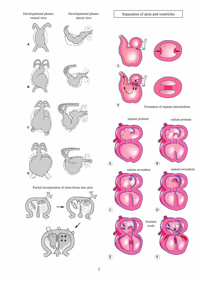
2
Separation of atria and ventricles
Developmental phases:
ventral view
Developmental phases:
lateral view
Formation of septum intermedium
septum primum
ostium secundum septum secundum
ostium primum
foramen
ovale
Partial incorporation of sinus-horns into atria

3
Separation of the truncus arteriosus
Detachment of aorta and truncus pulmonalis
elkülönülése
Formation of papillary muscles and atrioventricular cusps
Formation of
semilunar
valves

4
Aortic arches
eq bo su
ca
aortic arches with n. vagus
and n. laryngeus recurrens
left
side
right
side
ventral view
During development these structure disappear: - I., II., V. and VI. aortic arches on the right side;
- I., II. and V. aortic arches on the left side;
- connection between III. and IV. aortic arches;
- connection between right dorsal aorta and the unpaired aorta.

5
Development of venous system
Changes in sinus-horns
dorsal view
sub
card
inal
vei
ns
left left
sup
raca
rdin
al v
ein
s d
isap
pea
rance
of
caud
al c
ard
inal
is v
eins
left

6
Changes in circulation
Developmental anomalies
Key points: intracardial changes (foramen ovale), pulmonary circulation and ductus arteriosus (Botallo-duct),
ductus venosus (Arantius-duct), a. umbilicalis (nutrients- and oxygen-poor blood transport to placenta), v. umbilicalis (fresh blood to fetus)
pat
ent
duct
us
arte
rio
sus
(PD
A)
pulm
onar
y
sten
osi
s
interatrial
septal defect
Fal
lot-
tetr
alo
gy
aort
aste
no
sis
interventricular
septal defect
persistent right aortic arch (PRAA)
together with left lig. arteriosum, and
consequential compression of oesophagus pulmonary stenosis + overriding aorta +
interventricular septal defect + dilatation and
hypertrophy of the right ventricle