Embed Size (px)
Citation preview
Virginia Commonwealth UniversityVCU Scholars Compass
Neurology Publications Dept. of Neurology
2014
Development of status epilepticus, sustainedcalcium elevations and neuronal injury in a ratsurvival model of lethal paraoxon intoxicationLaxmikant Sudhir DeshpandeVirginia Commonwealth University, [email protected]
Dawn S. CarterVirginia Commonwealth University
Kristin PhillipsVirginia Commonwealth University, [email protected]
Robert E. BlairVirginia Commonwealth University, [email protected]
Robert J. DeLorenzoVirginia Commonwealth University, [email protected] this and additional works at: http://scholarscompass.vcu.edu/neurology_pubs
Part of the Neurology Commons
© 2014 Elsevier Inc. All rights reserved. NOTICE: this is the author’s version of a work that was accepted for publicationin NeuroToxicology. Changes resulting from the publishing process, such as peer review, editing, corrections, structuralformatting, and other quality control mechanisms may not be reflected in this document. Changes may have been madeto this work since it was submitted for publication. A definitive version was subsequently published in NeuroToxicologyVolume 44, September 2014, Pages 17–26, doi:10.1016/j.neuro.2014.04.006.
This Article is brought to you for free and open access by the Dept. of Neurology at VCU Scholars Compass. It has been accepted for inclusion inNeurology Publications by an authorized administrator of VCU Scholars Compass. For more information, please contact [email protected].
Downloaded fromhttp://scholarscompass.vcu.edu/neurology_pubs/9
brought to you by COREView metadata, citation and similar papers at core.ac.uk
provided by VCU Scholars Compass
1
Development of status epilepticus, sustained calcium elevations and
neuronal injury in a rat survival model of lethal paraoxon
intoxication
Laxmikant S. Deshpande*, Dawn S. Carter*, Kristin F. Phillips*, Robert E. Blair* and
Robert J. DeLorenzo1*†‡
Departments of Neurology*, Pharmacology and Toxicology†, and Molecular Biophysics and
Biochemistry‡. Virginia Commonwealth University, Richmond, Virginia 23298, USA
To whom correspondence should be addressed: 1Robert J. DeLorenzo, M.D., Ph.D., M.P.H.
Virginia Commonwealth University
School of Medicine
PO Box 980599
Richmond, VA 23298
Phone: 804-828-3392
Fax: 804-828-6432 E-mail: [email protected]
2
Abstract
Paraoxon (POX) is an active metabolite of organophosphate (OP) pesticide parathion that
has been weaponized and used against civilian populations. Exposure to POX produces high
mortality. OP poisoning is often associated with chronic neurological disorders. In this study, we
optimize a rat survival model of lethal POX exposures in order to mimic both acute and long-
term effects of POX intoxication. Male Sprague-Dawley rats injected with POX (4 mg/kg, ice-
cold PBS, s.c.) produced a rapid cholinergic crisis that evolved into status epilepticus (SE) and
death within 6-8 min. The EEG profile for POX induced SE was characterized and showed
clinical and electrographic seizures with 7-10 Hz spike activity. Treatment of 100% lethal POX
intoxication with an optimized three drug regimen (atropine, 2 mg/kg, i.p., 2-PAM, 25 mg/kg,
i.m. and diazepam, 5 mg/kg, i.p.) promptly stopped SE and reduced acute mortality to 12% and
chronic mortality to 18%. This model is ideally suited to test effective countermeasures against
lethal POX exposure. Animals that survived the POX SE manifested prolonged elevations in
hippocampal [Ca2+
]i (Ca2+
plateau) and significant multifocal neuronal injury. POX SE induced
Ca2+
plateau had its origin in Ca2+
release from intracellular Ca2+
stores since inhibition of
ryanodine/ IP3 receptor lowered elevated Ca2+
levels post SE. POX SE induced neuronal injury
and alterations in Ca2+
dynamics may underlie some of the long term morbidity associated with
OP toxicity.
Keywords: paraoxon, status epilepticus, survival model, Ca2+
dynamics, Sprague-Dawley rats
3
Introduction
Paraoxon (POX) is an active metabolite of organophosphate (OP) insecticide parathion
that has been weaponized and has caused human casualties (Federation of American Scientists,
1998; Gould and Folb, 2003; Croddy et al., 2004; Moorcraft and McLaughlin, 2008). POX is a
potent acetylcholinesterase (AChE) inhibitor that prevents the breakdown and causes rapid
buildup of the neurotransmitter acetylcholine at the synapse, resulting in a “cholinergic crisis”
associated with numerous physiological symptoms, including excessive salivation, respiratory
depression, bradycardia and prolonged seizures (status epilepticus, SE) (Bajgar, 2004; Hoffmann
and Papendorf, 2006). These symptoms of POX exposure, if left untreated can result in the rapid
demise of the OP exposed individuals (McDonough et al., 1995). Survivors of SE and OP
compounds poisoning suffer from significant morbidities including spontaneous recurrent
seizures, deficits in cognition and depression (Steenland et al., 1994; Garcia et al., 2003; Ruckart
et al., 2004; Johnson et al., 2009; de Araujo Furtado et al., 2010; de Araujo Furtado et al., 2012;
Rauh et al., 2012; Terry et al., 2012). It is important to develop animal survival models for lethal
exposures to evaluate treatments to reduce acute mortality and prevent chronic morbidity after
exposure to potent OP agent.
There are studies reporting models of acute POX toxicity and survival shortly after
exposure (Petrikovics et al., 2004; Albuquerque et al., 2006; Todorovic et al., 2012); however, to
our knowledge there are no studies that evaluate long-term survival after lethal POX induced SE.
The goal of this study is to further develop a reliable rat survival model for lethal POX exposure
SE that will replicate both the acute and long-term effects of this agent. Such a model could be
used screen medical countermeasures to improve survival and also help in studying molecular
mechanisms underlying the development of long-term morbidity following OP exposures. Here

4
we utilized POX to mimic lethal OP poisoning and have followed surviving animals for up to
one year after POX intoxication. The mortality, behavioral manifestations and EEG profile for
acute POX exposure are characterized in this study as evaluated for other nerve agent and
pesticide exposures (McDonough and Shih, 1993; Shih et al., 2007; Deshpande et al., 2010). Our
results show that POX exposure produced a reliable response mimicking all the signs and
symptoms of OP intoxication. Development of this model resulted in high survival rates
following lethal POX exposures as a result of an improved regimen of treatment with atropine, 2-
PAM and diazepam. This model could provide a reproducible method to mimic the human
survival of POX toxicity.
It has been established that one of the long-term molecular changes that occurs following
the survival of SE from either exposure to the OP DFP (Deshpande et al., 2010) or
chemoconvulsant pilocarpine (Raza et al., 2004) is the development of sustained elevations in
([Ca2+
]i) termed the “Ca2+
plateau”. These alterations in Ca2+
dynamics along with the
associated neuronal injury are thought to underlie the chronic effects of SE on producing
acquired epilepsy and behavioral and cognitive impairment (Rice et al., 1998; Raza et al., 2004;
Deshpande et al., 2008b). This study will evaluate the effects of POX induced SE on the
development of the Ca2+
plateau and neuronal damage to determine if the POX SE model
produces similar effects on these processes as seen with pilocarpine and DFP SE. We will also
investigate the contribution of ryanodine receptor (RyR) or inositol trisphosphate receptor
(IP3R)-dependent regulation of [Ca2+
]i towards the maintenance of the Ca2+
plateau utilizing
select inhibitors that have been shown to lower elevated Ca2+
levels following SE (Nagarkatti et
al., 2008; Nagarkatti et al., 2010). Being able to reverse the Ca2+
plateau after survival of POX
SE with the Ca2+
induced Ca2+
release (CICR) inhibitors could provide new targets for the

5
development of agents to reverse the Ca2+
plateau and offer a unique insight into the
pathophysiology of POX toxicity.
Materials and methods
Animals
All animal use procedures were in strict accordance with the National Institute of Health
Guide for the Care and Use of Laboratory Animals and approved by Virginia Commonwealth
University’s Institutional Animal Care and Use Committee. Male Sprague-Dawley rats (Harlan,
Indianapolis, IN) weighing ~250-300 g and 10-weeks age were used in this study. Animals were
housed two per cage at 20-22oC with a 12 Light: 12 Dark hour cycle (lights on 0600-01800 h)
and free access to food and water.
Chemicals
All the chemicals were obtained from Sigma Aldrich Company (St. Louis, MO) unless
otherwise noted. POX was prepared by dissolving in ice-cold phosphate buffered saline (PBS)
just before the experiment and injected subcutaneously (s.c.) and was kept on ice until the time
of injections. Time on ice (between diluting in saline to injections) was never more than five
minutes. Atropine sulfate and pralidoxime chloride (2-PAM) were dissolved in saline (0.9%
NaCl) and sterile filtered. Diazepam injection-USP was obtained from VCU health system
pharmacy. All the drugs were prepared fresh on the day of experiment. Dantrolene was dissolved
in DMSO. It was applied to the neurons via bath perfusion. Response to dantrolene was recorded
for up to 10-mins following the bath perfusion. It took approximately 1 min for the complete
exchange of bath solution. Levetiracetam tablets (Lupin; 250 mg) were obtained by VCU
division of animal resources from local pharmacy. The water soluble tablets were crushed and
6
dissolved in saline and sterile filtered to remove the non-soluble binding agents and
pharmaceutical excipients before i.p. injection. Carisbamate (RWJ 333369) was a gift from
Johnson & Johnson, Pharmaceutical Research & Development L.L.C. (Titusville, NJ, USA), and
was suspended in 0.2% methylcellulose for i.p. administration.
Electrode implantation and seizure monitoring
Rats were stereotaxically implanted with three skull surface electrode screws attached to
teflon insulated stainless steel MedWire® (Plastics One, Roanoke, VA, USA.) under general
anesthesia with isoflurane/O2 (5% induction; 2.5% maintenance). Electrode screws were
positioned through burr holes above the right and left frontal cortices (AP, 3 mm and ML, ±3
mm from bregma); the third surface electrode screw was positioned over the cerebellum to serve
as reference and two additional (non-electrode) skull screws were inserted for structural support.
The electrode screws were seated to contact, but not penetrate the dura mater. Female amphenol
terminal pins connected to the electrode wire were seated into an electrode pedestal (Plastics
One, Roanoke, VA) and this assembly was secured to the skull with Cerebond™ adhesive
(Plastics One, Roanoke, VA). Rats were allowed 1 week of recovery time before the start of the
experiment. Wire leads were securely connected into the threaded electrode pedestal on the rat
and then connected to an electrical-swivel commutator (Plastics One, Roanoke, VA) to allow for
free movement of the rat while maintaining continuity of EEG signals. EEG signals were
amplified using a Grass model 8-10D (Grass Technologies, West Warwick, RI) and digitized
using a Powerlab 16/30 data acquisition system (AD Instruments, Colorado Springs, CO).
Evaluation of digitally acquired EEG was performed with Labchart (AD Instruments, Colorado
Springs, CO) (Raza et al., 2004; Deshpande et al., 2010).

7
POX induced status epilepticus
One minute following POX injection animals received 2-PAM (50 mg/kg, i.m.) and
atropine (2 mg/kg, s.c.). Rats underwent convulsions and SE-like activity within 5-7 minutes
following POX, and behavioral observations and Racine scores were made. Onset of SE was
determined by the presence of continuous class 4-5 level seizures as assessed using a modified
Racine scale (Racine, 1972), which were objectively and independently determined by three
trained observers (Raza et al., 2004; Deshpande et al., 2010). Racine scores were averaged for
each animal. Following onset of POX SE, animals were injected with diazepam (5 mg/kg, i.p.)
and 2-PAM (25 mg/kg, i.m.) at 1, 3 and 5 h to terminate and control seizures. Surviving animals
were then injected with saline (3cc/animal, i.p.) and fed lactose milk as part of supportive care
and returned to home cage. Rats may lose up to 10-15 g weight due to fluid loss associated with
SE. Animal health was reassessed every day for the next three days and wet-chow and saline was
administered if weight gain did not occur. By the fourth-day all the SE rats had recovered from
POX induced cholinergic crisis and were ambulatory. Surviving rats were housed individually in
temperature and light controlled vivarium. All the rats were visually monitored once a week till
their use in behavioral experiments. For Ca2+
studies, animals were sacrificed at 1 h following
SE onset or at 1, 7 or 30 days after SE. Mortality was assessed at 24, 48 and 72-h. For
mechanism of Ca2+
plateau studies, levetiracetam (50 mg/kg, i.p.) or carisbamate (90 mg/kg, i.p.)
was injected at 1, 6 and 18-h following onset of SE.
Isolation of Hippocampal CA1 Neurons and Loading with Fura-2
Acute isolation of CA1 hippocampal neurons was performed by established procedures
(Raza et al., 2004; Deshpande et al., 2010). At specified times following onset of POX induced
SE, animals were anesthetized with isoflurane and decapitated. Brains were rapidly dissected and

8
placed in 4°C oxygenated (95% O2/5% CO2) artificial cerebrospinal fluid (aCSF) consisting of
(in mM): 201.5 sucrose, 10 glucose, 1.25 NaH2PO4, 26 NaHCO3, 3 KCl, 7 MgCl2, and 0.2
CaCl2). MK-801 (1 M) was added to all solutions to increase cell viability and was removed 15
min prior to imaging. Hippocampal slices (450 m) were cut on a vibrating microtome (Leica
Microsystems, Wetzlar, Germany) and then equilibrated for 10 min at 34°C in a piperazine-N,N'-
bis[2-ethanesulfonic acid] (PIPES)-aCSF solution containing (in mM): 120 NaCl, 25 glucose, 20
PIPES, 5 KCl, 7 MgCl2, and 0.1 CaCl2. Slices were then treated with 8 mg/ml protease in
PIPES-aCSF for 6 min at 34°C and rinsed. Enzyme treated slices were visualized on a dissecting
microscope to excise the CA1 hippocampal layer which was then triturated with a series of
Pasteur pipettes of decreasing diameter in cold (4°C) PIPES-aCSF solution containing 1 M
Fura-2 AM (Invitrogen, Carlsbad, CA). The cell suspension was placed in the middle of 2 well
glass-bottomed chambers (Nunc, Thermo Scientific). These glass chambers were previously
treated overnight with 0.05 mg/ml poly-L-lysine followed by multiple rinses with distilled water
and then further treated with Cell-Tak (BD-Biosciences, San Jose, CA) biocompatible cellular
adhesive (3.5 g/cm2) for 30-min, rinsed and air-dried. Neuronal suspension placed in the center
of adhesive coated dishes when settled firmly adhered to the bottom. This technique simplified
further manipulations on the dissociated neurons. Plates were then incubated at 37°C in a 5%
CO2/95% air atmosphere for 45 min. Fura-2 was washed off with PIPES-aCSF and plates were
incubated an additional 15 min to allow for complete cleavage of the AM moiety from Fura-2.
Measurement of [Ca2+
]i
Fura-2 loaded cells were transferred to a 37°C heated stage (Harvard Apparatus,
Hollington, MA) on an Olympus IX-70 inverted microscope coupled to a fluorescence imaging
system (Olympus America, Center Valley, PA) and subjected to [Ca2+
]i measurements by

9
procedures well established in our laboratory (Raza et al., 2004; Deshpande et al., 2010). All
experiments were performed using a 20X, 0.7 N.A. water immersion objective and images were
recorded by an ORCA-ER high-speed digital CCD camera (Hammamatsu Photonics K.K.,
Japan). Fura-2 was excited with a 75 W xenon arc lamp (Olympus America, Center Valley, PA).
Ratio images were acquired by alternating excitation wavelengths (340/380 nm) by using a
Lambda 10-2 filter wheel (Sutter Instruments Co., Novato, CA) and a Fura filter cube at 510/540
emission with a dichroic at 400 nm. All image acquisition and processing was controlled by a
computer connected to the camera and filter wheel using Metafluor Software ver 7.6 (MDS
Analytical Technologies, Downington, PA). Image pairs were captured every 5 s and the images
at each wavelength were averaged over 10 frames. Background fluorescence was obtained by
imaging a field lacking Fura-2.
Hippocampal CA1 neurons were identified based on their distinct morphology. These
neurons displayed pyramidal shaped cell body, long axon and dendrites and have been
demonstrated to be devoid of immunoreactivity for specific protein markers for interneurons,
including parvalbumin, cholecystokinin, vasoactive intestinal peptide, somatostatin, and
neuropeptide Y (Raza et al., 2004; Deshpande et al., 2010). The process of enzymatic treatment
and mechanical trituration can add minimal stress during acute dissociation of neurons.
However, we have shown previously that the neurons isolated using these procedures exhibit
electrophysiological properties identical neurons in slices or in cultures (Raza et al., 2007), are
viable, and not apoptotic or necrotic (Sun et al., 2008).
Calcium calibration
We performed Ca2+
calibration determinations as described previously (Raza et al., 2004;
Deshpande et al., 2010) to provide estimates of absolute [Ca2+
]i concentrations from the 340/380

10
ratio values. A Ca2+
calibration curve was constructed using solutions of calibrated Ca2+
buffers
ranging from 0 Ca2+
(Ca2+
free) to 39 M Ca2+
(Invitrogen, Carlsbad, CA). Values from the
calibration curve were used to convert fluorescent ratios to [Ca2+
]i. Final [Ca2+
]i were calculated
from the background corrected 340/380 ratios using the equation (Grynkiewicz et al., 1985):
[Ca2+
]i = (Kd x Sf2/ Sb2) x (R - Rmin)/ (Rmax- R)
where R is the 340/380 ratio at any time; Rmax is the maximum measured ratio in saturating Ca2+
solution (39 M free Ca2+
); Rmin is the minimal measured ratio Ca2+
free solution; Sf2 is the
absolute value of the corrected 380-nm signal at Rmin; Sb2 is the absolute value of the corrected
380-nm signal at Rmax; the Kd value for Fura 2 is 224 nM.
Fluoro-Jade staining
Animals were sacrificed 48-h following POX exposure and received all the antidotes at
the appropriate times as outlined in POX SE section above. Briefly, deep anesthesia was induced
in rats with ketamine/xylazine (75mg/kg/7.5mg/kg i.p.) mixture. Anesthetized animals were
flushed transcardially with saline and perfused with 4% paraformaldehyde in a 100 mM sodium
phosphate buffer (pH 7.4). Fixed brains were removed and post-fixed in 4%
paraformaldehyde/phosphate buffer overnight, cryoprotected in 30% sucrose/phosphate buffer
(pH 7.4) (48 h), flash frozen in isopentane and stored at -80C until used for sectioning. Coronal
sections (40 µm) were cut on a cryostat (Leica Microsystems, Wetzlar, Germany) and stored at -
20C in a cryoprotectant solution of 0.1 M phosphate buffer containing 30% sucrose, 30%
ethylene glycol and 1% polyvinyl-pyrrolidone (PVP-40). Sections were washed three times in a
0.1 % triton in PBS and then float-mounted onto microscope slides (Trubond 380; Tru Scientific
LLC, Bellingham, WA) and allowed to dry at room temperature overnight prior to staining.

11
Slides were incubated in a solution of 1% NaOH in 80% ethanol for 5 minutes followed by
hydration in a 70% ethanol and then ddH2O for 2 minutes each. Slides were then incubated in a
0.06% KMnO4 solution for 10 min followed by washing in ddH2O for 2 min. Slides were then
stained in a 0.0004% fluoro-Jade C solution in 0.1% acetic acid for 20 min. Stained slides
underwent three washes in ddH2O for 2 min each and then dried in a desiccant chamber at 55C
for 30 min. Stained slides were then cleared with xylene for 5 min and cover slipped with DPX
mounting agent (Sigma Chem. Co., St. Louis, MO). Stained sections were evaluated with a
fluorescent IX70 inverted microscope with a 20X (UApo 340, 0.7 n.a., water) objective
(Olympus America, Center Valley, PA) and excitation/emission filters for visualization of FITC.
Greyscale digital images (1324x1024, 16-bit, 1X1 binning) of Fluoro-Jade C staining for select
brain regions including dentate gyrus hilus, were acquired with a Hamamatsu ORCA-ER camera
(Hamamatsu Photonics, Japan).
Data analysis
In dose-response experiments, the response to POX is expressed as the percentage of
animals exhibiting SE or mortality. LD50 values representing the estimated doses causing SE or
mortality in 50% of animals with their 95% confidence limits (95% CL) were determined by log-
probit analysis using the Litchfield and Wilcoxon method.
For comparison of [Ca2+
]i between sham and POX-treated animals, Student’s t-test or
One Way Analysis of Variance (ANOVA) was applied when appropriate to compare [Ca2+
]i. The
Tukey test was used for all post-hoc comparisons. For comparing the distributions of [Ca2+
]i
levels a Chi-square test was used. Statistical tests were run and graphs generated with SigmaPlot
12.5 (SPSS Inc, Chicago, IL). p<0.05 was considered significant.
12
We used 5-9 animals for each experimental condition or time point studied. Dissociating
the hippocampal slices routinely yielded 20-25 healthy, phase-bright neurons that were used for
the recordings. The means of each group of neurons from each animal were used to evaluate
results and conduct statistical analysis. Viable neurons included in the study had a smooth
surface and a pyramidal-like morphology with processes. Non-viable neurons were swollen or
circular and the surface was uneven and irregular. Data from each animal were pooled together
in respective groups and ultimately represented as total number of cells studied.
Analysis of digital images to count FluoroJade C positive cell staining was carried out
with ImageJ (ImageJ, U. S. National Institutes of Health, Bethesda, MD) by thresholding for
specific stain and obtaining positive cell counts using the particle analysis component (size range
in pixel: 25-1000). All parameters for digital acquisition and analysis of staining remained
constant throughout. Representative digital images were processed with Adobe Photoshop for
figures (Adobe Systems Inc., San Jose, CA).
Results
Effects of POX administration
Paraoxon (POX, 0.1-4 mg/kg, s.c., n= 5-8) was injected and behavioral responses were
observed. Rats manifested gross behavioral changes such as increased grooming, sniffing and
chewing that were rapidly followed by tremors, wet-dog shakes and Straub tail response.
Cholinergic symptoms appeared shortly thereafter which included respiratory distress (croaking,
bradypnea), ex-opthalmus, salivation, defecation and urination. Following higher doses of POX,
tonic-clonic seizures appeared and rats rapidly went into status epilepticus (SE). Without
antidotal treatment of the severe cholinergic crisis, death ensued rapidly in all POX SE animals,
demonstrating the lethal effects of this dose of POX exposure. POX induced a dose-dependent
13
increase in the incidence of SE and mortality. The LD50 for induction of seizure was 0.39 mg/kg
(95% CL: 0.32-0.47) and for mortality it was 0.85 mg/kg (95% CL: 0.32-2.19). In addition, the
time to onset of POX SE was also dose-dependent. At low dose (0.5 mg/kg) it was 18.7 ± 1.1
min, whereas at high dose (4 mg/kg) it was 3.2 ± 0.2 min. The seizure responses were scored on
a five point Racine scale (see methods). At doses 1 mg/kg and above a maximum Racine score of
5 was noted. At low POX doses not associated with mortality, animals spent 40 min in SE in a 1-
h observation period. At POX doses associated with mortality, the time of death was also dose-
dependent. This data is summarized in Table 1 and 2.
Effects of atropine, 2-PAM and diazepam following acute POX poisoning
The three drug regimen consisting of atropine to treat cholinergic symptoms, 2-PAM as
an antidote to AChE inhibition and diazepam for stopping seizures is FDA approved for OP
poisoning and being used by military to treat nerve agent exposure (Chemical Hazards
Emergency Medical Management, 2013). We investigated the efficacy of these agents in our rat
model of lethal POX poisoning. For these experiments we tested the highest dose of POX (4
mg/kg) as this ensured that 100% animals would undergo SE and without intervention resulted in
100% mortality. Such criteria allowed for rigorous testing of the three drug intervention.
Mortality was assessed at 1-h, 24-h, 48-h and 72-h following POX injection.
Atropine (2 mg/kg, saline, i.p.) was injected 1-min after POX injection (4 mg/kg, ice-cold
PBS, s.c.). All rats (n= 24) went into SE. Mortality was 30% 1-h after SE. At 24-h following SE,
mortality was 60% and it was 70% and 90% at 48 and 72-h respectively. Thus, while atropine
relieved the cholinergic symptoms and helped in reducing early mortality, the progressive AChE
inhibition and SE still led to significant mortality.
14
Rats (n= 24) were then injected with 2-PAM (25 mg/kg, saline, i.m.) along with atropine
(2 mg/kg, i.p.) 1-min after POX injection (4 mg/kg, s.c.). Mortality was almost halved to 15% 1-
h after SE when 2-PAM was introduced. At 24-h following SE, mortality was 50% and it was
60% and 80% at 48 and 72-h respectively. Thus, while atropine and 2-PAM administration
dramatically lowered the early mortality, the unremitting seizures and SE led to greater mortality
in the subsequent days. Racine scores were not affected in the presence of the atropine and 2-
PAM either alone or in combination suggesting that these agents did not affect severity of SE.
Rats (n= 24) were administered diazepam (5 mg/kg, i.p.) at 1, 3 and 5-h following POX
(4 mg/kg, s.c.) induced SE, in addition to atropine and 2-PAM administration 1-min following
POX injection. Mortality rate of 12% at 1-h following POX induced SE was in a similar range as
observed above. However, when the SE was terminated with sequential diazepam injections,
mortality was dramatically reduced to 8% at 24-h and 17% at 48 and 72-h. These findings
suggested that stopping the OP induced SE is critical to reduce the mortality and improve long-
term survival. This survival data has been summarized in Table 3. Rats surviving lethal POX
exposure at the 72-h time point were returned to the vivarium and housed one per cage. POX
treated rats were re-assessed for mortality at 3, 6 and 12-months post rescue from POX SE.
Taking into account the normal age-related attritions (<5%) in our rat populations, no further
increases in mortality were observed in POX SE survivors. Thus, this lethal POX survival model
is ideally suited to prepare animals to study long term effects of POX exposure.
EEG characterization of POX induced SE
POX induced seizures were also confirmed with EEG recordings. Baseline EEG was
recorded for 30-min before POX injection (4 mg/kg, s.c.). A representative recording is shown in
Fig. 1. POX induced seizure onset was very rapid and the tonic-clonic seizure activity occurred
15
in parallel with high rhythmic epileptiform EEG activity as shown in Fig 1a (expanded portion).
Within 7-8 minutes, animals progressed into electrographic SE. The SE was intense, did not wax
or wane and did not lose its intensity throughout the one-hour of seizure activity displaying 7-10
Hz spike activity (Fig. 1a). Animals were injected with diazepam (5 mg/kg) plus 2-PAM (25
mg/kg) at 1, 3 and 5-h following onset of POX SE. This dose of diazepam was effective in
completely terminating both behavioral and electrographic SE (Fig. 1, DZP). The EEG pattern
for SE termination for up to 24-h following diazepam is shown in Fig. 1. It demonstrates the
effectiveness of optimized 3-dose regimen in that seizures remained suppressed upon drug
intervention and did not reoccur in days following SE termination.
The electrophysiological progression of POX induced SE is depicted in Fig. 2. EEG
alterations following POX administration were rapid and correlated with behavioral seizure
manifestations. Fig. 2A shows a baseline EEG recording before POX injection. Low-voltage fast
activity started to appear within 2-3 min of POX administration (Fig. 2B). High-voltage slow
activity (Fig. 2C) appeared shortly thereafter and rapidly progressed into high-voltage spiking in
correlating with behavioral seizure activity by approximately 5-6 minutes (Fig. 2E). By this time
animals were in electrographic SE with fully developed seizures. SE continued unabated for one
hour, after which time they were terminated using sequential diazepam injections.
POX induced SE caused elevations in hippocampal neuronal [Ca2+
]i
To investigate if POX exposure caused long lasting changes in hippocampal neuronal
[Ca2+
]i, similar to other OPs and chemoconvulsants (Raza et al., 2004; Deshpande et al., 2010),
we measured neuronal [Ca2+
]i after 1-h of SE using the high affinity, ratiometric Ca2+
indicator
Fura-2 in acutely isolated hippocampal neurons from control and POX exposed SE animals.
Representative pseudocolor images of neurons from control and POX-SE rats are shown in Fig.
16
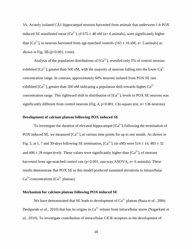
3A. Acutely isolated CA1 hippocampal neurons harvested from animals that underwent 1-h POX
induced SE manifested mean [Ca2+
]i of 675 48 nM (n= 6 animals), were significantly higher
than [Ca2+
]i in neurons harvested from age-matched controls (163 16 nM, n= 5 animals) as
shown in Fig. 3B (p<0.001, t-test).
Analysis of the population distributions of [Ca2+
]i revealed only 5% of control neurons
exhibited [Ca2+
]i greater than 500 nM, with the majority of neurons falling into the lower Ca2+
concentration range. In contrast, approximately 60% neurons isolated from POX SE rats
exhibited [Ca2+
]i greater than 500 nM indicating a population shift towards higher Ca2+
concentration range. This rightward shift in distribution of [Ca2+
]i levels in POX SE neurons was
significantly different from control neurons (Fig. 4, p<0.001, Chi-square test, n= 136 neurons).
Development of calcium plateau following POX induced SE
To investigate the duration of elevated hippocampal [Ca2+
]i following the termination of
POX induced SE, we measured [Ca2+
]i at various time points for up to one month. As shown in
Fig. 5, at 1, 7 and 30-days following SE termination, [Ca2+
]i (in nM) were 514 14, 483 32
and 406 28 respectively. These values were significantly higher than [Ca2+
]i of neurons
harvested from age-matched control rats (p<0.001, one-way ANOVA, n= 6 animals). These
results demonstrate that POX SE in this model produced sustained elevations in intracellular
Ca2+
concentrations (Ca2+
plateau).
Mechanism for calcium plateau following POX induced SE
We have demonstrated that SE leads to development of Ca2+
plateau (Raza et al., 2004;
Deshpande et al., 2010) that has its origins in Ca2+
release from intracellular stores (Nagarkatti et
al., 2010). To investigate contribution of intracellular CICR receptors in the development of

17
POX Ca2+
plateau, [Ca2+
]i were measured in neurons obtained from POX SE rats in the presence
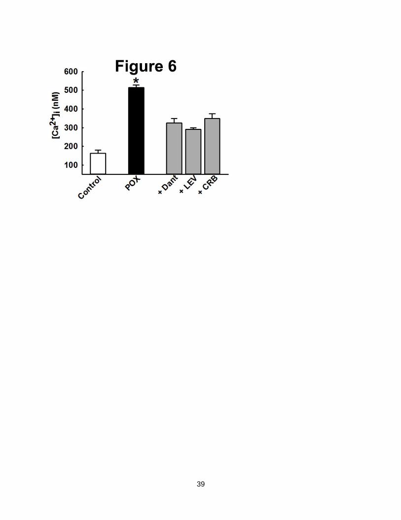
of RyR blocker dantrolene (Nagarkatti et al., 2010). As shown in Fig. 6, application of
dantrolene (50 M) to hippocampal neurons isolated from rats 24-h following POX SE resulted
in a significant drop in [Ca2+
]i from 514 14 nM (no drug) to 324 24 nM (p<0.01, n= 5 rats).
This represent almost 60% reduction in [Ca2+
]i following dantrolene application suggesting that
RyRs are significantly contributing to maintaining the POX SE induced Ca2+
elevations.
Following the efficacy of dantrolene in lowering POX SE induced Ca2+
elevations in
vitro, we next investigated the effects of in vivo administration of levetiracetam and carisbamate.
We have shown these drugs to target RyR/ IP3 receptors and also produce an antiepileptogenic
response following SE-like injury (Deshpande et al., 2008a; Nagarkatti et al., 2008). As shown
in Fig. 6, [Ca2+
]i in hippocampal neurons isolated from rats treated with three doses of
levetiracetam (50 mg/kg, i.p., 1, 6 and 18-h post POX SE onset) were 290 10 nM that were
significantly lower than [Ca2+
]i in neurons isolated from untreated POX SE rats (p<0.001, n= 5
rats). Similarly, [Ca2+
]i in hippocampal neurons isolated from rats treated with three doses of
carisbamate (90 mg/kg, i.p., 1, 6 and 18-h post SE onset) were 348 26 nM that were
significantly lower than [Ca2+
]i in neurons isolated from untreated POX SE rats (p<0.001, n= 5
rats) as shown in Fig. 6.
Neuronal injury associated with acute POX intoxication
To assess neuronal injury in animals surviving exposure, brain sections from animals
injected with POX or vehicle were labeled with Fluoro-Jade C (FJC) (Li et al., 2011). Across all
brain regions examined, there was negligible FJC labeling in brain sections obtained from
vehicle controls. In contrast, after POX treatment, FJC-positive cells were observed in select
regions throughout the forebrain. Within the hippocampus, FJC-positive staining was observed

18
in the polymorphic layer and along the hilus/granule cell border of the dentate gyrus.
Additionally, FJC-positive stained neurons were observed throughout layers II and III of the
parietal cortex (Fig. 7A). The quantification of the neuronal injury expressed as FJC positive
cells is shown in Fig. 7B.
Discussion
The ready availability of pesticides and other toxic agricultural and industrial chemicals
make these agents attractive target for terrorists to weaponize them and cause civilian causalities
(Federation of American Scientists, 1998; Gould and Folb, 2003; Central Intelligence Agency,
2004; Croddy et al., 2004; Moorcraft and McLaughlin, 2008). It is therefore important to study
mechanisms for reducing mortality and morbidity upon exposure to OP agents. Compounds like
DFP and POX mimic the actions of sarin, soman and VX, and are therefore utilized in a
controlled experimental setting to model the effects of nerve agent exposure in civilian
laboratories (Kadriu et al., 2009; Wright et al., 2009; Zaja-Milatovic et al., 2009; Deshpande et
al., 2010; Li et al., 2011; Todorovic et al., 2012). The POX lethal exposure and survival model
optimized in this study provides an ideal animal model to develop medical counter measures to
POX toxicity in a reliable and cost effective manner. Our survival model after lethal POX
exposure offers a valuable contribution to the field by providing a reproducible system to study
the chronic effects of POX toxicity and the development of OP exposure co-morbidities. In
addition, this study demonstrates comparable findings to other OP agents such as DFP (Kadriu et
al., 2009; Wright et al., 2009; Zaja-Milatovic et al., 2009; Deshpande et al., 2010; Li et al.,
2011), the chemoconvulsant pilocarpine (Raza et al., 2004), and chemical threat agents such as
Tetramethylenedisulfotetramine (TETS) (Cao et al., 2012), that POX exposure produces
elevated neuronal [Ca2+
]i and injury. These changes in intracellular Ca2+
concentrations
19
persisted for over a month following the termination of POX SE, and in combination with the
observed with the multifocal neuronal injury, may underlie some of the long-term effects of OP
toxicity on neuronal function and behavioral impairments (DeLorenzo et al., 2005; Filbert et al.,
2005; de Araujo Furtado et al., 2012). Furthermore, identification of mechanisms responsible for
injury-induced Ca2+
plateau may elucidate novel targets and candidate drugs for the potential
treatment of long-term morbidity associated with POX and other OP exposures.
Following high dosages of POX, the enzyme AChE is rapidly inhibited, resulting in a
massive buildup of acetylcholine at the synapses. A cholinergic crisis ensues that result in the
progression of bradypnea, bradycardia and convulsions, followed by rapid death (Bajgar, 2004;
Hoffmann and Papendorf, 2006). The three drug regimen presented in this study consisting of
atropine, 2-PAM and diazepam is FDA approved and used by US military (Mark I kit), and is the
mainstay treatment for OP poisoning/ nerve agent toxicity (Chemical Hazards Emergency
Medical Management, 2013). A higher survival rate was obtained following lethal POX
exposures by optimizing the standard three drug regimens used for treating OP poisoning. The
mortality following lethal POX intoxication was most effectively reduced by atropine, in
agreement with previous nerve agent studies (Shih et al., 2007). A lower dose of atropine (0.1-
0.2 mg/kg), similar to auto injector doses used in human pre-exposure situations for OP
intoxication, was effective in reducing mortality by 20% in our animal model (data not shown).
However, since we wanted to develop an animal model of lethal OP exposure survival in order to
study long-term morbidities, we increased atropine concentration to 2 mg/kg. This higher dose of
atropine, along with 2-PAM and diazepam intervention, resulted in a significantly higher
survival rate of 85%. This is in line with findings from our previous report following lethal DFP
exposure (Deshpande et al., 2010). Our results show that while each drug by itself is critical to

20
limit acute mortality, stopping the SE with diazepam is by far the most important intervention to
significantly reduce long-term mortality. This is in agreement with the clinical literature that
reports a high mortality in patients undergoing SE (DeLorenzo et al., 2009).
POX induced SE produced prolonged elevations in neuronal Ca2+
concentrations similar
to those seen with DFP and pilocarpine (Raza et al., 2004; Deshpande et al., 2010). These
elevations were sustained and lasted for up to one-month following the termination of SE. This
Ca2+
plateau had its origin in Ca2+
release from intracellular stores, since intervention with CICR
receptor inhibitors dantrolene (Nagarkatti et al., 2010) or levetiracetam (Nagarkatti et al., 2008)
abolished the Ca2+
plateau when administered in vitro or in vivo. The Ca2+
plateau following SE
is thought to trigger plasticity changes during the period of epileptogenesis that ultimately results
in the development of acquired epilepsy (DeLorenzo et al., 2005). Indeed, treatment with the
purported antiepileptogenic agent carisbamate prevented the development of spontaneous
epileptiform discharges in hippocampal neurons following SE (Deshpande et al., 2008a). These
epileptiform discharges are a result of altered Ca2+
dynamics following SE-like injury
(DeLorenzo et al., 2005). In line with these studies, treatment with carisbamate lowered POX SE
mediated elevations in intracellular Ca2+
levels. It will be interesting to evaluate if Ca2+
levels
revert back to higher concentrations following removal of drugs. This may necessitate multiple
drug injections to maintain lower Ca2+
levels post SE in order to prevent development of Ca2+
mediated altered synaptic plasticity.
It has been demonstrated that SE is associated with significant cell death in various brain
regions (DeLorenzo et al., 2005). This neuronal death is thought to be Ca2+
dependent since
manipulations that lower neuronal Ca2+
entry before or during SE are effective in producing
significant neuroprotection. Thus, NMDA receptor antagonist MK-801 given before SE prevents

21
the protracted elevations in [Ca2+
]i inhibits epileptogenesis and other SE related morbidities
(Rice et al., 1998; Raza et al., 2004; DeLorenzo et al., 2005; Deshpande et al., 2010). We have
also demonstrated that while NMDA receptor mediated Ca2+
entry is essential for generation of
the Ca2+
plateau; it does not play a role in its maintenance (Raza et al., 2004; Deshpande et al.,
2010). Thus, MK-801 administered 1-h after SE cannot prevent the development of Ca2+
plateau
and does not afford neuroprotection (Raza et al., 2004; DeLorenzo et al., 2005). However, CICR
from intracellular stores contributes to the maintenance of the Ca2+
plateau such that inhibition of
RyRs not only abolishes Ca2+
plateau, but it also prevents the development of SE-like injury
induced spontaneous epileptiform discharges in hippocampal neurons in vitro (Nagarkatti et al.,
2010). We hypothesize that it is this Ca2+
entry occurring during SE and not the Ca2+
plateau
generated after one-hour of SE that contributes to neuronal death. It will be interesting to
investigate if the other brain regions associated with neuronal death also manifest similar Ca2+
elevations. The CICR agents used in this study are administered one-hour after SE at which point
they may not be able to prevent the on-going neuronal death processes. However, by inhibiting
the sustained Ca2+
elevations post SE we predict that they may be able lower the morbidities
associated with SE. This possibility is currently being evaluated by our laboratory.
We have observed POX intoxicated rats for up to one-year following their rescue with the
three-drug regimen optimized in this study. After taking into consideration the normal age-
related attrition in the population of rats housed in our vivarium, we did not observe increases in
mortality percentage in POX SE rescued rats. To our knowledge this is the first study that has
followed the mortality in rats surviving lethal POX exposure for an extended period of time out
to one year. An SE survival model of POX intoxication should also exhibit long-term
consequences of OP exposure indicative of cognitive deficits and co-morbidities (Steenland et
22
al., 1994; Brown and Brix, 1998; Ivens et al., 1998; Wesseling et al., 2002; Ruckart et al., 2004;
Edwards and Tchounwou, 2005; de Araujo Furtado et al., 2012; Terry et al., 2012). In our
preliminary results we have observed the development of chronic seizures, depressive symptoms
and memory impairments in POX SE survivor rats (data not shown). The complete assessment of
chronic neurological disorders in POX survivors is currently underway in our laboratory.
However, these preliminary observations further validate our proposed model.
We have demonstrated that CNS injuries such as SE, stroke or traumatic brain injury
result in neuronal injury and protracted elevations in hippocampal [Ca2+
]i (Rice et al., 1998; Raza
et al., 2004; Deshpande et al., 2008b; Deshpande et al., 2010). Since Ca2+
is a major second
messenger molecule, such sustained increases in its levels activate multiple signaling cascades,
affect protein transcription and alter gene expression of proteins involved in controlling
membrane excitability and synaptic plasticity (DeLorenzo et al., 2005). These Ca2+
mediated
alterations are thought to underlie the long-term morbidity following CNS insults (DeLorenzo et
al., 2005; Deshpande et al., 2008b). OP exposure has been reported to be associated with
neurological problems such as seizures, cognitive deficits and other behavioral disorders (de
Araujo Furtado et al., 2010; de Araujo Furtado et al., 2012). Inhibition of the POX SE induced
Ca2+
plateau with a regimen of FDA approved drugs could prove to be novel countermeasure
agents to potentially prevent the morbidity associated with OP poisoning.
23
Funding
This work was supported by the CounterACT Program, National Institutes of Health
Office of the Director (NIH OD), and the National Institute of Neurological Disorders and Stroke
[Grant Number U01NS058213 and R21NS072061] to RJD. Its contents are solely the
responsibility of the authors and do not necessarily represent the official views of the federal
government.

24
References:
Albuquerque, E.X., Pereira, E.F., Aracava, Y., Fawcett, W.P., Oliveira, M., Randall, W.R.,
Hamilton, T.A., Kan, R.K., Romano, J.A., Jr., Adler, M., 2006. Effective countermeasure against
poisoning by organophosphorus insecticides and nerve agents. Proc Natl Acad Sci U S A 103,
13220-13225.
Bajgar, J., 2004. Organophosphates/nerve agent poisoning: mechanism of action, diagnosis,
prophylaxis, and treatment. Adv Clin Chem 38, 151-216.
Brown, M.A., Brix, K.A., 1998. Review of health consequences from high-, intermediate- and
low-level exposure to organophosphorus nerve agents. J Appl Toxicol 18, 393-408.
Cao, Z., Hammock, B.D., McCoy, M., Rogawski, M.A., Lein, P.J., Pessah, I.N., 2012.
Tetramethylenedisulfotetramine alters Ca(2)(+) dynamics in cultured hippocampal neurons:
mitigation by NMDA receptor blockade and GABA(A) receptor-positive modulation. Toxicol
Sci 130, 362-372.
Central Intelligence Agency, 2004. Terrorist CBRN: Materials and Effects.
Chemical Hazards Emergency Medical Management, 2013. Nerve Agents - Emergency
Department/Hospital Management, pp.
Croddy, E., Wirtz, J., Larsen, J., 2004. Weapons of Mass Destruction: An Encyclopedia of
Worldwide Policy, Technology, and History ABC-CLIO, Santa Barbara, CA.
de Araujo Furtado, M., Lumley, L.A., Robison, C., Tong, L.C., Lichtenstein, S., Yourick, D.L.,
2010. Spontaneous recurrent seizures after status epilepticus induced by soman in Sprague-
Dawley rats. Epilepsia 51, 1503-1510.
de Araujo Furtado, M., Rossetti, F., Chanda, S., Yourick, D., 2012. Exposure to nerve agents:
from status epilepticus to neuroinflammation, brain damage, neurogenesis and epilepsy.
Neurotoxicology 33, 1476-1490.
DeLorenzo, R.J., Kirmani, B., Deshpande, L.S., Jakkampudi, V., Towne, A.R., Waterhouse, E.,
Garnett, L., Ramakrishnan, V., 2009. Comparisons of the mortality and clinical presentations of
status epilepticus in private practice community and university hospital settings in Richmond,
Virginia. Seizure 18, 405-411.
DeLorenzo, R.J., Sun, D.A., Deshpande, L.S., 2005. Cellular mechanisms underlying acquired
epilepsy: the calcium hypothesis of the induction and maintainance of epilepsy. Pharmacol Ther
105, 229-266.
Deshpande, L.S., Carter, D.S., Blair, R.E., DeLorenzo, R.J., 2010. Development of a prolonged
calcium plateau in hippocampal neurons in rats surviving status epilepticus induced by the
organophosphate diisopropylfluorophosphate. Toxicol Sci 116, 623-631.

25
Deshpande, L.S., Nagarkatti, N., Ziobro, J.M., Sombati, S., DeLorenzo, R.J., 2008a. Carisbamate
prevents the development and expression of spontaneous recurrent epileptiform discharges and is
neuroprotective in cultured hippocampal neurons. Epilepsia 49, 1795-1802.
Deshpande, L.S., Sun, D.A., Sombati, S., Baranova, A., Wilson, M.S., Attkisson, E.A., Hamm,
R.J., Delorenzo, R.J., 2008b. Alterations In Neuronal Calcium Levels Are Associated With
Cognitive Deficits After Traumatic Brain Injury. Neurosci Lett 441, 115-119.
Edwards, F.L., Tchounwou, P.B., 2005. Environmental toxicology and health effects associated
with methyl parathion exposure--a scientific review. Int J Environ Res Public Health 2, 430-441.
Federation of American Scientists, 1998. Truth and Reconciliation Commission. FINAL
REPORT: Volume TWO Chapter SIX. Presented to President Nelson Mandela on 29 October
1998.
Filbert, M., Levine, E., Ballough, G., 2005. Neuroprotection for nerve agent-induced brain
damage by blocking delayed calcium overload: a review J Med CBR Def 3.
Garcia, S.J., Abu-Qare, A.W., Meeker-O'Connell, W.A., Borton, A.J., Abou-Donia, M.B., 2003.
Methyl parathion: a review of health effects. J Toxicol Environ Health B Crit Rev 6, 185-210.
Gould, C., Folb, P.I., 2003. Project Coast: Apartheid's Chemical and Biological Warfare
Programme, United Nations.
Grynkiewicz, G., Poenie, M., Tsien, R.Y., 1985. A new generation of Ca2+ indicators with
greatly improved fluorescence properties. J Biol.Chem. 260, 3440-3450.
Hoffmann, U., Papendorf, T., 2006. Organophosphate poisonings with parathion and dimethoate.
Intensive Care Med 32, 464-468.
Ivens, I.A., Schmuck, G., Machemer, L., 1998. Learning and memory of rats after long-term
administration of low doses of parathion. Toxicol Sci 46, 101-111.
Johnson, F.O., Chambers, J.E., Nail, C.A., Givaruangsawat, S., Carr, R.L., 2009. Developmental
chlorpyrifos and methyl parathion exposure alters radial-arm maze performance in juvenile and
adult rats. Toxicol Sci 109, 132-142.
Kadriu, B., Guidotti, A., Costa, E., Auta, J., 2009. Imidazenil, a non-sedating anticonvulsant
benzodiazepine, is more potent than diazepam in protecting against DFP-induced seizures and
neuronal damage. Toxicology 256, 164-174.
Li, Y., Lein, P.J., Liu, C., Bruun, D.A., Tewolde, T., Ford, G., Ford, B.D., 2011. Spatiotemporal
pattern of neuronal injury induced by DFP in rats: a model for delayed neuronal cell death
following acute OP intoxication. Toxicol Appl Pharmacol 253, 261-269.
McDonough, J.H., Jr., Dochterman, L.W., Smith, C.D., Shih, T.M., 1995. Protection against
nerve agent-induced neuropathology, but not cardiac pathology, is associated with the
anticonvulsant action of drug treatment. Neurotoxicology 16, 123-132.
26
McDonough, J.H., Jr., Shih, T.M., 1993. Pharmacological modulation of soman-induced
seizures. Neurosci Biobehav Rev 17, 203-215.
Moorcraft, P., McLaughlin, P., 2008. The Rhodesian War: A Military History., Yorkshire: Pen
& Sword.
Nagarkatti, N., Deshpande, L.S., Carter, D.S., DeLorenzo, R.J., 2010. Dantrolene inhibits the
calcium plateau and prevents the development of spontaneous recurrent epileptiform discharges
following in vitro status epilepticus. Eur J Neurosci 32, 80-88.
Nagarkatti, N., Deshpande, L.S., DeLorenzo, R.J., 2008. Levetiracetam inhibits both ryanodine
and IP3 receptor activated calcium induced calcium release in hippocampal neurons in culture.
Neurosci Lett 436, 289-293.
Petrikovics, I., Papahadjopoulos, D., Hong, K., Cheng, T.C., Baskin, S.I., Jiang, J., Jaszberenyi,
J.C., Logue, B.A., Szilasi, M., McGuinn, W.D., Way, J.L., 2004. Comparing therapeutic and
prophylactic protection against the lethal effect of paraoxon. Toxicol Sci 77, 258-262.
Racine, R.J., 1972. Modification of seizure activity by electrical stimulation. II. Motor seizure.
Electroencephalogr Clin Neurophysiol 32, 281-294.
Rauh, V.A., Perera, F.P., Horton, M.K., Whyatt, R.M., Bansal, R., Hao, X., Liu, J., Barr, D.B.,
Slotkin, T.A., Peterson, B.S., 2012. Brain anomalies in children exposed prenatally to a common
organophosphate pesticide. Proc Natl Acad Sci U S A 109, 7871-7876.
Raza, M., Blair, R.E., Sombati, S., Carter, D.S., Deshpande, L.S., DeLorenzo, R.J., 2004.
Evidence that injury-induced changes in hippocampal neuronal calcium dynamics during
epileptogenesis cause acquired epilepsy. Proc Natl Acad Sci U S A 101, 17522-17527.
Raza, M., Deshpande, L.S., Blair, R.E., Carter, D.S., Sombati, S., DeLorenzo, R.J., 2007. Aging
is associated with elevated intracellular calcium levels and altered calcium homeostatic
mechanisms in hippocampal neurons. Neurosci Lett 418, 77-81.
Rice, A.C., Floyd, C.L., Lyeth, B.G., Hamm, R.J., DeLorenzo, R.J., 1998. Status epilepticus
causes long-term NMDA receptor-dependent behavioral changes and cognitive deficits.
Epilepsia 39, 1148-1157.
Ruckart, P.Z., Kakolewski, K., Bove, F.J., Kaye, W.E., 2004. Long-term neurobehavioral health
effects of methyl parathion exposure in children in Mississippi and Ohio. Environ Health
Perspect 112, 46-51.
Shih, T.M., Rowland, T.C., McDonough, J.H., 2007. Anticonvulsants for nerve agent-induced
seizures: The influence of the therapeutic dose of atropine. J Pharmacol Exp Ther 320, 154-161.
Steenland, K., Jenkins, B., Ames, R.G., O'Malley, M., Chrislip, D., Russo, J., 1994. Chronic
neurological sequelae to organophosphate pesticide poisoning. Am J Public Health 84, 731-736.

27
Sun, D.A., Deshpande, L.S., Sombati, S., Baranova, A., Wilson, M.S., Hamm, R.J., DeLorenzo,
R.J., 2008. Traumatic brain injury causes a long-lasting calcium (Ca2+)-plateau of
elevated intracellular Ca levels and altered Ca2+ homeostatic mechanisms in
hippocampal neurons surviving brain injury. Eur J Neurosci 27, 1659-1672.
Terry, A.V., Jr., Beck, W.D., Warner, S., Vandenhuerk, L., Callahan, P.M., 2012. Chronic
impairments in spatial learning and memory in rats previously exposed to chlorpyrfos or
diisopropylfluorophosphate. Neurotoxicol Teratol 34, 1-8.
Todorovic, M.S., Cowan, M.L., Balint, C.A., Sun, C., Kapur, J., 2012. Characterization of status
epilepticus induced by two organophosphates in rats. Epilepsy Res 101, 268-276.
Wesseling, C., Keifer, M., Ahlbom, A., McConnell, R., Moon, J.D., Rosenstock, L., Hogstedt,
C., 2002. Long-term neurobehavioral effects of mild poisonings with organophosphate
and n-methyl carbamate pesticides among banana workers. Int J Occup Environ Health 8,
27-34.
Wright, L.K., Liu, J., Nallapaneni, A., Pope, C.N., 2009. Behavioral sequelae following acute
diisopropylfluorophosphate intoxication in rats: Comparative effects of atropine and
cannabinomimetics. Neurotoxicol Teratol 32, 329-335.
Zaja-Milatovic, S., Gupta, R.C., Aschner, M., Milatovic, D., 2009. Protection of DFP-induced
oxidative damage and neurodegeneration by antioxidants and NMDA receptor antagonist.
Toxicol Appl Pharmacol 240, 124-131.

28
Figure Legends
Figure 1. A representative continuous 24-h EEG recording from a rat before, during and after
POX induced SE. Baseline EEG activity is noted before POX exposure. This leads to intense SE
minutes after POX administration (4 mg/kg, s.c.). (a) Expanded Depicts an expanded EEG
segment of SE characterized by high frequency spiking. SE is robust, intense and doesn’t wax
and wane for the entire 1-h. SE is stopped at 1-h following administration of diazepam (DZP).
(b) Expanded EEG segment after 1st DZP (5 mg/kg, i.p.) depicting rapid termination of SE. Two
additional DZP and 2-PAM injections are administered at 3 and 5-h after SE onset. SE remained
suppressed and seizures did not reoccur for the 24-h period following POX toxicity symptoms.
Traces are representative of n= 6 animals.
Figure 2. EEG progression of POX induced SE. (A) Baseline activity prior to POX injection. (B)
Low-voltage fast activity appears within 2-3 min of POX administration. (C) High-voltage slow
activity appears within the next minute that progresses to (D) high frequency and high-voltage
spiking and (E and F) sustained continuous seizure activity. EEG activity at 15-min following
POX administration (F) demonstrates electrographic SE with fully developed continuous
seizures. Spike frequency during SE was sustained at greater than 7- 10 Hz. SE was induced by
POX within 5-7 min following injections. EEG activity correlated with behavioral observations.
Figure 3. Effect of POX induced SE on [Ca2+
]i. (A) Pseudocolor ratiometric images of
representative control and POX treated isolated neurons. Control neurons had bluish color that
corresponds to lower Fura-2 ratio while POX-SE neurons had orange-red color that corresponds
to higher Fura-2 ratio. (B) Elevated [Ca2+
]i in CA1 hippocampal neurons acutely isolated from

29
animals that had experienced 1-h of POX induced SE compared to neurons from control animals.
(*p<0.001, t-test, n= 6 and 5 animals respectively). Data are represented as mean SEM.
Figure 4. Distribution of [Ca2+
]i for control and POX-SE hippocampal neurons. Control neurons
demonstrated a normal distribution for [Ca2+
]i with approximately 95% of neurons exhibiting
[Ca2+
]i less than 500 nM and only 5% neurons exhibiting very high [Ca2+
]i. In contrast, POX-SE
neurons demonstrated a rightward shift towards higher [Ca2+
]i with approximately 60% neurons
exhibiting [Ca2+
]i greater than 500 nM (n= 136 neurons).
Figure 5. Development of Ca2+
plateau following POX induced SE. CA1 hippocampal [Ca2+
]i
from control (white bar) and POX rats were isolated immediately after (1-h) and 1, 7 and 30 days
after SE (black bars). [Ca2+
]i in control animals at each time point were not significantly different
from the control values shown and thus were omitted from the graph for clarity. [Ca2+
]i in
neurons isolated from POX-SE rats was significantly higher than control values (*p<0.05, one-
way ANOVA, post hoc Tukey test, n= 6 rats at each time). Data represented as mean SEM.
Figure 6. Mechanism of Ca2+
plateau following POX SE. CA1 hippocampal [Ca2+
]i from control
(white bar) and POX rats were isolated 24-h after SE (black bars). Dantrolene (Dant, 50 M,
bath application) caused a significant decrease in elevated post SE Ca2+
levels. In separate
experiments, rats were injected with either levetiracetam (LEV, 50 mg/kg, i.p.) or carisbamate
(CRB, 90 mg/kg, i.p.) at 1, 6 and 18-h post SE onset. CA1 hippocampal neurons were isolated
30-mins post the last drug injection. [Ca2+
]i in neurons isolated from POX-SE rats treated with
either LEV or CRB were significantly lower than comparable POX SE rats (no drugs) values at
the respective time point. (*p<0.05, compared to POX, one-way ANOVA, post-hoc Tukey test,
n= 5 animals for each treatment). Data represented as mean SEM.

30
Figure 7. POX SE induced neuronal injury. A. Representative photomicrographs of Fluoro-Jade
C (FJC) staining in the (a) dentate gyrus-hilus region, (b) thalamus, (c) parietal cortex and (d)
temporal/ peri-rhinal cortex of a control rat (left panel, c) and a POX rat 2 days after POX SE (4
mg/kg, s.c.) Scale bars, 200 μm. B. Quantitative analyses of FJC labeling. FJC positive cells
indicative of neuronal injury were observed in hilus, thalamus and cortex of POX rats 48-h after
SE termination. Control rats did not exhibit any FJC labeling. (*p<0.05 compared to control, t-
test, n= 4-6 rats).
31
Table 1: Dose dependent effects of POX on SE and mortality
Dose of POX
(mg/kg, s.c.,
n= 5-8)
% SE % Mortality
0.1 7 ± 2 0
0.5 42 ± 7 26 ± 5
0.8 71 ± 6 50 ± 6
1 81 ± 4 88 ± 4
2 90 ± 2 100
4 100 100
8 100 100

32
Table 2: Characteristics of POX induced SE
POX dose
(mg/kg, s.c., n=8)
SE onset time
(min.)
Racine Score
(scale: 0-5)
Time of death
(min.)
0.1 37 ± 3 2 -
0.5 19 ± 1 2 32 ± 3
0.8 11 ± 1 4 26 ± 4
1 7 ± 0.5 5 12 ± 1
2 5 ± 0.5 5 7 ± 0.5
4 3 ± 0.2 5 5 ± 0.2
8 2 ± 0.4 5 3 ± 0.2

33
Table 3: Role of three-drug regimen in promoting survival following lethal POX
intoxication
Time post SE Percent survival following POX SE in the presence of
No drug Atropine Atropine +
2-PAM
Atropine + 2-PAM
+ diazepam
1-h 0 71 ± 4 83 ± 6 88 ± 4
24-h - 41 ± 4 50 ± 6 83 ± 6
48-h - 33 ± 6 41 ± 4 79 ± 8
72-h - 12 ± 4 21 ± 4 79 ± 8
n= 8 for no drug condition and n= 24 for each separate drug combination

34
35

36
37

38
39

40
![Focal hemodynamic patterns of status epilepticus detected ... · epilepticus or subtle status epilepticus [4]. Electroenceph-alogram (EEG), the diagnostic gold standard, may not be](https://img.pdfslide.us/doc/110x75/6074493ed430437ef144c30f/focal-hemodynamic-patterns-of-status-epilepticus-detected-epilepticus-or-subtle.jpg)