Embed Size (px)
Citation preview
Alfonso Maria Lostia, Maria Chiara Zorzoli, Salvador Fortaner Torrent, Emilio Mendoza, Mounir Bouhifd, Sandra Coecke
Development of an in vitro metabolic hepatic clearance method
2016
EUR 28354 EN
This publication is a Technical report by the Joint Research Centre (JRC), the European Commission’s science and
knowledge service. It aims to provide evidence-based scientific support to the European policy-making process.
The scientific output expressed does not imply a policy position of the European Commission. Neither the
European Commission nor any person acting on behalf of the Commission is responsible for the use which might
be made of this publication.
Contact information
Name: Alfonso Maria Lostia
Address: Joint Research Centre, via E. Fermi 2749, I-21027 Ispra (VA), Italy
E-mail: [email protected]
Tel.: +39 0332 78 5259
JRC Science Hub
https://ec.europa.eu/jrc
JRC104213
EUR 28354 EN
PDF ISBN 978-92-79-64627-0 ISSN 1831-9424 doi: 10.2788/886451
Luxembourg: Publications Office of the European Union, 2016
© European Union, 2016
Reproduction is authorised provided the source is acknowledged.
How to cite: Lostia AM et al.; Development of an in vitro metabolic hepatic clearance method; EUR: 28354 EN doi: 10.2788/886451
All images © European Union 2016
Table of contents
Abstract ............................................................................................................... 1
1 Introduction .................................................................................................... 3
2 Materials and methods ..................................................................................... 7
2.1 General experimental layout ........................................................................ 7
2.2 Materials ................................................................................................... 7
2.2.1 Chemicals: ......................................................................................... 7
2.2.2 Cells, cell media and additives: ............................................................. 8
2.2.3 Plastic ware and disposable: ................................................................. 8
2.2.4 Technical equipment: .......................................................................... 8
2.2.5 Reagents used for chemical analysis: .................................................... 9
2.2.6 Analytical equipment ........................................................................... 9
2.3 Preparation of cell assay medium, stock and working solutions ........................ 9
2.3.1 Cell medium for incubation assay: ........................................................ 9
2.3.2 Cell medium and acetonitrile mixture (50:50): ..................................... 10
2.3.3 Stop solutions using specific internal standards .................................... 10
2.3.4 Stock concentration of 10mM for each chemical to be tested .................. 11
2.3.5 Working concentration of 2µM for each chemical to be tested ................. 11
2.3.6 Analytical standards for the calibration curve for the LC-MS analysis ....... 12
2.4 Human in vitro hepatic metabolic clearance method ..................................... 12
2.4.1 Study design of the in vitro human hepatic metabolic clearance method (or in vitro incubation assay) ................................................................................ 12
2.4.1.1 Prepare equipment for incubation assay ........................................ 14
2.4.1.2 Thawing and reconstitution of cryopreserved human hepatocytes ..... 14
2.4.1.3 Incubation assay ........................................................................ 15
2.4.1.4 Preparation of analytical standards ............................................... 16
2.4.2 LC-MS/MS analysis ............................................................................ 17
2.4.3 Calculation of in vitro intrinsic clearance (CLint) ..................................... 18
3 Results ......................................................................................................... 20
3.1 LC-MS analysis ........................................................................................ 20
3.2 In vitro hepatic metabolic clearance ........................................................... 21
4 Conclusion .................................................................................................... 25
References ......................................................................................................... 26
List of abbreviations and definitions ....................................................................... 27
List of figures ...................................................................................................... 28
List of tables ....................................................................................................... 29
1
Abstract
There is increasing demand from in vitro method developers, validation bodies, in vitro method end-users as well as receiving authorities and
OECD to integrate kinetics information in toxicity testing in order to improve chemical risk assessment. A strategic way to do this is detailed in
the "EURL ECVAM strategy for achieving 3Rs impact in the assessment of toxicokinetics and systemic toxicity" (Bessems J et al. 2015). One of the
strategic aims of this report urges for the development and applied use of in vitro kinetic methods for absorption, distribution, metabolism and
excretion to be used in the future risk assessment approaches using new advanced methodologies (NAMs).
Information on metabolism has been identified as a critical piece of information in integrated test strategies based on non-animal methods.
Therefore, the purpose of this report is to describe the preliminary
experimental work towards a representative in vitro human hepatic metabolic clearance method that can be ultimately used in future defined
approaches for toxicological risk assessment.
The proposed method is a result of the efforts of the EURL ECVAM
laboratory team combined with knowledge gathered during (1) a literature search on in vitro human hepatic metabolic clearance methods,
(2) an EURL ECVAM call for procedures detailing in vitro human hepatic metabolic clearance methods and (3) an expert meeting to establish the
elements critical for in vitro methods that predict human hepatic metabolic clearance. The representative in vitro human hepatic metabolic
clearance method was used to assess the in vitro intrinsic clearance of the following three chemicals: Acetaminophen, Diclofenac and Verapamil.
Each chemical was added at one concentration to cryopreserved primary human hepatocytes (pooled from 10 donors) and the cells were exposed
at different incubation time-points up to 2 h. The concentrations of each
chemical over-time were measured by using Ultra-Performance Liquid-Chromatography (UPLC) coupled with (Quadrupole Time-of-Flight) Q-Tof
mass spectrometer.
The results obtained show good within-laboratory reproducibility (e.g.
between different runs on different days), of the calculated in vitro intrinsic clearance.
The results and experience gained with this experimental in-house effort by Directorate F – Health, Consumers and Reference Materials, F.3
Chemicals Safety and Alternative Methods will support the definition of an EURL ECVAM recommendation on human hepatic metabolic clearance and
the subsequent drafting of a proposal for an OECD Guidance Document with the objective to characterise and describe in vitro hepatic metabolic
2
clearance methods in order to facilitate their regulatory uptake and use to
support chemical risk assessment.
3
1 Introduction
Kinetics describes the concentration-time profile of a chemical in a biological system (e.g. human body, organ, tissue, cell). In other words,
it describes what the body (or cell) does to a chemical and it is the opposite of dynamics which describes what a chemical does to the body
(or cell).
Kinetics is the result of 4 processes: Absorption, Distribution, Metabolism
and Excretion (ADME). Metabolism (chemical bio-transformation or 'metabolic clearance') is a critical factor in determining kinetic behaviour
of compounds of various kinds. Metabolism can occur in several organs and tissues in the body. Nevertheless, the liver is the main site
responsible for metabolic clearance.
In short, human hepatic metabolic clearance represents in many cases one of the two most important determinants of the concentration-time
profile of a chemical, the other one being the human absorption kinetics. Therefore, hepatic metabolic clearance data can represent an
indispensable information source to support the chemical risk assessment.
In fact, there is increasing demand (from in vitro method developers, validation bodies, in vitro method end-users as well as receiving
authorities and OECD) to integrate kinetics information in toxicity testing in order to improve chemical risk assessment. (EURL-ECVAM TK Strategy,
Bessems J et al. 2015 [1]).
Exposure to a chemical does not automatically mean that all of the dose
will be bioavailable and therefore able to cause a specific toxicity. Hence the knowledge of the chemical concentration-time profile in a biological
system (therefore the chemical kinetics) can assist to better design
toxicity tests (both in vivo and in vitro) and interpret toxicological findings by:
selecting the most relevant doses to be administered (e.g. avoid doses which can cause saturation of metabolism which then will
lead to general systemic toxicity effects);
selecting the most sensitive animal species which can provide
valuable information relevant to human biology (e.g. improve the extrapolation from animal toxicological data and assess relevance
for humans);
waiving toxicity tests which can reduce the use of laboratory
animals (e.g. if kinetics data indicate that a chemical bio-accumulates in the body then chronic toxicity studies are more
relevant than acute-studies);
improving the design on in vitro tests and the interpretation of
the data generated (e.g. evaluate if in vitro metabolism is similar
to the in vivo counterpart to then assess the relevance of the data when making in vitro to in vivo extrapolation).
4
There are several other applications where knowledge of kinetics can
support and improve chemical risk assessment and a more detailed discussion can be found in EURL-ECVAM TK Strategy (Bessems J et al.
2015 [1]). In several EU chemical management legislations (REACH [2], CPR [3], PPPR [4], BPR [5]) it is required or recommended information
on obtaining kinetics data. A more detailed overview was published in the EURL-ECVAM TK Strategy (Bessems J et al. 2015 [1]).
Since modern toxicology relies more on the use of alternative methods to animal toxicity testing, methods are being developed/used to provide
kinetics information.
Therefore, considering that human hepatic metabolic clearance plays a
key role in determining kinetics, there are already several non-guideline in vitro methods to measure in vitro human hepatic metabolic clearance
and these methods can significantly vary for the experimental-settings, stage of development, intended use, reliability, relevance, etc.
Following the above considerations, the purpose of this project was to
implement in EURL ECVAM laboratory facility an in vitro metabolic hepatic clearance method in order to build the in-house expertise to perform this
type of studies which can serve multiple research works focused on supporting chemical risk assessment by using alternative to animal
toxicity testing methods.
The development of an in vitro human hepatic metabolic clearance method (or in vitro clearance method) in the EURL ECVAM laboratory
facility was based on previous work which is described in more details in the following two deliverables recently published:
EURL ECVAM literature survey to identify publically available in vitro human hepatic metabolic clearance/ stability methods
(2014) [6]
EURL ECVAM web survey on in vitro human hepatic metabolic
clearance/ stability methods (2014) [7]
Briefly, the EURL ECVAM established a process to map existing in vitro human hepatic metabolic clearance methods in order to evaluate the
experimental conditions used across various settings.
The ultimate goal (which is part of the Work Programme of Directorate F – Health, Consumers and Reference Materials, F.3 Chemicals Safety and
Alternative Methods and object of future planned deliverables) is to develop a Guidance Document (to be submitted to the OECD) which
serves to: i) characterise and report the most important elements and attributes of in vitro hepatic metabolic clearance methods; ii) evaluate
5
their performance and iii) report all this information in a structured and
easily accessible way.
The reason to have such Guidance is that there are several in vitro
metabolic clearance methods which differ in their experimental design and for the application they are used for. Therefore there is a guidance is
needed on how to evaluate if the available (or new) methods produce relevant information that can be used to support chemical risk
assessment.
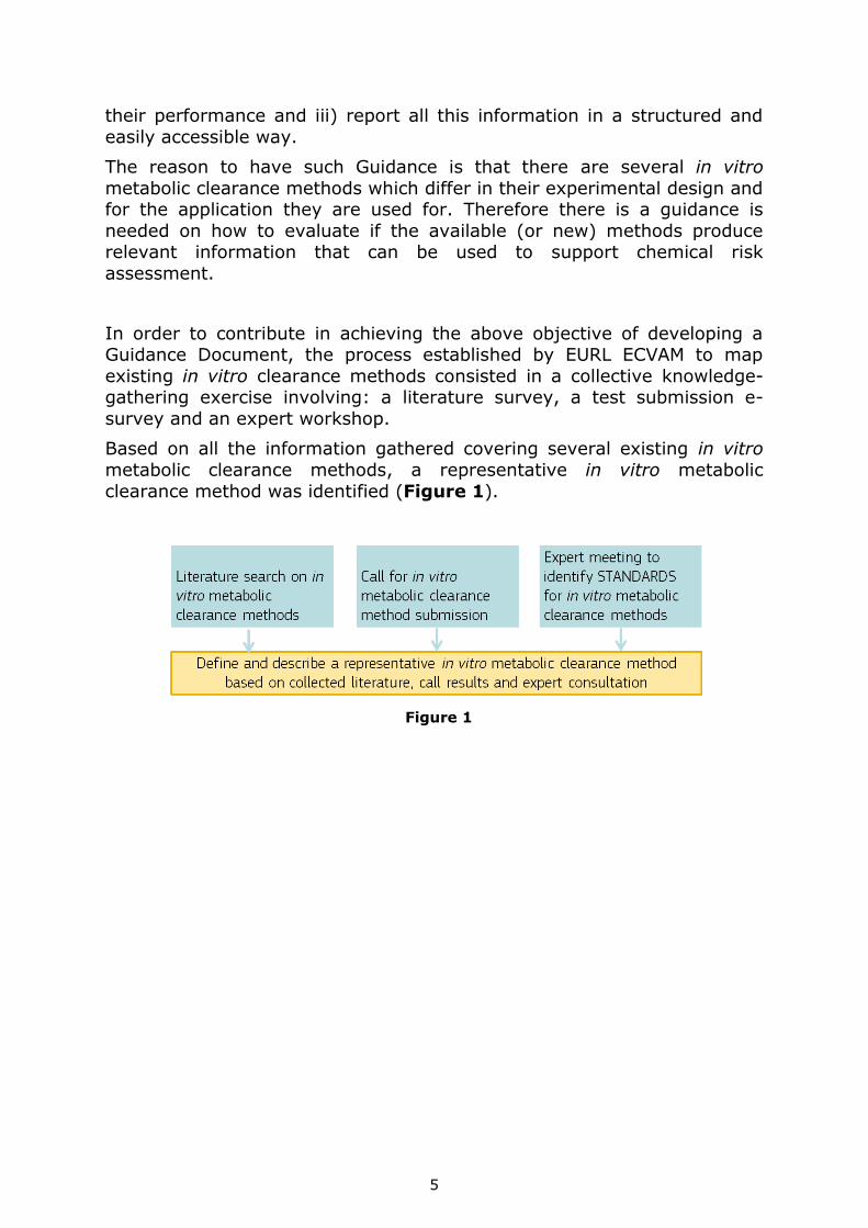
In order to contribute in achieving the above objective of developing a Guidance Document, the process established by EURL ECVAM to map
existing in vitro clearance methods consisted in a collective knowledge-gathering exercise involving: a literature survey, a test submission e-
survey and an expert workshop.
Based on all the information gathered covering several existing in vitro
metabolic clearance methods, a representative in vitro metabolic
clearance method was identified (Figure 1).
Figure 1
6
This representative method is not meant to be the "best possible" in vitro
human hepatic metabolic clearance method but simply indicates the most common used experimental layout (fitted for specific purposes) built on
existing experience collected from available in vitro clearance methods.
Therefore the representative methods can be considered as having a set
of experimental elements that, based on practical experience, have proven to generate reliable and relevant data.
Before describing in the Materials and Methods section the work done to implement the representative method, the following discussion will
provide a general description of possible approaches to develop in vitro hepatic metabolic clearance methods.
In vitro human hepatic metabolic clearance is commonly measured using hepatocytes suspension cultures (generally collected from human liver
which are called "primary hepatocytes" or from cancer cells) or hepatic microsomes (sub-cellular fractions). In contrast to microsomes
(representing only the Phase I biotransformation enzymes of human liver
since they are lacking conjugation activities), the use of primary human hepatocytes (fresh or cryopreserved) is widely accepted as the most
appropriate in vitro system since all the metabolic enzymes (Phase I and Phase II biotransformation enzymes) but also transporters are present in
a physiological relevant set-up. A typical experiment consists in exposing the hepatocytes to one concentration of the chemical to be tested (the
concentration should be below cytotoxicity) in order to determine intrinsic clearance (CLint).
CLint is expressed as μL/(min*millions of cells) and indicates the volume cleared by the chemical (due to metabolism) per minute and per million
of hepatocytes used in the experiment. Therefore CLint refers to enzyme-mediated clearance that would occur without physiological limitations
(e.g. protein binding, hepatic blood flow).
In general, two approaches are used to determine CLint: metabolite
formation and substrate depletion. For metabolite formation, the chemical
is exposed to hepatocytes at one concentration over several time-points and the formation of the metabolites is measured. This requires the
knowledge of which metabolites are formed and therefore this approach is technically demanding and not always feasible when there prior
knowledge of the metabolites is lacking.
The second approach, named substrate depletion, entails the incubation
of the hepatocytes exposed to the test compound at one concentration and the measurement of its disappearance over time. This approach is
technically easier to be performed and in fact it is the most common in vitro metabolic clearance method.
The method implemented in the EURL ECVAM laboratory facilities made
use of a suspension culture of pooled cryopreserved primary human
7
hepatocytes (collected from 10 different donors). The CLint was measured
by using the substrate depletion approach for 3 monoconstituent substrates used as reference chemicals with available in vivo hepatic
clearance data, e.g. acetaminophen, diclofenac and verapamil.
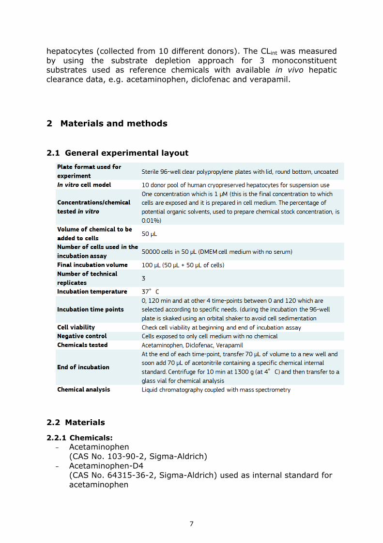
2 Materials and methods
2.1 General experimental layout
2.2 Materials
2.2.1 Chemicals:
− Acetaminophen
(CAS No. 103-90-2, Sigma-Aldrich)
− Acetaminophen-D4 (CAS No. 64315-36-2, Sigma-Aldrich) used as internal standard for
acetaminophen
8
− Diclofenac sodium salt
(CAS No. 15307-79-6, Sigma-Aldrich) − Diclofenac 13C6 sodium salt 4.5 hydrate
(CAS No. 1261393-73-0, Sigma-Aldrich) used as internal standard for diclofenac
− Verapamil hydroxychloride (CAS No. 152-11-4, Sigma-Aldrich)
− Verapamil-D7 Hydrochloride (CAS No. 1188265-55-5, LGC Standards) used as internal standard
for verapamil hydroxychloride
2.2.2 Cells, cell media and additives:
− 10 donor pool of cryopreserved primary human hepatocytes for suspension use (provider Bioreclamation-ITV, X008001)
− InVitroGRO Hepatocytes Thawing medium (BioreclamationITV, Z99019)
− Williams’ Medium E (W1878 Sigma) − Insulin, Transferrin, Selenium (ITS) (41400045 Gibco)
− L-Glutamine 200mM (25030149 Gibco) − Hepes, (1M, pH 7.4) (Gibco 15630080)
2.2.3 Plastic ware and disposable:
− Sterile 96-well clear polypropylene plates with lid, round bottom, uncoated
− Sterile 15-mL polypropylene centrifuge tubes − Sterile 50-mL polypropylene centrifuge tubes
− Sterile polypropylene reagent reservoir of at least 20 mL size − Adjustable pipette 2-20 μL
− Adjustable pipette 10-100 μL
− Adjustable pipette 100-1000 μL − Sterile pipette tips
2.2.4 Technical equipment:
− Water bath able to maintain a temperature of 37°C − Cell incubator with 5 ± 1% CO2 atmosphere, 37± 2°C temperature
and with water-saturated atmosphere − Orbital shaker able to operate at water-saturated atmosphere
− Balance with at least 0.01 mg readability
− Centrifuge appropriate for 96-well plates and able to maintain a temperature of 4 oC
− Heating plate with internal regulator to maintain 37oC
9
2.2.5 Reagents used for chemical analysis:
− Acetonitrile (UPLC grade)
− Formic acid (UPLC grade) − Ultrapure water 18.2MΩ.cm
2.2.6 Analytical equipment
UPLC Acquity coupled with Xevo G2-S QTof (Waters)
2.3 Preparation of cell assay medium, stock and working
solutions
The following sections describe the preparation of the different reagents and solutions used to perform the in vitro metabolic hepatic clearance
method.
Table 1 summarises the reagents and solutions to be prepared and the
time for their preparation.
Table 1
To be prepared the day before the experiment
The cell medium to be used for incubation assay (section 2.3.1)
The mixture of cell medium and acetonitrile (50:50) (section 2.3.2)
The stop solutions with specific internal standard at 1 µM (section 2.3.3)
The5 mL stock concentration of 10 mM for each chemical to be tested (section 2.3.4.)
To be prepared the day of the experiment
The working concentration of 2 µM for each chemical to be tested (section 2.3.5)
The chemical standards for the calibration curve for LC-MS analysis (section 2.3.6)
2.3.1 Cell medium for incubation assay:
In a bottle containing 500mL of Williams's Medium E the following steps were done:
Addition of 0.5mL of ITS (Insulin, Transferrin, Selenium)
Addition of 5mL of L-Glutamine 200mM
Addition of 7.5mL of HEPES
Thorough shaking and labelling as “Cell medium for incubation
assay”
An expiry date of 1 month was assigned
10
2.3.2 Cell medium and acetonitrile mixture (50:50):
To obtain the mixture the following procedure was adopted:
Addition of 10mL of "Cell medium for incubation assay" into a 20mL volumetric flask and making up to the mark with acetonitrile
Thorough shaking and labelling as “Cell medium-acetonitrile 50:50”
An expiry date of 1 day was assigned
2.3.3 Stop solutions using specific internal standards
Note: For each of the 3 chemicals tested, a specific internal standard was used to then performed the chemical analysis by using mass
spectrometry
To have a 2mM concentration, a defined amount of mg of the
desired Internal Standard was weighted and transferred in a 5mL volumetric flask
About 3mL of acetonitrile (or another solvent depending on the solubility) were added and checked to ensure that all the powder
was dissolved. Then making up to the mark with acetonitrile
Then a thorough shaking was done and after transferred in a glass tube and labelled as "Internal standard (either for Acetaminophen,
Diclofenac or Verapamil) 2mM in acetonitrile"
An expiry date of 6 months was assigned
In a 10mL volumetric flask was added acetonitrile (about 9mL) until almost the mark
5µL of "Internal standard 2mM in acetonitrile" were added in the volumetric flask, shaken thoroughly and made up to the mark with
acetonitrile
Finally, an "Internal standard 1µM in acetonitrile" label was applied
and an expiry date of 6 months was assigned
11
2.3.4 Stock concentration of 10mM for each chemical to be tested
In order to have a stock solution of 10mM concentration, a defined
amount of mg of the desired chemical was weighted and
transferred in a 5mL volumetric flask
About 3mL of acetonitrile were added and checked to ensure that all the powder was dissolved. Then maked up to the mark with
acetonitrile
Thoroughly shaken and transferred in a glass tube and labelled as
“Chemical Name (either Acetaminophen, Diclofenac or Verapamil) 10mM in acetonitrile”
An expiry date of 6 months was assigned
2.3.5 Working concentration of 2µM for each chemical to be tested
Note: the working concentration was the one to which the cells were exposed for the incubation assay to measure in vitro metabolic hepatic
clearance. When the working concentration was added to the cells, the final
concentration was 1µM. It was also important that the percentage of organic solvent was less than 0.1% to avoid potential interference with
the cells. In this case the percentage of acetonitrile was 0.01% when
added to the cells.
Using a calibrated glass syringe, 200µL of a specific chemical stock concentration of 10mM were transferred in 1mL volumetric flask,
and then made up to the mark with "cell medium-acetonitrile
50:50" solution.
After a thorough shaking, the solution was transferred in a glass tube and labelled as “Chemical Name (Acetaminophen, Diclofenac
or Verapamil) 2mM”. An expiry date of 1 day was assigned.
Using a calibrated glass syringe, 5µL of "Chemical Name 2mM"
previously prepared were transferred in 5mL volumetric flask, and then made up to the mark with "cell medium for incubation assay"
solution.
Shaken thoroughly, the solution was then transferred in a glass tube and labelled as “Chemical Name 2µM in cell medium”. An
expiry date of 1 day was also assigned.
12
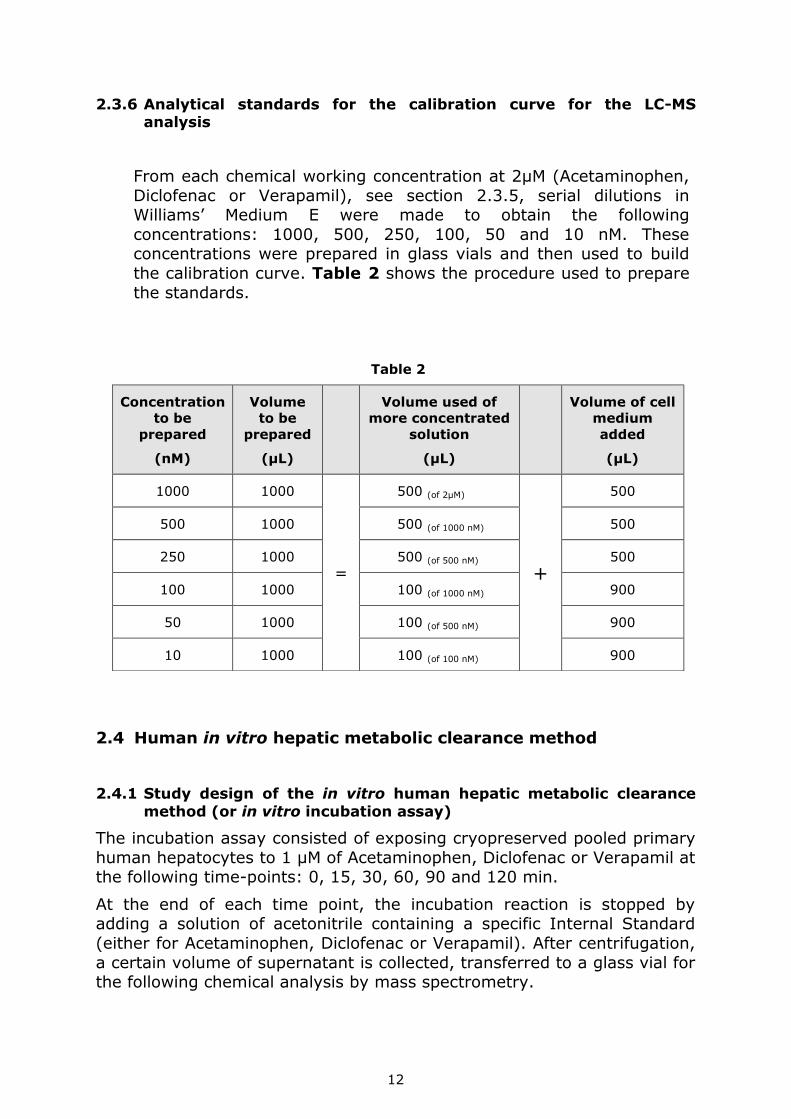
2.3.6 Analytical standards for the calibration curve for the LC-MS analysis
From each chemical working concentration at 2µM (Acetaminophen,
Diclofenac or Verapamil), see section 2.3.5, serial dilutions in Williams’ Medium E were made to obtain the following
concentrations: 1000, 500, 250, 100, 50 and 10 nM. These concentrations were prepared in glass vials and then used to build
the calibration curve. Table 2 shows the procedure used to prepare
the standards.
2.4 Human in vitro hepatic metabolic clearance method
2.4.1 Study design of the in vitro human hepatic metabolic clearance
method (or in vitro incubation assay)
The incubation assay consisted of exposing cryopreserved pooled primary
human hepatocytes to 1 µM of Acetaminophen, Diclofenac or Verapamil at the following time-points: 0, 15, 30, 60, 90 and 120 min.
At the end of each time point, the incubation reaction is stopped by adding a solution of acetonitrile containing a specific Internal Standard
(either for Acetaminophen, Diclofenac or Verapamil). After centrifugation,
a certain volume of supernatant is collected, transferred to a glass vial for the following chemical analysis by mass spectrometry.
Table 2
Concentration
to be
prepared
(nM)
Volume
to be
prepared
(µL)
Volume used of
more concentrated
solution
(µL)
Volume of cell
medium
added
(µL)
1000 1000
=
500 (of 2µM)
+
500
500 1000 500 (of 1000 nM) 500
250 1000 500 (of 500 nM) 500
100 1000 100 (of 1000 nM) 900
50 1000 100 (of 500 nM) 900
10 1000 100 (of 100 nM) 900
13
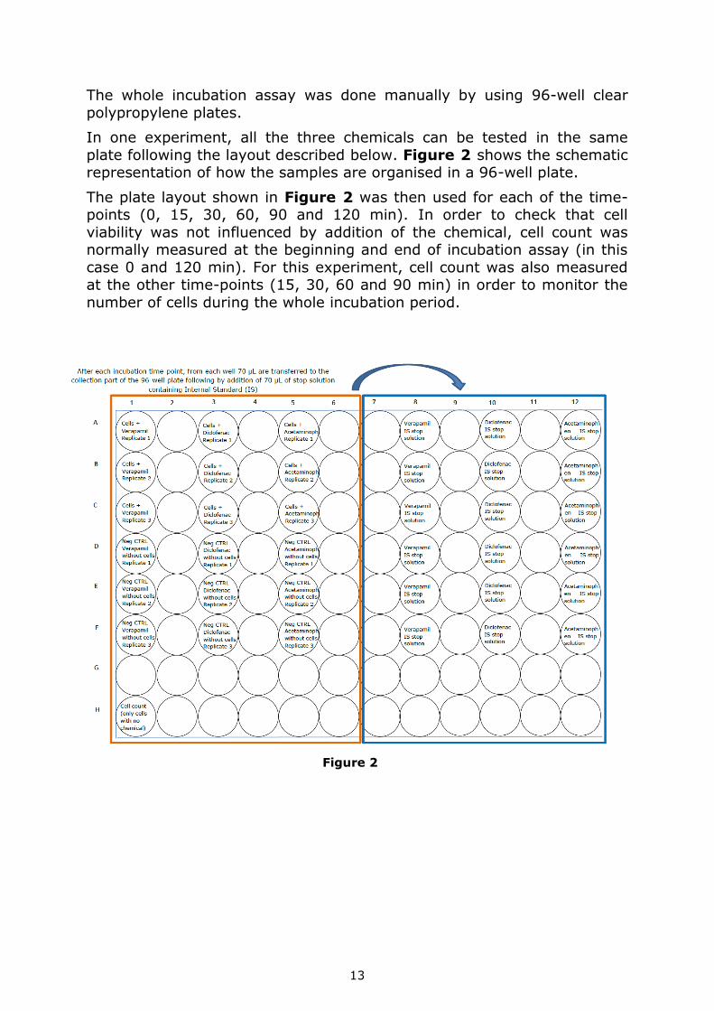
The whole incubation assay was done manually by using 96-well clear
polypropylene plates.
In one experiment, all the three chemicals can be tested in the same
plate following the layout described below. Figure 2 shows the schematic representation of how the samples are organised in a 96-well plate.
The plate layout shown in Figure 2 was then used for each of the time-points (0, 15, 30, 60, 90 and 120 min). In order to check that cell
viability was not influenced by addition of the chemical, cell count was normally measured at the beginning and end of incubation assay (in this
case 0 and 120 min). For this experiment, cell count was also measured at the other time-points (15, 30, 60 and 90 min) in order to monitor the
number of cells during the whole incubation period.
Figure 2
14
2.4.1.1 Prepare equipment for incubation assay
6 sterile 96-well clear polypropylene plates with lid, round bottom, uncoated were taken. For each plate, the lid was labelled writing
the corresponding incubation time (e.g. 0, 15, 30, 60, 90 or 120 min)
To each plate, 50µL of "Verapamil (or another chemical) working concentration of 2µM" (see section 2.3.5) were added to 3 wells,
as shown in Figure 2. The position of these wells, which were used as technical replicates, was marked
To each plate, 50µL of "Verapamil (or another chemical) working
concentration of 2µM" (see section 2.3.5) and then 50µL of cell medium (see section 2.3.1) were added to 3 wells, as shown in
Figure 2. Also in this case, the position of these wells which were
used as negative controls was marked
All 96-well plates were put in the incubator at 37°C and 5% CO2 for 10-15min prior to cell addition
2.4.1.2 Thawing and reconstitution of cryopreserved human hepatocytes
The cell medium was pre-warmed (see section 2.3.1) to 37°C for
about 20-30min
The following steps have taken about 30 min.
Notes:
* The thawing procedure is stressful to frozen cells, and working quickly ensures that a high proportion of the cells survive the
procedure.
* 1 vial contained about 5 million of frozen hepatocytes. Since, for the
incubation assay, each well contained 50000 cells, plus 3 chemicals in triplicate, 6 time points and controls (50000 x 3 chemicals x 3
replicates x 6 time points = >2700000 cells), 1 vial was enough to perform an experiment following the layout shown in Figure 2.
48mL of thawing medium (see section 2.2.2) were transferred to a sterile 50mL polystyrene tube
The thawing medium was prewarmed to 37°C for about 10min
After, the vial from the shipping container or cryostorage was carefully removed. If the vial has been stored in the liquid phase,
it is necessary to ensure that liquid nitrogen is removed from the vial before warming up and check that the cap is firmly closed.
The vial was immersed into a 37°C water bath and gently shaken for about 1.5 minutes until the ice was detached from the plastic
15
The vial was transferred into the laminar flow hood and disinfected
with an absorbent paper containing isopropanol or ethanol
From the vial, all hepatocytes cell suspension were transferred into the pre-warmed 50mL polystyrene tube containing 48mL of
thawing medium
From the 50mL polystyrene tube, 1mL of solution was used to
rinse the vial once to take what remains of cells. Then, transferred back into the 50mL polystyrene tube
The hepatocytes suspension was reached by gently inverting the
50mL polystyrene tube 3 times
The 50mL polystyrene tube was centrifuged at 50g at room
temperature during 5 minutes
The supernatant was discarded by either pouring in one motion (partial pouring and re-inverting the centrifuge tube is
inadequate), or aspirating using a vacuum pump
4mL of pre-warmed cell medium were added to the 50mL
polystyrene tube. Then, a pipette 100-1000µL was used to pipette gently for loosening the pellet
The total cell count and the percentage of viable cells were
determined using the Trypan Blue exclusion method
After determination of the cell concentration (expressed as
cells/mL), a dilution with pre-warmed cell medium was made to obtain the desired concentration of cells (for the incubation assay
50000 cells in 50µL are needed. Therefore the desired final concentration is 1 million cells in 1mL of cell medium)
Acceptance criteria to use hepatocytes for incubation assay
Two conditions must be taken into account when performing the
incubation assay:
A minimum cell viability of 80% after thawing must be obtained.
After reconstitution, cell suspensions can be used for up to 4 hours.
2.4.1.3 Incubation assay
Firstly, from the incubator, only the 96-well plate labelled on the
lid as "0min" was removed
50µL of viable hepatocytes (1 million cells/mL) were added only to the 3 wells used as technical replicates (see section 2.4.1.1)
Immediately, from each of the 6 wells of the first column containing Verapamil-exposed samples (see Figure 2), 70µL were
transferred, by using a multichannel pipette, into the corresponding new column as shown in Figure 2. Then, 70µL of
16
"stop solution with Verapamil Internal Standard (see section
2.3.3) were immediately added to each well
The previous step was repeated for the remaining two chemicals: Diclofenac and Acetaminophen and consequently followed the
complete procedure as described below
The 96-well plate was centrifuged at 1300g for 10min at 4°C.
Then, from each well 100µL of supernatant were transferred in the corresponding previously-labelled glass vials which were stored at
low temperature (-70°C) until LC-MS analysis
From the incubator, all the remaining 96-well plates were removed and placed on the heating plate (those labelled on the lid as "15,
30, 60, 90 and 120 min")
In order to start the incubation reaction, 50µL of viable
hepatocytes (1 million cells/mL) were added, only to the wells used as technical replicates (see Figure 2). Furthermore, 50µL of
viable hepatocytes were added to each plate, into one well which was used for cell-count (Figure 2)
The 96-well plates were returned to the orbital shaker in the
incubator at 37°C and the shaker speed was adjusted at 300 rpm
At each time-point of 15, 30, 60, 90 and 120 min, the
corresponding 96-well plate was removed from the incubator.
Immediately, from each of the 6 wells of the first column containing Verapamil-exposed samples (see Figure 2), 70µL were
transferred, by using a multichannel pipette, into the corresponding new column as shown in Figure 2. Then, 70µL of
"stop solution with Verapamil Internal Standard (see section
2.3.3) were immediately added to each well
The previous step was repeated for the remaining two chemicals: Diclofenac and Acetaminophen
Note: it was important to record the exact time when the stop solution
was added to the samples
The 96-well plate was centrifuged at 1300g for 10min at 4°C.
Then, 100µL of supernatant from each well were transferred in the corresponding previously-labelled glass vials which were stored at
low temperature (-70°C) until LC-MS analysis.
2.4.1.4 Preparation of analytical standards
For each chemical, the prepared standards (see section 2.3.6) were obtained following the same procedure used for the preparation of the
samples as described in section 2.4.1.3.
Briefly, from each standard 70µL were transferred to a new glass vial, previously labelled. Then 70µL of "stop solution with corresponding
Internal Standard (see section 2.3.3)" were added. The glass vials were
17
then centrifuged at 1300g for 10min at 4°C. Then, 100µL of supernatant
were transferred in a new previously-labelled glass vials which were stored at -70°C until LC-MS analysis.
2.4.2 LC-MS/MS analysis
The mass spectrometer was a Xevo G2-S QTof (Waters) coupled with an
Acquity Ultra Performance Liquid Chromatography (UPLC) system (Waters) and interfaced with an ElectroSpray Ion source (ESI).
For all 3 chemicals the column used was a 50×2.1mm, Acquity UPLC-BEH
C18 1.7μm. Mobile phase A was an aqueous solution of 0.1% formic acid and as mobile phase B was used 100% acetonitrile containing 0.1% of
formic acid. The chromatographic conditions for Diclofenac and Verapamil were the
same as shown in Table 3.
Table 3
Time (min) Flow rate (mL/min) A (%) B (%)
0 0.4 98 2
0.5 0.4 98 2
2.5 0.4 20 80
2.51 0.4 2 98
4.5 0.4 2 98
4.51 0.4 98 2
6 0.4 98 2
The chromatographic conditions for acetaminophen are shown in Table 4.
Table 4
Time (min) Flow rate (mL/min) A (%) B (%)
0 0.4 98 2
0.4 0.4 98 2
2.2 0.4 2 98
3 0.4 2 98
3.1 0.4 98 2
4 0.4 98 2
For Diclofenac and Verapamil the instrument operated in MS/MS mode in
which the parent ion was selected in the quadrupole and then
fragmented. The resulting product ions were monitored for the quantitative analysis. For Acetaminophen the instrument operated in MSe
mode and no specific product ions were selected for the quantitative analysis.

18
Table 5 summarises the source parameters optimised for each chemicals.
Table 5
Instrumental Parameters Settings Test Chemical
Acetaminophen Diclofenac Verapamil
Polarity ionisation Positive Negative Positive
Capillary (kV) 1 0.5 0.4
Cone (V) 40 100 40
Source Temperature (°C) 120 120 120
Desolvation Temperature (°C) 350 500 500
Cone Gas Flow (L/h) 50 50 50
Desolvation Gas Flow (L/h) 1000 1000 1000
Table 6 summarises the precursor- and product-ion(s) monitored for
each chemical and used for the quantitative analysis. Diclofenac
quantification, the precursor/product ion pair m/z 250/214 was used as quantifier, while the pair at m/z 455/303 was used for verapamil.
Table 6
Chemical Polarity MS mode Precursor ion
(m/z)
Product ion
(m/z)
Acetaminophen positive MSe 151.06 -
Acetaminophen-D4 positive MSe 155.08 -
Diclofenac negative MS/MS 250.01 178.06
214.04
Diclofenac 13C6 negative MS/MS 256.08 220.11
Verapamil positive MS/MS 455.29 165.09
303.20
Verapamil-D7 positive MS/MS 462.33 310.29
2.4.3 Calculation of in vitro intrinsic clearance (CLint)
The calculated concentrations, expressed as nM, have been transformed
into a natural logarithmic scale (ln). Then the elimination rate constant (k) was calculated from the slope of
ln(Ct/C0) vs t (min), with a 1/y weighting, according to equation 1.
ln(𝐶𝑡
𝐶0) = −𝑘𝑡 (eq. 1)
Ct = the substrate concentration in the incubation well at time t (min)
C0 = the substrate concentration in the incubation well at time t= 0
19
k = the elimination rate constant
The intrinsic clearance in vitro (CLint in vitro) was calculated according to
the following equation:
𝐶𝐿𝑖𝑛𝑡 = 𝑘𝑉
𝑁 μL/ (min*millions of cells)
Where:
𝑘 = 0.639
𝑡1/2
V = incubation volume (100 μL)
N = number of cells (x106) in the incubation (50000 cells used in this assay)
20
3 Results
3.1 LC-MS analysis
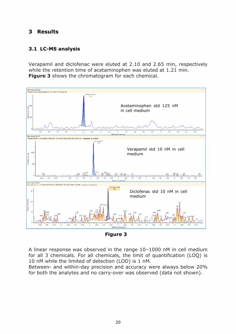
Verapamil and diclofenac were eluted at 2.10 and 2.65 min, respectively
while the retention time of acetaminophen was eluted at 1.21 min. Figure 3 shows the chromatogram for each chemical.
A linear response was observed in the range 10–1000 nM in cell medium
for all 3 chemicals. For all chemicals, the limit of quantification (LOQ) is 10 nM while the limited of detection (LOD) is 1 nM.
Between- and within-day precision and accuracy were always below 20% for both the analytes and no carry-over was observed (data not shown).
Acetaminophen std 125 nM
in cell medium
Verapamil std 10 nM in cell medium
Diclofenac std 10 nM in cell medium
Figure 3
21
3.2 In vitro hepatic metabolic clearance
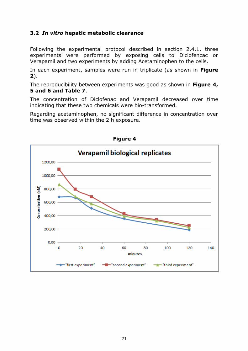
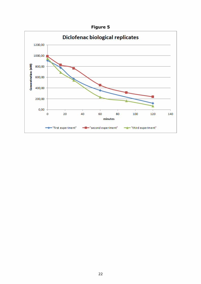
Following the experimental protocol described in section 2.4.1, three experiments were performed by exposing cells to Diclofencac or
Verapamil and two experiments by adding Acetaminophen to the cells.
In each experiment, samples were run in triplicate (as shown in Figure
2).
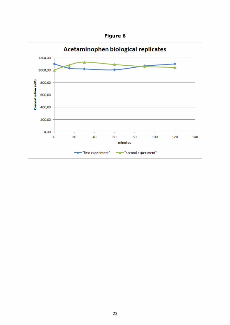
The reproducibility between experiments was good as shown in Figure 4,
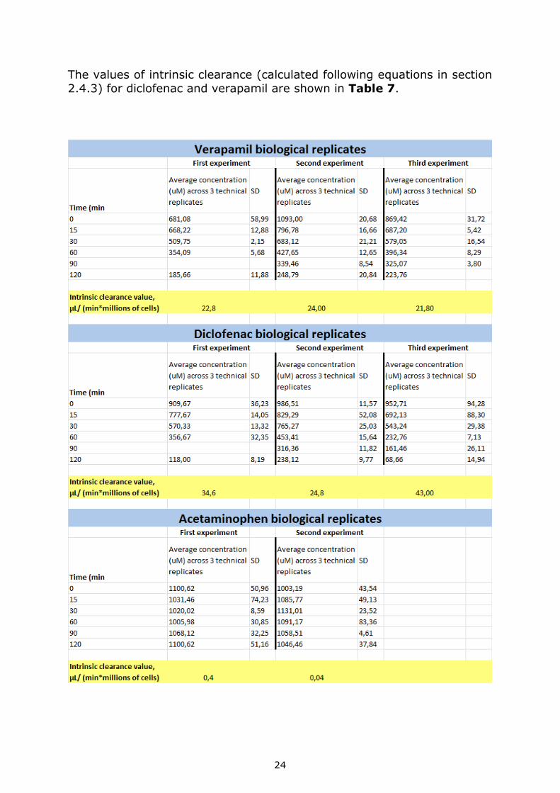
5 and 6 and Table 7.
The concentration of Diclofenac and Verapamil decreased over time indicating that these two chemicals were bio-transformed.
Regarding acetaminophen, no significant difference in concentration over time was observed within the 2 h exposure.
Figure 4
22
Figure 5
23
Figure 6
24
The values of intrinsic clearance (calculated following equations in section
2.4.3) for diclofenac and verapamil are shown in Table 7.
25
4 Conclusion
Knowledge of in vitro human hepatic metabolic clearance can improve the
prediction of chemical's toxicity by using alternatives to animal testing methods and therefore better support chemical risk assessment.
This report describes the preliminary work done at the EURL ECVAM laboratory facility to describe an experimental procedure towards a
representative in vitro human hepatic metabolic clearance method that can be ultimately used in future defined approaches for toxicological risk
assessment.
The method implemented was based on a collective knowledge-gathering
exercise in which existing available in vitro human hepatic metabolic
clearance methods were mapped and compared. The method implemented, represented the most common experimental features used
by the existing methods gathered while doing the mapping exercise.
As preliminary work, three chemicals with known in vivo human hepatic
metabolic clearance data were used to challenge the in vitro method implemented in the EURL ECVAM laboratory.
The scope of the project was to evaluate the reproducibility of the data (concentration-time curves and calculated in vitro intrinsic clearance)
between the different experimental runs performed.
The data obtained clearly show good reproducibility.

26
References
1. EURL ECVAM strategy for achieving 3Rs impact in the assessment of toxicokinetics and systemic toxicity. (2015) Bessems J., Coecke S.,
Gouliarmou V., Whelan M., Worth A.
2. Regulation (EC) No 1907/2006 of the European Parliament and of the
Council of 18 December 2006 concerning the Registration, Evaluation,
Authorisation and Restriction of Chemicals (REACH).
3. Regulation (EC) No 1223/2009 of the European Parliament and of the Council of 30 November 2009 on cosmetic products.
4. Regulation (EC) No 1107/2009 of the European Parliament and of the
Council of 21 October 2009 concerning the placing of plant protection products on the market.
5. Regulation (EU) No 528/2012 of the European Parliament and of the
Council of 22 May 2012 concerning the making available on the market and use of biocidal products.
6. EURL ECVAM literature survey to identify publically available in vitro
human hepatic metabolic clearance/ stability methods. (2014)
Gouliarmou V, Coecke S.
7. EURL ECVAM web survey on in vitro human hepatic metabolic clearance/ stability methods. (2014, limited distribution) Gouliarmou
V, Coecke S.
27
List of abbreviations and definitions
ADME: Absorption, Distribution, Metabolism, Excretion BPR: Biocidal Products Regulation
CLint: intrinsic clearance
CPR: Cosmetic Products Regulation LC-MS: liquid chromatography-mass spectrometry
NAMs: new advanced methodologies OECD: Organisation for Economic Co-operation and Development
PPPR: Plant Protection Products Regulation Q-Tof: quandrupole-time of flight
REACH: Registration, Evaluation, Authorisation and Restriction of Chemicals
TK: toxicokinetics UPLC: ultraperformance liquid chromatography
28
List of figures
Figure 1. Schematic representation of the process followed to collect knowledge on existing in vitro hepatic metabolic clearance methods in
order to then define the representative method to be implemented in the
laboratory facility.
Figure 2. Plate layout used in the in vitro hepatic metabolic clearance method.
Figure 3. UPLC chromatograms of Acetaminophen, Diclofenac and
Veraplamil.
Figure 4. Results of three experiments performed to measure Verapamil in vitro hepatic metabolic clearance.
Figure 5. Results of three experiments performed to measure Diclofenac
in vitro hepatic metabolic clearance.
Figure 6. Results of two experiments performed to measure
Acetaminophen in vitro hepatic metabolic clearance.
29
List of tables
Table 1. List of reagents and solutions to be prepared and the time for their preparation.
Table 2. Procedure followed to prepare the chemical standards to be
used for LC-MS analysis.
Table 3. Chromatographic conditions used for diclofenac and verapamil.
Table 4. Chromatographic conditions used for acetaminophen.
Table 5. Q-Tof source parameters optimised for acetaminophen,
diclofenac and verapamil.
Table 6. Precursor- and product-ion(s) monitored for acetaminophen,
diclofenac and verapamil and used for LC-MS analysis.
Table 7. Intrinsic clearance values calculated for Diclofenac and Verapamil for each of the three experiments performed.

Europe Direct is a service to help you find answers to your questions about the European Union
Free phone number (*): 00 800 6 7 8 9 10 11
(*) Certain mobile telephone operators do not allow access to 00 800 numbers or these calls may be billed.
A great deal of additional information on the European Union is available on the Internet.
It can be accessed through the Europa server http://europa.eu
How to obtain EU publications
Our publications are available from EU Bookshop (http://bookshop.europa.eu),
where you can place an order with the sales agent of your choice.
The Publications Office has a worldwide network of sales agents.
You can obtain their contact details by sending a fax to (352) 29 29-42758.
XX-N
A-x
xxxx-E
N-N
doi: 10.2788/886451
ISBN 978-92-79-64627-0
LB-N
A-2
8354-E
N-N