Embed Size (px)
Citation preview
1
Please note that this is an author-produced PDF of an article accepted for publication following peer review. The definitive publisher-authenticated version is available on the publisher Web site.
International Journal of Food Microbiology June 2015, Volume 203, Pages 55-62 http://dx.doi.org/10.1016/j.ijfoodmicro.2015.03.005 http://archimer.ifremer.fr/doc/00254/36551/ © 2015 Elsevier B.V. All rights reserved.
Achimer http://archimer.ifremer.fr
Development of a real-time PCR method coupled with a selective pre-enrichment step for quantification of
Morganella morganii and Morganella psychrotolerans in fish products
Podeur Gaetan 1, 2, 3
, Dalgaard Paw 4, Leroi Francoise
1, Prevost Hervé
2, 3, Emborg Jette
4, Martinussen
Jan 5, Hansen Lars Hestbjerg
6, Pilet Marie France
2, 3, *
1 Ifremer, Laboratory of Microbial Ecosystem and Marine Molecules for Biotechnology, Nantes, France
2 LUNAM Université, Oniris, UMR1014, Secalim, Nantes, France
3 INRA, Nantes, France
4 National Food Institute (DTU Food), Technical University of Denmark, Kgs. Lyngby, Denmark
5 DTU System Biology, Technical University of Denmark, Kgs. Lyngby, Denmark
6 Department of Environmental Science, Aarhus University, Roskilde, Denmark
* Corresponding author : Marie-France Pilet, tel.: + 33 2 40 68 78 11 ; email address : [email protected]
Abstract : Histamine fish poisoning is common and due to toxic concentrations of histamine often produced by Gram-negative bacteria in fin-fish products with a high content of the free amino acid histidine. The genus Morganella includes two species previously reported to cause incidents of histamine fish poisoning. Morganella morganii and Morganella psychrotolerans are both strong producer of histamine. However, little is known about the occurrence and critical stages for fish contamination with these bacteria. To elucidate contamination routes of Morganella, specific real-time quantitative PCR (RTi qPCR) methods for quantification of M. morganii and M. psychrotolerans have been developed. Selective primers amplified a 110 bp region of the vasD gene for M. psychrotolerans and a 171 bp region of the galactokinase gene for M. morganii. These primer-sets showed high specificity as demonstrated by using purified DNA from 23 other histamine producing bacteria and 26 isolates with no or limited histamine production. The efficiency of the qPCR reactions on artificially contaminated fish samples were 100.8% and 96.3% respectively. The limit of quantification (LOQ) without enrichment was 4 log CFU/g. A quantitative enrichment step with a selective medium was included and improved the sensitivity of the methods to a LOQ of below 50 CFU/g in seafood. RTi qPCR with or without enrichment were evaluated for enumeration of Morganella species in naturally contaminated fresh fish and lightly preserved seafood from Denmark. These new methods will contribute to a better understanding of the occurrence and histamine production by Morganella species in fish products, information that is essential to reduce the unacceptably high frequency of histamine fish poisoning.
2
Please note that this is an author-produced PDF of an article accepted for publication following peer review. The definitive publisher-authenticated version is available on the publisher Web site.
Highlights
► Quantification of Morganella species in fish products by RTi qPCR ► Sensitivity of the assay was improved with a quantitative selective enrichment step to 50 CFU/g ► The assays accurately quantified Morganella in fresh and lightly preserved fish ► Development of a specific tool to quantify main histamine producing bacteria in fish
Keywords : histamine-producing bacteria, tuna, selective medium, Enterobacteriaceae, galactokinase, typeVI secretion system
ACC
EPTE
D M
ANU
SCR
IPT
ACCEPTED MANUSCRIPT
3
1. Introduction
Histamine fish poisoning (HFP) is common worldwide and due to consumption of a range of
dark-fleshed fin-fishes (Dalgaard et al., 2008). Between 2008-2010, HFP was responsible for
51% of all seafood related outbreaks of disease in France and in Europe and for 20% in
United States (CDC, 2011; Helwigh et al., 2012; InVS, 2011). HFP is typically caused by
consumption of fish muscle containing high concentration of histamine (> 500 mg/kg) which
can be observed in fish species with high level of histidine such as Scombridae (tuna,
mackerel...), Clupeidae (herring, sardine...) and other species like Coryphaena hippurus
(mahi-mahi) or Belone belone (garfish) (Hungerford, 2010). HFP is a relatively mild illness
with allergy-like symptoms that appear some minutes to few hours after consumption of the
food. They are mainly characterized by rash, diarrhea, nausea, headache, flushing and
sweating (Prester, 2011).
Histamine in fish flesh is produced by decarboxylation of free histidine by bacterial
decarboxylase. Gram-negative marine and enteric bacteria have been identified as the main
bacterial groups responsible for HFP in fish products (Bjornsdottir et al., 2009; Dalgaard et
al., 2008; Veciana-Noguès et al., 2004). Histamine producing bacteria (HPB) can be sub-
divided into low and high producers of histamine based on the formation of histamine in a
broth culture medium containing histidine. The high histamine producers include mesophilic
species such as M. morganii, Enterobacter aerogenes, Hafnia alvei, Raoultella planticola and
Photobacterium damselae, which are able to produce more than 1000 mg/l of histamine in
tryptone soy broth supplemented with 2% histidine after 24-48 h incubation at temperatures
above 15°C (Bjornsdottir et al., 2009). High histamine-producing and psychrotolerant
bacteria have also been described, including Photobacterium phosphoreum (Kanki et al.,
2004) and M. psychrotolerans (Emborg et al., 2006). Both species produce histamine at low
temperature until 0°C in fish products (Dalgaard et al., 2006; Emborg et al., 2005).

ACC
EPTE
D M
ANU
SCR
IPT
ACCEPTED MANUSCRIPT
4
Whereas all Morganella isolates seem to be strong histamine producers, intra-species
variability exists for other species including P. phosphoreum (Dalgaard et al., 2006; Emborg
et al. 2005; Kim et al., 2002; Klausen et al., 1987). Both M. morganii and M. psychrotolerans
have been isolated from fish products (fresh tuna, tuna in sauce, cold-smoked tuna)
responsible for outbreaks (for a review, see Dalgaard et al., 2008). Despites those facts, there
is a lack of data on prevalence of Morganella spp. in fish products, probably due to the
absence of sufficiently sensitive and specific enumeration method. Previous experiments of
detection of HPB have been performed on differential media (Mavromatis and Quantick,
2002; Tao et al., 2009) based on color modification of pH indicators once histidine is
converted to histamine during bacterial growth. However, these methods are time consuming
and unreliable (Bjornsdottir et al., 2009). Nowadays, molecular methods based on RTi qPCR
are more reliable for detection, identification and quantification of bacteria but their lack of
sensitivity when used with food products remains a problem (Postollec et al., 2011).
Bjornsdottir-Butler et al. (2011a) developed a RTi qPCR method based on the primers of
Takahashi et al. (2003) designed on the histidine decarboxylase gene to quantify Gram-
negative and high HPB. That method has been set-up for mesophilic species but cannot detect
P. phosphoreum or M. psychrotolerans (experiments in our laboratory, data not shown). More
recently, RTi qPCR methods for quantification of M. morganii (Ferrario et al., 2012a, 2012b)
and viable P. phoshoreum (Macé et al., 2013) in fish products have been proposed. However,
in those studies the detection limit of RTi qPCR was typically between 3-4 log CFU/g in food
products. This limit is too high to study the occurrence of Morganella spp. in fish products in
which expected contamination level is most likely less than 100 CFU/g. To overcome this
problem of the sensitivity of RTi qPCR methods, enrichment steps with selective media have
been proposed, allowing quantification of 1-10 CFU/g for e.g. the pathogenic bacteria Listeria
ACC
EPTE
D M
ANU
SCR
IPT
ACCEPTED MANUSCRIPT
5
monocytogenes (O’ Grady et al., 2008) and Salmonella (McGuinness et al., 2009) in different
food products.
The objectives of the present study were to develop two specific and sensitive RTi qPCR
methods for quantification of M. morganii and M. psychrotolerans in fish products. Selective
primers for both species have been designed and evaluated against isolates of HPB and non-
HBP isolates. These RTi qPCR methods used in combination with a new enrichment step
reduced the LOQ and increase the methods field of application for fish products.
2. Materials & methods
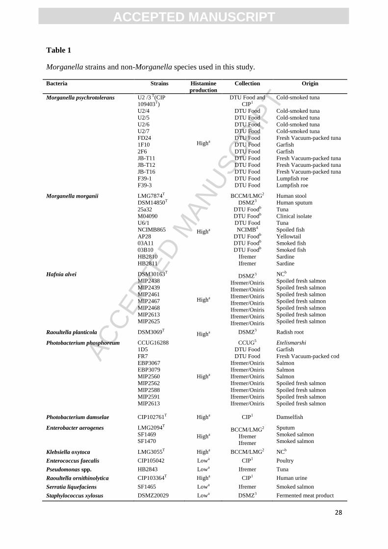
2.1.Bacterial strains and pre-culturing
Bacterial strains used in this study are listed in Table 1. Strains were grown in Brain Heart
Infusion (BHI, Biokar Diagnostics, Beauvais, France) at 20°C during 24 h, for Morganella
strains, and during 24-48 h for other strains, except for P. phosphoreum which was cultivated
at 15°C in BHI with 2% NaCl. The strains were stored at -80°C in their culture medium with
10% glycerol.
2.2.DNA extraction
For bacterial cultures, DNA extraction was performed on 1.5 ml cultures that have reached a
concentration of at least 8 log CFU/ml. After centrifugation during 10 min at 8500 x g, the
chromosomal DNA of all bacterial isolates was extracted using the Qiagen DNeasy Blood and
Tissue Kit (Qiagen, S.A., Courtaboeuf, France).
The DNA extraction on fish tissue was adapted from a protocol developed for raw salmon to
quantify P. phosphoreum by RTi qPCR (Macé et al., 2013). Briefly, 30 g portion of seafood
samples (tuna, mackerel or herring) was aseptically weighed in a sterile stomacher bag and 5-
fold diluted with sterile peptone-salt water (0.1% peptone, 0.85% salt). Ten milliliters of
ACC
EPTE
D M
ANU
SCR
IPT
ACCEPTED MANUSCRIPT
6
homogenized suspension were filtered on a Nucleospin Filter L (Macherey-Nagel, Hoerdt,
France). The following extraction of DNA was performed as described by Macé et al. (2013).
DNA was purified using the DNeasy Blood & Tissue Kit as described in the Qiagen
instruction manual.
2.3.Genomics data and primer design
Shotgun sequencing was performed in Denmark using Roche FLX 454 pyrosequencing on
DNA from the M. morganii strain U6/1 and on DNA from the type strain of M.
psychrotolerans U2/3T = LMG 23374
T = DSM 17886
T (Emborg et al., 2006; Meyer et al.,
2008). Sequencing was done using the FLX Titanium sequencing kit and 1 region of an
XLR70 pico titre plate per strain. Contigs for each strain were assembled using the Newbler
assembler software version 2.0.01.14 provided with the GS FLX instrument and annotated by
using the RAST annotation server (Aziz et al., 2008). Genome sequencing and assembly of
the M. morganii U6/1 and M. psychrotolerans U2/3T resulted in 51 and 28 x coverage on 3,9
Mb and 4,2 Mb size genomes, respectively. Genomes were assembled into 123 (M. morganii
U6/1) and 292 (M. psychrotolerans U2/3T) large contigs (>500 bp).
Thirty primers pairs were designed for M. psychrotolerans and M. morganii using the
Geneious Software (Geneious version 6.1, Biomatters Ltd.) based on the Primer3 calculation
method (Untergasser et al., 2007) and the Primer-Blast software (NCBI, UK). Primers pairs
were initially evaluated in silico by using the nucleotide Blast program to check their
specificity for M. morganii or M. psychrotolerans against all genomic data of GenBank
(NCBI, UK) and tested in vitro as described in 2.4 using appropriate hybridization
temperatures. The selected primers VasD-F4 (5'-AAATCGCCATCACACTCCTTG-3') and
VasD-R4 (5'-TTCAAAACGGGAGTCCTCACTG-3') were designed on the vasD gene from
the Type VI secretion system of M. psychrotolerans. This primer set matched respectively
ACC
EPTE
D M
ANU
SCR
IPT
ACCEPTED MANUSCRIPT
7
position 146-166 and 234-255 of the M. psychrotolerans U2/3T vasD gene (Genbank
accession number KP069481). For M. morganii, the primers GalK-F4 (5'-
ACAGTGCTTCGGCGCATCCC-3') and GalK-R4 (5'-GCAGCCACCACGCAGACCTT-3')
were obtained on the galactokinase gene (galK) and matched respectively position 39-58 and
190-209 of the galactokinase gene of M. morganii U6/1 (Genbank accession number
KP069480).
2.4.Real-time PCR amplification
Inclusivity and exclusivity of primers (TAG Copenhagen, Denmark or Invitrogen, Illkirch,
France) designed for M. morganii and M. psychrotolerans were tested on bacterial DNA
extracted from the strains listed in Table 1. Genomic DNA was measured using a Nanovalue
(Applied Biosystem, Saint-Aubin, France) and diluted to 4 ng/µl. Specificity of the RTi qPCR
assay was tested using 4 ng of DNA.
Real-time qPCR was conducted in a 15 µl reaction volume using the following reaction
mixture: 1 U of Iq SYBR® Green Supermix (Biorad, Hercules, US), 300 nM of each VasD or
GalK forward and reverse primers, nuclease free H2O and 1 µl of DNA template. Real-time
PCR cycling was performed using a CFX-96 instrument (Biorad, Marnes-la-Coquette,
France) or a Mx3000P thermocycler (Stratagene, AH Dianostics, Aarhus, Denmark). The
cycling parameters were: 95°C hold for 180 s for initial denaturation and activation of the hot-
start polymerase, followed by 40 cycles of amplification of 95°C for 15 s, 60°C for 30 s for
M. morganii, or 62°C for 30 s for M. psychrotolerans. Fluorescence was read at the end of
each amplification cycle. At the end of the 40 cycles, a melting curve was conducted between
55°C and 95°C with a 0.5°C/5 s increment read. The cycle threshold (CT) value was
determined using a background limit of 0.02.
ACC
EPTE
D M
ANU
SCR
IPT
ACCEPTED MANUSCRIPT
8
To obtain standard curves relating CT-values and cell concentrations (log CFU/g), ten-fold
serially dilutions of a culture of the type strains M. psychrotolerans U2/3T and M. morganii
LMG7874T = CIP A231
T (Table 1) were inoculated on pieces of canned tuna flesh. DNA
extraction was performed on each piece of tuna inoculated with decreasing known
concentrations of each strain between 2 to 8 log CFU/g. Specific RTi qPCR was performed as
described above and enumeration of each strain was made on BHI agar incubated 24 h at
20°C.
For quantification in naturally contaminated products a positive control was made with DNA
extracted as described in section 2.2 from 30 g of fish inoculated with M. morganii
LMG7874T or M. psychrotolerans U2/3
T (9 log CFU/g). Negative controls consisted in DNA
extracted from canned tuna and sterile water.
Development of a Morganella enrichment (MoE) medium
To improve sensitivity of the RTi qPCR methods, enrichment steps were added. A selective
enrichment medium was developed to allow optimal growth of Morganella species and to
inhibit most of the other HPB. High histamine-producer M. psychrotolerans U2/3T, F39-1,
JB-T11, M. morganii LMG7874T, DSM14850
T, 03A11, R. planticola DSM3069
T, E.
aerogenes LMG2094T, K. oxytoca LMG3055
T and H. alvei DSM30163
T (Table 1) were
selected to test the effect of 15 antibiotics and of the 2-deoxy-D-galactose substrate. This
substrate was tested as the hexose analog can be lethal for microorganisms able to metabolize
galactose (Alper and Ames, 1975). Tests were performed in honeycomb 2 microplates with
100 wells (Thermo Electron Oy, Vantaa, Finland), each well being filled with 300 µl of
Nutrient Broth (NB, CM0001, Oxoid, Basingstoke, UK) with or without different
concentrations of antibiotic (Table 2) or with 2-deoxy-D-galactose. NB was supplemented
with 0.2 mg/ml of 2-deoxy-D-galactose and incubated with or without an overlay of sterile
ACC
EPTE
D M
ANU
SCR
IPT
ACCEPTED MANUSCRIPT
9
paraffin oil. Wells were inoculated with an overnight pre-culture of each strain at 25°C in NB,
at a final concentration of 102 CFU/ml. A negative control was performed using non-
inoculated NB broth. Growth was followed during 72 h at 25°C by absorbance measurement
at 540 nm and with measurements every 20 minutes (Bioscreen C, Labsystem, Helsinki,
Finland). Plates were shaked 10 s at medium speed before each measurement. Experiments
were done in duplicate and growth data were analyzed by Excel (Microsoft Corporation,
Redmond, US) to determine the growth inhibiting effect of the studied antimicrobials.
The final MoE medium was adapted from Emborg and Dalgaard (2008) and consisted of
buffered Lucia-Bertani broth added L-histidine and colistin: 10.0 g/l bacto tryptone (211705,
Becton and Dickinson Company, Sparks, MD, USA), 5.0 g/l yeast extract (212750, BD), 7.0
g/l KH2PO4 (1.04873, Merck, Darmstadt, Germany), 7.0 g/l K2HPO4 (1.05104, Merck) and
10.0 g/l L-histidine monohydrochloride monogydrate (Sigma, H8125). The autoclaved
(121°C, 15 min) and chilled medium was added a filter sterilized solution of colistin-sulfate
salt (Sigma, C4461) to a final concentration of 32 mg/l. pH was adjusted to 6.50.
2.5. Development of a quantitative enrichment procedure
To calculate the concentration of M. psychrotolerans or M. morganii in fish product from
concentrations determined by RTi qPCR after enrichment in MoE, the maximum specific
growth rate (µmax) of each Morganella species in the MoE medium were determined. Pre-
cultures (NB, 25 °C, 12 h) of, respectively, four M. psychrotolerans strains (MIX-Mp: U2/3T,
JB-T11, JB-T12, U2/5) or four M. morganii strains (MIX-Mm: 25a32, AP28, LMG7874T,
DSM14850T) were mixed. Appropriate dilutions were inoculated in peptone-salt water,
canned tuna and cooked shrimp to obtain a concentration of 250 CFU/ml or 250 CFU/g.
Thirty grams of inoculated seafood were five-fold diluted with peptone-salt water and
homogenized in a stomacher 400 (Seward Medical, London, UK). 1.00 ml of the homogenate
ACC
EPTE
D M
ANU
SCR
IPT
ACCEPTED MANUSCRIPT
10
was then transferred to 9.00 ml of MoE medium and growth was determined in duplicate by
viable counting during storage at 10°C for M. psychrotolerans and at 37°C for M. morganii.
These temperatures for specific incubation were selected for the two species based on the
known effect of temperature on the growth rate of M. psychrotolerans and M. morganii
(Emborg and Dalgaard, 2008). In the same way, the peptone-salt water solution with 250
CFU/ml was five-fold diluted and then 1 ml was inoculated in the MoE medium. Samples
were removed for bacterial enumeration and DNA extraction after 0, 14, 24, 38, 49, 63 and 86
h for M. psychrotolerans and after 0, 1, 3, 4, 6, 7 and 9 h for M. morganii. Enumeration of
Morganella was done on Tryptone Soya Agar (TSA, CMO131, Oxoid) at 25°C after 36 h of
incubation. DNA extraction was done with 1 ml of MoE as described above (See 2.2). For
each food matrix, the maximum specific growth rate (µmax) of M. psychrotolerans and M.
morganii in MoE was determined by fitting growth data using an exponential model (Eq. 1).
Log(Nt) = Log(N0) + (µmax x t)/Ln(10) (1)
where N0 and Nt are cell concentrations (CFU/ml) at time zero and at time t, respectively.
2.6.Quantification of Morganella species in natural contaminated fish products by RTi
qPCR
The RTi qPCR methods were used to quantify M. psychrotolerans and M. morganii on
different fish products from Denmark (Table 3). Tuna loins from the local fish market and
herring provided by local fisherman were transported to the laboratory and kept overnight in
ice before being processed and analysed. Tuna loins were cut in 8 pieces of 60 g and then
vacuum packed. Herrings were eviscerated, tail and head were removed and filets were
vacuum packed using a packaging film with low gas permeability. Frozen tuna steaks, hot
smoked mackerel, cold-smoked herring and Matjes herring fillets bought in supermarket were
already vacuum- or modified atmosphere packed. The fresh products were then stored for one
ACC
EPTE
D M
ANU
SCR
IPT
ACCEPTED MANUSCRIPT
11
week at 2°C whereas previously frozen tuna and lightly preserved products were kept at 10°C.
The products were analyzed in triplicate (3 packs) on the day of processing and after one
week of storage. Thirty grams of product were 5-fold diluted with chilled peptone-salt water,
homogenized with stomacher whereafter RTi qPCR was performed directly as described in
2.4. In addition, for each pack, two times 1.00 ml of the homogenized solution was added to
two times 9.00 ml of MoE medium (two tubes) and then incubated respectively during 72 h at
10°C or 7 h at 37°C before DNA extraction and quantification by the RTi qPCR assay (see
2.4). Total viable counts were determined at each time of analysis on spread plates of Long &
Hammer agar (L&H) incubated 5 days at 15°C (NMKL, 2006; Van Spreekens, 1974). Plates
were also observed in the dark to enumerate luminous P. phosphoreum colonies.
Enterobacteriaceae were enumerated using Tryptone Soya Agar/Violet Red Bile Glucose
(TSA/VRBG) agar as previously described (Emborg and Dalgaard, 2008). 24 colonies from
the TSA/VRBG agar were isolated and identified by using simple biochemical tests (Dalgaard
et al. 2006).
3- Results
3.1 Specificity of the real-time PCR assay
A set of 30 primers pairs has been designed and tested on M. pyschrotolerans and M.
morganii strains. Of these primers, only the VasD-FR4 and the GalK-FR4 primers were
specific for M. psychrotolerans and M. morganii, respectively. Establishment of the
specificity of the VasD and GalK primers were tested in silico by sequence alignment using
the GenBank database and by RTi qPCR on 13 M. psychrotolerans, 11 M. morganii and 49
HPB or non-HPB isolates (Table 1). The GalK-FR4 primers presented a 100% homology
sequence with the M. morganii related gene available in the GenBank database (MU9_2965).
For the VasD-FR4 primers, partial sequence alignments were obtained with 14 non-
ACC
EPTE
D M
ANU
SCR
IPT
ACCEPTED MANUSCRIPT
12
Enterobacteriaceae vasD sequences. In vitro RTi qPCR tests indicated a good specificity of
the primers for the 13 M. psychrotolerans and the 11 M. morganii isolates. The mean CT
values were 16.8 ± 0.3 (n = 13) and 15.2 ± 0.5 (n = 11) with 4 ng of DNA extracted from
broth cultures of M. psychrotolerans and M. morganii, respectively. RTi qPCR exclusivity
test on 49 HPB or non-HPB isolates resulted in CT values higher than 29. That was considered
as the CT threshold for specific detection of Morganella species obtained on pure cultures in
liquid medium. This threshold was increased to 31 CT when bacterial DNA was extracted
from fish samples (tuna) (data not shown). The melting temperature calculated at the end of
each real-time PCR assay was 83.5°C for M. psychrotolerans and 86°C for M. morganii.
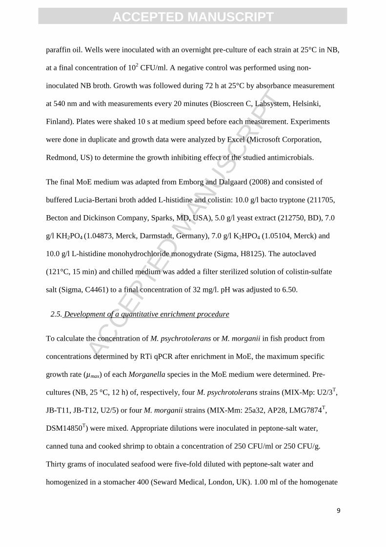
3.2 Efficiency and detection range of the RTi qPCR assay
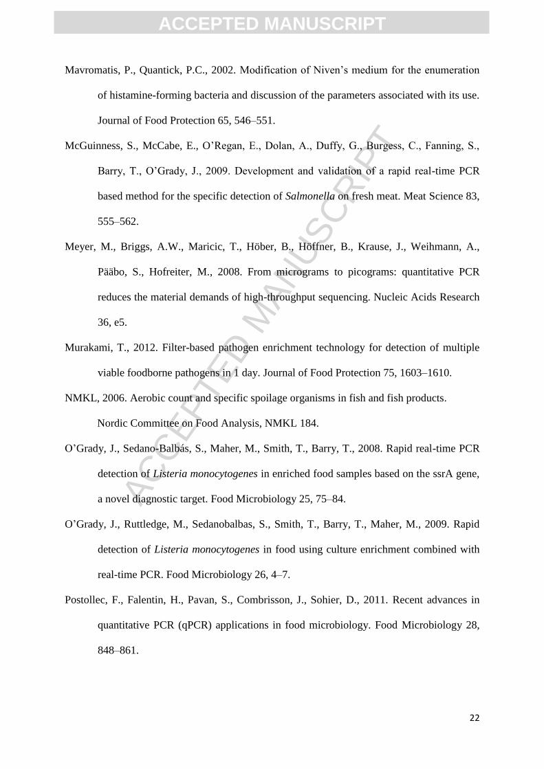
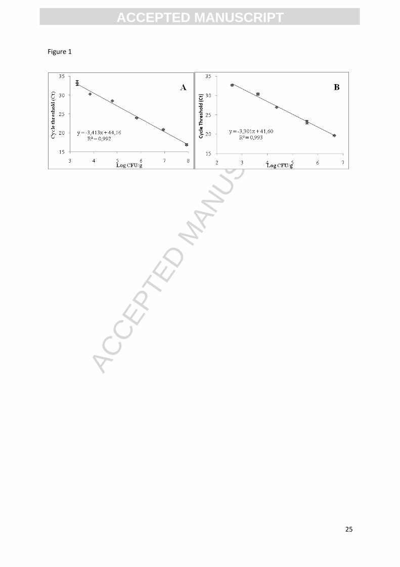
The standard curve showed a linear relation between cell concentrations (log CFU/g) and CT
values for type strains of both Morganella species in canned tuna (Fig. 1). No signal was
detected by RTi qPCR for the negative control. The linear relation was determined on a 5 log
(CFU/g) range from ca. 3 to ca. 8 log CFU/g for both species (Fig. 1). The linear relations was
CT = -3.30 x viable count (log CFU/g) + 41.60 (R²=0.99) for M. psychrotolerans, with an
efficiency of 100.8% on inoculated fish. For M. morganii the standard curve was CT = -3.413
x viable count (log CFU/g) + 44.16 (R²=0.99) with an efficiency of 96.3% (Fig. 1).
Concentrations of Morganella lower than 4 log (CFU/g) resulted in CT values higher than 31
and therefore could not be distinguished from non-specific reactions with other HPB or non-
HPB. For that reason, the lower and upper LOQ of each Morganella species in fish products
using the RTi qPCR method have been set to 4 and 8 log CFU/g respectively.
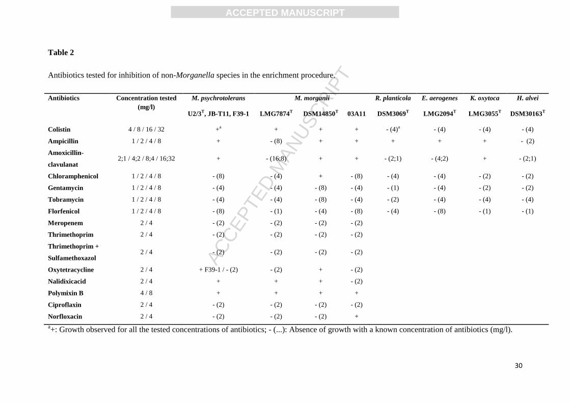
3.3 Development of an enrichment medium
The substrate 2-deoxy-D-Galactose showed an inhibition of M. psychrotolerans growth and
was not further studied. Most antibiotics, in the concentrations tested, inhibited growth of
ACC
EPTE
D M
ANU
SCR
IPT
ACCEPTED MANUSCRIPT
13
Morganella strains and had a species specific inhibitory effect on non-Morganella HPB
(Table 2). In contrast, colistin inhibited the growth of all non-Morganella HPB and
concentrations up to 32 mg/l did not inhibit growth the Morganella isolates (Table 2).
3.4 RTi qPCR quantification using an enrichment step
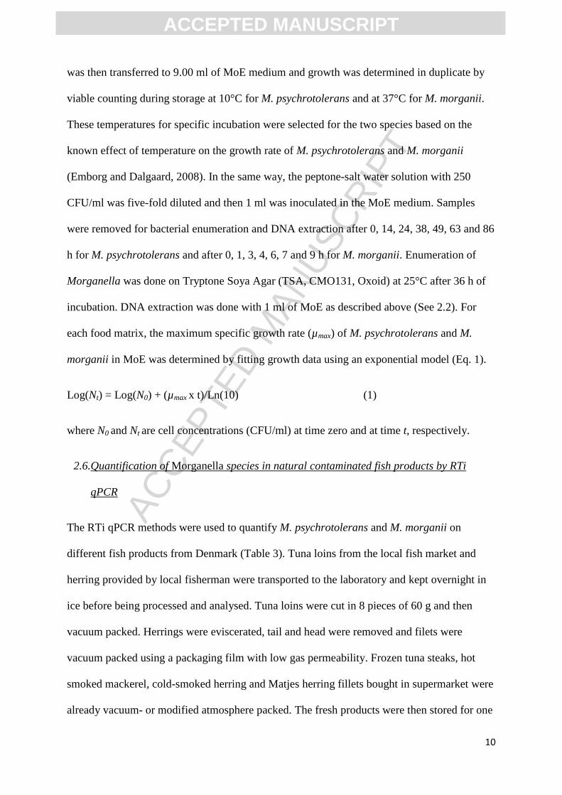
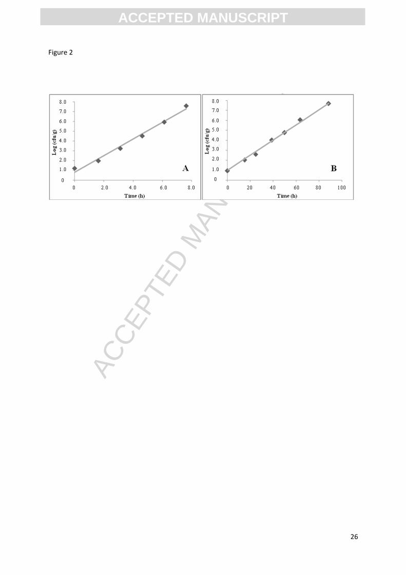
Three matrixes (peptone-salt water, canned tuna and cooked shrimp) have been used to study
the subsequent growth kinetics of Morganella strains in MoE. The µmax-values of M.
psychrotolerans in MoE at 10°C were 0.182 h-1
for MIX-Mp inoculated from peptone-salt
water, 0.180 h-1
from canned tuna and 0.180 h-1
from cooked shrimp (Fig. 2). Statistical
analysis showed no significant difference between growth rates obtained on the three matrixes
(p > 0.05), thus growth rate of M. psychrotolerans has been set to 0.180 h-1
in fish products.
The µmax-values of MIX-Mm at 37°C were 1.939 h-1
from peptone-salt water, 2.002 h-1
from
canned tuna, and 2.130 h-1
from cooked shrimp. Again, values were not significantly different
and growth rate for M. morganii has been set at 2.024 h-1
.
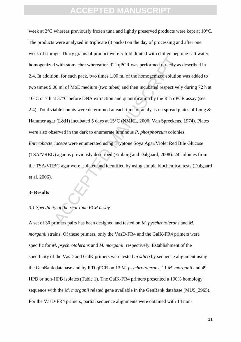
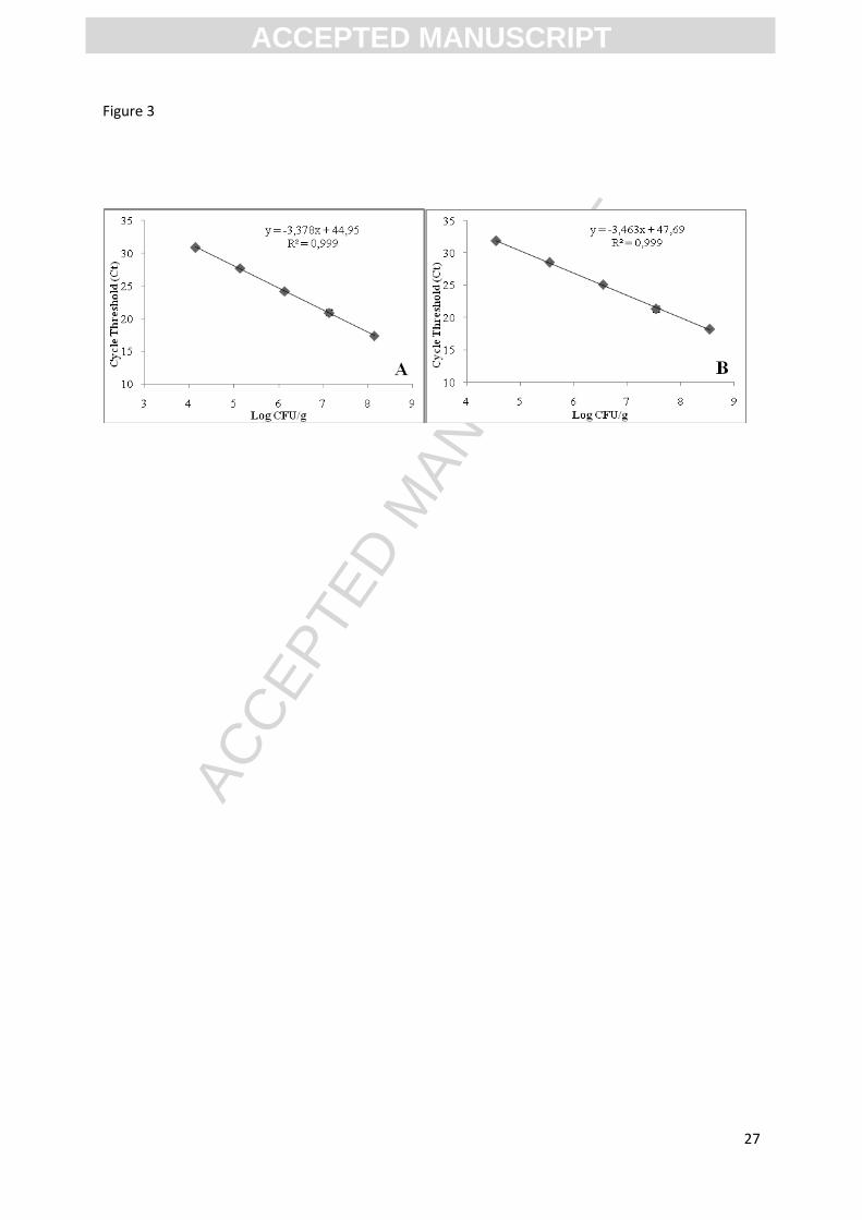
To complete the enrichment method, standard curves to determine the relation between CT
obtained by RTi qPCR and cell concentrations (log CFU/g) obtained by classical plate count
were performed for each species when growing in the MoE medium. The linear relation
determined for both species was scaled from 4.0 ± 0.5 to 8.0 ± 0.5 log CFU/ml. For M.
morganii, the equation was CT = -3.378 x viable count (log CFU/ml) + 44.95 (R²=0.99) with
an efficiency of 97.71%. For M. psychrotolerans, the equation was CT = -3.464x viable count
(log CFU/ml) + 47.69 (R²=0.99) with an efficiency of 94.54% (Fig. 3).
After enrichment, the initial cell concentration in fish is calculated from the concentration
obtained by RTi qPCR in the MoE medium and by taking into account the µmax-value of M.
psychrotolerans or M. morganii, the time of enrichment and the dilution of the fish sample
(Eq. 2):
ACC
EPTE
D M
ANU
SCR
IPT
ACCEPTED MANUSCRIPT
14
IC = CaE - ((µmax x ET) / ln10) + log(DF) (2)
where IC is the initial cell concentration in the fish product (log CFU/g), CaE the
concentration in MoE medium after enrichment as determined by RTi qPCR (log CFU/ml),
ET the enrichment time (h) and DF the total dilution factor resulting from homogenization
and transfer of the homogenate to the MoE medium.
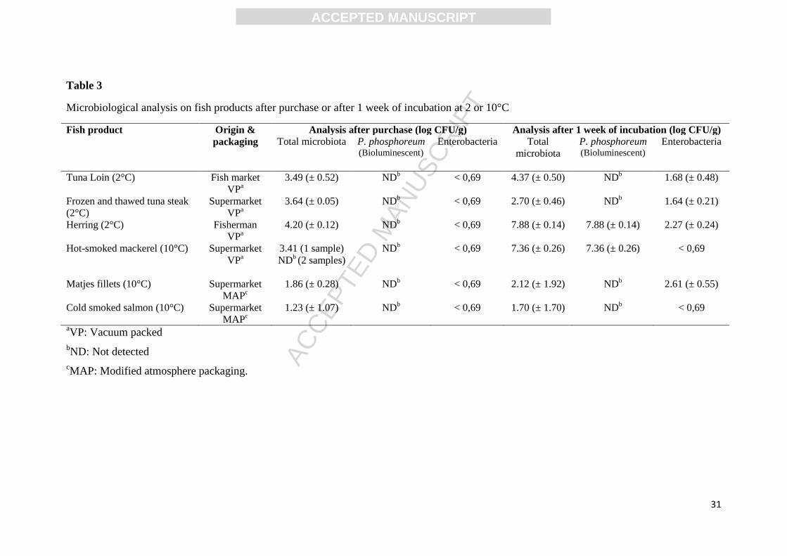
3.5 Quantification of Morganella in fish products by RTi qPCR
Just after processing of the studied seafoods, the viable counts on L&H were under 4.2 log
CFU/g for fresh fish and under 1.9 log CFU/g for lightly preserved products (Table 3). After
storage during one week this concentration increased up to 7 log CFU/g in herring fillets and
hot smoked mackerel. Most colonies on L&H agar plates were bioluminescent and the
microbiota therefore seemed to be dominated by P. phosphoreum. Viable counts of the four
others fish product remained under 4.3 log CFU/g. Enterobacteriaceae were not detected after
processing and remained under 2.6 log CFU/g after one week (Table 3).
RTi qPCR enumeration with or without the enrichment step did not allow to detect the
presence of M. psychrotolerans or M. morganii in the studied fish samples. To confirm these
results the MoE medium was plated on TSA/VRBG agar. No Enterobacteriaceae was
detected in MoE after 7 h enrichment at 37°C, confirming the absence of M. morganii in the
fish samples. Enumeration on TSA/VRBG plates after 72 h of enrichment at 10°C, for tuna
loins, herring fillets and hot smoked mackerel showed bacterial growth up to 8 log CFU/ml..
However, these bacteria on TSA/VRBG agar plates were Pseudomonas/Shewanella-like and
not Morganella/Enterobacteriaceae. These results support those obtained by RTi qPCR on
MoE medium with CT-values higher than 30 and corresponding to concentrations of M.
psychrotolerans below 5.1 log CFU/ml in the MoE medium and below 1.2 log CFU/g in
ACC
EPTE
D M
ANU
SCR
IPT
ACCEPTED MANUSCRIPT
15
herring fillets and hot smoked mackerel after storage for one week at respectively, 2°C or
10°C.
4 Discussion
After 2009, RTi qPCR methods have been developed for HPB detection and quantification in
seafood. These methods have focused on mesophilic HPB species and have not been tested
for M. psychrotolerans and P. phosphoreum (Bjornsdottir-Butler et al., 2011a, 2011b;
Ferrario et al., 2012a, 2012b). RTi qPCR methods have previously been used in combination
with an enrichment step to qualitatively detect pathogenic bacteria including Listeria
monocytogenes (O’Grady et al., 2008; O’Grady et al., 2009) and Escherichia coli (Chern et
al., 2011; Taskin et al., 2011) in food products. Spoilage bacteria responsible for sensory
defect have also been quantified by RTi qPCR, e.g. Brochotrix thermosphacta (Mamlouk et
al., 2012) and P. phosphoreum (Macé et al., 2013).
The present study focused on quantification of Morganella species in fish products as these
are strongly histamine producing bacteria and have been responsible for HFP outbreaks.
Specific and sensitive quantification methods were missing to study their occurrence and to
help management of histamine formation in seafood. Available methods are limited in
specificity or in sensitivity to obtain reliable results to improve our knowledge on these HPB.
Recently, a RTi qPCR method was developed for quantification of M. morganii by Ferrario et
al.(2012a), but this method was not tested for selectivity in relation to M. psychrotolerans
although the type strain (U2/3T) has been available in culture collections since 2007 (Emborg
et al., 2006). Primers from Ferrario et al.(2012b) were checked in silico in the present study
and might lead to unspecific reaction with M. psychorotolerans. To obtain a specific
quantification method for each Morganella species primers for M. psychrotolerans have been
designed on the vasD gene as part of the Type VI secretion system, recently discovered in

ACC
EPTE
D M
ANU
SCR
IPT
ACCEPTED MANUSCRIPT
16
Vibrio cholera by Filloux et al. (2008). In the M. psychrotolerans genome, the type VI
secretion system is composed of 17 genes which are missing on the M. morganii genome and
this allowed the developed RTi qPCR method to specifically detect the Morganella
psychrotolerans . For M. morganii, the choice of primer cites were the galactokinase gene due
to absence of this gene in M. psychrotolerans, unable to ferment D-galactose (Emborg et al.,
2006). These two pairs of primers were 100% inclusive for their target species. Without
enrichment the developed RTi qPCR methods can be used for specific detection and
quantification of Morganella species in samples with high concentrations of these bacteria,
for example fish products responsible of HFP. This is the first method develop for
quantification of M. psychrotolerans and it seems useful to increase our understanding of the
relative importance of psychrotolerant and mesophilic bacteria responsible for histamine
formation e.g. in relation to outbreaks of HFP.
Using a specific enrichment medium, the developed RTi qPCR methods may be sufficiently
sensitive to enumerate Morganella species in newly processed seafood and within the seafood
processing environment. Colistin allowed growth of Morganella and reduced growth of some
of the other well known and strongly histamine producing Enterobacteriaceae during
enrichment (Table 2). Furthermore, data for fresh herring and hot smoked mackerel indicated
that enrichment in MoE without NaCl limited growth of P. phosphoreum and thereby the
potential interference from this marine bacterium that often are present in high concentration
in fresh and lightly preserved seafood (Dalgaard et al., 1997). Nevertheless, selectivity of the
MoE medium in relation to other microorganisms that may influence growth of the
Morganella species during enrichment deserves further study.
Enrichment at 37°C for M. morganii allowed DNA extraction and quantification within one
working day. In contrast, for M. psychrotolerans, incubation at 10°C was chosen to reduce the
growth of M. morganii in the MoE medium. Clearly, the suggested enrichment and RTi qPCR
ACC
EPTE
D M
ANU
SCR
IPT
ACCEPTED MANUSCRIPT
17
method for M. psychrotolerans is not a rapid method. However, it is the only method
available for specific enumeration M. psychrotolerans and with a LOQ below 50 CFU/g in
fish products. This method seems valuable to obtain information about the occurrence and
growth of this strongly histamine producing bacteria that has been responsible for outbreaks
of HFP. M. psychrotolerans has been isolated sporadically in seafood worldwide (Emborg et
al., 2006; Macé et al., 2012; Torodo et al., 2014). Available information on its occurrence is
far from sufficient to quantitatively evaluate its contribution to the risk of histamine formation
and HFP. The enrichment and RTi qPCR method suggested in the present study has the
potential to overcome this problem.
Lag phases of the Morganella species during growth in the MoE have not been studied. For
enrichment from fresh or lightly preserved seafood lag phases are likely to be of little
practical importance. However, for frozen seafood and more preserved, like salted and/or
dried, products significant lag times may be observed during enrichment in MoE. Using eq. 2
to calculate cell concentration in fish products will result in underestimation of cell
concentrations when lag times in the MoE medium are observed and this aspect needs further
study for the suggest enrichment procedures at 10°C or 37°C. Enrichment steps have
previously been proposed to lower the detection limit of RTi qPCR methods (O’Grady et al.,
2009, Taskin et al., 2011). However in those studies, the enrichment step did not allow
quantification of the initial concentration of the bacteria detected. In the present study, the
determination of the growth rate in the enrichment media made it possible to calculate the
initial concentration from different enrichment times using Eq. (2). This approach resembles
quantitative incubation methods that are known for their ability to enumerate low
concentrations of bacteria as shown e.g. for P. phosphoreum in fish samples (Dalgaard et al.,
1996).
ACC
EPTE
D M
ANU
SCR
IPT
ACCEPTED MANUSCRIPT
18
The LOQ for the developed RTi qPCR methods was limited by the 50-fold dilution during
homogenization (x5) and inoculation of the MoE medium (x10). A direct enrichment with 30
g of fish flesh homogenized in 120 ml of MoE medium may lower the LOQ but need further
study to evaluate if the growth rate during enrichment will depend on the type of seafood e.g.
for fish products with different pH and salt content. Otherwise, a filtration method has already
shown good results on pathogenic bacteria and might be tested to increase sensitivity to 1
CFU/g of products (Murakami, 2012).
The methods developed in this study for quantification of M. psychrotolerans or M. morganii
in fish products may be used to survey occurrence of these important HPB in fish products
and to improve management of histamine formation.
5 Acknowledgments
Gaëtan Podeur was the recipient of a Ph.D fellowship from the French Ministry of Higher
Education and Research.
6 References
Alper, M.D., Ames, B.N., 1975. Positive selection of mutants with deletions of the gal-chl
region of the Salmonella chromosome as a screening procedure for mutagens that
cause deletions. Journal of Bacteriology 121, 259–266.
Aziz, R.K., Bartels, D., Best, A.A., DeJongh,M., Disz, T., Edwards, R.A., Formsma, K.,
Gerdes, S., Glass, E.M., Kubal, M., Meyer, F., Olsen, G.J., Olson, R., Osterman, A.L.,
Overbeek, R.A., McNeil, L.K., Paarmann, D., Paczian, T., Parrello, B., Pusch, G.D.,
Reich, C., Stevens, R., Vassieva, O., Vonstein, V., Wilke, A., Zagnitko, O., 2008. The
RAST Server: rapid annotations using subsystems technology. BMC Genomics 9, 75.
ACC
EPTE
D M
ANU
SCR
IPT
ACCEPTED MANUSCRIPT
19
Bjornsdottir, K., Bolton, G.E., McClellan-Green, P.D., Jaykus, L.-A., Green, D.P., 2009.
Detection of Gram-negative histamine-producing bacteria in fish: a comparative study.
Journal of Food Protection 72, 1987–1991.
Bjornsdottir-Butler, K., Jones, J.L., Benner, R., Burkhardt, W., 2011a. Development of a real-
time PCR assay with an internal amplification control for detection of Gram-negative
histamine-producing bacteria in fish. Food Microbiology 28, 356–363.
Bjornsdottir-Butler, K., Jones, J.L., Benner, R.A., Burkhardt, W., 2011b. Quantification of
total and specific gram-negative histamine-producing bacteria species in fish using an
MPN real-time PCR method. Food Microbiology 28, 1284–1292.
CDC, 2011. Outbreak Surveillance Data. http://wwwn.cdc.gov/foodborneoutbreaks/
Chern, E.C., Siefring, S., Paar, J., Doolittle, M., Haugland, R.A., 2011. Comparison of
quantitative PCR assays for Escherichia coli targeting ribosomal RNA and single copy
genes. Letters in Applied Microbiology 52, 298–306.
Dalgaard, P., Mejlholm, O., Huss, H.H., 1996. Conductance method for quantitative
determination of Photobacterium phosphoreum in fish products. Journal of Applied
Bacteriology, 81, 57-64.
Dalgaard, P., Mejlholm, O., Christiansen, T.J., Huss, H.H., 1997. Importance of
Photobacterium phosphoreum in relation to spoilage of MAP fish products. Letters in
Applied Microbiology 24, 373-378.
Dalgaard, P., Madsen, H.L., Samieian, N., Emborg, J., 2006. Biogenic amine formation and
microbial spoilage in chilled garfish (Belone belone belone) – effect of modified
atmosphere packaging and previous frozen storage. Journal of Applied Microbiology
101, 80–95.
Dalgaard, P., Emborg, J., Kjølby, A., Sørensen, N.D., Ballin, N.Z., 2008. Histamine and
biogenic amines - formation and importance in seafood. In: Børresen, T. (Ed.).
ACC
EPTE
D M
ANU
SCR
IPT
ACCEPTED MANUSCRIPT
20
Improving seafood products for the consumer. Woodhead Publishing Ltd., Cambridge,
England, pp. 292-324.
Emborg, J., Dalgaard, P., Ahrens, P., 2006. Morganella psychrotolerans sp. nov., a histamine-
producing bacterium isolated from various seafoods. International Journal of
Systematic and Evolutionnary Microbiology 56, 2473–2479.
Emborg, J., Laursen, B.G., Dalgaard, P., 2005. Significant histamine formation in tuna
(Thunnus albacares) at 2°C—effect of vacuum- and modified atmosphere-packaging
on psychrotolerant bacteria. International Journal of Food Microbiology 101, 263–
279.
Emborg, J., Dalgaard, P., 2008. Growth, inactivation and histamine formation of Morganella
psychrotolerans and Morganella morganii– development and evaluation of predictive
models. International Journal of Food Microbiology 128, 234-243.
Ferrario, C., Pegollo, C., Ricci, G., Borgo, F., Fortina, M.G., 2012a. PCR Detection and
identification of histamine-forming bacteria in filleted tuna fish samples. Journal of
Food Science 77, M115–M120.
Ferrario, C., Ricci, G., Borgo, F., Fortina, M.G., 2012b. Species-specific DNA probe and
development of a quantitative PCR assay for the detection of Morganella morganii.
Letters in Applied Microbiology 54, 292–298.
Filloux, A., Hachani, A., Bleves, S., 2008. The bacterial type VI secretion machine: yet
another player for protein transport across membranes. Microbiology 154, 1570–1583.
Helwigh, B., Korsgaard, H., Gronlund, A.J., Sorensen, A.H., Nygaard Jensen, A., Boel, J.,
BorckHog, B., 2012. Microbiological contaminants in food in the European Union in
2004-2009.Technical University of Denmark.
http://www.efsa.europa.eu/en/supporting/doc/249e.pdf
Hungerford, J.M., 2010. Scombroid poisoning: A review. Toxicon 56, 231–243.
ACC
EPTE
D M
ANU
SCR
IPT
ACCEPTED MANUSCRIPT
21
InVS (Institut de Veille Sanitaire), 2011. Surveillance des toxi-infections alimentaires
collectives. Données de la déclaration obligatoire 2011.
http://www.invs.sante.fr/Dossiers-thematiques/Maladies-infectieuses/Maladies-a-
declaration-obligatoire/Toxi-infections-alimentaires-collectives/Donnees-
epidemiologiques. Accessed on 19/02/ 2015.
Kanki, M., Yoda, T., Ishibashi, M., Tsukamoto, T., 2004. Photobacterium phosphoreum
caused a histamine fish poisoning incident. International Journal of Food
Microbiology 92, 79–87.
Kim, S.H., Price, R.J., Morrissey, M.T., Field, K.G., Wei, C.I., An, H., 2002. Histamine
production by Morganella morganii in mackerel, albacore, mahi-mahi, and salmon at
various storage temperatures. Journal of Food Science 67, 1522–1528.
Klausen, N.K., Huss, H.H., 1987. Growth and histamine production by Morganella morganii
in various temperature conditions. International Journal of Food Microbiology 5, 147–
156.
Macé, S, Cornet, J, Chevalier, F, Cardinal, M, Pilet, M-F, Dousset, X, Joffraud, J-J., 2012.
Characterisation of the spoilage microbiota in raw salmon (Salmo salar) steaks stored
under vacuum or modified atmosphere packaging combining conventional methods
and PCR-TTGE. Food Microbiology. 30, 164 –172.
Macé, S., Mamlouk, K., Chipchakova, S., Prévost, H., Joffraud, J.-J., Dalgaard, P., Pilet, M.-
F., Dousset, X., 2013. Development of a rapid real-Time PCR method as a tool to
quantify viable Photobacterium phosphoreum in salmon (Salmo salar) steaks. Applied
and Environnemental Microbiology 79, 2612-2619.
Mamlouk, K., Macé, S., Guilbaud, M., Jaffrès, E., Ferchichi, M., Prévost, H., Pilet, M.-F.,
Dousset, X., 2012. Quantification of viable Brochothrix thermosphacta in cooked
shrimp and salmon by real-time PCR. Food Microbiology 30, 173–179.
ACC
EPTE
D M
ANU
SCR
IPT
ACCEPTED MANUSCRIPT
22
Mavromatis, P., Quantick, P.C., 2002. Modification of Niven’s medium for the enumeration
of histamine-forming bacteria and discussion of the parameters associated with its use.
Journal of Food Protection 65, 546–551.
McGuinness, S., McCabe, E., O’Regan, E., Dolan, A., Duffy, G., Burgess, C., Fanning, S.,
Barry, T., O’Grady, J., 2009. Development and validation of a rapid real-time PCR
based method for the specific detection of Salmonella on fresh meat. Meat Science 83,
555–562.
Meyer, M., Briggs, A.W., Maricic, T., Höber, B., Höffner, B., Krause, J., Weihmann, A.,
Pääbo, S., Hofreiter, M., 2008. From micrograms to picograms: quantitative PCR
reduces the material demands of high-throughput sequencing. Nucleic Acids Research
36, e5.
Murakami, T., 2012. Filter-based pathogen enrichment technology for detection of multiple
viable foodborne pathogens in 1 day. Journal of Food Protection 75, 1603–1610.
NMKL, 2006. Aerobic count and specific spoilage organisms in fish and fish products.
Nordic Committee on Food Analysis, NMKL 184.
O’Grady, J., Sedano-Balbás, S., Maher, M., Smith, T., Barry, T., 2008. Rapid real-time PCR
detection of Listeria monocytogenes in enriched food samples based on the ssrA gene,
a novel diagnostic target. Food Microbiology 25, 75–84.
O’Grady, J., Ruttledge, M., Sedanobalbas, S., Smith, T., Barry, T., Maher, M., 2009. Rapid
detection of Listeria monocytogenes in food using culture enrichment combined with
real-time PCR. Food Microbiology 26, 4–7.
Postollec, F., Falentin, H., Pavan, S., Combrisson, J., Sohier, D., 2011. Recent advances in
quantitative PCR (qPCR) applications in food microbiology. Food Microbiology 28,
848–861.
ACC
EPTE
D M
ANU
SCR
IPT
ACCEPTED MANUSCRIPT
23
Prester, L., 2011. Biogenic amines in fish, fish products and shellfish: a review. Food
additives & contaminants: Part A 28, 1547–1560.
Takahashi, H., Kimura, B., Yoshikawa, M., Fujii, T., 2003. Cloning and sequencing of the
histidine decarboxylase genes of gram-negative, histamine-producing bacteria and
their application in detection and identification of these organisms in fish. Applied and
Environmental Microbiology 69, 2568–2579.
Tao, Z., Sato, M., Abe, N., Yamaguchi, T., Nakano, T., 2009. Simple and rapid detection of
histamine-forming bacteria by differential agar medium. Food Control 20, 903–906.
Taskin, B., Gozen, A.G., Duran, M., 2011. Selective quantification of viable Escherichia coli
bacteria in biosolids by quantitative PCR with propidium monoazide modification.
Applied and Environmental Microbiology 77, 4329–4335.
Torido, Y., Ohshima, C., Takahashi, H., Miya, S., Iwakawa, A., Kuda, T., Kimura, B., 2014.
Distribution of psychrotolerant and mesophilic histamine-producing bacteria in retail
fish in Japan. Food Control 46, 328-342.
Untergasser, A., Nijveen, H., Rao, X., Bisseling, T., Geurts, R., Leunissen, J.A.M., 2007.
Primer3Plus, an enhanced web interface to Primer3. Nucleic Acids Research 2007 35,
W71-W74.
Van Spreekens, K.J.A., 1974. The suitability of a modification of long and hammer’s medium
for the enumeration of more fastidious bacteria from fresh fishery products. Archiv für
Lebensmittel hygiene 10, 213-219.
Veciana-Nogués, M.T., Bover-Cid, S., Mariné-Font, A., Vidal-Carou, M.C., 2004. Biogenic
amine production by Morganella morganii and Klebsiella oxytoca in tuna. European
Food Research and Technology 218, 284–288.
ACC
EPTE
D M
ANU
SCR
IPT
ACCEPTED MANUSCRIPT
24
Figure 1: Standard curve showing the relationship between CT values and log CFU/g for
serially diluted culture of M. morganii (A) and M. psychrotolerans (B) inoculated on canned
tuna.
Figure 2: Growth of M. morganii (A) and M. psychrotolerans (B) respectively at 37°C and
10°C in MoE medium followed by plate count on TSA agar as a function of the time. The line
represents a fitted exponential growth model.
Figure 3: Standard curve showing the liner relationship between CT values and bacterial
enumeration (log CFU/ml) of serially diluted M. morganii (A) and M. psychrotolerans (B) in
MoE medium.
ACC
EPTE
D M
ANU
SCR
IPT
ACCEPTED MANUSCRIPT
25
Figure 1
ACC
EPTE
D M
ANU
SCR
IPT
ACCEPTED MANUSCRIPT
26
Figure 2
ACC
EPTE
D M
ANU
SCR
IPT
ACCEPTED MANUSCRIPT
27
Figure 3
ACC
EPTE
D M
ANU
SCR
IPT
ACCEPTED MANUSCRIPT
28
Table 1
Morganella strains and non-Morganella species used in this study.
Bacteria Strains Histamine
production
Collection Origin
Morganella psychrotolerans U2 /3 T(CIP
109403T)
U2/4
U2/5
U2/6
U2/7
FD24
1F10
2F6
JB-T11
JB-T12
JB-T16
F39-1
F39-3
Higha
DTU Food and
CIP1
DTU Food
DTU Food
DTU Food
DTU Food
DTU Food
DTU Food
DTU Food
DTU Food
DTU Food
DTU Food
DTU Food
DTU Food
Cold-smoked tuna
Cold-smoked tuna
Cold-smoked tuna
Cold-smoked tuna
Cold-smoked tuna
Fresh Vacuum-packed tuna
Garfish
Garfish
Fresh Vacuum-packed tuna
Fresh Vacuum-packed tuna
Fresh Vacuum-packed tuna
Lumpfish roe
Lumpfish roe
Morganella morganii LMG7874T
DSM14850T
25a32
M04090
U6/1
NCIMB865
AP28
03A11
03B10
HB2810
HB2811
Higha
BCCM/LMG2
DSMZ3
DTU Foodb
DTU Foodb
DTU Food
NCIMB4
DTU Foodb
DTU Foodb
DTU Foodb
Ifremer
Ifremer
Human stool
Human sputum
Tuna
Clinical isolate
Tuna
Spoiled fish
Yellowtail
Smoked fish
Smoked fish
Sardine
Sardine
Hafnia alvei DSM30163T
MIP2438
MIP2439
MIP2461
MIP2467
MIP2468
MIP2613
MIP2625
Higha
DSMZ3
Ifremer/Oniris
Ifremer/Oniris
Ifremer/Oniris
Ifremer/Oniris
Ifremer/Oniris
Ifremer/Oniris
Ifremer/Oniris
NCb
Spoiled fresh salmon
Spoiled fresh salmon
Spoiled fresh salmon
Spoiled fresh salmon
Spoiled fresh salmon
Spoiled fresh salmon
Spoiled fresh salmon
Raoultella planticola DSM3069T Higha DSMZ3 Radish root
Photobacterium phosphoreum CCUG16288
1D5
FR7
EBP3067
EBP3079
MIP2560
MIP2562
MIP2588
MIP2591
MIP2613
Higha
CCUG5
DTU Food
DTU Food
Ifremer/Oniris
Ifremer/Oniris
Ifremer/Oniris
Ifremer/Oniris
Ifremer/Oniris
Ifremer/Oniris
Ifremer/Oniris
Etelismarshi
Garfish
Fresh Vacuum-packed cod
Salmon
Salmon
Salmon
Spoiled fresh salmon
Spoiled fresh salmon
Spoiled fresh salmon
Spoiled fresh salmon
Photobacterium damselae CIP102761T Higha CIP1 Damselfish
Enterobacter aerogenes LMG2094T
SF1469
SF1470 Higha
BCCM/LMG2
Ifremer
Ifremer
Sputum
Smoked salmon
Smoked salmon
Klebsiella oxytoca LMG3055T Higha BCCM/LMG2 NCb
Enterococcus faecalis CIP105042 Lowa CIP1 Poultry
Pseudomonas spp. HB2843 Lowa Ifremer Tuna
Raoultella ornithinolytica CIP103364T Higha CIP1 Human urine
Serratia liquefaciens SF1465 Lowa Ifremer Smoked salmon
Staphylococcus xylosus DSMZ20029 Lowa DSMZ3 Fermented meat product
ACC
EPTE
D M
ANU
SCR
IPT
ACCEPTED MANUSCRIPT
29
Listeria monocytogenes RF190 Lowa Ifremer Shrimp
Shewanella putrefaciens CIP6929
RF47
RF49
Lowa CIP1
Ifremer
Ifremer
NCb
Vacuum-packed trout
Cod fillets
Psychrobacter spp. CCUG 42949
EBP3029
Lowa CCUG5
Ifremer
Desalted cod
Salmon
Psychrobacter aquaticus MIP2412 Lowa Ifremer/Oniris Spoiled fresh salmon
Acinetobacter spp. EBP3044 Lowa Ifremer/Oniris Salmon
Vibrio cholerae RF184 Lowa Ifremer Cooked shrimp
Carnobacterium maltaromaticum V1 Lowa Ifremer/Oniris Fish
Vibrio parahemolyticus RF179 Lowa Ifremer Water
Enterococcus faecium CIP 54.33 Lowa CIP1 Canned fish
Brochotrix thermosphacta EBP3084 Lowa Ifremer/Oniris Salmon
Carnobacterium jeotgali KCTC 13251 Lowa KCTC6 Shrimp
Leuconostoc gelidum LHIS2959 Lowa Ifremer/Oniris Mackerel fillets
Carnobacterium divergens V41 Lowa Ifremer/Oniris Fish viscera
Escherichia coli CIP 76.24 Lowa CIP1 NCb
Lactobacillus curvatus LHIS2886 Lowa Ifremer/Oniris Fresh sardine
Lactobacillus fuchuensis LHIS2997 Lowa Ifremer/Oniris Fresh mackerel
Lactobacillus sakei LHIS2855 Lowa Ifremer/Oniris Fresh sardine a High: Production of histamine between 200-5000 mg/l in histidine broth. Low: 0 to 200 mg/l of histamine in histidine.
broth.
b See Emborg et al. (2006) for the origin and isolation of these isolates. NC: Not communicated
1CIP : Collection de l’Institut Pasteur ; 2BCCM/LGM : Belgian Coordinated Collections of Microorganisms, 3DSMZ :
Deutsche Sammlung von Mikroorganismen und Zellkulturen; 4NCIMB : National Collections of Industrial, Marine and Food
Bacteria, 5CCUG : Culture Collection, University of Göteborg, Sweden, 6KCTC : Korean Collection for Type Cultures.
,.
ACC
EPTE
D M
ANU
SCR
IPT
ACCEPTED MANUSCRIPT
30
Table 2
Antibiotics tested for inhibition of non-Morganella species in the enrichment procedure.
Antibiotics Concentration tested
(mg/l)
M. psychrotolerans M. morganii R. planticola E. aerogenes K. oxytoca H. alvei
U2/3T, JB-T11, F39-1 LMG7874
T DSM14850
T 03A11 DSM3069
T LMG2094
T LMG3055
T DSM30163
T
Colistin 4 / 8 / 16 / 32 +a
+ + + - (4)a
- (4) - (4) - (4)
Ampicillin 1 / 2 / 4 / 8 + - (8) + + + + + - (2)
Amoxicillin-
clavulanat 2;1 / 4;2 / 8;4 / 16;32 + - (16;8) + + - (2;1) - (4;2) + - (2;1)
Chloramphenicol 1 / 2 / 4 / 8 - (8) - (4) + - (8) - (4) - (4) - (2) - (2)
Gentamycin 1 / 2 / 4 / 8 - (4) - (4) - (8) - (4) - (1) - (4) - (2) - (2)
Tobramycin 1 / 2 / 4 / 8 - (4) - (4) - (8) - (4) - (2) - (4) - (4) - (4)
Florfenicol 1 / 2 / 4 / 8 - (8) - (1) - (4) - (8) - (4) - (8) - (1) - (1)
Meropenem 2 / 4 - (2) - (2) - (2) - (2)
Thrimethoprim 2 / 4 - (2) - (2) - (2) - (2)
Thrimethoprim +
Sulfamethoxazol 2 / 4 - (2) - (2) - (2) - (2)
Oxytetracycline 2 / 4 + F39-1 / - (2) - (2) + - (2)
Nalidixicacid 2 / 4 + + + - (2)
Polymixin B 4 / 8 + + + +
Ciproflaxin 2 / 4 - (2) - (2) - (2) - (2)
Norfloxacin 2 / 4 - (2) - (2) - (2) +
a+: Growth observed for all the tested concentrations of antibiotics; - (...): Absence of growth with a known concentration of antibiotics (mg/l).
ACC
EPTE
D M
ANU
SCR
IPT
ACCEPTED MANUSCRIPT
31
Table 3
Microbiological analysis on fish products after purchase or after 1 week of incubation at 2 or 10°C
Fish product
Origin &
packaging
Analysis after purchase (log CFU/g) Analysis after 1 week of incubation (log CFU/g)
Total microbiota
P. phosphoreum (Bioluminescent)
Enterobacteria
Total
microbiota
P. phosphoreum (Bioluminescent)
Enterobacteria
Tuna Loin (2°C) Fish market
VPa
3.49 (± 0.52) NDb
< 0,69 4.37 (± 0.50) NDb
1.68 (± 0.48)
Frozen and thawed tuna steak
(2°C)
Supermarket
VPa
3.64 (± 0.05) NDb
< 0,69 2.70 (± 0.46) NDb
1.64 (± 0.21)
Herring (2°C) Fisherman
VPa
4.20 (± 0.12) NDb
< 0,69 7.88 (± 0.14) 7.88 (± 0.14) 2.27 (± 0.24)
Hot-smoked mackerel (10°C) Supermarket
VPa
3.41 (1 sample)
NDb (2 samples)
NDb
< 0,69 7.36 (± 0.26) 7.36 (± 0.26) < 0,69
Matjes fillets (10°C) Supermarket
MAPc
1.86 (± 0.28) NDb
< 0,69 2.12 (± 1.92) NDb
2.61 (± 0.55)
Cold smoked salmon (10°C) Supermarket
MAPc
1.23 (± 1.07) NDb
< 0,69 1.70 (± 1.70) NDb
< 0,69
aVP: Vacuum packed
bND: Not detected
cMAP: Modified atmosphere packaging.