Embed Size (px)
Citation preview

Department of Medicinal Biochemistry and Biophysics
Karolinska Institute, Stockholm, Sweden
DEVELOPMENT OF A NEW PNA ANALOGUE AS A POTENTIAL
ANTISENSE DRUG AND TOOL FOR LIFE-SCIENCE STUDIES
Andis Slaitas
Stockholm 2004

All previously published papers were reproduced with permission from the publisher. Published and printed by Karolinska University Press Box 200, SE-171 77 Stockholm, Sweden © Andis Slaitas, 2004 ISBN 91-7349-642-1

3
ABSTRACT The work described in this thesis focuses on applying synthetic organic chemistry methods (supported by modern synthesizers and analytical techniques) for the preparation of peptides and modified nucleic acids in order to affect certain properties in these biomolecules, which can lead to their application as drugs and/or tools for life sciences studies. The first part of the thesis reports the design and synthesis of a novel pyrrolidine-based peptide nucleic acid (PNA). Two enantiomers of the chiral pyrrolidine-containing unit were chemically synthesized and further incorporated into PNA fragments using modern automated solid-phase assembly methods. By comparison of their binding affinities to both DNA and RNA targets, it was found that such PNAs, due to their enhanced rigidity are able to recognize and bind to the complementary RNA strands with significantly larger affinity than to the complementary DNA. These findings may be utilized in the development of RNA-specific molecular probes, binding assays for cell extracts that contain mixtures of RNA and DNA, or targeting of viral RNAs, i.e. act as basis for the development of antisense drugs. With the rapid advancement of biochemistry and related sciences, there is an increasing demand for synthetic peptides. The development and application of a new type of peptide-coupling agent is described. This coupling agent is superior in some ways to the most commonly used commercial agents, since peptides obtained by its action are generally of higher enantiopurity, i.e. a significant reduction of racemization of the amino acid during coupling is obtained. The potential use of this agent in linking peptide segments has been studied and discussed. So called ‘segment coupling’ becomes economically valuable when either large synthetic peptides are to be made, or when a number of peptides, consisting mainly of highly conserved parts with differences in only a few amino acid residues, are desired. Alternatively, this coupling agent could be used for solid phase synthesis of PNA sequences containing racemization-prone units. As a spin-off from the above project, new sulfilimine derivatives of natural nucleosides have been successfully synthesized, isolated, characterized and studied. The synthesis of deoxycytidine and deoxyadenosine sulfilimine dimethyl and diphenyl sulfilimines is reported and the results of tests of their stability under a variety of conditions – mostly those relevant to oligonucleotide chemistry are discussed. The results of these stability studies lead to the conclusion that the sulfilimine group can serve as UV-detectable labels for short oligonucleotides. This label can be either transient or permanent (dimethyl or diphenyl sulfilimine, respectively) and can simplify the detection of tagged oligonucleotides during their purification and in assays, which can otherwise be a complicated task. In the final part of this thesis it is demonstrated how a sequence-specific PNA clamp is used to suppress a dsDNA-digesting enzyme Exonuclease III. The rate of the enzymatic degradation has been studied in different environments and added-PNA concentrations. It was found that PNA indeed specifically suppresses the action of the enzyme, in addition to non-specific inhibition. This, previously non-documented finding, helps to better understand the mechanism of action of these enzymatic processes, as well as having potential applications within both DNA sequencing and gene therapy.

4
LIST OF PUBLICATIONS
I
Andis Slaitas, Esther Yeheskiely. Synthesis and Hybridization of Novel
Chiral Pyrrolidine Based PNA Analogue. Nucleosides, Nucleotides & Nucleic
Acids 2001, 20, 1377–1379.
II
Andis Slaitas, Esther Yeheskiely. A Novel N-(Pyrrolidinyl-2-methyl)glycine-
Based PNA with a Strong Preference for RNA over DNA. Eur. J. Org. Chem.
2002, 2391-2399.
III Fast and Efficient Peptide Bond Formation Using bis-[α,α-bis(trifluoromethyl)-
benzyloxy]diphenylsulfur. Part I. J. Peptide Res. 2002, 60, 283-291.
IV Synthesis and Reactivity of Nucleoside Sulfilimines. Phosphorus, Sulfur, and
Silicon 2004, 179, 153–171.
V
Andis Slaitas, Charlotte Ander, Zeno Földes-Papp, Rudolf Rigler, Esther
Yeheskiely. Suppression of Exonucleolytic Degradation of Double-Stranded
DNA and Inhibition of Exonuclease III by PNA. Nucleosides, Nucleotides &
Nucleic Acids 2003, 22, 1603-1605.

5
TABLE OF CONTENTS
ABSTRACT................................................................................................................. 3
LIST OF PUBLICATIONS........................................................................................ 4
TABLE OF CONTENTS............................................................................................ 5
LIST OF ABBREVIATIONS .................................................................................... 7
1. GENERAL INTRODUCTION.......................................................................... 9 NUCLEIC ACIDS .......................................................................................................... 9
Native nucleic acids - DNA and RNA ................................................................... 9 PNA – an artificial nucleic acid.......................................................................... 10 PNA Synthesis ..................................................................................................... 11 Properties of PNA ............................................................................................... 15 Advantages and disadvantages of PNA compared to native nucleic acids......... 17 PNA modifications .............................................................................................. 18 Applications of PNA............................................................................................ 21
2. N-(PYRROLIDINYL-2-METHYL)GLYCINE-BASED PNA (PAPERS I AND II)............................................................................................................... 22
Synthesis of monomers ........................................................................................ 23 PNA synthesis...................................................................................................... 27 Purification ......................................................................................................... 31 UV thermal melting experiments......................................................................... 31 Summary.............................................................................................................. 32
3. SULFURANE-MEDIATED PEPTIDE BOND FORMATION (PAPER III) ………………………………………………………………………………….34
Peptide coupling agents ...................................................................................... 34 Racemization during peptide coupling ............................................................... 35 BTBDS - Martin Sulfurane Dehydrating agent................................................... 35 Yield optimisation of BTBDS couplings.............................................................. 36 Racemization in BTBDS-mediated coupling....................................................... 38 Proposed mechanism of action of BTBDS .......................................................... 41 Summary.............................................................................................................. 43
4. NUCLEOSIDE SULFILIMINES (PAPER IV).............................................. 45
Sulfilimines.......................................................................................................... 45 Synthesis of sulfilimines ...................................................................................... 46 Synthesis of nucleoside sulfilimines .................................................................... 47 Synthesis of a sulfilimine-containing dinucleotide.............................................. 49 Stability of nucleoside sulfilimines...................................................................... 50 Summary.............................................................................................................. 54
5. SUPPRESSION OF EXONUCLEOLYTIC DEGRADATION OF DOUBLE-STRANDED DNA AND INHIBITION OF EXONUCLEASE III BY PNA (PAPER V) ......................................................................................... 56
Synthesis of PNA ................................................................................................. 57

6
UV thermal melting experiments......................................................................... 60 Enzymatic degradation studies ........................................................................... 61 Summary.............................................................................................................. 62
CONNECTIONS AND CONCLUSIONS ............................................................... 63
ACKNOWLEDGEMENTS...................................................................................... 64
REFERENCES.......................................................................................................... 65

7
LIST OF ABBREVIATIONS Ac Acetyl Ade Adenine (6-aminopurine) Aeg N-(2-Aminoethyl)glycine All Allyl Alloc Allyloxycarbonyl Base Heterocyclic nucleobase (where appropriate) Bhoc Benzhydryloxycarbonyl (diphenylmethyloxycarbonyl) Boc t-Butyloxycarbonyl BTBDS [α,α-bis(trifluoromethyl)benzyloxy]diphenylsulfur Bz Benzoyl Bzl Benzyl Cbz Benzyloxycarbonyl Cyt Cytosine (4-aminopyrimidin-2one) DCA Dichloroacetic acid DCE 1,2-Dichloroethane DCM Dichloromethane DIEA Diisopropylethylamine (Hünig’s base) DMF N,N-Dimethylformamide DMT Dimethoxytrityl [bis(4-methoxyphenyl)(phenyl)methyl] DNA Deoxyribonucleic acid ds Double-stranded Gua Guanine (2-aminopurin-6-one) Fmoc 9-Fluorenylmethyloxycarbonyl HATU O-(7-Azabenzotriazol-1-yl)-N,N,N´,N´-tetramethyluronium
hexafluorophosphate HBTU O-(Benzotriazol-1-yl)-N,N,N´,N´-tetramethyluronium
hexafluorophosphate HMBA 4-Hydroxymethylbenzoic acid HOBt 1-Hydroxybenztriazole LNA ‘Locked’ nucleic acid MMT Monomethoxytrityl [(4-methoxyphenyl)(diphenyl)methyl] Ms Mesyl (methanesulfonyl) NCS N-Chlorosuccinimide PG Protecting group Pmg Pyrrolidin-2-yl methyl glycine PNA Peptide (polyamide) nucleic acid PS Polystyrene (as in ‘PS solid support’) PyBroP® Bromotripyrrolidinophosphonium hexafluorophosphate RNA Ribonucleic acid RP Reversed-phase ss Single-stranded TEA Triethylamine TEAB Triethylammonium bicarbonate Tf Trifluoromethanesulfonyl (triflyl) TFA Trifluoroacetic acid THF Tetrahydrofuran Thy Thymine (5-methylpyrimidin-2,4-dione) Trt Trityl (triphenylmethyl) Ts 4-Methylbenzenesulfonyl (tosyl)

8

9
1. GENERAL INTRODUCTION
Nucleic acids
Native nucleic acids - DNA and RNA
DNA and RNA – carriers of the genetic information, have fascinated scientists for
now nearly a century.
Nucleic acids are built from repeating units – nucleotides, which consist of a
phosphorylated sugars (ribose or deoxyribose) attached to a heterocycle – adenine,
cytosine, guanine and thymine or uracil. Nucleotides are linked as phosphate diesters
to form the chains of DNA or RNA (Figure 1.1).
BaseO
O
O
OH
O
O
P
BaseO
OH
O
O-OH P
R
R
Nucleobases
(deoxy)ribose
phosphatediester linker
R = H deoxyribose (DNA)R = OH ribose (RNA)
5´
3´
NH
NH
O
O
RN
NH
O
NH2
N
N
N
NH
NH2
NH
N
N
NH
O
NH2
Cytosine Uracil (R = H)Thymine (R = Me)
Adenine Guanine
Figure 1.1. Chemical structure of DNA (RNA).
The chemical structure alone cannot be credited for the unique properties of nucleic
acids. In the first half of the 20th century it was found that the amount of adenine in
DNA is equal to the amount of thymine, and the amount of cytosine to that of
guanine. This important finding made perfect sense after the proposed DNA structure
by Watson and Crick in 1953. By their model the DNA structure is a double helix,
formed from two nucleic acid strands with the heterocyclic nucleobases positioned
inside the helices and sugar phosphates on the outside. The structure is held together

10
by hydrogen bond interactions of the heterocyclic bases. These interactions are the
strongest between the adenine-thymine pair and the cytosine-guanine pair (
Figure 1.2).
This ability of nucleic acids to form duplexes with their complementary counterparts
is the key to the storage and replication of the genetic information.
N
N
O
OR
H
N
N
N
N
NH
H R
T
AN
N
N
N
O
NHH
H
R
N
N
O
NHH
R
G
C
Figure 1.2. H-bonding in nucleobases leading to Watson-Crick complementarity. R = ribose or deoxyribose
PNA – an artificial nucleic acid
Peptide nucleic acids were first described by P. E. Nielsen et al. in 1991.1 PNA was
designed by a computer-assisted modelling of triple helices. The third strand (also
known as the Hoogsteen strand) was stripped of its deoxyribose backbone and a new
– pseudopeptidic N-(2-aminoethyl)glycine (Aeg) backbone was constructed, thus
obtaining a DNA mimic, which is not charged and achiral. The nucleobases are
attached to the Aeg backbone via a methylene carbonyl linkage (Figure 1.3).

11
BaseO
O
O
OH
O
O
P
BaseO
OH
O
O-OH P
Base
N
NH
CH2
O
CH2
Base
N
NH2
O
OH
O
O
NH2
N
NH
O
Base
O
N
OH
O
Base
O
DNA PNA PNA
Figure 1.3. The chemical structure of PNA and its similarity to DNA.
PNA Synthesis
PG1NH
NOH
OO
BasePG2
Figure 1.4. General structure of a PNA monomer
A PNA monomer consists of N-protected (2-aminoethyl)glycine (PG1) to which a
protected nucleobase (PG2) is attached. These two protecting groups have to be
orthogonal i.e. PG2 must be stable to the conditions used to remove PG1 (Figure 1.4).
There are several combinations of protecting groups reported for the PNA synthesis,
and the most commonly used are summarized in Table 1.1.
12
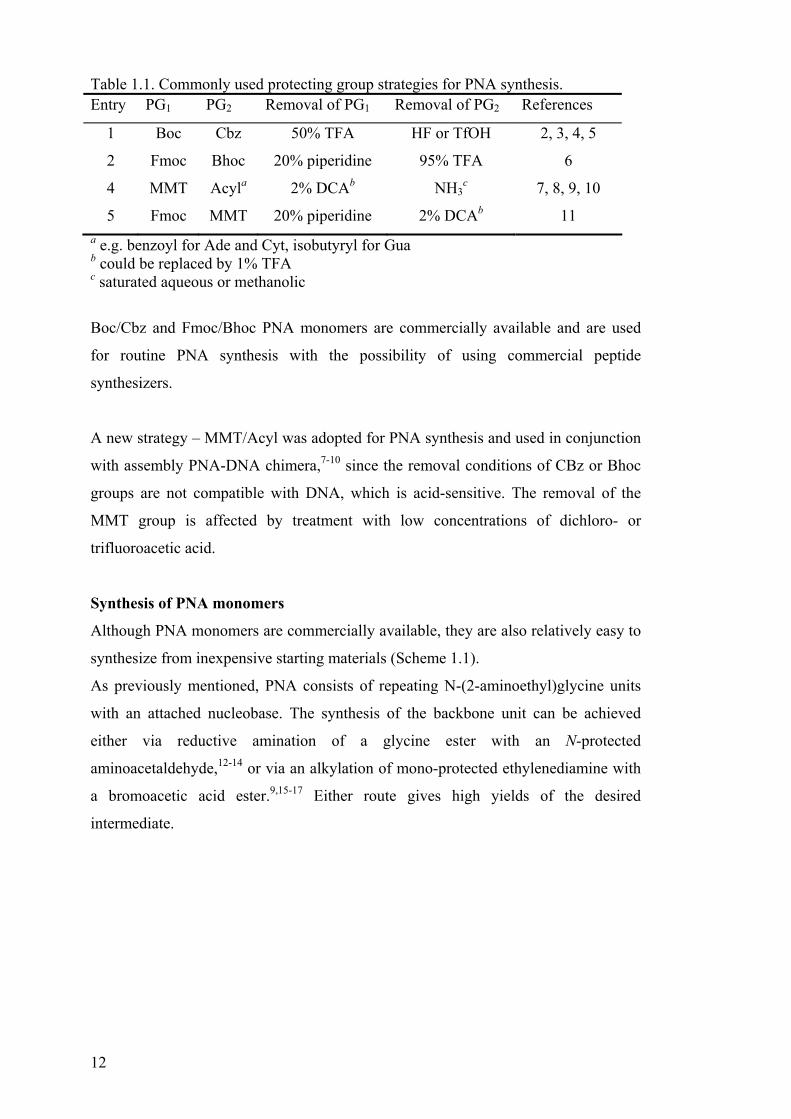
Table 1.1. Commonly used protecting group strategies for PNA synthesis. Entry PG1 PG2 Removal of PG1 Removal of PG2 References
1 Boc Cbz 50% TFA HF or TfOH 2, 3, 4, 5
2 Fmoc Bhoc 20% piperidine 95% TFA 6
4 MMT Acyla 2% DCAb NH3c 7, 8, 9, 10
5 Fmoc MMT 20% piperidine 2% DCAb 11 a e.g. benzoyl for Ade and Cyt, isobutyryl for Gua b could be replaced by 1% TFA c saturated aqueous or methanolic
Boc/Cbz and Fmoc/Bhoc PNA monomers are commercially available and are used
for routine PNA synthesis with the possibility of using commercial peptide
synthesizers.
A new strategy – MMT/Acyl was adopted for PNA synthesis and used in conjunction
with assembly PNA-DNA chimera,7-10 since the removal conditions of CBz or Bhoc
groups are not compatible with DNA, which is acid-sensitive. The removal of the
MMT group is affected by treatment with low concentrations of dichloro- or
trifluoroacetic acid.
Synthesis of PNA monomers
Although PNA monomers are commercially available, they are also relatively easy to
synthesize from inexpensive starting materials (Scheme 1.1).
As previously mentioned, PNA consists of repeating N-(2-aminoethyl)glycine units
with an attached nucleobase. The synthesis of the backbone unit can be achieved
either via reductive amination of a glycine ester with an N-protected
aminoacetaldehyde,12-14 or via an alkylation of mono-protected ethylenediamine with
a bromoacetic acid ester.9,15-17 Either route gives high yields of the desired
intermediate.

13
NH
OPG + NH2
OR
O
NH
NH
PGOR
O
NH
NH2PG + BrOR
O
Na(CN)BH3
Et3N
Scheme 1.1. Synthesis of protected (2-aminoethyl)glycine
In the next step protected nucleobases are attached via an amide bond using a
coupling agent, e.g. DCC18 or HBTU.19 (Scheme 1.2).
NH
NH
PG1OR
O
+ BaseOH
OPG2
NH
NPG1OR
OO
BasePG2
HBTU
Scheme 1.2. Synthesis of Aeg-PNA monomers
Solid phase PNA synthesis
The assembly of PNA, using either of the above-described monomers, is usually
performed on a solid support, preferably via automated synthesis.
The solid support is usually functionalised polystyrene to which an N-protected
amino acid (e.g. glycine) is attached via its C-terminus to a cleavable linker. It is not
advisable to link the PNA monomers directly onto the linker since, due to the
potential formation of a ketopiperazine during the first deprotection step, the support
may get de-functionalised.20

14
LinkerO
O
NPS
O
Base
NH2
LinkerOH PS
NH
N
O
O
Base
+
Scheme 1.3. De-functinalization of support due to cyclization.
The choice of the linker is very important and depends on the protecting group
strategy used. For instance, a Wang linker [4-(hydroxylmethyl)phenol)] is used for
Fmoc/Bhoc chemistry and the product is cleaved from the resin by acid treatment, for
instance using TFA,21 while a more acid stable HMBA [4-(hydroxymethyl)benzoic
acid] linker is used for MMT/Acyl chemistry.9,22
The loading of the solid support can range from 30 µM/g to >1 mM/g. Low loading
resins are usually used in the synthesis of PNA-DNA chimera.9
The PNA synthesis cycle consists of the following operations:
• Chain elongation (coupling step)
• Wash
• Removal of the transient protecting group
• Wash
The chain elongation is the critical step in the PNA assembly. The efficiency of the
elongation step, and thus the choice of the coupling agent, is crucial for the overall
yield and purity of the desired PNA, especially when somewhat longer fragments are
to be assembled.
HATU is probably the most commonly used coupling agent for PNA synthesis. It is
reported to give better than 99% coupling yields in a relatively short time (15-30
min).23
In our work we used the less expensive and more available coupling agent HBTU and
still managed to maintain the same efficiency.
The intermediate wash steps are necessary for the removal of unreacted monomers,
coupling agent and other non-resin bound material from the polystyrene resin. The
wash procedure is usually performed by alternating resin-swelling and non-swelling
15
solvents e.g. DMF and acetonitrile. Insufficient washing can have a devastating effect
on both the quality and the quantity of the desired end product.
Normally the size of the PNA fragments that are constructed on a solid-support do
not exceed 16 bases. There are two main reasons behind this: first, a 16-mer PNA has
usually already high affinity to both DNA and RNA, greatly surpassing the affinities
of the native nucleic acids; second, being rather lipophilic, PNA has a tendency to
self-aggregate both on the solid support during synthesis, leading to low yields of the
desired product as well as in solution24 leading to precipitation and non sequence-
specific interactions.25,26
After the synthesis the PNA is usually purified by reversed-phase HPLC, using
conditions similar to those utilized in the purification of small peptides i.e. a low pH
buffer.8
Properties of PNA
What made the discovery of PNA invaluable is that, although being an artificial
nucleic acid, PNA was binding DNA and especially RNA with an affinity greater
than that of native nucleic acids. For instance a pentadecamer H-tgt acg tca caa cta-
NH2 was forming a duplex with complementary anti-parallel DNA (i.e. the amino
terminus of PNA is facing the 3´ end of the DNA) with a Tm† of 69.5 °C, while the
corresponding DNA-DNA duplex has a Tm of 53.3 °C.27 The duplex with a
complementary anti-parallel RNA was even more stable with a measured Tm of
72.3°C, compared to the DNA-RNA duplex with the Tm of 50.6 °C. 27 Interestingly,
PNA is also able to bind to a parallel DNA targets (i.e. the amino terminus of PNA
facing the 5´-end of the DNA), although with lower affinity (56.1 °C). Kinetic
binding studies have shown that the binding of PNA to anti-parallel DNA is much
faster (< 30 s) than compared to parallel targets.28
The influence of single-point mismatches is more pronounced in PNA•DNA than
DNA•DNA duplexes. For example, a single Cyt to Gua mismatch in the middle of a
DNA pentadecamer showed a Tm depression of 9 °C when hybridized to DNA, but 16
°C in the case of PNA.27
† Tm denotes thermal melting, i.e. the temperature at which 50% of the duplex is dissociated, as measured by an increase in the UV absorption. A higher Tm value means higher duplex stability and vice versa.

16
The increased stability of PNA•DNA and PNA•RNA duplexes in comparison to
DNA•DNA(RNA) duplexes is mainly attributed to the lack of electrostatic repulsion
between the two strands. This is supported by experiments showing that the thermal
melting stability of DNA-DNA duplexes increases with increasing ionic strength of
the medium and becomes equal to that of a PNA•DNA duplex at ionic strength above
1 M NaCl. At the same time the change in ionic strength has little effect on the
stability of PNA•DNA duplexes.27
Homopyrimidine PNAs or PNAs with a high pyrimidine:purine ratio bind to
complementary DNA via the formation of (PNA)2•DNA triplexes.1,29 These
complexes are very stable and are dependent on the length of the oligomers, an
average increase of 10 °C per base pair is observed.29 Triplex formation involving
Cyt is pH dependent, in accordance to the Hoogsteen binding model, i.e. cytosine
needs to be protonated at N3 in order to form a hydrogen bond to the N7 of guanine.
N
N
O H
N
N
O
O
R
H
N
N
NH
NH
NH R
H
O
R
T
A
T N
N+
H
NH
HO
R
N
N
NH
N
O
NHH
H
R
N
NH
O
NHH
R
Hoogsteenbinding
G
C
C+
Figure 1.5. Hoogsteen binding model (triple strand formation), R = ribose or deoxyribose
Thermal melting of (PNA)2•DNA hybrids exhibits very pronounced hysteresis, i.e.
the difference between the melting (higher) and annealing temperatures (lower),
indicating that the rate of formation of the triplex is very slow.30,31

17
Strand invasion
Homopyrimidine PNA oligomers, when targeting dsDNA, displaces the pyrimidine
strand of the dsDNA and binds to the purine strand forming a (PNA)2•DNA triplex
and a looped out ssDNA – so called P-loop.1,32 This process is unique for PNA and
takes place only in a low ionic strength buffer (< 50 mM). However, once pre-formed
in low salt buffer, the P-loop structures are stable in salt concentrations as high as 500
mM.33,34
The strand invasion concept has been proved by several techniques. For example,
when a dsDNA target was incubated with a homothymine PNA 10-mer, a
footprinting experiment showed a protection of a d(A)10 target, while a d(T)10 region
of the same target was cleaved by Staphylococcus nuclease as well as S1 nuclease.
Both these nucleases are known to prefer ssDNA. In addition the displaced d(T)10
strand was probed with KMnO4, which oxidizes the C5-to-C6 double bonds in
thymine residues not involved in the base-pairing.1
Advantages and disadvantages of PNA compared to native nucleic acids
PNA has many advantages over oligodeoxy- and oligoribonucleotides.
• PNA is generally more stable than DNA or RNA fragaments. Being a
polyamide-based molecule, PNA is very stable under acidic and moderately
stably under basic conditions, as well elevated temperatures.
• PNA is not a substrate for proteases, peptidases or nucleases. All these
characteristics facilitate synthesis, purification, storage and application of
PNA fragments.
• The lack of charge in the PNA backbone, results in the lack of charge
repulsion between the strands, and thus a greater affinity towards its targets.
Actually, introduction of a positive charge in the PNA strand can be beneficial
for the formation and stability of triple helices.35
• PNA is a more specific binder, single-point mismatches are better pronounced
in PNA than DNA or RNA.27
• PNA forms stable triplexes with DNA, while the (DNA)3 hybrids are rather
unstable.
• PNA binding to its complementary target is rather unaffected by the ionic
strength of the medium.27,36

18
PNA has also some shortcomings:
• PNA has a lower solubility compared to both DNA and RNA due to the lack
of charge in the molecule.2
• PNA has very low cellular permeability, thus limiting its applications for
antigene or antisense therapies (vide infra).37
• PNA is rather non-selective binding DNA vs. RNA.27
• In biological systems, the triplex formation is limited only to guanine-poor
targets, since the physiological pH does not affect the protonation of cytidine
residues.38
In order to overcome these limitations of PNA, and to add more positive features,
further modifications are being developed. For instance, the solubility issue has been
addressed by adding hydrophilic amino acids or short peptides at the end of PNA
fragments. These modifications greatly enhance the solubility of the PNA, but usually
have no negative effect on its binding ability.1,39 In order to achieve hybridization to
the nuclear DNA, The cell permeability has been increased by conjugating the PNA
with peptides.40,41
PNA modifications
Being of a relatively simple structure, PNA monomers are good targets for chemical
modifications. By PNA modifications one usually understands changes in the (2-
aminoethyl)glycine backbone and the methylene carbonyl linkage from the backbone
to the nucleobase.
The scientific literature has an ample range of examples reporting PNA
modifications. The aim of these modifications has been mostly to further improve the
properties of PNA, such as binding affinity and solubility, and to synthesize new
DNA mimics in order to get a better understanding of the structural and biological
features of the native nucleic acids.

19
NH
NNH
O
Base
O
RR
chain extensions not permitted
required elements
modifications may be tolorated
Figure 1.6. Towards the SAR of an acyclic backbone-based PNA.
Modifications to the aminoethylglycine backbone are not very well tolerated in terms
of effect on the hybridization properties of PNA (Figure 1.6). Changes in the
distances between the N-atoms, as well as the distance from the tertiary amide
nitrogen to the secondary amide (glycine fragment) or to the nucleobase are generally
not favoured.42,43 Increasing the linkage length by one carbon and incorporation in the
middle (shown bold) of Aeg-PNA decamer ‘H-gta gat cac t-NH2’ resulted a Tm
depression of 8-20 °C.43 The tertiary amide nitrogen is a required structural element,
since the reduction to the tertiary amine resulted in a Tm depression of 22 °C.44 In
these, so-called 1st generation modifications only the α-position of the glycine
tolerates substitution. For example, introducing a D or L-alanine instead of the
glycine, resulted in slight depression of Tm in the binding of DNA (0.7 and 1.8 °C,
respectively).42 Introduction of D-lysine resulted in a slight increase of the binding
affinity towards DNA (∆Tm = +1.0 °C), while the L-isomer had the opposite effect – a
1 °C destabilization.42
In more recent years much work has been done on investigating various cyclic
backbone-based PNAs.43-49 By linking various parts of the PNA backbone, the
influence of conformational restriction and chirality on the binding affinity of PNA
can be evaluated. Table 1.2 shows the most successful modifications made as well as
commenting on the actual advantages.

20
Table 1.2. Cyclic backbone-based PNAs. (A review by V. A. Kumar gives an in-depth analysis of cyclic backbone PNAs.45) Entry Structure Features Reference
1
Aminoprolyl PNA
N
O
BaseNH
O
Single modification inserted at
the N-terminus of Aeg PNA
shows discrimination of anti-
parallel vs. parallel binding to
the target DNA. Fully modified
sequence does not bind target.
Alternating Aeg and aminoprolyl
PNA shows higher binding
affinity than pure Aeg-PNA
46, 47,
48, 49
2
Aminoethylprolyl PNA
(Aep-PNA)
NH+
O
BaseNH
Backbone protonated at
physiological pH. The oligomers
containing 4S, 2-S/R Aep-
thymine units showed favourable
binding properties towards target
sequences without affecting the
specificity. The stereochemistry
at C2 did not have any effect on
the binding abilities.
50
3
Pyrrolidine PNA (PyrPNA)
N
NH
O
Base
O
All four diastereomers of
adenine-9-yl pyr-PNA have been
synthesized. The oligomers
incorporating (3S,5R) isomer
were shown to have the highest
affinity towards RNA in
comparison with DNA. The fully
modified decamer was binding
rU10 with a small decrease in
efficiency compared to Aeg-
PNA.
51

21
Applications of PNA
The unique properties of PNA make it an attractive tool for a variety of applications.
In theory, PNA could be used wherever there is a demand for sequence-specific
recognition of nucleic acid (DNA or RNA) fragments, and especially in applications
where DNA or RNA would have limited use e.g. cell extracts containing nucleases.
The high affinity of PNA has been utilized in designing hybridization assays, for
example, the presence of Mycobacterium tuberculosis was detected by the
hybridization of the microorganisms mRNA to a fluorescently labelled PNA.52
Another interesting application is PCR-clamping, where a PNA fragment binds the
PCR primer site, thus effectively blocking the PCR product, while a sequence with a
single mutation binds the site and is PCR-amplified.53 Following a similar strategy,
point mutations were detected in the K-ras gene in the presence of excess amounts of
non-mutant DNA.54
The so-called biosensor approach entails monitoring the hybridization of DNA targets
to an immobilized PNA probe by means of measurable signals produced by the
probe.55-57
Perhaps even more interesting is the use of PNA for the inhibition of transcription
(antigene) or translation (antisense) of genetic material.
A site-specific termination of reverse transcription as well as in vitro translation at the
point of formation of a PNA-RNA duplex has been reported.58
More recently it has been shown that an 11-mer PNA directed toward the initiation
codon, dose-dependently inhibited the expression of the neurotrophin receptor
p75NTR (associated with both neurodevelopment and neurodegenerative disorders).59
In comparison a 19-mer phosphorothioate sequence failed to show any activity
against the target.
Numerous other examples of inhibition of expression,60-62 reverse transcription63-65 or
replication66,67 have been reported.

22
2. N-(PYRROLIDINYL-2-METHYL)GLYCINE-BASED PNA (PAPERS I AND II)
PNAs, which are based on amino acids other than glycine, can form stable complexes
with complementary DNA and RNA.68-72 Reports on these compounds demonstrate
that the chirality of the backbone is an important factor determining the stability of
the complexes. In addition, recent studies indicate that introduction of conformational
restriction into oligonucleotides,73,74 PNA-DNA75,76 chimera and modified PNAs77,78
has a beneficial effect on their hybridization properties. It is believed that
conformational restriction in the nucleic acid backbone should result in a better
positioning relative to the target strand, thus giving higher affinity. It would be
interesting to investigate the hybridization properties of a non-charged PNA
modification that will combine conformation restriction with chirality. Therefore, a
new chiral PNA analogue, the backbone of which contains both isomers of N-(2-
pyrrolidine-methyl)glycine (Pmg) I (Figure 2.1), was designed and prepared.
N N
O
Base
N
ON
O
Base
O
Pmg-PNA, I
NH
N
Base
ONH
N
Base
O
N
NH
O
Base
NH
N
Base
OY
Y
Aep-PNA, II Pyr-PNAs, III (Y=O) and IV (Y=H2)
Figure 2.1. Structures of Pmg-PNA, aminoethylprolyl PNA, and pyrrolidine PNAs
23
It was reasoned that the pyrrolidine moiety will introduce restraints and chirality,
while the backbone would still contain the requisite six covalent bonds in the
monomeric unit as the Aeg-PNA. Fragments would be constructed by introduction of
an amide bond between the carboxyl and the pyrrolidine amino functions of two
monomers. The second amino group in the Pmg monomeric unit would be the
attachment point (carbonyl methylene) of the nucleobase. Unlike other pyrrolidine-
based PNAs, e.g. aminoethylprolyl PNA79 (Aep-PNA) II (Figure 2.1) and pyrrolidine
PNA80 (IV), which are positively charged, the Pmg backbone will remain non-
charged at physiological pH. Pyrrolidine PNA III is also non-charged at neutral pH,
but the insertion of a single modification caused a Tm drop of 3.5 °C, while a fully
modified decamer showed a Tm depression of 1 °C per modification.81
Poly-cationic species are generally known to form stable complexes with DNA or
RNA due to charge attraction; however, non-specific binding may be a
consequence.82
Studying the hybridization of Pmg-PNA will permit the determination of the
conformational restrictions and chirality effects on duplex formation in the absence of
positive charges. This may provide useful information for the development of new
PNA analogues.
Synthesis of monomers
A first step, prior to the assembly of Pmg-containing PNA fragments on a solid
support, is the preparation of properly protected Pmg building blocks.
Disconnection of the Pmg fragment leads to a number of valid synthetic routes
(Scheme 2.1). It would reasonable to introduce the 2-methylene-pyrrolidine fragment
from a proline derivative, since there are many L and D-proline derivatives
commercially available as pure enantiomers.

24
NH
NH
OR
O
N NH2
PG
OR
O
X+
A
N X
PG
OR
O
NH2
+
B
NPG O
OR
O
NH2
+
CD
N NH2
PG
OR
O
O+
N NH
OR
O
OPG
E
N OH
PG
OR
O
NHTs
+F
Scheme 2.1. Disconnections of Pmg fragment.
In analogy to the synthesis of the Aeg backbone unit (See Chapter 1), the Pmg
fragment could be obtained through alkylation of the selectively protected diamine,
i.e. 2-aminomethylpyrrolidine (Pathway A).
Alternatively, a glycine ester could be alkylated with an N-protected (2-
halomethyl)pyrrolidine (Pathway B).
Another approach would be to introduce the secondary exocyclic amino function via
a reductive amination, either using N-protected prolinal (pyrrolidine 2-carboxy-
aldehyde) and a glycine ester, or 2-aminomethyl-pyrrolidine and a glyoxalic ester.
(Pathways C and D).
Considering the structural similarity of Pmg to a dipeptide, one could attempt to
perform a selective reduction of the amide bond of the properly protected Pro-Gly
dipeptide (Pathway E).83
During the course of this study another suitable route was published, i.e. an alkylation
of N-tosyl or p-nitrobenzenesulfonyl derivatized glycine with prolinol under
Mitsunobu conditions (Pathway F).84
Of the above mentioned routes several were tested in the synthesis of a protected Pmg
backbone. Alkylation of glycine benzyl ester (Pathway B) with 2-iodomethyl-N-Boc-
pyrrolidine (2.1) did not yield the expected product 2.2, however a new product was

25
observed, which was later, according to its 1H NMR spectrum, identified as N-Boc-2-
iodopiperidine (2.3). Such ring expansion via a strained aziridine intermediate has
been previously reported.85 In comparison, the alkylation of glycine ester with Boc
protected 2-iodoethylamine 2.4 proceeded smoothly, giving the expected N-(2-
aminoethyl)glycine 2.5.
OBzl
OH2N
OBzl
ONH
NH
Boc
N NH
OBzl
O
Boc
+NBoc
I
not observed major prod.
major prod.
NH
IBoc
NBoc
I
2.4 2.5
2.12.2 2.3
Scheme 2.2. Alkylations of glycine ester.
Reduction of the amide bond of a dipeptide, Boc-Pro-Gly-OEt, was attempted
(Pathway E) with THF or Me2S-complexed diborane. Performing the reduction at low
temperatures gave a poor yield of the desired product Boc-Pmg-OEt (2.6), and
increasing the temperature gave side products, mostly the reduction of the ester to an
alcohol 2.7. Although borane is generally more reactive towards amides than esters, it
was reported that amino acid esters can be reduced by borane.86
N NH
OPGOBzl
O B2H6 • THF or
N NH
PGOBzl
O+
N NH
OPGOH
PG = Boc, Trtminor major
2.6 2.7
B2H6 • Me2S
Scheme 2.3. Dipeptide reduction with borane.
Reductive amination of prolinal (Pathway C) was not attempted since the reaction
could lead to a certain degree of racemization at C2 of pyrrolidine.87

26
The route that gave the best results was alkylation of N-Boc-(2-
aminomethyl)pyrrolidine (2.8) with allyl bromoacetate (Pathway A). Following this
route a protected S-Pmg backbone unit 2.9 was obtained in 65% isolated yield using
THF as a solvent and Et3N as base (Scheme 2.4).
N NH2
Boc
+ BrO
O THFEt3N N N
H
BocO
O
2.8 2.9
Scheme 2.4. Alkylation of 2-(aminomethyl)pyrrolidine.
Both isomers of N-Boc-(2-aminomethyl)pyrrolidine are commercially available,88
however, due to their relatively high cost and long delivery time, they were
synthesized from the more available prolinol (2-hydroxymethyl-pyrrolidine, 2.10).
The key step was a nucleophilic displacement of prolinol mesylate 2.12 with lithium
azide, furnishing 2-azidomethylpyrrolidine (2.13), which was subsequently reduced
to the corresponding amine 2.8 through action of triphenylphosphine in the presence
of water (Scheme 2.5). These reaction conditions are mild, allowing the use of other
N-protecting groups than Boc for N1 of pyrrolidine, such as Fmoc, Trt, Alloc.89
NH
OH N OR
BocR = H 2. 11
R = Ms 2.12
R = N3 2.13
N R
Boc2.10
R = NH2 2.8
Boc2O
MsCl
LiN3
PPh3
H2O
Scheme 2.5. Synthesis of 2-(aminomethyl)pyrrolidine.
Having made the R-Pmg backbone, the R-Pmg-PNA monomers 2.14a-c were
synthesized by acylation of the free secondary amino function of compound 2.9 with
protected nucleobase acetic acids 2.15a-c in conjunction with HBTU, followed by
cleavage of the the allyl esters 2.16a-c (See Scheme 2.6). Although there are
numerous methods available for making an amide bond, HBTU was selected because
of its rapid action and easy availability.

27
NNH
OAll
O
Boc
NN
OR
O
Boc
O
Thy
R = allyl 2.16a
R = OH 2.14a
R = allyl 2.16b
R = OH 2.14b
R = allyl 2.16c
R = OH 2.14c
2.9
Thy
O
OH
NN
OR
O
Boc
O
AdeBz
NN
OR
O
Boc
O
CytBz
Cyt
O
OH
BzAde
O
OH
Bz
2.15a 2.15b 2.15c
i i i
ii iii iii
Scheme 2.6. Synthesis of Pmg-PNA monomers. (i) HBTU, DIEA, DMF; (ii) NaOH, dioxane; (iii) Bu3SnH, [Pd(PPh3)2]Cl2, AcOH, DCM.
Allyl esters have an advantage over the more commonly used methyl/ethyl or benzyl
esters, since the allyl esters can be cleaved both under basic (base hydrolysis) or
neutral (catalytic hydrostannylation) conditions.90 The latter comes in handy when
there are other base-sensitive functionalities present in the molecule, like the benzoyl
groups on the exocyclic amines of adenine and cytosine. Besides the above-
mentioned advantages, the monomers, after hydrostannolytic cleavage and workup,
are obtained as triethylammonium salts, which allows their purification via standard
flash silica chromatography. An alternative method, specially used in the synthesis of
MMT-protected Aeg-PNA monomers, is cleavage of the allyl esters by aqueous
tetrabutylammonium hydroxide in methanol (producing Bu4N+-salts), which can be
contaminated with an excess of Bu4N+OH¯ and are purified by laborious multiple
extractions with water.91
PNA synthesis
The PNA sequences that we wished to synthesize, and test, were 10-mers with one or
two Pmg monomers included in the middle of the Aeg-PNA strand. The assembly
was performed on an automated DNA synthesizer (Gene Assembler) using a highly
crosslinked polystyrene functionalized with Fmoc-glycine via a 4-
(hydroxymethyl)benzoic acid (HMBA) linker.

28
Since the MMT protected Aeg-PNA monomers were already available (Paper V), the
assembly of the Pmg-containing PNA would utilize three different protecting groups,
i.e. Fmoc, MMT and Boc. Thus, the synthesizer was programmed to execute the
following steps:
1. Removal of Fmoc group (deprotection of support-bound glycine)
2. Coupling
3. Removal of MMT group (deprotection of Aeg-PNA units)
4. Removal of Boc group (deprotection of Pmg-PNA unit(s))
5. Intermediate washes
Assembly of PNA decamers including Pmg
Four PNA decamers VI-IX were constructed containing one or two, S- or R-Pmg
units in the middle of the PNA strand. All PNA decamers were terminated with two
consecutive lysines, in order to enhance the solubility of the assembled PNAs in
water at pH 7.92
Table 2.1. Steps involved in the solid phase synthesis using a mixed protection group strategy.
Step Function Solvents and reagents Time, min.
1 Fmoc deprotection of resin-bound glycine and terminal lysines
22 % piperidine in NMP 2×3.5
2 Single coupling MMT Aeg-PNA monomers[b], HBTU[c], DIEA[c] 17.5
3 MMT deprotection
during chain elongation
1 % TFA/DCE 3
4 Double coupling Boc Pmg-PNA monomer[c], HBTU[b], DIEA[c] 2×17.5
5 Boc deprotection 50 % TFA/DCM 2×15
6 Lysine termination Fmoc-L-Lys(Boc)-OH[b], HBTU[b], DIEA[c] 2×17.5
[a] Synthesis was performed on 1 µmol scale; [b] 0.3 M solution in NMP; [c] 0.4 M solution in NMP.

29
The post-synthetic procedures included removal of the Nε-Boc groups from the
terminal lysines, and the release of the synthesized PNA fragments from the solid
support by ammonolysis. This provided crude PNAs with glycine amide at their C-
termini (Scheme 2.7).
Fmoc-Gly-HMBA-PS
a, b
MMT-(Aeg-PNA)n-Gly-HMBA-PS
Boc-(Pmg-PNA)m-(Aeg-PNA)n-Gly-HMBA-PS
MMT-(Aeg-PNA)k-(Pmg-PNA)m-(Aeg-PNA)n-Gly-HMBA-PS
H-(Lys)2-(Aeg-PNA)k-(Pmg-PNA)m-(Aeg-PNA)n-Gly-NH2
c, d
e, b
c, f, a, f, a, g, h
c, b
e, d
c, b
Scheme 2.7. Solid phase synthesis of Pmg-PNA using a mixed protecting group strategy; reagents: (a) 22% piperidine/NMP; (b) MMT-Aeg-PNA monomer, DIEA, HBTU; (c) 1% TFA/DCE; (d) Boc-Pmg-PNA monomer, DIEA, HBTU; (e) 50% TFA/DCM; (f) Fmoc-L-Lys(Boc)-OH, DIEA, HBTU; (g) 95% aq. TFA; (h) NH3/MeOH.
In addition to the four PNA fragments, it was also decided to assemble a fully
modified PNA decamer, i.e. having a uniform Pmg-backbone.
The preliminary thermal melting data indicated that the PNA fragments containing
the R-Pmg modification show greater binding affinity to their complementary targets
in comparison to S-Pmg containing PNAs. At this point it was decided to prepare
only the fully modified R-Pmg PNA.
Further, it was understood that having Boc protection is inconvenient since now all
the deprotection steps would have to be done manually. Therefore, the Boc group was

30
replaced with Fmoc, which is removed under basic conditions usually using about
20% piperidine solution. Thus, the existing Boc-Pmg monomers were first treated
with 50% TFA in DCM resulting in cleavage of the Boc group and then treated with
FmocOSu (Scheme 2.8) and purified by crystallization giving the desired thymine,
N6-benzoyladenine, and N4-benzoylcytosine building blocks 17a-c in excellent yields.
NN
OH
O
Boc
O
Base
1) TFA
2) FmocOSu NN
OH
O
Fmoc
O
Base
2.14a-c 2.17a-c
Scheme 2.8. Synthesis of Fmoc R-Pmg monomers; Reagents: (a) TFA/DCM/MeOH/H2O 16:16:1:1; (b) FmocOSu, DIEA, THF.
Once the Fmoc R-Pmg monomers were prepared an automated solid phase synthesis
of the fully modified R-Pmg-PNA decamer X was performed (Scheme 2.9).
Fmoc-Gly-HMBA-PSa, b
Fmoc-(Pmg-PNA)n-Gly-HMBA-PS
Fmoc-(Lys)2-(Pmg-PNA)10-Gly-HMBA-PS
a, c
H-(Lys)2-(Pmg-PNA)10-Gly-NH2
a, d, e
a, b
Scheme 2.9. Synthesis of all-R-Pmg PNA decamer X; reagents: (a) 22%
piperidine/NMP; (b) Fmoc-Pmg-PNA monomer, DIEA, HBTU; (c) Fmoc-L-
Lys(Boc)-OH, DIEA, HBTU; (d) 95% aq. TFA; (e) NH3/MeOH.

31
Table 2.2. Steps involved in solid phase synthesis of PNA X[a]
Step Function Solvents and reagents Time,
min.
1 Fmoc
deprotection 22 % piperidine in NMP 2×3.5
2 Coupling Fmoc Pmg-PNA monomer[b],
HBTU[b], DIEA[c] 2×17.5
3 Lysine
termination
Fmoc-L-Lys(Boc)-OH[b], HBTU[b],
DIEA[c] 2×17.5
[a] Synthesis was performed on 1 µmol scale; [b] 0.3 M solution in NMP; [c] 0.4 M solution in NMP.
Purification
The purifications of the PNAs were performed by RP-HPLC using a C18 column
running a gradient of acetonitrile in a low pH buffer (0.1% TFA). Major peaks were
collected and analyzed by MS and the product-containing fractions were lyophilized
to give pure PNAs.
UV thermal melting experiments
Thermal melting values of the corresponding DNA•DNA and DNA•RNA duplexes
were recorded and used as references.
The ability of the Pmg containing PNA fragments to form duplexes with
complementary DNA and RNA was evaluated by UV thermal melting experiments.
The data obtained from these experiments are collected in Table 2.3.

32
Table 2.3. UV thermal melting data (λ = 260 nm). Entry PNA
#
Sequences Tm, °C ∆Tm(RNA vs.
DNA),
°C
∆Tm/mod.,
°C
1 d(TCACTTCCAT):DNA 35.0
2 d(TCACTTCCAT):RNA 38.0 3.0
3 V tcacttccat : DNA 40.0
4 V tcacttccat : RNA 53.0 13.0
5 VI tcactTSccat : DNA 21.0 – 19.0[a]
6 VI tcactTSccat : RNA 39.0 18.0 – 14.0[b]
7 VIII tcactTRccat : DNA 24.0 – 16.0[a]
8 VIII tcactTRccat : RNA 44.0 20.0 – 9.0[b]
9 VII tcacTSTSccat : DNA – [c] – [c]
10 VII tcacTSTSccat : RNA 27.0 – – 13.0[b]
11 IX tcacTRTRccat : DNA 5.0 – 17.5[a]
12 IX tcacTRTRccat : RNA 36.0 31.0 – 8.5[b]
13 X (TCACTTCCAT)R : DNA – [c] – [c]
14 X (TCACTTCCAT)R : RNA – [c] – – [c]
All PNA sequences are of the following structure (N → C): H-(Lys)2-(PNA)10-Gly-NH2. Lowercase letters in PNA sequences denote Aeg-PNA (a, c, t) and bold uppercase denotes Pmg-PNA (TX) where the superscript letter indicates the isomer. [a] Compared to the Tm of PNA:DNA duplex; [b] compared to the Tm of PNA:RNA duplex; [c] no sigmoidal transition was observed.
Summary
In summary, very clear trends can be observed: a) R-Pmg containing PNAs form
more stable duplexes both with DNA and RNA, compared to S-Pmg containing
PNAs, but less stable than those obtained with non-modified Aeg-PNA; b) both the
R- and the S- Pmg containing PNAs form tighter duplexes with complementary RNA
than with DNA; c) both the S- and the R- Pmg PNA exhibit substantially stronger
discrimination between RNA and DNA compared to all-Aeg-PNA and native DNA;
d) fully modified ‘all-Pmg’ PNA does not form tight duplexes with either DNA or
RNA.

33
This enhanced selectivity, especially of the R-Pmg-containing fragments towards
RNA, can be utilized in several possible ways. For instance, by increasing the Aeg-
PNA part around R-Pmg based units, a more stable RNA-preferring nucleic acid
analogue can be created. This analogue could be used to label or identify specific
RNA in cell extracts, gels and may have other applications in studies where
discrimination between RNA and DNA is desired.

34
3. SULFURANE-MEDIATED PEPTIDE BOND FORMATION (PAPER III)
Peptide coupling agents
Amide bond formation plays a central role in the synthesis of peptides and numerous
other biologically active molecules.93,94 The introduction of amide bonds is not only
crucial for the construction of peptide backbones, but it is also an essential step in the
protection of both amino and carboxylic acid functions95-98 both of which can be
rendered relatively non-reactive by converting them into amides. Over the past
decades a large number of reagents have been developed for the introduction of
amide (peptide) linkages.99-106 A useful coupling agent should fulfil the following
criteria: rapid action, high coupling yield and low racemization (in the condensation
of chiral components), as well as shelf-stability and ease to handle.
A large number of the commonly used coupling agents are uronium (I), phosphonium
(II) or immonium (III) salts, which can be derived from N,N-dialkylated ureas,101,102
tris-(N,N-dialkylamino) phosphanes (phosphamides)103,104 or N,N-dialkyl
carboxyamides 105,106 respectively (Scheme 3.1).
R2N N+
X
R
RAn–
R2N NR2
O
P+
R2N NR2
X
R2NPR2N NR2
O
R2N
R´ N+
X
R
RR´ NR2
O
I
II
III
An–
An–
Scheme 3.1. Commonly used peptide coupling agents and their synthetic precursors.

35
Racemization during peptide coupling
It is well established within peptide chemistry that racemization occurs mainly during
activation of the carboxyl function due to base-mediated deprotonation of an
azalactone intermediate.107,108 It has also been reported that using mild bases such as
N-methylmorpholine or 2,4,6-trimethylpyridine instead of the commonly applied
N,N-diisopropylethylamine (DIEA) results in reduced racemization, although the
yields and time of couplings are somewhat compromised.109-113 Not many procedures
are reported in which the external base is completely excluded.114,115 From previous
studies directed towards the applications and mechanism of action of BTBDS116-127 it
seemed plausible that the sulfurane functionality could be used for the construction of
an amide bond without an external base. In this work the possibility of using BTBDS
for amide bond formation was investigated. The extent of racemization of BTBDS-
mediated coupling was also studied and compared to that of two of the most
commonly used coupling agents - HATU and HBTU.
S
ORf
ORfPh
PhRf = –C(CF3)2Ph
3.1
Figure 3.1. Structure of bis-[α,α-bis(trifluoromethyl)benzyloxy]diphenylsulfur (BTBDS)
BTBDS - Martin Sulfurane Dehydrating agent
BTBDS is a commercially available reagent, sold by Aldrich Chemical Co. under the
name ‘Martin Sulfurane Dehydrating agent’. As the name suggests, BTBDS is used
for dehydrations of a wide variety of substrates, including but not limited to
secondary and tertiary alcohols producing alkenes,117,118 formation of cyclic ethers
from diols (epoxides from 1,2-diols)119 and preparation of nitriles from aminomethyl
aryls.
In the current study BTBDS was used for activation of an N-protected amino acid in
order to promote a reaction with another amino acid, thus forming a peptide bond.
36
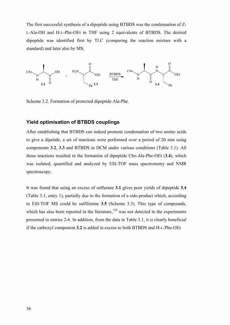
The first successful synthesis of a dipeptide using BTBDS was the condensation of Z-
L-Ala-OH and H-L-Phe-OEt in THF using 2 equivalents of BTBDS. The desired
dipeptide was identified first by TLC (comparing the reaction mixture with a
standard) and later also by MS.
O
NH
OHCbz+
NH2
Ph
O
OEt
O
NH
NH
CbzOEt
O
Ph
BTBDSTHF
3.2 3.3 3.4
Scheme 3.2. Formation of protected dipeptide Ala-Phe.
Yield optimisation of BTBDS couplings
After establishing that BTBDS can indeed promote condensation of two amino acids
to give a dipetide, a set of reactions were performed over a period of 20 min using
components 3.2, 3.3 and BTBDS in DCM under various conditions (Table 3.1). All
these reactions resulted in the formation of dipeptide Cbz-Ala-Phe-OEt (3.4), which
was isolated, quantified and analyzed by ESI-TOF mass spectrometry and NMR
spectroscopy.
It was found that using an excess of sulfurane 3.1 gives poor yields of dipeptide 3.4
(Table 3.1, entry 1), partially due to the formation of a side-product which, according
to ESI-TOF MS could be sulfilimine 3.5 (Scheme 3.3). This type of compounds,
which has also been reported in the literature,120 was not detected in the experiments
presented in entries 2-6. In addition, from the data in Table 3.1, it is clearly beneficial
if the carboxyl component 3.2 is added in excess to both BTBDS and H-L-Phe-OEt.
37
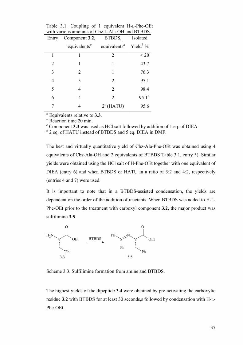
Table 3.1. Coupling of 1 equivalent H-L-Phe-OEt with various amounts of Cbz-L-Ala-OH and BTBDS. Entry Component 3.2,
equivalentsa
BTBDS,
equivalentsa
Isolated
Yieldb %
1 1 2 < 20
2 1 1 43.7
3 2 1 76.3
4 3 2 95.1
5 4 2 98.4
6 4 2 95.1c
7 4 2d (HATU) 95.6a Equivalents relative to 3.3. b Reaction time 20 min. c Component 3.3 was used as HCl salt followed by addition of 1 eq. of DIEA. d 2 eq. of HATU instead of BTBDS and 5 eq. DIEA in DMF.
The best and virtually quantitative yield of Cbz-Ala-Phe-OEt was obtained using 4
equivalents of Cbz-Ala-OH and 2 equivalents of BTBDS Table 3.1, entry 5). Similar
yields were obtained using the HCl salt of H-Phe-OEt together with one equivalent of
DIEA (entry 6) and when BTBDS or HATU in a ratio of 3:2 and 4:2, respectively
(entries 4 and 7) were used.
It is important to note that in a BTBDS-assisted condensation, the yields are
dependent on the order of the addition of reactants. When BTBDS was added to H-L-
Phe-OEt prior to the treatment with carboxyl component 3.2, the major product was
sulfilimine 3.5.
NH2
Ph
OEt
O
BTBDSN
Ph
OEt
O
SPh
Ph
3.3 3.5
Scheme 3.3. Sulfilimine formation from amine and BTBDS.
The highest yields of the dipeptide 3.4 were obtained by pre-activating the carboxylic
residue 3.2 with BTBDS for at least 30 seconds,s followed by condensation with H-L-
Phe-OEt.

38
Racemisation in BTBDS-mediated coupling
In the next stage, after optimization of the reaction conditions, it was important to
determine the extent of racemization caused by BTBDS activation of Cbz-L-alanine.
For this purpose 1H and 13C NMR spectra of dipeptide 3.4 obtained by BTBDS-
mediated formation were recorded. The spectra indicated the presence of only one
compound, which was identical to the Cbz-L-Ala-L-Phe-OEt prepared by HATU-
assisted condensation. The detection limit of this method was found to be about 3%.
This led to the conclusion that racemization in the BTBDS-assisted formation of
compound 3.4 did not exceed the level of racemization observed in a HATU-
mediated coupling (Table 3.1, entries 5, 6 and 7, respectively).
The results obtained from NMR spectroscopy stimulated us to investigate the
racemization in more detail. It was performed by analyzing the coupling products of
racemization-sensitive amino acids, i.e. serine 110,121 and cysteine,109,122 with
benzylamine using HPLC analysis with a chiral-phase column.
NH
OH
O
Boc
OBzl
BTBDS
BzlNH2 NH
NHBzl
O
Boc
OBzl
3.6 3.7
Scheme 3.4. Formation of the protected serine benzylamide
At first, condensation of Boc-L-Ser(Bzl)-OH with benzyl amine was investigated
(Scheme 3.4). Treatment of the above amino acid with HBTU, HATU or BTBDS in
DCM followed by addition of benzyl amine gave the protected serine benzyl amide
3.7.123 DCM was selected as solvent, since it is known that the rate of racemization is
substantially lower in DCM compared to polar solvents, such as DMF.109 In addition,
our previous experiments showed that DCM is a suitable solvent for the BTBDS
mediated couplings (See Table 1.) The reactions were allowed to proceed for 20 min,
after which an aliquot was withdrawn and quenched in the HPLC mobile phase. The
crude mixture was then analyzed by HPLC.
HBTU- and HATU-assisted condensations were performed by reacting the carboxyl
component, amino nucleophile, coupling agent and DIEA in a ratio of 1:1:1:2,

39
respectively. These conditions are often used to effect fast and efficient amide bond
formation utilizing the above-mentioned uronium salts. The reactions involving
BTBDS were performed under the conditions specified in Table 3.1, entry 5. It was
observed that after 20 min HBTU, HATU and BTBDS couplings gave 5.5 %, 2.2 %
and 1.8 % D-isomer of amide 3.7 and that the total yield of both isomers was 93 %,
100 % and 99 %, respectively. An additional experiment was executed using BTBDS,
showing that the extent of racemization is concentration dependent, i.e. higher
concentrations of the components resulted in increased racemization.
Experiments were then conducted to determine the degree of epimerization during the
condensation of Boc-L-Ser(Bzl)-OH and Boc-L-Cys(Trt)-OH with the less basic
amino component 3.3. The resulting diastereomeric compositions of dipeptides Boc-
Ser(Bzl)-Phe-OEt and Boc-Cys(Trt)-Phe-OEt were analyzed using chiral-phase
HPLC as described above for the synthesis and analysis of compound 3.7. The
coupling conditions and their respective outcomes are summarized in Table 3.2 and
Table 3.3.
NH
OH
O
PG
R
NH
NHBzl
O
PG
R
BTBDS
H-L-Phe-OEt
PG = Boc, R = OBzl 3.6PG = Boc, R = STrt 3.8PG = Bz, R = Ph 3.11
PG = Boc, R = OBzl 3.6PG = Boc, R = STrt 3.10PG = Bz, R = Ph 3.12
Scheme 3.5. Formation of dipeptides Boc-AA-Phe-OEt.
Table 3.2. Coupling of Boc-L-Ser(Bzl)-OH with H-L-Phe-OEt.
Equivalents Entry Coupling
agent C N CP DIEA
CC , M Preact.
time, min.
Racem.
% D,L
Relative
Yield, %
1. HBTU 1 1 1 2 0.13 15 8.3 99
2. HATU 1 1 1 2 0.13 4 3.7 100
3. BTBDS 4 1 2 0 0.14 4 - a > 99
4. BTBDS 4 1 2 5 0.14 4 2.1 > 99
C, N and CP in the table denote carboxyl component, amino component and coupling agent, respectively; CC is the concentration of the carboxyl component. a Below the detection limit.

40
The results depicted in Table 3.2 and Table 3.3 indicate that BTBDS-mediated
couplings of Boc-L-Ser(Bzl)-OH and Boc-L-Cys(Trt)-OH to H-L-Phe-OEt proceeded
in high yields and with undetectable or low racemization.
Table 3.3. Coupling of Boc-L-Cys(Trt)-OH with H-L-Phe-OEt.
Equivalents Entry
Coupling
agent C N CP DIEACC, M
Preact.
time, min.
Racem. %
D,L
Relative
Yield, %
1. HBTU 1 1 1 2 0.07 15 6.1 96
2. HATU 1 1 1 2 0.07 4 4.1 100
3. BTBDS 4 1 2 0 0.14 4 0.9 92
4. BTBDS 4 1 2 0 0.07 4 - 75
They also show that in the BTBDS-mediated formation of dipeptide 3.10, the
concentration affects not only the extent of racemization, but also the yield. The
results given in Table 3.3 suggest that, in comparison to the HBTU- and HATU-
mediated formation of dipeptide 3.10, the BTBDS reaction requires higher
concentrations or longer reaction time.
The low or virtually non-existent racemization in BTBDS-assisted formation of
dipeptide 3.9, versus the 1.8 % D-isomer found in serine benzylamide 3.7 can be
attributed to two factors. The first is the difference in basicity of these amines.
Benzylamine has a pKa value of 9.36124 while that of H-L-Phe-OEt is 2 units lower
(7.23).125 This pKa difference makes the environment of the Boc-L-Ser(Bzl)-
OH/benzylamine mediated condensation more basic than that of the Boc-L-Ser(Bzl)-
OH/H-L-Phe-OEt (given in Table 3.2, entry 3), with the consequence that
racemization increased to 1.8%. The second factor that influences racemization is the
pre-activation time. The higher level of racemization can be the outcome of the longer
pre-activation time, i.e. 15 vs. 4 min for the formation of compounds 3.7 and 3.9,
respectively. As expected, racemization also increased when DIEA was added in
excess to the C-component to the BTBDS coupling mixture, as indicated in Table 3.2,
entry 4. The absence of racemization in the condensation of urethane-protected amino
acids prompted us to extend our investigations to a system in which the α-amino
function has been converted to an amide.
It is well established that urethane-type protecting groups like Boc, Fmoc and Cbz
suppress the formation of azalactones (main factor in racemization) of activated
amino acids.126 However, it is not always possible to use urethane-linked amino acids.

41
In segment condensations, the amino function adjacent to the activated carboxyl
moiety is part of the peptide backbone i.e. an N-acyl (amide) component. Activation
of such C-terminal amino acids mostly leads to substantial racemization.127
Stimulated by the performance of BTBDS we decided to apply it in the coupling of
Bz-L-Phe-OH (3.11), which could be considered as a model for a carboxylic function
in a peptide fragment. Compound 3.11 was coupled to H-L-Phe-OEt as described
below in Table 3.4.
Table 3.4. Coupling of Bz-L-Phe-OH with H-L-Phe-OEt.
Equivalents Entry Coupling
agent C N CP DIEA
CC M Preact.
time, min.
Racem.
% D,L
Relative
Yield, %
1 HBTU 1 1 1 2 0.13 15 47.4 75a
2 HBTU 1 1 1 2 0.14 15 47.8 92b
3 HATU 1 1 1 2 0.14 4 36.7 91a
4 HATU 1 1 1 2 0.14 4 40.7 100b
5 BTBDS 4 1 2 0 0.14 4 13.2 64a
6 BTBDS 4 1 2 0 0.14 4 19.2 94b a 20 min reaction time. b 100 min reaction time.
HBTU- and HATU-mediated amide bond formations resulted in almost complete
racemization of Bz-Phe in dipeptide 3.12 (48% and 41% of D,L-diastereomer,
respectively, Table 3.4, entries 2 and 4), while BTBDS gave 19 % of the D,L-
diastereomer (entry 6). In all cases a longer time (100 min) was required to drive the
reactions to completion.
As mentioned earlier, the coupling of Bz-Phe-OH with an amino nucleophile can be
considered to mimic segment condensation. The promising results shown in Table 3.4
suggest that sulfurane-based coupling of properly protected peptide fragments is
worth further investigation.
Proposed mechanism of action of BTBDS
Formation of an amide bond between a carboxylic acid and an amine generally
requires activation of the acid. Taking into account that BTBDS has two easily
exchangeable ligands - α,α-bis(trifluoromethyl)benzyloxy groups - the first step of
the reaction could be the following:
42
ORf
S
ORf
Ph
Ph
OH
R
O
+
ORf
S
OPh
Ph
O
R
+ RfOH
3.13
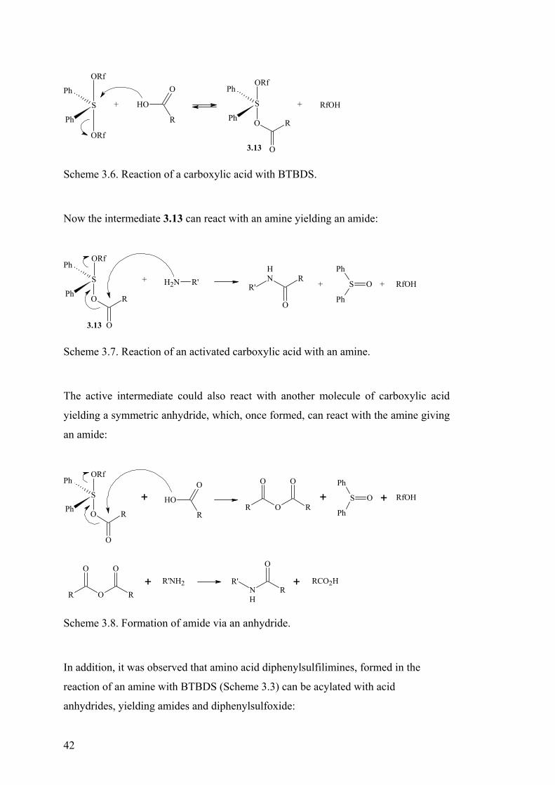
Scheme 3.6. Reaction of a carboxylic acid with BTBDS.
Now the intermediate 3.13 can react with an amine yielding an amide:
ORf
S
OPh
Ph
O
R
+ NH2 R' NH
O
RR' + S O
Ph
Ph
+ RfOH
3.13
Scheme 3.7. Reaction of an activated carboxylic acid with an amine.
The active intermediate could also react with another molecule of carboxylic acid
yielding a symmetric anhydride, which, once formed, can react with the amine giving
an amide:
ORf
S
OPh
Ph
O
R
+ + S O
Ph
Ph+ RfOHOH
R
O
R O
O
R
O
R O
O
R
O
+ R'NH2NH
R
O
R' + RCO2H
Scheme 3.8. Formation of amide via an anhydride.
In addition, it was observed that amino acid diphenylsulfilimines, formed in the
reaction of an amine with BTBDS (Scheme 3.3) can be acylated with acid
anhydrides, yielding amides and diphenylsulfoxide:

43
SNPh
Ph
OEt
O
Ph
+ + Ph2SO(Z-L-Ala)2O Z-Ala-Phe-OEt[H2O]
3.5 3.4
Scheme 3.9. Acylation of an amino acid sulfilimine with acid anhydride.
In order to get more information on the possible mechanistic pathways, an additional
experiment was performed: phenylalanine ethyl ester was acylated with 2 equivalents
of Z-alanine anhydride under conditions maximally similar to those used in BTBDS
reactions, i.e. solvent, component concentration, reaction time. This reaction would
be similar to the one described in Table 3.1, entry 5, if the condensation reaction
proceeds via the symmetric anhydride.
The yield of the fully protected dipeptide was 84%, compared to 98% in the case of
BTBDS-mediated coupling. This means that it is feasible that the BTBDS-mediated
coupling proceeds through the symmetric anhydride. It does, moreover, not exclude
that the reaction could proceed wholly or at least partially through the active
intermediate 3.13 (Scheme 3.7). The level of racemization in the above-described
sym-anhydride reaction did not exceed the ones observed for BTBDS-mediated
coupling.
Summary
In summary, this initial investigation concerning the development of sulfurane-based
coupling agents shows that bis-[α,α-bis(trifluoromethyl)benzyloxy]diphenylsulfur
can be used to effect fast and efficient condensations of urethane protected amino
acids with remarkably low racemization. The yields and racemization levels were
compared to two of the most commonly used uronium salt (i.e. HBTU and HATU)
and it is shown that in all cases BTBDS-mediated coupling in the absence of a tertiary
external base gives better results. These data, obtained from the condensation
experiments executed on relatively racemization-sensitive amino acids, i.e. serine and
cysteine, suggest that similar outcomes may be achieved for other properly protected
amino acids. Also the coupling studies of Bz-L-Phe-OH indicate that, after additional
improvement, sulfuranes could become useful for segment condensation. In
conclusion, the sulfurane concept is worth further exploration and extension in

44
several directions, such as testing of other sulfuranes for their ability to serve as
peptide coupling agents, studies of sulfurane mediated peptide segment condensation
and sulfurane-based amide bond formation of hindered amino acids as well as in the
synthesis of chiral PNAs.
45
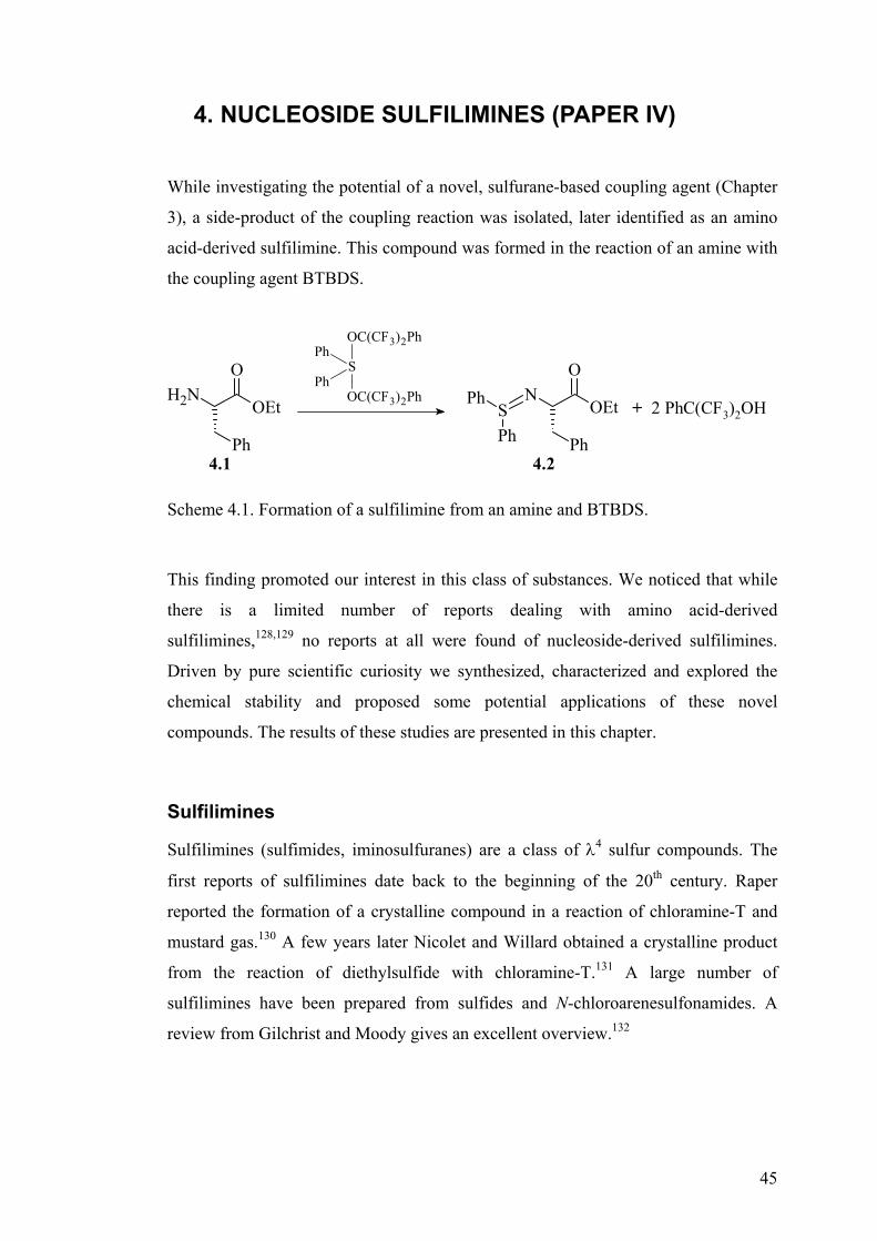
4. NUCLEOSIDE SULFILIMINES (PAPER IV)
While investigating the potential of a novel, sulfurane-based coupling agent (Chapter
3), a side-product of the coupling reaction was isolated, later identified as an amino
acid-derived sulfilimine. This compound was formed in the reaction of an amine with
the coupling agent BTBDS.
NH2
Ph
OEt
ON
Ph
OEt
O
SPh
Ph+ 2 PhC(CF3)2OH
4.1 4.2
OC(CF3)2Ph
SPh
OC(CF3)2Ph
Ph
Scheme 4.1. Formation of a sulfilimine from an amine and BTBDS.
This finding promoted our interest in this class of substances. We noticed that while
there is a limited number of reports dealing with amino acid-derived
sulfilimines,128,129 no reports at all were found of nucleoside-derived sulfilimines.
Driven by pure scientific curiosity we synthesized, characterized and explored the
chemical stability and proposed some potential applications of these novel
compounds. The results of these studies are presented in this chapter.
Sulfilimines
Sulfilimines (sulfimides, iminosulfuranes) are a class of λ4 sulfur compounds. The
first reports of sulfilimines date back to the beginning of the 20th century. Raper
reported the formation of a crystalline compound in a reaction of chloramine-T and
mustard gas.130 A few years later Nicolet and Willard obtained a crystalline product
from the reaction of diethylsulfide with chloramine-T.131 A large number of
sulfilimines have been prepared from sulfides and N-chloroarenesulfonamides. A
review from Gilchrist and Moody gives an excellent overview.132

46
N S
R
R
R
N-
S+
R
R
R
Figure 4.1. Structure of sulfilimines.
The data on sulfilimines, including X-ray structural analysis lead to description of
sulfilimines as resonance hybrids. The substituents have very little effect on the
bonding and all examined sulfilimines show similar features.133,134
Synthesis of sulfilimines
Sulfilimines can be obtained in a variety of ways. The first method is a reaction of
sulfides with N-halo compounds. A variety of N-halo compounds have been utilized,
for example, N-chloroarenesulfonamides,135,136 N-chloroamides,137,138 and N-
chloroanilines.139,140
R1
SR2
+Cl
NH
R3
SN
R3
R1
R2
Scheme 4.2. Formation of sulfilimines in a reaction between sulfide and N-haloamine.
Sulfilimines are also formed in reactions of sulfides, amines and reagents such as lead
(IV) acetate,141 NCS142 or sulfuryl chloride.139
Me2S + TsNH2 Me2S NTsPb(OAc)4
Scheme 4.3. Formation of tosyl-dimethlsulfilimine.
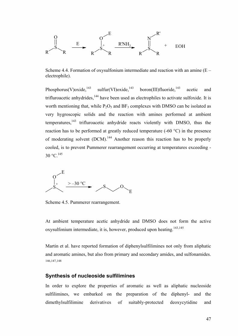
Swern et al.143,144 have published a number of reports on the synthesis of sulfilimines
utilizing sulfoxide as the source of sulfur. Intially, the sulfoxide is converted into a
oxysulfonium species, which is further allowed to react with an amine.
47
RS
+
R
OE
RS
R
OE R'NH2
RS
R
NR'
+ EOH
Scheme 4.4. Formation of oxysulfonium intermediate and reaction with an amine (E – electrophile).
Phosphorus(V)oxide,143 sulfur(VI)oxide,143 boron(III)fluoride,143 acetic and
trifluroacetic anhydrides,144 have been used as electrophiles to activate sulfoxide. It is
worth mentioning that, while P2O5 and BF3 complexes with DMSO can be isolated as
very hygroscopic solids and the reaction with amines performed at ambient
temperatures,143 trifluroacetic anhydride reacts violently with DMSO, thus the
reaction has to be performed at greatly reduced temperature (-60 °C) in the presence
of moderating solvent (DCM).144 Another reason this reaction has to be properly
cooled, is to prevent Pummerer rearrangement occurring at temperatures exceeding -
30 °C.145
S+
OE
S OE
> –30 °C
Scheme 4.5. Pummerer rearrangement.
At ambient temperature acetic anhydride and DMSO does not form the active
oxysulfonium intermediate, it is, however, produced upon heating.143,145
Martin et al. have reported formation of diphenylsulfilimines not only from aliphatic
and aromatic amines, but also from primary and secondary amides, and sulfonamides.
146,147,148
Synthesis of nucleoside sulfilimines
In order to explore the properties of aromatic as well as aliphatic nucleoside
sulfilimines, we embarked on the preparation of the diphenyl- and the
dimethylsulfilimine derivatives of suitably-protected deoxycytidine and

48
deoxyadenosine. Our intention is to use the information obtained from investigating
these compounds as the basis for a broader study of the properties of nucleoside
sulfilimines as potential protecting groups for nucleobases. For the synthesis of the
nucleoside diphenylsulfilimine, BTBDS was the reagent of choice, while for making
the nucleoside dimethylsulfilimines a protocol employing dimethylsulfoxide/
trifluoroacetic anhydride was followed.144 This method relies on the activation of
dimethylsulfoxide (DMSO) followed by addition of an amine. For the synthesis of
dimethylsulfilimines 4.4a and 4.4b, Tfa2O was chosen, since it was reported to
perform better than P2O5, BF3 and SO3.13
OB
RO
RO
NH2OC(CF3)2Ph
SPh
OC(CF3)2Ph
PhO
B
RO
RO
N=SPh2
CH2Cl2
4.3a B = Ade, R = Ac4.3b B = Cyt, R = TBDMS
4.4a B = Ade, R = Ac4.4b B = Cyt, R = TBDMS
4.4c B = Ade, R = OH4.4d B = Cyt, R = OH4.4e B = Cyt, R = Ac
iiiii
i
Scheme 4.6. Synthesis of nucleoside diphenylsulfilimines. (i) NH3/MeOH; (ii) Et3N × 3 HF/THF; (iii) Ac2O/Py.
S
O
CH2Cl2, -60 °C
Tfa2O
S+
OTfa
TfaO–
OB
RO
RO
NH2O
B
RO
RO
N=SMe2
i4.5a B = Ade, R = Ac4.5b B = Cyt, R = TBDMS4.5c B = Ade, R = TBDMS
iiiii
4.3a B = Ade, R = Ac4.3b B = Cyt, R = TBDMS4.3c B = Ade, R = TBDMS
[Me2S-OTfa]+
CH2Cl2, -45 °C
4.5d B = Ade, R = OH4.5e B = Cyt, R = OH4.5f B = Cyt, R = Ac
Scheme 4.7. Synthesis of nucleoside dimethylsulfilimines. (i) NH3/MeOH; (ii) Et3N × 3 HF/THF; (iii) Ac2O/Py.
The treatment of 3´,5´-O-protected deoxynucleosides 4.3a149 and 4.3b150 with bis-
[α,α-bis(trifluoromethyl)benzyloxy]diphenylsulfur in DCM resulted in the formation
of diphenylsulfilimines 4.4a and 4.4b in 82% and 88% yield, respectively (Scheme
4.7). It should be mentioned, that protection of the free hydroxyl functions of the
nucleosides during the BTBDS and the DMSO/trifluoroacetic anhydride reactions is

49
mandatory, since BTBDS can cause elimination of alcohols to produce alkenes,151,152
and the DMSO/trifluoroacetic anhydride mixture can bring about their
oxidation.153,154 Next, compound 4.4c was obtained by ammonolysis of diacetylated
4.4a in anhydrous NH3/MeOH. Fluoride-ion assisted cleavage of the t-
butyldimethylsilyl (TBDMS) groups from 4.4b resulted in desilylated compound
4.4d, which was further acetylated with acetic anhydride in pyridine to give, after
work-up and purification, 4.4e (92%).
Nucleoside dimethylsulfilimines 4.5a, 4.5b, and 4.5c were prepared from 4.3a, 4.3b,
and 4.3c150 in 70%, 62% and 63% yield, respectively (Scheme 4.7) by the reactions of
the properly bis-O-protected nucleosides with DMSO/ trifluoroacetic anhydride in
CH2Cl2. Dimethylsulfilimine derivative 4.5a was further ammonolyzed as mentioned
above for bis-acetylated 4.4a, while 4.5f was prepared by removal of the TBDMS
group from 4.5b with Et3N × 3 HF for 18 h (70% isolated yield), followed by
acetylation with acetic anhydride in pyridine, which proceeded in quantitative yield.
The dimethylsulfilimine group was completely intact during the desilylation and the
acetylation reactions.
While engaged in the synthesis of 4.4a-e and 4.5a-f, we found that the sulfilimines
have characteristic UV absorbance maxima at 284-296 nm. Within this wavelength
range, the nucleobases have an absorbance minimum.155 This UV absorbance
simplified purification and analysis of the above-mentioned compounds, since it
enabled a fast and clear identification of sulfilimine-containing compounds in the
reaction mixtures.
Synthesis of a sulfilimine-containing dinucleotide
In the next stage after the preparation of the completely protected nucleoside
sulfilimines, we focused our attention on testing of their stability during
oligonucleotide synthetic conditions for the H-phosphonate approach. In the course of
their preparation, it became apparent that dimethylsulfilimines 4.5a-f are less stable
than their diphenyl counterparts 4.4a-e. Consequently, a successful application of the
former derivatives in the assembly of dimer 4.8 will indicate that diphenylsulfilimine
will also survive this procedure. Therefore, deoxycytidine dimethylsulfilimine 4.5e
was selected as starting material in the preparation of dinucleotide 4.8 as outlined in
Scheme 4.8.
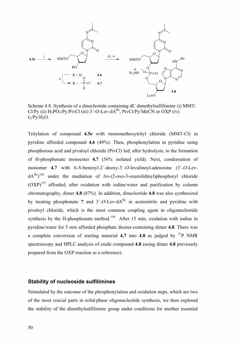
50
N
N
N
N
NH
O
O
N
N
N
O
MMTO
S
PO
O
OO
LevO
Bz
Et3NH+ –
iii, iviO
RO
N
N
N
O
MMTO
S
iiR = H
R = PO
OH
–
4.6
4.7
4.5e
4.8
Scheme 4.8. Synthesis of a dinucleotide containing dC dimethylsulfilimine (i) MMT-Cl/Py (ii) H3PO3/Py/PivCl (iii) 3´-O-Lev-dABz, PivCl/Py/MeCN or OXP (iv) I2/Py/H2O.
Tritylation of compound 4.5e with monomethoxytrityl chloride (MMT-Cl) in
pyridine afforded compound 4.6 (49%). Then, phosphonylation in pyridine using
phosphorous acid and pivaloyl chloride (PivCl) led, after hydrolysis, to the formation
of H-phosphonate monoester 4.7 (56% isolated yield). Next, condensation of
monomer 4.7 with 6-N-benzoyl-2´-deoxy-3´-O-levulinoyl-adenosine (3´-O-Lev-
dABz)156 under the mediation of bis-(2-oxo-3-oxazolidinyl)phosphoryl chloride
(OXP)157 afforded, after oxidation with iodine/water and purification by column
chromatography, dimer 4.8 (87%). In addition, dinucleotide 4.8 was also synthesized
by treating phosphonate 7 and 3´-O-Lev-dABz in acetonitrile and pyridine with
pivaloyl chloride, which is the most common coupling agent in oligonucleotide
synthesis by the H-phosphonate method.158 After 15 min, oxidation with iodine in
pyridine/water for 5 min afforded phosphate diester-containing dimer 4.8. There was
a complete conversion of starting material 4.7 into 4.8 as judged by 31P NMR
spectroscopy and HPLC analysis of crude compound 4.8 (using dimer 4.8 previously
prepared from the OXP reaction as a reference).
Stability of nucleoside sulfilimines
Stimulated by the outcome of the phosphonylation and oxidation steps, which are two
of the most crucial parts in solid-phase oligonucleotide synthesis, we then explored
the stability of the dimethylsulfilimine group under conditions for another essential

51
step, i.e. detritylation.158 This is a repeated mild acidolysis of the di- or
monomethoxytrityl groups that serve as temporary protection of the 5´-hydroxy
functions of the nucleosides (at the extension point of the oligonucleotide chain). In
addition, the iodine oxidation158 was further investigated in more detail. Moreover,
although a capping159 (acetylation) protocol is not regularly used in the H-
phosphonate methodology, acetylation is often performed in order to protect the
hydroxyl functions of the nucleosides and is also standard in the amidite approach. It
was therefore included in the stability studies. The results of these tests are
summarized in Table 4.1.
Table 4.1. Stability of adenosine and cytidine dimethylsulfilimines.a
2% DCA/DCE 0.01 M Iodineb Capping mixturec Time,
min dA 4.5a dC 4.5f dA 4.5c dC 4.5b dA 4.5c dC 4.5b
5 99 ~100 ~100 ~100 ~100 90
15 99 97 ~100 90 ~100 90
30 92 96 ~100 88 ~100 79
60 90 94 99 81 ~100 76
120 83 91 99 77 ~100 72 a The values given in the table correspond to the percentage of the intact sulfilimine as determined by straight-phase HPLC (mean error ± 3%). b Solution in collidine/water/MeCN. c Ac2O/collidine/MeCN/DMAP.
The data in Table 4.1 show that the dimethylsulfilimine function of nucleosides 4.5a
and 4.5f was not stable in 2% dichloroacetic acid (DCA) in 1,2-dichloroethane
(DCE). Some degradation was observed already after 30 min for both compounds
4.5a and 4.5f. This implies that the dimethylsulfilimine group on deoxyadenosine
and deoxycytidine may not survive the repeated detritylation steps. Nonetheless, it
may be stable during the several detritylations necessary for the preparation of a short
oligonucleotide fragment. In contrast to the observed lability under acidic conditions
of compound 4.5a, the dimethylsulfilimine of derivative 4.5c was not degraded when
exposed to the conditions required for the oxidation of the phosphonate diester
function into a phosphate diester158,160 or to the capping solution (Ac2O/sym-
collidine/DMAP/MeCN) commonly used in some solid-phase nucleic acid synthesis
protocols.161 On the other hand, when cytidine dimethylsulfilimine 4.5b was
dissolved in the above mentioned iodine solution, it was intact for the first 5 min.

52
However, slightly longer treatment led to cleavage of the sulfilimine (10% within the
15 min) as indicated inTable 4.1. Moreover, exposure of 4.5b to the capping mixture
proved to have a deleterious effect on the sulfilimine integrity. In this case, 10% of
nucleoside 4.5b was affected within 5 min. The information summarized in Table 4.1
suggests that only properly protected dimethylsulfilimine derivatives of
deoxyadenosine may endure oligonucleotide synthesis protocols that require repeated
oxidation steps or oxidation times longer than 5 min. This is also correct for methods
that endorse repeated capping step.
Next, the diphenylsulfilimino group of compounds 4.4a, 4.4b, and 4.4e was subjected
to the same conditions as described in Table 4.1 for the dimethylsulfilimine function.
The values obtained from these stability tests are given in Table 4.2, and they indicate
that, in contrast to their dimethyl counterparts, compounds 4.4a and 4.4b were stable
during treatment with 2% DCA in DCE for 120 min. Prolonged exposure (24 h) to
this mixture showed that the deoxycytidine derivative 4.4d remained unaffected,
while Ac2dA=SPh2 (4.4a) was less stable and was partially degraded (9%).
Table 4.2. Stability of adenosine and cytidine diphenylsulfilimines.
2% DCA/DCE 0.01 M Iodine Capping mixture
Time,
min Ac2dA-
SPh2
4.4a
Ac2dC-
SPh2
4.4e
Ac2dA-
SPh2
4.4a
Si2dC-
SPh2
4.4b
Ac2dA-
SPh2
4.4a
Si2dC-
SPh2
4.4b
5 ~100 ~100 ~100 ~100 ~100 ~100
15 ~100 ~100 ~100 ~100 ~100 ~100
30 ~100 ~100 ~100 ~100 ~100 ~100
60 ~100 ~100 99 ~100 ~100 ~100
120 ~100 99 94 ~100 ~100 ~100
The diphenylsulfilimine moiety was also found to be much more stable in the
oxidation mixture of I2/collidine in aqueous acetonitrile. After 2 h, the deoxycytidine
derivative 4.4b was still completely intact in contrast to 4.4a which was partially
cleaved (6%) as shown in Table 4.2 Furthermore, no cleavage of the
diphenylsulfilimine function in 4.4a and 4.4b was observed in a mixture of
Ac2O/collidine/DMAP in MeCN for at least 2 h.

53
The stability of the diphenylsulifilimino group towards the conditions listed in Table
4.2 suggests that it will endure the assembly of longer nucleic acid fragments on a
solid support using the H-phosphonate approach. A successful application of
nucleoside diphenylsulfilimines in the phosphoroamidite chemistry is also very
likely, provided they would survive the repeated phosphonylation step.
In the following stage, the stability of dA=SMe2, dC=SMe2, dA=SPh2 and dC=SPh2
(4.5d, 4.5e, 4.4c and 4.4d, respectively) in an aqueous environment and during
ammonolysis, which is the final step in oligonucleotide synthesis,158 employed to
remove the protecting groups from the nucleobases and detach the oligonucleotide
fragment from the solid-support, was examined.
Exposure to 33% aq. NH3 for 16 h at 21 °C showed that 4.4c, 4.4d, and 4.5d were un-
degraded while 4.5e lost 12% of the dimethylsulfilimine group. Upon treatment with
33% aq. NH3 at elevated temperature, the diphenylsulfilimine derivatives 4c and 4d
remained unaffected, but compounds 4.5d and 4.5e were almost completely
converted into the corresponding deoxyadenosine and deoxycytidine, respectively. In
anhydrous NH3 in methanol, at 21 °C, all four nucleoside sulfilimines 4.5d, 4.5e,
4.4c, and 4.4d were found to be stable for at least 2 h.
The increased stability of the diphenylsulfilimines 4.4c and 4.4d, compared to the
dimethyl- 4.5d and 4.5e, is also evident in water and 50 mM TRIS buffer (pH 9.0).
The diphenylsulfilimines were stable in aqueous solutions at 21 ºC for 16 hours,
while the dimethyl analogues were substantially degraded under the same conditions.
In H2O at elevated temperature however, only dA=SPh2 (4.4c) was stable, while
compound dC=SPh2 (4.4d) lost 37% of the sulfilimino group, and derivatives 4.5d
and 4.5e were only detected in trace amounts.
The outcome of the stability tests in aqueous solutions is consistent with the results of
the tests reported above, which show the increased stability of the diphenylsulfilimine
function compared to the dimethylsulfilimine. This outcome leads to the conclusion
that the diphenyl-sulfilimine group would not be cleaved at the final stage of nucleic
acids synthesis, i.e. the deprotection of the nucleobases and detachment from the
solid-support which is brought about by ammonolysis either at ambient temperature
or at 60 °C.
This stability to ammonolysis and to the above-mentioned conditions depicted in
Table 4.2 permits retaining the diphenylsulfilimine group during and after
oligonucleotide synthesis, a fact that can make them useful for the introduction of
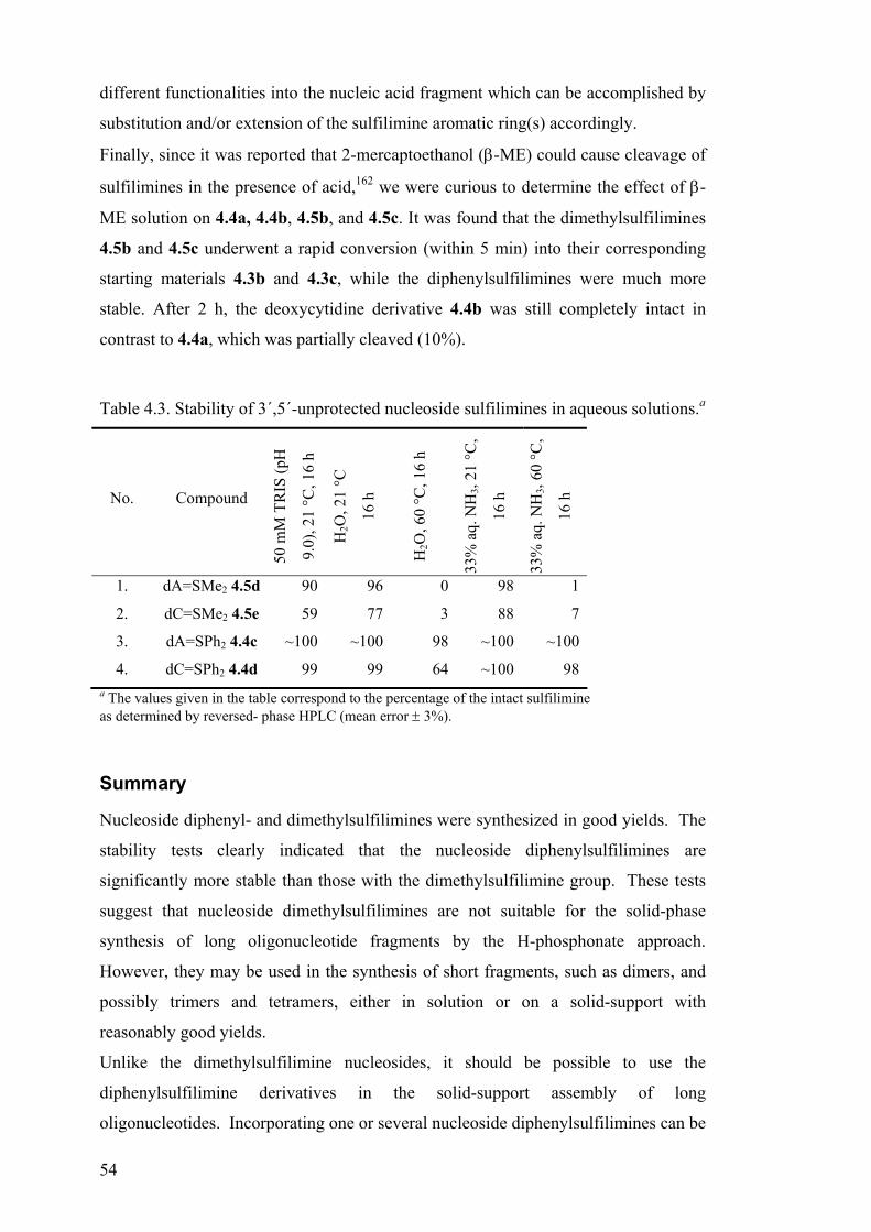
54
different functionalities into the nucleic acid fragment which can be accomplished by
substitution and/or extension of the sulfilimine aromatic ring(s) accordingly.
Finally, since it was reported that 2-mercaptoethanol (β-ME) could cause cleavage of
sulfilimines in the presence of acid,162 we were curious to determine the effect of β-
ME solution on 4.4a, 4.4b, 4.5b, and 4.5c. It was found that the dimethylsulfilimines
4.5b and 4.5c underwent a rapid conversion (within 5 min) into their corresponding
starting materials 4.3b and 4.3c, while the diphenylsulfilimines were much more
stable. After 2 h, the deoxycytidine derivative 4.4b was still completely intact in
contrast to 4.4a, which was partially cleaved (10%).
Table 4.3. Stability of 3´,5´-unprotected nucleoside sulfilimines in aqueous solutions.a
No. Compound
50 m
M T
RIS
(pH
9.0)
, 21
°C, 1
6 h
H2O
, 21
°C
16 h
H2O
, 60
°C, 1
6 h
33%
aq.
NH
3, 21
°C,
16 h
33%
aq.
NH
3, 60
°C,
16 h
1. dA=SMe2 4.5d 90 96 0 98 1
2. dC=SMe2 4.5e 59 77 3 88 7
3. dA=SPh2 4.4c ~100 ~100 98 ~100 ~100
4. dC=SPh2 4.4d 99 99 64 ~100 98a The values given in the table correspond to the percentage of the intact sulfilimine as determined by reversed- phase HPLC (mean error ± 3%).
Summary
Nucleoside diphenyl- and dimethylsulfilimines were synthesized in good yields. The
stability tests clearly indicated that the nucleoside diphenylsulfilimines are
significantly more stable than those with the dimethylsulfilimine group. These tests
suggest that nucleoside dimethylsulfilimines are not suitable for the solid-phase
synthesis of long oligonucleotide fragments by the H-phosphonate approach.
However, they may be used in the synthesis of short fragments, such as dimers, and
possibly trimers and tetramers, either in solution or on a solid-support with
reasonably good yields.
Unlike the dimethylsulfilimine nucleosides, it should be possible to use the
diphenylsulfilimine derivatives in the solid-support assembly of long
oligonucleotides. Incorporating one or several nucleoside diphenylsulfilimines can be

55
advantageous due to the fact that they are unaffected by ammonolysis and that the
sulfilimine moiety absorbs at 290-300 nm. Thus, they could be used as UV tags for
oligonucleotides via pinpointing of their location in the presence of unlabeled
fragments. Another possible advantage is that by modifying one or both of the
phenyl rings accordingly, diarylsulfilimine-containing nucleotides could be used to
introduce functionalities, such as, for example an intercalator, at a predetermined
position in a nucleic acid fragment.
Changing the substituents on the sulfur, for example, by synthesizing methyl-phenyl
sulfilimines, could tune the stability of the nucleoside sulfilimines. The aim of these
modifications would be to obtain enhanced stability towards the oligosynthesis
conditions, while still preserving the ease of removal. The use of sulfilimines as
potential UV tags and/or protecting groups is not limited to DNA synthesis, but
extendable to RNA, PNA and PNA-DNA chimera.

56
5. SUPPRESSION OF EXONUCLEOLYTIC DEGRADATION OF DOUBLE-STRANDED DNA AND INHIBITION OF EXONUCLEASE III BY PNA (PAPER V)
A new method of single-molecule DNA sequencing, is based upon the detection and
identification of single fluorescently labelled mononucleotides released by the
exonucleolytic degradation of single-stranded DNAs. 163 The key element in this
novel DNA sequencing approach is the suppression of the degradation of double-
stranded DNA. As a part of a collaboration project, we investigated the ability of
PNA to suppress the degradation of dsDNA by Exonuclease III.
First, it was necessary to ascertain that PNA can indeed suppress the degradation of a
double-stranded DNA in a sequence-specific manner as well as to determine the
efficacy of the suppression.
Thus, two shorter fragments of the master DNA (from the sequencing project), a
30mer/45mer were chemically synthesized.♣ The formation of a duplex from these
fragments will create a protruding 3´-end that is adjacent to the anti-parallel (-) DNA
strand, which is not to be cleaved by 3´→5´ exonucleases that are double strand
specific (See Figure 5.1). This will ensure that the exonucleolytic degradation will be
only mono-directional i.e. 3´→5´, thus simplifying the quantification of the degradation.
The quantification of the nucleotides released by the degradation in the presence and
in the absence of PNA will provide the answers to the questions about the binding
stability of such PNA complexes.
5´
5´
3´
3´
5´
3´
dsDNA
+
apPNA
Figure 5.1. System design. dsDNA consists of 30- and 45-mer∗ fragments (3´-overhang); apPNA is a 16-mer anti-parallel PNA.
♣ Purchased from TAG Copenhagen. ∗ 5´-TCT-TCA-CCT-CTC-TCT-CTT-TCT-GTC-TCT-CTC-TTT-CTT-TGT-CCT-CTT-3´
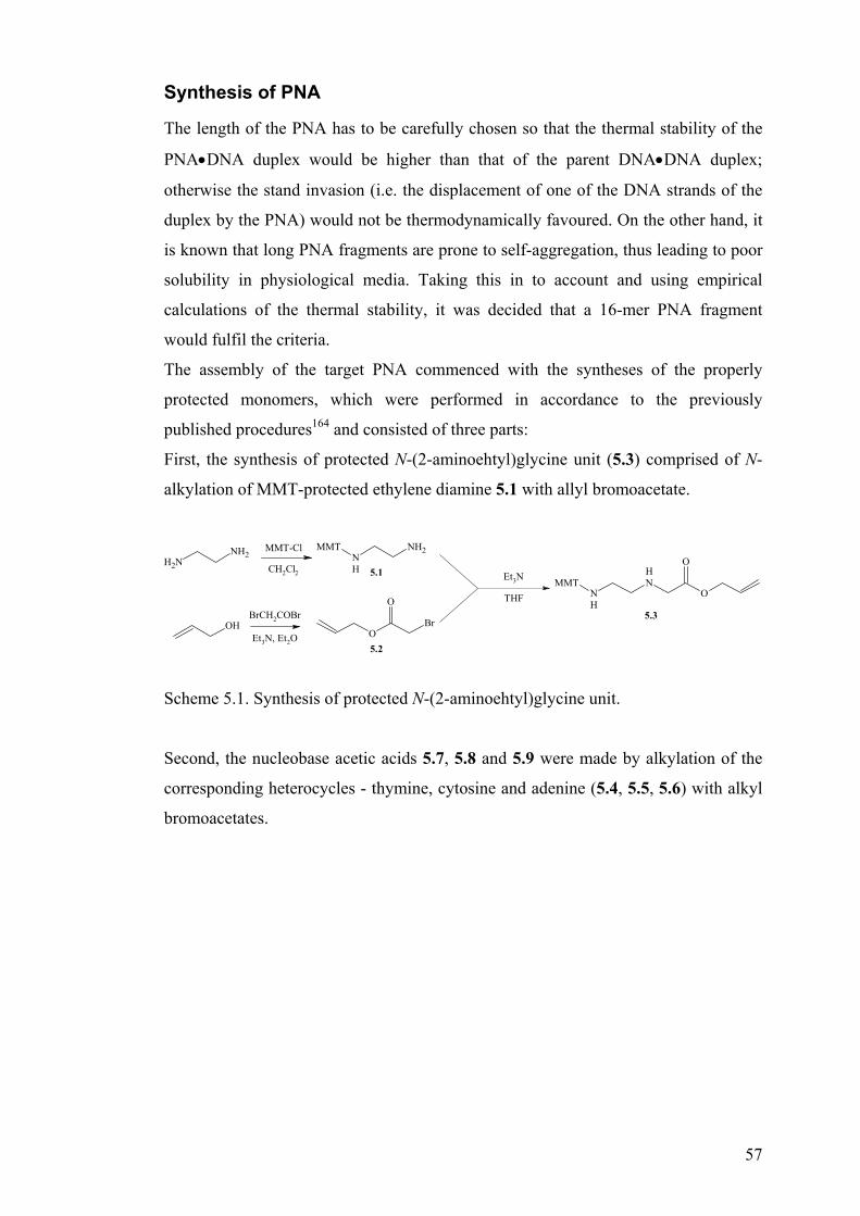
57
Synthesis of PNA
The length of the PNA has to be carefully chosen so that the thermal stability of the
PNA•DNA duplex would be higher than that of the parent DNA•DNA duplex;
otherwise the stand invasion (i.e. the displacement of one of the DNA strands of the
duplex by the PNA) would not be thermodynamically favoured. On the other hand, it
is known that long PNA fragments are prone to self-aggregation, thus leading to poor
solubility in physiological media. Taking this in to account and using empirical
calculations of the thermal stability, it was decided that a 16-mer PNA fragment
would fulfil the criteria.
The assembly of the target PNA commenced with the syntheses of the properly
protected monomers, which were performed in accordance to the previously
published procedures164 and consisted of three parts:
First, the synthesis of protected N-(2-aminoehtyl)glycine unit (5.3) comprised of N-
alkylation of MMT-protected ethylene diamine 5.1 with allyl bromoacetate.
NH2
NH2MMT-Cl
CH2Cl2NH
NH2MMT
OHBrCH2COBr
Et3N, Et2OO
O
Br
Et3N
THF O
O
NH
NH
MMT5.1
5.2
5.3
Scheme 5.1. Synthesis of protected N-(2-aminoehtyl)glycine unit.
Second, the nucleobase acetic acids 5.7, 5.8 and 5.9 were made by alkylation of the
corresponding heterocycles - thymine, cytosine and adenine (5.4, 5.5, 5.6) with alkyl
bromoacetates.

58
NH
NH
O
O
BrCH2CO2Me
K2CO3, DMF
NH
N
O
O
OMe
O
1) aq. NaOH / MeOH
2) aq. HCl
NH
N
O
O
OH
O
N
NH
NH2
O
NaH, DMF
BrCH2CO2tBu
N
N
NH2
O
OtBu
O
1) Bz2O, Py
2) 50% TFA/CH2Cl2
N
N
NH
O
OH
O
Bz
N
N
N
NH
NH2
NaH, DMF
BrCH2CO2tBu
N
N
N
N
NH2
tBuO
O
1) BzCl, Py
2) 50% TFA/CH2Cl2
N
N
N
N
NH
OH
O
Bz
5.4
5.5
5.6 5.9
5.8
5.7
Scheme 5.2. Synthesis of protected nucleobase acetic acids.
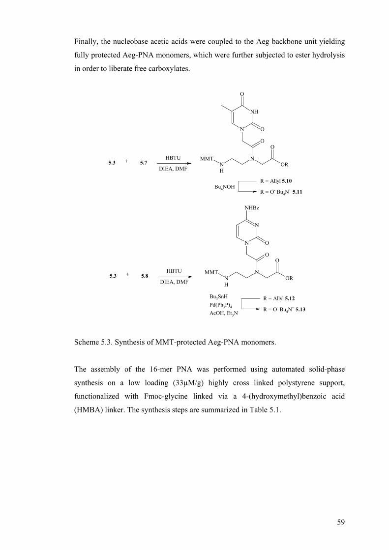
59
Finally, the nucleobase acetic acids were coupled to the Aeg backbone unit yielding
fully protected Aeg-PNA monomers, which were further subjected to ester hydrolysis
in order to liberate free carboxylates.
OR
O
NNH
MMT
NH
N
O
O
O
R = Allyl 5.10
R = O- Bu4N+ 5.11
Bu4NOH
OR
O
NNH
MMT
N
N
NHBz
O
O
R = Allyl 5.12
R = O- Bu4N+ 5.13
Bu3SnHPd(Ph3P)4
AcOH, Et3N
+ HBTU
DIEA, DMF5.3 5.7
+ HBTU
DIEA, DMF5.3 5.8
Scheme 5.3. Synthesis of MMT-protected Aeg-PNA monomers.
The assembly of the 16-mer PNA was performed using automated solid-phase
synthesis on a low loading (33µM/g) highly cross linked polystyrene support,
functionalized with Fmoc-glycine linked via a 4-(hydroxymethyl)benzoic acid
(HMBA) linker. The synthesis steps are summarized in Table 5.1.

60
Table 5.1. Solid phase synthesis of PNA.
Step Function Solvents and reagents Time,
min.
Fmoc deprotection of
resin-bound glycine
and terminal lysines
22 % piperidine in NMP 2×3.5
Coupling MMT Aeg-PNA monomers[a],
HBTU[a], DIEA[c] 17.5
MMT deprotection
during chain
elongation
1 % TFA/DCE 3
Boc deprotection 50 % TFA/DCM 2×15
Lysine termination Fmoc-L-Lys(Boc)-OH[a],
HBTU[a], DIEA[b] 2×17.5
[a] As a 0.3 M solution in NMP [b] As a 0.4 M solution in NMP
A pure PNA 16-mer was obtained by reverse-phase HPLC purification of the crude
PNA after detachment of the PNA from the resin.
UV thermal melting experiments
Prior to the enzymatic degradation experiments, the ability of a 16-mer PNA
fragment, complementary and anti-parallel to the 30-mer, to hybridize to ssDNA and
dsDNA was evaluated (Table 5.2).
Table 5.2. UV thermal melting data of the nucleic acid components.
No. Componentsa Tm, °Cb
1. DNA (45) : DNA (30) 57
2. DNA (30) : apPNA 74
3. dsDNA : apPNA 37, 74
As expected, it was found that the PNA•DNA duplex was more stable than the
DNA•DNA duplex (∆Tm = 17 °C), which should allow the strand invasion to occur.

61
Enzymatic degradation studies
The following experiments were set up – 1 nmol of dsDNA in a TRIS buffer (66 mM,
pH 8.0), containing 0.66 mM Mg2+ and 2-mercaptoethanol (1 mM) were mixed with
various amounts of apPNA (0, 1, 5, and 7 nmol), briefly heated up to 90 °C and
cooled to ambient temperature over a period of 90 min. Exonuclease III (2 units†) was
added and the assay was incubated at 37 °C. Aliquots were withdrawn at fixed time
intervals (30 and 60 min) and analyzed for the amounts of the formed
mononucleotides by reverse-phase HPLC. The amounts of the mononucleotides
released from the degradation of dsDNA with no apPNA added were assigned to
100% and used as a reference point.
0%
20%
40%
60%
80%
100%
120%
dGMP 100% 65% 37% 18% 70%
dsDNA a b c d
a) dsDNA + 10 nM apPNA; b) dsDNA + 50 nM apPNA; c) dsDNA + 70 nM apPNA; d) dsDNA + 70 nM non-complementary PNA 10-mer.
Figure 5.2. Relative amount of degradation of dsDNA by Exonuclease III after 60 min (by the amount of the released dGMP).
By plotting the amounts of the released mononucleotides versus the concentration of
apPNA, it was apparent that increasing the concentration of apPNA from 10 to 70 nM
resulted in increased suppression of degradation (Figure 5.2, entries a-c).
Further, it was important to determine whether the observed suppression of
degradation is specific due to the base pairing of PNA with the dsDNA (strand
invasion) or non-specific, due to the interactions of PNA with the DNA duplex or the
† One unit is defined as the amount of enzyme required to produce 1nmol of acid-soluble nucleotides from double-stranded DNA in 30 minutes at 37°C.

62
enzyme. Thus, when a non-complementary PNA 10-mer was used instead of the
apPNA, the enzymatic activity was observed to be 70% (Figure 5.2, entry ‘d’).
Summary
It can be concluded that anti-parallel PNA does indeed suppress the activity of
Exonuclease III in a concentration-dependant and sequence-specific manner, in
addition to some degree of non-specific inhibition, which is less pronounced than the
specific suppression. The extent of the suppression could be further increased by
optimizing the binding affinity of the PNA. Factors such as length of PNA, guanine-
cytosine content, ionic strength of the media, incubation temperature and time,
concentration of Mg2+, use of various additives could be changed in order to achieve
maximum specific suppression values, while keeping the non-specific inhibition low.

63
CONNECTIONS AND CONCLUSIONS
The work described in the above five chapters deals with various aspects of the
development of new potential molecular tools for biochemistry and related
disciplines.
A new PNA modification has been introduced, which has an enhanced ability in its
binding to discriminate between complementary DNA and RNA. The assembly of
backbone-modified PNAs can be problematic due to potentially lower reactivity of
the monomers in which novel structural elements are included. To deal with such
problems, the use of a new type of amide bond-forming reagent has been proposed
and investigated.
The right choice of protecting groups for the chemical synthesis of nucleic acid
fragments is one of the keys for success. Often the problems are caused by premature
removal of protecting groups or on the contrary – partial loss of product because of
incomplete deprotection. A new type of potential protecting group and/or UV label
has been introduced and their stability towards a variety of conditions have been
tested and reported.
Further, a PNA fragment has been successfully applied as a means to enhance the
stability of a DNA strand against enzymatic degradation, a finding that may have an
application in a novel sequencing approach.

64
ACKNOWLEDGEMENTS
I would like to express my gratitude to:
Dr. Esther Yeheskiely, my supervisor, for accepting me as a post-graduate
student, for her never-ending interest in this work and for all the help in
preparation of this thesis.
Professor Roger Strömberg for his advice in the lab work, and help with
preparation of manuscripts and this thesis.
Dr. Susannah Sigurdsson and Dr. Esmail Yousefi-Salakdeh for all the fun in
the lab, for your support even when things didn’t go so well and for being
such great friends.
Charlotte Ander for her help with instruments and for being such a nice
colleague.
Dr. Petri Heinonen for his friendship and help with the NMR.
Dr. Martin Bollmark and Simone Zaramella for scrupulous reading of this
thesis.
All the people in the lab I had privilege working with – Anna, Annemieke,
Ethel, Hans, Jessica, Robert, Stefan, Viji, Vilnis.
All the nice people from other departments.
And last but not least … Evita and Linda, for all their love and support during
these years. I would have never made it without you.
Mr. E. Blackadder and S. Baldrick for cheering me up.

65
REFERENCES 1 Nielsen, P. E.; Egholm, M.; Berg, R. H.; Buchardt, O. Science, 1991, 254, 1497. 2 Egholm, M.; Buchardt, O.; Nielsen, P. E.; Berg, R. H. J. Am. Chem. Soc. 1992, 114,
1895-1897. 3 Dueholm, K. L.; Egholm, M.; Behrens, C.; Christensen, L.; Hassen, H. F.; Vulpius,
T. et al. J. Org. Chem. 1994, 59, 5767-5773. 4 Christensen, L.; Fitzpatrick, R.; Gildea, B.; Petersen, K. H.; Hansen, H. F.; Koch, T.
et al. J. Pept. Sci. 1995, 3, 175-183. 5 Koch, T.; Hansen, H. F.; Andersen, P.; Larsen, T, Batz, H. G.; Ottensen, K.; Ørum,
H. J. Pept. Res. 1997, 49, 80-88. 6 Thomson, S. A.; Josey, J. A.; Cadilla, R.; Gaul, M. D.; Hassman, C. F.; Luzzio, M.
J. et al. Tetrahedron 1995, 51, 6179-6194. 7 Will, D. W.; Breipohl, G.; Langner, D.; Knolle, J.; Uhlmann, E. Tetrahedron, 1995,
51, 12069-12082. 8 van der Laan, A. C.; Meeuwenoord, N. J.; Kuyl-Yeheskiely, E.; Oosting, R. S.;
Brands, R.; van Boom, J. H. Recl. Trav. Chim. Pays-Bas, 1995, 114, 295-297. 9 van der Laan, A. C.; Brill, R.; Kuimelis, R. G.; Kuyl-Yeheskiely, E.; van Boom, J.
H.; Andrus, A.; Vinayak, R. Tetrahedron Lett. 1997, 38, 2249-2252. 10 van der Laan, A. C.; Havenaar, P.; Oosting, R. S.; Kuyl-Yeheskiely, E.; Uhlmann,
E.; van Boom, J. H. Bioorg. Med. Chem. Lett. 1998, 8, 663-668. 11 Breipohl, G.; Knolle, J.; Langner, D.; O’Malley, G.; Uhlmann, E. Bioorg. Med.
Chem. Lett. 1996, 6, 665-670. 12 Salvi, J-P.; Walchshofer, N.; Paris, J. Tetrahedron Lett. 1994, 35, 1181-1184. 13 Farese, A.; Patino, N.; Condom, R.; Dalleu, S.; Guedj, R. Tetrahedron Lett. 1996,
37, 1413-1416. 14 Falkiewicz, B.; Wisniowski, W.; Kolodziejczyk, A. S.; Wisniewski, K. Nucleosides
Nucleotides 2001, 20, 1393-1398. 15 Fader, L. D.; Boyd, M.; Tsantrizos, Y. S. J. Org. Chem. 2001, 66, 3372-3379. 16 Clivio, P.; Guillaume, D.; Adeline, M-T.; Hamon, J.; Riche, C.; Fourrey, J-L. J.
Am. Chem. Soc. 1998, 120, 1157-1166. 17 Efimov, V. A.; Buryakova, A. A.; Choob, M. V.; Chakhmakhcheva, O. G. Russ. J.
Bioorg. Chem. (Engl. Transl.) 1998, 24, 618 - 630. 18 Egholm, M.; Nielsen, P. E.; Buchardt, O.; Berg, R. H. J. Am. Chem. Soc. 1992,
114, 9677-9678. 19 Thomson, S. A.; Josey, J. A.; Cadilla, R.; Gaul, M. D.; Hassman, C. Fred; et al.
Tetrahedron 1995, 51, 6179-6194. 20 Thomson, S. A.; Josey, J. A.; Cadilla, R.; Gaul, M. D.; Hassman, C. F.; Luzzio, M.
J.; et al. Tetrahedron 1995, 51, 6179-6194. 21 Wang, S. S. J. Am. Chem. Soc. 1973, 95, 1328-1333. 22 Sheppard, R. C.; Williams, B. J. Int. J. Peptide Protein Res. 1982, 20, 451-454. 23 Corradini, R.; Sforza, S.; Dossena, A.; Palla, G.; Rocchi, R.; Filira, F.; Nastri, F.;
Marchelli, R. J. Chem. Soc. Perkin Trans. 1 2001, 20, 2690-2696. 24 Bergmann, F.; Bannwarth, W.; Tam, S. Tetrahedron Lett. 1995, 36, 6823-6826. 25 Tackett A. J; Corey, D. R; Raney, K. D. Nucleic Acids Res. 2002, 30, 950-957 26 Gangamani, B. P.; Kumar, V. A.; Ganesh, K. N. Biochem. Biophys. Res. Commun.
1997, 240, 778. 27 Egholm, M.; Buchardt, O.; Christensen, L.; Behrens, C.; Freier, S. M.; Driver, D.
A.; et al. Nature, 1993, 365, 566-568. 28 Rose, D. J. Anal. Chem. 1993, 65, 3545-3549.
66
29 Egholm, M.; Buchardt, O.; Nielsen, P. E.; Berg, R. H. J. Am. Chem. Soc. 1992,
114, 1895-1897. 30 Egholm, M.; Chistensen, L.; Dueholm, K. L.; Buchardt, O.; Coull, J.; Nielsen, P. E.
Nucleic Acids Res. 1995, 23, 217. 31 Griffith, M. C.; Risen, L. M.; Grieg, M. J.; Lesnik, E .A.; Sprankle, K. G.; Griffey,
R. H.; et al. J. Am. Chem. Soc. 1995, 117, 831. 32 Demidov, V.; Frank-Kamenetskii, M. D.; Egholm, M.; Buchardt, O.; Nielsen, P. E.
Nucleic Acids Res. 1993, 21, 2103-2107. 33 Cherny, D. Y.; Belotserkovskii, B. P.; Frank-Kamenetskii, M. D.; Egholm, M.;
Buchardt, O.; Berg, R. H.; Nielsen, P. E. Proc. Natl. Acad. Sci. U.S.A. 1993, 90, 1667-1670.
34 Peffer, N. J.; Hanvey, J. C.; Bisi, J. E.; Thomson, S. A.; Hassman, C. F.; Noble, S. A.; Babiss, L. E. Proc. Natl. Acad. Sci. U.S.A. 1993, 90, 10648-10652.
35 Kosaganov, Y. N.; Stetsenko, D. A.; Lubyako, E. N.; Kvitko, N. P.; Lazurkin, Y. S.; Nielsen, P. E. Biochemistry 2000, 39, 11742-11747.
36 Almarsson, Ö.; Bruice, T. C.; Kerr, J.; Zuckermann, R. N. Proc. Natl. Acad. Sci. USA 1993, 90, 7518-7522.
37 Wittung, P.; Kajanus, J.; Edwards, K.; Nielsen, P. E.; Nordén, B.; Malmstöm, B. G. FEBS Lett. 1995, 365, 27-29.
38 Nielsen, P. E.; Egolm, M.; Berg, R. H.; Buchardt, O. Anti-Cancer Drug Design 1993, 8, 53-63.
39 Gildea, B. D.; Casey, S.; NacNeill, J.; Perry-O'Keefe, H.; Sorensen, D.; Coull, J. M. Tetrahedron Lett. 1998, 39, 7255-7258.
40 Li, X.; Zhang, L.; Lu, J.; Chen, Y.; Min, J.; Zhang, L. Bioconjugate Chem. 2003, 14, 153-157.
41 Simmons, C. G.; Pitts, A. E.; Mayfield, L. D.; Shay, J. W.; Corey, D. R. Bioorg. Med. Chem. Lett. 1997, 7, 3001-3006.
42 Haaima, G.; Lohse, A.; Buchardt, O.; Nielsen, P. E. Angewandte, 1996, 35, 1939-1941
43 Hyrup, B.; Egholm, M.; Nielsen, P. E.; Wittung, P.; Norden, B.; Buchardt, O. J. Am. Chem. Soc. 1994, 116, 7964-7970.
44 Hyrup, B.; Egholm, M.; Buchardt, O.; Nielsen, P. E. Bioorg. Med. Chem. Lett. 1996, 6, 1083-1088.
45 Kumar, V. A. Eur. J. Org. Chem. 2002, 13, 2021-2032. 46 Gangamani, B. P.; Kumar, V. A.; Ganesh, K. N. Tetrahedron 1996, 52, 15017-
15030. 47 Gangamani, B. P.; Kumar, V. A.; Ganesh, K. N. Tetrahedron 1999, 55, 177-192. 48 Gangamani, B. P.; D’Costa, M.; Kumar, V. A.; Ganesh, K. N.Nucleoside
Nucleotides 1999, 18, 1409 -1011. 49 Jordan, S.; Schwemler, C.; Kosch, W.; Kretschmer, A.; Schwenner, E.; Milke, B.
Bioorg. Med. Chem. Lett. 1997, 7, 687-692. 50 D’Costa, M.; Kumar, V. A.; Ganesh, K. N. Org. Lett. 1999, 1, 1513-1516. 51 Puschl, A.; Boesen, T.; Zuccarello, G.; Dahl, O.; Pitsch, S.; Nielsen, P. E. J. Org.
Chem. 2001, 66, 707-712. 52 Stender, H.; Lund, K.; Petersen, K. H.; Rasmussen, O. F.; Hongmanee, P.; Miorner,
H.; Godtfredsen, S. E. J. Clinical Microbiol. 1999, 37, 2760-2765. 53 Orum, H.; Nielsen, P. E.; Egholm, M.; Berg, R. H.; Buchardt, O.; Stanley, C.
Nucleic Acids Res. 1993, 21, 5332-5336. 54 Sotlar, K.; Escribano, L.; Landt, O.;Mohrle, S.; Herrero, S.; Torrelo, A.; Lass, U.;
Horny, H.-P.; Bultmann, B. Am. J. Pathol. 2003, 162, 737 - 746.

67
55 Wang, J.; Palecek, E.; Nielsen, P. E.; Rivas, G.; Cai, X.; Shiraishi, H.; Dontha, N.;
Luo, D.; Farias, P. A. M. J. Am. Chem. Soc. 1996, 118, 7667-7670. 56 Arlinghaus, H. F.; Kwoka, M. N.; Jacobson, K. B. Anal. Chem. 1997, 69, 3747-
3753. 57 Feriotto, G.; Corradini, R.; Sforza, S.; Bianchi, N.; Mischiati, C.; Marchelli, R.;
Gambari, R. Lab. Invest. 2001, 81, 1415-1427. 58 Hanvey, J. C.; Peffer, N. J.; Bisi, J. E.; Thomson, S. A.; Cadilla, R. ; Josey, J. A.; et
al. Science 1992, 258, 1481-1485. 59 Cheah, I. K. Cheema, S. S.; Langford, S. J.; Lopes, E. C.; MacFarlane, K. J.;
Petratos, S. Bioorg. Med. Chem. Lett. 2003, 13, 2377-2380. 60 Sun, L.; Fuselier, J. A.; Murphy, W. A.; Coy, D. H. Peptides 2002, 23, 1557-1565. 61 Bonnard, E.; Mazarguil, H.; Zajac, J-M. Peptides 2002, 23, 1107-1113. 62 Pooga, M.; Langel, U. Curr. Cancer Drug Targets 2001, 1, 231-239. 63 Koppelhus, U.; Zachar, V.; Nielsen, P. E.; Liu, X.; Eugen-Olsen, J.; Ebbesen, P.
Nucleic Acids Res. 1997, 25, 2167-2173. 64 Boulme, F.; Freund, F.; Moreaul, S.; Nielsen, P. E.; Gryaznov, S.; Toulme, J-J.;
Litvak, S. Nucleic Acids Res. 1998, 26, 5492-5500. 65 Lee, R.; Kaushik, N.; Modak, M. J.; Vinayak, R.; Pandey, V. N. Biochemistry
1998, 37, 900-910. 66 Kaushik, N.; Pandey, V. N. Virology 2002, 303, 297-308. 67 Diviacco, S.; Rapozzi, V.; Xodo, L.; Helene, C.; Quadrifoglio, F.; Giovannangeli,
C. FASEB 2001, 15, 2660-2668 68 van der Laan, A. C.; van Amsterdam, I.; Tesser, G. I.; van Boom, J. H.; Kuyl-
Yeheskiely, E.; Nucleosides Nucleotides 1998, 17, 219-231. 69 Sforza, S.; Haaima, G.; Marchelli, R.; Nielsen, P. E.; Eur. J. Org. Chem. 1999, 1,
197-204. 70 Zhang, L.; Min, J.; Zhang, L.; Bioorg. Med. Chem. Lett. 1999, 9, 2903-2908. 71 Hoffmann, M.; Brückner, A. M.; Hupp, T.; Engels, B.; Diederichsen, U.; Helv.
Chim. Acta 2000, 83, 2580-2593. 72 Sforza, S.; Corradini, R.; Ghirardi, S.; Dossena, A.; Marchelli, R.; Eur. J. Org.
Chem. 2000, 16, 2905-2913. 73 Koshkin, A. A.; Singh, S. K.; Nielsen, P.; Rajwanshi, V. K.; Kumar, R.;
Meldgaard, M.; Olsen, C. E.; Wengel, J.; Tetrahedron 1998, 54, 3607-3630. 74 Wengel, J.; Acc. Chem. Res. 1999, 32, 301-310. 75 Verheijen, J. C.; van Roon, A-M. M.; van der Laan, A. C.; van der Marel, G. A.;
van Boom, J. H.; Nucleosides Nucleotides 1999, 18, 493-508. 76 Kumar, V.; Pallan, P. S.; Meena; Ganesh, K. N.; Org. Lett. 2001, 3, 1269-1272. 77 Lowe, G.; Vilaivan, T.; J. Chem. Soc., Perkin Trans. 1 1997, 1, 555-560. 78 Lowe, G.; Vilaivan, T.; Westwell, M.; Bioorg. Chem. 1997, 25, 321-329. 79 D’Costa, M.; Kumar, V. A.; Ganesh, K. N.; Org. Lett. 1999, 1, 1513-1516. 80 Püschl, A.; Tedeschi, T.; Nielsen, P. E.; Org. Lett. 2000, 2, 4161-4163. 81 Puschl, A.; Boesen, T.; Zuccarello, G.; Dahl, O.; Pitsch, S.; Nielsen, P.E.; J. Org.
Chem. 2001, 66, 707-712. 82 Deng, H.; Bloomfield, V. A.; Benevides, J. M.; Thomas, G. J. Jr.; Nucleic Acids
Res. 2000, 17, 3379-3385. 83 Roeske, R. W.; Weitl, F. L.; Prasad, K. U.; Thompson, R. M.; J. Org. Chem. 1976,
41, 1260-1261. 84 Falkiewicz, B.; Kolodziejczyk, A. S.; Liberek, B.; Wisniewski, K.; Tetrahedron,
2001, 57, 7909-7917. 85 Hammer, C. F.; Weber, J. D.; Tetrahedron, 1981, 37, 2173-2180. 86 Kornet, M. J.; Thio, P. A.; Tanpp, S. I.; J. Org. Chem. 1668, 33, 3637-3639.

68
87 Jurczak, J.; Golebiowski, A.; Chem. Rev. 1989, 89, 149-164. 88 Available from AstaTech, Inc., U.S.A. 89 Greene, T.; Wuts, P. G. M.; Protective Groups in Organic Synthesis, 3rd Edn., John
Wiley & Sons, Inc., 1999. 90 Dangles, O.; Guibe, F.; Balavoine, G.; Lavielle, S.; Marquet, A.; J. Org. Chem.
1987, 52, 4984-4993. 91 van der Laan, A. C.; Strömberg, R.; van Boom, J. H.; Kuyl-Yeheskiely, E.; Efimov,
V. A.; Chakhmakhcheva, O. G.; Tetrahedron Lett. 1996, 37, 7857-7860. 92 Nielsen, P. E.; Egholm, M.; Berg, R. M.; Buchardt, O.; Science 1991, 254, 1497-
1500. 93 Wentworth P. Jr. Trends Biotechnol. 1999, 17, 448-452. 94 Barlos, K.; Gatos, D. Biopolymers 1999, 51, 266-278. 95 Barret, A. G. M.; Lana, J. C. A. J. Chem. Soc., Chem. Commun. 1978, 11, 471-472. 96 Pyne, S. G. Tetrahedron Lett. 1987, 28, 4737-4740. 97 Ames, D. E.; Islip, P. J. J. Chem. Soc. 1961, 351-356. 98 Kende, A. S.; Bentley, T. J.; Draper, R. W.; Jenkins, J. K.; Joyeux, M.; Kubo, I.
Tetrahedron Lett. 1973, 16, 1307-1310. 99 Albericio, F.; Chinchilla, R.; Dodsworth, D.; Nájera, C. Org. Prep. Proced. Int.
2001, 33, 203-303. 100 Bailén, M. A.; Chinchilla, R.; Dodsworth, D.; Nájera, C. J. Org. Chem. 1999, 64,
8936-8939. 101 König, W.; Geiger, R. Chem. Ber. 1970, 103, 788-798. 102 Dourtoglou, V.; Ziegler, J. C.; Gross, B. Tetrahedron Lett. 1978, 19, 1269-1272. 103 Dormoy, J. R.; Castro, B. Tetrahedron Lett. 1979, 20, 3321-3322. 104 Rivero, A.; Somanathan, R.; Hellberg, L. H. Synth. Comm. 1995, 24, 2185-2188. 105 Li., P.; Xu, J. C. Tetrahedron Lett. 1999, 40, 3605-3608. 106 Li., P.; Xu, J. C. J. Org. Chem. 2000, 65, 2951-2958. 107 Kovacs, J.; Jham, G. N.; Hui, K. Y.; Holleran, E. M.; Kim, S. E.; Canavan, T. Int.
J. Peptide Protein Res. 1984, 24, 161-167. 108 Goodman, M.; Levine, L. J. Am. Chem. Soc. 1964, 86, 2918-2922. 109 Han, Y.; Albericio, F.; Barany, G. J. Org. Chem. 1997, 62, 4307-4312. 110 Di Fenza, A.; Tancredi, M.; Galoppiini, C.; Rovero, P. Tetrahedron Lett. 1998, 39,
8529-8532. 111 Carpino, L. A.; El-Faham, A. Tetrahedron 1999, 55, 6813-6830. 112 Johansson, A.; Åkerblom, E.; Ersmark, K.; Lindeberg, G.; Hallberg, A. J. Comb.
Chem. 2000, 2, 496-507. 113 Carpino, L. A.; Ionescu, D.; El-Faham, A. J. Org. Chem. 1996, 61, 2460-2465. 114 Carpino, L. A.; Beyermann, M.; Wenshuh, H.; Beinert, M. Acc. Chem. Res. 1996,
29, 268-274. 115 Bodanszky, M.; Bednarek, M. A.; Bodanszky, A. Int. J. Peptide Protein Res.
1982, 20, 387-395. 116 Martin, J. C.; Arhart, R. J. J. Am. Chem. Soc. 1971, 93, 2341-2342. 117 Arhart, R. J.; Martin, J. C. J. Am. Chem. Soc. 1972, 94, 5003-5010. 118 Kaplan, L. J.; Martin, J. C. J. Am. Chem. Soc. 1973, 95, 793-798. 119 Martin, J. C.; Franz, J. A.; Arhart, R. J. J. Am. Chem. Soc. 1974, 96, 4604-4611. 120 Franz, J. A.; Martin, J. C.; J. Am. Chem. Soc. 1975, 97, 583-591. 121 Romoff, T. T.; Goodman, M. J. Peptide Res. 1997, 49, 281-292. 122 Kaiser, T.; Nicholson, J. G.; Kohlbau, H. J.; Voelter, W. Tetrahedron Lett. 1996,
37, 1187-1190. 123 Li, H.; Jiang, X.; Ye, Y.; Fan, C.; Romoff, T.; Goodman, M. Org. Lett. 1999, 1,
91-93.
69
124 Arrowsmith, C. H.; Guo, H. X.; Kresge, A. J. J. Am. Chem. Soc. 1994, 116, 8890-
8894. 125 Hay, R. W.; Porter, L. J.; Morris, P. J. Aust. J. Chem. 1966, 19, 1197-1205. 126 Kemp, D. S. The Peptides (Gross, E., Meienhofer, J., Eds.) Academic Press: New
York, 1979, Vol. 1, pp-315-381. 127 Bergmann, M.; Zervas, L. Biochem. Z. 1928, 203, 280-292. 128 Shah, J. J.; Journal of the Tennessee Academy of Science 1976, 51, 93-96. 129 Tamura, Y.; Matsushima, H.; Ikeda, M.; Sumoto, K. Tetrahedron, 1976, 32, 431-
435. 130 Raper, H. S. Report to the British Chemical Warfare Department, May 1917. 131 Nicolet, B. H.; Willard, I. D. Science 1921, 53, 217. 132 Gilchrist, T. L.; Moody, C. J. Chem. Rev. 1977, 77, 409-435. 133 Cameron, A. F; Hair, N. J.; Morris, D. G. J. Chem. Soc., Perkin 2 1973, 14, 1951-
1954. 134 Kalman, A.; Duffin, B.; Kucsman, A. Acta Crystallographica, Section B 1971, 27,
586-594. 135 Schulz, G.; Kresze, G. Angew. Chem. Int. Ed. Engl. 1963, 2, 736- 136 Kucsman, A.; Ruff, F.; Kapovits, I. Tetrahedron 1966, 25, 1575-1585. 137 Kise, H; Whitfield, G. F.; Swern, D. J. Org. Chem. 1972, 37, 1121-1125. 138 Furukawa, N; Fukumura, M; Nishio, T; Oae, S. J. Chem. Soc., Perkin 1 1977, 2,
96-98. 139 Claus, P. K.; Rieder, W.; Hofbauer, P.; Vilsmaier, E. Tetrahedron 1975, 31, 505-
510. 140 Gilchrist, Thomas L.; Harris, C. John; Hawkins, David G.; Moody, Christopher J.;
Rees, Charles W. J. Chem. Soc., Perkin 1 1976, 20, 2166-2170. 141 Ohashi, T.; Matsunaga, K.; Okahara, M.; Komori, S. Synthesis 1971, 2, 96. 142 Haake, M.; Benack, H. Synthesis 1976, 308-310. 143 Varkey, T. E.; Whitfield, G. F.; Swern, D. J. Org. Chem. 1974, 39, 3365-3372. 144 Sharma, A. K.; Ku, T.; Dawson, A. D.; Swern, D. J. Org. Chem. 1975, 40, 2758-
2764. 145 Horner, L.; Kaiser, P. Justus Liebigs Ann. Chem. 1959, 626, 19-24. 146 Martin, J. C.; Franz, J. A. J. Am. Chem. Soc. 1975, 97, 6137-6144. 147 Franz, J. A.; Martin, J. C.; J. Am. Chem. Soc. 1975, 97, 583-591. 148 Franz, J. A.; Martin, J. C. J. Am. Chem. Soc. 1973, 95, 2017-2019. 149 Nair, V.; Chamberlain, S. D. Synthesis 1984, 5, 401. 150 Ogilvie, K. K. Can. J. Chem. 1973, 51, 3799. 151 Arhart, R. J.; Martin, J. C. J. Am. Chem. Soc. 1972, 94, 5003. 152 Kaplan, L. J.; Martin, J. C. J. Am. Chem. Soc. 1973, 95, 793. 153 Mancuso, A. J.; Swern, D. Synthesis 1981, 165. 154 Momotake, A.; Mito, J.; Yamaguchi, K.; Togo, H.; Yokoyama, M. J. Org. Chem.
1998, 63, 7207. 155 Blackburn, G. M.; Gait, M. J. Nucleic Acids in Chemistry and Biology, 2nd Edn.
1996, Oxford University Press, New York, ISBN 0-19-963533-1. p.22. 156 Reese, C. B.; Song, Q. J. Chem. Soc., Perkin I, 1999, 11, 1477. 157 Strömberg, R.; Stawinski, J. Nucleic Acids Res., Sym, Ser. 1987, 18, 185. 158 Garegg, P. J.; Lindh, I.; Regberg, T.; Stawinski, J.; Strömberg, R.; Henrichson, C.
Tetrahedron Lett. 1986, 27, 4051. 159 Koester, H.; Biernat, J.; McManus, J.; Wolter, A.; Stumpe, A.; Narang, C. K.;
Sinha, N. D.; Tetrahedron 1984, 40, 103. 160 Garegg, P. J.; Lindh, I.; Regberg, T.; Stawinski, J.; Henrichson, C.; Strömberg, R.
Tetrahedron Lett. 1986, 27, 4055.

70
161 Damha, M. J.; Ogilvie, K. K. Protocols for Oligonucleotides and Analogs (Editor
S. Agrawal, Methods in Molecular Biology, Vol. 20), ISBN 0-89603-247-7, p.85. 162 Young, P. R.; Reid, K. J. J. Org. Chem. 1987, 52, 2695. 163 Földes-Papp, Z.; Angerer, B.; Ankenbauer, W.; Rigler, R. J. Biotechnol. 2001, 86,
237. 164 van der Laan, A. C.; Meeuwenoord, N. J.; Kuyl-Yeheskiely, E.; Oosting, R. S.;
Brands, R.; van Boom, J. H. Recl. Trav. Chim. Pays-Bas, 1995, 114, 295-297.