Embed Size (px)
Citation preview
Contents lists available at ScienceDirect
Environmental Research
journal homepage: www.elsevier.com/locate/envres
Development and evaluation of a novel beneficent antimicrobial bioscaffoldbased on animal waste-fish swim bladder (FSB) doped with silvernanoparticlesFadak Howailia,1, Mansour Mashreghia,b,c,∗, Nasser Mahdavi Shahria, Ahmad Kompanyc,d,Razieh Jalalea Department of Biology, Faculty of Science, Ferdowsi University of Mashhad, Mashhad, 9177948974, Iranb Industrial Biotechnology Research Group, Institute of Biotechnology, Ferdowsi University of Mashhad, Mashhad, 9177948974, IrancNano Research Center, Ferdowsi University of Mashhad, Mashhad, 9177948974, IrandDepartment of Physics, Faculty of Science, Ferdowsi University of Mashhad, Mashhad, 9177948974, Irane Department of Chemistry, Faculty of Science, Ferdowsi University of Mashhad, Mashhad, 9177948974, Iran
A R T I C L E I N F O
Keywords:Fish swim bladderBioscaffoldAgNPsBiological activityWound dressing
A B S T R A C T
Treated fish wastes have found many applications in industry and medicine. Besides, nowadays low-cost scaffoldwith antimicrobial activity which can accelerates the process of wound healing is very demanding. In this studyfish swim bladder (FSB), taken from Rutilus frisii, which is a disposable waste was doped with silver nanoparticles(AgNPs) and evaluated as antimicrobial wound dressing. The scanning electron microscopy (SEM) micrographsshowed the presence of AgNPs on the scaffold. Histological observation confirmed cells and muscle removal fromFSB and collagen preservation. There was significant antibacterial activity even in 50 ppm AgNPs concentrationagainst pathogenic bacteria, swelling ratio was rather low, and cytotoxic assay revealed that the AgNPs-FSBscaffold had no toxic effect on human foreskin fibroblast (HFF) cells. Interestingly, despite the porous structure,the AgNPs-FSB scaffold was found to be a suitable barrier to microbial penetration even after 72 h. Further studyshowed the gradual release of AgNPs during 24 h. In conclusion, biofabricated FSB prepared in this study haveappropriate characteristics notably encompassing a high quantity of collagen and broad-spectrum antimicrobialactivity. Also, its porous structure made it suitable as a 3-D structure for the growth of cells and adding otherantimicrobial nano-sized materials.
1. Introduction
In the last decade, more research has been conducted to find a betterway of utilizing the waste product generated from food processing in-dustries. The increasing of industrial byproducts is one of the mainreasons for the conversion of these wastes into valuable products.Among the different valuable products from the waste, animal dis-posable organs could be utilizable wastes, due to their effective appli-cations in biomedical and pharmaceutical industries. One of the mostabundant sources is fish organs and its components. Many studies havedealt with the extraction and functional properties of collagen from fishwastes (Jeevithan et al., 2013; Li et al., 2018). These researches havegained enormous interests in the recent past due to the growing needfor novel health substances with the least side-effects. Also, fish by-
products are viewed as significant for their noteworthy potentiality astherapeutic compounds.
The swim bladder, gas bladder, fish maw or air bladder is an in-ternal organ that contributes to the ability of a fish to control its floatingin water (Finney et al., 2006). This tissue is always considered as gar-bage in the fishery, but has various applications. For example, bio-piezoelectric nanogenerator (BPNG) is fabricated from FSB in whichnatural collagen of this tissue act as nanofibrils. This nanogenerator isused as a sensor in the biomedical application (Ghosh and Mandal,2016). FSB is not only biocompatible and biodegradable, but also has ahigh content of collagen compared to other animal tissues studied so far(Hafidh et al., 2009; Naderi et al., 2013; Shahabipour et al., 2013).Collagen is the main component of the extracellular matrix (ECM) (Patiet al., 2010), this protein gives the skin its tensile strength and plays an
https://doi.org/10.1016/j.envres.2020.109823Received 14 January 2020; Received in revised form 21 May 2020; Accepted 9 June 2020
∗ Corresponding author. Department of Biology, Faculty of Science, Ferdowsi University of Mashhad, Mashhad, 9177948974, Iran.E-mail addresses: [email protected] (F. Howaili), [email protected] (M. Mashreghi), [email protected] (N.M. Shahri), [email protected] (A. Kompany),
[email protected] (R. Jalal).1 Present Address: Pharmaceutical Sciences Laboratory, Faculty of Science and Engineering, Åbo Akademi University, Turku, Finland.
Environmental Research 188 (2020) 109823
Available online 20 June 20200013-9351/ © 2020 Elsevier Inc. All rights reserved.
T
important role in the process of wound healing. Strikingly, collagenattracts cells, such as fibroblasts and keratinocytes to the wound, whichencourages debridement, angiogenesis, and re-epithelialization. In ad-dition, collagen provides a natural scaffold or substrate for new tissuegrowth (Miranda-Nieves and Chaikof, 2016).
The presence of bacteria delay the wound healing process (Fanet al., 2014). Therefore, preparing scaffolds with suitable antimicrobialactivity has been one of the most important concerns in recent researchstudies. Silver is known as an antimicrobial agent for centuries and hasbeen widely used in the treatment of clinical diseases, including new-born eye prophylaxis, topical burn wounds, and orthopedic infections(Klasen, 2000). Silver is a broad-spectrum antimicrobial agent that in-hibits yeast, mold, and bacteria, including methicillin-resistant Sta-phylococcus aureus (MRSA) and vancomycin-resistant enterococci(VRE), whenever provided at an appropriate concentration (Rai et al.,2014). Silver in nano size has larger effective surface comparing to itsbulk and therefore has more antimicrobial activity than its bulk shape(Rai et al., 2009). The bacterial cellulose, a biological structuresomehow similar to FSB, impregnated in AgNPs was used as an anti-microbial scaffold (Maneerung et al., 2008). Also, a scaffold made ofchitin with AgNPs added antimicrobial activity to the dressing(Madhumathi et al., 2010). In this study, a new bio-scaffold fabricatedfrom the extracellular matrix of fish swim bladder impregnated intocolloidal AgNPs to achieve antimicrobial activity for further applica-tions in wound healing and tissue engineering industries.
2. Experiment
2.1. Bioscaffold preparation
After removing the outer layer of FSB and cutting it into smallerpieces, a combination of physical and chemical decellularizationmethods, including snap freeze-thawing in liquid nitrogen, and treat-ment with sodium dodecyl sulfate (SDS) were performed to lyse the cellmembrane and cellular components for preventing immune response ofthe body (Crapo et al., 2011). After washing FSB tissue with normalsaline, the snap freeze-thawing method was applied using two differentprotocols. First FSB tissue was soaked in liquid nitrogen for 3 min, andthen soaked 5 min in distilled water for rapid thawing. In the secondprocedure, FSB tissue was soaked 5 min in liquid nitrogen and also5 min in distilled water. Both experiments were carried out in tripli-cates (Jackson et al., 1988). In the next step, FSB tissue was washed inphosphate-buffered saline (PBS) at 37 °C and treated with 0.5 and 1%sodium dodecyl sulfate (SDS) for 24 h in a shaker incubator (Crapoet al., 2011). Finally, the FSB scaffold was prepared by soaking thetreated tissue in PBS solution followed by 0.3% acetic acid addition.
2.2. Histological staining
To prepare the tissue for microscopic examination the followingsteps were performed: (i) fixation of FSB tissue specimen, before andafter decellularization, with Bouin's solution for 24 h to prevent theautolysis and tissue decomposition with bacteria, (ii) dehydration of thefixed specimen with ethylic alcohol to facilitate paraffin penetration,(iii) clarifying in n-butanol, (iv) embedding in paraffin blocks, and (v)sectioning with microtome (Letz, Australia). Finally, the tissue sectionswere put on the surface of gelatinated lam and was prepared for his-tological staining (Mohammadie et al., 2017).
The tissue section staining, with picro-fuchsin, was performed be-fore and after the decellularization to investigate the effect of decel-lularization on collagen and muscle loss. Hematoxylin and Eosin (H&E)staining was used for nucleus detection in the tissue sections (Lillieet al., 1976), while the collagen and elastin were treated by orcein-picroindigocarmine staining (Kiernan, 2001). The stained FSB tissuesections were observed with an optical microscope (Olympus, Ger-many).
2.3. AgNPs-FSB scaffold
Dispersed colloidal AgNPs with the size of 100–200 nm were pre-pared by sonicating AgNPs solution in a bath sonicator (GM2070,Bandelin Sonopuls, Germany) for 10 min at 25 °C. Then the treated FSBscaffold was soaked in the colloidal AgNPs in a shaking incubator at36 °C overnight (Wei et al., 2011). The prepared bioscaffold containingAgNPs was morphologically characterized using SEM. Also to find outthe amount of colloidal AgNPs loaded in the prepared FSB scaffold,AgNPs concentration was first determined by UV–visible spectro-photometer S2100 (Unico, Germany) at the absorbance wavelength of400 nm. Subsequently, one piece of the FSB scaffold was weighed upand immersed in 25 mL of colloidal AgNPs overnight under staticcondition. Then the scaffold piece was placed in a filter paper for de-watering. The amount of colloidal AgNPs absorbed into the preparedFSB scaffold was determined by subtracting AgNPs concentration of thecolloid before and after FSB soaking. Six pieces of FSB were used forestimating the loading capacity of each AgNPs concentration. Theloading capacity of AgNPs was calculated as the amount of the absorbedcolloidal AgNPs per square centimeter of FSB (mg/cm2) (Wei et al.,2011).
2.4. Swelling studies of FSB scaffold
The dry weight of the scaffolds were noted as Wi. The scaffolds werefirst immersed in deionized water overnight. Then, the scaffolds weretaken, blotted with a filter paper to remove the water adsorbed and thewet weight was recorded as Ws,s. The ratio of swelling was determinedusing the following formula: swelling ratio = (Ws,s −Wi)/Wi (Zhanget al., 2015).
2.5. In vitro release of AgNPs from FSB scaffold
FSB tissue sections were placed in Erlenmeyer flasks containing25 ml PBS solution and kept in shaking incubator with a shaking speedof 60 rpm at 37 °C for 24 h. Then, changes in the absorbance of PBSsolution were monitored spectrophotometrically at 400 nm wavelengthin every 2 h to determine the rate of AgNPs release from the FSBscaffold (Cheng et al., 2015). Experiments were carried out in triplicateand a sample (without AgNPs) was used as a control.
2.6. Antibacterial activity
The antibacterial properties of FSB scaffold containing AgNPs wereinvestigated against Gram-negative bacteria (Esherichia coli,Pseudomonas aeruginosa, Salmonella typhimurium) and Gram-positivebacteria (Staphylococcus aureus, Bacillus subtilis) using disk diffusion,bacterial growth inhibition, test tube dilution methods and microbialbarrier property (MBP).
Disc diffusion method was performed in tryptic soy agar (TSA) Petridish. The FSB tissue section was immersed in AgNPs colloid at variousconcentrations (30, 50, 70 and 100 ppm). The AgNPs laden FSB scaffoldsections were dried using filter paper at room temperature and ster-ilized by UV light irradiation for 20 min. The bacterial suspension(100 μl of 104–105 CFU ml−1) was applied on the surface of the TSAagar plate uniformly before placing bioscaffold disks on plates in-cubated at 37 °C for 24 h. The antibacterial activity of bioscaffoldsamples was determined by measuring the average diameter of theinhibition zone for each AgNPs concentration and each microbial strain,which based on three replicates (Unnithan et al., 2014).
In bacterial growth inhibition assay, the antibacterial activity ofAgNPs-FSB scaffold was determined using culture turbidity as a quali-tative measure of cell growth. To examine the bacterial growth rate orthe bacterial growth behavior in the presence of AgNPs-FSB scaffolds, asingle colony of E. coli, S. aureus, P. aeruginosa, S. typhimurium, and B.subtilis grown on agar culture medium, was transferred into 100 ml of
F. Howaili, et al. Environmental Research 188 (2020) 109823
2
liquid seed medium. After 12 h of agitated cultivation at 37 °C, 5 ml ofcell suspension from the seed culture was introduced into a 250 ml.Erlenmeyer flask containing 100 ml of tryptic soy broth (TSB) and thena piece of AgNPs-FSB scaffold was put in the Erlenmeyer flask. Theculture was kept at 37 °C for 24 h under agitation. The samples weredrawn every 2 h and analyzed by UV–visible spectrophotometer S2100(Unico, Germany) at the absorbance wavelength of 600 nm. The ex-periments also included a positive control (flask containing AgNPs-FSBscaffold and nutrient media, devoid of bacterial inoculum) and negativecontrol (flask containing bacterial inoculum and nutrient media, devoidof AgNPs-FSB scaffold). The negative controls indicated the bacterialgrowth profile in the absence of scaffold. The absorbance value forpositive control was subtracted from the experimental values (flaskscontaining nutrient media, bacterial inoculum, and AgNPs-FSB scaf-fold). The percentage of bacterial growth inhibition (GI %) of anti-microbial bioscaffold was evaluated to compare with its positive controlusing the following equation:
Percentage of growth inhibition (GI%) = 100 – (OD630 at the pre-sence of antibacterial agent/OD630 at the absence of antibacterialagent × 100) (Hosseini et al., 2016).
The minimum inhibitory concentration (MIC) of AgNPs-FSB at dif-ferent AgNPs concentrations (30, 50, 100, and 150 μg ml−1) were de-termined using the test tube dilution method, in which each tube in-oculated with the same amount of testing bacteria at 2.5 × 10 5 CFU/ml concentration. Afterward, test tubes were incubated for 24 h. Theconcentration in which AgNPs-FSB inhibits 99% of bacterial growth,compared with positive control, was determined based on turbidity.
The microbial barrier property (MBP) of studied bioscaffold wasalso investigated. First, four 10 ml tubes containing 5 ml nutrient brothwere prepared, kept at room temperature and checked for possiblecontamination for 24, 48 and 72 h. The positive control was an open-uptube without any cover, the negative control was a tube sealed with asterilized cork, two other tubes sealed with sterilized FSB scaffold andtop-covered with sterilized scaffold impregnated in AgNPs. The tur-bidity of each tube was monitored in time intervals and the ability ofbioscaffold to hinder microbial penetration was found as the result ofno turbidity after 72 h comparing the positive control tube (Augustineet al., 2015).
2.7. Cytotoxicity of FSB scaffold
The cytotoxicity of the different tested FSB scaffolds and AgNPs onhuman foreskin fibroblast (HFF) cells was determined by MTT (3-(4, 5-Dimethylthiazol-2-Yl)-2, 5- Diphenyltetrazolium Bromide) assay for 24,48 and 72 h after the treatment. HFF cells at a density of 3.2 × 10³ cellsper well were seeded in 96-well plates and incubated at 37 °C under 5%CO2 for 24 h. Then, the cells were treated with AgNPs (50 ppm) or5 mm wide ringlets of various tested scaffolds which were placed intothe center of each well. The tested FSB scaffolds included six groups:FSB normal tissue denoted as (Tissue), FSB normal tissue impregnatedin AgNPs (Tissue + N), decellularized FSB tissue with freeze-thawing
(TN2), FSB tissue section decellularized with freeze-thawing and 0.5%SDS (TN2+ SDS), FSB tissue decellularized with 0.5% SDS and freeze-thawing impregnated in Ag NPs (TN2+ SDS + N). The incubated cellswith the decellularized FSB tissue with freeze-thawing impregnated inthe AgNPs (TN2+ N) without SDS supplement and the cells withoutexposure to FSB tissue were taken as controls. All plates were incubatedin a CO2 incubator at 37 °C for 24, 48 and 72 h. The 20 μl of MTTreagent (0.5 mg/ml) was added to each plate and the plates were in-cubated for 3 h in a CO2 incubator. Then, 150 μl of DMSO (dimethylsulfoxide) was added to dissolve the formazan crystals and the absor-bance was measured at 570 nm using a microplate ELISA reader (Statfax 2100, Awareness Technology, Inc., USA). The cell viability wascalculated using the following equation:
% Cell viability = [(AT-AB) / (AC-AB)] × 100
where AT, AC, and AB are the absorbances of the cells incubated withthe tested FSB scaffolds or AgNPs, the cells without exposure with FSBtissue (control), and blank respectively, at 570 nm.
Statistical analysis of the data was performed by one way ANOVAand Tukey test in SPSS for comparing the viability of HFF cells in thepresence and absence of bioscaffolds. All experiments were accom-plished in triplicates. The obtained data are presented in terms of‘mean ± standard deviation’ values. Differences were considered sta-tistically significant at p < 0.05.
3. Results and discussion
3.1. Preparation and characterization of FBS scaffold
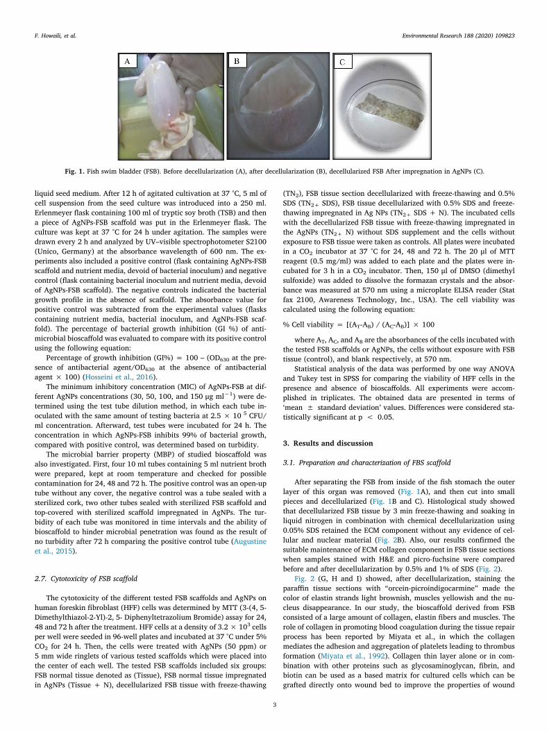
After separating the FSB from inside of the fish stomach the outerlayer of this organ was removed (Fig. 1A), and then cut into smallpieces and decellularized (Fig. 1B and C). Histological study showedthat decellularized FSB tissue by 3 min freeze-thawing and soaking inliquid nitrogen in combination with chemical decellularization using0.05% SDS retained the ECM component without any evidence of cel-lular and nuclear material (Fig. 2B). Also, our results confirmed thesuitable maintenance of ECM collagen component in FSB tissue sectionswhen samples stained with H&E and picro-fuchsine were comparedbefore and after decellularization by 0.5% and 1% of SDS (Fig. 2).
Fig. 2 (G, H and I) showed, after decellularization, staining theparaffin tissue sections with “orcein-picroindigocarmine” made thecolor of elastin strands light brownish, muscles yellowish and the nu-cleus disappearance. In our study, the bioscaffold derived from FSBconsisted of a large amount of collagen, elastin fibers and muscles. Therole of collagen in promoting blood coagulation during the tissue repairprocess has been reported by Miyata et al., in which the collagenmediates the adhesion and aggregation of platelets leading to thrombusformation (Miyata et al., 1992). Collagen thin layer alone or in com-bination with other proteins such as glycosaminoglycan, fibrin, andbiotin can be used as a based matrix for cultured cells which can begrafted directly onto wound bed to improve the properties of wound
Fig. 1. Fish swim bladder (FSB). Before decellularization (A), after decellularization (B), decellularized FSB After impregnation in AgNPs (C).
F. Howaili, et al. Environmental Research 188 (2020) 109823
3

dressing (Jeevithan et al., 2013).Also, the decellularization procedure which we used created more
porous scaffold. This facilitates the absorption of extra materials such asmetal nanoparticles for bioscaffold development and applications. Forexample, according to Zhou et al. studies porous hydroxyapatite scaf-fold could help the improvement of nanosilver doping (Zhou et al.,2015). In a study conducted by Shemsh et al., hematoxylin-eosin (H&E)stain was used for assessing the tissue morphology and the grade ofelastin degradation of ligamentum flavum of patients with lumbarspinal canal stenosis (Shemesh et al., 2017). Their results showed thesuitability of the staining method for elastin and other tissue component
evaluation as if used for FSB tissue staining procedures.SEM imaging analysis of FSB before decellularization and also
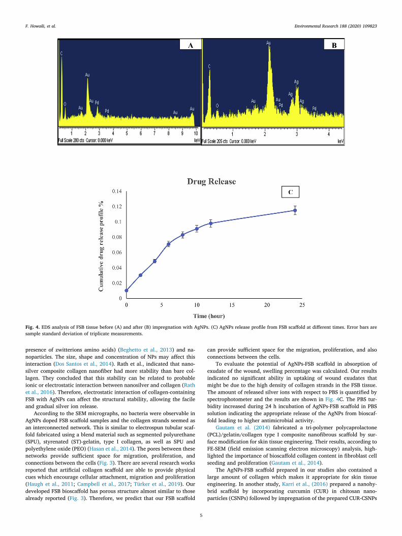
treated FSB tissue with various concentrations of AgNPs showed thepresence of polydispersed sphere AgNPs (100–200 nm) on the surfaceof the FSB scaffold (Fig. 3). Furthermore, energy dispersive spectro-scopy (EDS) analysis confirmed the presence of silver element on thesurface of the bioscaffold (Fig. 4A and B). AgNPs attached to FSBscaffold through tiny pores and its adsorption have probably been dueto electrostatic interactions.
Electrostatic interactions are possibly implicated in the bindingbetween collagen surface with zwitterionic functionality (due to
Fig. 2. FSB staining before and afterdecellularization with 0.5% and 1%SDS. H&E staining before decellular-ization (A); decellularization with 0.5%SDS (B); decellularization with 1% SDS(C); FSB tissue staining with picro-fuchsin (D); FSB tissue staining withpicro-fuchsin with 0.5% FSB (E); FSBstaining with picro-fuchsin with 1%SDS (F); FSB tissue staining with picroindigo carmine-orcein, which orceinstain the elastin fibers with dark redcolor and picroindigocarmin stain themuscles with yellowish brown color(G); FSB tissue staining with orcein-pi-croindigocarmine after decellulariza-tion with 0.5% SDS (H); FSB tissuestaining with orcein-picroindigo-carmine with 1% SDS (I). (For inter-pretation of the references to color inthis figure legend, the reader is referredto the Web version of this article.)
Fig. 3. SEM images of AgNPs (A); FSB tissue before decellularization (B); decellularized FSB tissue (C); decellularized FSB tissue after impregnation in AgNPs (D).
F. Howaili, et al. Environmental Research 188 (2020) 109823
4
presence of zwitterions amino acids) (Beghetto et al., 2013) and na-noparticles. The size, shape and concentration of NPs may affect thisinteraction (Dos Santos et al., 2014). Rath et al., indicated that nano-silver composite collagen nanofiber had more stability than bare col-lagen. They concluded that this stability can be related to probableionic or electrostatic interaction between nanosilver and collagen (Rathet al., 2016). Therefore, electrostatic interaction of collagen-containingFSB with AgNPs can affect the structural stability, allowing the facileand gradual silver ion release.
According to the SEM micrographs, no bacteria were observable inAgNPs doped FSB scaffold samples and the collagen strands seemed asan interconnected network. This is similar to electrospun tubular scaf-fold fabricated using a blend material such as segmented polyurethane(SPU), styrenated (ST)-gelatin, type I collagen, as well as SPU andpolyethylene oxide (PEO) (Hasan et al., 2014). The pores between thesenetworks provide sufficient space for migration, proliferation, andconnections between the cells (Fig. 3). There are several research worksreported that artificial collagen scaffold are able to provide physicalcues which encourage cellular attachment, migration and proliferation(Haugh et al., 2011; Campbell et al., 2017; Türker et al., 2019). Ourdeveloped FSB bioscaffold has porous structure almost similar to thosealready reported (Fig. 3). Therefore, we predict that our FSB scaffold
can provide sufficient space for the migration, proliferation, and alsoconnections between the cells.
To evaluate the potential of AgNPs-FSB scaffold in absorption ofexudate of the wound, swelling percentage was calculated. Our resultsindicated no significant ability in uptaking of wound exudates thatmight be due to the high density of collagen strands in the FSB tissue.The amount of released silver ions with respect to PBS is quantified byspectrophotometer and the results are shown in Fig. 4C. The PBS tur-bidity increased during 24 h incubation of AgNPs-FSB scaffold in PBSsolution indicating the appropriate release of the AgNPs from bioscaf-fold leading to higher antimicrobial activity.
Gautam et al. (2014) fabricated a tri-polymer polycaprolactone(PCL)/gelatin/collagen type I composite nanofibrous scaffold by sur-face modification for skin tissue engineering. Their results, according toFE-SEM (field emission scanning electron microscopy) analysis, high-lighted the importance of bioscaffold collagen content in fibroblast cellseeding and proliferation (Gautam et al., 2014).
The AgNPs-FSB scaffold prepared in our studies also contained alarge amount of collagen which makes it appropriate for skin tissueengineering. In another study, Karri et al., (2016) prepared a nanohy-brid scaffold by incorporating curcumin (CUR) in chitosan nano-particles (CSNPs) followed by impregnation of the prepared CUR-CSNPs
Fig. 4. EDS analysis of FSB tissue before (A) and after (B) impregnation with AgNPs. (C) AgNPs release profile from FSB scaffold at different times. Error bars aresample standard deviation of triplicate measurements.
F. Howaili, et al. Environmental Research 188 (2020) 109823
5
into collagen scaffold for tissue regeneration applications. They foundthat the synergistic combination of CUR (anti-inflammatory and anti-oxidant), chitosan (sustain drug carrier, wound healing) and collagen(established wound healer as a scaffold) are promising strategies toaddress various pathological manifestations of diabetic wounds, havingbetter wound healing capability (Karri et al., 2016).
AgNPs -FSB also can create a favorable 3-D bioscaffold for pro-mising wound dressing and tissue engineering applications. 3-D struc-ture scaffolds, mainly defined as device inclusive of polymeric bioma-terials or decellularized tissue possessing the superiority to affordstructural frame for cell attachment and tissue progress. There havebeen many attempts to mimic such a suitable 3-D environment for cellgrowth and proliferation (Goh et al., 2013; Mazza et al., 2015;Venugopal et al., 2008). Similarly, we developed a natural, decel-lularized scaffold, and the SEM images showed a 3-D interlinked porousstructure (Fig. 3). This porous structure provide suitable environmentfor cell culture. However, further studies are required to approvewhether this novel scaffold derived from fish swim bladder could pre-serve a qualified 3-D environment to promote adheration, multi-plication, or differentiation (Yang et al., 2008).
3.2. Antibacterial activity of FSB scaffold
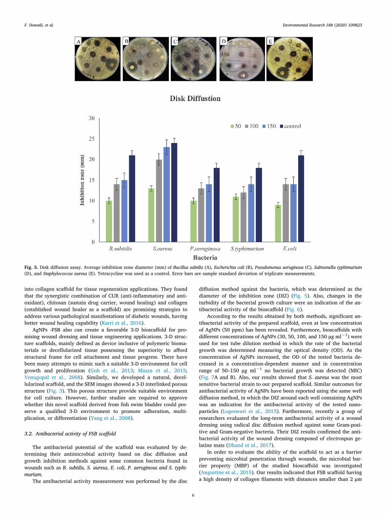
The antibacterial potential of the scaffold was evaluated by de-termining their antimicrobial activity based on disc diffusion andgrowth inhibition methods against some common bacteria found inwounds such as B. subtilis, S. aureus, E. coli, P. aeruginosa and S. typhi-murium.
The antibacterial activity measurement was performed by the disc
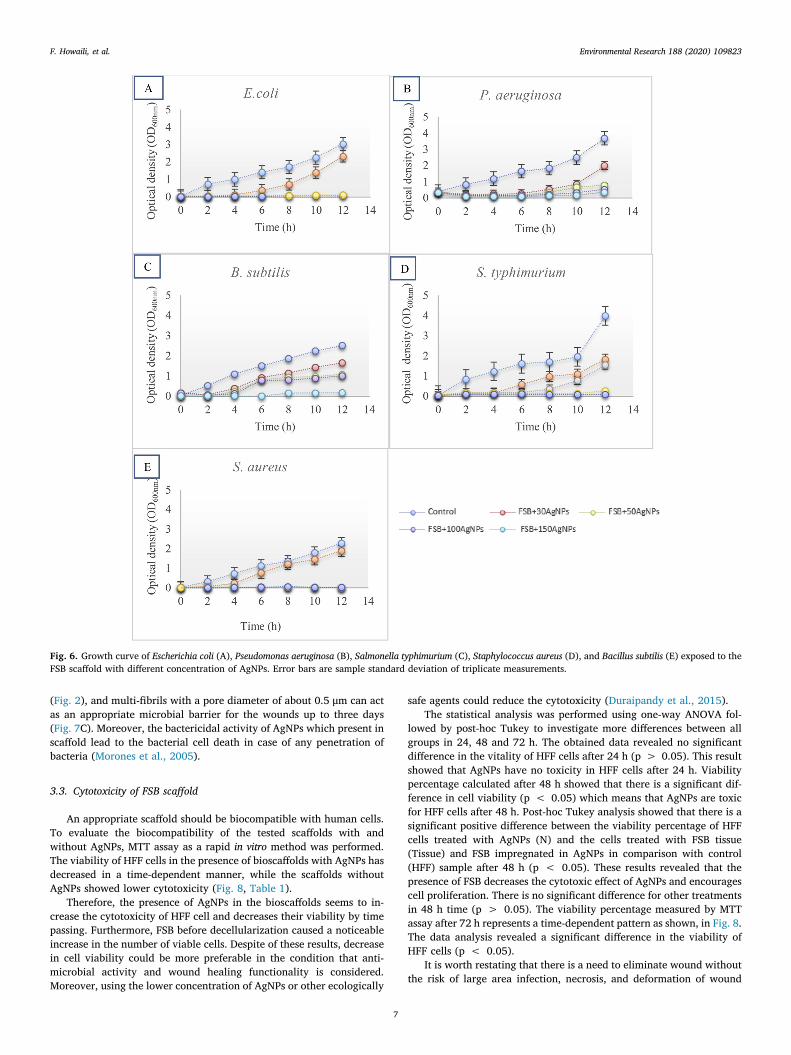
diffusion method against the bacteria, which was determined as thediameter of the inhibition zone (DIZ) (Fig. 5). Also, changes in theturbidity of the bacterial growth culture were an indication of the an-tibacterial activity of the bioscaffold (Fig. 6).
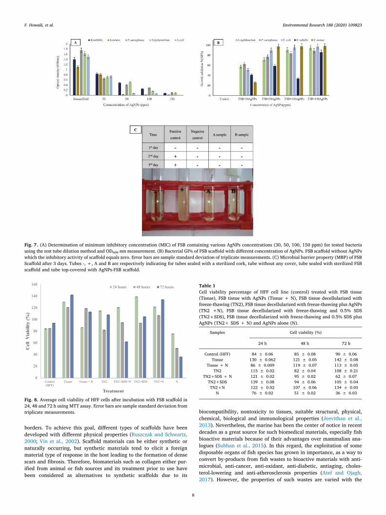
According to the results obtained by both methods, significant an-tibacterial activity of the prepared scaffold, even at low concentrationof AgNPs (50 ppm) has been revealed. Furthermore, bioscaffolds withdifferent concentrations of AgNPs (30, 50, 100, and 150 μg ml−1) wereused for test tube dilution method in which the rate of the bacterialgrowth was determined measuring the optical density (OD). As theconcentration of AgNPs increased, the OD of the tested bacteria de-creased in a concentration-dependent manner and in concentrationrange of 50–150 μg ml−1 no bacterial growth was detected (MIC)(Fig. 7A and B). Also, our results showed that S. aureus was the mostsensitive bacterial strain to our prepared scaffold. Similar outcomes forantibacterial activity of AgNPs have been reported using the same welldiffusion method, in which the DIZ around each well containing AgNPswas an indication for the antibacterial activity of the tested nano-particles (Logeswari et al., 2015). Furthermore, recently a group ofresearchers evaluated the long-term antibacterial activity of a wounddressing using radical disc diffusion method against some Gram-posi-tive and Gram-negative bacteria. Their DIZ results confirmed the anti-bacterial activity of the wound dressing composed of electrospun ge-latine mats (Dhand et al., 2017).
In order to evaluate the ability of the scaffold to act as a barrierpreventing microbial penetration through wounds, the microbial bar-rier property (MBP) of the studied bioscaffold was investigated(Augustine et al., 2015). Our results indicated that FSB scaffold havinga high density of collagen filaments with distances smaller than 2 μm
Fig. 5. Disk diffusion assay. Average inhibition zone diameter (mm) of Bacillus subtilis (A), Escherichia coli (B), Pseudomonas aeruginosa (C), Salmonella typhimurium(D), and Staphylococcus aureus (E). Tetracycline was used as a control. Error bars are sample standard deviation of triplicate measurements.
F. Howaili, et al. Environmental Research 188 (2020) 109823
6
(Fig. 2), and multi-fibrils with a pore diameter of about 0.5 μm can actas an appropriate microbial barrier for the wounds up to three days(Fig. 7C). Moreover, the bactericidal activity of AgNPs which present inscaffold lead to the bacterial cell death in case of any penetration ofbacteria (Morones et al., 2005).
3.3. Cytotoxicity of FSB scaffold
An appropriate scaffold should be biocompatible with human cells.To evaluate the biocompatibility of the tested scaffolds with andwithout AgNPs, MTT assay as a rapid in vitro method was performed.The viability of HFF cells in the presence of bioscaffolds with AgNPs hasdecreased in a time-dependent manner, while the scaffolds withoutAgNPs showed lower cytotoxicity (Fig. 8, Table 1).
Therefore, the presence of AgNPs in the bioscaffolds seems to in-crease the cytotoxicity of HFF cell and decreases their viability by timepassing. Furthermore, FSB before decellularization caused a noticeableincrease in the number of viable cells. Despite of these results, decreasein cell viability could be more preferable in the condition that anti-microbial activity and wound healing functionality is considered.Moreover, using the lower concentration of AgNPs or other ecologically
safe agents could reduce the cytotoxicity (Duraipandy et al., 2015).The statistical analysis was performed using one-way ANOVA fol-
lowed by post-hoc Tukey to investigate more differences between allgroups in 24, 48 and 72 h. The obtained data revealed no significantdifference in the vitality of HFF cells after 24 h (p > 0.05). This resultshowed that AgNPs have no toxicity in HFF cells after 24 h. Viabilitypercentage calculated after 48 h showed that there is a significant dif-ference in cell viability (p < 0.05) which means that AgNPs are toxicfor HFF cells after 48 h. Post-hoc Tukey analysis showed that there is asignificant positive difference between the viability percentage of HFFcells treated with AgNPs (N) and the cells treated with FSB tissue(Tissue) and FSB impregnated in AgNPs in comparison with control(HFF) sample after 48 h (p < 0.05). These results revealed that thepresence of FSB decreases the cytotoxic effect of AgNPs and encouragescell proliferation. There is no significant difference for other treatmentsin 48 h time (p > 0.05). The viability percentage measured by MTTassay after 72 h represents a time-dependent pattern as shown, in Fig. 8.The data analysis revealed a significant difference in the viability ofHFF cells (p < 0.05).
It is worth restating that there is a need to eliminate wound withoutthe risk of large area infection, necrosis, and deformation of wound
Fig. 6. Growth curve of Escherichia coli (A), Pseudomonas aeruginosa (B), Salmonella typhimurium (C), Staphylococcus aureus (D), and Bacillus subtilis (E) exposed to theFSB scaffold with different concentration of AgNPs. Error bars are sample standard deviation of triplicate measurements.
F. Howaili, et al. Environmental Research 188 (2020) 109823
7
borders. To achieve this goal, different types of scaffolds have beendeveloped with different physical properties (Ruszczak and Schwartz,2000; Vin et al., 2002). Scaffold materials can be either synthetic ornaturally occurring, but synthetic materials tend to elicit a foreignmaterial type of response in the host leading to the formation of densescars and fibrosis. Therefore, biomaterials such as collagen either pur-ified from animal or fish sources and its treatment prior to use havebeen considered as alternatives to synthetic scaffolds due to its
biocompatibility, nontoxicity to tissues, suitable structural, physical,chemical, biological and immunological properties (Jeevithan et al.,2013). Nevertheless, the marine has been the center of notice in recentdecades as a great source for such biomedical materials, especially fishbioactive materials because of their advantages over mammalian ana-logues (Subhan et al., 2015). In this regard, the exploitation of somedisposable organs of fish species has grown in importance, as a way toconvert by-products from fish wastes to bioactive materials with anti-microbial, anti-cancer, anti-oxidant, anti-diabetic, antiaging, choles-terol-lowering and anti-atherosclerosis properties (Atef and Ojagh,2017). However, the properties of such wastes are varied with the
Fig. 7. (A) Determination of minimum inhibitory concentration (MIC) of FSB containing various AgNPs concentrations (30, 50, 100, 150 ppm) for tested bacteriausing the test tube dilution method and OD600 nm measurement. (B) Bacterial GI% of FSB scaffold with different concentration of AgNPs. FSB scaffold without AgNPswhich the inhibitory activity of scaffold equals zero. Error bars are sample standard deviation of triplicate measurements. (C) Microbial barrier property (MBP) of FSBScaffold after 3 days. Tubes -, +, A and B are respectively indicating for tubes sealed with a sterilized cork, tube without any cover, tube sealed with sterilized FSBscaffold and tube top-covered with AgNPs-FSB scaffold.
Fig. 8. Average cell viability of HFF cells after incubation with FSB scaffold in24, 48 and 72 h using MTT assay. Error bars are sample standard deviation fromtriplicate measurements.
Table 1Cell viability percentage of HFF cell line (control) treated with FSB tissue(Tissue), FSB tissue with AgNPs (Tissue + N), FSB tissue decellularized withfreeze-thawing (TN2), FSB tissue decellularized with freeze-thawing plus AgNPs(TN2 +N), FSB tissue decellularized with freeze-thawing and 0.5% SDS(TN2+SDS), FSB tissue decellularized with freeze-thawing and 0.5% SDS plusAgNPs (TN2+ SDS + N) and AgNPs alone (N).
Samples Cell viability (%)
24 h 48 h 72 h
Control (HFF) 84 ± 0.06 85 ± 0.08 90 ± 0.06Tissue 130 ± 0.062 121 ± 0.05 142 ± 0.08
Tissue + N 86 ± 0.009 119 ± 0.07 113 ± 0.05TN2 115 ± 0.02 82 ± 0.04 108 ± 0.21
TN2+SDS + N 121 ± 0.02 95 ± 0.02 62 ± 0.07TN2+SDS 139 ± 0.08 94 ± 0.06 105 ± 0.04TN2+N 122 ± 0.02 107 ± 0.06 134 ± 0.05
N 76 ± 0.02 51 ± 0.02 36 ± 0.03
F. Howaili, et al. Environmental Research 188 (2020) 109823
8
origin of fish species and different parts of fish as well as living en-vironments of fish (Ideia et al., 2019). Thus, more investigations have tobe conducted for discovering the application of fish-byproducts prop-erties from all new sources. In this context, fish swim bladder due to theeasy availability, eco-friendly and natural-like behavior has been in-troduced as a suitable dressing for biomedical and pharmaceutical ap-plications. Also, its nanobiotechnological modification explored a newera of the potential application of FSB in tissue engineering which canprovide a base for pharmaceutical research in the near future.
4. Conclusions
A novel scaffold with antibacterial activity was successfully syn-thesized using decellularized fish swim bladder (FSB) and colloidalsilver nanoparticles (AgNPs). This scaffold contains a large amount ofthin collagen strands that increase migration and proliferation whichmake it an appropriate artificial scaffold for tissue engineering.Moreover, having prominent characteristics such as encompassing highdense collagen, appropriate antibacterial activity, suitable microbialbarrier, good flexibility, biocompatibility and biodegradability withease of formulation, we propose FSB as an advantageous bioresource forwound dressing.
Declaration of interest statement
We wish to confirm that there are no known conflicts of interestassociated with this publication and there has been no significant fi-nancial support for this work that could have influenced its outcome.
Credit Author Statement
Fadak Mousavi Howaili: Investigation, Methodology, Visualization,Writing - original draft, Writing - review & editing. Mansour Mashreghi:Supervision, Funding acquisition, Data curation, Validation, Writing -original draft, Writing - review & editing. Nasser Mahdavi Shahri:Conceptualization, Methodology, Formal analysis, Validation. Writing -Review & Editing. Ahmad Kompany: Validation. Project administration,Writing - review & editing. Razieh Jalal: Methodology, Formal analysis,Validation, Data curation.
Acknowledgments
We acknowledge financial support from the Ferdowsi University ofMashhad (grant number: 3/31140).
References
Atef, M., Ojagh, S.M., 2017. Health benefits and food applications of bioactive com-pounds from fish byproducts: a review. J. Funct. Foods 35, 673–681.
Augustine, R., Kalarikkal, N., Thomas, S., 2015. An in vitro method for the determinationof microbial barrier property (MBP) of porous polymeric membranes for skin sub-stitute and wound dressing applications. Tissue Eng. Regen. Med. 12, 12–19.
Beghetto, V., Zancanaro, A., Scrivanti, A., Matteoli, U., Pozza, G., 2013. The leather in-dustry: a chemistry insight Part I: an overview of the industrial process. Sci.Ca’Foscari.
Campbell, J.J., Husmann, A., Hume, R.D., Watson, C.J., Cameron, R.E., 2017.Development of three-dimensional collagen scaffolds with controlled architecture forcell migration studies using breast cancer cell lines. Biomaterials 114, 34–43.
Cheng, F., Gao, J., Wang, L., Hu, X., 2015. Composite chitosan/poly (ethylene oxide)electrospun nanofibrous mats as novel wound dressing matrixes for the controlledrelease of drugs. J. Appl. Polym. Sci. 132.
Crapo, P.M., Gilbert, T.W., Badylak, S.F., 2011. An overview of tissue and whole organdecellularization processes. Biomaterials 32, 3233–3243.
Dhand, C., Venkatesh, M., Barathi, V.A., Harini, S., Bairagi, S., Leng, E.G.T.,Muruganandham, N., Low, K.Z.W., Fazil, M.H.U.T., Loh, X.J., 2017. Bio-inspiredcrosslinking and matrix-drug interactions for advanced wound dressings with long-term antimicrobial activity. Biomaterials 138, 153–168.
Dos Santos, C.A., Seckler, M.M., Ingle, A.P., Gupta, I., Galdiero, S., Galdiero, M., Gade, A.,Rai, M., 2014. Silver nanoparticles: therapeutical uses, toxicity, and safety issues. J.Pharmacol. Sci. 103, 1931–1944.
Duraipandy, N., Lakra, R., Srivatsan, K.V., Ramamoorthy, U., Korrapati, P.S., Kiran, M.S.,
2015. Plumbagin caged silver nanoparticle stabilized collagen scaffold for wounddressing. J. Mater. Chem. B 3, 1415–1425.
Fan, Z., Liu, B., Wang, J., Zhang, S., Lin, Q., Gong, P., Ma, L., Yang, S., 2014. A novelwound dressing based on Ag/graphene polymer hydrogel: effectively kill bacteria andaccelerate wound healing. Adv. Funct. Mater. 24, 3933–3943.
Finney, J.L., Robertson, G.N., McGee, C.A.S., Smith, F.M., Croll, R.P., 2006. Structure andautonomic innervation of the swim bladder in the zebrafish (Danio rerio). J. Comp.Neurol. 495, 587–606. https://doi.org/10.1002/cne.20948.
Gautam, S., Chou, C.-F., Dinda, A.K., Potdar, P.D., Mishra, N.C., 2014. Surface mod-ification of nanofibrous polycaprolactone/gelatin composite scaffold by collagen typeI grafting for skin tissue engineering. Mater. Sci. Eng. C 34, 402–409.
Ghosh, S.K., Mandal, D., 2016. Efficient natural piezoelectric nanogenerator: electricitygeneration from fish swim bladder. Nanomater. Energy 28, 356–365.
Goh, S.-K., Bertera, S., Olsen, P., Candiello, J.E., Halfter, W., Uechi, G., Balasubramani,M., Johnson, S.A., Sicari, B.M., Kollar, E., 2013. Perfusion-decellularized pancreas asa natural 3D scaffold for pancreatic tissue and whole organ engineering. Biomaterials34, 6760–6772.
Hafidh, R.R., Abdulamir, A.S., Baker, F.A., 2009. Comparative histological and histo-chemical inter-species investigation of mammalian submandibular salivary glands.Era Mod. Diag. Pathol. 384.
Hasan, A., Memic, A., Annabi, N., Hossain, M., Paul, A., Dokmeci, M.R., Dehghani, F.,Khademhosseini, A., 2014. Electrospun scaffolds for tissue engineering of vasculargrafts. Acta Biomater. 10, 11–25.
Haugh, M.G., Murphy, C.M., McKiernan, R.C., Altenbuchner, C., O'Brien, F.J., 2011.Crosslinking and mechanical properties significantly influence cell attachment, pro-liferation, and migration within collagen glycosaminoglycan scaffolds. Tissue Eng.17, 1201–1208.
Hosseini, M., Mashreghi, M., Eshghi, H., 2016. Biosynthesis and antibacterial activity ofgold nanoparticles coated with reductase enzymes. Micro & Nano Lett. 11, 484–489.
Ideia, P., Pinto, J., Ferreira, R., Figueiredo, L., Spínola, V., Castilho, P.C., 2019. Fishprocessing industry residues: a review of valuable products extraction and char-acterization methods. Waste and Biomass Valorization 1–24.
Jackson, D.W., Grood, E.S., Wilcox, P., Butler, D.L., Simon, T.M., Holden, J.P., 1988. Theeffects of processing techniques on the mechanical properties of bone-anteriorcruciate ligament-bone allografts: an experimental study in goats. Am. J. Sports Med.16, 101–105.
Jeevithan, E., Qingbo, Z., Bao, B., Wu, W., 2013. Biomedical and pharmaceutical appli-cation of fish collagen and gelatin: a review. J. Nutr. Therapeut. 2, 218–227.
Karri, V.V.S.R., Kuppusamy, G., Talluri, S.V., Mannemala, S.S., Kollipara, R., Wadhwani,A.D., Mulukutla, S., Raju, K.R.S., Malayandi, R., 2016. Curcumin loaded chitosannanoparticles impregnated into collagen-alginate scaffolds for diabetic woundhealing. Int. J. Biol. Macromol. 93, 1519–1529.
Kiernan, J.A., 2001. Histological and Histochemical Methods. Theory Pract, Arnold Ed. .Klasen, H.J., 2000. A historical review of the use of silver in the treatment of burns. II.
Renewed interest for silver. Burns 26, 131–138.Li, J., Wang, M., Qiao, Y., Tian, Y., Liu, J., Qin, S., Wu, W., 2018. Extraction and char-
acterization of type I collagen from skin of tilapia (Oreochromis niloticus) and itspotential application in biomedical scaffold material for tissue engineering. ProcessBiochem. 74, 156–163.
Lillie, R.D., Pizzolato, P., Donaldson, P.T., 1976. Nuclear stains with soluble metachromemetal mordant dye lakes. Histochemistry 49, 23–35.
Logeswari, P., Silambarasan, S., Abraham, J., 2015. Synthesis of silver nanoparticles usingplants extract and analysis of their antimicrobial property. J. Saudi Chem. Soc. 19,311–317.
Madhumathi, K., Kumar, P.T.S., Abhilash, S., Sreeja, V., Tamura, H., Manzoor, K., Nair,S.V., Jayakumar, R., 2010. Development of novel chitin/nanosilver composite scaf-folds for wound dressing applications. J. Mater. Sci. Mater. Med. 21, 807–813.
Maneerung, T., Tokura, S., Rujiravanit, R., 2008. Impregnation of silver nanoparticlesinto bacterial cellulose for antimicrobial wound dressing. Carbohydr. Polym. 72,43–51.
Mazza, G., Rombouts, K., Hall, A.R., Urbani, L., Luong, T.V., Al-Akkad, W., Longato, L.,Brown, D., Maghsoudlou, P., Dhillon, A.P., 2015. Decellularized human liver as anatural 3D-scaffold for liver bioengineering and transplantation. Sci. Rep. 5, 13079.
Miranda-Nieves, D., Chaikof, E.L., 2016. Collagen and elastin biomaterials for the fabri-cation of engineered living tissues. ACS Biomater. Sci. Eng. 3, 694–711.
Miyata, T., Taira, T., Noishiki, Y., 1992. Collagen engineering for biomaterial use. Clin.Mater. 9, 139–148.
Mohammadie, Z.M., Parivar, K., Shahri, N.M., Fereidoni, M., Hayati-Roodbari, N., 2017.Decellularized bovine articular cartilage matrix reinforced by carboxylated-SWCNTfor tissue engineering application. Braz. Arch. Biol. Technol. 60.
Morones, J.R., Elechiguerra, J.L., Camacho, A., Holt, K., Kouri, J.B., Ramírez, J.T.,Yacaman, M.J., 2005. The bactericidal effect of silver nanoparticles. Nanotechnology16, 2346.
Naderi, S., Zadeh, J.K., Shahri, N.M., Abady, K.N.S., Cheravi, M., Baharara, J., Rad,S.A.B., Bahrami, A.R., 2013. Three-dimensional Scaffold from decellularized humanGingiva for cell cultures: glycoconjugates and cell behavior. Cell J 15, 166.
Pati, F., Adhikari, B., Dhara, S., 2010. Isolation and characterization of fish scale collagenof higher thermal stability. Bioresour. Technol. 101, 3737–3742. https://doi.org/10.1016/j.biortech.2009.12.133.
Rai, M., Kon, K., Ingle, A., Duran, N., Galdiero, S., Galdiero, M., 2014. Broad-spectrumbioactivities of silver nanoparticles: the emerging trends and future prospects. Appl.Microbiol. Biotechnol. 98, 1951–1961.
Rai, M., Yadav, A., Gade, A., 2009. Silver nanoparticles as a new generation of anti-microbials. Biotechnol. Adv. 27, 76–83.
Rath, G., Hussain, T., Chauhan, G., Garg, T., Goyal, A.K., 2016. Collagen nanofibercontaining silver nanoparticles for improved wound-healing applications. J. Drug
F. Howaili, et al. Environmental Research 188 (2020) 109823
9
Target. 24, 520–529.Ruszczak, Z., Schwartz, R.A., 2000. Modern aspects of wound healing: an update.
Dermatol. Surg. 26, 219–229.Shahabipour, F., Mahdavi-Shahri, N., Matin, M.M., Tavassoli, A., Zebarjad, S.M., 2013.
Scaffolds derived from cancellous bovine bone support mesenchymal stem cells'maintenance and growth. In Vitro Cell. Dev. Biol. Anim. 49, 440–448. https://doi.org/10.1007/s11626-013-9591-7.
Shemesh, S., Sidon, E., Kaisler, E., Sheinis, D., Velkes, S., Ohana, N., Benayahu, D., 2017.Diabetes mellitus is associated with increased elastin fiber loss in ligamentum flavumof patients with lumbar spinal canal stenosis: results of a pilot histological study. Eur.Spine J. 1–9.
Subhan, F., Ikram, M., Shehzad, A., Ghafoor, A., 2015. Marine collagen: an emergingplayer in biomedical applications. J. Food Sci. Technol. 52, 4703–4707.
Türker, E., Yildiz, Ü.H., Yildiz, A.A., 2019. Biomimetic hybrid scaffold consisting of co-electrospun collagen and PLLCL for 3D cell culture. Int. J. Biol. Macromol. 139,1054–1062.
Unnithan, A.R., Gnanasekaran, G., Sathishkumar, Y., Lee, Y.S., Kim, C.S., 2014.Electrospun antibacterial polyurethane–cellulose acetate–zein composite mats for
wound dressing. Carbohydr. Polym. 102, 884–892.Venugopal, J., Low, S., Choon, A.T., Ramakrishna, S., 2008. Interaction of cells and na-
nofiber scaffolds in tissue engineering. J. Biomed. Mater. Res. B Appl. Biomater. 84,34–48.
Vin, F., Teot, L., Meaume, S., 2002. The healing properties of Promogran in venous legulcers. J. Wound Care 11, 335–341.
Wei, B., Yang, G., Hong, F., 2011. Preparation and evaluation of a kind of bacterial cel-lulose dry films with antibacterial properties. Carbohydr. Polym. 84, 533–538.
Yang, Q., Peng, J., Guo, Q., Huang, J., Zhang, L., Yao, J., Yang, F., Wang, S., Xu, W.,Wang, A., 2008. A cartilage ECM-derived 3-D porous acellular matrix scaffold for invivo cartilage tissue engineering with PKH26-labeled chondrogenic bone marrow-derived mesenchymal stem cells. Biomaterials 29, 2378–2387.
Zhang, D., Zhou, W., Wei, B., Wang, X., Tang, R., Nie, J., Wang, J., 2015. Carboxyl-modified poly (vinyl alcohol)-crosslinked chitosan hydrogel films for potential wounddressing. Carbohydr. Polym. 125, 189–199.
Zhou, K., Dong, C., Zhang, X., Shi, L., Chen, Z., Xu, Y., Cai, H., 2015. Preparation andcharacterization of nanosilver-doped porous hydroxyapatite scaffolds. Ceram. Int. 41,1671–1676.
F. Howaili, et al. Environmental Research 188 (2020) 109823
10