Embed Size (px)
Citation preview
RESEARCH ARTICLE DEVELOPMENT AND STEM CELLS2878
Development 139, 2878-2890 (2012) doi:10.1242/dev.074765© 2012. Published by The Company of Biologists Ltd
INTRODUCTIONNervous system development involves complex extrinsic andintrinsic signaling pathways to precisely coordinate themaintenance of the necessary neural stem/progenitor (NS/P) pooland, at the same time, the orderly acquisition of neural cell fate.Neurons and glia, comprising astrocytes and oligodendrocytes,arise from multipotent neural stem cells in a temporally definedorder: generation of neurons precedes glia and generation ofastrocytes precedes oligodendrocytes (Edlund and Jessell, 1999;Okano and Temple, 2009; Qian et al., 2000). It is well establishedthat the coordinated activation or suppression of transcriptionalactivators and repressors, which regulate the expression of anetwork of lineage-specific genes, plays a major role in this process(Cheng et al., 2005; Edlund and Jessell, 1999; Ross et al., 2003).Nevertheless, the mechanism utilized by specific regulators tocontrol the proper progression of neural development remainsmostly unknown.
A potential candidate protein, which could play an essentialrole in this process, is the transcriptional repressor REST (RE1silencing transcription factor, also called NRSF) (Chong et al.,1995; Schoenherr and Anderson, 1995). REST regulates a largenumber of neuronal genes encoding fundamental neuronal traitsas well as non-coding brain-specific microRNA genes (Bruce etal., 2004; Johnson et al., 2007; Otto et al., 2007). In non-neural
cells, REST binds a conserved 23 bp element, RE1 (repressorelement 1, also called NRSE) (Chong et al., 1995; Schoenherrand Anderson, 1995), which is present in its target genes, andacts to suppress their expression via two distinct repressordomains (Andres et al., 1999; Ballas et al., 2001; Roopra et al.,2000). Our previous studies showed that, although REST, inconjunction with its co-repressor CoREST (RCOR1 – MouseGenome Informatics), silences neuronal genes in non-neuronalcells outside the nervous system, the same genes are repressedand poised for expression in pluripotent embryonic stem (ES)cells (Ballas et al., 2005; Ballas and Mandel, 2005). Thisdifferential regulation of the same sets of genes by REST ismediated by the differential recruitment of chromatin modifiersby CoREST (Ballas and Mandel, 2005). Importantly, REST itselfis differentially regulated throughout neural development;whereas REST is expressed at a high level in ES cells, it isdownregulated to a minimal level in NS/P cells. As NS/P cellsdifferentiate, REST remains present in glia (Abrajano et al.,2009; Dewald et al., 2011) but is largely absent in neurons,allowing the restricted expression of its target genes specificallyin neurons (Ballas et al., 2005). Whereas the high level of RESTexpression in ES cells is mediated by NANOG and OCT4(POU5F1 – Mouse Genome Informatics)) (Boyer et al., 2005;Loh et al., 2006), the low level in NS/P cells is mediated, at leastin part, by proteasomal degradation via the E3 ubiquitin ligase-TRCP (BTRC – Mouse Genome Informatics) (Ballas et al.,2005; Westbrook et al., 2008). However, the function andsignificance of this differential regulation of REST duringnervous system development and whether the normal low levelof REST present in NS/P cells has any roles in maintaining NS/Pcell self-renewal and/or differentiation along the different neurallineages remains largely unknown.
Department of Biochemistry and Cell Biology, Stony Brook University, Stony Brook,New York, NY 11794, USA.
*Author for correspondence ([email protected])
Accepted 18 May 2012
SUMMARYREST is a master repressor of neuronal genes; however, whether it has any role during nervous system development remains largelyunknown. Here, we analyzed systematically the role of REST in embryonic stem cells and multipotent neural stem/progenitor (NS/P)cells, including neurogenic and gliogenic NS/P cells derived from embryonic stem (ES) cells or developing mouse embryos. We showedthat REST-null ES cells remained pluripotent and generated teratomas consisting of the three germ layers. By contrast, multipotentNS/P cells lacking REST displayed significantly reduced self-renewal capacity owing to reduced cell cycle kinetics and precociousneuronal differentiation. Importantly, although early-born neurogenic NS/P cells that lack REST were capable of differentiating toneurons and glia, the neuronal and oligodendrocytic pools were significantly enlarged and the astrocytic pool was shrunken.However, gliogenic NS/P cells lacking REST were able to generate a normal astrocytic pool size, suggesting that the shrinkage of theastrocytic pool generated from neurogenic NS/P cells lacking REST probably occurs by default. Microarray profiling of early-bornNS/P cells lacking REST showed upregulation of neuronal as well as oligodendrocytic genes, specifically those involved in myelination.Furthermore, chromatin immunoprecipitation analyses showed that some of the upregulated oligodendrocytic genes contain anRE1 motif and are direct REST targets. Together, our data support a central role for REST during neural development in promotingNS/P cell self-renewal while restricting the generation and maturation of neurons and oligodendrocytes.
KEY WORDS: Embryonic stem cells, Neural development, Neural stem/progenitor cells, Neurons, Oligodendrocytes, REST, Mouse
REST regulates the pool size of the different neural lineagesby restricting the generation of neurons andoligodendrocytes from neural stem/progenitor cellsMatthew V. Covey, Jeffrey W. Streb, Roman Spektor and Nurit Ballas*
DEVELO
PMENT
2879RESEARCH ARTICLEREST regulates neural lineages pools
Here, we analyzed the function of REST at different stages ofneural development and show that, although the high level ofREST in ES cells is essential for its full functioning as a repressorof neuronal genes, neither REST nor CoREST is required formaintaining ES cell pluripotency. Furthermore, REST is notrequired for the conversion of ES cells to neurogenic or gliogenicNS/P cells; however, REST is crucial for maintaining NS/P cellself-renewal and proper ratios of neurons:glia duringdifferentiation. Although the loss of REST in neurogenic NS/P cellsaccelerated neuronal differentiation by depleting the NS/P andastrocytic pools, it had no effect on the pool size of astrocytesgenerated from gliogenic NS/P cells. Importantly, we show thatREST regulates genes encoding not only neuronal but alsooligodendrocyte specification and maturation and that the loss ofREST in NS/P cells resulted in de-repression of these two geneclasses and acceleration of both neuronal and oligodendrocytedifferentiation.
MATERIALS AND METHODSAnimal careAll experiments were performed in accordance with research guidelines setby the Institutional Animal Care and Use Committee (IACUC) of StonyBrook University.
ES cells and conversion to NS/P cellsWild-type (Rest+/+), heterozygous (Rest+/–) and homozygous (Rest–/–) EScells were described previously (Jorgensen et al., 2009b). ES cells werecultured as described (Ballas et al., 2005). Conversion of ES cells to NS/Pcells was performed using the ES cell-derived neurosphere system asdescribed (Okada et al., 2008) except that ultra low attachment dishes wereprepared with Glycosan HyStem (BioTime).
Teratoma formationTeratomas were generated from Rest+/+ and Rest–/– ES cells bysubcutaneous injection of 5�106 cells into the left or right side of the spineof B6.Cg-Foxn1nu/J nude mice (The Jackson Laboratory). After 3 weeks,the teratomas were dissected, fixed with 4% paraformaldehyde, embedded,sectioned and stained with Hematoxylin and Eosin using a standardprotocol.
Primary cultures of NS/P cellsNS/P cells were isolated from cortices of embryonic day (E) 12.5 mouseembryos as described (Ballas et al., 2005) and cultured in the presence of20 ng/ml epidermal growth factor (EGF; R&D Systems) and basicfibroblast growth factor (bFGF; Millipore). NS/P cells from cortices ofE17.5 mouse embryos were isolated in a similar manner except that theacutely isolated cortical NS/P cells were labeled with CD15 (LeX/SSEA1)-FITC (BD Biosciences) and sorted by fluorescence-activated cell sorting(FACS) for LeX-positive cells as described (Ballas et al., 2005).
Transduction of ES and NS/P cells with lenti virus-carrying shRNAThe different shRNAs were constructed in a lenti viral vector (a gift fromDr Lemischka, Mount Sinai Medical Center) under the human H1promoter as described (Ivanova et al., 2006). The vector also contained gfpcDNA driven by the ubiquitin C promoter and served as a marker for thetransduced cells. Sequences used for shRNA design: Rest shRNA#1, 5�-GCCGAATCTGAAGAGCAGT-3�; Rest shRNA#2, 5�-GTGTAATCTAC -AATACCA-3�; CoRest shRNA#1, 5�-AGTCTTATTTGAGCAAGCC-3�;CoRest shRNA#2, 5�-GTCTTATTTGAGCAAGCCT-3�; ScrambledshRNA, 5�-ATCATTCCGGATACTGCGA-3�. Mouse embryonic fibroblast(MEF)-free ES cells were transduced in suspension for 12 hours at 37°Cin the presence of polybrene (100 g/ml), washed, cultured on MEFs until50% confluent, FACS sorted for GFP+ cells, and cultured on MEFs. E12.5or E17.5 NS/P cells were dissociated at their first passage, plated oncoverslips or 60-mm dishes coated with poly-D-lysine/laminin (BDBiosciences) and transduced with lenti virus after one day in culture. Cellswere washed, cultured in NS/P media supplemented with EGF and FGF
for 6 days, FACS sorted for GFP and cultured as above. Unless otherwisespecified, shRNA#2 and shRNA#1 were used to knock down REST andCoREST, respectively.
Competition assayNS/P cells were transduced with lenti virus carrying scrambled or RestshRNA and gfp (H1PshRNA-UbiqC-EGFP) or with lenti virus carryingdsRed (H1P-UbiqC-dsRed). GFP- or dsRed-positive cells were FACS sortedafter 6 days in culture and replated at a 3:1 GFP:DsRed ratio for 7 days withfresh EGF and FGF. The cells were dissociated and FACS sorted. Thenumber of GFP and dsRed cells per sample was recorded and the mixed cellswere replated as above. This procedure was repeated for three passages.
Neurosphere formation assayFor primary neurospheres, NS/P cells transduced with lenti virus carryingRest or scrambled shRNA and FACS sorted for GFP were plated at adensity of 50,000 cells/well in a 12-well plate (BD Biosciences) with EGFand FGF for 6 days. For generation of secondary neurospheres, the primaryneurospheres were dissociated and replated at similar density as describedabove.
Alkaline phosphatase assayES cells were cultured on MEFs in a 12-well plate and fixed with 2%paraformaldehyde. Vector Red substrate working solution (VectorLaboratories) was applied for 30 minutes followed by washes with 100mM Tris-HCl buffer (pH 8.0).
ImmunocytochemistryImmunostaining of cells on coverslips was performed as described (Ballaset al., 2005). ES cell-derived neurospheres were fixed in 4%paraformaldehyde, cryoprotected in 30% sucrose in PBS overnight, washedin PBS, frozen in Neg 50 (Richard-Allan Scientific), sectioned at 10 mon a cryostat and mounted onto gelatin-coated slides. Primary antibodiesused were: chicken anti-GFP (1:500, AbCam); rabbit anti-GFAP (1:500,Dako); mouse anti-TUJ1 (1:500, Covance); mouse anti-CNPase (1:500,Sigma); mouse anti-nestin (1:500, Millipore); mouse anti-Ki67 (1:50, BDBiosciences); mouse anti-phosphohistone H3 (1:50, Cell Signaling); mouseanti-SSEA1 (CD15) (1:50, BD Biosciences); rabbit anti-OCT4 (1:100,AbCam). Secondary antibodies were conjugated to Alexa Fluor(Invitrogen). Images were collected on a Zeiss confocal laser scanningLSM 510 microscope.
Western blot analysisWhole cell extracts were prepared using a modified Dignam method asdescribed (Ballas et al., 2005). Primary antibodies used were: rabbit anti-REST-C (1:1000) and rabbit anti-CoREST (1:800) as described (Ballas etal., 2005); rabbit anti--actin (1:5000, Sigma); rabbit anti-Sin3A (1:1000,Santa Cruz).
Quantitative real-time RT-PCR analysisTotal RNA was isolated using Trizol (Invitrogen). Quantitative real-timePCR was performed following reverse transcription using a StepOnePlusRealtime PCR System (Applied Biosystems). The relative amount of thespecific mRNAs was normalized to the level of Gapdh mRNA. Thesequences of primers used are listed in supplementary material Table S1.
Microarray analysis of NS/P cellsMicroarray analysis was performed at the Bionomics Research andTechnology Center (BRTC), University of Medicine and Dentistry of NewJersey. E12.5 NS/P cells were transduced with lenti virus carrying Rest orscrambled shRNA, FACS sorted after 6 days in culture, and replated in thepresence of EGF and FGF for 3 days. Total RNA was isolated from threeindependent experiments and analyzed individually. Microarray data havebeen deposited in the Gene Expression Omnibus database under accessionnumber GSE38538.
Chromatin immunoprecipitation (ChIP)ChIP assays were performed on E12.5 NS/P cells cultured with EGF andFGF as previously described (Ballas et al., 2005). The sheared chromatinwas immunoprecipitated using rabbit anti-REST antibodies generated D
EVELO
PMENT
2880 RESEARCH ARTICLE Development 139 (16)
against the N terminus (anti-REST p73) (Chong et al., 1995) or the Cterminus (anti-REST-C) (Ballas et al., 2005). Normal rabbit IgG (SantaCruz) was used as a control. Primer sets designed to amplify the RE1-containing regions of the specific neuronal and oligodendrocytic geneswere as follows: Syt4, forward 5�-GCGTCAATTCGGGTTTCAGC -CACGTC-3�, reverse 5�-CAAGTTCAGCAACTTGCTCACCGAAT-3�;Snap25, forward 5�-GCTACAGGCGAGCATGTGCTGCAAAC-3�,reverse 5�-GTGAGGA AGAGAGCAGGCAATTGTC-3�; Mal, forward 5�-GTCTACTG AAAGAACGGGAACC-3�, reverse 5�-CTTTGGCAAGA -GTACTG GGAAC-3�; Myt1, forward 5�-CAAGCAGGAGCTGTTT -GAGTTGGT-3�, reverse 5�-TGACAGTGTTGGAGAACAAAGCGG-3�;Myt1L, forward 5�-CCCTCCCAAAGTTAATCACCCATC-3�, reverse 5�-CAGTGAACCTGTAGGAAGTCTCTG-3�.
Statistical analysisData were analyzed for statistical significance using Student’s t-test.Comparisons were interpreted as significant when associated with P<0.05.For the microarray, data were analyzed for statistical significance usingpaired t-test.
RESULTSREST functions as a repressor of neuronal genes inES cells but is not required for maintaining ES cellpluripotency or for their conversion to NS/P cellsBecause REST is a key regulator of neuronal genes and iscontrolled by pluripotent transcription factors, it could potentiallybe involved in maintaining ES cell pluripotency. Recent studiesattempting to analyze whether REST is involved in maintaining thepluripotency state of ES cells remain controversial (Buckley et al.,2009; Jorgensen et al., 2009a; Jorgensen and Fisher, 2010; Singhet al., 2008). Thus, we sought to analyze systematically the state ofES cells lacking REST. We compared homozygous Rest knockout(Rest–/–) ES cells, isolated from a previously generated Rest-nullmouse model (Chen et al., 1998), as well as heterozygous (Rest+/–)ES cells containing half of the normal amount of REST, to wild-
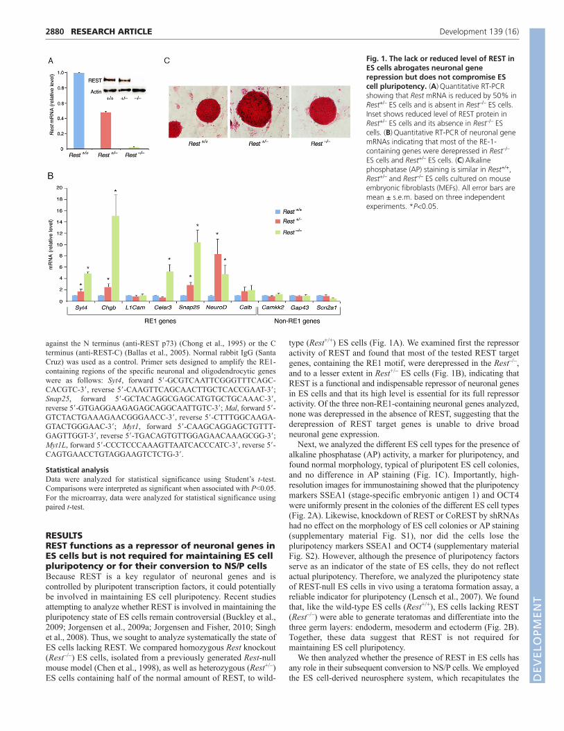
type (Rest+/+) ES cells (Fig. 1A). We examined first the repressoractivity of REST and found that most of the tested REST targetgenes, containing the RE1 motif, were derepressed in the Rest–/–,and to a lesser extent in Rest+/– ES cells (Fig. 1B), indicating thatREST is a functional and indispensable repressor of neuronal genesin ES cells and that its high level is essential for its full repressoractivity. Of the three non-RE1-containing neuronal genes analyzed,none was derepressed in the absence of REST, suggesting that thederepression of REST target genes is unable to drive broadneuronal gene expression.
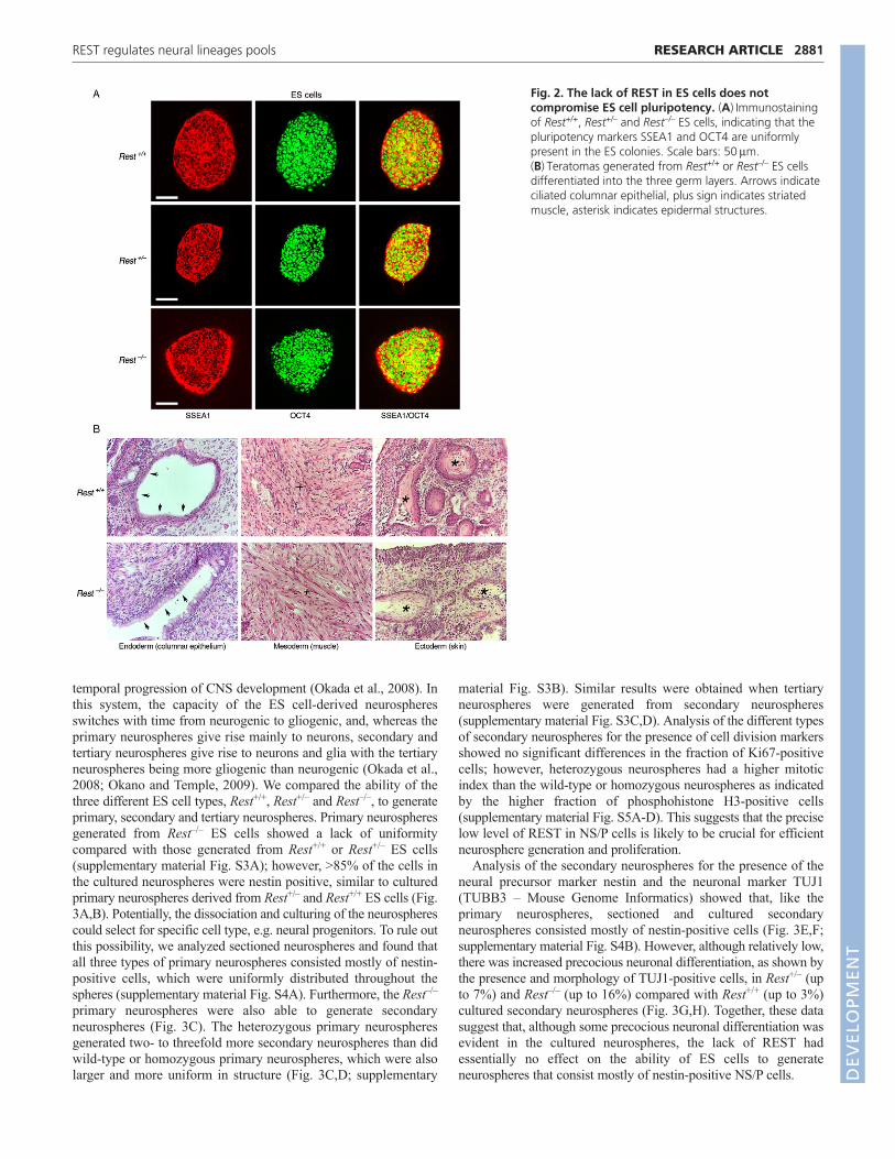
Next, we analyzed the different ES cell types for the presence ofalkaline phosphatase (AP) activity, a marker for pluripotency, andfound normal morphology, typical of pluripotent ES cell colonies,and no difference in AP staining (Fig. 1C). Importantly, high-resolution images for immunostaining showed that the pluripotencymarkers SSEA1 (stage-specific embryonic antigen 1) and OCT4were uniformly present in the colonies of the different ES cell types(Fig. 2A). Likewise, knockdown of REST or CoREST by shRNAshad no effect on the morphology of ES cell colonies or AP staining(supplementary material Fig. S1), nor did the cells lose thepluripotency markers SSEA1 and OCT4 (supplementary materialFig. S2). However, although the presence of pluripotency factorsserve as an indicator of the state of ES cells, they do not reflectactual pluripotency. Therefore, we analyzed the pluripotency stateof REST-null ES cells in vivo using a teratoma formation assay, areliable indicator for pluripotency (Lensch et al., 2007). We foundthat, like the wild-type ES cells (Rest+/+), ES cells lacking REST(Rest–/–) were able to generate teratomas and differentiate into thethree germ layers: endoderm, mesoderm and ectoderm (Fig. 2B).Together, these data suggest that REST is not required formaintaining ES cell pluripotency.
We then analyzed whether the presence of REST in ES cells hasany role in their subsequent conversion to NS/P cells. We employedthe ES cell-derived neurosphere system, which recapitulates the
Fig. 1. The lack or reduced level of REST inES cells abrogates neuronal generepression but does not compromise EScell pluripotency. (A)Quantitative RT-PCRshowing that Rest mRNA is reduced by 50% inRest+/– ES cells and is absent in Rest–/– ES cells.Inset shows reduced level of REST protein inRest+/– ES cells and its absence in Rest–/– EScells. (B)Quantitative RT-PCR of neuronal genemRNAs indicating that most of the RE-1-containing genes were derepressed in Rest–/–
ES cells and Rest+/– ES cells. (C)Alkalinephosphatase (AP) staining is similar in Rest+/+,Rest+/– and Rest–/– ES cells cultured on mouseembryonic fibroblasts (MEFs). All error bars aremean ± s.e.m. based on three independentexperiments. *P<0.05.
DEVELO
PMENT
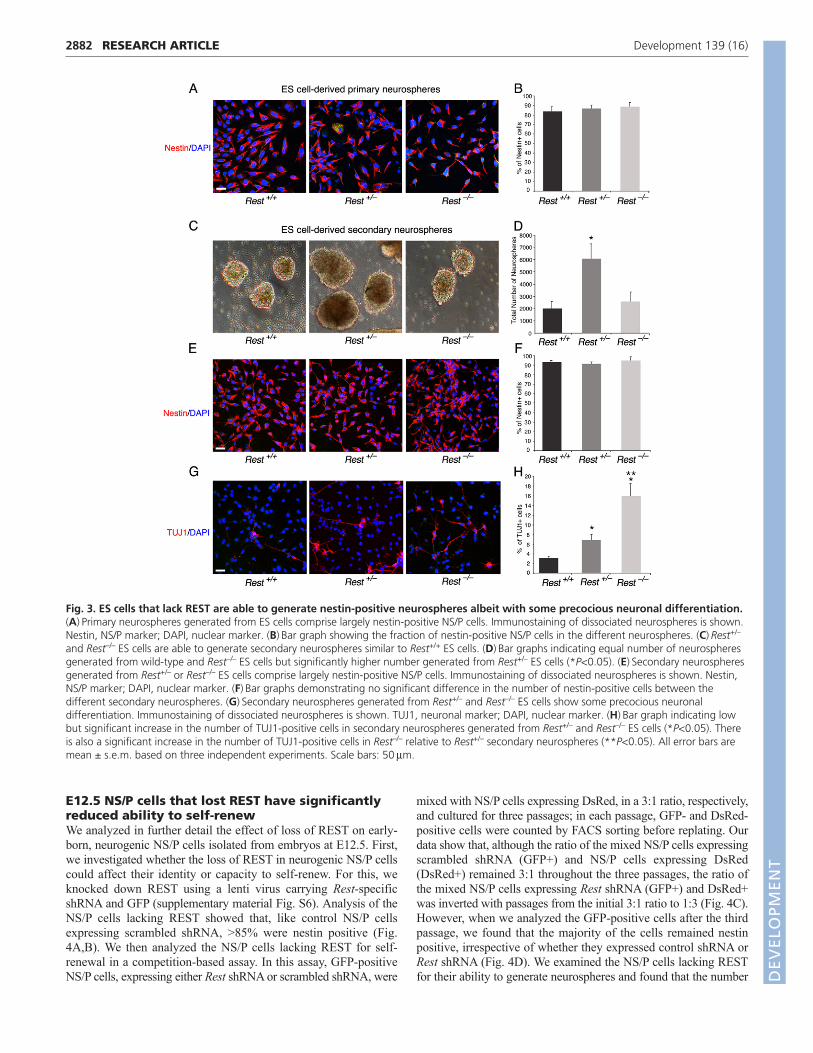
temporal progression of CNS development (Okada et al., 2008). Inthis system, the capacity of the ES cell-derived neurospheresswitches with time from neurogenic to gliogenic, and, whereas theprimary neurospheres give rise mainly to neurons, secondary andtertiary neurospheres give rise to neurons and glia with the tertiaryneurospheres being more gliogenic than neurogenic (Okada et al.,2008; Okano and Temple, 2009). We compared the ability of thethree different ES cell types, Rest+/+, Rest+/– and Rest–/–, to generateprimary, secondary and tertiary neurospheres. Primary neurospheresgenerated from Rest–/– ES cells showed a lack of uniformitycompared with those generated from Rest+/+ or Rest+/– ES cells(supplementary material Fig. S3A); however, >85% of the cells inthe cultured neurospheres were nestin positive, similar to culturedprimary neurospheres derived from Rest+/– and Rest+/+ ES cells (Fig.3A,B). Potentially, the dissociation and culturing of the neurospherescould select for specific cell type, e.g. neural progenitors. To rule outthis possibility, we analyzed sectioned neurospheres and found thatall three types of primary neurospheres consisted mostly of nestin-positive cells, which were uniformly distributed throughout thespheres (supplementary material Fig. S4A). Furthermore, the Rest–/–
primary neurospheres were also able to generate secondaryneurospheres (Fig. 3C). The heterozygous primary neurospheresgenerated two- to threefold more secondary neurospheres than didwild-type or homozygous primary neurospheres, which were alsolarger and more uniform in structure (Fig. 3C,D; supplementary
2881RESEARCH ARTICLEREST regulates neural lineages pools
material Fig. S3B). Similar results were obtained when tertiaryneurospheres were generated from secondary neurospheres(supplementary material Fig. S3C,D). Analysis of the different typesof secondary neurospheres for the presence of cell division markersshowed no significant differences in the fraction of Ki67-positivecells; however, heterozygous neurospheres had a higher mitoticindex than the wild-type or homozygous neurospheres as indicatedby the higher fraction of phosphohistone H3-positive cells(supplementary material Fig. S5A-D). This suggests that the preciselow level of REST in NS/P cells is likely to be crucial for efficientneurosphere generation and proliferation.
Analysis of the secondary neurospheres for the presence of theneural precursor marker nestin and the neuronal marker TUJ1(TUBB3 – Mouse Genome Informatics) showed that, like theprimary neurospheres, sectioned and cultured secondaryneurospheres consisted mostly of nestin-positive cells (Fig. 3E,F;supplementary material Fig. S4B). However, although relatively low,there was increased precocious neuronal differentiation, as shown bythe presence and morphology of TUJ1-positive cells, in Rest+/– (upto 7%) and Rest–/– (up to 16%) compared with Rest+/+ (up to 3%)cultured secondary neurospheres (Fig. 3G,H). Together, these datasuggest that, although some precocious neuronal differentiation wasevident in the cultured neurospheres, the lack of REST hadessentially no effect on the ability of ES cells to generateneurospheres that consist mostly of nestin-positive NS/P cells.
Fig. 2. The lack of REST in ES cells does notcompromise ES cell pluripotency. (A)Immunostainingof Rest+/+, Rest+/– and Rest–/– ES cells, indicating that thepluripotency markers SSEA1 and OCT4 are uniformlypresent in the ES colonies. Scale bars: 50m.(B)Teratomas generated from Rest+/+ or Rest–/– ES cellsdifferentiated into the three germ layers. Arrows indicateciliated columnar epithelial, plus sign indicates striatedmuscle, asterisk indicates epidermal structures.
DEVELO
PMENT
2882
E12.5 NS/P cells that lost REST have significantlyreduced ability to self-renewWe analyzed in further detail the effect of loss of REST on early-born, neurogenic NS/P cells isolated from embryos at E12.5. First,we investigated whether the loss of REST in neurogenic NS/P cellscould affect their identity or capacity to self-renew. For this, weknocked down REST using a lenti virus carrying Rest-specificshRNA and GFP (supplementary material Fig. S6). Analysis of theNS/P cells lacking REST showed that, like control NS/P cellsexpressing scrambled shRNA, >85% were nestin positive (Fig.4A,B). We then analyzed the NS/P cells lacking REST for self-renewal in a competition-based assay. In this assay, GFP-positiveNS/P cells, expressing either Rest shRNA or scrambled shRNA, were
RESEARCH ARTICLE Development 139 (16)
mixed with NS/P cells expressing DsRed, in a 3:1 ratio, respectively,and cultured for three passages; in each passage, GFP- and DsRed-positive cells were counted by FACS sorting before replating. Ourdata show that, although the ratio of the mixed NS/P cells expressingscrambled shRNA (GFP+) and NS/P cells expressing DsRed(DsRed+) remained 3:1 throughout the three passages, the ratio ofthe mixed NS/P cells expressing Rest shRNA (GFP+) and DsRed+was inverted with passages from the initial 3:1 ratio to 1:3 (Fig. 4C).However, when we analyzed the GFP-positive cells after the thirdpassage, we found that the majority of the cells remained nestinpositive, irrespective of whether they expressed control shRNA orRest shRNA (Fig. 4D). We examined the NS/P cells lacking RESTfor their ability to generate neurospheres and found that the number
Fig. 3. ES cells that lack REST are able to generate nestin-positive neurospheres albeit with some precocious neuronal differentiation.(A)Primary neurospheres generated from ES cells comprise largely nestin-positive NS/P cells. Immunostaining of dissociated neurospheres is shown.Nestin, NS/P marker; DAPI, nuclear marker. (B)Bar graph showing the fraction of nestin-positive NS/P cells in the different neurospheres. (C)Rest+/–
and Rest–/– ES cells are able to generate secondary neurospheres similar to Rest+/+ ES cells. (D)Bar graphs indicating equal number of neurospheresgenerated from wild-type and Rest–/– ES cells but significantly higher number generated from Rest+/– ES cells (*P<0.05). (E)Secondary neurospheresgenerated from Rest+/– or Rest–/– ES cells comprise largely nestin-positive NS/P cells. Immunostaining of dissociated neurospheres is shown. Nestin,NS/P marker; DAPI, nuclear marker. (F)Bar graphs demonstrating no significant difference in the number of nestin-positive cells between thedifferent secondary neurospheres. (G)Secondary neurospheres generated from Rest+/– and Rest–/– ES cells show some precocious neuronaldifferentiation. Immunostaining of dissociated neurospheres is shown. TUJ1, neuronal marker; DAPI, nuclear marker. (H)Bar graph indicating lowbut significant increase in the number of TUJ1-positive cells in secondary neurospheres generated from Rest+/– and Rest–/– ES cells (*P<0.05). Thereis also a significant increase in the number of TUJ1-positive cells in Rest–/– relative to Rest+/– secondary neurospheres (**P<0.05). All error bars aremean ± s.e.m. based on three independent experiments. Scale bars: 50m.
DEVELO
PMENT
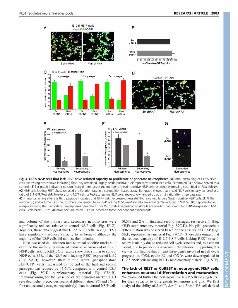
and volume of the primary and secondary neurospheres weresignificantly reduced relative to control NS/P cells (Fig. 4E-G).Together, these data suggest that E12.5 NS/P cells lacking RESThave significantly reduced capacity to self-renew, although themajority of the NS/P cells did not lose their identity.
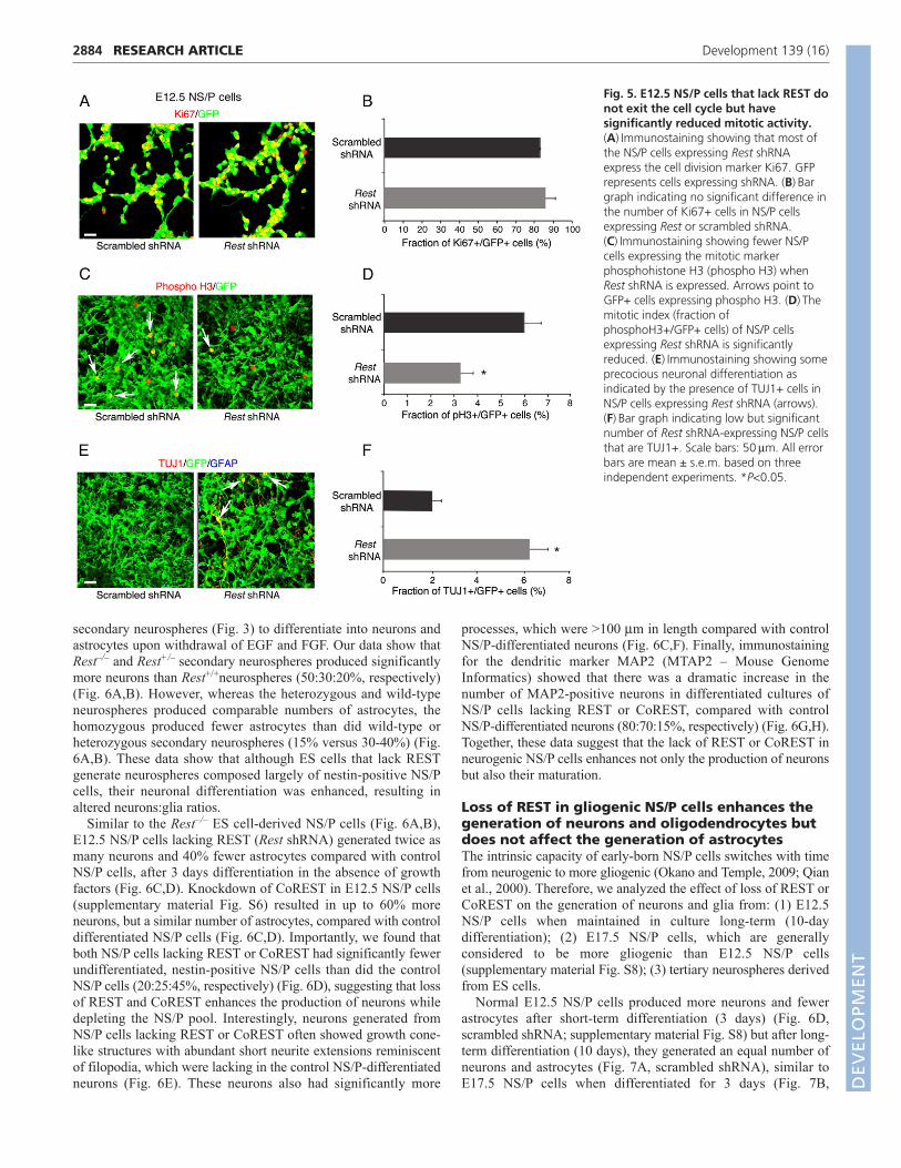
Next, we used cell division and neuronal-specific markers toexamine the underlying cause of reduced self-renewal of E12.5NS/P cells lacking REST. Our results show that, similar to controlNS/P cells, 85% of the NS/P cells lacking REST expressed Ki67(Fig. 5A,B); however, their mitotic index (phosphohistoneH3+/GFP+ cells), measured by the end of the first and secondpassages, was reduced by 45-50% compared with control NS/Pcells (Fig. 5C,D; supplementary material Fig. S7A,B).Immunostaining for the presence of the neuronal marker TUJ1revealed higher precocious neuronal differentiation (8% and 3% infirst and second passages, respectively) than in control NS/P cells
2883RESEARCH ARTICLEREST regulates neural lineages pools
(0.5% and 2% in first and second passages, respectively) (Fig.5E,F; supplementary material Fig. S7C,D). No glial precociousdifferentiation was observed based on the absence of GFAP (Fig.5E,F; supplementary material Fig. S7C,D). These data suggest thatthe reduced capacity of E12.5 NS/P cells lacking REST to self-renew is mainly due to reduced cell cycle kinetics and, to a certainextent, due to precocious neuronal differentiation. Supporting thisview is our finding that at least three genes involved in cell cycleprogression, Cdk6, cyclin B2 and Cdk1c, were downregulated inE12.5 NS/P cells lacking REST (supplementary material Fig. S7E).
The lack of REST or CoREST in neurogenic NS/P cellsenhances neuronal differentiation and maturationWe examined further the nestin-positive NS/P cells lacking RESTfor their capacity to differentiate to neurons and glia. We firstanalyzed the ability of Rest+/+, Rest+/– and Rest–/– ES cell-derived
Fig. 4. E12.5 N/SP cells that lack REST have reduced capacity to proliferate or generate neurospheres. (A)Immunostaining of E12.5 NS/Pcells expressing Rest shRNA indicating that they remained largely nestin positive. GFP represents transduced cells. Scrambled (Scr) shRNA served as acontrol. (B)Bar graph indicating no significant differences in the number of nestin-positive NS/P cells, whether expressing scrambled or Rest shRNA.(C)NS/P cells lacking REST show reduced proliferation rate in a competition-based assay. Bar graph shows that mixed NS/P cells initially cultured at aratio of 3:1 GFP/Rest shRNA-expressing NS/P cells:dsRed-expressing NS/P cells, respectively, ended up at a 1:3 ratio after three passages.(D)Immunostaining after the third passage indicates that GFP+ cells, expressing Rest shRNA, remained largely Nestin-positive NS/P cells. (E,F)Thenumber (E) and volume (F) of neurospheres generated from NS/P lacking REST (Rest shRNA) are significantly reduced. *P<0.05. (G)Representativeimages showing that secondary neurospheres generated from Rest shRNA-expressing NS/P cells are smaller than scrambled shRNA-expressing NS/Pcells. Scale bars: 50m. All error bars are mean ± s.e.m. based on three independent experiments.
DEVELO
PMENT
2884
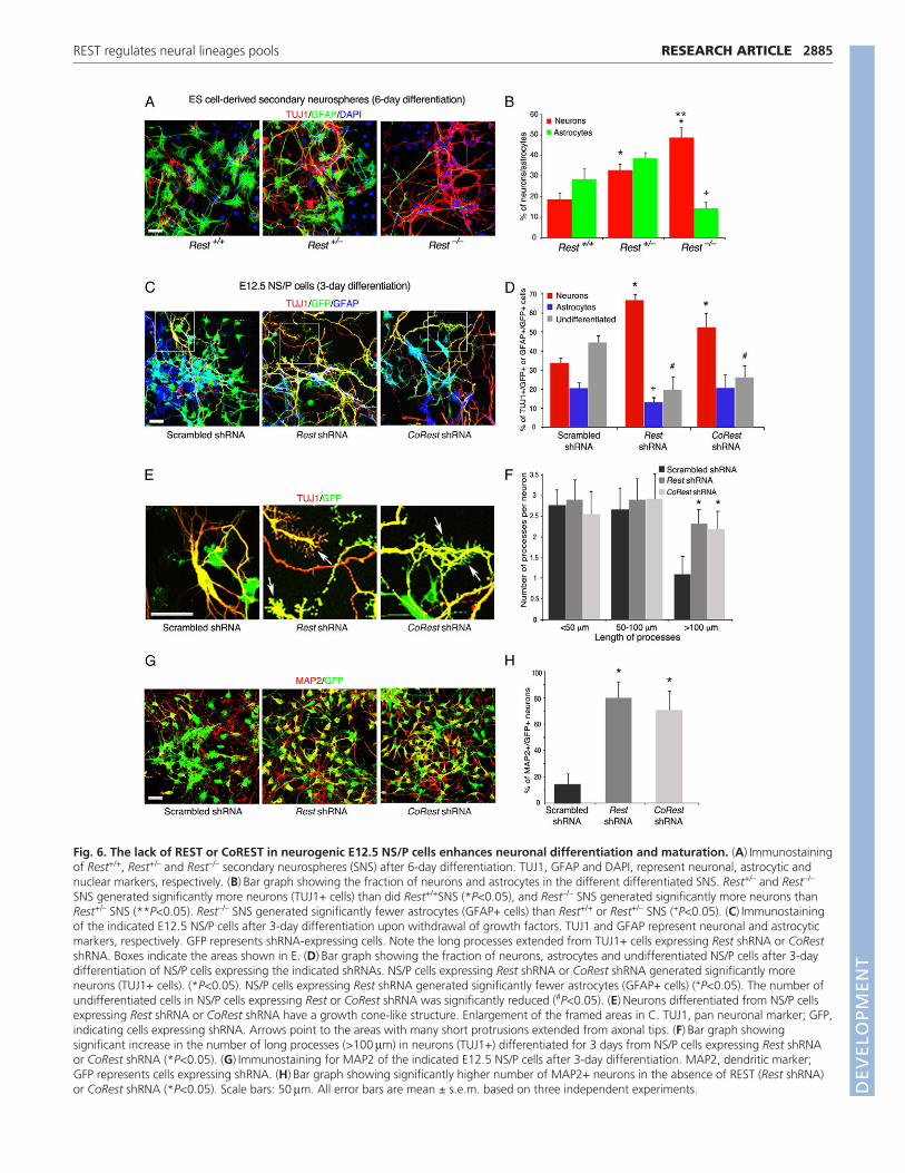
secondary neurospheres (Fig. 3) to differentiate into neurons andastrocytes upon withdrawal of EGF and FGF. Our data show thatRest–/– and Rest+/– secondary neurospheres produced significantlymore neurons than Rest+/+neurospheres (50:30:20%, respectively)(Fig. 6A,B). However, whereas the heterozygous and wild-typeneurospheres produced comparable numbers of astrocytes, thehomozygous produced fewer astrocytes than did wild-type orheterozygous secondary neurospheres (15% versus 30-40%) (Fig.6A,B). These data show that although ES cells that lack RESTgenerate neurospheres composed largely of nestin-positive NS/Pcells, their neuronal differentiation was enhanced, resulting inaltered neurons:glia ratios.
Similar to the Rest–/– ES cell-derived NS/P cells (Fig. 6A,B),E12.5 NS/P cells lacking REST (Rest shRNA) generated twice asmany neurons and 40% fewer astrocytes compared with controlNS/P cells, after 3 days differentiation in the absence of growthfactors (Fig. 6C,D). Knockdown of CoREST in E12.5 NS/P cells(supplementary material Fig. S6) resulted in up to 60% moreneurons, but a similar number of astrocytes, compared with controldifferentiated NS/P cells (Fig. 6C,D). Importantly, we found thatboth NS/P cells lacking REST or CoREST had significantly fewerundifferentiated, nestin-positive NS/P cells than did the controlNS/P cells (20:25:45%, respectively) (Fig. 6D), suggesting that lossof REST and CoREST enhances the production of neurons whiledepleting the NS/P pool. Interestingly, neurons generated fromNS/P cells lacking REST or CoREST often showed growth cone-like structures with abundant short neurite extensions reminiscentof filopodia, which were lacking in the control NS/P-differentiatedneurons (Fig. 6E). These neurons also had significantly more
RESEARCH ARTICLE Development 139 (16)
processes, which were >100 m in length compared with controlNS/P-differentiated neurons (Fig. 6C,F). Finally, immunostainingfor the dendritic marker MAP2 (MTAP2 – Mouse GenomeInformatics) showed that there was a dramatic increase in thenumber of MAP2-positive neurons in differentiated cultures ofNS/P cells lacking REST or CoREST, compared with controlNS/P-differentiated neurons (80:70:15%, respectively) (Fig. 6G,H).Together, these data suggest that the lack of REST or CoREST inneurogenic NS/P cells enhances not only the production of neuronsbut also their maturation.
Loss of REST in gliogenic NS/P cells enhances thegeneration of neurons and oligodendrocytes butdoes not affect the generation of astrocytesThe intrinsic capacity of early-born NS/P cells switches with timefrom neurogenic to more gliogenic (Okano and Temple, 2009; Qianet al., 2000). Therefore, we analyzed the effect of loss of REST orCoREST on the generation of neurons and glia from: (1) E12.5NS/P cells when maintained in culture long-term (10-daydifferentiation); (2) E17.5 NS/P cells, which are generallyconsidered to be more gliogenic than E12.5 NS/P cells(supplementary material Fig. S8); (3) tertiary neurospheres derivedfrom ES cells.
Normal E12.5 NS/P cells produced more neurons and fewerastrocytes after short-term differentiation (3 days) (Fig. 6D,scrambled shRNA; supplementary material Fig. S8) but after long-term differentiation (10 days), they generated an equal number ofneurons and astrocytes (Fig. 7A, scrambled shRNA), similar toE17.5 NS/P cells when differentiated for 3 days (Fig. 7B,
Fig. 5. E12.5 NS/P cells that lack REST donot exit the cell cycle but havesignificantly reduced mitotic activity.(A)Immunostaining showing that most ofthe NS/P cells expressing Rest shRNAexpress the cell division marker Ki67. GFPrepresents cells expressing shRNA. (B)Bargraph indicating no significant difference inthe number of Ki67+ cells in NS/P cellsexpressing Rest or scrambled shRNA.(C)Immunostaining showing fewer NS/Pcells expressing the mitotic markerphosphohistone H3 (phospho H3) whenRest shRNA is expressed. Arrows point toGFP+ cells expressing phospho H3. (D)Themitotic index (fraction ofphosphoH3+/GFP+ cells) of NS/P cellsexpressing Rest shRNA is significantlyreduced. (E)Immunostaining showing someprecocious neuronal differentiation asindicated by the presence of TUJ1+ cells inNS/P cells expressing Rest shRNA (arrows).(F)Bar graph indicating low but significantnumber of Rest shRNA-expressing NS/P cellsthat are TUJ1+. Scale bars: 50m. All errorbars are mean ± s.e.m. based on threeindependent experiments. *P<0.05.
DEVELO
PMENT
2885RESEARCH ARTICLEREST regulates neural lineages pools
Fig. 6. The lack of REST or CoREST in neurogenic E12.5 NS/P cells enhances neuronal differentiation and maturation. (A)Immunostainingof Rest+/+, Rest+/– and Rest–/– secondary neurospheres (SNS) after 6-day differentiation. TUJ1, GFAP and DAPI, represent neuronal, astrocytic andnuclear markers, respectively. (B)Bar graph showing the fraction of neurons and astrocytes in the different differentiated SNS. Rest+/– and Rest–/–
SNS generated significantly more neurons (TUJ1+ cells) than did Rest+/+SNS (*P<0.05), and Rest–/– SNS generated significantly more neurons thanRest+/– SNS (**P<0.05). Rest–/– SNS generated significantly fewer astrocytes (GFAP+ cells) than Rest+/+ or Rest+/– SNS (+P<0.05). (C)Immunostainingof the indicated E12.5 NS/P cells after 3-day differentiation upon withdrawal of growth factors. TUJ1 and GFAP represent neuronal and astrocyticmarkers, respectively. GFP represents shRNA-expressing cells. Note the long processes extended from TUJ1+ cells expressing Rest shRNA or CoRestshRNA. Boxes indicate the areas shown in E. (D)Bar graph showing the fraction of neurons, astrocytes and undifferentiated NS/P cells after 3-daydifferentiation of NS/P cells expressing the indicated shRNAs. NS/P cells expressing Rest shRNA or CoRest shRNA generated significantly moreneurons (TUJ1+ cells). (*P<0.05). NS/P cells expressing Rest shRNA generated significantly fewer astrocytes (GFAP+ cells) (+P<0.05). The number ofundifferentiated cells in NS/P cells expressing Rest or CoRest shRNA was significantly reduced (#P<0.05). (E)Neurons differentiated from NS/P cellsexpressing Rest shRNA or CoRest shRNA have a growth cone-like structure. Enlargement of the framed areas in C. TUJ1, pan neuronal marker; GFP,indicating cells expressing shRNA. Arrows point to the areas with many short protrusions extended from axonal tips. (F)Bar graph showingsignificant increase in the number of long processes (>100m) in neurons (TUJ1+) differentiated for 3 days from NS/P cells expressing Rest shRNAor CoRest shRNA (*P<0.05). (G)Immunostaining for MAP2 of the indicated E12.5 NS/P cells after 3-day differentiation. MAP2, dendritic marker;GFP represents cells expressing shRNA. (H)Bar graph showing significantly higher number of MAP2+ neurons in the absence of REST (Rest shRNA)or CoRest shRNA (*P<0.05). Scale bars: 50m. All error bars are mean ± s.e.m. based on three independent experiments. D
EVELO
PMENT
2886
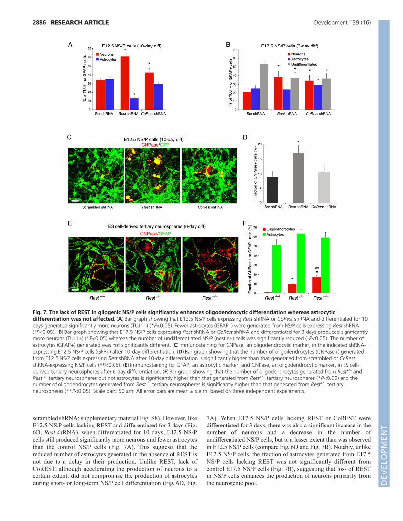
scrambled shRNA; supplementary material Fig. S8). However, likeE12.5 NS/P cells lacking REST and differentiated for 3 days (Fig.6D, Rest shRNA), when differentiated for 10 days, E12.5 NS/Pcells still produced significantly more neurons and fewer astrocytesthan the control NS/P cells (Fig. 7A). This suggests that thereduced number of astrocytes generated in the absence of REST isnot due to a delay in their production. Unlike REST, lack ofCoREST, although accelerating the production of neurons to acertain extent, did not compromise the production of astrocytesduring short- or long-term NS/P cell differentiation (Fig. 6D, Fig.
RESEARCH ARTICLE Development 139 (16)
7A). When E17.5 NS/P cells lacking REST or CoREST weredifferentiated for 3 days, there was also a significant increase in thenumber of neurons and a decrease in the number ofundifferentiated NS/P cells, but to a lesser extent than was observedin E12.5 NS/P cells (compare Fig. 6D and Fig. 7B). Notably, unlikeE12.5 NS/P cells, the fraction of astrocytes generated from E17.5NS/P cells lacking REST was not significantly different fromcontrol E17.5 NS/P cells (Fig. 7B), suggesting that loss of RESTin NS/P cells enhances the production of neurons primarily fromthe neurogenic pool.
Fig. 7. The lack of REST in gliogenic NS/P cells significantly enhances oligodendrocytic differentiation whereas astrocyticdifferentiation was not affected. (A)Bar graph showing that E12.5 NS/P cells expressing Rest shRNA or CoRest shRNA and differentiated for 10days generated significantly more neurons (TUJ1+) (*P<0.05). Fewer astrocytes (GFAP+) were generated from NS/P cells expressing Rest shRNA(+P<0.05). (B)Bar graph showing that E17.5 NS/P cells expressing Rest shRNA or CoRest shRNA and differentiated for 3 days produced significantlymore neurons (TUJ1+) (*P<0.05) whereas the number of undifferentiated NS/P (nestin+) cells was significantly reduced (+P<0.05). The number ofastrocytes (GFAP+) generated was not significantly different. (C)Immunostaining for CNPase, an oligodendrocytic marker, in the indicated shRNA-expressing E12.5 NS/P cells (GFP+) after 10-day differentiation. (D)Bar graph showing that the number of oligodendrocytes (CNPase+) generatedfrom E12.5 NS/P cells expressing Rest shRNA after 10-day differentiation is significantly higher than that generated from scrambled or CoRestshRNA-expressing NS/P cells (*P<0.05). (E)Immunostaining for GFAP, an astrocytic marker, and CNPase, an oligodendrocytic marker, in ES cell-derived tertiary neurospheres after 6-day differentiation. (F)Bar graph showing that the number of oligodendrocytes generated from Rest+/– andRest–/– tertiary neurospheres but not astrocytes is significantly higher than that generated from Rest+/+ tertiary neurospheres (*P<0.05) and thenumber of oligodendrocytes generated from Rest–/– tertiary neurospheres is significantly higher than that generated from Rest+/– tertiaryneurospheres (**P<0.05). Scale bars: 50m. All error bars are mean ± s.e.m. based on three independent experiments.DEVELO
PMENT
Surprisingly, our analysis of the long-term differentiated cultureof E12.5 NS/P cells showed that the production of oligodendrocytes,measured by the presence of CNPase (CNP – Mouse GenomeInformatics), was twofold higher in the absence of REST but was notaltered in the absence of CoREST (Fig. 7C,D). Enhanced productionof oligodendrocytes was also evident in differentiated tertiaryneurospheres derived from Rest+/– or Rest–/– ES cells, whichgenerated up to 10- and 20-fold, respectively, more oligodendrocytesthan from neurospheres derived from wild-type Rest+/+ ES cells (Fig.7E,F). Conversely, and like differentiated E17.5 N/P cells (Fig. 7B),the fractions of astrocytes generated from the three different types oftertiary neurospheres were indistinguishable from each other (Fig.7F). This suggests that in the absence of REST neurogenic NS/Pcells are able to acquire gliogenic capacity and that the loss of RESThas no effect on the production of astrocytes from NS/P cells, whichhad already acquired gliogenic fate.
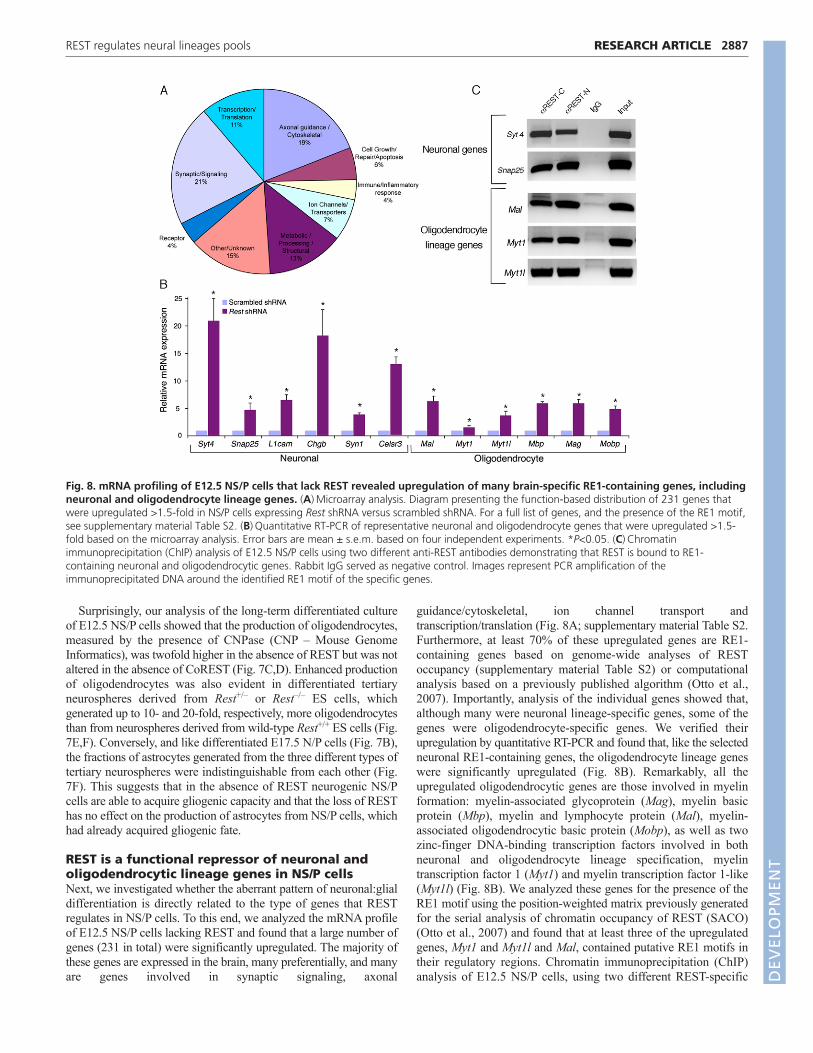
REST is a functional repressor of neuronal andoligodendrocytic lineage genes in NS/P cellsNext, we investigated whether the aberrant pattern of neuronal:glialdifferentiation is directly related to the type of genes that RESTregulates in NS/P cells. To this end, we analyzed the mRNA profileof E12.5 NS/P cells lacking REST and found that a large number ofgenes (231 in total) were significantly upregulated. The majority ofthese genes are expressed in the brain, many preferentially, and manyare genes involved in synaptic signaling, axonal
2887RESEARCH ARTICLEREST regulates neural lineages pools
guidance/cytoskeletal, ion channel transport andtranscription/translation (Fig. 8A; supplementary material Table S2.Furthermore, at least 70% of these upregulated genes are RE1-containing genes based on genome-wide analyses of RESToccupancy (supplementary material Table S2) or computationalanalysis based on a previously published algorithm (Otto et al.,2007). Importantly, analysis of the individual genes showed that,although many were neuronal lineage-specific genes, some of thegenes were oligodendrocyte-specific genes. We verified theirupregulation by quantitative RT-PCR and found that, like the selectedneuronal RE1-containing genes, the oligodendrocyte lineage geneswere significantly upregulated (Fig. 8B). Remarkably, all theupregulated oligodendrocytic genes are those involved in myelinformation: myelin-associated glycoprotein (Mag), myelin basicprotein (Mbp), myelin and lymphocyte protein (Mal), myelin-associated oligodendrocytic basic protein (Mobp), as well as twozinc-finger DNA-binding transcription factors involved in bothneuronal and oligodendrocyte lineage specification, myelintranscription factor 1 (Myt1) and myelin transcription factor 1-like(Myt1l) (Fig. 8B). We analyzed these genes for the presence of theRE1 motif using the position-weighted matrix previously generatedfor the serial analysis of chromatin occupancy of REST (SACO)(Otto et al., 2007) and found that at least three of the upregulatedgenes, Myt1 and Myt1l and Mal, contained putative RE1 motifs intheir regulatory regions. Chromatin immunoprecipitation (ChIP)analysis of E12.5 NS/P cells, using two different REST-specific
Fig. 8. mRNA profiling of E12.5 NS/P cells that lack REST revealed upregulation of many brain-specific RE1-containing genes, includingneuronal and oligodendrocyte lineage genes. (A)Microarray analysis. Diagram presenting the function-based distribution of 231 genes thatwere upregulated >1.5-fold in NS/P cells expressing Rest shRNA versus scrambled shRNA. For a full list of genes, and the presence of the RE1 motif,see supplementary material Table S2. (B)Quantitative RT-PCR of representative neuronal and oligodendrocyte genes that were upregulated >1.5-fold based on the microarray analysis. Error bars are mean ± s.e.m. based on four independent experiments. *P<0.05. (C)Chromatinimmunoprecipitation (ChIP) analysis of E12.5 NS/P cells using two different anti-REST antibodies demonstrating that REST is bound to RE1-containing neuronal and oligodendrocytic genes. Rabbit IgG served as negative control. Images represent PCR amplification of theimmunoprecipitated DNA around the identified RE1 motif of the specific genes.
DEVELO
PMENT
2888
antibodies, showed that, like in the neuronal RE1-containing genesSyt4 and Snap25, REST is bound to the RE1 motif of theoligodendrocyte genes Mal, Myt1 and Myt1l (Fig. 8C). Together, ourresults suggest that REST regulates neuronal and oligodendrocytespecific genes, and that the absence of REST in NS/P cells promotesthe upregulation of these target genes and, consequently, enhancesthe generation of neurons and oligodendrocytes.
DISCUSSIONOur data show that REST is a functional repressor of neuronal genesin ES cells and that even a 50% reduction in the REST level issufficient to abrogate neuronal gene repression. This suggests thatthe high level of REST in ES cells, which is mediated by pluripotenttranscription factors, is required for its full repressor activity. Theseresults, which corroborate our previous findings that neuronal genesin ES cells are repressed but their chromatin is poised for activation(Ballas et al., 2005), suggest that this type of repressor mechanismdepends mainly on the function of the REST repressor complex.Nevertheless, the derepressed neuronal genes in the absence ofREST, including the transcription factor NeuroD, which potentiallycould drive neuronal differentiation (Cho and Tsai, 2004), wereunable to compromise ES cell pluripotency. It is likely that, in EScells, neuronal genes are also regulated post-transcriptionally suchthat the elevated neuronal transcripts are not translated into functionalproteins. Previous studies addressing the role of REST in maintainingES cell pluripotency have been controversial. Whereas Singh et al.(Singh et al., 2008) have shown that REST is required for ES cellpluripotency based on reduced levels of pluripotency markers,Jorgensen et al. and Buckley et al. have reported that levels ofpluripotency factors remain unchanged in the absence of REST(Buckley et al., 2009; Jorgensen et al., 2009a). It remains unclearwhether different experimental conditions used in these studies couldhave contributed to this discrepancy. However, our analyses using amore direct approach, i.e. teratoma formation, in addition topluripotency markers, corroborate the studies by Jorgenson et al. andBuckley et al. (Buckley et al., 2009; Jorgensen et al., 2009a) and
RESEARCH ARTICLE Development 139 (16)
suggest that REST is not part of the transcription factor network thatregulates ES cell pluripotency. That the loss of function of REST inES cells does not compromise their pluripotency is further supportedby earlier work showing that REST knockout mice died at E11(Chen et al., 1998), well past the ES cell stage (E4-E5).
In contrast to the lack of a role for REST in maintaining ES cellpluripotency, our results point to a central role for REST past theES cell stage, during neural development. ES cells lacking RESTwere able to convert to neurospheres with a high percentage ofnestin-positive NS/P cells; however, precocious neuronaldifferentiation was evident, similar to E12.5 NS/P cells lackingREST. The reduced cell cycle kinetics together with precociousneuronal differentiation in E12.5 NS/P cells lacking REST resultedin depletion of the NS/P pool. Recent studies addressing thefunction of REST in adult hippocampal neurogenesis show thatloss of REST also results in precocious neurogenesis and depletionof the adult NS/P pool (Gao et al., 2011), together suggesting ageneral and crucial role for REST in maintaining both embryonicand adult NS/P cell self-renewal. The number and size ofneurospheres generated from Rest–/– ES cells were alsosignificantly lower than those generated from Rest+/– ES cells, butnot different from those generated from Rest+/+ ES cells. Thismight reflect differences between direct conversion of ES cells toNS/P cells via neurospheres in vitro, and NS/P cells isolated fromdeveloping embryos. This is because the level of Rest mRNA inE12.5 NS/P (Ballas et al., 2005) is more similar to the level of RestmRNA in neurospheres derived from Rest+/– ES cells rather thanneurospheres derived from Rest+/+ ES cells, which was twofoldhigher (data not shown). In this scenario, the higher level of RESTin neurospheres derived from Rest+/+ relative to those derived fromRest+/– ES cells or E12.5 NS/P cells, probably delayed neurospheregeneration. Interestingly, similar to the loss of function of REST inE12.5 NS/P cells, our recent data show that E12.5 NS/P cellsoverexpressing REST remain mostly nestin positive and do not exitthe cell cycle, although reduced cell proliferation was evident(Mandel et al., 2011) (data not shown). These data further suggest
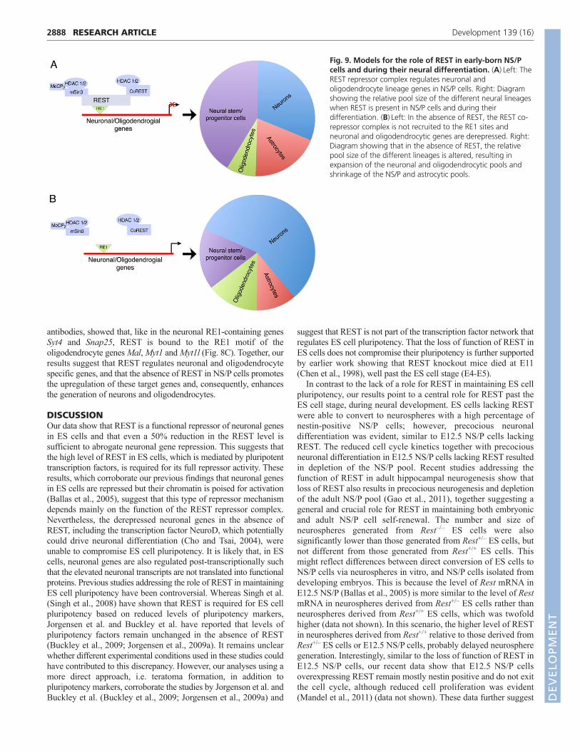
Fig. 9. Models for the role of REST in early-born NS/Pcells and during their neural differentiation. (A)Left: TheREST repressor complex regulates neuronal andoligodendrocyte lineage genes in NS/P cells. Right: Diagramshowing the relative pool size of the different neural lineageswhen REST is present in NS/P cells and during theirdifferentiation. (B)Left: In the absence of REST, the REST co-repressor complex is not recruited to the RE1 sites andneuronal and oligodendrocytic genes are derepressed. Right:Diagram showing that in the absence of REST, the relativepool size of the different lineages is altered, resulting inexpansion of the neuronal and oligodendrocytic pools andshrinkage of the NS/P and astrocytic pools.
DEVELO
PMENT
that the precisely downregulated level of REST in multipotent stemcells is crucial, and too much or too little of REST negativelyaffects NS/P cell self-renewal.
Consistent with the precocious neuronal differentiation of NS/Pcells lacking REST, their default differentiation resulted in asignificantly larger pool of neurons and smaller pools ofundifferentiated NS/P cells and astrocytes. This suggests thatdysfunction of the REST repressor complex enhances neurogenesiswhile depleting the NS/P pool (Fig. 9). The shrinkage of theastrocytic pool in the absence of REST could be due to thedepletion of neurogenic NS/P cells for the accelerated generationof neurons, which in turn depletes the gliogenic NS/P pool that isnormally generated with time from the neurogenic NS/P pool.Alternatively, the absence of REST might drive neuronaldifferentiation not only from neurogenic, but also from gliogenic,NS/P cells and could thus deplete both the neurogenic andgliogenic NS/P pools, resulting in generation of fewer astrocytes.That the production of neurons from gliogenic NS/P cells lackingREST was accelerated whereas the production of astrocytes wasunaffected supports the view that the loss of REST probablyenhances the production of neurons only from the neurogenic NS/Ppool and does not switch gliogenic NS/P cells to neurogenic.Furthermore, lack of REST also did not affect the transition ofneurogenic to gliogenic NS/P cells in the presence of growthfactors. Interestingly, our microarray analysis of genes that are up-or downregulated in NS/P cells that lack REST did not identifygenes involved in astrocytic lineage specification. Together, thesedata support the view that depletion of the astrocytic pool duringdifferentiation of neurogenic NS/P cells lacking REST probablyoccurs indirectly.
Loss of CoREST in NS/P cells also accelerated neuronaldifferentiation and maturation; however, this effect was mildercompared with the loss of REST and therefore did not affect theastrocytic pool. Although CoREST could potentially be part ofother repressor complexes, the close similarity in the phenotypeobserved in the absence of REST or CoREST suggests that thepredominant function of CoREST during neural development isprobably in conjunction with the REST repressor complex.
Our recent data show that gain of function of REST inneurogenic NS/P cells during neocortical development delays, butdoes not compromise, neuronal differentiation (Mandel et al.,2011). Importantly, gain of function of REST in neurogenicprogenitors did not result in generation of glia even though RESTremains present in glia and absent in neurons (Mandel et al., 2011).These studies further support the view that loss or gain of functionof REST in NS/P cells is incapable of switching NS/P cells fromgliogenic to neurogenic or from neurogenic to gliogenic.
Our microarray, ChIP and differentiation analyses of NS/P cellsall point to a role for REST not only in neurogenesis but also inoligodendrogenesis (Fig. 9). The absence of REST in NS/P cellsresulted in upregulation of neuronal genes as well asoligodendrocyte lineage genes, specifically those involved inmyelination. At least three of the six upregulated oligodendrocyte-associated genes (Mal, Myt1 and Myt1l) contain the RE1 motif,which also binds REST. Supporting our data, ChIP-ChIP analysisof REST/CoREST target genes in premature or matureoligodendrocytes identified genes involved in oligodendrocytespecification and maturation (Abrajano et al., 2009).
Collectively, our data support a central role for the RESTrepressor complex during neural development in maintaining NS/Pcell self-renewal and in regulating the generation of proper poolsize of the different neural lineages, including neurons, astrocytes
2889RESEARCH ARTICLEREST regulates neural lineages pools
and oligodendrocytes (Fig. 9). By regulating genes involved inneuronal and oligodendrocyte specification and maturation, theREST repressor complex restrains neuronal and oligodendrocytegeneration and thus regulates the precise pools of the differentneural lineages. Further studies are warranted to understandwhether such regulation of neuronal and oligodendrocytegeneration and maturation by the REST repressor complex, isdynamically involved in pathological conditions such as acuteinjury and repair or chronic neurological disorders.
AcknowledgementsWe thank Dr Sally Temple for valuable discussion about the results and advice;Dr Sean McCorkle for providing his expertise with the bioinformatics; Dr GailMandel for providing the anti-REST antibodies; Drs Amanda Fisher, HelleJorgenson and Zhou-Feng Chen for sharing and providing the different Rest-knockout ES cells; Dr Thomas Zarembinski and BioTime for the generous giftof Glycosan HyStem; the Pathology Translational Research Laboratory forassistance with the teratoma analysis.
FundingThis work was supported by a grant from the National Institutes of Health[NS060797 to N.B.]. Deposited in PMC for release after 12 months.
Competing interests statementThe authors declare no competing financial interests.
Supplementary materialSupplementary material available online athttp://dev.biologists.org/lookup/suppl/doi:10.1242/dev.074765/-/DC1
ReferencesAbrajano, J. J., Qureshi, I. A., Gokhan, S., Zheng, D., Bergman, A. and
Mehler, M. F. (2009). Differential deployment of REST and CoREST promotesglial subtype specification and oligodendrocyte lineage maturation. PLoS ONE 4,e7665.
Andres, M. E., Burger, C., Peral-Rubio, M. J., Battaglioli, E., Anderson, M. E.,Grimes, J., Dallman, J., Ballas, N. and Mandel, G. (1999). CoREST: afunctional corepressor required for regulation of neural- specific geneexpression. Proc. Natl. Acad. Sci. USA 96, 9873-9878.
Ballas, N. and Mandel, G. (2005). The many faces of REST oversee epigeneticprogramming of neuronal genes. Curr. Opin. Neurobiol. 15, 500-506.
Ballas, N., Battaglioli, E., Atouf, F., Andres, M. E., Chenoweth, J., Anderson,M. E., Burger, C., Moniwa, M., Davie, J. R., Bowers, W. J. et al. (2001).Regulation of neuronal traits by a novel transcriptional complex. Neuron 31,353-365.
Ballas, N., Grunseich, C., Lu, D. D., Speh, J. C. and Mandel, G. (2005). RESTand its corepressors mediate plasticity of neuronal gene chromatin throughoutneurogenesis. Cell 121, 645-657.
Boyer, L. A., Lee, T. I., Cole, M. F., Johnstone, S. E., Levine, S. S., Zucker, J. P.,Guenther, M. G., Kumar, R. M., Murray, H. L., Jenner, R. G. et al. (2005).Core transcriptional regulatory circuitry in human embryonic stem cells. Cell 122,947-956.
Bruce, A. W., Donaldson, I. J., Wood, I. C., Yerbury, S. A., Sadowski, M. I.,Chapman, M., Gottgens, B. and Buckley, N. J. (2004). Genome-wide analysis of repressor element 1 silencing transcription factor/neuron-restrictivesilencing factor (REST/NRSF) target genes. Proc. Natl. Acad. Sci. USA 101,10458-10463.
Buckley, N. J., Johnson, R., Sun, Y. M. and Stanton, L. W. (2009). Is REST aregulator of pluripotency? Nature 457, E5-6; discussion E7.
Chen, Z. F., Paquette, A. J. and Anderson, D. J. (1998). NRSF/REST is required invivo for repression of multiple neuronal target genes during embryogenesis. Nat.Genet. 20, 136-142.
Cheng, L. C., Tavazoie, M. and Doetsch, F. (2005). Stem cells fromepigeneticsto microRNAs. Neuron 46, 363-367.
Cho, J. H. and Tsai, M. J. (2004). The role of BETA2/NeuroD1 in the developmentof the nervous system. Mol. Neurobiol. 30, 35-47.
Chong, J. A., Tapia-Ramirez, J., Kim, S., Toledo-Aral, J. J., Zheng, Y.,Boutros, M. C., Altshuller, Y. M., Frohman, M. A., Kraner, S. D. andMandel, G. (1995). REST: a mammalian silencer protein that restricts sodiumchannel gene expression to neurons. Cell 80, 949-957.
Dewald, L. E., Rodriguez, J. P. and Levine, J. M. (2011). The RE1 bindingprotein REST regulates oligodendrocyte differentiation. J. Neurosci. 31, 3470-3483.
Edlund, T. and Jessell, T. M. (1999). Progression from extrinsic to intrinsicsignaling in cell fate specification: a view from the nervous system. Cell 96, 211-224. D
EVELO
PMENT
2890 RESEARCH ARTICLE Development 139 (16)
Gao, Z., Ure, K., Ding, P., Nashaat, M., Yuan, L., Ma, J., Hammer, R. E. andHsieh, J. (2011). The master negative regulator REST/NRSF controls adultneurogenesis by restraining the neurogenic program in quiescent stem cells. J.Neurosci. 31, 9772-9786.
Ivanova, N., Dobrin, R., Lu, R., Kotenko, I., Levorse, J., DeCoste, C., Schafer,X., Lun, Y. and Lemischka, I. R. (2006). Dissecting self-renewal in stem cellswith RNA interference. Nature 442, 533-538.
Johnson, D. S., Mortazavi, A., Myers, R. M. and Wold, B. (2007). Genome-wide mapping of in vivo protein-DNA interactions. Science 316, 1497-1502.
Johnson, R., Teh, C. H., Kunarso, G., Wong, K. Y., Srinivasan, G., Cooper, M.L., Volta, M., Chan, S. S., Lipovich, L., Pollard, S. M. et al. (2008). RESTregulates distinct transcriptional networks in embryonic and neural stem cells.PLoS Biol. 6, e256.
Jorgensen, H. F. and Fisher, A. G. (2010). Can controversies be put to REST?Nature 467, E3-4; discussion E5.
Jorgensen, H. F., Chen, Z. F., Merkenschlager, M. and Fisher, A. G. (2009a). IsREST required for ESC pluripotency? Nature 457, E4-5; discussion E7.
Jorgensen, H. F., Terry, A., Beretta, C., Pereira, C. F., Leleu, M., Chen, Z. F.,Kelly, C., Merkenschlager, M. and Fisher, A. G. (2009b). REST selectivelyrepresses a subset of RE1-containing neuronal genes in mouse embryonic stemcells. Development 136, 715-721.
Kent, W. J., Sugnet, C. W., Furey, T. S., Roskin, K. M., Pringle, T. H., Zahler,A. M. and Haussler, D. (2002). The human genome browser at UCSC.Genome Res. 12, 996-1006.
Lattin, J. E., Schroder, K., Su, A. I., Walker, J. R., Zhang, J., Wiltshire, T.,Saijo, K., Glass, C. K., Hume, D. A., Kellie, S. et al. (2008). Expression analysisof G protein-coupled receptors in mouse macrophages. Immunome Res. 4, 5.
Lensch, M. W., Schlaeger, T. M., Zon, L. I. and Daley, G. Q. (2007). Teratomaformation assays with human embryonic stem cells: a rationale for one type ofhuman-animal chimera. Cell Stem Cell 1, 253-258.
Loh, Y. H., Wu, Q., Chew, J. L., Vega, V. B., Zhang, W., Chen, X., Bourque,G., George, J., Leong, B., Liu, J. et al. (2006). The Oct4 and Nanogtranscription network regulates pluripotency in mouse embryonic stem cells.Nat. Genet. 38, 431-440.
Mandel, G., Fiondella, C. G., Covey, M. V., Lu, D. D., Loturco, J. J. and Ballas,N. (2011). Repressor element 1 silencing transcription factor (REST) controlsradial migration and temporal neuronal specification during neocorticaldevelopment. Proc. Natl. Acad. Sci. USA 108, 16789-16794.
Okada, Y., Matsumoto, A., Shimazaki, T., Enoki, R., Koizumi, A., Ishii, S.,Itoyama, Y., Sobue, G. and Okano, H. (2008). Spatiotemporal recapitulationof central nervous system development by murine embryonic stem cell-derivedneural stem/progenitor cells. Stem Cells 26, 3086-3098.
Okano, H. and Temple, S. (2009). Cell types to order: temporal specification ofCNS stem cells. Curr. Opin. Neurobiol. 19, 112-119.
Otto, S. J., McCorkle, S. R., Hover, J., Conaco, C., Han, J. J., Impey, S.,Yochum, G. S., Dunn, J. J., Goodman, R. H. and Mandel, G. (2007). A newbinding motif for the transcriptional repressor REST uncovers large genenetworks devoted to neuronal functions. J. Neurosci. 27, 6729-6739.
Qian, X., Shen, Q., Goderie, S. K., He, W., Capela, A., Davis, A. A. andTemple, S. (2000). Timing of CNS cell generation: a programmed sequence ofneuron and glial cell production from isolated murine cortical stem cells. Neuron28, 69-80.
Roopra, A., Sharling, L., Wood, I. C., Briggs, T., Bachfischer, U., Paquette, A.J. and Buckley, N. J. (2000). Transcriptional repression by neuron-restrictivesilencer factor is mediated via the Sin3-histone deacetylase complex. Mol. Cell.Biol. 20, 2147-2157.
Ross, S. E., Greenberg, M. E. and Stiles, C. D. (2003). Basic helix-loop-helixfactors in cortical development. Neuron 39, 13-25.
Schoenherr, C. J. and Anderson, D. J. (1995). The neuron-restrictive silencerfactor (NRSF): a coordinate repressor of multiple neuron-specific genes. Science267, 1360-1363.
Singh, S. K., Kagalwala, M. N., Parker-Thornburg, J., Adams, H. andMajumder, S. (2008). REST maintains self-renewal and pluripotency ofembryonic stem cells. Nature 453, 223-227.
Su, A. I., Wiltshire, T., Batalov, S., Lapp, H., Ching, K. A., Block, D., Zhang, J.,Soden, R., Hayakawa, M., Kreiman, G. et al. (2004). A gene atlas of themouse and human protein-encoding transcriptomes. Proc. Natl. Acad. Sci. USA101, 6062-6067.
Waterston, R. H., Lindblad-Toh, K., Birney, E., Rogers, J., Abril, J. F.,Agarwal, P., Agarwala, R., Ainscough, R., Alexandersson, M., An, P. et al.(2002). Initial sequencing and comparative analysis of the mouse genome.Nature 420, 520-562.
Westbrook, T. F., Hu, G., Ang, X. L., Mulligan, P., Pavlova, N. N., Liang, A.,Leng, Y., Maehr, R., Shi, Y., Harper, J. W. et al. (2008). SCFbeta-TRCPcontrols oncogenic transformation and neural differentiation through RESTdegradation. Nature 452, 370-374.
DEVELO
PMENT