Embed Size (px)
Citation preview

ANALYTICAL SCIENCES JANUARY 2018, VOL. 34 91
Introduction
In the last decade, paper substrates have emerged as simple, low cost and portable platforms for the development of analytical devices.1–3 Paper is composed of cellulose and lignin and presents a tridimensional porous structure making possible the spontaneous fluidic transport under capillary action.4 This kind of substrate has become increasingly popular for the scientific community due to its operational simplicity, low instrumental requirements and, most importantly, the possibility to be used in point-of-care testing with the sample-in:answer-out capability.1–4 Recent examples of paper-based analytical devices (PADs) for lateral flow or spot test assays,5–7 flow-injection analysis,8,9 chromatographic and electrophoretic separations,10,11 digital microfluidics,12 direct analysis using paper spray ionization mass spectrometry13–15 and electroanalytical measurements16,17 have been successfully reported.
The integration of electrochemistry on paper substrates has received noticeable attention in the last years due to its remarkable advantages, including compatibility with miniaturization, adjustable selectivity, inherent portability, high sensitivity and low cost.18–21 The development of electrochemical paper-based analytical devices (ePADs) was first described in 2009 by Dungchai and co-workers.22 In this pioneering report, carbon working electrodes were fabricated by screen-printing
method using carbon ink containing Prussian blue. The authors successfully demonstrated the simultaneous detection of uric acid, glucose and lactate in biological samples. In 2010, Carvalhal and co-workers23 reported the integration of electrochemical detection on paper to monitor the chromatographic separation of ascorbic acid (AA) and uric acid (UA). For this purpose, gold electrodes were deposited on polyester films by e-beam evaporation and integrated with a paper separation device. Since the disclosure of these two first reports, different designs, approaches and protocols have been proposed for developing ePADs. The chronological summary regarding the electrode fabrication methods to be incorporated on paper devices can be found in excellent review articles recently reported.16–20,24 In general, electrodes can be fabricated on paper by screen-printing, metallic deposition through sputtering or evaporation, microwires, stencil-printing, inkjet-printing, graphite pencil leads and pencil-drawing methods.18–20,25
Pencil-drawn paper supported electrodes were firstly proposed by Dossi and co-workers.26 Basically, the desirable electrochemical cell can be manually drawn inside a small area defined with hydrophobic barrier-like wax. The authors demonstrated the analytical feasibility of these electrodes for voltammetric measurements as well as detectors for monitoring chromatographic separation of ascorbic acid and sunset yellow on paper devices. In this fabrication method, the graphite is deposited on the paper surface due to the friction promoted by the pencil movement that allows for the creation of continuous patterns. Consequently, the graphite amount to be deposited on the paper surface depends on the pencil hardness.25
2018 © The Japan Society for Analytical Chemistry
† To whom correspondence should be addressed.E-mail: [email protected]
Determination of Ascorbic Acid in Commercial Tablets Using Pencil Drawn Electrochemical Paper-based Analytical Devices
Virgilio X. G. OLIVEIRA,* Anderson A. DIAS,* Leandro L. CARVALHO,* Thiago M. G. CARDOSO,* Flavio COLMATI,* and Wendell K. T. COLTRO*,**†
* Instituto de Química, Universidade Federal de Goiás, Goiânia, GO, 74690-900, Brazil ** Instituto Nacional de Ciência e Tecnologia de Bioanalítica-INCTBio, Campinas, SP, 13083-970, Brazil
This study describes the use of electrochemical paper-based analytical devices (ePADs) drawn with graphite pencil for the determination of ascorbic acid (AA) in commercial tablets. ePADs were fabricated using vegetal paper and graphite pencil. First, the three-electrode electrochemical cell drawn using a graphical software and toner lines were laser printed on the vegetal paper surface to delimit the electrode areas. Then, the electrode regions were manually painted with graphite pencil. Afterwards, the pseudo-reference electrode was defined with the deposition of silver ink over the graphite surface. Cyclic voltammetry and square wave voltammetry (SWV) experiments were performed to optimize the electroanalytical parameters as well as to quantitatively determine the AA concentration in two commercial tables. ePADs exhibited linear behavior for a concentration range between 0.5 and 3.0 mmol L–1. The achieved limit of detection and sensitivity were 70 μmol L–1 and 0.47 μA/mmol L–1, respectively. The AA concentration levels found by SWV experiments in both CenevitTM and Energil CTM were 2.80 ± 0.02 and 3.10 ± 0.01 mmol L–1, respectively. The accuracy of the proposed devices was investigated through recovery experiments in three concentration levels and it presented values between 95 and 115%.
Keywords Carbon electrodes, food science, microfluidic paper-based analytical devices, vitamin C
(Received September 17, 2017; Accepted October 19, 2017; Published January 10, 2018)
92 ANALYTICAL SCIENCES JANUARY 2018, VOL. 34
Many publications have been reported in literature showing the use of pencil-drawn electrodes for direct electroanalytical measurements,26,27 strain sensors,28 flexible sensors24 or even as detectors for analytical separations on microfluidic devices.29 Two of the remarkable advantages of pencil electrodes are certainly the simplicity and the low cost. Graphite electrodes are simply drawn on paper using a day-to-day writing tool that costs ca. $3 per unit, and the production of hundreds of electrodes with a single piece is made possible.
In this current report, we describe the use of pencil electrodes directly drawn on paper substrates for the direct determination of L-ascorbic acid (AA) in commercial tablets. Is well known, AA is an antioxidant usually found in foods and dietary supplements and its determination is of paramount importance for quality control in the food industry. In literature, many reports can be found showing the electrochemical determination of AA including the pioneering report from Dossi and co-workers.26 However, most of them require chemically modified working electrodes or large volume electrochemical cells. In our study, AA was directly detected on ePADs prepared with pencil electrodes without any previous treatment using a sample volume of 50 μL.
Experimental
Chemicals, materials and solutionsL-Ascorbic acid, monobasic and dibasic sodium phosphate
salts and potassium chloride were acquired from Sigma Aldrich Co. (Saint Louis, MO, USA). All chemicals were of analytical grade and used as received, without further purification. Analytical solutions were prepared using ultrapure water processed through a water purification system (Direct-Q® 3, Millipore, Darmstadt, Germany) with resistivity equal to or higher than 18.2 MΩ cm. Commercial effervescent tablets of vitamin C (without sugar) with 1000 mg of ascorbic acid per tablet were purchased at the local drugstore (Goiânia, GO, Brazil).
Commercial pencil (Model KOH I NOOR 8800/6B) and vegetal paper sheet (Marpax, A4 size, 90 g m–2) were received from Fruto de Arte (São Paulo, SP, Brazil). Silver ink (Model 6130M, lot number 150902) was acquired from Methode Development Co. (Chicago, IL, USA).
For electrochemical experiments, a solution of 1 mol L–1 KCl was used as the supporting electrolyte. A stock solution of ascorbic acid was prepared at 10 mmol L–1 concentration in the presence of the supporting electrolyte.
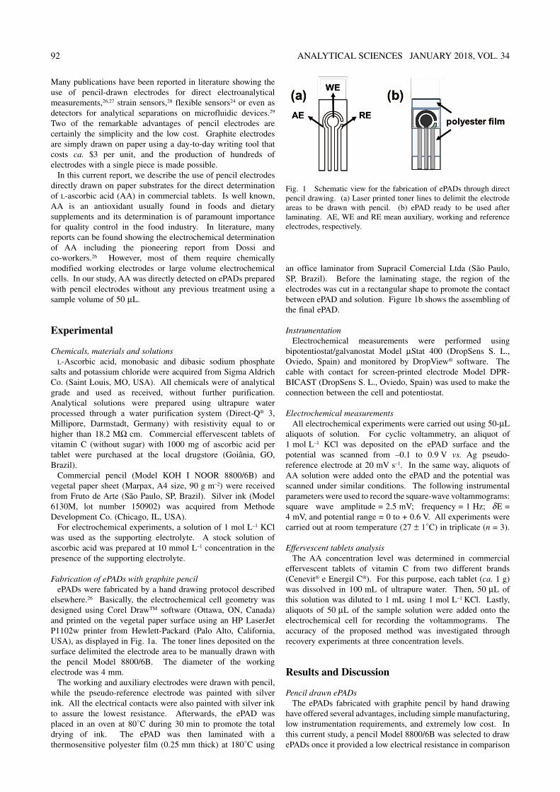
Fabrication of ePADs with graphite pencilePADs were fabricated by a hand drawing protocol described
elsewhere.26 Basically, the electrochemical cell geometry was designed using Corel DrawTM software (Ottawa, ON, Canada) and printed on the vegetal paper surface using an HP LaserJet P1102w printer from Hewlett-Packard (Palo Alto, California, USA), as displayed in Fig. 1a. The toner lines deposited on the surface delimited the electrode area to be manually drawn with the pencil Model 8800/6B. The diameter of the working electrode was 4 mm.
The working and auxiliary electrodes were drawn with pencil, while the pseudo-reference electrode was painted with silver ink. All the electrical contacts were also painted with silver ink to assure the lowest resistance. Afterwards, the ePAD was placed in an oven at 80°C during 30 min to promote the total drying of ink. The ePAD was then laminated with a thermosensitive polyester film (0.25 mm thick) at 180°C using
an office laminator from Supracil Comercial Ltda (São Paulo, SP, Brazil). Before the laminating stage, the region of the electrodes was cut in a rectangular shape to promote the contact between ePAD and solution. Figure 1b shows the assembling of the final ePAD.
InstrumentationElectrochemical measurements were performed using
bipotentiostat/galvanostat Model μStat 400 (DropSens S. L., Oviedo, Spain) and monitored by DropView® software. The cable with contact for screen-printed electrode Model DPR-BICAST (DropSens S. L., Oviedo, Spain) was used to make the connection between the cell and potentiostat.
Electrochemical measurementsAll electrochemical experiments were carried out using 50-μL
aliquots of solution. For cyclic voltammetry, an aliquot of 1 mol L–1 KCl was deposited on the ePAD surface and the potential was scanned from –0.1 to 0.9 V vs. Ag pseudo-reference electrode at 20 mV s–1. In the same way, aliquots of AA solution were added onto the ePAD and the potential was scanned under similar conditions. The following instrumental parameters were used to record the square-wave voltammograms: square wave amplitude = 2.5 mV; frequency = 1 Hz; δE = 4 mV, and potential range = 0 to + 0.6 V. All experiments were carried out at room temperature (27 ± 1°C) in triplicate (n = 3).
Effervescent tablets analysisThe AA concentration level was determined in commercial
effervescent tablets of vitamin C from two different brands (Cenevit® e Energil C®). For this purpose, each tablet (ca. 1 g) was dissolved in 100 mL of ultrapure water. Then, 50 μL of this solution was diluted to 1 mL using 1 mol L–1 KCl. Lastly, aliquots of 50 μL of the sample solution were added onto the electrochemical cell for recording the voltammograms. The accuracy of the proposed method was investigated through recovery experiments at three concentration levels.
Results and Discussion
Pencil drawn ePADsThe ePADs fabricated with graphite pencil by hand drawing
have offered several advantages, including simple manufacturing, low instrumentation requirements, and extremely low cost. In this current study, a pencil Model 8800/6B was selected to draw ePADs once it provided a low electrical resistance in comparison
Fig. 1 Schematic view for the fabrication of ePADs through direct pencil drawing. (a) Laser printed toner lines to delimit the electrode areas to be drawn with pencil. (b) ePAD ready to be used after laminating. AE, WE and RE mean auxiliary, working and reference electrodes, respectively.

ANALYTICAL SCIENCES JANUARY 2018, VOL. 34 93
with other Models (data not shown). It is important to highlight that ca. 100 ePADs can be prepared from a single vegetal paper sheet at a cost of ca. $0.05 per unit. Considering the manual processing, the time required to complete the manufacturing of each ePAD is around 10 min. The lamination of the ePADs with thermosensitive polyester films provided higher robustness for analytical procedures. In addition, this strategy has allowed the adjustment of the best layout for integrating with a portable support for electrochemical measurements. Due to the inherent versatility of the fabrication process, the layout of ePADs can be easily configured to meet the requirements for integrating with portable electrochemical readers or even quick response (QR) codes.30 The association of both portability and disposability are quite attractive for on-site applications. One of the main limitations of the proposed devices is associated with the hand drawing steps, which may affect the reproducibility. However, this drawback was recently overcome by Dossi et al.,31 who explored an adapted printer to automatically draw electrodes on paper. This adapted printer can enable the use of this direct draw protocol in large scale.
Electrochemical behavior of ascorbic acid in pencil drawn ePADs
Figure 2 shows a cyclic voltammogram of AA recorded on the ePADs designed with graphite pencil. As it can be noted, the voltammogram for the supporting electrolyte revealed a behavior similar to carbon with no faradaic event in the selected potential window.32 On the other hand, the AA solution exhibited electrochemical activity on the proposed device. As denoted in Fig. 2, an anodic current peak increasing was observed at 0.4 V vs. pseudo-reference electrode composed of Ag. No reduction peak was observed, thus suggesting an irreversible electrochemical reaction. The recorded data are in good agreement with the results reported by Qin et al.33 and Pisoschi et al.,34 who explored rGO modified screen-printed electrodes and platinum working electrode, respectively.
Detection of ascorbic acid by square wave voltammetrySquare wave voltammetry (SWV) was selected to detect
ascorbic acid due to its superior sensitivity when compared to other voltammetric techniques. Moreover, other advantages of this technique include the minimal adsorption of non-electroactive products on the electrode surface, thus preventing
electrode poisoning.35
Figure 3 shows the SWV recorded for AA solutions at different concentrations. The current peak intensity revealed a good linear correlation when AA concentration ranged from 0.5 to 3.0 mmol L–1. The correlation coefficient was equal to 0.998. The achieved values for limit of detection (LOD) and sensitivity were 70 μmol L–1 and 0.47 μA/mmol L–1, respectively. The LOD was calculated based on the ratio between three times the standard deviation of the blank and the slope of the analytical curve. In comparison with the data reported in the literature, the achieved LOD is lower than the value found by Thiagarajan et al.36 and higher than the LODs found by other authors, including da Silva et al.,37 Kalimuthu et al.,38 Qi et al.,39 Zheng et al.,40 Wang et al.,41 Zhang et al.,42 and Taei et al.43 It is important to highlight that all mentioned reports explored modified electrodes for detecting AA. On the other hand, the proposed ePADs were used without any modification, making the electrode fabrication simpler, easier and cheaper than previous studies. Furthermore, ePADs designed with pencil can be used as disposable electrodes, thus preventing their fouling or poisoning.
Once we demonstrated the electroanalytical feasibility of the proposed ePADs, two commercial tablets were analyzed as previously mentioned. Figure 4 shows the recorded voltammograms for both samples. Considering the dilution and the analytical curve, the concentration of AA in both tablets were estimated to be 2.80 ± 0.02 and 3.10 ± 0.01 mmol L–1 for CenevitTM and Energil CTM, respectively. The expected concentration for both tablets was 3 mmol L–1. In this way, it is possible to predicate that the error between the expected and the experimental values was relatively low and acceptable for quantitative measurements. The accuracy of the proposed devices was investigated through recovery experiments by spiking the commercial sample solutions with known standard concentrations. The recovery ranged from 95 to 115% (n = 3). Based on the reported data, the proposed ePADs can be explored as powerful platforms for the quality control of commercial tablets and even for authenticity investigation of commercialized products.
Fig. 2 Cyclic voltammograms recorded for supporting electrolyte and ascorbic acid on ePADs vs. Ag pseudo-reference electrode at 20 mV s–1.
Fig. 3 Analytical curve for the detection of ascorbic acid on pencil drawn ePADs. Experimental conditions: amplitude = 2.5 mV and frequency = 1 Hz, from 0 to 0.6 V vs. Ag pseudo-reference electrode at 20 mV s–1.

94 ANALYTICAL SCIENCES JANUARY 2018, VOL. 34
Conclusions
In summary, we described in this report the use of ePADs prepared by a pencil drawing protocol for the determination of AA in commercial tablets. One of the remarkable features of our devices refers to the disposability of ePADs designed with graphite pencil. Based on the achieved data, AA was successfully detected on pencil electrodes without any modification. This achievement is quite important because it prevents electrode poisoning or fouling. The found LOD was sufficient to ensure the determination of this compound in two commercial tablets with high reliability. Considering the instrumental simplicity, easy of fabrication, short time, global affordability of both paper and pencil, low cost and robustness ensured by the lamination, the proposed ePADs can be a good alternative to commercial electrodes for quality control testing in food sciences.
Acknowledgements
This project was supported by Conselho Nacional de Desenvolvimento Científico e Tecnológico (CNPq) (grant 448089/2014-9), Coordenação de Aperfeiçoamento de Pessoal de Nível Superior (CAPES), Fundação de Amparo à Pesquisa do Estado de Goiás (FAPEG) and Instituto Nacional de Ciência e Tecnologia de Bioanalítica (INCTBio). CAPES and CNPq are also acknowledged for the scholarships and researcher fellowship (grant 308140/2016-8) granted to the authors.
References
1. A. W. Martinez, S. T. Phillips, G. M. Whitesides, and E. Carrilho, Anal. Chem., 2010, 82, 3.
2. D. M. Cate, J. A. Adkins, J. Mettakoonpitak, and C. S. Henry, Anal. Chem., 2015, 87, 19.
3. W. K. Tomazelli Coltro, C.-M. Cheng, E. Carrilho, and D. P. de Jesus, Electrophoresis, 2014, 35, 2309.
4. R. Pelton, TrAC Trends Anal. Chem., 2009, 28, 925. 5. G. G. Morbioli, T. Mazzu-Nascimento, L. A. Milan, A. M.
Stockton, and E. Carrilho, Anal. Chem., 2017, 89, 4786. 6. T. Mazzu-Nascimento, G. G. Morbioli, L. A. Milan, F. C.
Donofrio, C. A. Mestriner, and E. Carrilho, Anal. Chim.
Acta, 2017, 950, 156. 7. T. M. G. Cardoso, R. B. Channon, J. A. Adkins, M.
Talhavini, W. K. T. Coltro, and C. S. Henry, Chem. Commun., 2017, 53, 7957.
8. K. L. Dornelas, N. Dossi, and E. Piccin, Anal. Chim. Acta, 2015, 858, 82.
9. J. Lankelma, Z. Nie, E. Carrilho, and G. M. Whitesides, Anal. Chem., 2012, 84, 4147.
10. L. Y. Shiroma, M. Santhiago, A. L. Gobbi, and L. T. Kubota, Anal. Chim. Acta, 2012, 725, 44.
11. C. L. S. Chagas, F. R. de Souza, T. M. G. Cardoso, R. C. Moreira, J. A. F. da Silva, D. P. de Jesus, and W. K. T. Coltro, Anal. Methods, 2016, 8, 6682.
12. D. G. Rackus, R. P. S. de Campos, C. Chan, M. M. Karcz, B. Seale, T. Narahari, C. Dixon, M. D. Chamberlain, and A. R. Wheeler, Lab Chip, 2017, 17, 2272.
13. H. Wang, J. Liu, R. G. Cooks, and Z. Ouyang, Angew. Chem., 2010, 122, 889.
14. H. V. Pereira, V. S. Amador, M. M. Sena, R. Augusti, and E. Piccin, Anal. Chim. Acta, 2016, 940, 104.
15. T. C. Colletes, P. T. Garcia, R. B. Campanha, P. V. Abdelnur, W. Romão, W. K. T. Coltro, and B. G. Vaz, Analyst, 2016, 141, 1707.
16. F. Terzi, B. Zanfrognini, S. Ruggeri, N. Dossi, G. M. Casagrande, and E. Piccin, Sens Actuators, B, 2017, 245, 352.
17. S. Nantaphol, R. B. Channon, T. Kondo, W. Siangproh, O. Chailapakul, and C. S. Henry, Anal. Chem., 2017, 89, 4100.
18. J. Mettakoonpitak, K. Boehle, S. Nantaphol, P. Teengam, J. A. Adkins, M. Srisa-Art, and C. S. Henry, Electroanalysis, 2016, 28, 1420.
19. J. Adkins, K. Boehle, and C. Henry, Electrophoresis, 2015, 36, 1811.
20. J.-M. Oh and K.-F. Chow, Anal. Methods, 2015, 7, 7951.21. M. Santhiago, J. Bettini, S. R. Araújo, and C. C. B. Bufon,
ACS Appl. Mater. Interfaces, 2016, 8, 10661.22. W. Dungchai, O. Chailapakul, and C. S. Henry, Anal.
Chem., 2009, 81, 5821.23. R. F. Carvalhal, M. Simão Kfouri, M. H. de Oliveira
Piazetta, A. L. Gobbi, and L. T. Kubota, Anal. Chem., 2010, 82, 1162.
24. M. Santhiago, M. Strauss, M. P. Pereira, A. S. Chagas, and C. C. B. Bufon, ACS Appl. Mater. Interfaces, 2017, 9, 11959.
25. N. Dossi, F. Terzi, E. Piccin, R. Toniolo, and G. Bontempelli, Electroanalysis, 2016, 28, 250.
26. N. Dossi, R. Toniolo, A. Pizzariello, F. Impellizzieri, E. Piccin, and G. Bontempelli, Electrophoresis, 2013, 34, 2085.
27. N. Dossi, R. Toniolo, F. Terzi, E. Piccin, and G. Bontempelli, Electrophoresis, 2015, 36, 1830.
28. C.-W. Lin, Z. Zhao, J. Kim, and J. Huang, Sci. Rep., 2014, 4, 3812.
29. C. L. S. Chagas, L. Costa Duarte, E. O. Lobo-Júnior, E. Piccin, N. Dossi, and W. K. T. Coltro, Electrophoresis, 2015, 36, 1837.
30. M. Santhiago, C. S. Henry, and L. T. Kubota, Electrochim. Acta, 2014, 130, 771.
31. N. Dossi, S. Petrazzi, R. Toniolo, F. Tubaro, F. Terzi, E. Piccin, R. Svigelj, and G. Bontempelli, Anal. Chem., 2017, 89, 10454.
32. R. G. C. S. dos Reis and F. Colmati, J. Solid State Electrochem., 2016, 20, 2559.
33. Q. Qin, X. Bai, and Z. Hua, J. Electroanal. Chem., 2016, 782, 50.
Fig. 4 Square wave voltammograms for the determination of ascorbic acid in two commercial tablets. Experimental conditions: see Fig. 3.

ANALYTICAL SCIENCES JANUARY 2018, VOL. 34 95
34. A. M. Pisoschi, A. Pop, A. I. Serban, and C. Fafaneata, Electrochim. Acta, 2014, 121, 443.
35. A. Chen and B. Shah, Anal. Methods, 2013, 5, 2158.36. S. Thiagarajan and S.-M. Chen, Talanta, 2007, 74, 212.37. R. P. da Silva, A. W. O. Lima, and S. H. P. Serrano, Anal.
Chim. Acta, 2008, 612, 89.38. P. Kalimuthu and S. A. John, Talanta, 2010, 80, 1686.39. S. Qi, B. Zhao, H. Tang, and X. Jiang, Electrochim. Acta,
2015, 161, 395.40. X. Zheng, Y. Guo, J. Zheng, X. Zhou, Q. Li, and R. Lin,
Sens. Actuators, B, 2015, 213, 188.41. L. Wang, C. Gong, Y. Shen, W. Ye, M. Xu, and Y. Song,
Sens. Actuators, B, 2017, 242, 625.42. D. Zhang, L. Li, W. Ma, X. Chen, and Y. Zhang, Mater. Sci.
Eng. C, 2017, 70, 241.43. M. Taei and M. S. Jamshidi, Microchem. J., 2017, 130, 108.