Embed Size (px)
Citation preview
Vol. 39: 159-167, 2000 DISEASES OF AQUATIC ORGANISMS
Dis Aquat Org Published February 9
Detection of Australian gill-associated virus (GAV) and lymphoid organ virus (LOV) of Penaeus
monodon by RT-nested PCR
Jeff A. Cowley*, Christine M. Dimmock, Kirsten M. Spann, Peter J. Walker
Cooperative Research Centre for Aquaculture, CSIRO Tropical Agriculture, PMB3, Indooroopilly 4068, Australia
ABSTRACT: A highly sensitive test based on reverse transcription followed by nested polymerase chain reaction (RT-nPCR) was developed to detect the Australian yellow-head-like viruses, gill-associ- ated virus (GAV) and lymphoid organ virus (LOY) of Penaeus rnonodon. The RT-nPCR detected viral RNA in as little as 10 fg lymphoid organ total RNA isolated from GAV-infected P. monodon. Amplifica- tion of serial dilutions of a GAV cDNA clone showed that the nested PCR was sufficiently sensitive to detect a single genome equivalent using a DNA template. The specificity and sensitivity of the RT- nPCR was also demonstrated using experimentally infected P. (Marsupenaeus) japonicus, where GAV sequences could be amplified from lymphoid organ and haemocyte RNA as early as 6 h post infection (p.i.), and from gills by 24 h p.i. In contrast, transmission electron microscopy (TEM) identified nucleo- capsids and virions in lymphoid organ cells and haemocytes from Days 3 and 6 p i . , respectively, while there was no evidence of infection in gill cells at any time. The practical application of the RT-nPCR was demonstrated by screening healthy wild-caught P monodon broodstock. The high prevalence (>g8 %) of broodstock that were positive by RT-nPCR suggests that LOV is endemic in northern Queensland. In addition, results with lymphoid organ, gill and haemocyte RNA suggest that small gill biopsies may be best suited to the non-sacrificial testing of valuable broodstock. The speed and sensitivity of the RT- nPCR make it a useful adjunct to TEM for diagnosing LOV/GAV infection of P. monodon. with the addi- tional benefit that screening of gill biopsies may facilitate selection of LOV-free broodstock.
KEY WORDS: Yellow head virus (YHV) . Lymphoid organ virus (LOV) . Gill-associated virus (GAV) Penaeus monodon RT-PCR . Penaeid shrimp - Prawns
INTRODUCTION
We have recently described a rod-shaped, gill-asso- ciated virus (GAV) which was associated with mor- talities of Penaeus monodon prawns at 4 farms in Queensland, Australia, in 1996 (Spann et al. 1997). GAV is highly pathogenic, inducing mortalities from 4 to 5 d following experimental infection of subadult P. monodon. In diseased prawns, large numbers of heli- cal nucleocapsids and enveloped virions appear in gill and lymphoid organ cells. Invariably, the lymphoid organ displays extensive cellular necrosis and struc- tural degeneration (Spann et al. 1997). In morphology and cytopathology, GAV closely resembles yellow
head virus (YHV) from Thailand (Boonyaratpalin et al. 1993, Chantanachookin et al. 1993, Wongteerasupaya et al. 1995). However, GAV does not induce the pale to yellow body colouration described for YHV, and mortality is usually preceded by a pink to red body colouration. Prior to the identification of GAV, a virus with similar morphology was reported to be common in healthy P. monodon in Queensland (Spann et al. 1995). Unlike GAV, lymphoid organ virus (LOV) infection appeared to be restricted to hypertrophied cells in discrete foci or 'spheroids' in the lymphoid organ. LOV is non-pathogenic, does not cause exten- sive cellular necrosis and has not been observed in gill tissue.
Recent molecular studies of YHV and GAV have shown that they are closely related but distinct viruses which are likely to be classified in the family Coron-
O Inter-Research 2000 Resale of full article not permitted
160 Dis Aquat Org 39: 159-167,2000
aviridae (Cowley et al. 1999). Wongteerasupaya et al. (1995) demonstrated that YHV contains a single- stranded RNA genome. Nadala et al. (1997a) subse- quently showed that the YHV genome is -22 kb long and identified 4 major virion proteins (170, 135, 67 and 22 kDa) of which the 135 kDa protein is glycosylated. Tang & Lightner (1999) have amplified reverse tran- scription-polymerase chain reaction (RT-PCR) prod- ucts from cDNA synthesised using a primer of opposite sense to a putative coding sequence to show that YHV genomic RNA is likely to be of positive polarity. We have recently determined the nucleotide sequence of the GAV genome and established that it is indeed a (+) RNA virus which contains sequence homologies and a genome organisation, including large overlapping ORFla and ORFlb polyprotein genes translated via a -1 ribosomal frameshift site, characteristic of toro- and coronaviruses (Cowley et al. unpubl.). Comparative sequence analysis of 3 corresponding regions of the ORFlb gene has indicated that YHV and GAV are closely related viruses that may be regarded as differ- ent geographic topotypes (Cowley et al. 1999).
In this paper, we describe a sensitive and specific RT-nested PCR (RT-nPCR) for detecting GAV in infected prawn tissues. The test is applied to the detec- tion of GAV in total RNA isolated from lymphoid organ, gill and haemocytes of healthy and clinically diseased Penaeus monodon and from P. (Marsupe- naeus) japonicus infected experimentally with GAV. Sequence analysis of PCR products amplified from healthy P, monodon indicates that LOV is a non-patho- genic variant of GAV. In addition, screening of hatch- ery broodstock suggests that LOV infection may be widespread in wild P. monodon from northern Queens- land.
MATERIALS AND METHODS
Prawns. Healthy Penaeus monodon and P. japonicus were obtained from farms and hatcheries in south-east and northern Queensland between December 1996 and March 1998. P. monodon broodstock were cap- tured from coastal waters in the Cairns, Innisfail and Townsville regions of northern Queensland. The broodstock were obtained either directly from a com- mercial trawler operator and tested prior to spawning or from hatcheries or CSIRO Marine Research, Cleve- land, and tested subsequent to spawning. Prawns were sampled within 1 to 2 d of their acquisition. Diseased P. monodon, displaying characteristic clinical symptoms and histological evidence of GAV infection (Spann et al. 1997), were obtained from a pond at a farm in south-east Queensland that had experienced mortali- ties in March 1997.
Virus infections. The prototype GAV isolate used to generate cDNA clones originated from clinically dis- eased Penaeus monodon collected from farm A in northern Queensland in 1996 (Spann et al. 1997). Healthy P. monodon (15 to 20 g) were injected with 5 p1 g-' body weight of head extracts of GAV-infected prawns as described previously (Spann et al. 1997 ). P. monodon deaths began at 4 d post infection (pi . ) , and at 6 d p.i. lymphoid organ and gill tissues were iso- lated, snap frozen on dry ice and stored in liquid nitro- gen until used for RNA isolation. Haemolymph (0.5 to 1.0 ml) was collected by cardiac puncture directly into 1 m1 ice-cold modified arsenals anticoagulant (19.3 mM sodium citrate. 239.8 mM NaC1, 182.5 mM glucose, 6.2 mM EDTA, pH 7.3) (van de Braak et al. 1996). Haemocytes were collected by microcentrifuga- tion at 5000 X g for 2 min, resuspended in 1 m1 phos- phate-buffered saline (PBS), recentrifuged and the haemocyte pellet was stored at -70°C.
To follow the progress of GAV infection in a suscep- tible LOV-free host, juvenile Penaeus japonicus (12 to 14 g) were inoculated as described above. Lymphoid organs, gills and haemocytes were pooled from 2 prawns sampled at 6, 24, and 48 h and on Days 3, 6, 9, 12 and 15 p.i. as described above and stored under liq- uid nitrogen until used for total RNA isolation. At the same times p i , haemocytes and cephalothorax tissue samples from 2 prawns were also processed for histol- ogy and transmission electron microscopy (TEM).
Light and electron microscopy. Histological exami- nations of prawn cephalothorax tissues fixed in David- son's fixative were conducted as described previously (Spann et al. 1997). Lymphoid organ, gill and haemo- cyte tissues were also fixed in modified Karnovsky's reagent and processed for TEM to confirm the pres- ence of LOVIGAV and to quantify nucleocapsids and virions (Spann et al. 1997).
RNA isolation. In general, total RNA was extracted from 50 to 100 mg lymphoid organ, gill or haemocytes of healthy and GAV-infected prawns by homogen- isation in 750 p1 TRIzol-LSTM (Life Technologies) using a pellet pestle according to the manufacturer's in- structions. RNA was resuspended in diethylpyrocarbo- nate (DEPC)-water, quantified by spectrophotometry (A260nm) and stored at -70°C. All reagents and tissue samples were handled in a separate laboratory using a laminar flow cabinet and aerosol-resistant barrier pipette tips to prevent carry-over contamination with PCR products.
Random PCR amplification and cloning of GAV cDNA. Lymphoid organ totaI RNA was isolated from Penaeus monodon infected experimentally with GAV as described above. RNAs were resolved in 0.6% LMP agarose (Life Technologies)-TAE gels containing 0.5 pg ml-' ethidium bromide (Sambrook et al. 1989).
Cowley et al.: RT-PCR detection of GAV and LOV 161
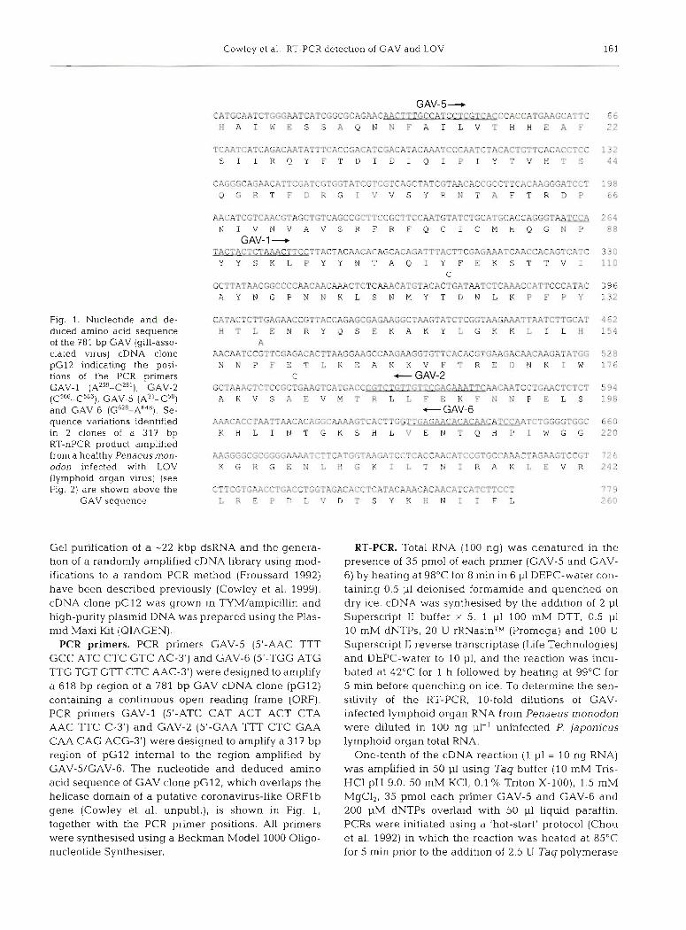
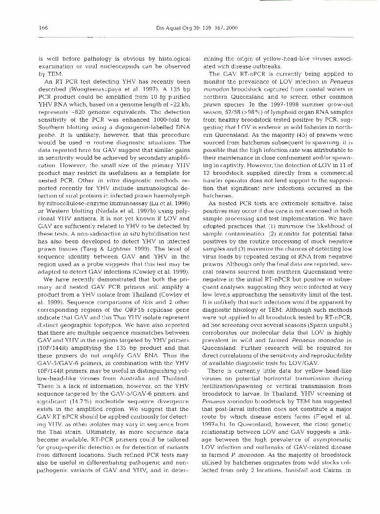
Fig. 1. Nucleotide and de- duced amino acid sequence of the 781 bp GAV (gill-asso- ciated virus) cDNA clone pG12 indicating the posi- tions of the PCR primers GAV-1 ( A ~ ~ ~ - C * ~ ' ) , GAV-2 (C566-C586), GAV-5 (A~'-C'') and GAV-6 (G62a-~648). Se- quence variations identified in 2 clones of a 317 bp RT-nPCR product amplified from a healthy Penaeusmon- odon infected with LOV (lymphoid organ virus) (see Fig. 2) are shown above the
GAV sequence
GAV-5- CATGCAATCTGGGAATCATCGGCGCAGAAC-CCTCGTCACCCACCATGAAGCATTC H A I W E S S A Q N N F A I L V T H H E A F
TCAATCATCAGACAATATTTCACCGACATCGACATACAAATCCCAATCTACACTGTTCACACCTCC S I I R Q Y F T D I D I Q I P I Y T V H T S
CAGGGCAGAACATTCGATCGTGGTATCGTCGTCAGCTATCGTAACACCGCCTTCACAAGGGATCCT Q G R T F D R G I V V S Y R N T A F T R D P
AACATCGTCAACGTAGCTGTCAGCCGCTTCCGCTTCCRATGTATCTGCATGCACCAGGGT~ N I V N V A V S R F R F Q C I C M H Q G N P
GAV-1- TACTACTCTAAACTTCCTTACTACAACACAGCACAGATTTACTTCGAGAAATCAACCACAGTCATC Y Y S K L P Y Y N T A Q I Y F E K S T T V I
C GCTTATAACGGCCCCAACAACAAACTCTCAAACATGTACACTGfiTMTCTCMCCATTCCCATAC A Y N G P N N K L S N M Y T D N L K P F P Y
CATACTCTTGAGAACCGTTACCAGAGCGAGAAGGCTAAGTATCTCGGTAAGAAATTAATCTTGCAT H T L E N R Y Q S E K A K Y L G K K L I L H
A AACAATCCGTTCGAGACACTTAAGGAAGCCAAGAAGGTGTTCACACGTGmGACAACMGATATGG N N P F E T L K E A K K V F T R E D N K I W
C t GAV-2 GCTAAAGTCTCCGCTGAAGTCATGACCCGTCTGTTrTTCGPr~AACAATCCTGMCTCTCT A K V S A E V M T R L L F E K F N N P E L S - GAV-6
A A A C A C C T A A T T A A C A C A G G C A A A A G T C A C T T G G T T G 3 m T C T G G G G T G G C K H L I N T G K S H L V E N T Q H P I W G G
AAGGGGCGCGGGGAAAATCTTCATGGTAAGATCCTCACCAACATCCGTGCCAAACTAGAAGTCCGT K G R G E N L H G K I L T N I R A K L E V R
CTTCGTGAACCTGACCTCGTAGACACCTCATACAAACACAACATCATCTTCCT L R E P D L V D T S Y K H N I I F L
Gel purification of a -22 kbp dsRNA and the genera- tion of a randomly amplified cDNA library using mod- ifications to a random PCR method (Froussard 1992) have been described previously (Cowley et al. 1999). cDNA clone pG12 was grown in TYM/ampicillin and high-purity plasmid DNA was prepared using the Plas- mid Maxi Kit (QIAGEN).
PCR primers. PCR primers GAV-5 (5'-AAC TTT GCC ATC CTC GTC AC-3') and GAV-6 (5'-TGG ATG TTG TGT GTT CTC AAC-3') were designed to amplify a 618 bp region of a 781 bp GAV cDNA clone (pG12) containing a continuous open reading frame (ORF). PCR primers GAV-l (5'-ATC CAT ACT ACT CTA AAC TTC C-3') and GAV-2 (5'-GAA T l T CTC GAA CAA CAG ACG-3') were designed to amplify a 317 bp region of pG12 internal to the region amplified by GAV-5/GAV-6. The nucleotide and deduced amino acid sequence of GAV clone pG12, which overlaps the helicase domain of a putative coronavirus-like ORFlb gene (Cowley et al. unpubl.), is shown in Fig. 1, together with the PCR primer positions. All primers were synthesised using a Beckman Model 1000 OLigo- nucleotide Synthesiser.
RT-PCR. Total RNA (100 ng) was denatured in the presence of 35 pm01 of each primer (GAV-5 and GAV- 6) by heating at 98'C for 8 min in 6 p1 DEPC-water con- taining 0.5 p1 deionised formamide and quenched on dry ice. cDNA was synthesised by the addition of 2 p1 Superscript I1 buffer X 5, 1 p1 100 mM DTT, 0.5 p1 10 mM dNTPs, 20 U rRNasinTM (Promega) and 100 U Superscript I1 reverse transcriptase (Life Technologies) and DEPC-water to 10 p1, and the reaction was incu- bated at 42°C for 1 h followed by heating at 99°C for 5 min before quenching on ice. To determine the sen- sitivity of the RT-PCR, 10-fold dilutions of GAV- infected lymphoid organ RNA from Penaeus monodon were diluted in 100 ng p1-' uninfected P. japonicus lymphoid organ total RNA.
One-tenth of the cDNA reaction (1 p1 = 10 ng RNA) was amplified in 50 p1 using Tag buffer (10 mM Tris- HCl pH 9.0, 50 mM KCl, 0.1 % Triton X-loo), 1.5 mM MgC12, 35 pm01 each primer GAV-5 and GAV-6 and 200 pM dNTPs overlaid with 50 p1 liquid paraffin. PCRs were initiated using a 'hot-start' protocol (Chou et al. 1992) in which the reaction was heated at 85OC for 5 min prior to the addition of 2.5 U Tag polymerase
162 Dis Aquat Org 39: 159-167,2000
(Promega). DNA was amplified by 30 cycles of 95"C/1 min, 58"C/1 min, and 72"C/40 S, followed by 72"C/10 min final extension and 20°C hold using either a Corbett Research or Omnigene (Hybaid) thermal cycler. PCR products (10 p1) were resolved in 2 % agarose-TAE gels containing 0.5 pg ml-' ethidium bro- mide.
Nested PCR. When the result of the primary RT-PCR was negative or inconclusive, 0.5 p1 of the primary PCR was amplified by nested PCR as above in a 50 p1 reac- tion volume using primers GAV-1 and GAV-2. In some cases, 5 p1 of the RT-PCR was used. Nested PCR condi- tions were as for the primary PCR except that the extension time was reduced to 30 s and number of cycles was reduced to 20. Nested PCR aliquots (10 p1) were analysed in 2 % agarose-TAE gels. When a result was still negative, the complete RT-nPCR was re- peated with up to 5 aliquots of the total RNA sample. All PCR reagents were handled in a laminar flow cabi- net using aerosol-resistant tips to avoid contamination. Primary RT-PCR products were handled in a separate work area to that in which nested PCRs were per- formed.
Sequencing analysis. For sequencing, nested PCR products were purified using a QIAquick column (QIAGEN) and cloned by ligation into pGEM-T and transformation of competent Escherichia coli DH5-a host cells as described previously (Cowley et al. 1999). Plasmid DNA was prepared using a RPM kit (B10 101) and automated sequencing of inserts was conducted using universal M13/pUC forward and reverse primers, ThermoSequenaseTM dye-terminator reagent (Amersham) and an automated AB1 Model- 377 sequencing apparatus (Applied Biosystems Inc.) at the Australian Genome Research Facility, Univer- sity of Queensland.
Gill biopsies. Six male and 4 female healthy, mature Penaeusmonodon (28 to 35 g), which had not spawned in captivity, were collected from a hatchery in northern Queensland and held in the aquarium facility at CSIRO Tropical Agriculture, Brisbane, for 25 d. Gill biopsies were performed on 5 of the 10 prawns (3 males and 2 females) by holding the prawns firmly with the dorsal side against a flat surface and gently lifting the left gill cover to expose the 2 posterior pri- mary gill filaments. The inner filament was removed using flame-sterilised scissors, leaving approximately 1.5 to 2 mm of the filament base. Prawns were moni- tored for mortality and wound healing for 5 wk. Total RNA was extracted using TRIzol-LSTV from gill biop- sies: (1) pooled from the 2 females; (2) pooled from 2 males; and (3) from the remaining male. RT-nPCRs were performed and analysed as described above except that 1 1-19 instead of 100 ng RNA was used for cDNA synthesis.
RESULTS
Specificity of the RT-nPCR
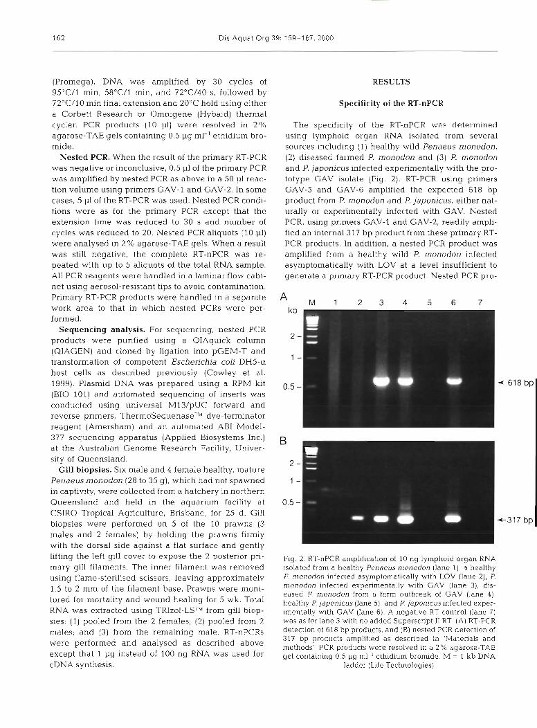
The specificity of the RT-nPCR was determined using lymphoid organ RNA isolated from several sources including (1) healthy wild Penaeus monodon, (2) diseased farmed P. monodon and (3) P. monodon and P. japonicus infected experimentally with the pro- totype GAV isolate (Fig. 2). RT-PCR using primers GAV-5 and GAV-6 amplified the expected 618 bp product from P. monodon and P. japonicus, either nat- urally or experimentally infected with GAV. Nested PCR, using primers GAV-l and GAV-2, readily ampli- fied an internal 317 bp product from these primary RT- PCR products. In addition, a nested PCR product was amplified from a healthy wild P. monodon infected asymptomatically with LOV at a level insufficient to generate a primary RT-PCR product. Nested PCR pro-
Fig. 2. RT-nPCR amplification of 10 ng lymphoid organ RNA isolated from a healthy Penaeus monodon (lane l ) , a healthy P. monodon infected asymptomatically with LOV (lane 2), P. rnonodon infected experimentally with GAV (lane 3), dis- eased P monodon from a farm outbreak of GAV (lane 41, healthy P japonicus (lane 51, and P. japonicus infected exper- mentally w t h GAV (lane 6). A negative RT control (lane 7) was as for lane 3 with no added Superscript I1 RT (A) RT-PCR detection of 618 bp products, and (B) nested PCR detection of 317 bp products amplified as described in 'Materials and methods' PCR products were resolved in a 2 % agarose-TAE gel containing 0.5 pg ml-' ethidium bromide. M = 1 kb DNA
ladder (Life Technologies)
Cowley et al.: RT-PCR detection of GAV and LOV 163
ducts were cloned and sequenced and the A sequences compared to that of pG12 generated kb
M 1 2 3 4 5 6 7 8 9
from the 22 kbp dsRNA of the prototype GAV iso- late. Sequences of 2 clones from P. monodon and
-
2 from P. japonicus infected with the prototype 1 - GAV were identical to the pG12 sequence. The sequences of 2 clones from the healthy P. mon- 0.5 - 4- 61 8 bp
odon harbouring LOV were identical and dis- played only 1.1% (3/274) nucleotide variation from ~ G 1 2 . None of the vanations altered the coding sequence (see Fig. 1). No products were amplified from uninfected P. japonicus or from another overtly healthy P. monodon. RNA from GAV-infected P. monodon also failed to produce RT-nPCR products when reverse transcriptase was omitted, demonstrating that the primers were not amplifying an extraneous DNA template. In addition, the absence of RT-PCR inhibitors and the integrity of the control P. japonicus RNA were confirmed by RT-PCR amplification of an 848 bp fragment of 18s ribosomal RNA using primers designed for the rRNA sequence of P, aztecus (Kim & Abele 1990) (data not shown).
I t was not possible to test for cross-reactivity with a comprehensive range of viral pathogens of Penaeus monodon. However, nested PCR with the
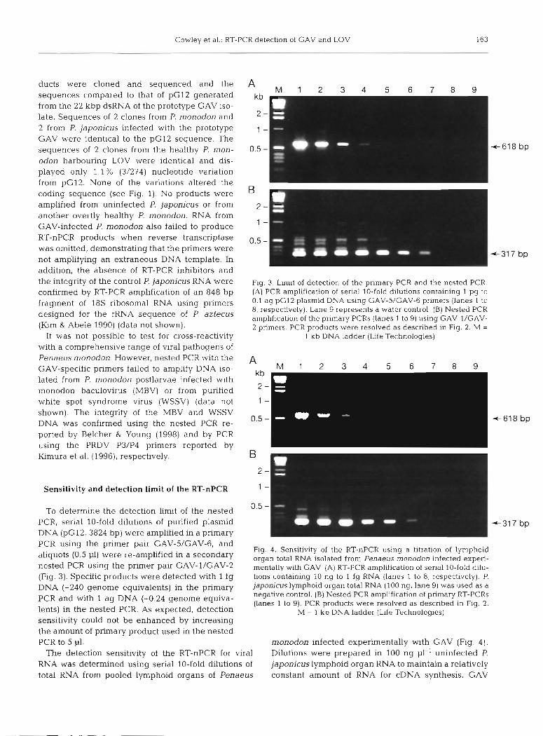
Fig. 3. Limit of detection of the primary PCR and the nested PCR. (A) PCR amplification of serial 10-fold dilutions containing 1 pg to 0.1 ag pG12 plasmid DNA using GAV-5/GAV-6 primers (lanes 1 to 8, respectively). Lane 9 represents a water control. (B) Nested PCR amplification of the primary PCRs (lanes 1 to 9) using GAV-l/GAV- 2 primers. PCR products were resolved as described in Fig. 2. M =
1 kb DNA ladder (Life Technologies)
I \
GAV-specific primers failed to amplify DNA iso- lated from P. monodon postlarvae infected with monodon baculovirus (MBV) or from purified white spot syndrome virus (WSSV) (data not shown). The integrity of the MBV and WSSV DNA was confirmed using the nested PCR re- 0.5 -
ported by Belcher & Young (1998) and by PCR using the PRDV P3/P4 primers reported by
PCR using the primer pair GAV-5/GAV-6, and Fig. 4. Sensitivity of the RT-nPCR using a titration of lymphoid
aliquots (0'5 pl) were re-amplified in a secondary organ total RNA isolated from Penaeus monodon infected experi- nested PCR using the primer pair GAV-1/GAV-2 mentally with GAV. (A) RT-PCR ampl~fication of serial 10-fold dilu- (Fig. 3). Specific products were detected with 1 fg tions containing 10 ng to 1 fg RNA (lanes 1 to 8, respectively). P.
DNA (-240 genonle equivalents) in the primary japonicus lymphoid organ total RNA (100 ng, lane 9) was used as a negative control. (B) Nested PCR amplification of primary RT-PCRs and with ag DNA (-0'24 genOme equiva- (lanes 1 to 9). PCR products were resolved as described in Flg. 2.
lents) in the nested PCR. As expected, detection M = 1 kb DNA ladder (Life Technologies) sensitivity could not be enhanced by increasing the amount of primary product used in the nested PCR to 5 p1. monodon infected experimentally with GAV (Fig. 4).
The detection sensitivity of the RT-nPCR for viral Dilutions were prepared in 100 ng p1-' uninfected P. RNA was determined using serial 10-fold dilutions of japonicus lymphoid organ RNA to maintain a relatively total RNA from pooled lymphoid organs of Penaeus constant amount of RNA for cDNA synthesis. GAV
164 Dis Aquat Org 39: 159-167, 2000
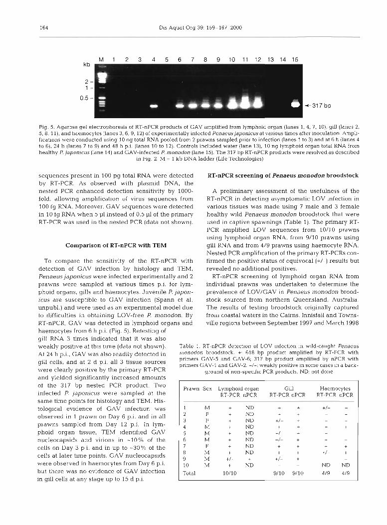
Fig. 5. Agarose gel electrophoresis of RT-nPCR products of GAV amplified from lymphoid organ (lanes 1, 4, 7, 10), gill (lanes 2, 5, 8, 1 l ) , and haemocytes (lanes 3, 6, 9, 12) of experimentally Infected Penaeus japonicus at various times after inoculation. Ampli- fication~ were conducted using 10 ng total RNA pooled from 2 prawns sampled prior to infection (lanes 1 to 3) and at 6 h (lanes 4 to 6), 24 h (lanes 7 to 9) and 48 h p.i. (lanes 10 to 12). Controls included water (lane 13), 10 ng lymphoid organ total RNA from healthy P. japonicus (lane 14) and GAV-infected P. monodon (lane 15). The 317 bp RT-nPCR products were resolved as described
in Fig. 2. M = 1 kb DNA ladder (Life Technologies)
sequences present in 100 pg total RNA were detected RT-nPCR screening of Penaeus monodon broodstock by RT-PCR. As observed with plasmid DNA, the nested PCR enhanced detection sensitivity by 1000- A preliminary assessment of the usefulness of the fold, allowing amplification of virus sequences from RT-nPCR in detecting asymptomatic LOV infection in 100 fg RNA. Moreover, GAV sequences were detected various tissues was made using 7 male and 3 female in 10 fg RNA when 5 p1 instead of 0.5 p1 of the primary healthy wild Penaeus monodon broodstock that were RT-PCR was used in the nested PCR (data not shown). used in captive spawnings (Table 1). The primary RT-
PCR amplified LOV sequences from 10/10 prawns using lymphoid organ RNA, from 9/10 prawns using
Comparison of RT-nPCR with TEM gill RNA and from 4/9 prawns using haemocyte RNA. Nested PCR amplification of the primary RT-PCRs con-
To compare the sensitivity of the RT-nPCR with firmed the positive status of equivocal (+/-) results but detection of GAV infection by histology and TEM, revealed no additional positives. Penaeus japonicus were infected experimentally and 2 RT-nPCR screening of lymphoid organ RNA from prawns were sampled at various times p.i. for lym- individual prawns was undertaken to determine the phoid organs, gills and haemocytes. Juvenile P. japon- prevalence of LOV/GAV in Penaeus monodon brood- icus are susceptible to GAV infection (Spann et al. stock sourced from northern Queensland, Australia. unpubl.) and were used as an experimental model due The results of testing broodstock originally captured to difficulties in obtaining LOV-free P. monodon. By from coastal waters in the Cairns, Innisfail and Towns- RT-nPCR, GAV was detected in lymphoid organs and ville regions between September 1997 and March 1998 haemocytes from 6 h p.i. (Fig. 5). Retesting of gill RNA 3 times indicated that it was also weakly positive at thls time (data not shown). Table 1. RT-nPCR detection of LOV Infection in wild-caught Penaeus
~t 24 h p,i,, GAV was also readily detected in monodon broodstock. +: 618 bp product amplified by RT-PCR with primers GAV-5 and GAV-6; 317 bp product amplified by nPCR with
and at p.i' tissue pnmers GAV-1 and GAV-2. +/-: weakly positive in some cases in a back- were clearly positive by the primary RT-PCR ground of non-speclfic PCR products. ND: not done and yielded significantly increased amounts of the 317 bp nested PCR product. Two infected P. japonicus were sampled at the same time points for histology and TEM. His- tological evidence of GAV infection was observed in 1 prawn on Day 6 p.i. and in all prawns sampled from Day 12 p.i. In lym- phoid organ tissue, TEM identified GAV nucleocapsids and virions in -10% of the cells on Day 3 p.i. and in up to -30% of the cells at later time points. GAV nucleocapsids were observed in haemocytes from Day 6 p.i. but there was no evidence of GAV infection in gill cells at any stage up to 15 d p.i.
Prawn Sex Lymphoid organ Gill Haemocytes RT-PCR nPCR RT-PCR nPCR RT-PCR nPCR
1 M + ND + + + + 2 F + ND + + P -
3 F + ND +/- + -
4 M + ND + + + + 5 M + ND +/L
+ - P
6 IM + ND +/- + - -
7 F + ND + + + + 8 M + ND + + +/- + 9 M +/- + +/- + - -
10 M + ND - - ND ND
Total 10/10 9/10 9/10 4/9 4/9
Cowley et al.. RT-PCR detection of GAV and LOV 165
Wild Hatchery Date Broodstock RT-nPCR source tested positive
A CSIRO 25Sep1997 10 10 A Hatchery 1 13 Oct 1997 12 12 B Hatchery 2 14 Oct 1997 12 12 C Hatchery 3 27 Mar 1998 12 12 A Trawlera 5Mar1998 12 11 Total 58 57
aPrawns supplied direct from a trawler
Table 2. Prevalence of LOV/GAV in Penaeus monodon brood- quence. More extensive sequence comparisons, in- stock determined by RT-nPCR testing of lymphoid organ cluding 3 additional CAV isolates and >20 LOV iso- RNA. Wild source: hatchery records indicated that prawns lates from healthy prawns collected from the wild in were captured from the (A) Innisfail, (B) Cairns and
(C) Townsville regions of northern Queensland, Australia different locations in northern Queensland, have iden- tified similar high levels of sequence identity (Cowley et al. unpubl.). These findings strongly suggest that GAV and LOV represent variants of the same virus. As we have reported previously, this level of sequence homology contrasts with the 76.3 % (209/274) nucleo- tide sequence identity in the same genomic region of GAV and a Thai YHV isolate, which represent distinct but closely related topotypes (Cowley et al. 1999).
Analysis of many captured and farmed Penaeus monodon infected asymptomatically with LOV has indicated that the quantities of the 618 bp RT-PCR product can sometimes match levels obtained from GAV-infected diseased prawns (Cowley et al. un-
are shown in Table 2. Of 58 broodstock tested, includ- publ.). However, some prawns are infected with LOV ing the 10 reported above, 57 (98%) were positive by at levels below the detection limit of the primary RT- RT-nPCR. PCR. Nested amplification of an internal 317 bp prod-
uct provides additional specificity and enhanced sen- sitivity. The nested PCR is 103- to 104-fold more
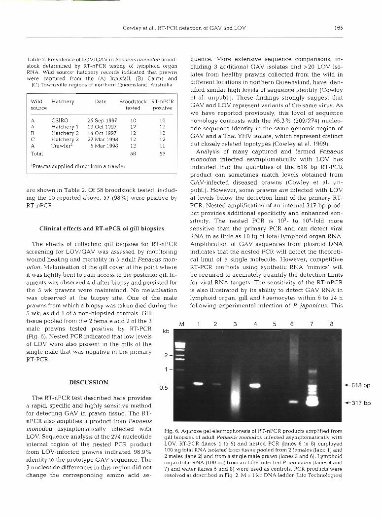
Clinical effects and RT-nPCR of gill biopsies sensitive than the primary PCR and can detect viral RNA in as little as 10 fg of total lymphoid organ RNA.
The effects of collecting gill biopsies for RT-nPCR Amplification of GAV sequences from plasmid DNA screening for LOV/GAV was assessed by monitoring indicates that the nested PCR will detect the theoreti- wound healing and mortality in 5 adult Penaeus mon- cal limit of a single molecule. However, competitive odon. Melanisation of the gill cover at the point where RT-PCR methods using synthetic RNA 'mimics' will it was lightly bent to gain access to the posterior gill fil- be required to accurately quantify the detection Limits aments was observed 4 d after biopsy and persisted for for viral RNA targets. The sensitivity of the RT-nPCR the 5 wk prawns were maintained. No melanisation is also illustrated by its ability to detect GAV RNA in was observed at the biopsy site. One of the male lymphoid organ, gill and haemocytes within 6 to 24 h prawns from which a biopsy was taken died during the following experimental infection of P. japonicus. This 5 wk, as did 1 of 5 non-biopsied controls. Gill tissue pooled from the 2 female and 2 of the 3 M 1 2 3 4 5 6 7 8 male prawns tested positive by RT-PCR kb (Fig. 6). Nested PCR indicated that low levels of LOV were also present in the gills of the single male that was negative in the primary
2 - RT-PCR.
1 -
DISCUSSION 0.5 -
The RT-nPCR test described here provides bp
a rapid, specific and highly sensitive method I 4 3 1 7 bp
for detecting GAV in prawn tissue. The RT- nPCR also amplifies a product from Penaeus
asymptomatically infected with Fig. 6. Agarose gel electrophoresls of RT-nPCR products amplified from LOV. Sequence analysis of the 274 nucleotide gill biopsies of adult Penaeus rnonodon infected asymptomatically with internal region of the nested PCR product LOV. RT-PCR (lanes 1 to 5) and nested PCR (lanes 6 to 8) employed
from ~ ~ ~ - i ~ f ~ ~ ~ ~ d prawns indicated 98.9 % 100 ng total RNA isolated from tissue pooled from 2 females (lane 1) and 2 males (lane 2) and from a single male prawn (lanes 3 and 6). Lymphoid
identity to the prototype sequence' The organ total RNA (100 ng) from a n LOV-infected P. monodon (lanes 4 and 3 nucleotide differences in this region did not 7) and water (lanes 5 and 8) were used as controls. PCR products were change the corresponding amino acid se- resolved as described in Fig. 2. M = 1 kb DNA ladder (Life Technologies)
166 Dis Aquat Org 39: 159-167, 2000
is well before pathology is obvious by histological examination or viral nucleocapsids can be observed by TEM.
An RT-PCR test detecting YHV has recently been described (Wongteerasupaya et al. 1997). A 135 bp PCR product could be amplified from 10 fg purified YHV RNA which, based on a genome length of -22 kb, represents -820 genome equivalents. The detection sensitivity of the PCR was enhanced 1000-fold by Southern blotting using a digoxigenin-labelled DNA probe. It is unlikely, however, that this procedure would be used in routine diagnostic situations. The data reported here for GAV suggest that similar gains in sensitivity would be achieved by secondary amplifi- cation. However, the small size of the primary YHV product may restrict its usefulness as a template for nested PCR. Other in vitro diagnostic methods re- ported recently for YHV include immunological de- tection of viral proteins in infected prawn haemolymph by nitrocellulose-enzyme immunoassay (Lu et al. 1996) or Western blotting (Nadala et al. 1997b) using poly- clonal YHV antisera. It is not yet known if LOV and GAV are sufficiently related to YHV to be detected by these tests. A non-radioactive in situ hybridisation test has also been developed to detect YHV in infected prawn tissues (Tang & Lightner 1999). The level of sequence identity between GAV and YHV in the region used as a probe suggests that this test may be adapted to detect GAV infections (Cowley et al. 1999).
We have recently demonstrated that both the pri- mary and nested GAV PCR primers will amplify a product from a YHV isolate from Thailand (Cowley et al. 1999). Sequence comparisons of this and 2 other corresponding regions of the ORFlb replicase gene indicate that GAV and this Thai YHV isolate represent distinct geographic topotypes. We have also reported that there are multiple sequence mismatches between GAV and YHV in the regions targeted by YHV primers (10F/144R) amplifying the 135 bp product and that these primers do not amplify GAV RNA. Thus the GAV-5/GAV-6 primers, in combination with the YHV 10F/144R primers, may be useful in distinguishing yel- low-head-like viruses from Australia and Thailand. There is a lack of information, however, on the YHV sequence targeted by the GAV-5/GAV-6 primers, and significant (14.7%) nucleotide sequence divergence exists in the amplified region. We suggest that the GAV RT-nPCR should be applied cautiously for detect- ing YHV, as other isolates may vary in sequence from the Thai strain. Ultimately, as more sequence data become available, RT-PCR primers could be tailored for group-specific detection or for detection of variants from different locations. Such refined PCR tests may also be useful in differentiating pathogenic and non- pathogenic variants of GAV and YHV, and in deter-
mining the origin of yellow-head-like viruses associ- ated wlth disease outbreaks.
The GAV RT-nPCR is currently being applied to monitor the prevalence of LOV infection in Penaeus monodon broodstock captured from coastal waters in northern Queensland and to screen other common prawn species. In the 1997-1998 summer grow-out season, 57/58 (> 98%) of lymphoid organ RNA samples from healthy broodstock tested positive by PCR, sug- gesting that LOV is endemic in wild fisheries in north- ern Queensland. As the majority (45) of prawns were sourced from hatcheries subsequent to spawning, it is possible that the high infection rate was attributable to their maintenance in close confinement and/or spawn- ing in captivity. However, the detection of LOV in 11 of 12 broodstock supplied directly from a commercial trawler operator does not lend support to the supposi- tion that significant new infections occurred in the hatcheries.
As nested PCR tests are extremely sensitive, false positives may occur if due care is not exercised in both sample processing and test implementation. We have adopted practices that (1) minimize the likelihood of sample contamination, (2) monitor for potential false positives by the routine processing of mock negative samples and (3) maximize the chances of detecting low virus loads by repeated testing of RNA from negative prawns. Although only the final data are reported, sev- eral prawns sourced from northern Queensland were negative in the initial RT-nPCR but positive in subse- quent analyses, suggesting they were infected at very low levels approaching the sensitivity limit of the test. It is unlikely that such infections would be apparent by diagnostic histology or TEM. Although such methods were not applied to all broodstock tested by RT-nPCR, ad hoc screening over several seasons (Spann unpubl.) corroborates our molecular data that LOV is highly prevalent in wild and farmed Penaeus monodon in Queensland. Further research will be required for direct correlations of the sensitivity and reproducibility of available diagnostic tests for LOV/GAV.
There is currently little data for yellow-head-like viruses on potential horizontal transmission during fertilization/spawning or vertical transmission from broodstock to larvae. In Thailand, YHV screening of Penaeus rnonodon broodstock by TEM has suggested that post-larval infection does not constitute a major route by which disease enters farms (Flegel et al. 1997a,b) In Queensland, however, the close genetic relationship between LOV and GAV suggests a link- age between the high prevalence of asymptomatic LOV infection and outbreaks of GAV-related disease in farmed P. monodon. As the m.ajority of broodstock utilised by hatcheries originates from wild stocks col- lected from only 2 locations, Innisfail and Cairns, in
Cowley et al.: RT-PCR detection of GAV and LOV 167
northern Queensland, this would have significant ram- ifications for the industry. The RT-nPCR test will assist in addressing the important issues relating to vertical transmission of LOV/GAV and in identifying LOV-free populations of P. monodon that may exist at other loca- tions in northern Australia.
The discovery that 9 out of 10 gill RNA samples from healthy Penaeus monodon tested positive by RT-nPCR contrasts with the complete lack of TEM evidence of LOV infection in gill tissue (Spann et al. 1995, 1997). LOV may be present at very low levels either in very few gill cells or in haemolymph or circulating haemo- cytes that infiltrate the tissue. Alternatively, LOV may establish a persistent infection similar to that reported recently for a mammalian coronavirus (mouse hepatitis virus) in which low levels of viral RNA replication occur in the absence of infectious virus (Bergmann et al. 1998). We have also found that the RT-nPCR can detect LOV RNA in single gill filaments biopsied from healthy adult P. monodon. Moreover, the prawns appear not to be adversely affected by the biopsy. Although effects on spawning efficiency need to be determined, the good correlation between detection of LOV in lymphoid organs and gills suggests that RT- nPCR testing of gill biopsies may find use in screening broodstock prior to spawning or in programs to estab- lish specific pathogen-free (SPF) hatchery stocks.
Acknowledgements. The authors wish to thank CSIRO Marine Research at Cleveland. Gold Coast Marine Aquacul- ture, Rocky Point Prawn Hatchery, Seafarm Pty. Ltd and Tomei Australia for supplying prawns, Dr Parichart Burns for the dsRNA strand-separation protocol, Dr Iain East for sup- plying MBV-infected prawn DNA, Dr Tracey Harvey for sup- plying WSSV DNA. Mr Alan Donaldson for maintaining prawns and aquarium facilities and Mr Lee Cadogan for tech- nical assistance.
LITERATURE CITED
Belcher CR, Young PR (1998) Colourmetric PCR based detec- tion of monodon baculovirus (MBV) in whole Penaeus monodon postlarvae. J Virol Methods 74:21-20
Bergmann C, Dirnacali E, Stohl S, Lai MMC, Tahara S. Marten N (1998) Varlabllity of persistent MHV RNA sequences constltutlng immune and replicative-relevant domains. Virology 244:563-572
Boonyaratpalin S, Supamattaya K, Kasornchandra J , Direk- busaracom S, Aekpanithanpong U, Chantanachookin C (1993) Non-occluded baculo-like virus, the causative agent of yellow-head disease in the black tiger shrimp (Penaeus monodon). Fish Pathol 28: 103-109
Chantanachookin C, Boonyaratpalin S, Kasornchandra J. Sat- aporn D, Ekpanithanpong U, Supamataya K, Sriurairatana S, Flegel TW (1993) Histology and ultrastructure reveal a new granulosis-like virus in Penaeus monodon affected by yellow-head disease. Dls Aquat Org 17:145-157
Editorial responsibihty: Timothy Flegel, Bangkok, Thailand
Chou Q, Russell M, Birch DE, Raymonmd J , Bloch W (1992) Prevention of pre-PCR mispriming and primer dimerisa- tion improves low-copy number amplifications. Nucleic Acids Res 20:1717-1723
Cowley JA, Dimmock CM, Wongteerasupaya C, Boonsaeng V, Panyim S, Walker PJ (1999) Yellow head virus from Thailand and gill-associated virus from Australia are closely related but distinct viruses. Dis Aquat Org 36: 153-157
Flegel TW, Sriurairatana S, Morrison DJ , Waiyakrutha N (1997a) Penaeus monodon captured broodstock surveyed for yellow-head virus and other pathogens by electron mlcroscopy. In: Flegel TW, Menasveta P, Paisarnrat S (eds) Shrimp biotechnoloqy in Thailand. National Center for ~ e n e t i ; ~ n ~ i n e e r i n g and Biotechnology, Bangkok, p 37-43
~ l e g e l TW. Boonyaratpalin S, Withyachumnarnkul B (1997b) Progress in research on yellow-head virus and white-spot virus in Thailand. In: Flegel TW, MacRae IH (eds) Dis- eases in Asian Aquaculture 111. Fish Health Section, Asian Fisheries Society, Manila, p 285-295
Froussard P (1992) A random-PCR method (rPCR) to construct whole cDNA library from low amounts of RNA. Nucleic Acids Res 20:2900
Klm W, Abele LG (1990) Molecular phylogeny of selected decapod crustaceans based on 18s rRNA nucleotide sequences. J Crustac Biol 10:l-13
Klmura T, Yamano K, Nakano H, Momoyama K, Hiraoka M, lnouye K (1996) Detection of penaeid rod-shaped DNA virus (PRDV) by PCR. Fish Pathol 31:93-98
Lu Y. Tapay LM, Loh PC (1996) Development of a nitrocellu- lose-enzyme immunoassay for the detection of yellow- head virus from penaeid shrimp. J Fish Dis 19:9-13
Nadala ECB, Tapay LM, Loh PC (1997a) Yellow-head virus: a rhabdovirus-like pathogen of penaeid shrimp. Dis Aquat Org 31:141-146
Nadala ECB, Tapay LM, Cao S, Loh PC (1997b) Detection of yellowhead virus and Chinese baculovirus in penaeid shrimp by a Western blot technique. J Virol Methods 69: 39-44
Sambrook J, Fritsch EF, Maniatis T (eds) (1989) Molecular cloning: a laboratory manual, 2nd edn. Cold Spring Har- bor Laboratory Press, Cold Spring Harbor
Spann KM, Vickers JE, Lester RJG (1995) Lymphoid organ virus of Penaeus monodon from Australia. Dis Aquat Org 23:127-134
Spann KM, Cowley JA, Walker PJ, Lester RJG (1997) A yel- low-head-like virus from Penaeus monodon cultured in Australia. Dis Aquat Org 31:169-179
Tang KFJ, Lightner DV (1999) A yellow head virus probe: nucleotide sequence and application to in situ hybridiza- tion. Dis Aquat Org 35-165-173
van de Braak CBT, Faber R, Boon JH (1996) Cellular and humoral characteristics of Penaeus monodon (Fabricius, 1798) haemolymph. Comp Haematol Int 6:194-203
Wongteerasupaya C, Sriurairatana S, Vickers JE, Akrajan~orn A, Boonsaeng V, Panylm S, Tassanakajon A, Withyachum- narnjul B, Flegel TW (1995) Yellow-head virus of Penaeus monodon is an RNA virus. Dis Aquat Org 22:45-50
Wongteerasupaya C, Tongcheua W, Boonsaeng V, Panyim S. Tassanakajon A, Withyachumnarnjul B. Flegel TW (1997) Detection of yellow-head virus (YHV) of Penaeus mon- odon by RT-PCR amplification. Dis Aquat Org 31: 181-186
Submitted: May 12, 1999; Accepted: August 31, 1999 Proofs received from author(s): January 11, 2000