Embed Size (px)
Citation preview
Bull. Mus. natn. Hist, nat., Paris, 4 e ser., 5, 1983,section A, n° 2 : 543-560.
Desmoscolecids from the Demerara abyssal basin
off french Guiana (Nematoda, Desmoscolecida)
b y Wilfrida D E C R A E M E R
Résumé. — Quatre nouvelles espèces de desmoscolecides sont décrites : Desmoscolex deme-rarae sp. nov., Quadricomoides trilabiata sp. nov., Q. labiosus sp. nov. et Spinodesmoscolex coro-natus gen. n., sp. nov. Le nouveau genre Spinodesmoscolex est caractérisé par les anneaux du corpsde forme desmoscolecoide, portant des rangées transversales de soies épineuses, et par la tête à extrémité antérieure triradiée avec trois zones labiales.
Abstract. — Four new species of desmoscolecids are described : Desmoscolex demerarae sp. nov.,Quadricomoides trilabiata sp. nov., Q. labiosus sp. nov. and Spinodesmoscolex coronatus gen. n.,sp. nov., the latter belonging to a new genus Spinodesmoscolex. Spinodesmoscolex is characterizedby desmoscolecoid body rings with transverse rows of spine-like setae and by the head with trira-dial anterior end provided with three labial areas.
W . D E C R A E M E R , Koninklijk Belgisch Insti'uut voor N atuurwetenschappen, Vautierstraat 29, B-1040 Brussel, Belgium.
This paper deals with a study of desmoscolecids collected during the " Demeraby "cruise in the Demerara abyssal basin near the Amazone cone, organized b y C N E X O -COB with the collaboration of the Muséum national d'Histoire naturelle, Paris. Theprogram of the " Demeraby " mission concerns deep-sea ecology of an environment expectedto be submitted to large continental influence near the Amazone cone. A large numberof samples were taken from two stations, A and B, respectively at about 4 400 m and4 800 m depth. The material was kindly put at m y disposal b y Dr M. SEGONZAC 1 .
The Demerara abyssal benthic fauna is rich in peculiar and very interesting species.Four new species were found : Desmoscolex demerarae sp. nov. , Quadricomoides trilabiata sp. nov. , Q. labiosus sp. nov . and Spinodesmoscolex coronatus gen. n., sp. nov. , belongingto a new genus Spinodesmoscolex.
MATERIAL AND METHODS
The samples with desmoscolecids were taken by box corers (KG) with large surface (USNEL0.25 m2) or by a beam-trawl with 6 m opening (CP). In order to recover the organisms of meio-faunal size, the sediments collected by these gears were sieved on a 250 u.m mesh. Desmoscolecidspecimens were found in the samples from station B, Demerara abyssal basin, listed in table I.
All type material is deposited in the Muséum national d'Histoire naturelle, Paris (see ontable Î).
1. Head of the " Centre national de tri d'océanographie biologique (CENTOB) " , Brest, France.
- 544 -
T A B L E I. — Location of species.
№ SLIDESCOL. M N H N
c M E T H O D SAMPLING T D E P T HSAMPLE LOCATION , ,
LENGTH DREDGING m SPECIES
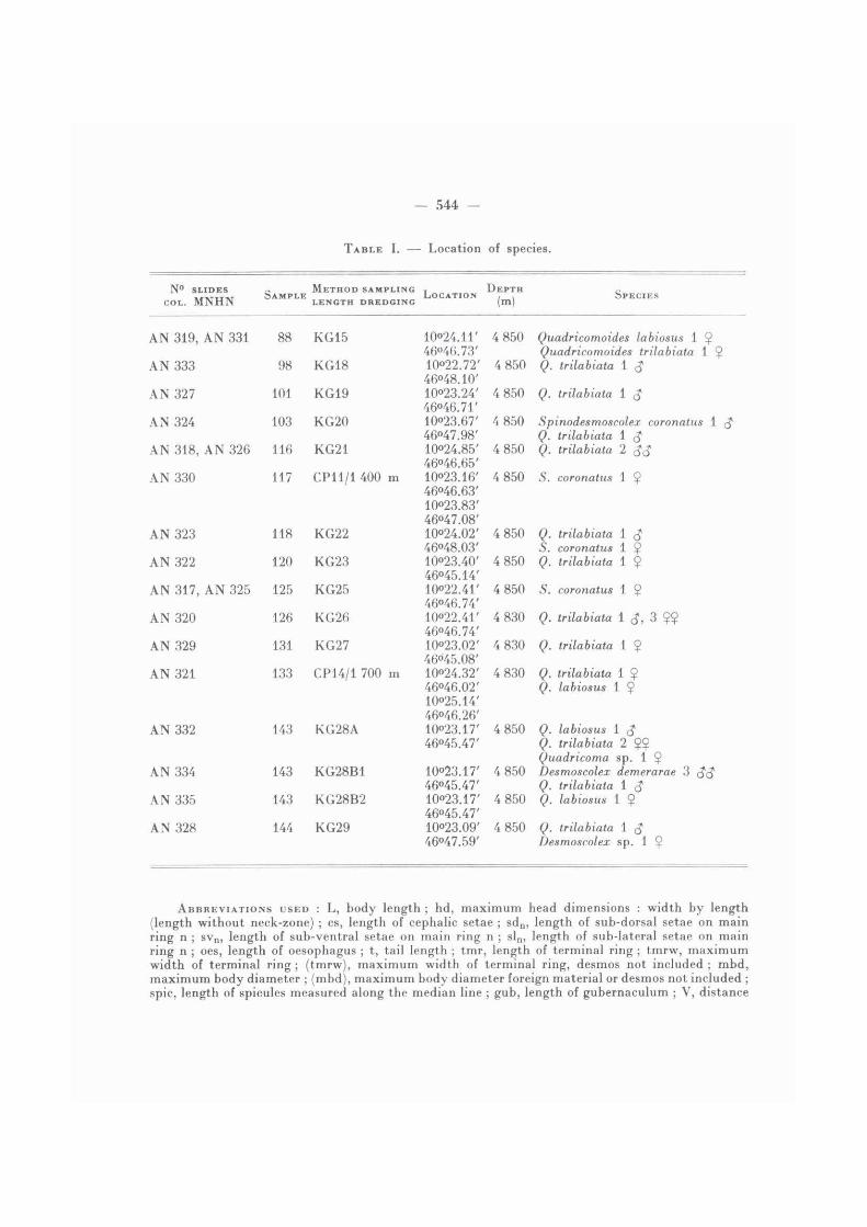
A N 3 1 9 , A N 3 3 1 8 8 K G 1 5 10<>24.11' 4 8 5 0 Quadricomoides labiosus 1 $ A N 3 1 9 , A N 3 3 14 6 ° 4 6 . 7 3 ' Quadricomoides trilabiata 1 $
A N 3 3 3 98 K G 1 8 1 0 ° 2 2 . 7 2 ' 4 8 5 0 Q. trilabiata 146048.10'
Q. trilabiata 1
A N 3 2 7 1(11 K G 1 9 1 0 0 2 3 . 2 4 ' 4 8 5 0 Q. trilabiata 1 <J46046.71'
Q. trilabiata 1 <J
A N 3 2 4 1 0 3 K G 2 0 1 0 0 2 3 . 6 7 '46047.98'
4 8 5 0 Spinodesmoscolex coronatus 1 <JQ. trilabiata 1 (J
A N 3 1 8 , A N 3 2 6 1 1 6 K G 2 1 1 0 0 2 4 . 8 5 ' 4 8 5 0 Q. trilabiata 2A N 3 1 8 , A N 3 2 646046.65'
Q. trilabiata 2
A N 3 3 0 1 1 7 C P U / 1 4 0 0 m 1 0 0 2 3 . 1 6 ' 4 8 5 0 S. coronatus 1 Ç C P U / 1 4 0 0 m 46046.63'1 0 0 2 3 . 8 3 '46047.08'
A N 3 2 3 1 1 8 K G 2 2 1 0 0 2 4 . 0 2 '46048.03'
4 8 5 0 Q. trilabiata 1 $ S. coronatus 1 $
A N 3 2 2 1 2 0 K G 2 3 1 0 0 2 3 . 4 0 ' 4 8 5 0 Q. trilabiata 1 Ç 46045.14'
Q. trilabiata 1 Ç
A N 3 1 7 , A N 3 2 5 1 2 5 K G 2 5 1 0 « 2 2 . 4 1 ' 4 8 5 0 5 . coronatus 1 Ç A N 3 1 7 , A N 3 2 546046.74'
5 . coronatus 1 Ç
A N 3 2 0 1 2 6 K G 2 6 1 0 ° 2 2 . 4 1 ' 4 8 3 0 Q. trilabiata 1 3 Ç$46046.74'
Q. trilabiata 1 3 Ç$
A N 3 2 9 131 K G 2 7 1 0 0 2 3 . 0 2 ' 4 8 3 0 Q. trilabiata 1 Ç 46045.08'
Q. trilabiata 1 Ç
A N 3 2 1 1 3 3 C P 1 4 / 1 7 0 0 m 1 0 0 2 4 . 3 2 ' 4 8 3 0 Q. trilabiata 1 Ç C P 1 4 / 1 7 0 0 m 4 6 ° 4 6 . 0 2 ' Q. labiosus 1 $ 1 0 0 2 5 . 1 4 '
Q. labiosus 1 $
46046.26'A N 3 3 2 1 4 3 K G 2 8 A 1 0 0 2 3 . 1 7 '
46045.47'4 8 5 0 Q. labiosus 1 <J
Q. trilabiata 2 $ $Quadricoma sp. 1 Ç
A N 3 3 4 1 4 3 K G 2 8 B 1 1 0 0 2 3 . 1 7 '46045.47'
4 8 5 0 Desmoscolex demerarae 3Q. trilabiata 1 $
A N 3 3 5 1 4 3 K G 2 8 B 2 1 0 0 2 3 . 1 7 ' 4 8 5 0 Q. labiosus 1 $ 46045.47'
Q. labiosus 1 $
A N 3 2 8 1 4 4 K G 2 9 1 0 0 2 3 . 0 9 '46047.59'
4 8 5 0 Q. trilabiata 1 £ Desmoscolex sp. 1 $
ABBREVIATIONS USED : L, body length ; hd, maximum head dimensions : width by length(length without neck-zone) ; cs, length of cephalic setae ; sdn , length of sub-dorsal setae on mainring n ; sv n , length of sub-ventral setae on main ring n ; sln, length of sub-lateral setae on mainring n ; oes, length of oesophagus ; t, tail length ; tmr, length of terminal ring ; tmrw, maximumwidth of terminal ring ; (tmrw), maximum width of terminal ring, desmos not included ; mbd,maximum body diameter ; (mbd), maximum body diameter foreign materiat or desmos not included ; spie, length of spicules measured along the median line ; gub, length of gubernaculum ; V, distance
- 545 -
of vulva from anterior body end as percentage of total body length ; a, b , c, proportions of de Man.All measurements are in micrometers (um).
D E S C R I P T I O N S
Subfamily D E S M O S C O L E C I N A E Shipley
Genus SPINODESMOSCOLEX gen. n.
DIAGNOSIS : Desmoscolecinae. Desmoscolecoid body rings, each ring with a transverse rowof spine-like setae surrounded by concretion ; head with triradial anterior end composed of threelabial areas, each area with two papillae ; oesophagus about cylindrical and very short.
T Y P E SPECIES : Spinodesmoscolex coronatus sp. nov.
Spinodesmoscolex coronatus sp. nov.
(Figs. 1-2)
MATERIAL : 1 $ holotype (slide AN 323). — Paratypes : 1 Ç (slide AN 330), 1 <$ (slide AN 324),1 Ç (slide AN 325) anterior body region with head cut off, head female (slide AN 317).
MEASUREMENTS : Holotype female : L = 1245, hd = 46 X 54, sdj = 75, sd3 = 65, sd5 = 59,sd7 = 62, sd 9 = 42, sdjj = 57, sd 1 3 = 50, sd 1 7 = 54, sd 2 2 = 95, sv 2 = 33, sv4 = 37, sv 6 = 42,sv8 = 44, sv 1 0 = 51, sv 1 2 = 52, sv 1 4 = 58, sv 1 6 = 42, oes = 59, t = 325, tmr = 129, mbd = 119,(mbd) = 92 ; b = 21.1, c = 3.5. — Paratype female (n = 1) : L = 1255, hd = 48 X 55, cs = 32,sdj = 86, sd 2 3 = 110, sv2 = 37, sv4 = 39, sv7 = 47, sv B = 48, sv 1 3 = 53, sv 1 7 - 74, oes = 62,t = 332, tmr = 135, mbd = 124, (mbd) = 102 ; b = 20.2, c = 3.5. — Paratype male (n = 1) : L = 915, hd = 36 X 43, cs = 29, sdx = 66, sd 3 = 60, sd5 = 54, sd7 = 52, sd 9 = 52, s d n = 45,sd 1 3 = 52, sd 1 7 = 52, sd 2 1 = 81, sv 2 = 30, sv4 = 36, svg = 38, sv8 = 37, sv 1 0 = 37, sv 1 2 = 38,sv 1 4 = 44, sv 1 6 = 43, oes = 53, t = 260, tmr = 99, mbd = 108, (mbd) = 93, spie = 72, gub =35 ; b = 17.2, c = 3.9.
DESCRIPTION
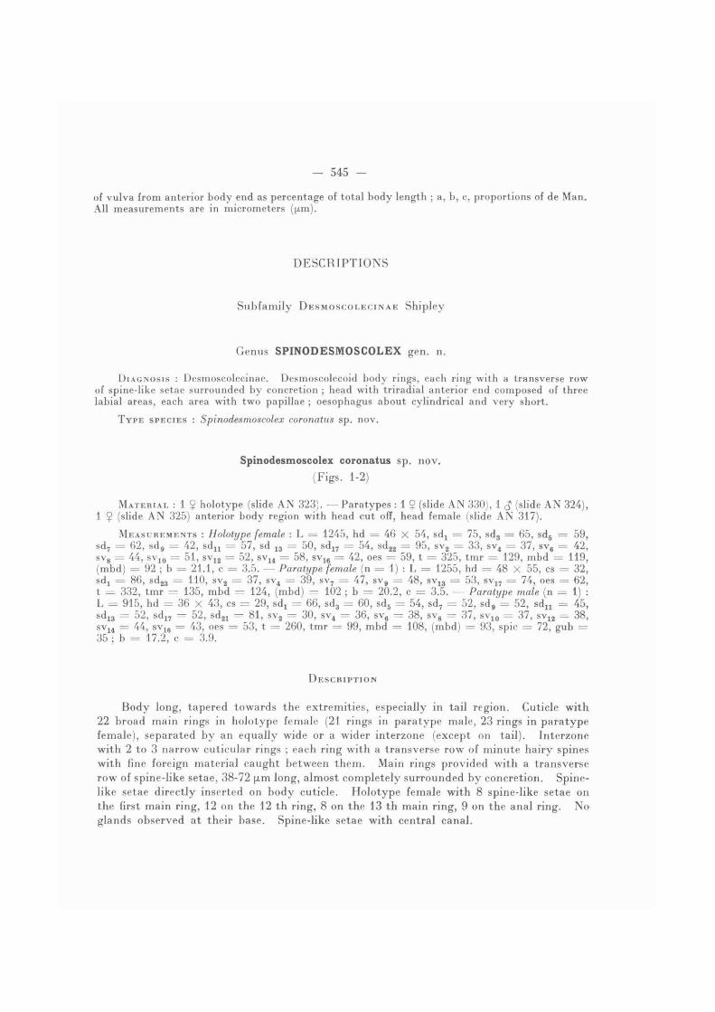
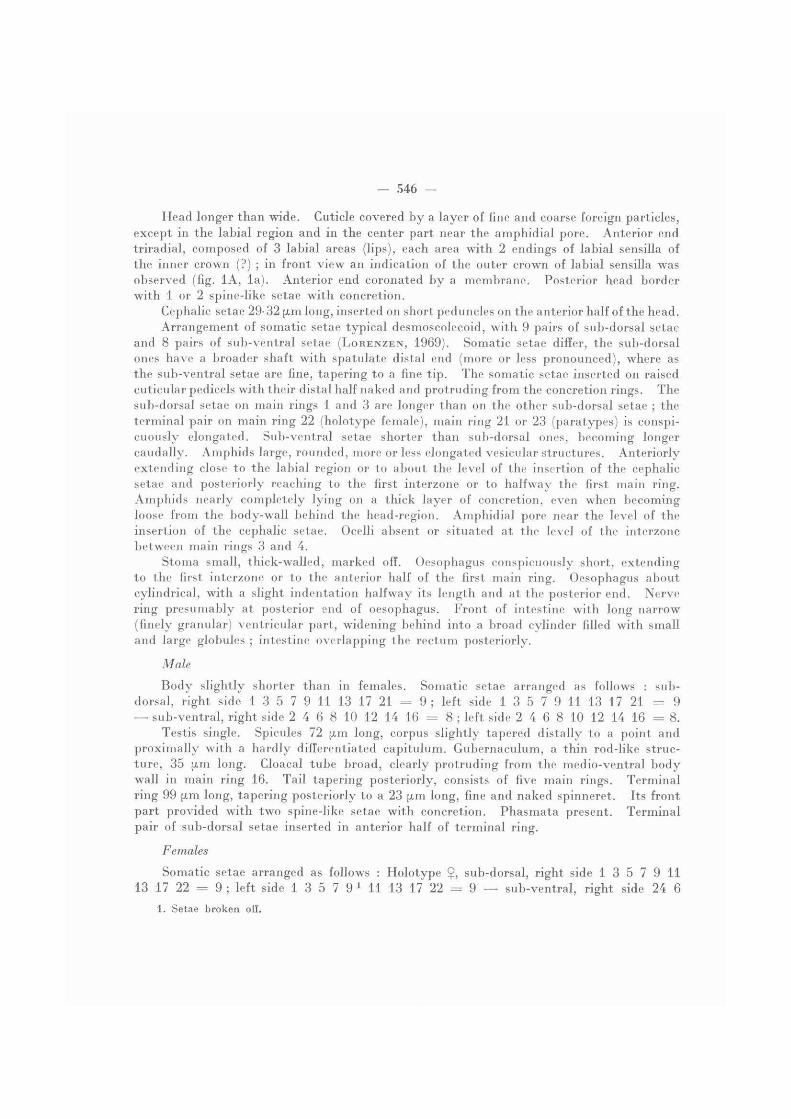
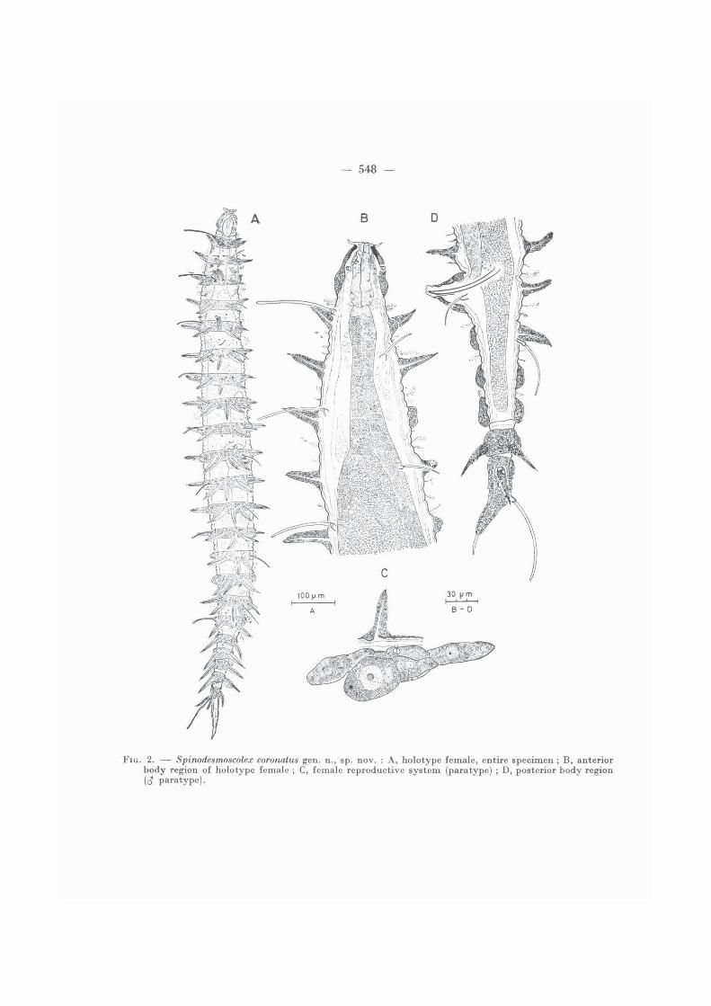
Body long, tapered towards the extremities, especially in tail region. Cuticle with22 broad main rings in holotype female (21 rings in paratype male, 23 rings in paratypefemale), separated by an equally wide or a wider interzone (except on tail). Interzonewith 2 to 3 narrow cuticular rings ; each ring with a transverse row of minute hairy spineswith fine foreign material caught between them. Main rings provided with a transverserow of spine-like setae, 38-72 pm long, almost completely surrounded by concretion. Spine-like setae directly inserted on b o d y cuticle. Holotype female with 8 spine-like setae onthe first main ring, 12 on the 12 th ring, 8 on the 13 th main ring, 9 on the anal ring. Noglands observed at their base. Spine-like setae with central canal.
- 546 -
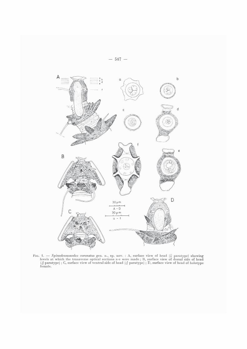
Head longer than wide. Cuticle covered b y a layer of fine and coarse foreign particles,except in the labial region and in the center part near the amphidial pore. Anterior endtriradial, composed of 3 labial areas (lips), each area with 2 endings of labial sensilla ofthe inner crown (?) ; in front view an indication of the outer crown of labial sensilla wasobserved (fig. 1A, l a ) . Anterior end coronated b y a membrane. Posterior head borderwith 1 or 2 spine-like setae with concretion.
Cephalic setae 29-32 pm long, inserted on short peduncles on the anterior half of the head.Arrangement of somatic setae typical desmoscolecoid, with 9 pairs of sub-dorsal setae
and 8 pairs of sub-ventral setae ( L O R E N Z E N , 1969). Somatic setae differ, the sub-dorsalones have a broader shaft with spatulate distal end (more or less pronounced), where asthe sub-ventral setae are fine, tapering to a fine tip. The somatic setae inserted on raisedcuticular pedicels with their distal half naked and protruding from the concretion rings. Thesub-dorsal setae on main rings 1 and 3 are longer than on the other sub-dorsal setae ; theterminal pair on main ring 22 (holotype female), main ring 21 or 23 (paratypes) is conspi-cuously elongated. Sub-ventral setae shorter than sub-dorsal ones, becoming longercaudally. Amphids large, rounded, more or less elongated vesicular structures. Anteriorlyextending close to the labial region or to about the level of the insertion of the cephalicsetae and posteriorly reaching to the first interzone or to halfway the first main ring.Amphids nearly completely lying on a thick layer of concretion, even when becomingloose from the body-wall behind the head-region. Amphidial pore near the level of theinsertion of the cephalic setae. Ocelli absent or situated at the level of the interzonebetween main rings 3 and 4.
Stoma small, thick-walled, marked off. Oesophagus conspicuously short, extendingto the first interzone or to the anterior half of the first main ring. Oesophagus aboutcylindrical, with a slight indentation halfway its length and at the posterior end. Nervering presumably at posterior end of oesophagus. Front of intestine with long narrow(finely granular) ventricular part, widening behind into a broad cylinder filled with smalland large globules ; intestine overlapping the rectum posteriorly.
Male
B o d y slightly shorter than in females. Somatic setae arranged as follows : sub-dorsal, right side 1 3 5 7 9 11 13 17 21 = 9 ; left side 1 3 5 7 9 11 13 17 21 = 9 — sub-ventral, right side 2 4 6 8 10 12 14 16 = 8 ; left side 2 4 6 8 10 12 14 16 = 8.
Testis single. Spicules 72 pm long, corpus slightly tapered distally to a point andproximally with a hardly differentiated capitulum. Gubernaculum, a thin rod-like struc-ture, 35 pm long. Cloacal tube broad, clearly protruding from the medio-ventral b o d ywall in main ring 16. Tail tapering posteriorly, consists of five main rings. Terminalring 99 pm long, tapering posteriorly to a 23 pm long, fine and naked spinneret. Its frontpart provided with two spine-like setae with concretion. Phasmata present. Terminalpair of sub-dorsal setae inserted in anterior half of terminal ring.
Females
Somatic setae arranged as follows : Holotype $, sub-dorsal, right side 1 3 5 7 9 1113 17 22 = 9 ; left side 1 3 5 7 9 1 11 13 17 22 = 9 — sub-ventral, right side 24 6
1. Setae broken off.
- 547 -
FIG. 1. — Spinodesmoscolex coronatus gen. n., sp. nov. : A, surface view of head (£ paratype) showinglevels at which the transverse optical sections a-e were made ; B, surface view of dorsal side of head((J paratype) ; C, surface view of ventral side of head (<J paratype) ; D, surface view of head of holotypefemale.
- 548 -
FIG. 2. —• Spinodesmoscolex coronatus gen. n., sp. nov. : A, holotype female, entire specimen ; B, anteriorbody region of holotype female ; C, female reproductive system (paratype) ; D, posterior body region(<J paratype).
- 549
8 10 12 14 16 = 8 ; left side 2 4 6 8 10 12 14 16 = 8. — Paratype $, sub-dorsal, rightside 1 3 1 51 8 1 101 121 141 191 24 = 9 ; left side 1 31 51 8 1 101 121 141 191 24 = 9 — sub-ventral, right side 2 4 7 9 111 13 151 17 = 8 ; left side 2 4 7 9 11 13 151 17 = 8.
Reproductive system didelphic-amphidelphic. Both branches of the genital systemoverlapping each other at the level of the vulva. Two spermathecae. Vulva situated at theanterior end of the interzone between main rings 10 and 11 (holotype) or 11 and 12 (para-type) . Anal tube large, protruding from the medio-ventral body-wall at posterior halfof main ring 16 (holotype) or 17 (paratype). Tail tapering posteriorly, consisting of 6 mainrings. Terminal ring, 129-135 u,m long, anteriorly with a transverse row of five spine-like setae (holotype) or two (paratype), tapering posteriorly to a spinneret. Phasmatapresent in posterior half of the terminal ring.
T Y P E L O C A L I T Y : Demerara abyssal basin off French Guiana, station B, at 10o24.02'/46o48.03',at 4 850 m depth, collected on 25-IX-1980.
DIAGNOSIS : Spinodesmoscolex coronatus gen. n., sp. nov. has 21-23 main body rings providedwith large spine-like setae surrounded by concretion, an anterior head-end with three lips coronatedby a membrane, a typical desmoscolecoid setal pattern in male and female, with a conspicuouslyelongated pair of terminal setae and a very short oesophagus almost restricted to the head-region.
R E M A R K S
The character of ' triradial anterior head-end with three lips ' observed in Spinodes-moscolex was known only from Quadricomoides Decraemer, 1976 (Meyliidae, Tricominae).Its presence in Spinodesmoscolex is the first record of this character within the Desmocole-cidae.
The presence of large spine-like setae on the main rings was never recorded beforefor the Desmoscolecida. These spine-like setae possess a fine inner canal ; no glandularstructures nor nerve endings in connection with them were found. However, the spine-like setae may be homologue with the fine tubes or spines found in the middle of the mainrings (after removal of the desmos) in several desmoscolecids e.g. in Desmoscolex apud asetosus in D E C R A E M E R (19756).
Genus DESMOSCOLEX Claparede
Desmoscolex Claparede, 1863 : 59.
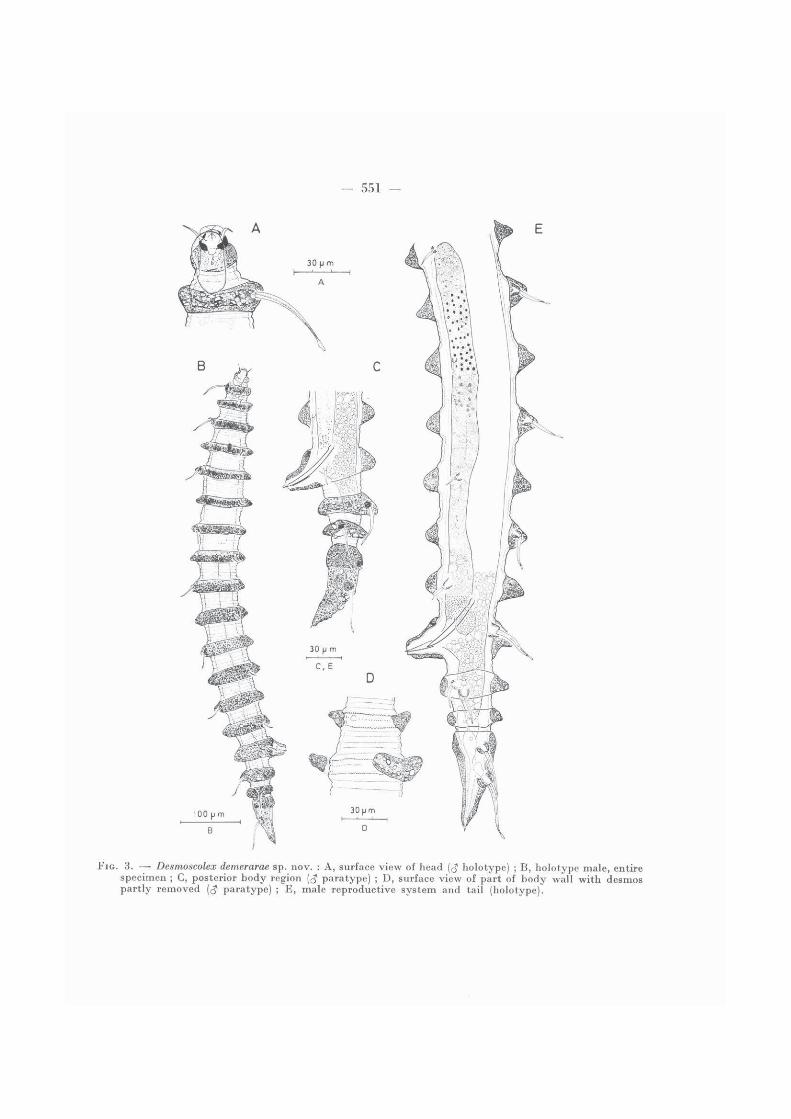
Desmoscolex demerarae sp. nov.
(Fig. 3)
MATERIAL : 1 $ holotype (slide AN 334). — Paratypes : 2 ^ (slide AN 334).
MEASUREMENTS : Holotype male : L = 800, hd = 27 X 27, cs = 14, sdx = 49, sd3 = 44,sd 5 = 32, sd7 = 34, sd 9 = 34, s d n = 29, sd 1 3 == 32, sd 1 5 = 38, sd 1 8 = 43, sd 1 9 = 54, sl2 = 13,
1. Setae broken off.
- 550 -
sv4 = 18, sv6 = 18, sv8 = 18, sv 1 0 = 19, sv 1 2 = 14, sv 1 4 = 16, sv 1 6 = 22, sl 1 6 = 25, t = 160,tmr = 83, spinneret = 5, spic = 54, gub = 22, oes = 50, mbd = 92, (mbd) = 62. — Paratype males (n = 2) : L = 730-810, hd = 23-24 X 24, cs = 10-13, sdj = 39-45, sd3 = 36, sd5 = 32-33,sd7 = 30-32, sd 9 = 28-33, s d u = 29-34, sd 1 3 = 29-33, sd 1 6 = 36-37, sd 1 9 = 38, sl2 = 14,sv 4 = 17, sv6 = 17, sv8 = 18-19, s v 1 0 = 15-17, sv 1 2 = 15-16, sv 1 4 = 17-18, sv 1 5 = 14-16, sv 1 6 = 20, sl 1 7 = 18, t = 146-162, tmr = 79, spinneret = 3-3.5, spic = 55-56, gub = 20, oes = 42-48, mbd = 80-92, (mbd) = 51-65.
D E S C R I P T I O N
Male
B o d y long, tapered at bo th ends. Cuticle with 18 main rings separated from b ybroad interzones, usually formed b y four annules ; the anteriormost interzones and thoseon the tail are narrower, with two to three annules. Main rings with fine and coarseconcretion material, interzones often covered with fine particles. In one male specimenthe concretion material (desmos) of some main rings became loose and separated from thecuticle (fig. 3 D ) . Underneath these concretion rings we observed three more or less swollenannules, the outer annules with the border folded, the middle annule bearing fine thorns.
Somatic setae arranged as follows : Holotype (J, sub-dorsal, right side 1 3 5 7 9 1113 16 18 1 18 = 10 ; left side 1 3 5 7 9 11 13 15 18 18 = 10 — sub-ventral, right side2 4 6 8 10 12 14 15 17 = 9 ; left side 2 4 6 8 10 12 14 16 16 = 9 (with sub-ventralsetae on main rings 2 and 16, 17 in sub-lateral position). •—• Paratype 1, sub-dorsal, rightside 1 3 5 7 9 11 13 16 18 (?) 19 = 10 ; left side 1 3 5 7 9 11 13 16 18 (?) 19 = 10— sub-ventral, right side 2 4 6 8 10 12 14 15 17 = 9 ; left side 2 4 6 8 10 12 14 1517 = 9 (with sub-ventral setae on main rings 2 and 17 in sub-lateral position). Somaticsetae inserted on low peduncles. The sub-dorsal setae with large basal shaft ending ona small spatulate tip ; the sub-ventral setae being smaller, ending on a fine open tip. Thesub-dorsal setae on the first main ring and on the terminal ring are elongated ; the sub-ventral setae become slightly longer posteriorly.
Head as wide as long, broadly rounded and anteriorly tapered ; its cuticle thickenedand sclerotized in the narrower anterior part, in the posterior part covered by a thicklayer of secretion and fine foreign material (except in the middle of the amphidial region).Labial region surrounded b y a membrane. Cephalic setae, short, with fine central canal.They are inserted far anteriorly on the head, almost without peduncle. Amphids, largevesicular structures, extending from the labial region to the first main ring. The verysmall amphidial pore is posteriorly connected with a canal ending on an elevated cuticularizedstructure (bar) (fig. 3A) . Ocelli, dark yel low rounded structures, situated at the levelof main ring 4.
Stoma small. Oesophagus typical desmoscolecoid, terminally surrounded b y thenerve ring. Oesophago-intestinal junction between main rings 1 and 2 or at the beginningof main ring 2 . Front of intestine with narrower ventricular part, widening behind intoa broad cylinder with small and large globules ; intestine overlapping the rectum posteriorly(fig. 3B, 3E) .
1. Setae broken off.
- 551 -
- 552 -
Reproductive system typical with one testis ( D E C R A E M E R , 1 9 7 5 ) . Spicules 5 4 pm long( 5 5 - 5 6 pm in paratypes), almost straight, narrowing distally to a pointed tip and proximallywith a slightly marked capitulum. Gubernaculum 22 pm long (20 pm in a male paratype),thin structure parallel to the spicules ; may be rather obscure. Cloacal tube largely protru-ding from the ventral b o d y wall in main ring 1 5 .
Tail with three main rings. Terminal ring with an indication of a non-separatedmain ring (see also the number of sub-dorsal setae). Three caudal glands observed.Phasmata not visible.
Female : not found.
T Y P E LOCALITY : Demerara abyssal basin off French Guiana, station B , at 1 0 ° 2 3 . 1 7 ' / 4 6 ° 4 5 . 4 7 ' ,at 4 8 5 0 m depth, collected on 2 9 - I X - 1 9 8 0 .
DIAGNOSIS : Desmoscolex demerarae sp. nov. is characterized by its head, anteriorly surroundedby a labial membrane and by the cuticularized structures in connection with the amphidial pores.It can also be distinguished by the number of main rings ( 1 8 ) and by its setal pattern with 1 0 pairsof sub-dorsal and 9 pairs of sub-ventral setae.
Subfamily T R I C O M I N A E Lorenzen
Genus QUADRICOMOIDES Decraemer
Quadricomoides Decraemer, 1 9 7 6 : 9 0 .
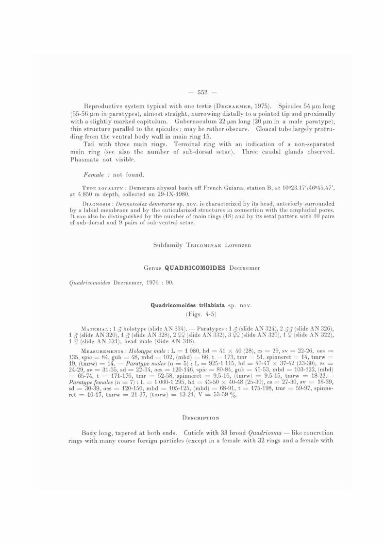
Quadricomoides trilabiata sp. nov.
(Figs. 4 - 5 )
MATERIAL : 1 holotype (slide A N 3 3 4 ) . — Paratypes : 1 $ (slide A N 3 2 4 ) , 2 S3 (slide A N 3 2 6 ) ,1 <? (slide A N 3 2 0 ) , 1 S (slide A N 3 2 8 ) , 2 $$ (slide A N 3 3 2 ) , 3 Ç ? (slide A N 3 2 0 ) , 1 $ (slide A N 3 2 2 ) ,1 ? (slide A N 3 2 1 ) , head male (slide A N 3 1 8 ) .
MEASUREMENTS : Holotype male : L = 1 0 8 0 , hd = 4 1 X 4 0 ( 2 8 ) , cs = 2 9 , sv = 2 2 - 2 6 , oes =1 3 5 , spic = 8 4 , gub = 4 8 , mbd = 1 0 2 , (mbd) = 6 6 , t = 1 7 3 , tmr = 5 1 , spinneret = 1 4 , tmrw =1 9 , (tmrw) = 14 . — Paratype males (n = 5 ) : L = 9 2 5 - 1 1 1 5 , hd = 4 0 - 4 7 X 3 7 - 4 2 ( 2 3 - 3 0 ) , cs =2 4 - 2 9 , sv = 3 1 - 3 5 , sd = 2 2 - 3 4 , oes = 1 2 0 - 1 4 6 , spic = 8 0 - 8 4 , gub = 4 5 - 5 3 , mbd = 1 0 3 - 1 2 2 , (mbd)= 6 5 - 7 4 , t -- 1 7 1 - 1 7 6 , tmr = 5 2 - 5 8 , spinneret = 9 . 5 - 1 6 , (tmrw) = 9 . 5 - 1 5 , tmrw = 1 8 - 2 2 . —Paratype females (n = 7) : L = 1 0 6 0 - 1 2 9 5 , hd = 4 3 - 5 0 X 4 0 - 4 8 ( 2 5 - 3 0 ) , cs = 2 7 - 3 0 , sv = 1 6 - 3 9 ,sd = 3 0 - 3 9 , oes = 1 2 0 - 1 5 0 , mbd = 1 0 5 - 1 2 5 , (mbd) = 6 8 - 9 1 , t = 1 7 5 - 1 9 8 , tmr = 5 9 - 9 7 , spinne-ret : 1 0 - 1 7 , tmrw = 2 1 - 3 7 , (tmrw) = 1 3 - 2 1 , V = 5 5 - 5 9 % .
D E S C R I P T I O N
Body long, tapered at both ends. Cuticle with 33 broad Quadricoma — like concretionrings with many coarse foreign particles (except in a female with 32 rings and a female with
- 553 -
34 rings). Inversion of direction of the concretion rings occurs within rings 22 or 23 ; the inversion, however, may be difficult to determine.
Head broad, tapered anteriorly to a large truncated end. Its naked cuticle is thick-ened and sclerotized, forming a kind of helmet. The head is followed b y a narrower' neck-zone ' with thin cuticle covered b y foreign material. Anterior end of head trira-dially symmetric with three labial areas (lips). In ' en face ' view the head is roundedtriangular (fig. 4a). A large triradial mouth-opening nearly reaches the border of thehelmet and divides the head terminally in three triangular sectors or lips (fig. 4c) . Theinner margins of these sectors bear minute spines, not observed in lateral view. Thesclerotized outer margin of the lip-sectors is slightly indented opposite to the endings ofthe six labial sensilla, two in each sector. At the level of the insertion of the cephalicsetae, the head is more or less quadrangular. Underneath the amphids, the head-cuticleis irregular, lumpy (fig. 4d).
Cephalic setae, about as long as the head (neck-zone not included), inserted on verylow peduncles halfway along the head length. They taper distally to an open tip andare apparently flanked over their whole length b y a membrane (fig. 4d) . At their basethey are in connection with glandular structures. Somatic setae homogeneous, with fine central canal and open tip, inserted on peduncles surrounded b y foreign material. Somaticsetae often broken off in the specimens available, and due to the large amount of foreignmaterial the insertion became obscure. Consequently, the arrangement of somatic setaecannot be given with certainty. The largest number of somatic setae observed on eachb o d y side is 10 sub-ventral setae in both sexes and 6 sub-dorsal setae in male, 8 sub-dorsalsetae in female. The somatic setae become longer posteriorly.
Amphids broad, rounded, thick-walled vesicular structures, surrounding nearly com-pletely the head (neck-zone excluded). Amphidial pores situated at the posterior borderof the sclerotized head wall. Ocelli large, rounded, brownish structures at level of concre-tion rings 6 or 7.
Oesophagus typical for the genus ( D E C R A E M E R , 1976) : consisting of a thin-walledstomatal part, a broader cylindrical anterior part reaching the level of the nerve ring,and a posterior part with asymmetric bulb or swelling, consisting of an internal differen-tiation and an outer muscular wall. The nerve ring surrounds the oesophagus at theend of the second or at the third concretion ring. The excretory duct of the dorsal oeso-phageal gland is conspicuously swollen in the anterior part of the oesophagus and occupiesnearly the entire dorsal wall. At the level of the asymmetric bulb the oesophageal lumenis shifted ventrally. The oesophago-intestinal junction occurs at the end of concretionring 4 or opposite concretion ring 5. A large and pale rounded organ lies dorsally alongthe narrower anterior part of the intestine. Posterior to this structure the intestine becomeswider. Intestine without post-rectal blindsac. From the posterior end of the ocelli on,or shortly behind them, the ventral intestinal wall contains dark red-brownish pigmentedgranules, forming a ventral strain along the intestine up to the level of concretion ring 27,i.e. shortly in front of the anus or cloaca (fig. 5B, 5E) . This ventral strain of pigment-granules in the intestinal wall may be enlarged at the beginning and at the end. Thispigment-strain was present in all specimens available.
Tail tapered posteriorly. Terminal ring about double the length of the former ring,ending on a naked narrow spinneret, 9.5-17 u,m long. Phasmata not observed.
2, 10

— 554 -
FIG. 4. —• Quadricomoides trilabiata sp. nov. : A, surface view of head and anterior body rings (<$ holo-type) ; B, surface view of ventral side of head (? paratype) ; C, head region ($ paratype) ; D, surfaceview of ventral side of head paratype) showing levels at which the transverse optical sections a — d were made.
- 555 -
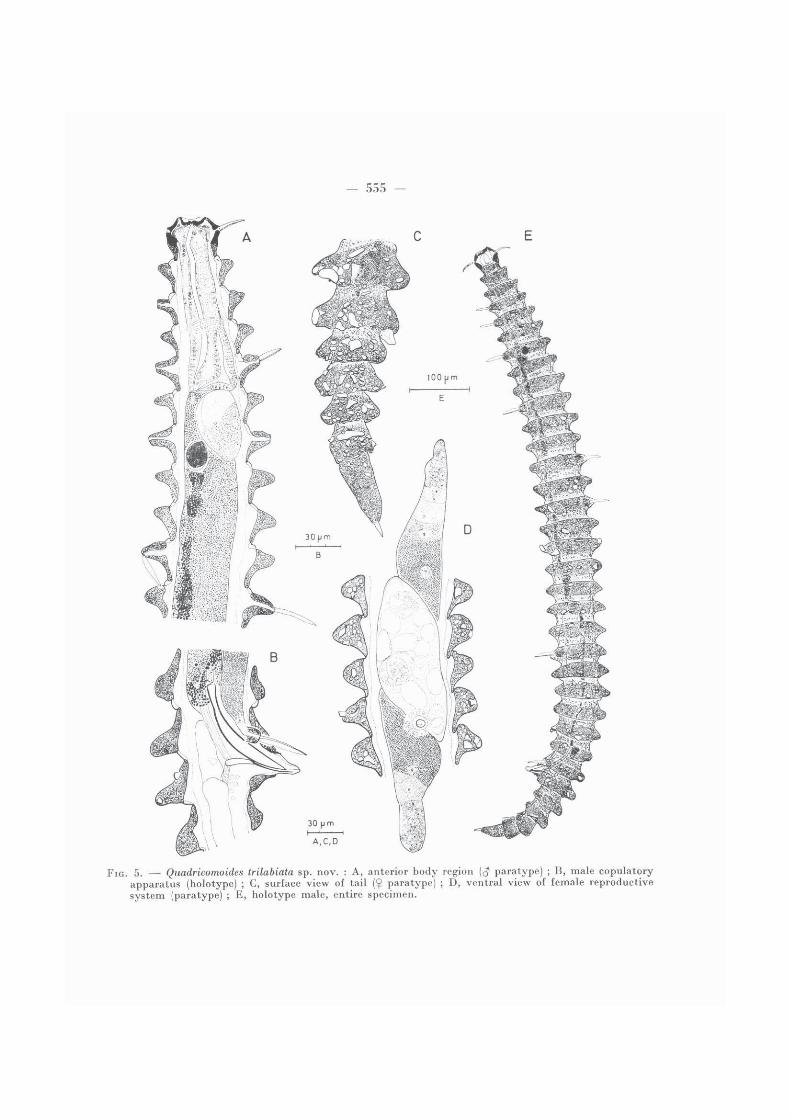
FIG. 5. — Quadricomoides trilabiata sp. nov. : A, anterior body region (<$ paratype) ; B, male copulatoryapparatus (holotype) ; C, surface view of tail (<j> paratype) ; D , ventral view of female reproductivesystem (paratype) ; E, holotype male, entire specimen.
- 5 5 6 -
MalesTwo testes, right one reflexed. Spicules broad, arcuate, proximally tapered to a
slightly marked capitulum, 84 pm long (80-84 pm in paratypes). Gubernaculum consistingdistally of a thin part along the spicules and proximally of a larger apophyse, orientateddorso-caudally and 17 pm long (14-16 pm in paratypes). Cloaca between concretion rings 27-28. Tail with six concretion rings.
FemalesReproductive system didelphic-amphidelphic. T wo spermathecae. Both uteri jo in
in a large sac with large cells. Vulva located between concretion rings 19 and 20, i.e. at55-59 % of the total b o d y length from the anterior end. Anus in main ring 29. Tailwith 5 concretion rings (6 rings in a female with 34 b o d y rings, 4 rings in a female with32 rings).
T Y P E LOCALITY : Demerara abyssal basin off French Guiana, at 10o23.17'/46°45.47', at 4 850 m depth, collected on 29-IX-1980.
DIAGNOSIS : Quadricomoides trilabiata sp. nov. has 33 Quadricoma-\ike concretion rings, a broad rectangular sclerotized head with covered " neck-region " , three lips and fine spines liningthe buccal cavity. It is also characterized by its long body, the broad spicules with slenderercapitulum, a gubernaculum with apophyses and sexual dimorphism in the number of concretionrings on the tail : 6 in males, 5 in females.
D I F F E R E N T I A L DIAGNOSIS
Quadricomoides trilabiata sp. nov . closely resembles Q. pedunculata Decraemer, 1976,in having a similar head-structure and a comparable copulatory apparatus. It differsfrom Q. pedunculata b y the structure and length of the oesophagus (measured in numberof concretion rings) and b y its longer b o d y length, longer spicules and gubernaculum andb y the absence of conspicuously high peduncles of insertion of somatic setae.
Q. trilabiata is comparable with Q. coomansi Decraemer, 1976, in the structure of theoesophagus and of the female reproductive system. It differs from Q. coomansi in head-shape, in a longer b o d y and in the structure of the copulatory apparatus.
Quadricomoides labiosus sp. nov.
(Figs. 6-7)
MATERIAL : 1 $ holotype (slide AN 335). — Paratypes : 1 $ (slide AN 319), 1 $ (slide AN 321),1 ¿ (slide AN 332), head female (slide AN 319).
MEASUREMENTS : Holotype female : L = 1 645, hd = 44 X 46 (29), cs = 23, sd 2 3 = 25, sd 3 4 = 25, sv2 = 16, sv4 = 24, s v u = 30, sv 2 9 = 30, sv 3 2 = 25, s v M = 24, oes = 265, t = 255, tmr = 54,mbd = 142, (mbd) = 93, V = 52 %. — Paratype females (n = 2) : L = 1 375-1 590, hd = 44-45 X 48-49 (31), cs = 20, sv2 = 14, s v n = 27, sv 1 9 = 26, sv 2 6 = 27, sv 3 1 = 24, sv 3 4 = 21, sd 1 5 = 22,sd 1 9 = 25, sd 3 2 ( 3 3 ) = 22-29, oes, = 205-270, t = 210-220, tmr = 40-64, tmrw = 22-31, (tmrw) = 12-21. — Paratype male (n = 1) : L = 1 350, hd = 45 X 44 (28), cs = 26, sv 2 = 16, sv 1 0 = 25,sv 1 3 = 26, sv 2 2 = 27, sdj, = 24, sd 1 7 = 23, oes = 240, t = 205, tmr = 56, tmrw = 28, (tmrw) = 16, spic = 86, gub = 53* mbd = 137, (mbd) = 87.
- 557 -
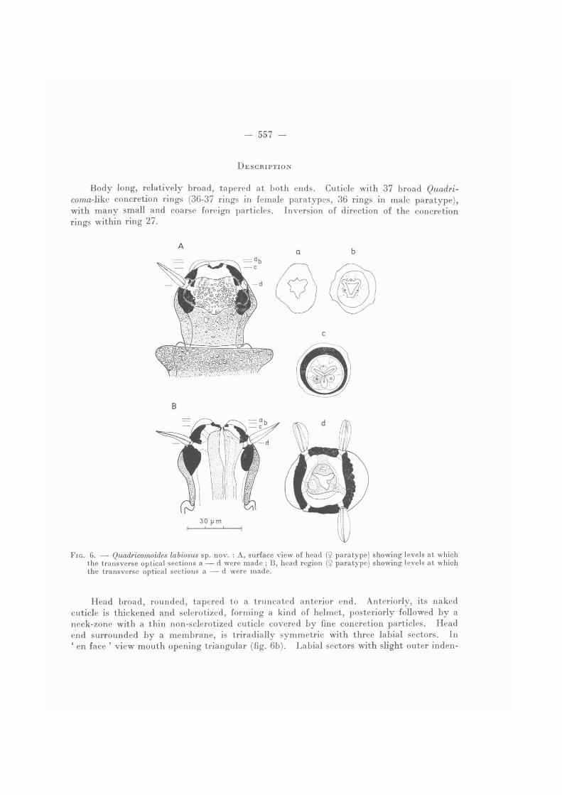
D E S C R I P T I O N
B o d y long, relatively broad, tapered at both ends. Cuticle with 37 broad Quadri-coma-like concretion rings (36-37 rings in female paratypes, 36 rings in male paratype),with many small and coarse foreign particles. Inversion of direction of the concretionrings within ring 27.
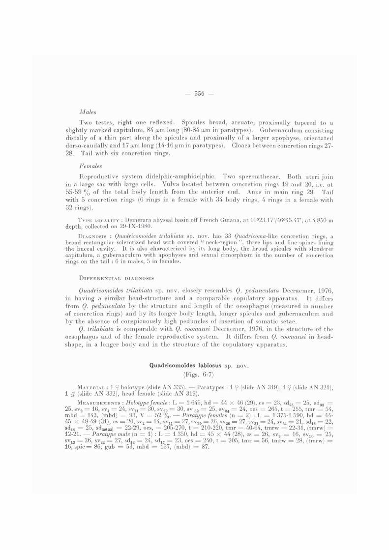
FIG. 6 . — Quadricomoides labiosus sp. nov. : A , surface view of head (? paratype) showing levels at whichthe transverse optical sections a —- d were made ; B, head region ($ paratype) showing levels at whichthe transverse optical sections a — d were made.
Head broad, rounded, tapered to a truncated anterior end. Anteriorly, its nakedcuticle is thickened and sclerotized, forming a kind of helmet, posteriorly followed b y a neck-zone with a thin non-sclerotized cuticle covered b y fine concretion particles. Headend surrounded b y a membrane, is triradially symmetric with three labial sectors. In' en face ' v iew mouth opening triangular (fig. 6b) . Labial sectors with slight outer inden-
- 558 -
tations for the endings of the six labial sensilla. At the level of the insertions of the cephalicsetae, the head is about quadrangular. Underneath the amphids the head-cuticle is con-spicuously irregular, lumpy (fig. 6A, 6d).
Cephalic setae, stout and short, inserted on minute peduncles, halfway the lengthof the sclerotized helmet. They taper distally to an open tip and are apparently surroundedover their whole length by a membrane (fig. 6d). Somatic setae inserted on low pedunclesare homogeneous. They are often broken off, however, the arrangement of the somaticsetae could be determined according to the setae and the insertion places.
Amphids broad rounded vesicular structures, largely covering the lateral sides ofthe helmet. Amphidial pore situated at the posterior border of the helmet. Ocelli, large,brownish structures lying opposite concretion rings 8 or concretion rings 8 and 9.
Oesophagus consisting of a thin-walled stomatal part, a wide cylindrical anterior partto the level of the nerve ring and a posterior part with dorsal asymmetric bulb or swelling.The nerve ring surrounds the oesophagus at the posterior end of concretion ring 4. Inconsecutive transverse optical sections of the stomatal region, three glandular structures(oesophageal glands ?) were observed outside the stomatal region (fig. 6c) , ending in thelip region (fig. 6b) . In lateral view excretory duct of dorsal oesophageal gland well discer-nible. Oesophago-intestinal junction occurs at the level of concretion ring 7. A palerounded organ lies dorsally along the beginning of the intestine. Intestine without post-rectal sac. From the level of the oesophagus the ventral intestinal wall contains a strainof small granules, pigmented or not, reaching to ring 29, close to the anus or cloaca.
Tail tapered posteriorly, with 6 concretion rings (except 5 rings in a female with36 b o d y rings). Terminal ring, twice as long as former ring, ends on a very short nakedspinneret. Phasmata not observed.
Females
Somatic setae arranged as follows : holotype female with 37 concretion rings : sub-dorsal, right side 3 7 15 17 24 28 31 = 7 ; left side 3 1 7 12 19 1 23 25 34 = 7 — sub-ventral, right side 2 4 1 6 9 11 13 16 19 23 26 29 31 34 = 13 ; left side 2 4 6 8 1116 18 21 24 27 29 32 34 = 13. — Paratype female with 36 concretion rings : sub-dorsal,right side 3 7 10 15 18 27 34 = 7 ; left side 3 7 1 11 1 17 22 27 32 = 7 — sub-ventral,right side 2 4 1 6 1 11 13 1 16 1 19 1 22 1 24 1 27 30 34 1 = 13 ; left side 2 4 1 6 1 8 1 1113 16 19 22 1 25 1 28 32 1 34 = 13.
Reproductive system didelphic-amphidelphic. Two spermathecae. Vulva situatedbetween concretion rings 19 and 20, i.e. at 52 % of the total b o d y length from anteriorend in the holotype. Anal tube protruding from the ventral b o d y wall in concretionring 31.
Male
Somatic setae arranged as follows : sub-dorsal, right side 2 5 — 16 23 28 34 = 6 ; left side 2 6 12 17 22 27 33 = 7 — sub-ventral, right side 2 4 7 9 11 13 16 18 21 2326 30 34 = 13 ; left side 2 4 1 5 1 7 8 10 11 12 14 16 18 20 22 26 30 1 34 = 16.
1. Setae broken off.
- 560 -
Two testes. Left (?) one reflexed. Spicules 86 u.m long, with slightly offset capitulumand distally tapered to a pointed tip. Gubernaculum consisting of a thin distal part alongthe spicules and a dorso-caudally orientated apophyse, 15 u,m long. Cloaca between con-cretion rings 30 and 31.
T Y P E LOCALITY : Demerara abyssal basin off French Guiana, at 10°23.17'/46°45.47', at 4 850 m depth, collected on 29-IX-1980.
DIAGNOSIS : Quadricomoid.es labiosus sp. nov. is characterized by its head structure withtriradially symmetric anterior end with three lip sectors, surrounded by a labial membrane ; by the lumpy outlook of the head cuticle underneath the amphids. It can also be distinguishedby the large body length, the structure of the oesophagus and oesophageal glands and by theshape of the copulatory apparatus.
R E M A R K
The position of Quadricomoides labiosus sp. nov. within the genus Quadricomoides isquestionable. Its peculiar structure of the labial and stomatal region and the very com-plicated structure of the oesophageal bulb and the oesophageal glands differentiate thisspecies from all other species of the genus. Due to the small number of specimens found,Quadricomoides labiosus is temporary classified within the genus Quadricomoides until moreinformation becomes available.
Acknowledgements
I wish to thank Dr SEGONZAC for the material he kindly put at my disposal. I am alsovery grateful to Dr GERAERT for reading of the manuscript.
L ITERATURE CfTED
CLAPAREDE, A. R. E., 1863. — Beobachtungen über Anatomie und Entwicklungsgeschichte wir-belloser Tiere, an der Küste der Normandie angestellt. Leipzig, W. Engelmann : 120 p.
DECHAEMER, W., 1957a. — Scientific report on the Belgian expedition to the Great Barrier Reefin 1967. Nematodes I : Desmoscolex-species (Nematoda — Desmoscolecida) from YongeReef, Lizard Island and Nymph Island with general characteristics of the genus Desmos-colex. Annls Soc. r. zool. Belg., 104 : 105-130.
— 19756. — Scientific report on the Belgian expedition to the Great Barrier Reef in 1967.Nematodes III : Further study of Desmoscolex-species (Nematoda — Desmoscolecida) fromYonge Reef, Lizard Island and Nymph Island. Cah. Biol, mar., 16 (2) : 269-284.
— 1976. — Scientific report on the Belgian expedition to the Great Barrier Reef in 1967.Nematodes VI. Morphological observations on a new genus Quadricomoides of marineDesmoscolecida. Aust. J. mar. Freshwat. Res., 27 : 89-115.
LORENZEN, S., 1969. — Desmoscoleciden (eine Gruppe freilebender Meeresnematoden) aus Küsten-salzwiesen. Vera ff. Inst. Meeresforsch., Rremerh., 12 : 169-203.
SHIPLEY, A. E., 1896. — Nemathelminthes. In : HARMER, S. F., & A. E. SHIPLEY (ed.), The Cam-bridge Natural History, vol. 2.