Embed Size (px)
Citation preview
Available online at www.sciencedirect.com
Bioorganic & Medicinal Chemistry 16 (2008) 890–901
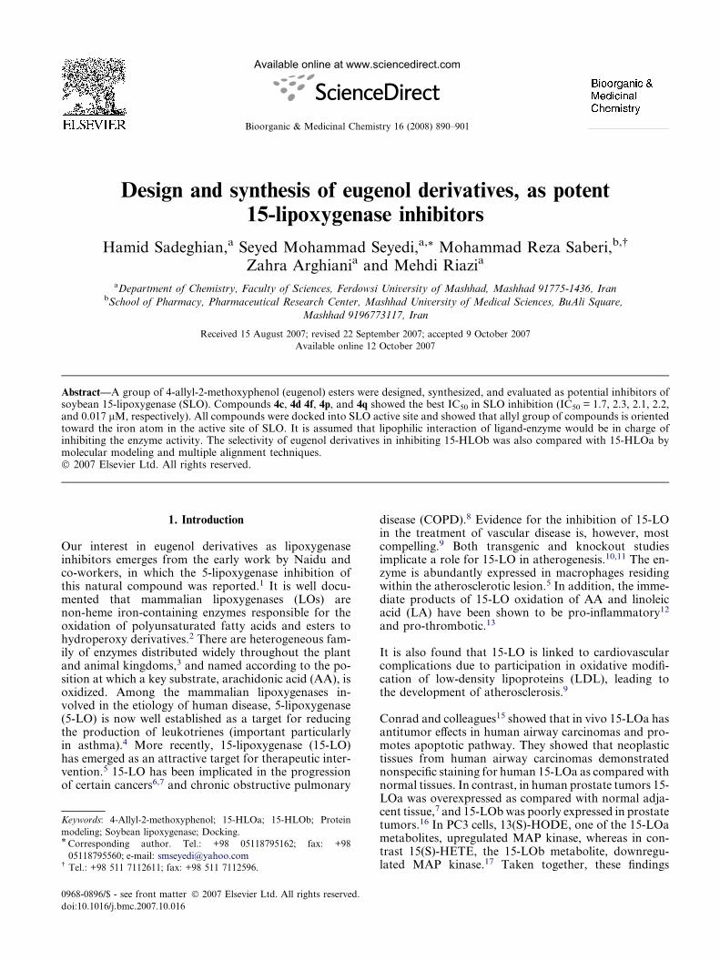
Design and synthesis of eugenol derivatives, as potent15-lipoxygenase inhibitors
Hamid Sadeghian,a Seyed Mohammad Seyedi,a,* Mohammad Reza Saberi,b,�
Zahra Arghiania and Mehdi Riazia
aDepartment of Chemistry, Faculty of Sciences, Ferdowsi University of Mashhad, Mashhad 91775-1436, IranbSchool of Pharmacy, Pharmaceutical Research Center, Mashhad University of Medical Sciences, BuAli Square,
Mashhad 9196773117, Iran
Received 15 August 2007; revised 22 September 2007; accepted 9 October 2007
Available online 12 October 2007
Abstract—A group of 4-allyl-2-methoxyphenol (eugenol) esters were designed, synthesized, and evaluated as potential inhibitors ofsoybean 15-lipoxygenase (SLO). Compounds 4c, 4d 4f, 4p, and 4q showed the best IC50 in SLO inhibition (IC50 = 1.7, 2.3, 2.1, 2.2,and 0.017 lM, respectively). All compounds were docked into SLO active site and showed that allyl group of compounds is orientedtoward the iron atom in the active site of SLO. It is assumed that lipophilic interaction of ligand-enzyme would be in charge ofinhibiting the enzyme activity. The selectivity of eugenol derivatives in inhibiting 15-HLOb was also compared with 15-HLOa bymolecular modeling and multiple alignment techniques.� 2007 Elsevier Ltd. All rights reserved.
1. Introduction
Our interest in eugenol derivatives as lipoxygenaseinhibitors emerges from the early work by Naidu andco-workers, in which the 5-lipoxygenase inhibition ofthis natural compound was reported.1 It is well docu-mented that mammalian lipoxygenases (LOs) arenon-heme iron-containing enzymes responsible for theoxidation of polyunsaturated fatty acids and esters tohydroperoxy derivatives.2 There are heterogeneous fam-ily of enzymes distributed widely throughout the plantand animal kingdoms,3 and named according to the po-sition at which a key substrate, arachidonic acid (AA), isoxidized. Among the mammalian lipoxygenases in-volved in the etiology of human disease, 5-lipoxygenase(5-LO) is now well established as a target for reducingthe production of leukotrienes (important particularlyin asthma).4 More recently, 15-lipoxygenase (15-LO)has emerged as an attractive target for therapeutic inter-vention.5 15-LO has been implicated in the progressionof certain cancers6,7 and chronic obstructive pulmonary
0968-0896/$ - see front matter � 2007 Elsevier Ltd. All rights reserved.
doi:10.1016/j.bmc.2007.10.016
Keywords: 4-Allyl-2-methoxyphenol; 15-HLOa; 15-HLOb; Protein
modeling; Soybean lipoxygenase; Docking.* Corresponding author. Tel.: +98 05118795162; fax: +98
05118795560; e-mail: [email protected]� Tel.: +98 511 7112611; fax: +98 511 7112596.
disease (COPD).8 Evidence for the inhibition of 15-LOin the treatment of vascular disease is, however, mostcompelling.9 Both transgenic and knockout studiesimplicate a role for 15-LO in atherogenesis.10,11 The en-zyme is abundantly expressed in macrophages residingwithin the atherosclerotic lesion.5 In addition, the imme-diate products of 15-LO oxidation of AA and linoleicacid (LA) have been shown to be pro-inflammatory12
and pro-thrombotic.13
It is also found that 15-LO is linked to cardiovascularcomplications due to participation in oxidative modifi-cation of low-density lipoproteins (LDL), leading tothe development of atherosclerosis.9
Conrad and colleagues15 showed that in vivo 15-LOa hasantitumor effects in human airway carcinomas and pro-motes apoptotic pathway. They showed that neoplastictissues from human airway carcinomas demonstratednonspecific staining for human 15-LOa as compared withnormal tissues. In contrast, in human prostate tumors 15-LOa was overexpressed as compared with normal adja-cent tissue,7 and 15-LOb was poorly expressed in prostatetumors.16 In PC3 cells, 13(S)-HODE, one of the 15-LOametabolites, upregulated MAP kinase, whereas in con-trast 15(S)-HETE, the 15-LOb metabolite, downregu-lated MAP kinase.17 Taken together, these findings

H. Sadeghian et al. / Bioorg. Med. Chem. 16 (2008) 890–901 891
including the upregulation of 15-LOa within the airwaytissue of smoking patients with chronic bronchitis pro-vided new evidence of possible acquired abnormalitieslinked to airway inflammation. The bronchial epitheliumis clearly a key player in inflammation and structuralchanges in airway diseases. Its rich content in 15-LOa–and 15-LOb–derived products highlights their potentialas new target for therapeutic interventions.
Three different strategies have been developed to inhibitthe LO’s pathway.18 They involve (i) redox inhibitors orantioxidants, which interfere with the redox cycle of 15-LO, (ii) iron-chelator agents, and (iii) non-redox com-petitive inhibitors, which compete with AA to bind theenzyme active site.
Eugenol (4-allyl-2-methoxyphenol) is naturally occur-ring phenolic compound in basil, cinnamon, and nut-meg, and the major component of clove oil. It iswidely used as component of zinc oxide eugenol cement
Figure 1. Clustal X (1.81) multiple alignment of SLO (green), 15-HLOa (blue
8 A are highlighted by yellow background.
in dentistry and is applied to the oral environment.19 Inaddition, eugenol is a flavoring agent in cosmetic andfood products.20 Eugenol has been shown to possessmany medicinal properties such as antispasmodic,21
antipyretic,22 anti-inflammatory,23 and antibacterialactivity.24 Recently it is reported that eugenol inhibits5-LO enzyme by non-competitive mechanism.1
In this work, seventeen ester derivatives of eugenol 4a–qwere designed, synthesized and their activities were iden-tified as the mean of IC50 on soybean 15-LO (SLO).There is reasonable homology between the soybeanLO and the human one (Fig. 1). This homology becomesmore identical (�50%) within 8A in the active site pock-et. Obviously soybean enzyme is much more accessiblethan the human one. Therefore, one can expect thatthe results can be extendable to human LO.
In this study, (i) common bonding model of 4-allyl-2-methoxyphenyl carboxylates in SLO active site,
), and 15-HLOb (red). The amino acids in the active site pocket within

892 H. Sadeghian et al. / Bioorg. Med. Chem. 16 (2008) 890–901
(ii) QSAR study of inhibitors to propose key features ofthis class of inhibitors, and (iii) theoretical potency ofsome of these compounds for inhibiting 15-HLOa and15-HLOb activity are reported.
2. Chemistry
4-Allyl-2-methoxyphenyl esters 4a–q (Scheme 1 and Ta-ble 1), according to the literature,21 started from eugenol2 and corresponding acid chlorides 3a–q which wereeither purchased or prepared (3b–d and 3q) by reactionof thionyl chloride and corresponding carboxylicacids.25 All desired esters were synthesized by the actionof acid chlorides in either aqueous solution of sodium 4-allyl-2-methoxyphenolate (4a, e–q) or hydrochloridesalts of pyridine carboxyl chloride 3b–d (prepared frompyridine carboxylic acids and thionyl chloride) in drypyridine.
Structural assignments of compounds 4a–q were basedupon the spectral and microanalytical data.
3. Molecular modeling, docking, and QSAR study
3.1. Multiple alignment
Highly conserved amino acids were identified throughmultiple alignment on clustalX 1.8126 software. Se-quences of lipoxygenase (LO) family were selected fromblasted sequences via ExPASY proteomics server27 withE-value < 0.02. Multiple alignment process was thencarried out on the selected sequences (protein weightmatrix: BLOSUM series, gap penalty = 10%).
3.2. Structure optimization
Structures 4a–q were simulated in chem3D professional;Cambridge software; using MM2 method (RMS gradi-ent = 0.05 kcal/mol).28 Output files were minimized un-der semi-empirical AM1 method in the second
OH
O
1) NaOH (aq)
2) RCOCl (3a,
2
N
O
OH N
O
Cl
SOCl2HCl
1b-d 3b-d
Scheme 1.
optimization (Convergence limit = 0.01; Iteration lim-it = 50; RMS gradient = 0.05 kcal/mol; Fletcher-Reevesoptimizer algorithm) in HyperChem7.5.29,30
Crystal structure of soybean lipoxygenase-3 (arachi-donic acid 15-lipoxygenase) complex with 13(S)-hydro-peroxy-9(Z)-2,11(E)-octadecadienoic acid was retrievedfrom RCSB Protein Data Bank (PDB entry: 1IK3).
3.3. Molecular docking
Automated docking simulation was implemented todock 4a–q into the active site of SLO with AutoDockversion 3.0331 using Lamarckian genetic algorithm.32
This method has been previously shown to producebonding modes similar to the experimentally observedmodes.30,32–34 The torsion angles of the ligands wereidentified, hydrogens were added to the macromolecule,bond distances were edited, and solvent parameters wereadded to the enzyme 3D structure. Partial atomiccharges were then assigned to the macromolecule as wellas ligands (Gasteiger for the ligands and Kollman forthe protein).
The regions of interest of the enzyme were defined byconsidering Cartesian chart 18.5, �2.0, and 20.4 as thecentral of a grid size of 40, 50, and 40 points in X, Y,and Z axises. The docking parameter files were gener-ated using Genetic Algorithm and Local Search Param-eters (GALS) while number of generations was set to 50.Compounds 4a–q were each docked into the active siteof LO enzyme and the simulations were composed of50 docking runs, each of 50 cycles containing a maxi-mum of 10,000 accepted and rejected steps. The simu-lated annealing procedure was started at hightemperature (RT = 616 kcal/mol, where R is the gasconstant and T is the steady-state temperature) andwas decreased by a fraction of 0.95 on each cycle.33
The 50 docked complexes were clustered with a root-mean-square deviation tolerance of 0.1 A. The programgenerated 50 docked conformers of 4a–q correspondingto the lowest-energy structures. After docking procedure
O
O
RO
3e-q)
4a, 4e-q
Eugenol / PyO
O
ON
4b-d

Table 1. For 4a, 4p, and 4q the IC50 values were calculated from dose–
response curves and values are given as means ± SEM of three
individual samples
O
O
RO
Compound Structure IC50 (lM)
Eugenol 34.6 ± 1.1
4a 4.4 ± 0.3
4b
N33.3 ± 1.5
4cN
1.7 ± 0.05
4dN
2.3 ± 0.2
4eF
6.7 ± 0.8
4f
F
2.1 ± 0.4
4gF
7.2 ± 0.5
4hCl
168.0 ± 13.5
4i
Cl
5.2 ± 0.2
4jCl
134.3 ± 7.5
4kH3C
77.2 ± 5.1
4l
CH3
11.4 ± 1.1
4mCH3
14.6 ± 1.0
(continued on next page)
Table 1 (continued)
Compound Structure IC50 (lM)
4n
OCH3
23.0 ± 1.3
4oOCH3
19.0 ± 1.3
4p 2.2 ± 0.3
4q 0.017 ± 0.001
H. Sadeghian et al. / Bioorg. Med. Chem. 16 (2008) 890–901 893
in AD3, docking results were submitted to WeblabViewerlite 4.035 and Swiss-PdbViewer 3.7 (spdbv)36 forfurther evaluations. The results of docking processing(DGb: estimated free energy of bonding, DGd: finaldocked energy, and Ki: estimated inhibition constant)are outlined in Table 2.
3.4. QSAR studies
QSAR studies were performed for optimized com-pounds 4a–q in DRAGON 2.1.37 In this study van derWaals volume (Sv)38 and Moriguchi octanol-water par-tition coefficient (log P)38 were determined (Table 2).
3.5. Protein modeling
Three-dimensional models of the 15-HLOa and 15-HLOb sequences were constructed by homology model-ing. BLAST sequence homology searches were per-formed in order to identify the template proteins. Thesoybean lipoxygenase3 complex with linoleic acid(PDB entry: 1IK3) was chosen as the template for mod-eling the proteins. Model building was performed in theprogram MODELLER9v139 using model-ligand algo-rithm. Several models at various refinement levels weregenerated and finally the refined structures involving lin-oleic acid in the active site pocket were minimized undermolecular mechanic AMBER method (RMS gradi-ent = 1) in HyperChem7.5.29 All models were validatedusing the program ERRAT at UCLA.40 The best modelhad an Errat score of 78–82%.
4. 15-LO inhibitory assessment
Lipoxygenase activity was measured in borate buffersolutions (0.1 M, pH 9) using the method described inthe literature,41,42 by measuring the absorbance at234 nm for 60 s after addition of the enzyme (soybean15-lipoxygenase), and linoleic acid (final concentration:134 lM) as substrate at 20 ± 1 �C. The final enzymeconcentration was 167 U/mL. Synthesized substanceswere added in DMSO solutions (final DMSO concentra-

Table 2. Data obtained from docking and QSAR analyses
Compound Ki DGb DGd Sv logP
4a 2.09e-6 �7.75 �9.49 7.79 2.255
4b 2.57e-6 �7.63 �8.66 7.19 0.860
4c 3.06e-6 �7.52 �9.34 7.19 0.860
4d 4.70e-6 �7.27 �8.62 7.19 0.860
4e 2.20e-6 �7.72 �9.46 7.90 2.702
4f 1.39e-6 �7.99 �9.78 7.90 2.702
4g 1.68e-6 �7.88 �9.33 7.90 2.702
4h 2.37e-4 �4.94 �3.65 8.53 2.786
4i 7.22e-7 �8.38 �9.17 8.53 2.786
4j 1.58e-6 �7.91 �9.79 8.53 2.786
4k 4.88e-6 �7.25 �9.03 9.39 2.871
4l 6.14e-7 �8.47 �10.29 9.39 2.871
4m 8.61e-7 �8.27 �10.19 9.39 2.871
4n 1.09e-6 �8.13 �10.07 9.90 1.859
4o 8.41e-7 �8.29 �10.25 9.90 1.859
4p 5.78e-7 �8.51 �9.38 9.59 3.516
4q 9.13e-9 �10.97 �12.31 14.78 4.823
DGb, estimated free energy of bonding; DGd, final docking energy, and Ki, estimated inhibition constant; Sv, van der Waals volume of benzene
derivatives; logP, Moriguchi octanol–water partition coefficient.
894 H. Sadeghian et al. / Bioorg. Med. Chem. 16 (2008) 890–901
tion 1%); whereas DMSO was added in control experi-ments with no inhibitor. The mixture of each inhibitorand linoleic acid was set as blank sample in testing step.At least six control test tubes and three tubes for eachinhibitor solution were measured. To ensure constantenzyme activity throughout the experiment, the enzymesolution was kept in ice, and controls were measured atregular intervals. Calculation of enzyme activity wascarried out as previously described42 and IC50 valueswere determined by linear interpolation between thepoints around 50% activity (Table 1).
5. Results and discussion
By considering Naidu’s work1 we tested the inhibitoryproperty of eugenol on the SLO (substrate: linoleicacid). The results showed IC50 = 34.6 lM for the men-tioned enzyme. Since a non-competitive mechanismhas been proved for inhibitory activity of eugenol ininhibition of 5-LO,1 we thought the mechanism mightgo through oxidation of hydroxyl group which mimicsthe fact of non-competitive theory reported by Naidu.1
To prove this idea the hydroxyl group was protectedby benzoate (a bulky and moderately lipophilic group).Unexpectedly the benzoate analog 4a showed a betteractivity (IC50 = 4.4 lM) which means that the activitystill exists and no other products such as hydroperoxyare isolated from action of the LO enzyme on 4a as sub-strate (assuming that hydroperoxy is supposed to be ob-tained if the redox pathway is blocked and the inhibitoracts through its allylic group in reaction with the enzymeactive site similar to the oxidation of natural unsatu-rated fatty acids).�
� Twenty milligrams of substrate was reacted with soybean LO enzyme
(1000 U/mL) in 30 mL borate buffer solution (0.1 M, pH 9) at 20 �C
for 1 h. The mixture was then extracted with dichloromethane
(2 · 15 mL) and analyzed by TLC.
Regarding the site-directed mutagenesis reported byKlinman, Minor, and Holman43,44 and docking pro-cedure in this study, one might conclude that eugenolis able to inhibit LO through allyl interaction withamino acids close to iron atom of the enzyme, mim-icking enzymic natural substrate (i.e., unsaturatedfatty acid).
Benzoate and cycloalkylate analogs 4b–q were designed,synthesized, and docked into the active site to supportthis mechanism. The esters of 4-allyl-2-methoxyphenol4a–q showed a broad range of inhibition activity onthe enzyme (IC50 = 0.017–168.0 lM; Table 1). Com-pound 4q having an adamantanecarboxylate substituentwas the most potent inhibitor at 17 nM, while the nico-tinate, 3-fluorobenzoate, cyclohexanecarboxylate, andisonicotinate analogs (4c, 4f, 4p, and 4d, respectively)presented less activity (IC50 = 1.7, 2.1, 2.2, and 2.3 lM,respectively). It was interesting to view 2- and 4-chloro-benzoate analogs (4h and 4j) as weak inhibitors of SLO(IC50 > 100 lM).
The experimental results matched with theoretical Ki ofdocking study for those models (Table 2) in which allylicdouble bond oriented toward iron atom similar toorientation of linoleic acid (LA) in the active site(Figs. 2 and 3).43 We generated 50 docked conformersof 4a–q corresponding to the lowest energy structuresin ADT software. A detailed inspection of each indepen-dent inhibitor conformer revealed that more than 40%of docking results had nearly identical orientations inwhich allyl group of each inhibitor oriented toward Fecore (except compounds 4h, 4i, 4k, and 4j: <20%). Oneconformer from each ester cluster which had more sim-ilarity with optimum conformer (lowest Ki) of benzoateanalog (4a) was adopted as the ‘consensus’ structure andused for further analysis.
It seems that the allyl group and phenyl core havehydrophobic interaction with Ile557, Leu565, Leu773,

Figure 2. X-ray presentation of SLO active site pocket complex with
13(S)-hydroperoxy-9(Z)-2,11(E)-octadecadienoic acid (green ball and
stick) (PDB entry: 1IK3). The conserved amino acids are presented in
blue and light brown color. Hydrogen bonds are shown by dashed
black lines.
H. Sadeghian et al. / Bioorg. Med. Chem. 16 (2008) 890–901 895
and Ile572, respectively, in such an orientation. The mostcritical residues, that is, Ile557, Leu565, Leu773, and Ile572,surprisingly appeared close to the active site (Fig. 2). X-ray presentation of LA into SLO43 indicates that Ile557,Leu565, and Leu773 lay within 4–6 A of Fe3+-OH andboth Leus are near the reactive C-11–C-13 of LA (C-11: hydrogen abstraction site, C-13: oxygenation site).Although Ile572 is far from Fe3+-OH (at 9 A), still formspart of the substrate-bonding cavity. Each of these resi-dues provides a large surface to interact with naturalsubstrate, particularly Leu565 and Leu773. Mutating
Figure 3. Superimposition of the bonding conformations of 4a–q in colored s
Gln716 and inhibitors are shown by dashed green lines.
large residues such as Ile or Leu to an Ala opens upspace within the bonding pocket of SLO, leading to al-tered H� transfer kinetics. The Ile557! Ala andIle572! Phe mutants decreased kcat by twofold fromWT (wild type), While Leu565! Ala and Leu773! Aladecreased kcat by 60- and 1000-fold, respectively, indi-cating that these hydrophobic residues (speciallyLeu565, and Leu773) contribute significantly to cataly-sis.44 According to the result of multiple alignment,three amino acids Ile557, Leu565, and Leu773 are foundto be conserved over all species.
We can also view in Figures. 3 and 4 that the proposedorientation of docked molecules has hydrophilic andhydrophobic interaction with conserved His513, Gln514,and Gln716. The amino acids Gln514 and Gln716 play akey role in oxidation potential of Fe3+ via hydrogenbonding with Asn713 and His518.45 This hydrogen bondnetwork is present in both SLO and 15-RLO (rabbit15-LO) structures and also plays a steric role in orient-ing the substrate and inhibitor bound to LO.45 TheC-3–C-8 hydrocarbon tail of LA is flanked by thehydrophobic portion of the Gln514 and Gln716 (Figs. 2and 4). Disrupting this bonding pocket by changingthe position of Gln514 and Glu716 may affect the properpositioning of the substrate for C-H bond cleavage sothat abstraction becomes more rate-limiting (as was ob-served in the Gln514! Ala, Gln716! Asn andGln716! Glu mutants by 4-, 3-, and 6-fold decrease inkcat from WT SLO, respectively,45). Proposed inhibitorymodel of docked molecules has hydrogen bond withGln716 via carbonyl group (Fig. 3). This can changethe oxidation potential of ferric ion by disrupting thehydrogen bond of Gln716 and Asn713. The aromaticand aliphatic part of carboxylate moiety in eugenolderivatives is flanked by the hydrophobic portion ofthe Gln716 side chain like LA (Fig. 4).
tick in the active site of SLO within 8 A. The hydrogen bonds between

Figure 4. Solvent surface view of conserved amino acids which have
interaction with 13(S)-hydroperoxy-9(Z)-2,11(E)-octadecadienoic acid
(green stick) and 4q (light brown stick).
896 H. Sadeghian et al. / Bioorg. Med. Chem. 16 (2008) 890–901
The Ki of proposed model of compounds, 4a, 4p, and 4q,have good relation with IC50 results (Fig. 5). This comesfrom tendency of the carboxylate moiety for filling all ofthe empty lipophilic space of Val372, Phe576, Ile770, andGln716 side chains (Fig. 6). This result can be clarifiedby considering lipophilic factor (log P) and van derWaals volume (Sv) of cyclohexyl, phenyl, and adaman-tyl groups (Table 2).
It is notable that in each group of isomeric inhibitors,the compounds with substituent or heteroatom in posi-tion 2 have lower activity in comparison with other iso-mers (4h, 4k, and 4b: IC50 = 168.0, 77.2, and 33.3 lM,respectively). Compound 4e (IC50 = 6.7 lM) does notfollow the above road map probably because of thehydrogen bonding of fluorine with conserved His513
(Fig. 7). Comparison of the calculated 1D-QSAR datawith IC50 values, showed linear and non-linear relationbetween Sv and IC50 values of inhibitors with substitu-
5 6 7 8 95
6
7
8
9
R2 = 0.987
4a 4p
4q
-log Ki
-log
IC50
Figure 5. Diagram of �log IC50 versus �log Ki for compounds 4a, 4p,
and 4q.
ent or heteroatom at para and meta position, respec-tively (Fig. 8a and b—compound 4j was excludedbecause of high deviation). Decreasing of Sv increasestendency of these compounds in SLO inhibition.
Due to lipophilic interaction area of carboxylate moiety(Sv and logP) in inhibition of SLO activity, we studiedthe tendency of compounds 4a, 4p, and 4q for inhibitingmodeled 15-HLOa and 15-HLOb (these compoundsshowed good relation between Ki and IC50 variationsfor SLO). The structures of modeled 15-HLOa and 15-HLOb demonstrate a high level of conservation of theoverall topology. Thus the structures of 15-HLOa couldbe superimposed on the 15-HLOb with RMS for the C-aatoms of around 0.95A. The largest differences betweenthe 15-HLOa and 15-HLOb were found in the regions ofhelix a2, a4-a5, a6-a7, and a15-a16 (residues 141–152,190–197, 233–246, 257–259, 321–327, and 565–571 for15-HLOa in contrast with 155–165, 204–207, 243–247,258–273, 336–339, and 578–588 for 15-HLOb, respec-tively). These residues do not build up any part of thesubstrate binding cleft. In the active site pocket of thetwo modeled proteins, the backbone of conserved aminoacids laying within 8 A of the Fe atom is well fitted butthis is not observed for other conserved amino acids inthis region. Free space of catalytic pocket seems to besmaller for 15-HLOa in comparison with 15-HLOb. Thislacking comes from steric occupation of aromatic sidechains of Phe352, Phe414, and Tyr551 in the cavity(Fig. 9b). After docking process on the modeled enzymes,4q showed better Ki for 15-HLOb than 15-HLOa by 100-fold in the same orientation which had been proved forSLO (Table 3 and Fig. 9). It may have something to dowith the smaller space of active site pocket of 15-HLOain comparison with 15-HLOb. Considering the lipophilic-ity of amino acids which surrounded the carboxylate moi-ety of inhibitors, these amino acids are more lipophilic in15-HLOb in comparison with 15-HLOa (Table 4). Thelipophilicity was taken from ExPASy27 (ProtScale) byapplying Hphob (Kyte and Doolittle hydropathicity).46
Therefore, we assume 4q a selective inhibitor for15-HLOb in comparison with 15-HLOa.
In summary, the present study introduces that large andlipophilic eugenol esters such as 4q can behave as SLOinhibitors (IC50 = 17 nM) and also as a selective inhibi-tor of 15-HLOb when compared with 15-HLOa. Theimportance of these compounds could be more high-lighted when we rank their easy synthesis pathway andtheir high yield.
6. Experimental
6.1. General procedures
Melting points were recorded on an Electrothermal type9100 melting point apparatus. The 1H NMR (100 MHz)spectra were recorded on a Bruker AC 100 spectrome-ter. Elemental analysis was obtained on a Thermo Finn-igan Flash EA microanalyzer. All measurements oflipoxygenase activities were carried out using an Agilent8453 spectrophotometer. The soybean 15-lipoxygenase

Figure 6. (a) Amino acids having lipophilic interaction with 4a, 4p, and 4q (The lipophilic parts of the side chains are distinguished by purple color).
Solvent surface of lipophilic amino acids interacting with carboxylate moiety of 4q, 4p, and 4a is shown in b, c, d, e, f, and g, respectively.
H. Sadeghian et al. / Bioorg. Med. Chem. 16 (2008) 890–901 897
and other chemicals were purchased from Sigma, Fluka,and Merck Co., respectively.
6.2. General procedure for preparation of 4-allyl-2-methoxyphenyl carboxylate 4b-d
Thionyl chloride (10 mL) was added to pyridine carbox-ylic acids 1b–d (20 mmol, 2.46 g). The mixture was
stirred and refluxed for 1 h. The thionyl chloride wasthen evaporated under reduced pressure giving a whitecrystalline residue of hydrochloride of 3b–d. The purityof 3b–d was quite sufficient to use the product directlyfor the following synthesis.
To a stirred solution of 3b–d (1.78 g, 10 mmol) in drypyridine (10 mL) was added eugenol (1.64 g,

Figure 7. Stick view of compound 4e interacting with His513 via
hydrogen bonding of fluorine atom (green color). Hydrogen bonds are
revealed by dashed black lines.
6 7 8 9 10 110
10
20
30MeanStd. Deviation
7.956
8.145
R2 = 0.9743
4c
4a
4f
4i
4l
4n
Sv
IC50
( μM
)
a
6 7 8 9 10 110
10
20
30MeanStd. Deviation
9.527
7.070
R2 = 0.9733
4d 4a
4g
4m
4o
Sv
IC50
( μM
)
b
Figure 8. Diagrams of measured IC50 versus van der Waals volume
(Sv) of benzoate moiety of eugenol esters with meta (a) and para
(b) substituent.
898 H. Sadeghian et al. / Bioorg. Med. Chem. 16 (2008) 890–901
10 mmol) dropwise at room temperature. The mix-ture was refluxed in oil bath while stirring for 4 h.After reaction completion, the pyridine wasevaporated under reduced pressure. The residue wastreated with 5% sodium carbonate (15 mL) andextracted with dichloromethane (2 · 15 mL). Theorganic extract was dried with anhydrous sodiumsulfate, concentrated under reduced pressure, andcrystallized from ethanol to provide the pure desiredcompounds 4b–d.
6.3. General procedure for preparation of 4-allyl-2-metho-xyphenyl carboxylate 4a and 4e-q
The acid chlorides 1a, e–q were synthesized via themethod described for compounds 2b–d.
To a stirred solution of sodium hydroxide (0.48 g,12 mmol) and eugenol (1.64 g, 10 mmol) in water(10 mL) were added acid chlorides 3a and 3e–q(10 mmol) dropwise at room temperature. After 30-min stirring at room temperature the mixture was ex-tracted with dichloromethane (2 · 15 mL), washed with5% sodium carbonate (2 · 15 mL), dried with anhydroussodium sulfate, and concentrated under reduced pres-sure to provide the desired compounds. All productswere crystallized from ethanol except 4h and 4k whichwere purified by column chromatography (silica gel 60;230–400, eluent: chloroform).
6.3.1. 4-Allyl-2-methoxyphenyl benzoate (4a). Whitecrystal. Yield: 87%; mp 55–56 �C. IR: 1727 cm�1
(C@O). 1H NMR (CDCl3): d 3.45 (d, 2, J = 6.6 Hz, –CH2–), 3.80 (s, 3, –OCH3), 5.01–5.25 (m, 2, H2C@),5.80–6.25 (m, 1, @CH), 6.82 (d, 1, J = 8.4, H-5), 6.85(s, 1, H-3), 7.10 (d, J = 8.4, H-6), 7.40–7.72 (m, 3, H-3 0, H-4 0, H-5 0), 8.22 (d, 2, J = 7.9, H-2 0, H-6 0). FoundC, 76.21; H, 6.03. C17H16O3 requires: C, 76.10; H,6.01%.
6.3.2. 4-Allyl-2-methoxyphenyl 2-pyridinecarboxylate(4b). Light brown crystal. Yield: 43%; mp 90–91 �C.IR: 1754 cm�1 (C@O). 1H NMR (CDCl3): d 3.42 (d, 2,J = 6.6 Hz, –CH2–), 3.80 (s, 3, –OCH3), 5.02–5.25 (m,2, H2C@), 5.72–6.22 (m, 1, @CH), 6.81 (d, 1, J = 8.4,H-5), 6.84 (s, 1, H-3), 7.12 (d, 1, J = 8.4, H-6), 7.54(m, 1, H-5 0), 7.89 (m, 1, H-4 0), 8.27 (d, 1, J = 7, H-3 0),8.84 (d, J = 4, H-6 0). Found C, 71.04; H, 5.65; N, 5.25.C16H15NO3 requires: C, 71.36; H, 5.61; N, 5.20%.
6.3.3. 4-Allyl-2-methoxyphenyl nicotinate (4c). Whitecrystal. Yield: 71%; mp 70–71 �C. IR: 1745 cm�1
(C@O). 1H NMR (CDCl3): d 3.42 (d, 2, J = 6.6 Hz,–CH2–), 3.81 (s, 3, –OCH3), 5.02–5.25 (m, 2, H2C@),5.72–6.22 (m, 1, @CH), 6.81 (d, 1, J = 8.4, H-5), 6.85(s, 1, H-3), 7.06 (d, J = 8.4, H-6), 7.45 (dd, 1, J = 7.8,4.9, H-5 0), 8.45 (d, 1, J = 7.8, H-4 0), 8.82 (d, J = 4.9,H-6 0), 9.40 (s, 1, H-2 0). Found C, 71.08; H, 5.58; N,5.19. C16H15NO3 requires: C, 71.36; H, 5.61; N, 5.20%.
6.3.4. 4-Allyl-2-methoxyphenyl isonicotinate (4d). Whitecrystal. Yield: 73%; mp 56–57 �C. IR: 1762 cm�1
(C@O). 1H NMR (CDCl3): d 3.41 (d, 2, J = 6.5 Hz,

Figure 9. Docking results of 4q in the active site of SLO (a), 15-HLOa (b), and 15-HLOb (c). The conserved and mutated amino acids are presented
by green and blue color, respectively.
Table 3. The estimated inhibition constant (Ki) of 4a, 4p, and 4q from
docking study on 15-HLOa and 15-HLOb
Compound Ki (15-HLOa) Ki (15-HLOb)
4a 1.90e-6 5.48e-6
4p 6.05e-7 1.86e-6
4q 1.96e-6 3.58e-8
H. Sadeghian et al. / Bioorg. Med. Chem. 16 (2008) 890–901 899
–CH2–), 3.81 (s, 3, –OCH3), 5.00–5.27 (m, 2, H2C@),5.80–6.20 (m, 1, @CH), 6.85 (d, 1, J = 8.4, H-5), 6.86(s, 1, H-3), 7.09 (d, J = 8.4, H-6), 8.00 (d, 2, J = 5, H-
3 0, H-5 0), 8.86 (d, 2, J = 5, H-2 0, H-6 0). Found C,71.19; H, 5.63; N, 5.22. C16H15NO3 requires: C, 71.36;H, 5.61; N, 5.20%.
6.3.5. 4-Allyl-2-methoxyphenyl 2-fluorobenzoate (4e).Light brown crystal. Yield: 63%; mp 55–56 �C. IR:1729 cm�1 (C@O). 1H NMR (CDCl3): d 3.40 (d, 2,J = 6.6 Hz, –CH2–), 3.81 (s, 3, –OCH3), 5.01–5.23 (m,2, H2C@), 5.77–6.26 (m, 1, @CH), 6.60 (m, 2, H-3 0,H-5 0), 6.81 (d, 1, J = 8.5, H-5), 6.84 (s, 1, H-3), 7.06
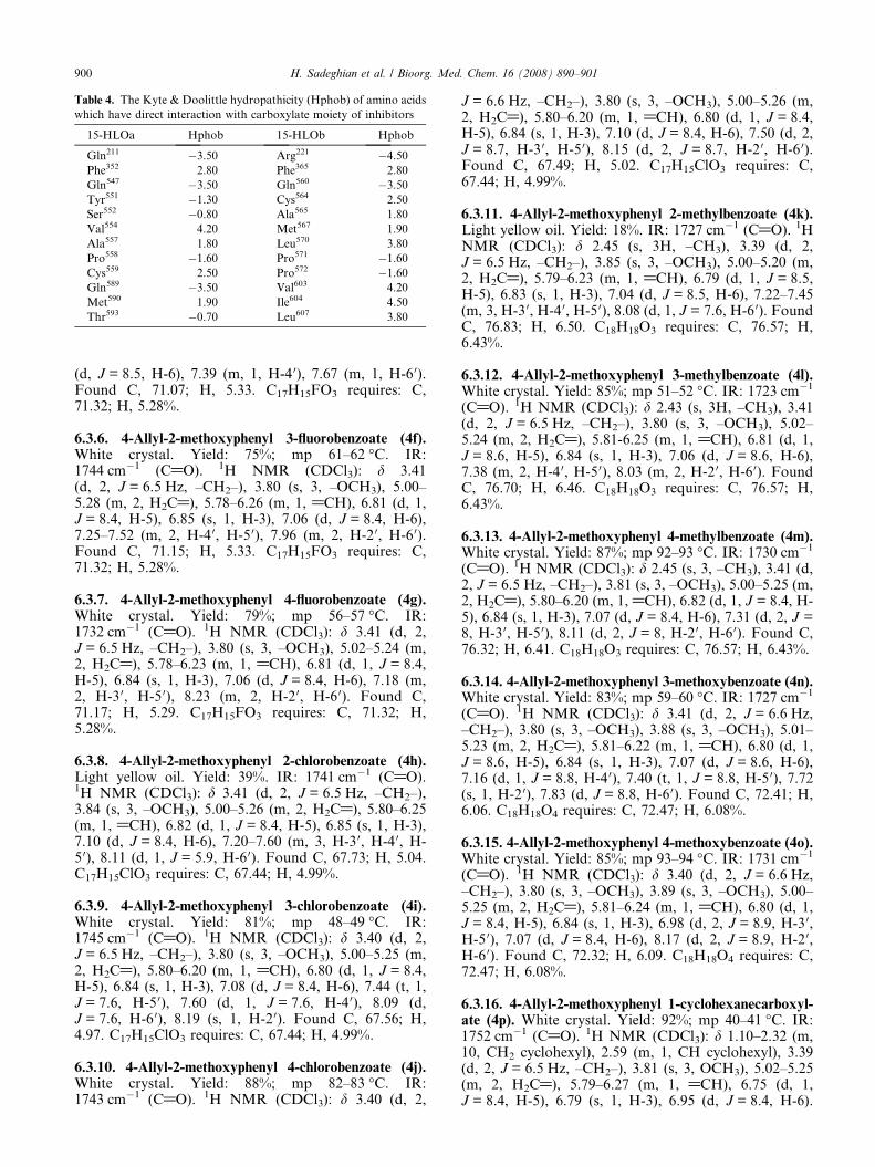
Table 4. The Kyte & Doolittle hydropathicity (Hphob) of amino acids
which have direct interaction with carboxylate moiety of inhibitors
15-HLOa Hphob 15-HLOb Hphob
Gln211 �3.50 Arg221 �4.50
Phe352 2.80 Phe365 2.80
Gln547 �3.50 Gln560 �3.50
Tyr551 �1.30 Cys564 2.50
Ser552 �0.80 Ala565 1.80
Val554 4.20 Met567 1.90
Ala557 1.80 Leu570 3.80
Pro558 �1.60 Pro571 �1.60
Cys559 2.50 Pro572 �1.60
Gln589 �3.50 Val603 4.20
Met590 1.90 Ile604 4.50
Thr593 �0.70 Leu607 3.80
900 H. Sadeghian et al. / Bioorg. Med. Chem. 16 (2008) 890–901
(d, J = 8.5, H-6), 7.39 (m, 1, H-4 0), 7.67 (m, 1, H-6 0).Found C, 71.07; H, 5.33. C17H15FO3 requires: C,71.32; H, 5.28%.
6.3.6. 4-Allyl-2-methoxyphenyl 3-fluorobenzoate (4f).White crystal. Yield: 75%; mp 61–62 �C. IR:1744 cm�1 (C@O). 1H NMR (CDCl3): d 3.41(d, 2, J = 6.5 Hz, –CH2–), 3.80 (s, 3, –OCH3), 5.00–5.28 (m, 2, H2C@), 5.78–6.26 (m, 1, @CH), 6.81 (d, 1,J = 8.4, H-5), 6.85 (s, 1, H-3), 7.06 (d, J = 8.4, H-6),7.25–7.52 (m, 2, H-4 0, H-5 0), 7.96 (m, 2, H-2 0, H-6 0).Found C, 71.15; H, 5.33. C17H15FO3 requires: C,71.32; H, 5.28%.
6.3.7. 4-Allyl-2-methoxyphenyl 4-fluorobenzoate (4g).White crystal. Yield: 79%; mp 56–57 �C. IR:1732 cm�1 (C@O). 1H NMR (CDCl3): d 3.41 (d, 2,J = 6.5 Hz, –CH2–), 3.80 (s, 3, –OCH3), 5.02–5.24 (m,2, H2C@), 5.78–6.23 (m, 1, @CH), 6.81 (d, 1, J = 8.4,H-5), 6.84 (s, 1, H-3), 7.06 (d, J = 8.4, H-6), 7.18 (m,2, H-3 0, H-5 0), 8.23 (m, 2, H-2 0, H-6 0). Found C,71.17; H, 5.29. C17H15FO3 requires: C, 71.32; H,5.28%.
6.3.8. 4-Allyl-2-methoxyphenyl 2-chlorobenzoate (4h).Light yellow oil. Yield: 39%. IR: 1741 cm�1 (C@O).1H NMR (CDCl3): d 3.41 (d, 2, J = 6.5 Hz, –CH2–),3.84 (s, 3, –OCH3), 5.00–5.26 (m, 2, H2C@), 5.80–6.25(m, 1, @CH), 6.82 (d, 1, J = 8.4, H-5), 6.85 (s, 1, H-3),7.10 (d, J = 8.4, H-6), 7.20–7.60 (m, 3, H-3 0, H-4 0, H-5 0), 8.11 (d, 1, J = 5.9, H-6 0). Found C, 67.73; H, 5.04.C17H15ClO3 requires: C, 67.44; H, 4.99%.
6.3.9. 4-Allyl-2-methoxyphenyl 3-chlorobenzoate (4i).White crystal. Yield: 81%; mp 48–49 �C. IR:1745 cm�1 (C@O). 1H NMR (CDCl3): d 3.40 (d, 2,J = 6.5 Hz, –CH2–), 3.80 (s, 3, –OCH3), 5.00–5.25 (m,2, H2C@), 5.80–6.20 (m, 1, @CH), 6.80 (d, 1, J = 8.4,H-5), 6.84 (s, 1, H-3), 7.08 (d, J = 8.4, H-6), 7.44 (t, 1,J = 7.6, H-5 0), 7.60 (d, 1, J = 7.6, H-4 0), 8.09 (d,J = 7.6, H-6 0), 8.19 (s, 1, H-2 0). Found C, 67.56; H,4.97. C17H15ClO3 requires: C, 67.44; H, 4.99%.
6.3.10. 4-Allyl-2-methoxyphenyl 4-chlorobenzoate (4j).White crystal. Yield: 88%; mp 82–83 �C. IR:1743 cm�1 (C@O). 1H NMR (CDCl3): d 3.40 (d, 2,
J = 6.6 Hz, –CH2–), 3.80 (s, 3, –OCH3), 5.00–5.26 (m,2, H2C@), 5.80–6.20 (m, 1, @CH), 6.80 (d, 1, J = 8.4,H-5), 6.84 (s, 1, H-3), 7.10 (d, J = 8.4, H-6), 7.50 (d, 2,J = 8.7, H-3 0, H-5 0), 8.15 (d, 2, J = 8.7, H-2 0, H-6 0).Found C, 67.49; H, 5.02. C17H15ClO3 requires: C,67.44; H, 4.99%.
6.3.11. 4-Allyl-2-methoxyphenyl 2-methylbenzoate (4k).Light yellow oil. Yield: 18%. IR: 1727 cm�1 (C@O). 1HNMR (CDCl3): d 2.45 (s, 3H, –CH3), 3.39 (d, 2,J = 6.5 Hz, –CH2–), 3.85 (s, 3, –OCH3), 5.00–5.20 (m,2, H2C@), 5.79–6.23 (m, 1, @CH), 6.79 (d, 1, J = 8.5,H-5), 6.83 (s, 1, H-3), 7.04 (d, J = 8.5, H-6), 7.22–7.45(m, 3, H-3 0, H-4 0, H-5 0), 8.08 (d, 1, J = 7.6, H-6 0). FoundC, 76.83; H, 6.50. C18H18O3 requires: C, 76.57; H,6.43%.
6.3.12. 4-Allyl-2-methoxyphenyl 3-methylbenzoate (4l).White crystal. Yield: 85%; mp 51–52 �C. IR: 1723 cm�1
(C@O). 1H NMR (CDCl3): d 2.43 (s, 3H, –CH3), 3.41(d, 2, J = 6.5 Hz, –CH2–), 3.80 (s, 3, –OCH3), 5.02–5.24 (m, 2, H2C@), 5.81-6.25 (m, 1, @CH), 6.81 (d, 1,J = 8.6, H-5), 6.84 (s, 1, H-3), 7.06 (d, J = 8.6, H-6),7.38 (m, 2, H-4 0, H-5 0), 8.03 (m, 2, H-2 0, H-6 0). FoundC, 76.70; H, 6.46. C18H18O3 requires: C, 76.57; H,6.43%.
6.3.13. 4-Allyl-2-methoxyphenyl 4-methylbenzoate (4m).White crystal. Yield: 87%; mp 92–93 �C. IR: 1730 cm�1
(C@O). 1H NMR (CDCl3): d 2.45 (s, 3, –CH3), 3.41 (d,2, J = 6.5 Hz, –CH2–), 3.81 (s, 3, –OCH3), 5.00–5.25 (m,2, H2C@), 5.80–6.20 (m, 1, @CH), 6.82 (d, 1, J = 8.4, H-5), 6.84 (s, 1, H-3), 7.07 (d, J = 8.4, H-6), 7.31 (d, 2, J =8, H-3 0, H-5 0), 8.11 (d, 2, J = 8, H-2 0, H-6 0). Found C,76.32; H, 6.41. C18H18O3 requires: C, 76.57; H, 6.43%.
6.3.14. 4-Allyl-2-methoxyphenyl 3-methoxybenzoate (4n).White crystal. Yield: 83%; mp 59–60 �C. IR: 1727 cm�1
(C@O). 1H NMR (CDCl3): d 3.41 (d, 2, J = 6.6 Hz,–CH2–), 3.80 (s, 3, –OCH3), 3.88 (s, 3, –OCH3), 5.01–5.23 (m, 2, H2C@), 5.81–6.22 (m, 1, @CH), 6.80 (d, 1,J = 8.6, H-5), 6.84 (s, 1, H-3), 7.07 (d, J = 8.6, H-6),7.16 (d, 1, J = 8.8, H-4 0), 7.40 (t, 1, J = 8.8, H-5 0), 7.72(s, 1, H-2 0), 7.83 (d, J = 8.8, H-6 0). Found C, 72.41; H,6.06. C18H18O4 requires: C, 72.47; H, 6.08%.
6.3.15. 4-Allyl-2-methoxyphenyl 4-methoxybenzoate (4o).White crystal. Yield: 85%; mp 93–94 �C. IR: 1731 cm�1
(C@O). 1H NMR (CDCl3): d 3.40 (d, 2, J = 6.6 Hz,–CH2–), 3.80 (s, 3, –OCH3), 3.89 (s, 3, –OCH3), 5.00–5.25 (m, 2, H2C@), 5.81–6.24 (m, 1, @CH), 6.80 (d, 1,J = 8.4, H-5), 6.84 (s, 1, H-3), 6.98 (d, 2, J = 8.9, H-3 0,H-5 0), 7.07 (d, J = 8.4, H-6), 8.17 (d, 2, J = 8.9, H-2 0,H-6 0). Found C, 72.32; H, 6.09. C18H18O4 requires: C,72.47; H, 6.08%.
6.3.16. 4-Allyl-2-methoxyphenyl 1-cyclohexanecarboxyl-ate (4p). White crystal. Yield: 92%; mp 40–41 �C. IR:1752 cm�1 (C@O). 1H NMR (CDCl3): d 1.10–2.32 (m,10, CH2 cyclohexyl), 2.59 (m, 1, CH cyclohexyl), 3.39(d, 2, J = 6.5 Hz, –CH2–), 3.81 (s, 3, OCH3), 5.02–5.25(m, 2, H2C@), 5.79–6.27 (m, 1, @CH), 6.75 (d, 1,J = 8.4, H-5), 6.79 (s, 1, H-3), 6.95 (d, J = 8.4, H-6).

H. Sadeghian et al. / Bioorg. Med. Chem. 16 (2008) 890–901 901
Found C, 74.48; H, 8.10. C17H22O3 requires: C, 74.42;H, 8.08%.
6.3.17. 4-Allyl-2-methoxyphenyl 1-admantanecarboxylate(4q). White crystal. Yield: 89%; mp 89–90 �C. IR:1738 cm�1 (C@O). 1H NMR (CDCl3): d 1.76 (m, 9,CH, CH2 adamantyl), 2.07 (m, 6, CH2 adamantyl),3.36 (d, 2, J = 6.6 Hz, –CH2–), 3.78 (s, 3, OCH3),4.97–5.22 (m, 2, H2C@), 5.82–6.20 (m, 1, @CH), 6.75(d, 1, J = 8.5, H-5), 6.78 (s, 1, H-3), 6.90 (d, J = 8.5,H-6). Found C, 77.58; H, 8.03. C21H26O3 requires: C,77.27; H, 8.03%.
Acknowledgments
We express our sincere gratitude to Dr. F. Hadizadehfor software supporting and Dr. A. Sadeghian for statis-tical analysis studies.
References and notes
1. Raghavenra, H.; Diwakr, B. T.; Lokesh, B. R.; Naidu, K.A. Prostaglandins, Leukotrienes Essential Fatty Acids2006, 74, 23–27.
2. Brash, A. R. J. Biol. Chem. 1999, 274, 23679–23682.3. Kuhn, H.; Thiele, B. J. FEBS Lett. 1999, 449, 7–11.4. Larsen, J. S.; Acosta, E. P. Ann. Pharmacother. 1993, 27,
898–903.5. Schewe, T. Biol. Chem. 2002, 383, 365–374.6. Kelavkar, U.; Glasgow, W.; Eling, T. E. Curr. Urol. Rep.
2002, 3, 207–214.7. Kelavkar, U. P.; Cohen, C.; Kamitani, H.; Eling, T. E.;
Badr, K. F. Carcinogenesis 2000, 21, 1777–1787.8. Zhu, J.; Kilty, I.; Granger, H.; Gamble, E.; Qiu, Y. S.;
Hattotuwa, K.; Elston, W.; Liu, W. L.; Liva, A.; Pauwels,R. A.; Kis, J. C.; De Rose, V.; Barnes, N.; Yeadon, M.;Jenkinson, S.; Jeffery, P. K. Am. J. Respir. Cell Mol. Biol.2002, 27, 666–677.
9. Zhao, L.; Funk, C. D. Trends Cardiovasc. Med. 2004, 14,191–195.
10. Cyrus, T.; Witztum, J. L.; Rader, D. J.; Tangirala, R.;Fazio, S.; Linton, M. F.; Funk, C. D. J. Clin. Invest. 1999,1597–1604.
11. Harats, D.; Shaish, A.; George, J.; Mulkins, M.; Kurihara,H.; Levkovitz, H.; Sigal, E. Arteriosler. Thromb. Vasc.Biol. 2000, 20, 2100–2105.
12. Sultana, C.; Shen, Y.; Rattan, V.; Kalra, V. J. J. Cell.Phys. 1996, 167, 467–487.
13. Setty, B. N.; Werner, M. H.; annun, Y. A.; Stuart, M.J. Blood 1992, 80, 2765–2773.
15. Chanez, P.; Bonnans, C.; Chavis, C.; Vachier, I. Am.J. Respir. Crit. Care Med. 2002, 27, 655–658.
16. Shappell, S. B.; Boeglin, W. E.; Olson, S. J.; Kasper, S.;Brash, A. R. Am. J. Pathol. 1999, 155, 235–245.
17. Hsi, L. C.; Wilson, L. C.; Eling, T. E. J. Biol. Chem. 2002,277, 40549–40556.
18. Charlier, C.; Michaux, C. Eur. J. Med. Chem. 2003, 38,645–659.
19. Markowitz, K.; Moynihan, M.; Liv, M.; Kim, S. OralSurg. Oral Pathol. 1992, 73, 729–737.
20. IARC, Monograph, Evaluation of Carcinogenic Risk ofChemicals to Humans. Lyon, France, 1985; vol. 36, pp 75–97.
21. Wagner, H.; Jurcic, K.; Deininger, R. Planta Med. 1979,37, 9–14.
22. Feng, J.; Lipton, J. M. Neuropharmacology 1987, 26,1775–1778.
23. Hume, W. R. J. Dent. Res. 1983, 62, 1013–1015.24. Moleyar, V.; Narasimham, P. Int. J. Food Microbiol. 1992,
16, 337–342.25. Villani, F. J.; King, M. S. Org. Syn. Coll. 1963, 4, 88–
89.26. Thompson, J. D.; Gibson, T. J.; Plewniak, F.; Jeanmou-
gin, F.; Higgins, D. G. Nucleic Acids Res. 1997, 24,4876–4882.
27. http://us.expasy.org/.28. ChemDraw� Ultra, Chemical Structure Drawing Standard,
CambridgeSoft Corporation, 100 Cambridge Park Drive,Cambridge, MA 02140 USA, http://www.cambrigesoft.com.
29. HyperChem� Release 7, Hypercube Inc., http://www.hyper.com/.
30. Bakavoli, M.; Nikpour, M.; Rahimizadeh, M.; Saberi, M.R.; Sadeghian, H. Bioorg. Med. Chem. 2007, 15, 2120–2126.
31. Auto Dock Tools (ADT), the Scripps Research Institute,10550 North Torrey Pines Road, La Jolla, CA 92037-1000, USA; (http://www.scripps.edu/pub/olson-web/doc/autodock/); Python, M.F.S.A programming language forsoftware integration and development. J. Mol. GraphicsMod. 1999, 17, 57-61.
32. Morris, G. M.; Goodsell, D. S.; Halliday, R. S.; Huey, R.;Hart, W. E.; Belew, R. K.; Olson, A. J. J. Comput. Chem.1998, 19, 1639–1662.
33. Sippl, W. J. Comput. Aided Mol. Des. 2000, 14,559–572.
34. Dym, O.; Xenarios, I.; Ke, H.; Colicelli, J. Mol. Pharma-col. 2002, 61, 20–25.
35. http://sunfire.vbi.vt.edu/gcg/seqweb-guides/WebLab_Viewer.html.
36. Swiss-pdbViewer 3.6, Glaxo Wellcome ExperimentalResearch, http://www.expasy.org/spdbv/.
37. DRAGON 2.1, Milano Chemometrics and QSAR ResearchGroup, Department pf Environmental Sciences, P.za DellaScienza, 1-20126 Milano, Italy, http://www.disat.unimib.it/chem/.
38. Todeschini, R.; Consonni, V. Handbook of MolecularDescriptors; Wiley-VCH, Weinheim, Germany.
39. Kumar, R.; Pavithra, S. R.; Tatu, U. J. Biosci. 2007, 32,531–536.
40. http://nihserver.mbi.ucla.edu/savs/.41. Malterud, K. E.; Rydland, K. M. J. Agric. Food Chem.
2000, 48, 5576–5580.42. Malterud, K. E.; Farbrot, T. L.; Huse, A. E.; Sund, R. B.
Pharmacology 1993, 47, 77–85.43. Skrzypczak-Jankun, E.; Bross, R.; Carroll, R. T.; Dun-
ham, W. R.; Funk, M. O., Jr. J. Am. Chem. Soc. 2001,123, 10814–10820.
44. Knapp, M. J.; Seebeck, F. P.; Klinman, J. P. J. Am. Chem.Soc. 2001, 123, 2931–2932.
45. Tomchick, D. R.; Phan, P.; Cymborowski, M.; Minor, W.;Holman, T. R. Biochemistry 2001, 40, 7509–7517.
46. Kyte, J.; Doolittle, R. F. J. Mol. Biol. 1982, 157,105–132.