Embed Size (px)
Citation preview
TO
RT
UR
E V
olu
me
16
, N
um
be
r 2
, 2
00
6108
Macroscopic changesThe significance of skin lesions is mostly related to the documentation of the history of torture. Acute lesions may give health problems, for example pain and secondary infections, including problems with healing, especially when located in an area with ven-ous or arterial insufficiency. Scars located close to a joint may induce contracture, decreased mobility of the joint and pain dur-ing activity. Apart from this, scars seldom inconvenience the patient, although they can sometimes be of cosmetic importance since they may be a reminder of the torture and add to the changed sense of identity induced by torture.
A detailed history of the alleged torture and of the symptoms it induced is important in order to evaluate the significance of the observed lesions on the skin. Information about the position of the victim and of the torturer during the torture is particularly important, as well as information about the shape of instruments in contact with the skin. In cases with no or uncharacteristic lesions, a characteristic history may be the
only support to the allegation of torture, as for example in some cases of electrical tor-ture. Also a history of skin diseases and non-torture-related lesions are of importance.
The examination should include the en-tire body surface to detect signs of:
1) Skin diseases2) Non-torture-related lesions3) Torture-related lesions
Torture sequelae related to the skin may be:
1) Lesions resulting from direct physical in-juries
2) The occurrence of new, or aggravation of existing, skin diseases, provoked by phys-ical or psychological trauma
When a doctor writes a certificate after conducting a medical examination of a per-son who alleges having been tortured, it is extremely important that the doctor states the degree of consistency with the history of torture1. A conclusion indicating the degree of support to the alleged history of torture should be based on a discussion of possible differential diagnoses (non-torture-related injuries, self-inflicted injuries included, and skin diseases). The degree of support should be indicated as follows:
Dermatological findings after alleged torture
Lis Danielsen, MD, DMSc* and Ole Vedel Rasmussen, MD, DMSC*
*)IRCT, Borgergade 13 P.O.Box 9049 DK-1022 Copenhagen K Denmark [email protected]
C L I N I C A L K N O W L E D G E
TO
RT
UR
E Vo
lum
e 1
6, N
um
be
r 2, 2
00
6109
1) A high degree of support2) Consistent with the alleged torture, mod-
erate degree of support3) Consistent with the alleged torture, slight
degree of support4) The changes cannot support the history
of torture
Acute lesions are often characteristic since they show a pattern of inflicted injury that differs from non-inflicted injuries, for exam-ple by their shape and distribution on the body. Since most lesions heal within a short period of time, leaving no or non-specific scars, a characteristic history of the acute lesions is important. Also a history of the de-velopment until healing is of importance.
Description of skin lesionsDescription of skin lesions should include the following points:
1) Localisation (use body diagram) sym-metrical, asymmetrical
2) Shape: round, oval, linear, etc.3) Size: use ruler4) Colour5) Surface: scaly, crusty, ulcerative, bullous,
necrotic6) Periphery: regular or irregular, zone in
the periphery7) Demarcation: sharply, poorly8) Level in relation to surrounding skin:
atrophic, hypertrophic, plane
The following findings are supportive of ex-ternal infliction:
1) Lack of symmetry (may also be the case for some skin diseases)
2) Linear lesions in irregular or criss-cross arrangements
3) A linear zone extending circularly around an extremity
4) A regular, narrow, hyperpigmented or hypertrophic zone surrounding a scar (sequels to an inflammatory zone around a necrotic area); this may also be the case with skin diseases with necrotic areas, for example necrotic vasculitis
Blunt traumaBlunt trauma may leave ecchymoses, contu-sions or lacerations with extravasation of blood in the skin and subcutaneous tissue, in some cases reflecting the shape of the instru-ment used, for example from beating with a stick (Figure 1).2 Two parallel linear le-
Figure 1. 1) Alleged torture involving beating with a stick on several areas of the skin, including the back of the thighs and the buttocks, five days previously. 2) Massive haematomas are seen in the gluteal regions and on the upper part of the back of the thighs, containing areas with parallel, linear, a few cm broad, haemorrhagic lesions circulating obliquely around the gluteal region and the up-per part of the thigh. 3) The lesions show signs of recent external inflictions from beating with a stick. No dermatological condition can explain the ob-lique, linear pattern. 4) Conclusion: A high degree of support to the history of torture because of the pattern of the lesions.2
C L I N I C A L K N O W L E D G E

TO
RT
UR
E V
olu
me
16
, N
um
be
r 2
, 2
00
6110
sions (“tramline bruises”) result from a blow with a rod or stick (figures 2, 3, 3a).3 The haemorrhagic areas often move down the body during the following days. Deep tissue
bruises might not be seen on the surface. The lesions change colour from dark red, to dusky purple, to brown, to green, to yel-low and to hyperpigmented brown, or they disappear. Severe beating on the soles of the feet, “Falanga”, may leave contusions in the arch of the feet and swelling of the feet ex-tending from the arch to the medial aspects of the feet and ankles (Figures 4, 5).4 Blunt trauma often leaves no or uncharacteristic
Figure 2. The formation of ”tramline” bruising from the application of a rectangular or cylindrical object.3
Figure 3. 1) Alleged torture involving beating with a broom handle. 2) Approximate parallel bruises are seen, and several of them, especially the lower-most, have a double ”tramline” appearance, typical of the impact of a round or square-section rod. 3) The lesions show signs of recent external infliction. The pressure in the centre may have compressed the vessels, so that they do not bleed. No derma-tological condition can explain the oblique, linear pattern. 4) Conclusion: A high degree of support to the history of torture, because of the pattern of the lesions.3
Figure 3a. 1) Unknown history of injuries. The inju-ries were observed on a detainee. 2) Several paral-lel bruises are seen on the back of the detainee in criss-cross arrangements; some of them with a double “tramline” appearance. 3) The lesions show signs of external infliction. 4) Conclusion: The lesions might be caused by a round or square-sec-tion rod. Published with permission from Red-Cross (ICRC)
C L I N I C A L K N O W L E D G E
TO
RT
UR
E Vo
lum
e 1
6, N
um
be
r 2, 2
00
6111
scars (Figure 6).5 Flogging or beating with canes or truncheons may, however, leave characteristic scars, for example asymmetric, linear, straight or curved or “tramline”-shaped scars, showing a pattern of external infliction.6-8 The scars may be hypertrophic with a narrow, regular, hyperpigmented area in the periphery, representing “arrowline” bruises or an inflammatory zone appearing around necrotic tissue in the acute phase (Figures 7-9).6 A differential diagnosis could be plant dermatitis, usually dominated, however, by shorter scars, with a narrow zone of hyperpigmentation in the periphery (Figure 10). In one case, the alleged tor-ture was beating and scalding on the back. Symmetrical, atrophic, depigmented, linear changes typical for striae distensae were observed on the back and in both axillary regions (Figures 11, 12).6 The skin changes could not support the history of torture. The patient, however, may have been unaware of the changes on the back before the tor-ture. Prolonged application of tight ligatures
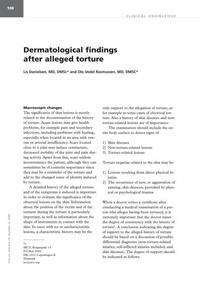
Figure 4. 1) Alleged torture involving ”Falanga”, i.e. severe beating on the soles of the feet. 2) Ery-thema with a slightly haemorrhagic appearance of the skin in the arch of the feet and a swelling of the feet extending from the arch to the medial aspects of the feet and ankles. 3) Typical for ”Fa-langa”, since it is unlikely that other types of blunt injuries could give such changes in that part of the foot, with oedema extending to the rest of the foot and ankle. 4) Conclusion: A high degree of support to the history of torture.4Reprintet with permission from the Danish Medical Association.
Figure 5. 1) Alleged torture involving ”Falanga”. 2) Haemorhagic areas are seen on the distal part of the sole of the foot, as well as dermatitis around the toes. 3) In this case, the patient had been walk-ing in the mountains without shoes, which could be a differential diagnosis. 4) Conclusion: Consist-ent with the alleged torture, but only slight degree of support because of the location of the injury to the distal part of the sole.
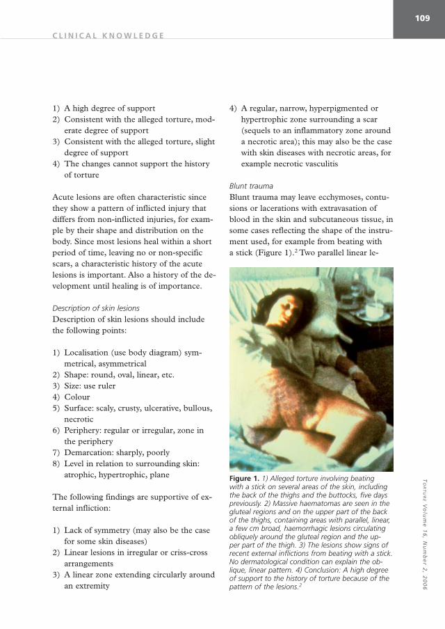
Figure 6. 1) Alleged torture involving blunt vio-lence from a blow five years previously. 2) Hyper-pigmented area with an irregular and indistinct limitation. 3) The lesion might be secondary to haemorrhage from ruptured blood vessels, but it lacks indication of the shape of the instrument used. 4) Conclusion: The lesion is consistent with the alleged torture, its support being of a slight degree.5Reprinted with permission from the Danish Medical Association.
C L I N I C A L K N O W L E D G E

TO
RT
UR
E V
olu
me
16
, N
um
be
r 2
, 2
00
6112
Figure 7. 1) Alleged torture involving flogging six months previously. 2) Long, straight or curved, linear scars in an asymmetric pattern on the back. They are curved particularly corresponding to the outlines of the body, where they have a broader, irregular end, and vertically directed in the centre. One straight scar is located vertically on the lower part of the trunk. The centres of the scars are depigmented, hypertrophic and surrounded by thin, hyperpigmented stripes. 3) The scars show signs of an external infliction with a pattern underlining the history of flogging. The torturer could have been standing behind the patient. The vertical direction of the scars in the centre of the trunk can, however, not exclude self-inflic-tion. A differential diagnosis could be plant dermatitis, but this shows shorter, linear scars with hyperpigmented stripes in the periphery. 4) Conclusion: The scars are consistent with the alleged torture, their support to the history being of a moderate degree, since self-infliction cannot be totally ruled out.6
Figure 8. Same patient as Figure 7.
Figure 9. Same patient as Figure 7.
Figure 10. Plant dermatitis with short, linear scars with a narrow zone of hyperpigmentation in the periphery. Differential diagnosis to Figure 7.6
C L I N I C A L K N O W L E D G E
TO
RT
UR
E Vo
lum
e 1
6, N
um
be
r 2, 2
00
6113
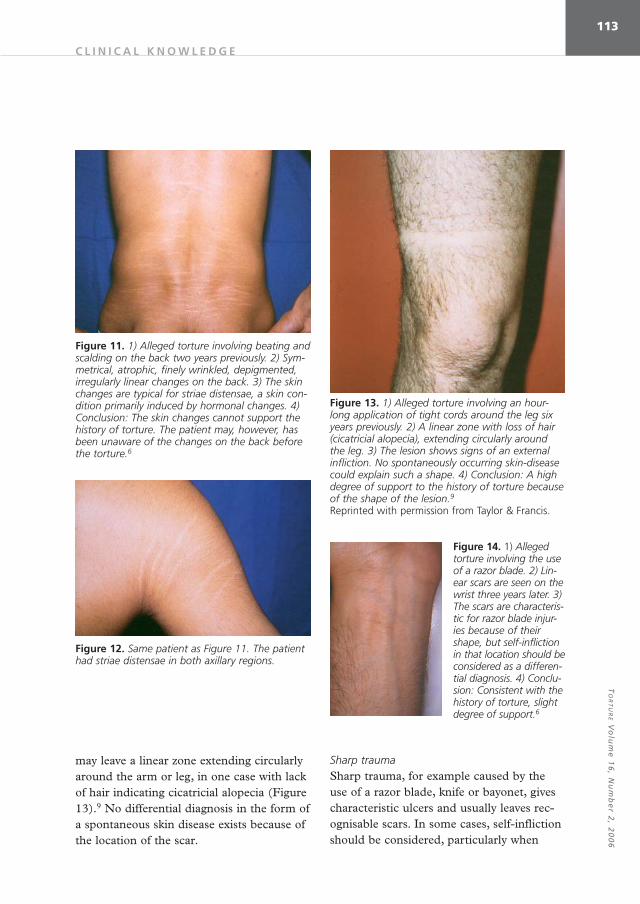
may leave a linear zone extending circularly around the arm or leg, in one case with lack of hair indicating cicatricial alopecia (Figure 13).9 No differential diagnosis in the form of a spontaneous skin disease exists because of the location of the scar.
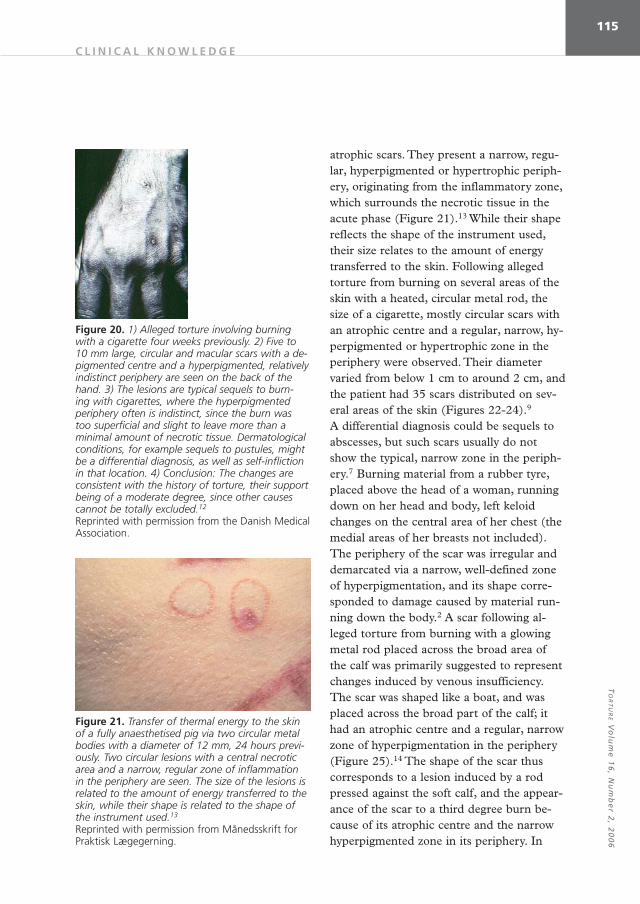
Sharp traumaSharp trauma, for example caused by the use of a razor blade, knife or bayonet, gives characteristic ulcers and usually leaves rec-ognisable scars. In some cases, self-infliction should be considered, particularly when
Figure 11. 1) Alleged torture involving beating and scalding on the back two years previously. 2) Sym-metrical, atrophic, finely wrinkled, depigmented, irregularly linear changes on the back. 3) The skin changes are typical for striae distensae, a skin con-dition primarily induced by hormonal changes. 4) Conclusion: The skin changes cannot support the history of torture. The patient may, however, has been unaware of the changes on the back before the torture.6
Figure 13. 1) Alleged torture involving an hour-long application of tight cords around the leg six years previously. 2) A linear zone with loss of hair (cicatricial alopecia), extending circularly around the leg. 3) The lesion shows signs of an external infliction. No spontaneously occurring skin-disease could explain such a shape. 4) Conclusion: A high degree of support to the history of torture because of the shape of the lesion.9Reprinted with permission from Taylor & Francis.
Figure 14. 1) Alleged torture involving the use of a razor blade. 2) Lin-ear scars are seen on the wrist three years later. 3) The scars are characteris-tic for razor blade injur-ies because of their shape, but self-infliction in that location should be considered as a differen-tial diagnosis. 4) Conclu-sion: Consistent with the history of torture, slight degree of support.6
Figure 12. Same patient as Figure 11. The patient had striae distensae in both axillary regions.
C L I N I C A L K N O W L E D G E

TO
RT
UR
E V
olu
me
16
, N
um
be
r 2
, 2
00
6114
located on a wrist (Figure 14).6,7 If pepper is applied to the open wounds, the scars may become hypertrophic (Figures 15-17).6 A differential diagnosis could be traditional healers, African ritual scar-tattoos or art on the body (Figures 18, 19).10 In one case, where the deepness of a scar, allegedly fol-lowing the use of a sword, was doubted, the use of a high-frequency ultrasound could demonstrate a considerable, deep scar.11 Afterwards, the patient was granted refugee status.
Thermal injuriesBurning with cigarettes, hot instruments or hot fluids leaves acute burns of varying de-grees. Burning is the form of torture that most frequently leaves scars, often of diag-nostic value. Cigarette burns often leave 5-10 mm large, circular and macular scars with a depigmented centre and a hyperpig-mented, relatively indistinct periphery (Fig-ure 20).12 Dermatological conditions, for ex-ample sequels to pustules, might be a differential diagnosis. Burning via the trans-fer of larger amounts of energy to the skin than that transferred when stubbing a ciga-rette on the skin often produces markedly
Figure 15. 1) Alleged torture involving the use of razor blades and the application of pepper to the open wounds two years previously, in Africa. 2) Numerous 5-15 mm long, 1-3 mm wide, linear or irregular scars on the side of the neck. (Similar scars on the other side of the neck). The scars are asymmetrically located and often irregular in shape. 3) A possible differential diagnosis is ritual tattooing with scars performed with razor blades and subsequent application of ashes, particularly on the neck. 4) Conclusion: The lesions are consist-ent with the history of torture, their support being of a slight degree, since a ritual tattooing cannot be excluded in Africa.6
Figure 16. Same patient as figure 15.
Figure 17. Same patient as patient 15.
Figure 18. Ritual scar tattoos. Differential diagnosis to patient 15.
Figure 19. The art on the body. Differential diagnosis to patient 15.10
Reprinted with per-mission from Søren Nancke-Krogh.
C L I N I C A L K N O W L E D G E
TO
RT
UR
E Vo
lum
e 1
6, N
um
be
r 2, 2
00
6115
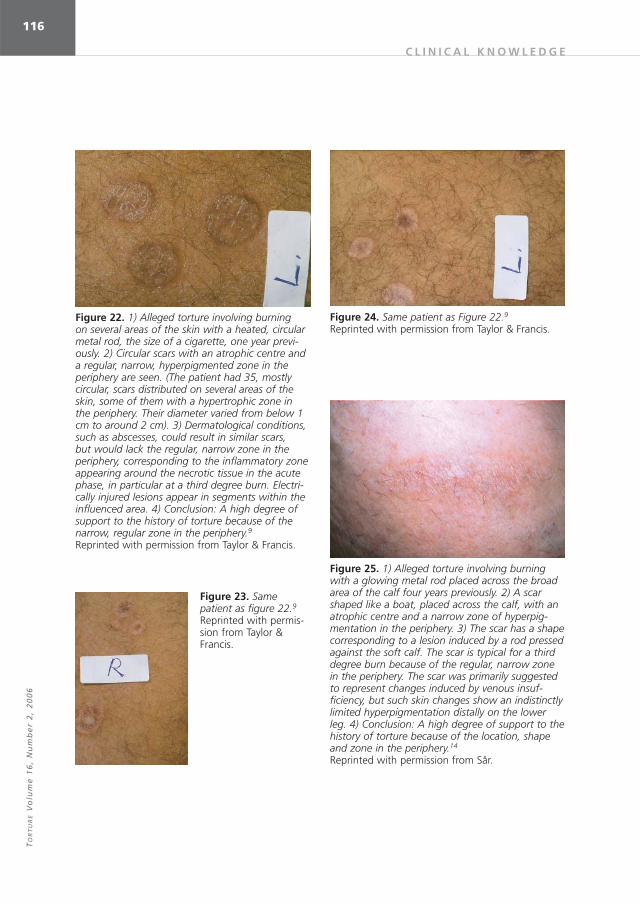
atrophic scars. They present a narrow, regu-lar, hyperpigmented or hypertrophic periph-ery, originating from the inflammatory zone, which surrounds the necrotic tissue in the acute phase (Figure 21).13 While their shape reflects the shape of the instrument used, their size relates to the amount of energy transferred to the skin. Following alleged torture from burning on several areas of the skin with a heated, circular metal rod, the size of a cigarette, mostly circular scars with an atrophic centre and a regular, narrow, hy-perpigmented or hypertrophic zone in the periphery were observed. Their diameter varied from below 1 cm to around 2 cm, and the patient had 35 scars distributed on sev-eral areas of the skin (Figures 22-24).9 A differential diagnosis could be sequels to abscesses, but such scars usually do not show the typical, narrow zone in the periph-ery.7 Burning material from a rubber tyre, placed above the head of a woman, running down on her head and body, left keloid changes on the central area of her chest (the medial areas of her breasts not included). The periphery of the scar was irregular and demarcated via a narrow, well-defined zone of hyperpigmentation, and its shape corre-sponded to damage caused by material run-ning down the body.2 A scar following al-leged torture from burning with a glowing metal rod placed across the broad area of the calf was primarily suggested to represent changes induced by venous insufficiency. The scar was shaped like a boat, and was placed across the broad part of the calf; it had an atrophic centre and a regular, narrow zone of hyperpigmentation in the periphery (Figure 25).14 The shape of the scar thus corresponds to a lesion induced by a rod pressed against the soft calf, and the appear-ance of the scar to a third degree burn be-cause of its atrophic centre and the narrow hyperpigmented zone in its periphery. In
Figure 20. 1) Alleged torture involving burning with a cigarette four weeks previously. 2) Five to 10 mm large, circular and macular scars with a de-pigmented centre and a hyperpigmented, relatively indistinct periphery are seen on the back of the hand. 3) The lesions are typical sequels to burn-ing with cigarettes, where the hyperpigmented periphery often is indistinct, since the burn was too superficial and slight to leave more than a minimal amount of necrotic tissue. Dermatological conditions, for example sequels to pustules, might be a differential diagnosis, as well as self-infliction in that location. 4) Conclusion: The changes are consistent with the history of torture, their support being of a moderate degree, since other causes cannot be totally excluded.12
Reprinted with permission from the Danish Medical Association.
Figure 21. Transfer of thermal energy to the skin of a fully anaesthetised pig via two circular metal bodies with a diameter of 12 mm, 24 hours previ-ously. Two circular lesions with a central necrotic area and a narrow, regular zone of inflammation in the periphery are seen. The size of the lesions is related to the amount of energy transferred to the skin, while their shape is related to the shape of the instrument used.13
Reprinted with permission from Månedsskrift for Praktisk Lægegerning.
C L I N I C A L K N O W L E D G E
TO
RT
UR
E V
olu
me
16
, N
um
be
r 2
, 2
00
6116
Figure 22. 1) Alleged torture involving burning on several areas of the skin with a heated, circular metal rod, the size of a cigarette, one year previ-ously. 2) Circular scars with an atrophic centre and a regular, narrow, hyperpigmented zone in the periphery are seen. (The patient had 35, mostly circular, scars distributed on several areas of the skin, some of them with a hypertrophic zone in the periphery. Their diameter varied from below 1 cm to around 2 cm). 3) Dermatological conditions, such as abscesses, could result in similar scars, but would lack the regular, narrow zone in the periphery, corresponding to the inflammatory zone appearing around the necrotic tissue in the acute phase, in particular at a third degree burn. Electri-cally injured lesions appear in segments within the influenced area. 4) Conclusion: A high degree of support to the history of torture because of the narrow, regular zone in the periphery.9Reprinted with permission from Taylor & Francis.
Figure 23. Same patient as figure 22.9Reprinted with permis-sion from Taylor & Francis.
Figure 24. Same patient as Figure 22.9Reprinted with permission from Taylor & Francis.
Figure 25. 1) Alleged torture involving burning with a glowing metal rod placed across the broad area of the calf four years previously. 2) A scar shaped like a boat, placed across the calf, with an atrophic centre and a narrow zone of hyperpig-mentation in the periphery. 3) The scar has a shape corresponding to a lesion induced by a rod pressed against the soft calf. The scar is typical for a third degree burn because of the regular, narrow zone in the periphery. The scar was primarily suggested to represent changes induced by venous insuf-ficiency, but such skin changes show an indistinctly limited hyperpigmentation distally on the lower leg. 4) Conclusion: A high degree of support to the history of torture because of the location, shape and zone in the periphery.14
Reprinted with permission from Sår.
C L I N I C A L K N O W L E D G E

TO
RT
UR
E Vo
lum
e 1
6, N
um
be
r 2, 2
00
6117
contrast, venous insufficiency leaves indis-tinctly limited hyperpigmentation and scars from ulcers located distally on the lower leg (Figure 26).14 Afterwards, the patient was granted refugee status. When the nail matrix is burnt, subsequent growth produces strip ed, thin, deformed nails, sometimes broken up in longitudinal segments. If the nail is also pulled off, an overgrowth of tis-sue may occur from the proximal nail fold (Figure 27).6 Changes caused by lichen pla-nus may be a relevant differential diagnosis, while fungus infection is characterised by thickened, yellowish, crumbling nails, differ-ent from those mentioned above (Figure 28).
Corrosive injuriesCorrosive injuries, caused by acid thrown against a victim, caused linear scars, a few cm wide, with a depigmented centre and a regular, narrow, hyperpigmented zone in the periphery, located on the thighs and buttocks (Figure 29).15 They were arranged in an asymmetric pattern, mostly obliquely directed down the legs. They showed signs of external infliction in agreement with a liquid running down the legs, and they indicated sequels to necrotic areas as expected follow-ing a corrosive injury.
Electrical injuriesElectric current follows the shortest route between the two electrodes through tissue with the lowest resistance, i.e. blood vessels, nerves and muscles.16 When using high-volt-age stun weapons, the current flow cannot, however, be limited to the pathway between the electrodes.17 The possibility of find-ing signs of electrical influence in the skin, particularly histological signs, is related to the type of electricity transferred, since the electrolytic action will be most pronounced by transfer of direct current and will not be present following transfer of high-frequency
Figure 26. Venous insufficiency with indistinctly limited hyperpigmentation distally on the lower leg. Differential diag-nosis to Figure 25.14
Reprinted with permis-sion from Sår.
Figure 27. 1) Alleged torture involving injury to the nail matrix caused by the pulling off of the toenails and burning with charcoal embers two years previ-ously. 2) Striped, deformed toenails, the left big toenail divided into three slightly curved longitu-dinal segments with an overgrowth of tissue from the proximal nail fold resulting in the formation of a pterygium. 3) When the nail matrix is injured as explained above, the nail may show such changes. A differential diagnosis could be lichen planus, but in that case, widespread skin affection would usu-ally be present. Fungus infection of toenails show thickened, yellowish nails, different from those mentioned above. 4) Conclusion: The changes are consistent with the history of torture, their support being of a slight degree, since the alterations can be caused by several injuries.6
Figure 28. Thickened, yellowish, crumbling toenail caused by fungus infection. Differential diagnosis to figure 27.6
C L I N I C A L K N O W L E D G E
TO
RT
UR
E V
olu
me
16
, N
um
be
r 2
, 2
00
6118
Figure 29. 1) Alleged torture involving acid thrown against the victim. 2) Linear scars, a few cm wide, with a depigmented centre and a regular, narrow, hyperpigmented zone in the periphery are seen on the thighs and buttocks. The scars appear in an asymmetric pattern, mostly obliquely directed down the legs. 3) The scars show signs of external infliction in agreement with a liquid running down the legs. They show sequels to necrotic areas with a narrow hyperpigmented zone in the periphery. 4) Conclusion: A high degree of support to the his-tory of torture because of the location, the shape and the narrow zone in the periphery.15
Reprinted with permission from Elsevier.
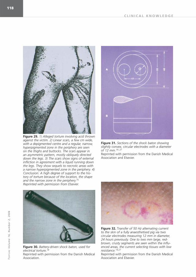
Figure 30. Battery-driven shock baton, used for electrical torture.18
Reprinted with permission from the Danish Medical Association.
Figure 31. Sections of the shock baton showing slightly convex, circular electrodes with a diameter of 12 mm.19, 27
Reprinted with permission from the Danish Medical Association and Elsevier.
Figure 32. Transfer of 50 Hz alternating current to the skin of a fully anaesthetised pig via two circular electrodes measuring 12 mm in diameter, 24 hours previously. One to two mm large, red-brown, crusty segments are seen within the influ-enced areas, the current selecting tissues with low resistance.19,27
Reprinted with permission from the Danish Medical Association and Elsevier.
C L I N I C A L K N O W L E D G E

TO
RT
UR
E Vo
lum
e 1
6, N
um
be
r 2, 2
00
6119
alternating current, where the concomi-tant heat generation dominates.16 Also, the amount of energy used plays a role for a domination of burn injuries in the lesions, particularly concerning low frequency alter-nating current. In some of the cases, electric torture leaves acute lesions on the skin. Un-like burn lesions, these lesions usually do not reflect the shape of the instrument used, but appear in segments within the influenced areas, since the current selects areas with low resistance (Figures 30-33).18,19 Electrical torture via electrodes shaped like a knitting needle, “Picana”, leaves clusters and linear arrangements of 1-5 mm wide lesions, cov-ered by red-brown crusts, sometimes sur-rounded by a 1-2 mm broad, erythematous zone with irregular and indistinct edges (Figure 34).2 Lesions in lines following a lin-ear application of the electrodes may also be seen. The crusts probably correspond to an electrical injury and may contain deposits of metal from the electrodes.20, 21 The concomi-tant heat development has not been suf-ficient to induce a regular inflammation in the periphery. Differential diagnosis may be insect bites or scratching. Many red lesions,
a few mm large, have been seen following the use of a battery-driven electrical instru-ment (Figure 35).22 A contact dermatitis may be a differential diagnosis. Well-demar-cated, serpiginous lesions, measuring 1-2 cm across, with an irregular, narrow, elevated, peripheral zone and a central area contain-ing several black spots, each measuring 1-2
Figure 33. Transfer of direct current to the skin of a fully anaesthetised pig via two circular electrodes measuring 12 mm in diameter, 24 hours previously. A few mm large, brown, crusty segments are seen within the anode area, while segments of a similar size with a necrotic centre and an inflammatory zone in the periphery are seen in the cathode area.
Figure 34. 1) Alleged torture involving ”Picana”, i.e. electrical torture via electrodes shaped like a knitting needle, 72 hours previously. 2) The skin of the frontal area of the trunk shows many erythematous lines, some 2-5 mm wide, mostly vertically arranged. Scattered among them are dark red, crusty spots. 3) The linear shape of the lesions indicates external infliction corresponding to a pointed electrode moved across the skin, the red crusty spots correspond to the entrance of the electrical current. The crusts probably correspond to an electrical injury. The concomitant heat devel-opment has not been sufficient enough to induce a regular inflammation in the periphery. An impor-tant differential diagnosis is scratching. 4) Conclu-sion: The lesions are consistent with the history of torture, their support being of a slight degree, since scratching cannot be excluded.2
C L I N I C A L K N O W L E D G E

TO
RT
UR
E V
olu
me
16
, N
um
be
r 2
, 2
00
6120
mm, have been observed shortly after elec-trical injuries on the left side of the chest and on the left arm (Figures 36, 37).23 The lesions show indication of electrical injury because of their appearance in 1-2 mm large segments and because of the involvement of blood vessels. Vasculitis or haemorrhagic herpes zoster might constitute a differential diagnosis. The location might be helpful since vasculitis is chiefly located at the lower extremities, is symmetrical and is sometimes more diffusely located, while herpes zoster is located in an area innervated by a single ganglion and is unilateral. Clusters of round, red macular scars, about 1 mm in diameter, have been observed four weeks after “Pi-cana” (Figure 38).12 Eight weeks later, many of the scars had disappeared. The remaining scars were small, white or red-brown spots. Among the skin diseases leaving pigmented scars is lichen planus leaving about 2 mm large scars.
Electrical torture has been reported to induce 6-8 mm large, irregular, red-brown, keloid scars on the helix of both ears.24 Dif-ferential diagnosis might be a chondroder-matitis helicis, but this is usually covered by a scale, and is pale and painful. Six months after the use of a 45 cm long stun gun, de-livering 150,000 V, with a screw 4 mm in
Figure 35. 1) Alleged torture involving a battery-driven electrical instrument, probably inducing high-frequency alternating current. 2) Many red lesions, a few mm large, are seen on the side of the trunk. 3) The appearance in segments and the red colour support the influence of electric current. A dermatitis might be a differential diagnosis. 4) Conclusion: Consistent with the history of torture, but only to a slight degree since contact dermatitis cannot be excluded.22
Reprinted with permission from TAT.
Figure 36. 1) Alleged torture involving electrical wires. 2) Shortly afterwards, well-demarcated, serpiginous lesions, measuring 1-2 cm across, with an irregular, narrow, elevated, peripheral zone and a central area containing several black spots, each measuring 1-2 mm, are seen. The lesions are seen on the left side of the chest and on the left arm. 3) The lesions show indications of electrical injury because of their irregular periphery and the 1-2 mm large, black segments at their centre, probably involving blood vessels. Electrical current follows the shortest route between the two electrodes through tissue with the lowest resistance, i.e. blood vessels, nerves and muscles. Vasculitis or haemorrhagic herpes zoster might be a differential diagnosis. However, vasculitis is chiefly located at the lower extremities and is symmetrical, while herpes zoster is located to an area innervated by a single ganglion. 4) Conclusion: A high degree of support to the history of torture because of its appearance in 1-2 mm black segments.
Figure 37. Same patient as Figure 36.23
Reprinted with permission from Lippincott, Wil-liams and Wilkins.
C L I N I C A L K N O W L E D G E

TO
RT
UR
E Vo
lum
e 1
6, N
um
be
r 2, 2
00
6121
diameter at its end and 12 small places from which electricity is also emitted from the lower part of its side, a sharply demarcated bluish line 1 mm across, forming a complete circle 5 mm in diameter and a second mark of similar characteristics completing only two-thirds of a circle, were observed25. Simi-lar fractions of a narrow red ring appearing in segments have been seen in the days after defibrillation using 2736 V along the periph-ery of the pad (Figures 39-41).26 They have been found to be due to a high current den-sity under the perimeter of the electrodes.
Skin diseasesAn example of a skin disease being psycho-logically provoked by torture may be the concomitant occurrence of an urticarial eruption. Physically provoked skin diseases may be the development of psoriasis or li-chen planus in the traumatised area, as a
Figure 38. 1) Alleged torture by ”Picana” four weeks previously. 2) One mm wide, macular, pig-mented scars in a group on the medial aspect of the right thigh. 3) The size of the scars is in agree-ment with damage caused by electric current via a needle-shaped electrode. Among the skin diseases leaving pigmented scars is lichen planus, leaving about 2 mm large scars, but these are often quad-ratic. 4) Conclusion: The scars are consistent with the history of torture, the support being of a slight degree because of their lack of specificity.12
Reprinted with permission from the Danish Medical Association.
Figure 39. Skin changes following high voltage defibrillation. At day 0, fractions of a red ring are seen corresponding to the periphery of the tinfoil electrode. The rings are a few mm broad and con-sist of a few mm long segments.26
Reprinted with permission from Elsevier.
Figure 40. Skin changes following high voltage defibrillation at day 7. Fractions of two red rings running into each other are seen26. Reprinted with permission from Elsevier.
Figure 41. Skin changes following high voltage defibrillation at day 7. Fractions of a red ring corre-sponding to the periphery of the ECG-electrode are seen. When using high-voltage electrical energy, the current flow cannot be limited to the pathway between the electrodes.26
Reprinted with permission from Elsevier.
C L I N I C A L K N O W L E D G E

TO
RT
UR
E V
olu
me
16
, N
um
be
r 2
, 2
00
6122
“Koebner reaction” (Figure 42).6 However, such skin changes have little diagnostic sig-nificance in relation to torture.
Microscopic changesIf a victim agrees, a 3-4 mm punch biopsy, under local anaesthesia, might be helpful in supporting an allegation of electrical torture (Figures 43-58).20,26-31 Previously, only a few cases of electrical torture have been studied histologically (Figures 59-64).22,23,32,33 In only one case, in which lesions were excised seven days after the injury, were alterations in the skin diagnostic of electrical injuries observed (deposition of calcium salts on dermal fibres in viable tissue located around necrotic tissue at the surface and on collagen fibres deep in the dermis). Lesions excised a few days after the alleged electric torture showed segmental changes and deposits of calcium salts on cellular structures, consist-ent with the influence of an electric current, but with only a moderate degree of support. A biopsy taken one month after the alleged electrical torture showed a conical scar, 1-2 mm broad, with an increased number of fi-
broblasts and tightly packed, thin collagen fi-bres arranged in parallel to the surface, con-sistent with electrical injury, but with only a slight degree of support. A biopsy taken five days after alleged electrical torture via the use of a battery-driven electrical instrument, probably delivering high-frequency alternat-ing current, where the concomitant heat de-velopment dominates, showed non-specific alterations with subepidermal bullae con-sistent with thermal injuries. Toxic contact dermatitis could be a differential diagnosis, the support to the history of torture being of a slight degree.
Even if an examination does not reveal any abnormal findings, the possible use of electrical torture cannot be excluded. The use of high-frequency ultrasound may be helpful in discovering the location of cal-cium deposits in order to select an area for biopsy.16
Figure 42. 1) Alleged torture via kicks on the leg 12 years previously. 2) Lichen planus element on the leg. 3) Lichen planus (like psoriasis) can be initi-ated by a trauma, known as a ”Koebner reaction”. 4) Conclusion: The plaque is consistent with the history of torture, the support is only of a slight degree because of its secondary nature.6
Figure 43 Epidermis, 24 hours after the transfer of thermal energy to the skin of a fully anaesthetised pig. The cytoplasm of the epidermal cells is granu-lar and fibrillar, the cells stretched with elongated, parallel nuclei. The changes have been found to be typical for thermal injuries in the first 3-4 days. In addition, a subepidermal bulla was seen in second degree burn lesions, and, following the highest temperatures, small areas with slightly pale, homo-geneous cytoplasm were seen.
C L I N I C A L K N O W L E D G E

TO
RT
UR
E Vo
lum
e 1
6, N
um
be
r 2, 2
00
6123
Figure 44. Epidermis, 24 hours after the transfer of electrical energy via direct current to the skin of a fully anaesthetised pig, the cathode area. ”Vesicular nuclei”, i.e. irregular and enlarged nuclei with clear nucleaplasm sometimes containing large, irregular clumps of chromatin, are seen. The cyto-plasm is pale and homogeneous. The changes have been found to be typical for electrical influence at the cathode in the first 3-4 days.27
Reprinted with permission from Elsevier.
Figure 45. Epidermis, 24 hours after the transfer of electrical energy via direct current to the skin of a fully anaesthetised pig, the anode area. In the stratum corneum, yellow iron containing clumps of keratin are seen. In the epidermis, small, round, ”empty nuclei” surrounded by a pale and homo-geneous cytoplasm are seen. These alterations are found to be typical of electrical influence at the anode in the first 3-4 days.
Figure 46. Epidermis, 24 hours after the transfer of high voltage direct current to the skin of a pa-tient during defibrillation, the anode area. The nu-clei are small, round and ”empty” and surrounded by pale homogeneous cytoplasm. The rise in tem-perature around the electrode foil edge was found to be between two and four degrees Celsius. Ther-mal influence via 50 degrees Celsius for 40 seconds did not leave epidermal changes in pig skin.26
Reprinted with permission from Elsevier.
Figure 47. The skin, 24 hours after transfer of electrical energy via direct current to the skin of a fully anaesthetised pig, the cathode area. The epi-dermis shows a pale and homogeneous cytoplasm and small, dark nuclei, ”white necrosis”. These changes have also been found to be typical for electrical influence at the cathode.19, 27
Reprinted with permission from the Danish Medical Association and Elsevier.
C L I N I C A L K N O W L E D G E

TO
RT
UR
E V
olu
me
16
, N
um
be
r 2
, 2
00
6124
Figure 48. The skin, 24 hours after the transfer of electrical energy via 50 Hz alternating current to the skin of a fully anaesthetised pig. A conical segment with ”white necrosis” in the epidermis and necrosis in the dermis is seen. Yellow, iron-containing clumps of keratin are seen in stratum corneum. Low frequency alternating current pro-duces a mixture of cathode and anode changes. A slight thermal influence may also occasionally be observed because of the concomitant heat genera-tion, particularly when large amounts of energy are used.19, 27
Reprinted with permission from the Danish Medical Association and Elsevier.
Figure 49. Dermis, 24 hours after the transfer of electrical energy via direct current to the skin of a fully anaesthetised pig, the cathode area. ”Vesicu-lar nuclei” are seen in a sweat duct, surrounded by unaffected connective tissue, the current selecting areas with low resistance.27
Reprinted with permission from Elsevier.
Figure 50. Dermis, 24 hours after the transfer of electrical energy via direct current to the skin of a fully anaesthetised pig, the cathode area. ”White necrosis” is seen in sweat glands.19, 27
Reprinted with permission from the Danish Medical Association and Elsevier.
Figure 51. The skin, four days after the transfer of electrical energy via direct current to the skin of a fully anaesthetised pig, the cathode area. Part of a conical segment of necrotic tissue is seen in the upper part of the skin. A narrow zone containing small, dark areas of calcified collagene-ous tissue is seen to surround the necrotic area at some distance. This is a typical finding at the cathode area.
C L I N I C A L K N O W L E D G E

TO
RT
UR
E Vo
lum
e 1
6, N
um
be
r 2, 2
00
6125
Figure 52. The skin, five days after the trans-fer of electrical energy via direct current to the skin of a fully anaesthe-tised pig, the cathode area. The dark, calci-fied area in the dermis is seen surrounded by normal connective tis-sue. A necrotic area is seen in the upper part of the skin.
Figure 53. The skin, two days after the transfer of electrical energy via direct current to the skin of a fully anaesthetised pig, the cathode area. A narrow zone of calcified collageneous tissue is seen to sur-round the necrotic area at the surface, separated from it by a zone of viable tissue. Alizarin red S stained section (a positive reaction for calcium salts).30
Reprinted with permission from Elsevier.
Figure 58. The dermis, seven days after the transfer of thermal energy to the skin of a fully anaesthetised pig. Deposits of calcium salts on cel-lular structures are seen. Can been seen after both electrical and thermal injury.
Figure 57. The skin, seven days after the transfer of electrical energy via 50 Hz alternating current to the skin of a fully anaesthetised pig. An area of calcified collageneous tissue is seen below the newly formed epidermis. Deposits of calcium salts on collagn fibres have only been seen in a few cases following 50 Hz alternating current. Alizarin red S stained section.29 Reprinted with permission from Lippincott, Wil-liams and Wilkins
Figure 59. 1) Alleged torture via electrical wires seven days previously (same patient as Figure 36). 2) Biopsy of the skin. Dark, calcified collageneous areas are seen below the newly-formed epidermis in the periphery of the lesion in both sides. 3) Diagnos-tic for electrical injury. Calcinosis cutis is a rare obser-vation, the calcium deposits usually not restricted to the collagen and elastic fibres. 4) Conclusion: A high degree of support to the history of torture.23
Reprinted with permission from Lippincott, Williams and Wilkins.
Figure 60. Same patient as Figure 59. Calcified collagen fibres are seen in an area deep in the dermis.23
Reprinted with permis-sion from Lippincott, Williams and Wilkins.
C L I N I C A L K N O W L E D G E

TO
RT
UR
E V
olu
me
16
, N
um
be
r 2
, 2
00
6126
Figure 61. Same section as Figure 60, in magnifi-cation.
Figure 62. Same patient as Figure 59. The current passed through the nerves to the heart. Tissue from the thoraxic cavity. Calcified collageneous tis-sure is seen close to a neuron.23
Reprinted with permission from Lippincott, Wil-liams and Wilkins.
Figure 63. Same patient as Figure 59. The thoraxic cavity. An area with calcified collageneous tissue is seen.
Figure 64. 1) Alleged electrical torture one month previously. 2) Skin biopsy showing a conical scar at the surface, 1-2 mm broad, with an increased number of fibroblasts and tightly-packed, thin col-lagen fibres arranged in parallel to the surface. 3) Other injuries may have caused a similar scar. 4) Consistent with the alleged torture because of the presence of a conical scar with signs of recent de-velopment, a slight degree of support.
References 1. Allden K, Baykal T, Iacopino V, Kirschner R,
Özkalipci Ö, Peel M et al, eds. Istanbul Protocol: manual on the effective investigation and docu-mentation of torture and other cruel, inhuman or degrading treatment or punishment. Geneva, Switzerland: United Nations, Office of the High Commissioner for Human Rights, 2001.
2. Rasmussen OV. Medical aspects of torture. Tor-ture types and their relation to symptoms and lesions in 200 victims, followed by a description of the medical profession in relation to torture. Doctoral dissertation. University of Copenhagen, Denmark. Dan Med Bull 1990;37(suppl 1).
3. Knight B. The pathology of wounds. In: Arnold E, ed. Forensic pathology. London, 1991:123-56.
4. Bro-Rasmussen F, Rasmussen OV. Falanga-tor-tur. Ugeskr Læger 1978;140:3197-201.
5. Cohn J, Jensen R, Severin B, Stadler H. Tortur i Argentina, Syrien og Zansibar. Ugeskr Læger 1978;140:3202-6.
6. Danielsen L. Skin changes after torture. Torture 1992;2(suppl 1):27-32.
7. Petersen HD, Rasmussen OV. Medical appraisal of allegations of torture and the involvement of doctors in torture. Forensic Sci Int 1992;53:97-116.
C L I N I C A L K N O W L E D G E

TO
RT
UR
E Vo
lum
e 1
6, N
um
be
r 2, 2
00
6127
8. Forrest DM. Examination for the late physi-cal after effects of torture. J Clin Forensic Med 1999;6:4-13.
9. Danielsen L, Berger P. Torture sequelae located to the skin. Acta Dermato-Vener (Stockh) 1981;61:43-6.
10. Nancke-Krogh S. Kunsten på kroppen (The art on the body). Copenhagen, 1985.
11. Gniadecka M, Danielsen L. High-frequency ultrasound for torture-inflicted skin lesions. Acta Dermato-Vener 1995;75:375-6.
12. Kjærsgaard Aa R, Genefke IK. Tortur i Uruguay og Argentina. Ugeskr Læger 1977;139:1057-9.
13. Danielsen L. Hudforandringer efter tortur (Skin changes following torture). Månedsskr Prakt Lægegern 1982;60:193-209.
14. Danielsen L. Hudforandringer efter tortur (Skin changes following torture). Sår 1995;3:80-3.
15. Gordon E, Mant,A K. Clinical evidence of tor-ture. Examination of a teacher from El Salvador. Lancet 1984(I):213-4.
16. Danielsen L. The examination and investigation of electric shock injuries. In: Peel M, Iacopino V, eds. The medical documentation of torture. Lon-don: Greenwich Medical Media, 2002:191-205.
17. USA - cruelty in control? The stun belt and other electro-shock equipment in law enforcement. AI Index AMR 51/54/99. London: Amnesty Interna-tional, 1999.
18. Dyhre-Poulsen P, Rasmussen L, Rasmussen OV. Undersøgelser af et instrument til elektrisk tor-tur. (Investigation of an instrument of electrical torture). Ugeskr Læger 1977;139:1054-6.
19. Danielsen L, Genefke IK, Karlsmark T, Loren-zen S, Nielsen KG, Nielsen O et al. Termiske og elektriske skader i svinehud. (Thermic and electric damages in pig skin). Ugeskr Læger 1978;140:3191-7.
20. Thomsen HK. Electrically induced epidermal changes. A morphological study of porcine skin after transfer of low-moderate amounts of elec-trical energy. Doctoral dissertation. University of Copenhagen, Denmark. FADL, 1984.
21. Jacobsen H. Electrically induced deposition of metal on the human skin. Forens Sci Int 1997;90:85-92.
22. TAT-Group against torture. Torture in Basque country. Report 2001. Gipuzkoa, Spain, 2001.
23. Danielsen L, Karlsmark T, Thomsen HK, Thom-sen JL, Balding LE. Diagnosis of electrical skin injuries: a review and a description of a case. Am J Forensic Med Pathol 1991;2:222-6.
24. Bork K, Nagel C. Long-standing pigmented kel-oid of the ears induced by electrical torture. J Am Acad Dermatol 1997;36:490-1.
25. Report to the government of the Netherlands on the visit to the Netherlands Antilles. Strassbourg, France: European Committee for the Prevention of Torture and Inhuman or Degrading Treatment or Punishment, 1998.
26. Danielsen L, Gniadecka M, Thomsen HK, Pedersen,F, Strange S, Nielsen KG et al. Skin changes following defibrillation. The effect of high voltage direct current. Forensic Sci Int 2003;134:134-41.
27. Danielsen L, Thomsen HK, Nielsen O, Aalund O, Nielsen KG, Karlsmark T et al. Electrical and thermal injuries in pig skin. Evaluated and compared by light microscopy. Forensic Sci Int 1978;12:211-25.
28. Thomsen HK, Danielsen L, Nielsen O, Aalund O, Nielsen KG, Karlsmark T et al. The effect of direct current, sodium hydroxide and hydrochlo-ric acid on pig epidermis. Acta Path Microbiol Immunol Scand 1983;91(sect A):307-16.
29. Karlsmark T, Thomsen HK, Danielsen L, Aalund O, Nielsen O, Nielsen KG et al. Tracing the use of electrical torture. Am J Forensic Med Pathol 1984;5:333-7.
30. Karlsmark T, Danielsen L, Aalund O, Thomsen HK, Nielsen O, Nielsen, KG et al. Electrically-induced collagen calcification in pig skin. A histopathologic and histochemical study. Forensic Sci Int 1988;39:163-74.
31. Karlsmark T. Electrically induced dermal changes. A morphological study of porcine skin after transfer of low-moderate amounts of electrical energy. Doctoral dissertation. Univer-sity of Copenhagen, Denmark. Dan Med Bull 1990;37:507-20.
32. Öztop F, Lök V, Baykal T, Tunca M. Signs of electrical torture on the skin. Treatment and re-habilitation centers report. Human Rights Foun-dation of Turkey Publications 1994;11:97-104.
33. Danielsen L, Karlsmark T, Thomsen HK. Diag-nosis of skin lesions following electrical torture. Romanian Journal of Legal Medicine 1997;5:15-20.
C L I N I C A L K N O W L E D G E


![Lasers in Dermatological Practice [UnitedVRG]](https://img.pdfslide.us/doc/110x75/577c83421a28abe054b44658/lasers-in-dermatological-practice-pdf-unitedvrg.jpg)