Embed Size (px)
Citation preview
Department of Rehabilitation Services
Reverse Shoulder Arthroplasty Protocol Copyright © 2011 The Brigham and Women's Hospital, Inc. Department of Rehabilitation Services. All rights
reserved.
1
Reverse Shoulder Arthroplasty Protocol:
General Information:
Reverse or Inverse Shoulder Arthroplasty (RSA) is designed specifically for the treatment of
glenohumeral (GH) arthritis when it is associated with irreparable rotator cuff damage, complex
fractures as well as for a revision of a previously failed conventional Total Shoulder
Arthroplasty (TSA) in which the rotator cuff tendons are deficient. It was initially designed and
used in Europe in the late 1980s by Grammont; and received FDA approval for use in the United
States in March of 2004.
The rotator cuff is either absent or minimally involved with the RSA; therefore, the
rehabilitation for a patient following the RSA is different than the rehabilitation following a
traditional TSA. The surgeon, physical therapist and patient need to take this into consideration
when establishing the postoperative treatment plan.
Important rehabilitation management concepts to consider for a postoperative physical therapy
RSA program are:
Joint protection: There is a higher risk of shoulder dislocation following RSA than a
conventional TSA.
o Avoidance of shoulder extension past neutral and the combination of
shoulder adduction and internal rotation should be avoided for 6 weeks
postoperatively.
o Patients with RSA don’t dislocate with the arm in abduction and external
rotation. They typically dislocate with the arm in internal rotation and
adduction in conjunction with extension. As such, tucking in a shirt or
performing bathroom/personal hygiene with the operative arm is an
especially dangerous activity particularly in the immediate post-operative
phase.
Deltoid function: Stability and mobility of the shoulder joint is now dependent upon the
deltoid and periscapular musculature. This concept becomes the foundation for the post-
operative physical therapy management for a patient that has undergone RSA.
Department of Rehabilitation Services
Reverse Shoulder Arthroplasty Protocol Copyright © 2011 The Brigham and Women's Hospital, Inc. Department of Rehabilitation Services. All rights
reserved.
2
Function: As with a conventional TSA, maximize overall upper extremity function,
while respecting soft tissue constraints.
ROM: Expectation for range of motion gains should be set on a case-by-case basis
depending upon underlying pathology. Normal/full active range of motion of the shoulder
joint following RSA is not expected.
Reverse Shoulder Arthroplasty Biomechanics
The RSA prosthesis reverses the orientation of the shoulder joint by replacing the glenoid fossa
with a glenoid base plate and glenosphere and the humeral head with a shaft and concave cup.
This prosthesis design alters the center of rotation of the shoulder joint by moving it medially and
inferiorly. This subsequently increases the deltoid moment arm and deltoid tension, which
enhances both the torque produced by the deltoid as well as the line of pull/action of the deltoid.
This enhanced mechanical advantage of the deltoid compensates for the deficient rotator cuff as
the deltoid becomes the primary elevator of the shoulder joint. This results in an improvement of
shoulder elevation as compared to pre-operative status and often individuals are able to raise their
upper extremity overhead.
Standard exercises for rotator cuff strengthening are not indicated or affective and may
cause excessive stress at the deltoid insertion. This could result in deltoid tendinopathy or
acromial stress fractures. Strengthening should focus on peri-scapular musculature, gentle
deltoid strengthening and isolated teres minor strengthening.
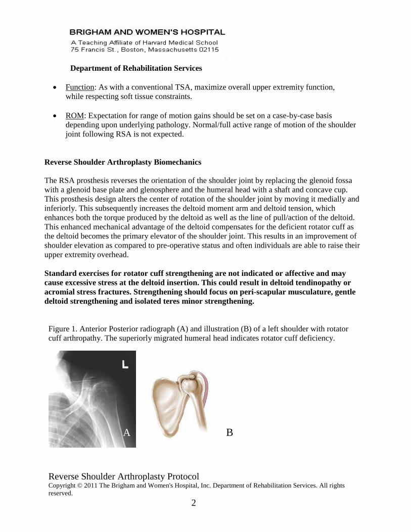
Figure 1. Anterior Posterior radiograph (A) and illustration (B) of a left shoulder with rotator
cuff arthropathy. The superiorly migrated humeral head indicates rotator cuff deficiency.
B A
Reverse Shoulder Arthroplasty Protocol Copyright © 2011 The Brigham and Women's Hospital, Inc. Department of Rehabilitation Services. All rights
reserved.
3
Department of Rehabilitation Services
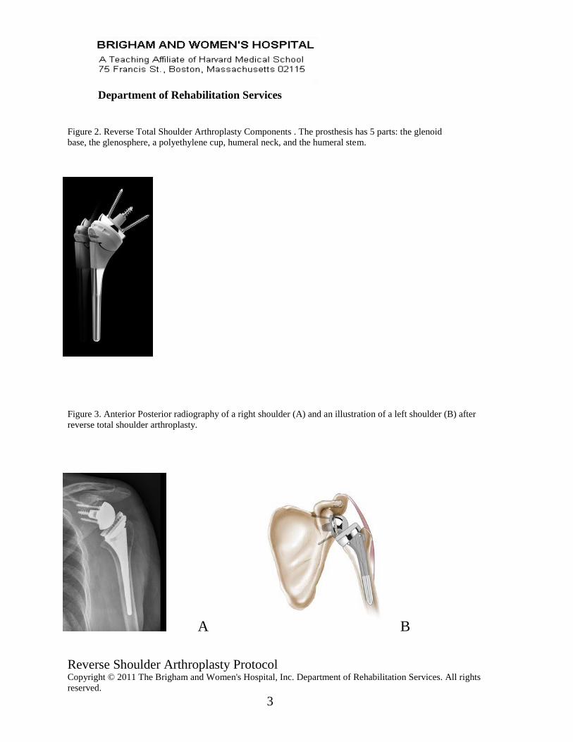
Figure 2. Reverse Total Shoulder Arthroplasty Components . The prosthesis has 5 parts: the glenoid
base, the glenosphere, a polyethylene cup, humeral neck, and the humeral stem.
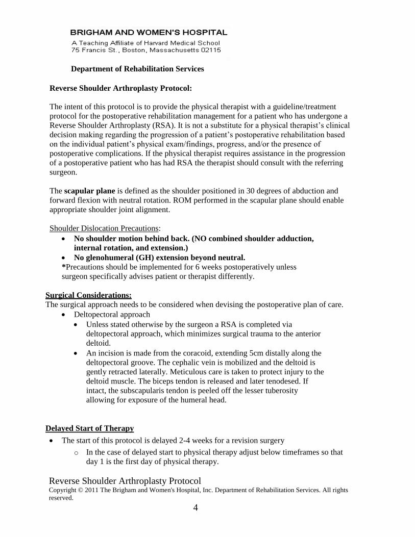
Figure 3. Anterior Posterior radiography of a right shoulder (A) and an illustration of a left shoulder (B) after
reverse total shoulder arthroplasty.
A B
Reverse Shoulder Arthroplasty Protocol Copyright © 2011 The Brigham and Women's Hospital, Inc. Department of Rehabilitation Services. All rights
reserved.
4
Department of Rehabilitation Services
Reverse Shoulder Arthroplasty Protocol:
The intent of this protocol is to provide the physical therapist with a guideline/treatment
protocol for the postoperative rehabilitation management for a patient who has undergone a
Reverse Shoulder Arthroplasty (RSA). It is not a substitute for a physical therapist’s clinical
decision making regarding the progression of a patient’s postoperative rehabilitation based
on the individual patient’s physical exam/findings, progress, and/or the presence of
postoperative complications. If the physical therapist requires assistance in the progression
of a postoperative patient who has had RSA the therapist should consult with the referring
surgeon.
The scapular plane is defined as the shoulder positioned in 30 degrees of abduction and
forward flexion with neutral rotation. ROM performed in the scapular plane should enable
appropriate shoulder joint alignment.
Shoulder Dislocation Precautions:
No shoulder motion behind back. (NO combined shoulder adduction,
internal rotation, and extension.)
No glenohumeral (GH) extension beyond neutral.
*Precautions should be implemented for 6 weeks postoperatively unless
surgeon specifically advises patient or therapist differently.
Surgical Considerations:
The surgical approach needs to be considered when devising the postoperative plan of care.
Deltopectoral approach
Unless stated otherwise by the surgeon a RSA is completed via
deltopectoral approach, which minimizes surgical trauma to the anterior
deltoid.
An incision is made from the coracoid, extending 5cm distally along the
deltopectoral groove. The cephalic vein is mobilized and the deltoid is
gently retracted laterally. Meticulous care is taken to protect injury to the
deltoid muscle. The biceps tendon is released and later tenodesed. If
intact, the subscapularis tendon is peeled off the lesser tuberosity
allowing for exposure of the humeral head.
Delayed Start of Therapy
The start of this protocol is delayed 2-4 weeks for a revision surgery
o In the case of delayed start to physical therapy adjust below timeframes so that
day 1 is the first day of physical therapy.
Reverse Shoulder Arthroplasty Protocol Copyright © 2011 The Brigham and Women's Hospital, Inc. Department of Rehabilitation Services. All rights
reserved.
5
Department of Rehabilitation Services
Progression to the next phase based on Clinical Criteria and Time Frames as
Appropriate.
Phase I- Immediate Post Surgical and Initiation of Range of Motion Phase
Day 1-6 weeks
Goals:
Patient and family independent with:
o Joint protection
o Passive range of motion (PROM)
o Assisting with putting on/taking off sling and clothing
o Assisting with home exercise program (HEP)
o Cryotherapy
Promote healing of soft tissue / maintain the integrity of the replaced joint.
Enhance PROM.
Restore active range of motion (AROM) of elbow/wrist/hand.
Independent with activities of daily living (ADL’s) with modifications.
Independent with bed mobility, transfers and ambulation or as per pre-admission
status.
Precautions:
Sling is worn for 2 weeks postoperatively and only removed for exercise and
bathing once able. The use of a sling maybe extended for a total of 6 weeks, if the
current RSA procedure is a revision surgery.
While lying supine, the distal humerus/elbow should be supported by a pillow or
towel roll to avoid shoulder extension. Patients should be advised to “always be
able to visualize their elbow while lying supine.”
No lifting of objects with operative extremity.
No supporting of body weight with involved extremity.
May shower at 3 days
Outside of showering, keep the incision clean and dry. No soaking/submerging
for 2 weeks; No whirlpool, fresh or salt water for 4 weeks.
Activity:
Day 1- 2 weeks
Insure patient is independent in bed mobility, transfers and ambulation
Insure proper sling fit/alignment/use. Active/Active Assisted ROM (AROM/AAROM) of cervical spine, elbow,

Reverse Shoulder Arthroplasty Protocol Copyright © 2011 The Brigham and Women's Hospital, Inc. Department of Rehabilitation Services. All rights
reserved.
6
Department of Rehabilitation Services
wrist, and hand.
Continuous cryotherapy for first 72 hours postoperatively, then apply as
needed for pain
2 weeks to 6 weeks:
Continue all exercises as above
Continue to maintain precautions of combined internal rotation and extension
(reaching behind back) as well as no lifting heavier than 1-2 lbs
Passive Range of Motion (PROM) typically begins at 2 weeks:
o Forward flexion and elevation in the scapular plane in supine to 120
degrees.
o ER in scapular plane to tolerance, respecting soft tissue constraints.
o No internal rotation
Gentle resisted exercise of elbow, wrist, and hand
May begin gentle pain free scapular pinches (Figure 1)
Begin to wean from sling at 2 weeks post-op
o May also begin pendulums (Figure 2a-2c) and pain free sub-max
deltoid isometrics at this time
o May use arm for pain free waist level activities
Active Assisted Range of Motion (AAROM) typically begins at 2 weeks
o Forward flexion and elevation in scapular plane in supine with
progression to lawn chair (figure 3) then to standing
o ER in scapular plane in supine
Active Range of motion (AROM) typically begins at 3 weeks
o Based on response to AAROM
o Progress from supine to lawn chair to standing
Manual Therapy
o Soft tissue massage upper trapezius, pec minor, scapular stabilizers
o Desensitization scar tissue
Criteria for progression to the next phase (Phase II):
Tolerates shoulder PROM, AAROM, AROM well with gradual
improvements.
Patient demonstrates the ability to isometrically activate all components of
the deltoid and periscapular musculature in the scapular plane.
Reverse Shoulder Arthroplasty Protocol Copyright © 2011 The Brigham and Women's Hospital, Inc. Department of Rehabilitation Services. All rights
reserved.
7
Department of Rehabilitation Services
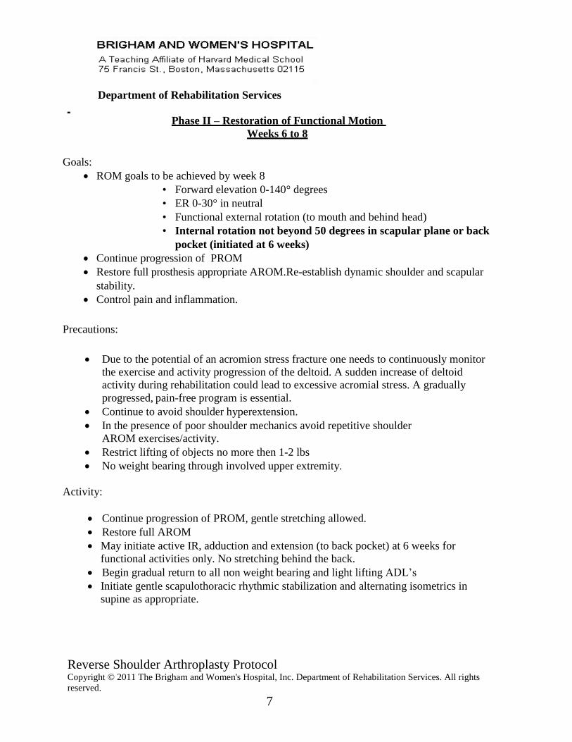
Phase II – Restoration of Functional Motion
Weeks 6 to 8
Goals:
ROM goals to be achieved by week 8
• Forward elevation 0-140° degrees
• ER 0-30° in neutral
• Functional external rotation (to mouth and behind head)
• Internal rotation not beyond 50 degrees in scapular plane or back
pocket (initiated at 6 weeks)
Continue progression of PROM
Restore full prosthesis appropriate AROM.Re-establish dynamic shoulder and scapular
stability.
Control pain and inflammation.
Precautions:
Due to the potential of an acromion stress fracture one needs to continuously monitor
the exercise and activity progression of the deltoid. A sudden increase of deltoid
activity during rehabilitation could lead to excessive acromial stress. A gradually
progressed, pain-free program is essential.
Continue to avoid shoulder hyperextension.
In the presence of poor shoulder mechanics avoid repetitive shoulder
AROM exercises/activity.
Restrict lifting of objects no more then 1-2 lbs
No weight bearing through involved upper extremity.
Activity:
Continue progression of PROM, gentle stretching allowed.
Restore full AROM
May initiate active IR, adduction and extension (to back pocket) at 6 weeks for
functional activities only. No stretching behind the back.
Begin gradual return to all non weight bearing and light lifting ADL’s
Initiate gentle scapulothoracic rhythmic stabilization and alternating isometrics in
supine as appropriate.
Reverse Shoulder Arthroplasty Protocol Copyright © 2011 The Brigham and Women's Hospital, Inc. Department of Rehabilitation Services. All rights
reserved.
8
Department of Rehabilitation Services
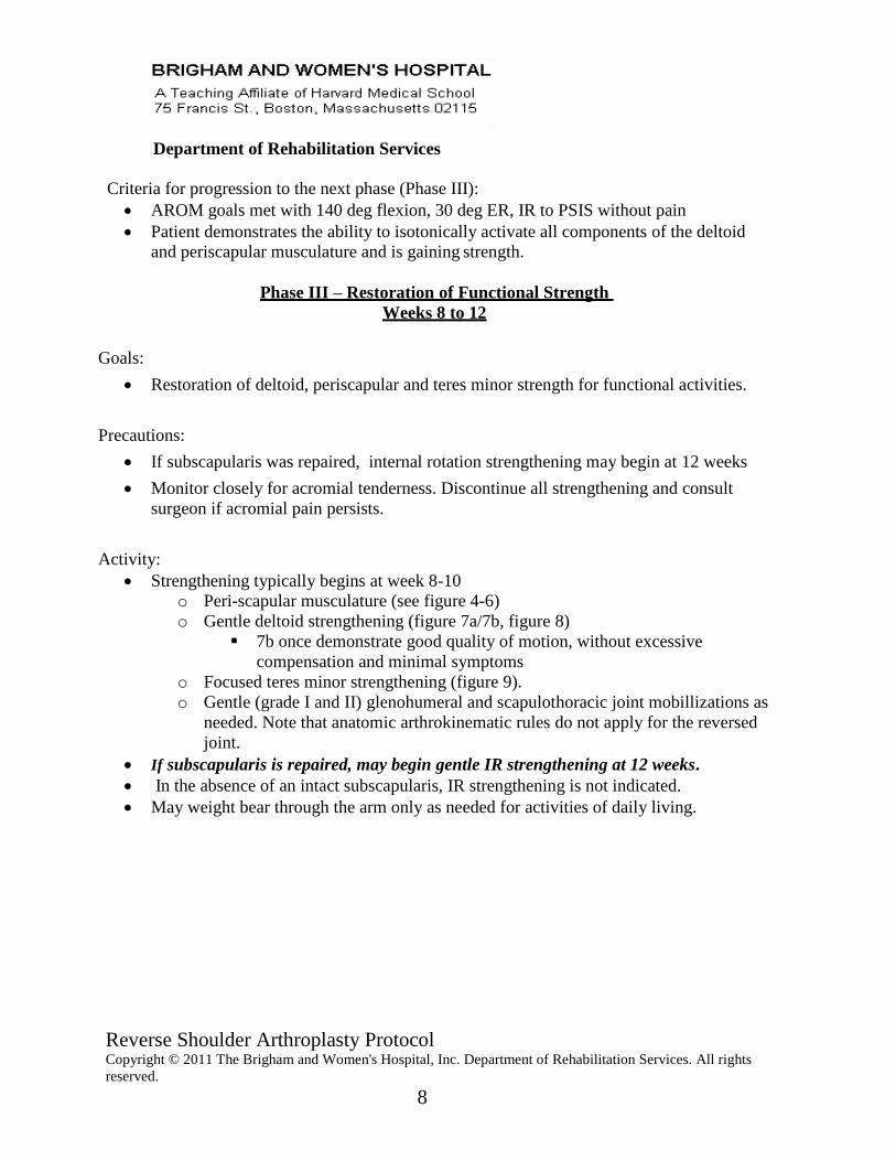
Criteria for progression to the next phase (Phase III):
AROM goals met with 140 deg flexion, 30 deg ER, IR to PSIS without pain
Patient demonstrates the ability to isotonically activate all components of the deltoid
and periscapular musculature and is gaining strength.
Phase III – Restoration of Functional Strength
Weeks 8 to 12
Goals:
Restoration of deltoid, periscapular and teres minor strength for functional activities.
Precautions:
If subscapularis was repaired, internal rotation strengthening may begin at 12 weeks
Monitor closely for acromial tenderness. Discontinue all strengthening and consult
surgeon if acromial pain persists.
Activity:
Strengthening typically begins at week 8-10
o Peri-scapular musculature (see figure 4-6)
o Gentle deltoid strengthening (figure 7a/7b, figure 8)
7b once demonstrate good quality of motion, without excessive
compensation and minimal symptoms
o Focused teres minor strengthening (figure 9).
o Gentle (grade I and II) glenohumeral and scapulothoracic joint mobillizations as
needed. Note that anatomic arthrokinematic rules do not apply for the reversed
joint.
If subscapularis is repaired, may begin gentle IR strengthening at 12 weeks.
In the absence of an intact subscapularis, IR strengthening is not indicated.
May weight bear through the arm only as needed for activities of daily living.
Reverse Shoulder Arthroplasty Protocol Copyright © 2011 The Brigham and Women's Hospital, Inc. Department of Rehabilitation Services. All rights
reserved.
9
Department of Rehabilitation Services
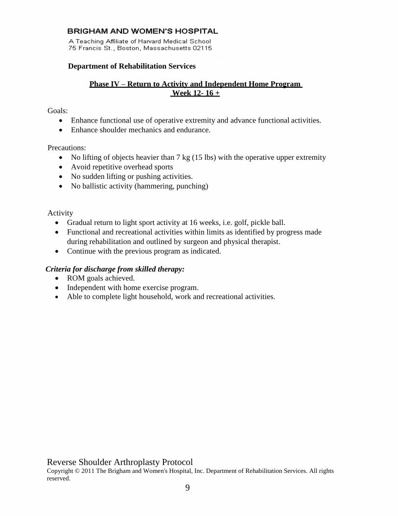
Phase IV – Return to Activity and Independent Home Program
Week 12- 16 +
Goals:
Enhance functional use of operative extremity and advance functional activities.
Enhance shoulder mechanics and endurance.
Precautions:
No lifting of objects heavier than 7 kg (15 lbs) with the operative upper extremity
Avoid repetitive overhead sports
No sudden lifting or pushing activities.
No ballistic activity (hammering, punching)
Activity
Gradual return to light sport activity at 16 weeks, i.e. golf, pickle ball.
Functional and recreational activities within limits as identified by progress made
during rehabilitation and outlined by surgeon and physical therapist.
Continue with the previous program as indicated.
Criteria for discharge from skilled therapy:
ROM goals achieved.
Independent with home exercise program.
Able to complete light household, work and recreational activities.
Reverse Shoulder Arthroplasty Protocol Copyright © 2011 The Brigham and Women's Hospital, Inc. Department of Rehabilitation Services. All rights
reserved.
10
Department of Rehabilitation Services
Authors: Reviewers:
Stephanie Boudreau, PT Janice McInnes, PT Ed Boudreau, PT Roya Ghazinouri, PT
Debbie Canoa, PT
Laurence D. Higgins, MD
Reg B.Wilcox III, PT
4/06
Reviewed: 6/1/06, 5/21/07, 6/13/07, 8/21/07, 9/8/07, 12/13/07
Updated by: Reviewed by:
Reg B. Wilcox III, PT Stephanie Boudreau, PT 3/11 Phil Kidd, PT
Laurence D. Higgins, MD
Updated by:
Eric Phillips, PT, DPT, OCS
Cristin Taylor, PA-C, PT
1/24/17
Reverse Shoulder Arthroplasty Protocol Copyright © 2011 The Brigham and Women's Hospital, Inc. Department of Rehabilitation Services. All rights
reserved.
11
Department of Rehabilitation Services
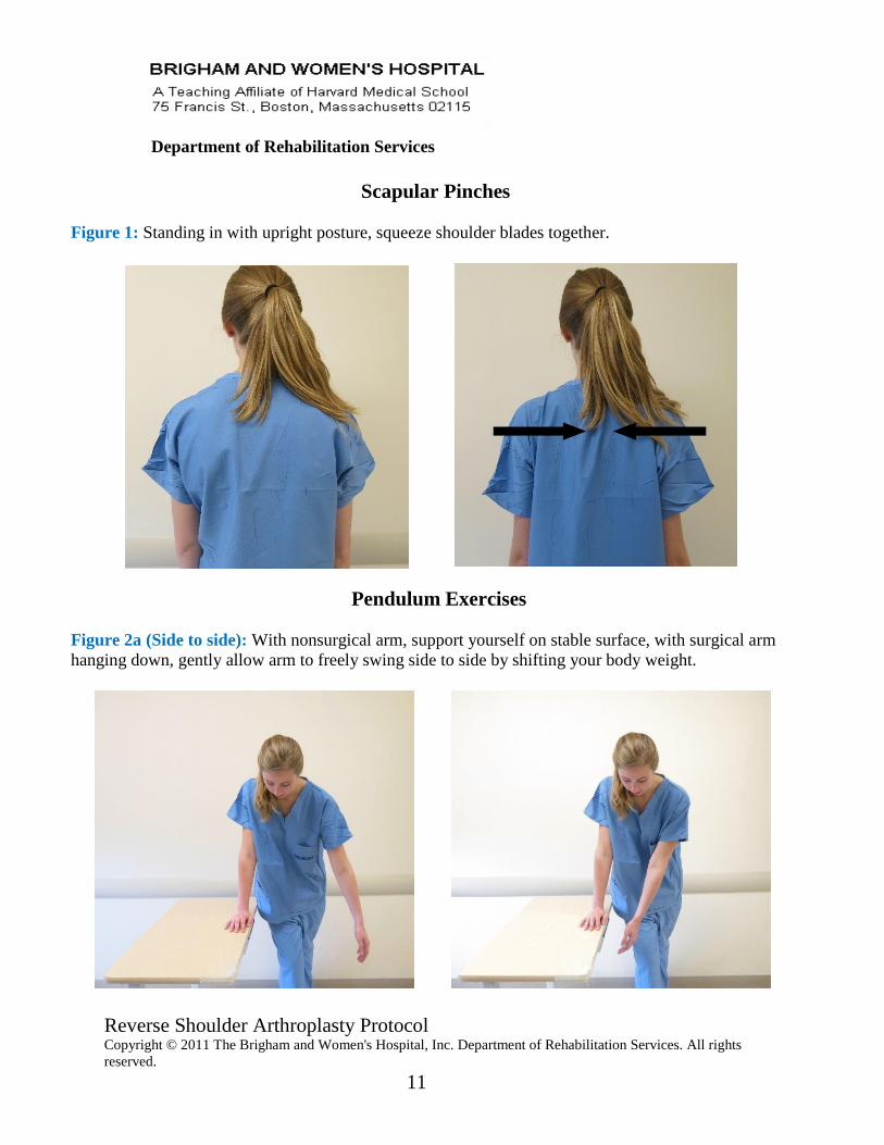
Scapular Pinches
Figure 1: Standing in with upright posture, squeeze shoulder blades together.
Pendulum Exercises
Figure 2a (Side to side): With nonsurgical arm, support yourself on stable surface, with surgical arm
hanging down, gently allow arm to freely swing side to side by shifting your body weight.
Reverse Shoulder Arthroplasty Protocol Copyright © 2011 The Brigham and Women's Hospital, Inc. Department of Rehabilitation Services. All rights
reserved.
12
Department of Rehabilitation Services
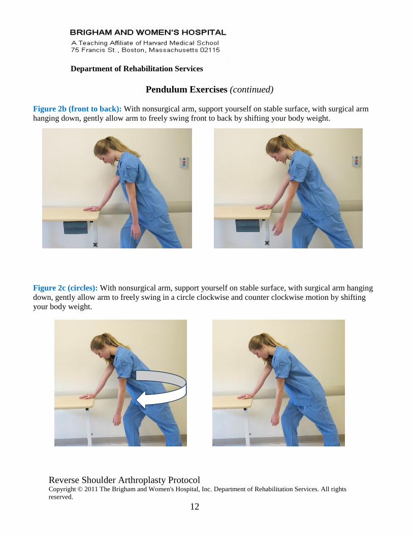
Pendulum Exercises (continued)
Figure 2b (front to back): With nonsurgical arm, support yourself on stable surface, with surgical arm
hanging down, gently allow arm to freely swing front to back by shifting your body weight.
Figure 2c (circles): With nonsurgical arm, support yourself on stable surface, with surgical arm hanging
down, gently allow arm to freely swing in a circle clockwise and counter clockwise motion by shifting
your body weight.
Reverse Shoulder Arthroplasty Protocol Copyright © 2011 The Brigham and Women's Hospital, Inc. Department of Rehabilitation Services. All rights
reserved.
13
Department of Rehabilitation Services
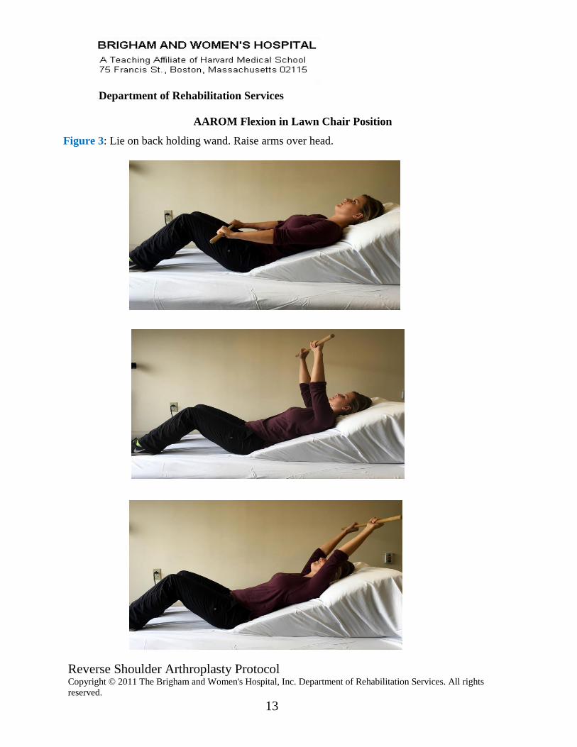
AAROM Flexion in Lawn Chair Position
Figure 3: Lie on back holding wand. Raise arms over head.
Reverse Shoulder Arthroplasty Protocol Copyright © 2011 The Brigham and Women's Hospital, Inc. Department of Rehabilitation Services. All rights
reserved.
14
Department of Rehabilitation Services
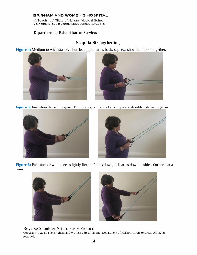
Scapula Strengthening
Figure 4: Medium to wide stance. Thumbs up, pull arms back, squeeze shoulder blades together.
Figure 5: Feet shoulder width apart. Thumbs up, pull arms back, squeeze shoulder blades together.
Figure 6: Face anchor with knees slightly flexed. Palms down, pull arms down to sides. One arm at a
time.
Reverse Shoulder Arthroplasty Protocol Copyright © 2011 The Brigham and Women's Hospital, Inc. Department of Rehabilitation Services. All rights
reserved.
15
Department of Rehabilitation Services
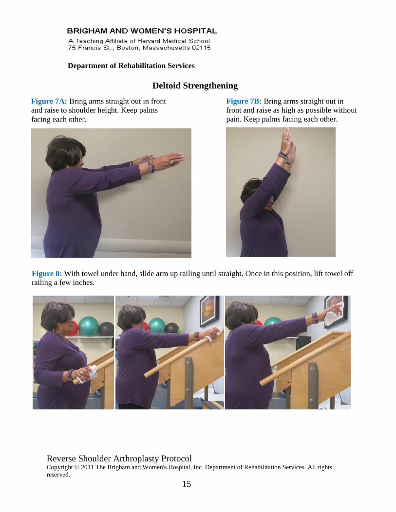
Deltoid Strengthening
Figure 8: With towel under hand, slide arm up railing until straight. Once in this position, lift towel off
railing a few inches.
Figure 7A: Bring arms straight out in front
and raise to shoulder height. Keep palms
facing each other.
Figure 7B: Bring arms straight out in
front and raise as high as possible without
pain. Keep palms facing each other.
Reverse Shoulder Arthroplasty Protocol Copyright © 2011 The Brigham and Women's Hospital, Inc. Department of Rehabilitation Services. All rights
reserved.
16
Department of Rehabilitation Services
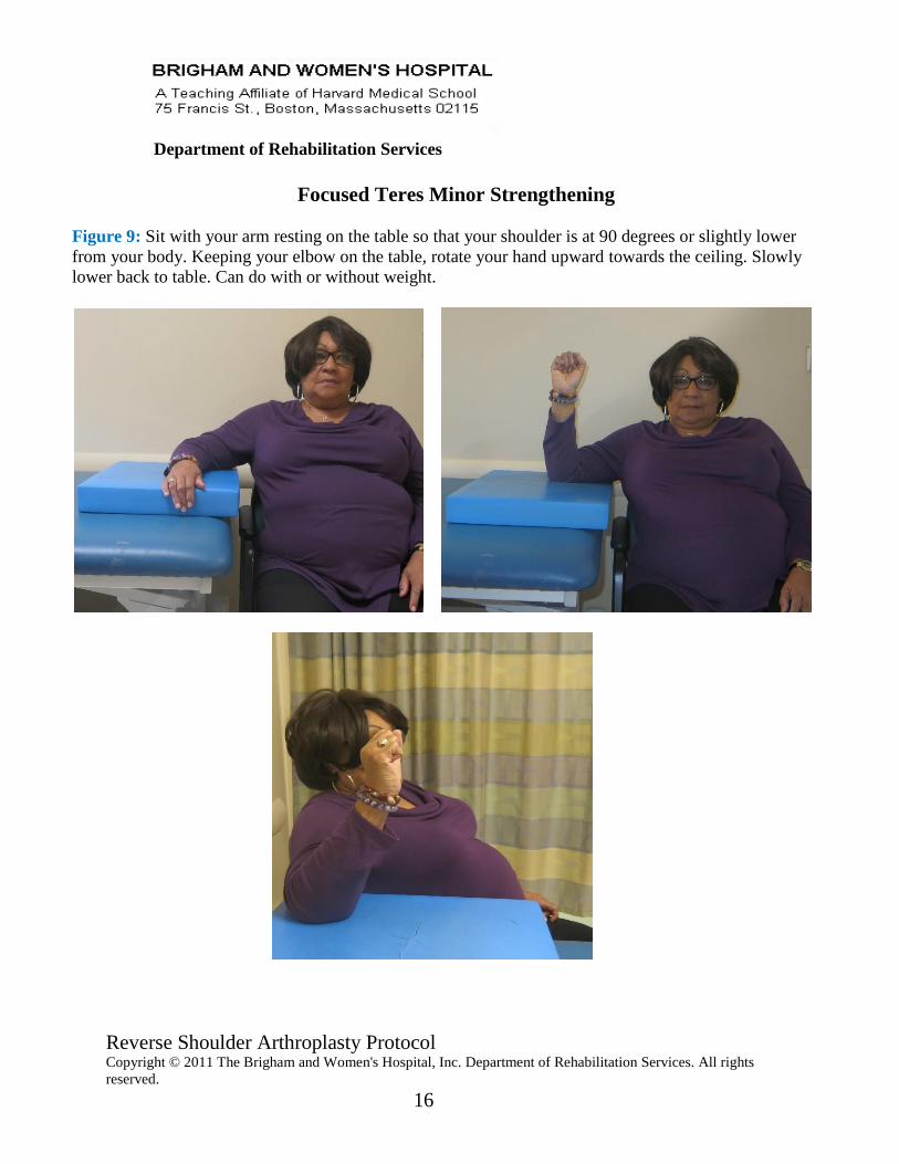
Focused Teres Minor Strengthening
Figure 9: Sit with your arm resting on the table so that your shoulder is at 90 degrees or slightly lower
from your body. Keeping your elbow on the table, rotate your hand upward towards the ceiling. Slowly
lower back to table. Can do with or without weight.