Embed Size (px)
Citation preview
DentistryKokovic et al., Dentistry 2012, 2:3
http://dx.doi.org/10.4172/2161-1122.1000124
Volume 2 • Issue 3 • 1000124DentistryISSN: 2161-1122 Dentistry, an open access journal
Open AccessCase Report
Implant Restoration in a Patient with Neurofibromatosis Type 1: A Case ReportVladimir Kokovic1*, Vojkan Lazic2, Milan Petrovic3, Drago Jelovac3 and Ana Todorovic4
1Associate Professor, Clinic of Oral Surgery, Faculty of Dentistry, University of Belgrade, Serbia2Associate Professor, Clinic of Prosthodontic, Faculty of Dentistry, University of Belgrade, Serbia3Clinic of Maxillofacial Surgery, Faculty of Dentistry, University of Belgrade, Serbia4Assitant Professor, Clinic of Prosthodontic, Faculty of Dentistry, University of Belgrade, Serbia
AbstractNeurofibromatosis type 1 (NF1), also known as von Recklinghausen’s disease, is a human genetic disorder. It is
probably the most commonly inherited disorder caused by a single gene.
This is a report of a 57-year-old man affected by NF1 who has severe atrophy of the jaws and extremely unsatisfactory anatomical conditions for conventional dental restauration. Radiographic and clinical evaluations showed inadequate quantity of bone for immediate implant rehabilitation. Delayed implant protocol was performed to obtain the correct bone volume and implants were inserted in the anterior parts of both jaws to support a prosthetic restoration.
*Corresponding author: Vladimir Kokovic, Associate Professor, Clinic of Oral Surgery, Faculty of Dentistry, University of Belgrade, Dr Subotica str. 4, 11000 Belgrade, Serbia, Tel: +38163324812, +381112685824; E-mail: [email protected]
Received December 19, 2011; Accepted March 06, 2012; Published March 09, 2012
Citation: Kokovic V, Lazic V, Petrovic M, Jelovac D, Todorovic A (2012) Implant Restoration in a Patient with Neurofibromatosis Type 1: A Case Report. Dentistry 2:124. doi:10.4172/2161-1122.1000124
Copyright: © 2012 Kokovic V, et al. This is an open-access article distributed under the terms of the Creative Commons Attribution License, which permits unrestricted use, distribution, and reproduction in any medium, provided the original author and source are credited.
Keywords: Neurofibromatosis type 1; Von Recklinghausen’s disease; Implant supported dentures
IntroductionNeurofibromatosis type 1 (NF1) is also known as von
Recklinghausen’s disease which results from mutation on the long arm of chromosome 17 [1]. This autosomal dominantly inherited disease is affecting approximately 1 in 3500 people [2]. The incidence of NF1 is about 97% of all cases of NF. Type 2 (NF2) is less frequent form and is associated with a mutation on the long arm of chromosome 22 [3,4].
The diagnosis of NF1 is based on clinical findings. Besides neurofibromas of certain skeletal malformations, café-au-lait spots and eye problems are diagnostic findings of NF1 [5]. Data from literature presented that the incidence of head and neck involvements with neurofibromatosis ranges from 1% to 22% [6,7]. Von Recklinghausen’s neurofibromatosis is mostly associated with extensive craniofacial manifestation which are usually resulting in facial asymmetry [8]. The plexiforme type is the mostly presented in oral and maxillofacial region. These tumors invade both the cheek and oral cavity. The presence of impacted and missing teeth in lower jaw as well as overgrowth of alveolar ridge are also oral manifestation of NF1 [5,9]. The radiographic manifestations of NF1 in maxillofacial region often include increase in bone density, hypoplasia of the mandibular body and ramus, abnormal coronoid process and presence of fat-like density tissue adjacent to the mandibular deformities [9,10]. The unilateral gingival hyperplasia has been described as quite common oral lesion in NF1 [11]. Taking into account of all the previously mentioned oral manifestations, prosthetic treatment of patients suffering from this disease requires a planning of therapy and multidisciplinary approach. The aim of this paper is to describe prosthetic restoration in a patient with neurofibromatosis type 1.
Case ReportIn the September 2007, a 57-year old male patient was referred to
the Clinic of Oral surgery by his dentist for rehabilitation with dental implants. In the order to restore intraoral masses of the right cheek, this patient was previously surgically multiply treated at the clinic of maxillofacial surgery. Considering the nature of disorder and lack of teeth, the patient was not able to adequately perform functions of chewing and deglutition. Furthermore, prosthetic rehabilitation with
classical protheses would not overcome the problem of proper stability during chewing function.
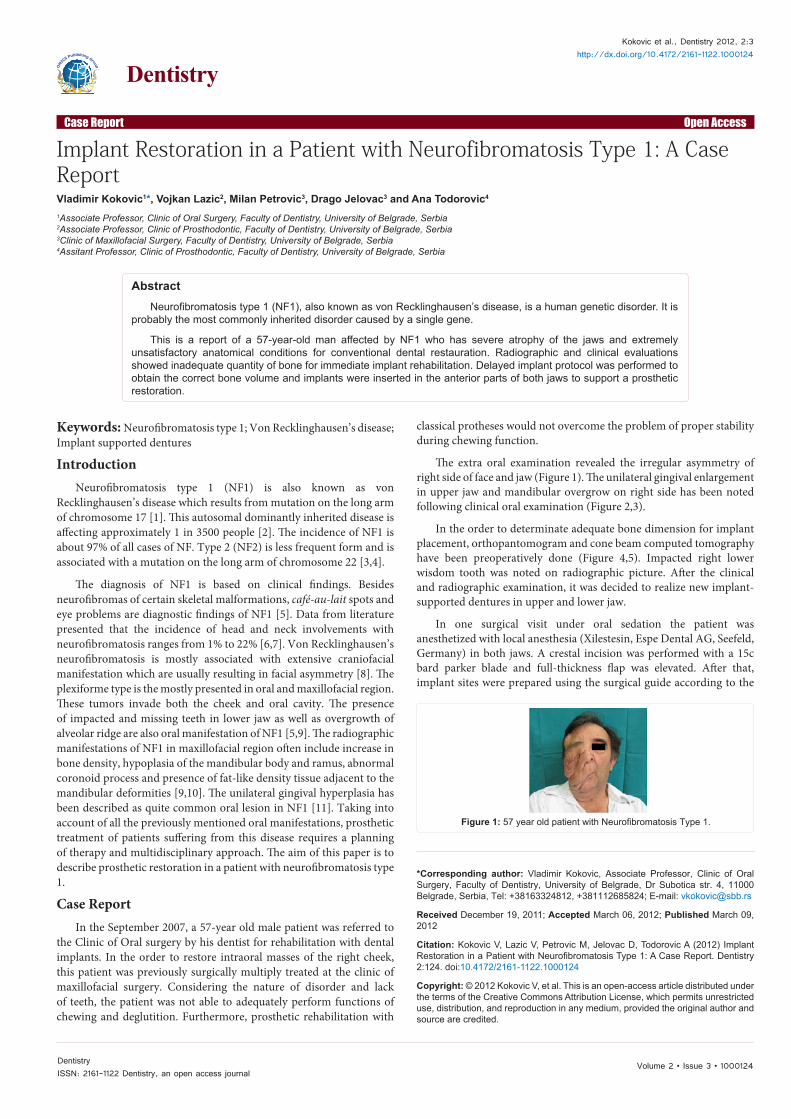
The extra oral examination revealed the irregular asymmetry of right side of face and jaw (Figure 1). The unilateral gingival enlargement in upper jaw and mandibular overgrow on right side has been noted following clinical oral examination (Figure 2,3).
In the order to determinate adequate bone dimension for implant placement, orthopantomogram and cone beam computed tomography have been preoperatively done (Figure 4,5). Impacted right lower wisdom tooth was noted on radiographic picture. After the clinical and radiographic examination, it was decided to realize new implant-supported dentures in upper and lower jaw.
In one surgical visit under oral sedation the patient was anesthetized with local anesthesia (Xilestesin, Espe Dental AG, Seefeld, Germany) in both jaws. A crestal incision was performed with a 15c bard parker blade and full-thickness flap was elevated. After that, implant sites were prepared using the surgical guide according to the
Figure 1: 57 year old patient with Neurofibromatosis Type 1.

Volume 2 • Issue 3 • 1000124DentistryISSN: 2161-1122 Dentistry, an open access journal
Citation: Kokovic V, Lazic V, Petrovic M, Jelovac D, Todorovic A (2012) Implant Restoration in a Patient with Neurofibromatosis Type 1: A Case Report. Dentistry 2:124. doi:10.4172/2161-1122.1000124
Page 2 of 3
procedure manufacture’s recommendations (Institute Straumann AG, Waldemburg, Switzerland). Four Straumann® SLActive implants were placed in the front area of upper jaw (4 Regular Neck 3.3ø x 12mm) and other four implants were inserted bilaterally in the premolar area of the mandible (3 Regular Neck 4.1ø and 1 Regular Neck 3.3ø x 10mm) . Implants were covered with closing screws and soft tissues were sutured. The patient was subjected to standard antibiotic (amoxicillin 1 gr twice per day for 5 days) and non-steroidal anti-inflammatory (nimesulid, 100mg twice per day for 3 days) therapies. Resonance frequency analysis (RFA) was used to determinate value of primary implant stability and for each implant it was > 60ISQ (Implant Stability Quotient). ISQ measurements were carried out in two horizontal directions: perpendicularly and parallel to the arch of jaw ovde idu slike ugradnje i merenja RFA. The maintenance of oral hygiene was improved with 0.12% chlorhexidine mouth rinses for 2 weeks.
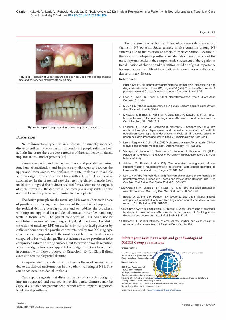
Due to inability to provide adequate retention of denture prostheses, during healing period patient didn’t use temporary prosthesis. Consequently, osseointegration of the implants and proper healing of soft tissues were allowed. Three months after surgery, uncovering procedure has been done and two weeks was necessary period for soft tissue maturation, prior to final impressions. Implant stability has been measured to assess readiness for loading protocol. Following healing period the patient has been sent to clinic of prosthetic for definitive restoration. The rigid retentive bars with metal clips tissues were fabricated to support the removable partial dentures (RPD) in upper and overdenture (OVD) in lower jaw (Figure 6). Exception was made in the upper jaw with using two retention systems for RPDs. Retention on right side has been provided with bar clip and on left with solitary ball attachments. Also, it was partially supported with few remained teeth in anterior area (Figure 7) [5,7].
Impression copings were placed on implants to achieve precise transfer of the intraoral location of the implants (position in situ) to the similar position on the laboratory cast. Heavier body impression material was used and impression was sent to dental laboratory.
The prefabricated plastic bars were attached parallel to abutments by surveyor and the abutments were shortened to proper vertical dimension. These plastic patterns and cast from Co-Cr alloy were burned out. After that procedure the casted bars were attached to implant body analogs on the models. The next step was to construct the framework for both prostheses.
An implant retained partial denture was designed in upper jaw, while in lower jaw we designed an overlay implant supported prostheses. After finishing the partial denture frameworks, their proper seat was checked out and the upper partial denture framework was physiologically adjusted. The metal matrices were positioned on implant supported bars and “O” ring type attachments which were attached by acrylic resin to retentive places of both frameworks on their basal sides. Due to presenting of unilateral gingival enlargement in upper jaw, short arch has been made on right side of partial denture. Conventional prosthodontic methods are followed to complete the prostheses. Delivery and adjustment are accomplished following acceptable prosthodontic guidelines (Figure 8).
In the follow-up period of two years checking control has been performed every three months.
Figure 2: Unsatisfactory clinical conditions in the upper jaw, buccal soft tissue masses of the right side of the cheek.
Figure 3: Limited anatomical conditions for implantation in the mandible, extensive soft tissue masses of the right cheek, narrow area of attached gingiva. Limited anatomical conditions for implantation in the mandible, extensive soft tissue masses of the right cheek, narrow area of attached gingiva.
Figure 4: Preoperative panoramic radiograph of the patient with 2D data planning for implantation; Characteristic finding- increase in bone density, enlarged mandibular foramen, lateral bowing of the mandibular ramus, increase in dimensions of the coronoid notch, and a decrease in the mandibular angle.
Figure 5: CT 3D reconstruction of the maxilla and mandible, enlarged mandibular-mental foramen.
Figure 6: The rigid retentive bars on lower jaw cast.
Volume 2 • Issue 3 • 1000124DentistryISSN: 2161-1122 Dentistry, an open access journal
Citation: Kokovic V, Lazic V, Petrovic M, Jelovac D, Todorovic A (2012) Implant Restoration in a Patient with Neurofibromatosis Type 1: A Case Report. Dentistry 2:124. doi:10.4172/2161-1122.1000124
Page 3 of 3
DiscussionNeurofibromatosis type 1 is an autosomal dominantly inherited
disease, significantly reducing the life comfort of people suffering from it. In the literature, there are very rare cases of the treatment with dental implants in this kind of patients [12].
Removable partial and overlay dentures could provide the desired functions of mastication and improves any discrepancy between the upper and lower arches. We preferred to unite implants in mandible with two rigid, precision – fitted bars, with retentive elements were attached to. In the presented case the retentive elements made from metal were designed also to direct occlusal forces down to the long axis of implant fixtures. The denture in the lower jaw is very stable and the occlusal forces are primarily supported by the implants.
The design principle for the maxillary RPD was to shorten the base of prostheses on the right side because of the insufficient support of the residual denture bearing surface and to stabilize the prosthesis with implant supported bar and dental connector over few remaining teeth in frontal area. The palatal connector of RPD could not be established because of remaining soft palatal structures. The distal extension of maxillary RPD on the left side was provided posterior by sufficient bone were the prostheses was retained by two “O” ring type attachments on implants with the most favorable stress distribution as compared to bar – clip design. These attachments allow prostheses to be compressed into the bearing surfaces, but to provide enough retention when dislodging forces are applied. The design principles have much in common with those proposed by Kratochvil [13] for Class II distal extension removable partial denture.
Adequate retention of denture prosthesis is the most current factor due to the skeletal malformation in the patients suffering of NF1. This can be achieved with dental implants.
Case report suggests that distal implants and a special design of implant supported and retained removable partial dentures may be especially suitable for patients who cannot afford implant-supported fixed dental prostheses.
Figure 7: Retention of upper denture has been provided with bar clip on right side and solitary ball attachments on left side.
Figure 8: Implant supported dentures on upper and lower jaw.
The disfigurement of body and face often causes depression and shame in NF patients. Social anxiety is also common among NF sufferers due to the reaction of others to their condition. Because of these reasons, adequate prosthetic rehabilitation could be one of the most important tasks in the comprehensive treatment of these patients. Rehabilitation of chewing and deglutition could be of great importance because the quality of life of these patients is sometimes very disturbed due to primary disease. References
1. Huson SM (1994) Neurofiromatosis: historical perspective, classification and diagnostic criteria. In : Huson SM, Hughes RA (eds). The Neurofibromatosis. A pathogenetic and Clinical Overview. London: Chapman & Hall 1-22.
2. Boyd KP, Korf BR, Theos A (2009) Neurofibromatosis type 1. J Am Acad Dermatol 61: 1-14.
3. Mulvihill JJ (1986) Neurofibromatosis. A genetic epidemiologist’s point of view. Ann N Y Acad Sci 486: 38-44.
4. Miyawaki T, Billings B, Har-Shai Y, Agbenorku P, Kokuba E, et al. (2007) Multicenter study of wound healing in neurofibromatosis and neurofibroma. J Craniofac Surg 18: 1008-1011.
5. Friedrich RE, Giese M, Schmelzle R, Mautner VF, Scheuer HA (2003) Jaw malformations plus displacement and numerical aberrations of teeth in neurofibromatosis type 1: a descriptive analysis of 48 patients based on panoramic radiographs and oral findings. J Craniomaxillofac Surg 31: 1-9.
6. Lee V, Ragge NK, Collin JR (2004) Orbitotemporal neurofibromatosis. Clinical features and surgical management. Ophthalmology 111: 382-388.
7. Visnapuu V, Peltonen S, Tammisalo T, Peltonen J, Happonen RP (2011) Radiographic Findings in the Jaws of Patients With Neurofibromatosis 1. J Oral Maxillofac Surg.
8. Adkins JC, Ravitch MM (1977) The operative management of von Recklinghausen’s neurofibromatosis in children, with special reference to lesions of the head and neck. Surgery 82: 342-348.
9. Lee L, Yan YH, Pharoah MJ (1996) Radiographic features of the mandible in neurofibromatosis: a report of 10 cases and review of the literature. Oral Surg Oral Med Oral Pathol Oral Radiol Endod 81: 361-367.
10. D’Ambrosio JA, Langlais RP, Young RS (1988) Jaw and skull changes in neurofibromatosis. Oral Surg Oral Med Oral Pathol 66: 391-396.
11. Bekisz O, Darimont F, Rompen EH (2000) Diffuse but unilateral gingival enlargement associated with von Recklinghausen neurofibromatosis: a case report. J Clin Periodontol 27: 361-365.
12. Ey-Chmielewska H, Sobolewska E, Fraczak B (2007) Description of prosthetic treatment in case of neurofibromatosis in the course of Recklinghausen disease. Case course. Ann Acad Med Stetin 53: 83-86.
13. Kratochvil FJ (1963) Influence of occlusal rest position and clasp design on movement of abutment teeth. J Prosthet Dent 13: 114-124.
Submit your next manuscript and get advantages of OMICS Group submissionsUnique features:
User friendly/feasible website-translation of your paper to 50 world’s leading languagesAudio Version of published paperDigital articles to share and explore
Special features:
200 Open Access Journals15,000 editorial team21 days rapid review processQuality and quick editorial, review and publication processingIndexing at PubMed (partial), Scopus, DOAJ, EBSCO, Index Copernicus and Google Scholar etcSharing Option: Social Networking EnabledAuthors, Reviewers and Editors rewarded with online Scientific CreditsBetter discount for your subsequent articles
Submit your manuscript at: http://www.omicsonline.org/submission