Embed Size (px)
Citation preview
Advances in Materials Research, Vol. 1, No. 1 (2012) 31-35 31
Dental arch wires with tooth-like color
Sinn-wen Chen*, Hsin-jay Wu, Chih-hao Liu, Yuan-chun Chien and Chih-chang Hu
Department of Chemical Engineering, National Tsing Hua University,
#101, Sec. 2, Kuang-Fu Road, Hsin-Chu 300, Taiwan
(Received July 1, 2011, Revised February 26, 2012, Accepted February 27, 2012)
Abstract. Unique tooth-like (milky white) color β-Ti dental arch wires are prepared by anodization in a1M H2SO4 electrolyte at 30oC and 30 V for 88 min and 40 s. Aggregates are formed on these surfaces ofthe anodized wires with tooth-like colors, and the results are different from those of the anodized wireswith monochromatic colors which have smooth oxide surfaces. Similar to the monochromatic wires, thecomposition of the oxide layer is TiO2-x and the x approaches zero at the outer layer. But different fromthe amorphous structure observed in monochromatic wires, the oxide layers are partially crystallized withan anatase structure. The milky white colors result from the rough and crystalized oxide layers, not by theinterference effect as observed in monochromatic wires.
Keywords: orthodontic materials; surface properties; anodization; tooth color
1. Introduction
Beta titanium, nickel-titanium and stainless steel dental arch wires are often used for orthodontic
treatments (Kusy and Stush 1987, Whitters et al. 1999, Kwon et al. 2005, Walker et al. 2007).
Colorization of dental arch wires has been reported previously (Yang et al. 2006, Wu et al. 2009),
with the different colors of anodized wires resulting from optical interference (Yang et al. 2006, Wu
et al. 2009, Aladjem 1973, Gaul 1993, Hrapovic et al. 2001). Anodization is a process of forming
oxide on the metallic anode (Aladjem 1973). It has been found for a long time that metals can be
colorized with anodization (Yang et al. 2006, Wu et al. 2009, Aladjem 1973, Gaul 1993, Hrapovic
et al. 2001). Primarily for cosmetic reasons, arch wires with tooth-like colors are needed. This study
investigates tooth-like (milky white) colors dental arch wires prepared by anodization (Chen et al.
2009). Since the interference effect does not produce milky white colors, the surfaces of the
anodized wires are analyzed and the colorization mechanisms are illustrated.
2. Experimental procedures
β-Ti dental arch wire (#101-011, Ortho-Organizers, Carlsbad, CA, USA) was first polished with
1 µm alumina slurry, and cleaned with 0.2 M HCl solution, acetone and de-ionized (DI) water. The
wire was then anodized with direct current (DC) in a 1 M H2SO4 electrolyte, and with Pt as the
*Corresponding author, Professor, E-mail: [email protected]
32 Sinn-wen Chen, Hsin-jay Wu, Chih-hao Liu, Yuan-chun Chien and Chih-chang Hu
cathode (Yang et al. 2006, Wu et al. 2009). The anodization was carried out at a constant voltage
and constant temperature with different lengths of anodization time. Optical microscope and SEM
(scanning electron microscopy, Hitachi, S-2500, Japan) were used to examine the microstructure and
surface morphology of the specimens. XPS (X-ray Photoelectron Spectroscopy, ESCA PHI 1600,
USA) was used to determine the chemical states of the elements in the oxide layer. Thickness of the
oxide layer was measured based on the bright-field images obtained by TEM (transmission electron
microscopy, Philips, TECNAI 20). The crystal structure of the oxide layer was characterized by
TEM and GIXRD (grazing incidence X-ray diffraction, MAC Science, MXP18, Japan).
3. Results and discussion
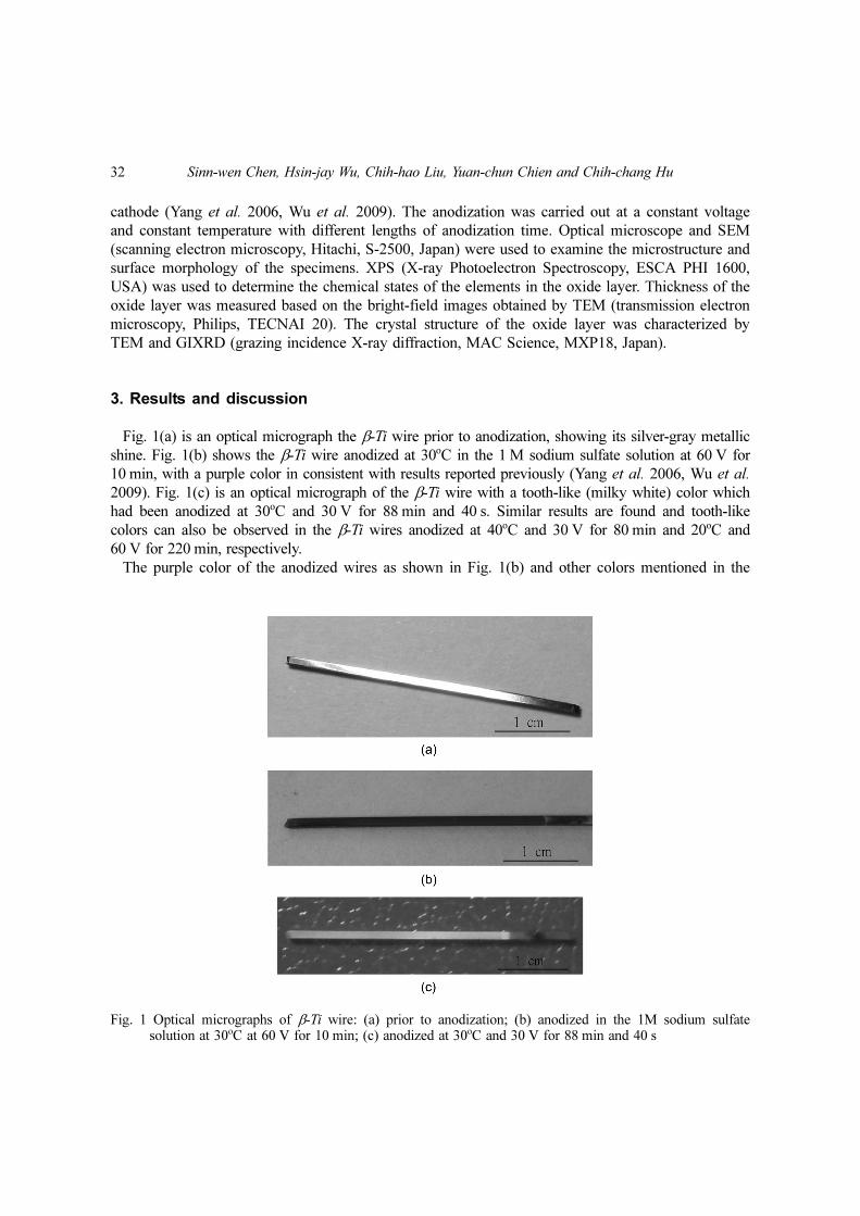
Fig. 1(a) is an optical micrograph the β-Ti wire prior to anodization, showing its silver-gray metallic
shine. Fig. 1(b) shows the β-Ti wire anodized at 30oC in the 1 M sodium sulfate solution at 60 V for
10 min, with a purple color in consistent with results reported previously (Yang et al. 2006, Wu et al.
2009). Fig. 1(c) is an optical micrograph of the β-Ti wire with a tooth-like (milky white) color which
had been anodized at 30oC and 30 V for 88 min and 40 s. Similar results are found and tooth-like
colors can also be observed in the β-Ti wires anodized at 40oC and 30 V for 80 min and 20oC and
60 V for 220 min, respectively.
The purple color of the anodized wires as shown in Fig. 1(b) and other colors mentioned in the
Fig. 1 Optical micrographs of β-Ti wire: (a) prior to anodization; (b) anodized in the 1M sodium sulfatesolution at 30oC at 60 V for 10 min; (c) anodized at 30oC and 30 V for 88 min and 40 s
Dental arch wires with tooth-like color 33
literature (Yang et al. 2006, Wu et al. 2009) are caused by the thin film interference effect. Through
the constructive and destructive interference effects, the anodized wires thus display monochromatic
colors (Yang et al. 2006, Wu et al. 2009). However, wires with a tooth-like (milky white) color,
caused by a mixture of light with different wave lengths, cannot result from the interference effect.
The β-Ti wire with a tooth-like color from anodization at 30oC and 30 V for 88 min and 40 s is then
further analyzed to understand the colorization mechanisms.
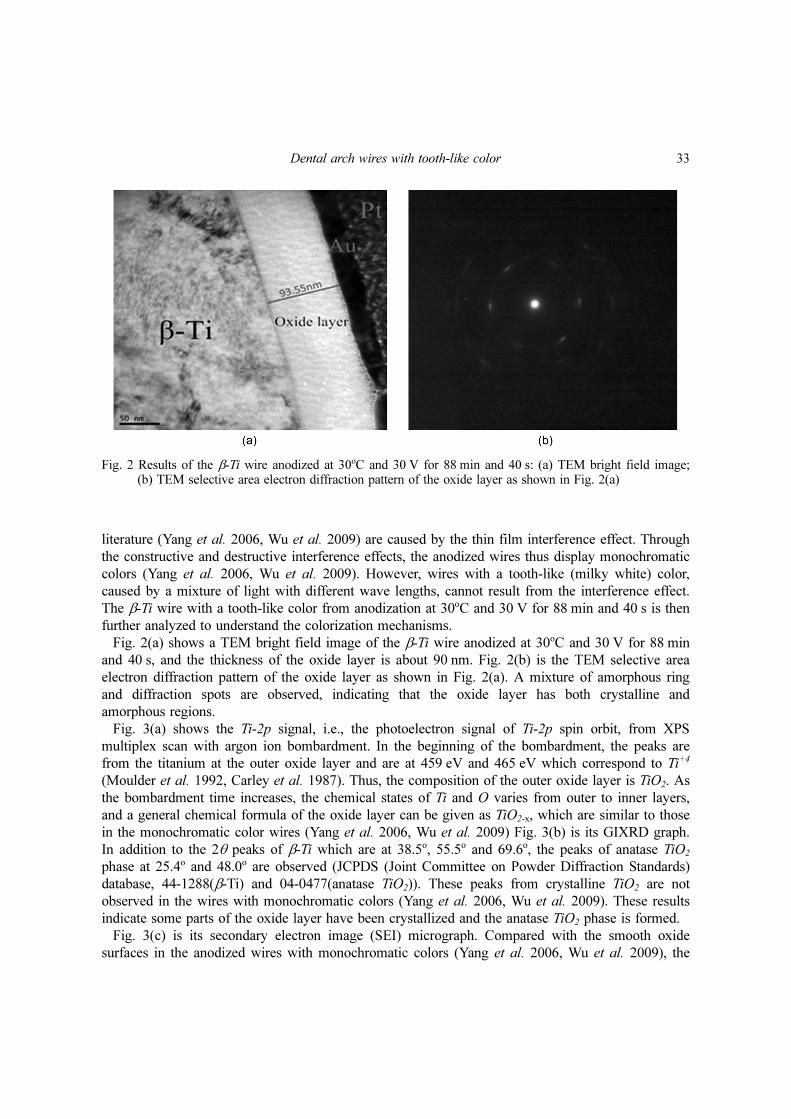
Fig. 2(a) shows a TEM bright field image of the β-Ti wire anodized at 30oC and 30 V for 88 min
and 40 s, and the thickness of the oxide layer is about 90 nm. Fig. 2(b) is the TEM selective area
electron diffraction pattern of the oxide layer as shown in Fig. 2(a). A mixture of amorphous ring
and diffraction spots are observed, indicating that the oxide layer has both crystalline and
amorphous regions.
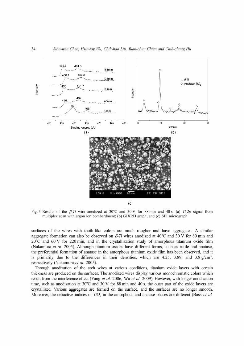
Fig. 3(a) shows the Ti-2p signal, i.e., the photoelectron signal of Ti-2p spin orbit, from XPS
multiplex scan with argon ion bombardment. In the beginning of the bombardment, the peaks are
from the titanium at the outer oxide layer and are at 459 eV and 465 eV which correspond to Ti+4
(Moulder et al. 1992, Carley et al. 1987). Thus, the composition of the outer oxide layer is TiO2. As
the bombardment time increases, the chemical states of Ti and O varies from outer to inner layers,
and a general chemical formula of the oxide layer can be given as TiO2-x, which are similar to those
in the monochromatic color wires (Yang et al. 2006, Wu et al. 2009) Fig. 3(b) is its GIXRD graph.
In addition to the 2θ peaks of β-Ti which are at 38.5o, 55.5o and 69.6o, the peaks of anatase TiO2
phase at 25.4o and 48.0o are observed (JCPDS (Joint Committee on Powder Diffraction Standards)
database, 44-1288(β-Ti) and 04-0477(anatase TiO2)). These peaks from crystalline TiO2 are not
observed in the wires with monochromatic colors (Yang et al. 2006, Wu et al. 2009). These results
indicate some parts of the oxide layer have been crystallized and the anatase TiO2 phase is formed.
Fig. 3(c) is its secondary electron image (SEI) micrograph. Compared with the smooth oxide
surfaces in the anodized wires with monochromatic colors (Yang et al. 2006, Wu et al. 2009), the
Fig. 2 Results of the β-Ti wire anodized at 30oC and 30 V for 88 min and 40 s: (a) TEM bright field image;(b) TEM selective area electron diffraction pattern of the oxide layer as shown in Fig. 2(a)
34 Sinn-wen Chen, Hsin-jay Wu, Chih-hao Liu, Yuan-chun Chien and Chih-chang Hu
surfaces of the wires with tooth-like colors are much rougher and have aggregates. A similar
aggregate formation can also be observed on β-Ti wires anodized at 40oC and 30 V for 80 min and
20oC and 60 V for 220 min, and in the crystallization study of amorphous titanium oxide film
(Nakamura et al. 2005). Although titanium oxides have different forms, such as rutile and anatase,
the preferential formation of anatase in the amorphous titanium oxide film has been observed, and it
is primarily due to the differences in their densities, which are 4.25, 3.89, and 3.8 g/cm3,
respectively (Nakamura et al. 2005).
Through anodization of the arch wires at various conditions, titanium oxide layers with certain
thickness are produced on the surfaces. The anodized wires display various monochromatic colors which
result from the interference effect (Yang et al. 2006, Wu et al. 2009). However, with longer anodization
time, such as anodization at 30oC and 30 V for 88 min and 40 s, the outer part of the oxide layers are
crystallized. Various aggregates are formed on the surface, and the surfaces are no longer smooth.
Moreover, the refractive indices of TiO2 in the amorphous and anatase phases are different (Bass et al.
Fig. 3 Results of the β-Ti wire anodized at 30oC and 30 V for 88 min and 40 s: (a) Ti-2p signal frommultiplex scan with argon ion bombardment; (b) GIXRD graph; and (c) SEI micrograph
Dental arch wires with tooth-like color 35
2010). The multi-phase and rough surfaces weaken the interference effect and enhance light scattering,
and tooth-like (milky white) colors are thus formed.
4. Conclusions
The β-Ti wires display monochromatic colors by anodization, and the colorization is caused by
interference effect of the smooth and amorphous oxide layer. The amorphous oxide layer begins to
crystallize with longer anodization time, and aggregates are formed on the surface. Because of the
extensive light scattering, the interference effect is no longer the dominating optical factor, and the
monochromatic colors of the anodized wires change to the tooth-like (milky white) colors.
Acknowledgments
This work was supported by the National Science Council of Taiwan (NSC96- 2218- E-007-012).
References
Aladjem, A. (1973), “Anodic oxidation of titanium and its alloys”, J. Mater. Sci., 8(5), 688-704.Bass, M., DeCusatis, C., Li, G., Mahajan, V.N. and Stryland, E.V. (2010), Handbook of Optics, Vol. IV: Optical
properties of materials, nonlinear optics and quantum optics, 3rd Edition, McGraw-Hill, New York.Carley, A.F., Chalker, P.R., Riviere, J.C. and Roberts, M.W. (1987), “The identification and characterization of
mixed oxidation states at oxidized titanium surfaces by analysis of X-ray photoelectron spectra”, J. Chem. Soc.Faraday Trans., 83, 351-370.
Chen, S.W., Wu, H.J., Huang, L.L. and Liou J.W.E. (2009), Metallic dental devices with tooth colors and theirpreparation methods, United Statues Patent Application Publication, Alexandria, NV.
Gaul, E. (1993), “Coloring titanium and related metals by electrochemical oxidation”, J. Chem. Educ., 70(3),176-178.
Hrapovic, S., Luan, B.L., D'Amours, M., Vatankhah, G. and Jerkiewicz, G. (2001), “Morphology, chemicalcomposition, and electrochemical characteristics of colored”, Langmuir, 17(10), 3051-3060.
JCPDS (Joint Committee on Powder Diffraction Standards) database, 44-1288(β-Ti) and 04-0477(anatase TiO2).Kusy, R.P. and Stush, A.M. (1987), “Geometric and material parameters of a nickel-titanium and a beta titanium
orthodontic arch wire alloy”, Dent. Mater., 3(4), 207-217.Kwon, Y.H., Seol, H.J., Kim, H.I., Hwang, K.J., Lee, S.G. and Kim, K.H. (2005), “Effect of acid fluoride
solution on beta titanium alloy wire”, J. Biomed. Mater. Res., B: Appl. Biomater., 73(2), 285-290.Moulder, J.F., Stickle, W.F., Sobol, P.E. and Bomben, K.D. (1992), Handbook of X-ray Photoelectron
Spectroscopy, Perkin-Elmer Corp., USA.Nakamura, T., Ichitsubo, T., Matsubara, E., Muramatsu, A., Sato, N. and Takahashi, H. (2005), “On the
preferential formation of anatase in amorphous oxide film”, Scripta Mater., 53(9), 1019-1023.Walker, M.P., Ries, D., Kula, K., Ellis, M. and Fricke, B. (2007), “Mechanical properties and surface
characterization of beta titanium and stainless steel orthodontic wire following topical fluoride treatment”,Angle Orthod., 77(2), 342-348.
Whitters, C. J., Strang, R. and Brown, D. (1999), “Dental materials: 1997 literature review”, J. Dent., 27(6), 401-435.Wu, H.J., Huang, L.L., Chen, S.W., Liou, J.W.E. and Lee, Y.T. (2009), “Surface characterization of anodized
dental archwires and miniscrews”, J. Taiwan Inst. Chem. Eng., 40(5), 563-572.Yang, C.L., Chen, F.L. and Chen, S.W. (2006), “Anodization of the dental arch wires”, Mater. Chem. Phys.,
100(2-3), 268-274.