Embed Size (px)
Citation preview
Density-Enhanced Protein Tyrosine Phosphatase 1 (DEP-1) Regulation of Epithelial Cell Adhesions
Jennifer Leigh Sallee
A dissertation submitted to the faculty of the University of North Carolina at Chapel Hill in partial fulfillment of the requirements for the degree of Doctor of Philosophy in the Department of Cell and Developmental Biology.
Chapel Hill
2008
Approved by
Keith Burridge, Ph.D. Alan Fanning, Ph.D. Lee M. Graves, Ph.D. Michael Schaller, Ph.D. Ellen Weiss, Ph.D.

ii
© 2008 Jennifer Leigh Sallee
ALL RIGHTS RESERVED
iii
Abstract
Jennifer Leigh Sallee:
Density-Enhanced Protein Tyrosine Phosphatase 1 (DEP-1) Regulation of Epithelial Cell
Adhesions
(Under the direction of Keith Burridge)
Cell-cell adhesions are critical to the development and maintenance of multicellular
organisms. Increased tyrosine phosphorylation of junctional proteins has been associated
with promoting disassembly of protein complexes at junctions and reducing cell-cell
adhesions. Levels of tyrosine phosphorylation reflect the balance between protein-tyrosine
kinase (PTK) and protein-tyrosine phosphatase (PTP) activity. DEP-1 is a receptor PTP
which localizes to cell-cell adhesions and has been implicated in regulating phosphorylation
of junctional proteins. The catalytically dead substrate-trapping mutant of DEP-1 (DEP-1
D/A) was used to identify additional substrates at cell-cell junctions. Members of the tight
junction, occludin and ZO-1, were found to be substrates of DEP-1. DEP-1 D/A was not
only able to bind these proteins in a tyrosine-phosphorylation dependent manner but wild
type DEP-1 was able to dephosphorylate them. Occludin and ZO-1 interactions with DEP-1
were mediated through binding to the catalytic domain of DEP-1 and not by other protein-
protein interaction motifs examined and appear to be specific to DEP-1. Over-expression of
DEP-1 increased transepithelial electrical resistance in confluent epithelial monolayers and
also reduced paracellular flux of FITC-dextran following a calcium switch. In addition, FAK
iv
and paxillin were also identified to be substrates of DEP-1 and indicate that DEP-1 could be
regulating integrin adhesion-mediated signaling as well. Future work will focus on mapping
the occludin residues necessary for DEP-1 interaction and will help to clarify potential
signaling pathways and kinases upstream of DEP-1 activity. By controlling phosphotyrosine
levels of tight junction proteins and perhaps focal adhesion proteins, DEP-1 may play a role
in regulating permeability and junction formation in epithelial cells.

v
To my parents Lynn and Linda Sallee, who have always believed in me and supported me

vi
Acknowledgements
I want to thank my advisor Keith Burridge for the opportunity to work in his lab,
under his guidance. It was a challenging, yet extremely rewarding experience which
developed my skills as an independent scientist and resulted in a stronger belief in myself
and my own capabilities. I am grateful to the members of my committee: Keith Burridge,
Alan Fanning, Lee Graves, Michael Schaller and Ellen Weiss for their advice, guidance and
support as my project developed. Particularly, I would like to thank Alan for his countless
meeting with me, sharing his vast knowledge of tight junctions.
My work would not have progressed without the support and technical assistance of
my Burridge-lab mates. I have had the opportunity to work with some very talented
scientists who are also wonderful people. Above all Kris DeMali and Erika Wittchen were
invaluable to my growth and development as a scientist and are awesome friends. My fellow
graduate students also made my time in the lab an enjoyable one, especially Amir
Aghajanian, Mike Allingham, and Adi Dubash.
Graduate school can be a tough time and having great friends who were going
through the same thing made it much easier to survive. I’m not sure what I would have done
without Laura DiMichele, Michelle Smith, Jessica Harrell, Rebecca Sayers, and Angie
Ponguta. I do not know if I can ever thank them enough for their friendships. My family has
also been a constant source of love and encouragement: Thanks Mom, Dad, Laura, John W.,
Kelly, John F., Doug, Pam, Tom, and Jillian (plus all of their kids!). My final and most
important thanks go to Michael Kerber, a man who took me by surprise and filled my life

vii
with unconditional love, support and happiness. I would not have survived the end of this
long journey without him.
viii
Table of Contents
Table of Contents………………………………………………………………………...viii
List of Tables………………………………………………………………………………xi
List of Figures……………………………………………………………………………..xii
List of Abbreviations……………………………………………………………………...xiv
Chapter 1: Introduction…………………………………………………………………....1
Regulation of cell adhesion by protein tyrosine phosphatases
1. Cell-Matrix adhesion………………………………………………….3
PTPs and adhesion to extracellular matrix………………..............3
Upstream regulation of Rho protein activity by protein
tyrosine phosphatases:………………………………………...5
PTPs acting proximal to integrins: ……………………................11
Differential PTP1B signaling in various cell types……................13
Focal Adhesion Disassembly: Downstream regulation by
PTPs………………………………………………………...15
2. Cell-Cell adhesion………………………………………………….....17
PTPs and cell-cell junctions……………………………………...17
Direct regulation of the cadherin-catenin complex………………18
Rho GTPases and cell-cell adhesion……………………………..22
Regulation of PTP activity…………………………………….....23
ix
DEP-1 (density enhanced protein tyrosine phosphatase-1)………………….…..26
Role of the extracellular domain of DEP-1………………………….…..29
DEP-1 regulation of growth factor receptors…………………………....30
DEP-1 in cancer…………………………………………………….…....32
DEP-1 in hematopoietic cells…………………………………………....35
Tight Junctions………………………………………………………………......37
Protein components of Tight Junctions……………………………….....37
Tyrosine phosphorylation of Tight Junctions…………………………....42
Conclusion…………………………………………………………………….....43
Chapter 2: DEP-1 regulates phosphorylation of tight junction proteins and enhances barrier function of epithelial cells…………….…………...45
Summary…………………………………………………………………………45
Introduction……………………………………………………………………....46
Results……………………………………………………………………………48
Discussion………………………………………………………………………..69
Materials and Methods……………………………………………………….......75
Chapter 3: Mapping the region of occludin necessary for DEP-1 interaction………......80
Summary………………………………………………………………………....80
Introduction……………………………………………………………………....80
Results…………………………………………………………………………....83
Discussion………………………………………………………………………..88
Materials and Methods…………………………………………………………...91
Chapter 4: Focal adhesion proteins FAK and paxillin are substrates of DEP-1…………93
Summary………………………………………………………………………....93
x
Introduction………………………………………………………………………93
Results……………………………………………………………………………94
Discussion………………………………………………………………………...99
Materials and Methods……………………………………………………….....104
Chapter 5: Conclusion and Future Directions………………………………………......106
DEP-1 as a regulator of leukocyte transmigration……………………………...107
The importance of RPTP extracellular domains………………………………..110
PTPs in cancer………………………………………………………………......113
PTP inhibitors…………………………………………………………………..119
Conclusion……………………………………………………………………...121
References……………………………………………………………………………....122
xi
List of Tables
Table 1. Regulation of Rho-GTPase activity by PTPs……………………………..7 Table 2. Binding partners and substrates for PTPs involved
in regulating cell-matrix adhesion…………………………………...……9 Table 3. Binding partners and substrates for PTPs involved in the
regulation of cell-cell junctions……………………………………….....20 Table 4. Tight Junction proteins interact with several other TJ proteins…….........39
xii
List of Figures
Figure 1.1 PTP regulation of integrin-mediated adhesion signaling and focal adhesions…………………………………………………….….6 Figure 1.2 Modes of regulation of receptor protein tyrosine
phosphatase activity……………………………………………………...25
Figure 1.3 Structure of receptor protein tyrosine phosphatases………………….….28 Figure 1.4 Polymorphisms of mouse and human PTPRJ……………………….…...34 Figure 1.5 Schematic representation of the basic components
of tight junctions………………………………………………………....38
Figure 2.1 The substrate-trapping mutant of DEP-1 binds tight junction proteins in a tyrosine phosphorylation-dependent manner………………50
Figure 2.2 The PDZ tail of DEP-1 is not responsible for the interactions with substrates…………………………………………………………....53
Figure 2.3 DEP-1 dephosphorylates occludin and ZO-1…………………………....54 Figure 2.4 Occludin and ZO-1 are specific substrates of DEP-1……………….…...56 Figure 2.5 Substrate trapping mutants of PTPs are able to bind tyrosine
Phosphorylated proteins………………………………………………….57 Figure 2.6 SAP-1 cannot bind occludin and ZO-1 but can dephosphorylate
occludin…………………………………………………………………..59
Figure 2.7 Src phosphorylation of occludin and induced interaction with DEP-1…………………………………………………………….....62
Figure 2.8 DEP-1 localizes to areas of cell-cell contact……………………………..64 Figure 2.9 DEP-1 enhances junctional integrity in epithelial cells……………….…67 Figure 2.10 Expression of DEP-1 enhances barrier function as
junctions reassemble………………………………………………….….68
Figure 2.11 Model of how DEP-1 may regulate tight junction integrity……..………74 Figure 3.1 Amino acid sequences for the intracellular C-terminal
tail of occludin………………………………………………….………..82
xiii
Figure 3.2 Connexin32 chimeras of occludin interact with DEP-1………….……...85 Figure 3.3 Schematic of myc-tagged truncation mutants of occludin…….….……..86 Figure 3.4 Truncation mutants of occludin interact with DEP-1 D/A……………....87 Figure 4.1 FAK and paxillin interact with substrate trapping mutants
of DEP-1……………………………………………………………....…96
Figure 4.2 FAK and paxillin interact with DEP-1 with a higher affinity than other PTP trapping mutants…………………………………..……..97
Figure 4.3 DEP-1 dephosphorylates FAK and paxillin in vitro……………….….....98 Figure 5.1 DEP-1 protein levels are increased in breast cancer cells
compared to normal-like breast epithelial cells…………………….…..117
Figure 5.2 DEP-1 from breast cancer cells has decreased PTP activity compared to normal breast epithelial cells…………………………..….118
xiv
Abbreviations
AJ adherens junction
DEP-1 density enhanced phosphatase 1
ECM extracellular matrix
FAK focal adhesion kinase
FITC fluorescein isothiocyanate
GAP GTPase activating protein
GEF guanine nucleotide exchange factor
GFP green fluorescent protein
GST glutathione S-transferase
HEK human embryonic kidney
LMW-PTP low-molecular weight protein tyrosine phosphatase
MAM meprin/A5/RPTPµ
MDCK Marin-Darby canine kidney
PDZ PSD-95/DlgA/ZO-1
PTK protein tyrosine kinase
PTP protein tyrosine phosphatase
PDGF platelet derived growth factor
ROS reactive oxygen species
RPTP receptor protein tyrosine phosphatase
SAP-1 stomach cancer-associated PTP-1
SFK Src family kinase
TEM transendothelial migration

xv
TER transepithelial electrical resistance
TJ tight junction
VE-cadherin vascular endothelial cadherin
VE-PTP vascular endothelial protein tyrosine phosphatase
ZO-1 zonula occludens-1
Chapter 1:
Introduction
The regulation of cell-cell adhesion is important in many physiological processes. In the
immune system, regulation of endothelial cell-cell junctions is critical for passage of white
blood cells from the blood to underlying tissue that may be damaged or infected. If
disassembly or reassembly is disregulated, you are left with either a weak immune response
or a constitutive response associated with several inflammatory disorders such as arthritis.
Another example of disregulated junctions is in epithelial-to-mesenchymal transition (EMT).
As cells become transformed, they can lose expression of cellular adhesion molecules,
resulting in loss of adhesion to matrix and to adjacent cells. These transformed cells now
have increased mobility and are able to travel to different sites (i.e. metastasis). One way in
which cell-matrix and cell-cell adhesions can be regulated is through the control of tyrosine
phosphorylation levels of proteins at points of cell adhesions. Protein tyrosine kinases (PTK)
and protein tyrosine phosphatases (PTP) are responsible for balancing phosphotyrosine
levels. The focus of my work has been on the role of PTPs, DEP-1 in particular, in
regulating cell-cell adhesions. In the next several pages I will give you an overview of the
regulation of cellular adhesions by classical PTPs.
Regulation of cell adhesion by protein tyrosine phosphatases
2
Cell adhesion and migration are two tightly coupled processes critical to normal
development and physiology. Two types of adhesion are usually distinguished: adhesion of
cells to the underlying extracellular matrix (ECM) and adhesion between adjacent cells.
Adhesions are more than simple physical links to the matrix or to other cells; they are also
sites where signals are initiated, allowing cells to monitor their immediate environment.
Prominent among the signaling pathways that emanate from adhesion sites are those
involving protein tyrosine phosphorylation. Tyrosine phosphorylation is a major post
translational modification that regulates many signal transduction pathways involved with
proliferation and differentiation, and in communication between adjacent cells and cell-
matrix interactions. The differential tyrosine phosphorylation of cell adhesion molecules and
their associated proteins is one means of altering the assembly and stability of adhesions.
Phosphotyrosine levels reflect the balance between protein tyrosine kinases (PTKs) and
protein tyrosine phosphatases (PTPs).
PTPs were discovered several years after PTKs and have been studied less
extensively. However, the number of genes encoding PTPs rivals that of PTKs, suggesting
that the functions of PTPs may be just as complex (1). In addition, the diversity of
phenotypes in knockout mice lacking various PTP genes demonstrates that many PTPs have
non-redundant functions. Although PTPs were first believed to behave as “housekeeping”
proteins, terminating signaling pathways initiated by PTKs, it is now appreciated that PTPs
can activate kinases and other enzymes by removing inhibitory phosphates, thus playing a
more active role in signaling pathways. Several families of PTPs have been identified,
including classical PTPs, dual specificity PTPs, myotubularins, PTEN-related PTPs and
aspartic acid-based PTPs (2). I will focus primarily on the classical PTPs and their functions
3
in cell-matrix and cell-cell adhesions. Classical PTPs contain a highly conserved catalytic
domain with a critical cysteine sulfhydryl in the catalytic site. They show considerable
diversity in their other domains, allowing for variations in binding partners, localization, and
function. In humans, 38 classical PTPs have been identified and these fall into two groups,
either transmembrane receptor-type PTPs (RPTPs) or cytoplasmic PTPs (2). RPTPs contain
extracellular domains often resembling adhesion receptors, a single transmembrane domain,
and either single or tandem catalytic domains in the intracellular sequence. Cytoplasmic
PTPs consist of a single catalytic domain with various amino- or carboxy- terminal protein-
binding motifs such as SH2 or FERM domains. The diversity in structure of the non-
catalytic domains of PTPs determines their classification into subgroups and can affect
variations in binding partners, localization and function.
1. Cell-Matrix adhesion PTPs and adhesion to extracellular matrix
Transmembrane receptors of the integrin family are responsible for adhesion to many
different ECM proteins, including fibronectin, laminin and collagen (3). For cells in tissue
culture, sites of strong adhesion to the ECM are known as focal adhesions and they serve to
anchor bundles of microfilaments (stress fibers) to the plasma membrane via integrins. Focal
adhesions not only play a structural role, but also act as scaffolds for numerous signaling
pathways downstream from integrin-mediated adhesion. Prominent among these signals is
tyrosine phosphorylation of proteins at the cytoplasmic face of focal adhesions catalyzed by
PTKs such as FAK and Src family kinases (SFKs) (4). Engagement of integrins in itself is
4
insufficient to induce the tyrosine phosphorylation and activation of FAK; integrin clustering
at focal adhesions is required (5). The aggregation of integrins and resulting tyrosine
phosphorylation at these sites is driven by myosin dependent cytoskeletal forces. This is, in
turn, stimulated by the RhoA/Rho kinase pathway or pathways activating myosin light chain
kinase (6). During integrin-induced adhesion, in parallel with the activation of FAK and
SFKs, there is a general inhibition of PTP activity (7).
In comparison with the large amount known about the role of PTKs in focal
adhesions, much less is known about PTPs. Several studies have reported changes in
tyrosine phosphorylation within focal adhesions in response to manipulating specific PTPs.
However, the identification of PTP targets within focal adhesions has been difficult and
sometimes contradictory. The problem arises because tyrosine phosphorylation is important
not only as a consequence of integrin-mediated adhesion, but it is also involved in many
upstream signaling pathways that affect integrin clustering and focal adhesion assembly.
Consequently, it is often difficult to discern whether manipulation of a PTP is directly
affecting the phosphorylation of a protein downstream from integrins, or whether it is
affecting focal adhesion assembly or disassembly, and thereby indirectly affecting tyrosine
phosphorylation of focal adhesion components. Strategies used for investigating the roles of
specific PTPs have included over-expression of wildtype PTPs, expression of mutant or
catalytically dead PTPs and elimination of specific PTPs, either by knockout or siRNA
strategies. However, all of these approaches may influence upstream signaling, and can be
misleading when the readout is the tyrosine phosphorylation of focal adhesion components.
One of the best approaches to identify specific targets is to use catalytically dead PTPs to
“trap” their substrates (8).
5
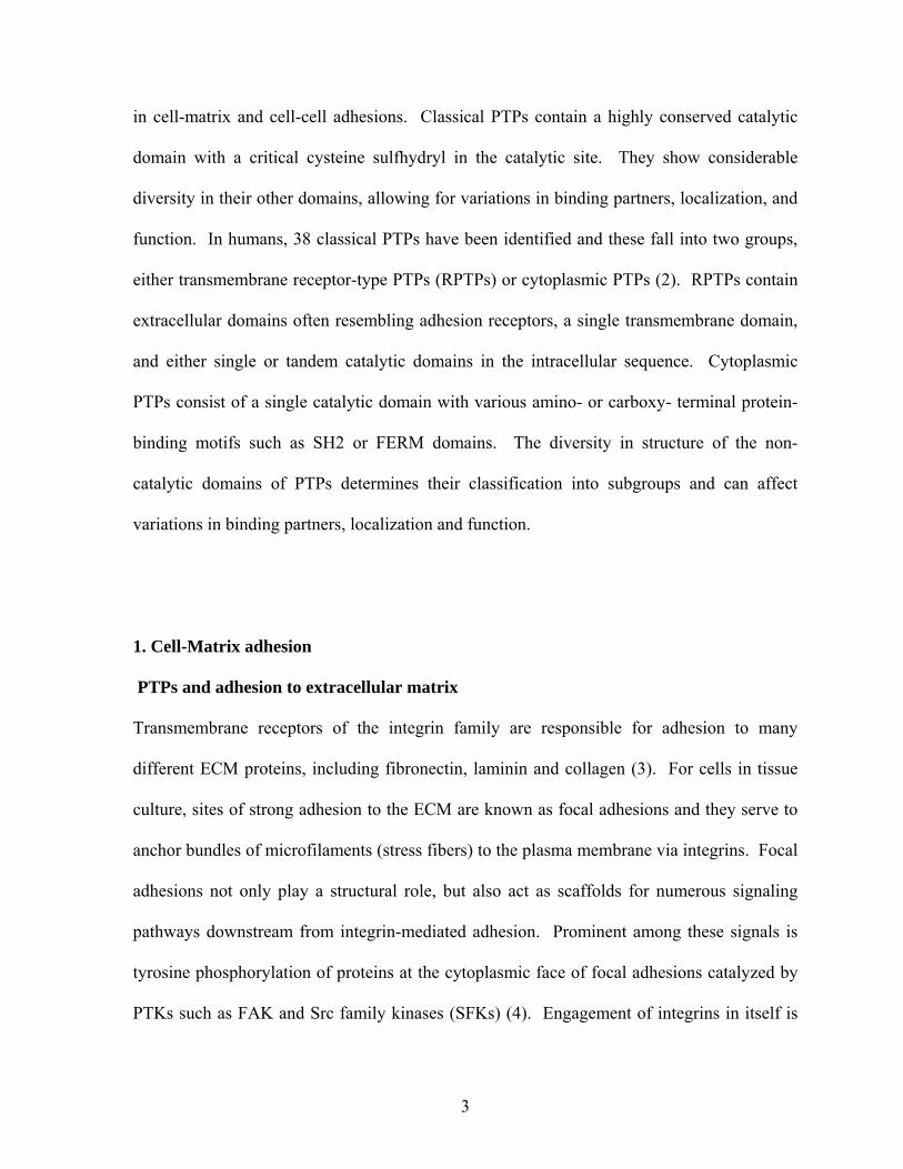
PTPs can affect integrin-mediated adhesion and the tyrosine phosphorylation that
occurs in focal adhesions by acting at least at three different levels. They can affect signaling
upstream, for example, by regulating the activities of GEFs and GAPs for Rho proteins; they
can act proximal to integrin engagement, for example by regulating Src kinase activity; or
they can dephosphorylate downstream targets, some of which may feedback to influence
upstream signaling pathways affecting focal adhesion assembly and turnover.
Upstream regulation of Rho protein activity by protein tyrosine phosphatases: PTP-PEST,
Shp-2 and LMW-PTP
Important upstream regulators of cell matrix adhesions are members of the Rho
family of small GTPases (9). In humans, this family of regulatory proteins includes
approximately 20 proteins, although most work has been focused on three ubiquitously
expressed members, RhoA, Rac1 and Cdc42. Like other G proteins, these proteins are active
in the GTP-bound form and are inactive when GTP is hydrolyzed to GDP. Activation of Rho
proteins is catalyzed by guanine nucleotide exchange factors (GEFs) which stimulate
exchange of bound GDP with GTP from the cytoplasmic pool. Most Rho proteins have
intrinsic GTPase activity, which is further stimulated by GTPase activating proteins (GAPs).
Many GEFs and GAPs are regulated by tyrosine phosphorylation. Consequently, PTPs can
profoundly influence the cycle of Rho protein activation by regulating the state of
phosphorylation of GEFs and GAPs (Figure 1.1). Examples of PTPs that have been reported
to regulate Rho protein activity are given in Table 1.
PTP-PEST is one PTP that affects adhesion and migration, in part by regulating the
activity of Rho proteins. Over-expression of PTP-PEST depresses membrane ruffling at the
6
Figure 1.1. PTP regulation of integrin-mediated adhesion signaling and focal adhesions. Upstream, the clustering of integrins is determined by RhoA-GTP levels (activity). PTPs can regulate the activity of Rho proteins by controlling the phosphorylation states of GEFs and GAPs. Downstream, integrin clustering leads to SFK and FAK activation. PTPs can both activate and inhibit SFKs by removing inhibitory or activating phosphorylations. Regulating the tyrosine phosphorylation of downstream targets such as FAK regulates the dynamics and disassembly of focal adhesions.
7
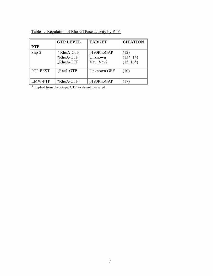
Table 1. Regulation of Rho-GTPase activity by PTPs
PTP
GTP LEVEL TARGET CITATION
Shp-2 ↑ RhoA-GTP ↑RhoA-GTP ↓RhoA-GTP
p190RhoGAP Unknown Vav, Vav2
(12) (13*, 14) (15, 16*)
PTP-PEST ↓Rac1-GTP
Unknown GEF (10)
LMW-PTP ↑RhoA-GTP p190RhoGAP (17) * implied from phenotype, GTP levels not measured
8
leading edge of cells due to decreased Rac1 activity (10). Conversely, in PTP-PEST-/- cells
Rac1 activity is elevated and sustained in cells plated on fibronectin (11).
Several pathways by which PTP-PEST might regulate Rac activity are suggested
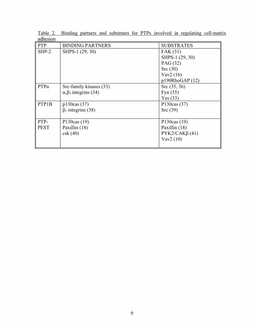
from published studies. PTP-PEST has been shown to bind and act on both p130Cas and
paxillin (18, 19) (Table 2). Both of these proteins, when tyrosine phosphorylated, interact
with Rac GEFs. Tyrosine phosphorylated p130Cas and paxillin bind the adapter Crk (20-
22), which in turn recruits the Rac GEF DOCK180 (23, 24). In addition, paxillin binds the
Rac GEF PIX via the adapter protein Pkl/Git (25). Therefore, a loss of PTP-PEST may
increase Rac activity by increasing the pool of phosphorylated p130cas and paxillin, thus
preserving their interactions with and regulation of their GEF binding partners. Recent work
has revealed that trapping mutants of PTP-PEST also bind Vav2 (11), a ubiquitously
expressed GEF regulated by tyrosine phosphorylation (26). This observation raises the
possibility that PTP-PEST may directly regulate Rac activity by controlling the
phosphorylation state of this GEF without the need for adaptor proteins such as
p130cas/paxillin.
The increased Rac1 activity found in PTP-PEST-/- fibroblasts would be predicted to
increase migration, but these cells actually have reduced migration rates (27). Examination
of cell morphology reveals prominent ruffling membranes/lamellipodia (hallmarks of active
Rac1), but the cells develop elongated tails, indicative of problems in detaching from the
substrate (11). Elongated tails are often associated with low RhoA activity (28). Through
interactions with both a Rac GEF and an as yet unidentified Rho GAP, PTP-PEST may
regulate the activities of both GTPases, thereby influencing migration by controlling
membrane ruffling and tail retraction. However, an inability to disassemble focal adhesions
9
Table 2. Binding partners and substrates for PTPs involved in regulating cell-matrix adhesion PTP BINDING PARTNERS SUBSTRATES SHP-2 SHPS-1 (29, 30) FAK (31)
SHPS-1 (29, 30) PAG (32) Src (30) Vav2 (16) p190RhoGAP (12)
PTPα Src-family kinases (33) αvβ3 integrins (34)
Src (35, 36) Fyn (35) Yes (33)
PTP1B p130cas (37) β1 integrins (38)
P130cas (37) Src (39)
PTP-PEST
P130cas (19) Paxillin (18) csk (40)
P130cas (19) Paxillin (18) PYK2/CAKβ (41) Vav2 (10)
10
in the rear would also account for the phenotype of the PTP-PEST null cells. Focal adhesion
disassembly is regulated by tyrosine phosphorylation as well and will be discussed below.
These results point to the complexity of phenotypes generated by PTP knockouts due to the
actions of PTPs on multiple targets.
Another PTP implicated in regulating Rho protein activity is Shp-2. Here, conflicting
results have been obtained, with some groups reporting that Shp-2 inhibits RhoA activity (15,
16), while other groups find that Shp-2 stimulates RhoA activity (13, 14), and still others
suggest that Shp-2 can exert both positive and negative regulatory effects on RhoA activity
(42). One target for Shp-2 is p190RhoGAP, a widely expressed GAP for RhoA (43) (Table
2). The activity of p190RhoGAP is stimulated by tyrosine phosphorylation (12, 43). By
dephosphorylating p190RhoGAP and so suppressing its GAP activity, Shp-2 can elevate
RhoA GTP levels (i.e. activate RhoA). Paradoxically, Shp-2 is one of the PTPs that
stimulates Src activity (see below), and Src is responsible for phosphorylation and activation
of p190RhoGAP (44). Consequently, Shp-2 can act on both sides of the phosphorylation
equation regulating p190RhoGAP activity. In addition, Shp-2 may be one of the PTPs that
inactivates Rho GEFs that are regulated by tyrosine phosphorylation (15, 16). Thus, the
apparently conflicting role of Shp-2 with regards to RhoA activity could be reconciled by the
differential action of the PTP on targets that can either positively or negatively regulate
RhoA activity.
One non-classical PTP that warrants mentioning in the context of RhoA activity is
LMW-PTP. It has been reported to act downstream of Src to regulate the phosphorylation
state of p190RhoGAP, thereby controlling Rho-mediated cytoskeletal rearrangement (45).
LMW-PTP has also been implicated in the crosstalk between Rac1 and RhoA, in which Rac1
11
mediated generation of reactive oxygen species was observed to inhibit LMW-PTP. This
elevated p190RhoGAP tyrosine phosphorylation and activity, suppressing RhoA activity
(17).
PTPs acting proximal to integrins: Shp-2, PTPα, and PTP1B
What is the initiating signal downstream from integrin-mediated adhesion? Several
studies have implicated Src family kinases (SFKs) in some of the earliest steps downstream
from integrins and preceding the activation of FAK (44, 46, 47). SFKs have been shown to
bind β-integrin cytoplasmic domains (48, 49), which has prompted investigation into how
these kinases are regulated in response to interaction of the integrins with their ligands.
SFKs are held in an inhibitory state by two intramolecular interactions. One interaction
involves the SH3 domain binding to the linker region between the kinase and SH2 domains.
This constraint may be removed by association of the SFK with integrin cytoplasmic
domains (39). The second intramolecular constraint to SFK activity involves binding of the
SH2 domain to the phosphorylated C-terminal tyrosine residue (Y527 in avian, Y529 in
mammalian cells). This inhibitory phosphorylation of Src is catalyzed by C-terminal Src
kinase (Csk). Csk complexes with Src and inactive integrin αIIb/β3 (49). A PTP must
dephosphorylate the C-terminal tyrosine in order for SFK activation. In addition, for full
activity, phosphorylation of Y416 must occur in the activation loop within the kinase
domain. Activation by removal of the C-terminal phosphate raises the possibility that PTPs
may be involved in the initiation of the signaling downstream from integrin engagement or
clustering. Several PTPs are capable of activating SFKs and, in the context of integrins,
three have been studied, Shp-2, PTPα, and PTP1B (30, 31, 33, 36, 39).
12
Cells expressing a truncated form of Shp-2 (lacking the N-terminal SH2 domain),
reveal diminished activation of Src and elevated phosphorylation of the inhibitory site, Y529,
in response to adhesion to ECM (30, 32). These cells spread more slowly and display
reduced tyrosine phosphorylation of FAK, paxillin and p130Cas (30, 31). In addition, Shp-2
may indirectly regulate Src activity by regulating the recruitment of Csk to the membrane.
Csk is recruited to the membrane via association with tyrosine phosphorylated PAG
(phosphoprotein associated with glycosphingolipid-enriched microdomains), a
transmembrane glycoprotein. Shp-2 dephosphorylates PAG and abolishes the Csk binding
site, resulting in a reduction in membrane associated Csk and a reduction of Csk-mediated
Src inhibition (32). Thus Shp-2 may activate Src both by directly acting on its C-terminal
phosphorylation site and by inhibiting Csk recruitment. A protein that may act in parallel to
PAG is SHPS-1 (SIRPα1). Like PAG, SHPS-1 recruits Shp-2 to the membrane and is a
target for its activity (29, 30).
PTPα is a receptor type, transmembrane PTP involved in the activation of SFKs and
in integrin signaling pathways. Ectopic expression of PTPα enhances the dephosphorylation
of the c-terminal Y529, strongly activating src and fyn kinases (36). Cells lacking PTPα
spread more slowly and contain decreased tyrosine phosphorylation of focal adhesion
components (35, 50). The decrease in tyrosine phosphorylation of FAK, especially at
autophosphorylation site Y397, in PTPα-/- cells suggests that this phosphatase lies between
integrins and the activation of FAK (51). PTPα and the integrin αvβ3 co-immunoprecipitate
from cells spreading on ECM substrates (34). This association has been shown to be
involved in the activation of SFKs following integrin engagement, which, in turn, is involved
in the reinforcement of integrin-cytoskeletal forces in response to tension (34). In this study,
13
the SFK involved was Fyn rather than Src. No evidence for an interaction between β1
integrins and PTPα was seen, but because similar downstream responses are observed for β1
and β3 integrins, it seems likely that parallel pathways may operate, possibly involving
different PTPs and different SFKs.
In platelets, activation of Src occurs rapidly in response to integrin engagement,
whereas FAK activation is a relatively late event (46). The association of Src with the β3
cytoplasmic domain involves binding via its SH3 domain, relieving one of the inhibitory
constraints on Src (39). With platelet αIIb/β3, the activation involves release of associated
Csk from the integrin/SFK complex and the subsequent recruitment of PTP1B. Interestingly,
the recruitment of PTP1B requires tyrosine phosphorylation of PTP1B and is blocked by Src
inhibitors (39). This implies that some level of Src activation must precede the recruitment
of PTP1B. Shattil and colleagues propose a model in which binding of αIIb/β3 to fibrinogen
induces micro-clustering of αIIb/β3, juxtaposing Src molecules so that these cross-
phosphorylate on Y416. It is suggested that this results in initial activation sufficient to
phosphorylate and recruit PTP1B, which, by removing the C-terminal phosphorylation of Src
results in full activity. In this model, many of the subsequent tyrosine phosphorylations,
including FAK activation, are triggered downstream from these initial events (39).
Differential PTP1B signaling in various cell types
With regard to its role in ECM adhesion, conflicting results have been reported for
PTP1B. The finding that PTP1B binds and acts on p130Cas (37) led to experiments
investigating the effects of expressing wildtype or mutant forms of PTP1B unable to bind to
p130Cas in cell adhesion situations. Expression of wildtype but not mutant PTP1B slowed
14
fibroblast spreading and depressed tyrosine phosphorylation of p130Cas and other proteins in
response to adhesion (52). In addition, the expression of wildtype PTP1B enhanced the
assembly of focal adhesions with short thick stress fibers, and decreased cell migration.
Consistent with these findings, depressing PTP1B expression in vascular smooth muscle
enhanced tyrosine phosphorylation of p130Cas and stimulated migration (53). Seemingly
contradictory results, however, were obtained in another study in which wildtype or
catalytically dead PTP1B were expressed in L cells (38). In these experiments, expression of
the wildtype PTP1B did not depress tyrosine phosphorylation in response to adhesion to
fibronectin, whereas expression of a catalytically dead mutant did. Expression of the inactive
mutant also suppressed Src activity and depressed cell attachment to a fibronectin
substratum. Additionally, the cells expressing the mutant PTP1B displayed an elongated
morphology, with focal adhesions that were reduced in size and number. A third study
examined the behavior of fibroblasts derived from PTP1B null mice (54). In this work, the
cells lacking PTP1B exhibited delayed spreading on a fibronectin-coated surface, but
surprisingly little effect was found in terms of tyrosine phosphorylation in response to
adhesion to fibronectin. However, when wildtype and PTP1B null cells were transformed
with SV40Tag, effects on tyrosine phosphorylation were seen. Now the transformed null
cells exhibited decreased tyrosine phosphorylation of p130Cas relative to transformed
wildtype cells following short periods (20 min) of adhesion to fibronectin. These null cells
also showed hyperphosphorylation of the inhibitory site in Src (Y527) in some situations.
Notably, the SV40Tag-transformed fibroblasts revealed an increased expression of PTP1B
relative to primary mouse embryo fibroblasts, possibly accounting for the differences
between the transformed and primary cells.
15
Can these apparently conflicting observations with PTP1B be reconciled? As the
authors have suggested, cell type differences may be critical, especially given that SV40Tag-
transformed cells elevate expression of PTP1B (54). Cell types may also diverge both as to
where PTP1B is acting in adhesion signaling pathways and in the degree of compensation by
other PTPs. For example, in some cell types PTP1B may have a major role regulating the C-
terminal inhibitory phosphorylation site of SFKs, whereas in other cell types different PTPs
(e.g. PTPα or Shp-2) may be more important. P130cas is a major substrate for Src and so in
cells in which PTP1B is regulating Src activity, one would predict lower phosphorylation of
p130Cas when PTP1B is absent or inactive (38, 54). However, in other cell types where
different PTPs may be more critical in regulating SFK activity, the effect of depressing
PTP1B activity would be predicted to be less significant in terms of p130Cas
phosphorylation. Since PTP1B can also dephosphorylate p130Cas, it would not be surprising
in these cells to observe that over-expression of wildtype PTP1B decreases the
phosphorylation of this target protein (52). One of the striking observations from Chernoff’s
group is that the cells over-expressing PTP1B revealed enhanced focal adhesions and stress
fibers (52). This is suggestive of increased RhoA activity and could arise because of
dephosphorylation and inactivation of a regulatory protein such as p190RhoGAP. However,
this phenotype could also result from defective focal adhesion disassembly (see below).
Focal adhesion disassembly: Downstream regulation by PTPs
With the discovery of FAK activation in response to integrin-mediated adhesion, it
was widely interpreted that tyrosine phosphorylation promoted focal adhesion assembly.
However, the phenotype of FAK knockout cells, as well as cells in which FAK was displaced
16
from focal adhesions, revealed robust and stable focal adhesions in the absence of FAK and
tyrosine phosphorylation detectable by immunofluorescence (55, 56). Rather than assembly
of focal adhesions, FAK activity correlated with focal adhesion turnover and disassembly (4).
While several pathways downstream of FAK could contribute to focal adhesion disassembly
(4, 25, 57-61), a novel, endocytic pathway (62) suggests a potentially important role for an as
yet unidentified PTP. In this work, tyrosine phosphorylated FAK was found to recruit
dynamin to focal adhesions (62). Dynamin is a protein involved in endocytosis and
expression of a dominant negative form of dynamin that inhibits endocytosis blocked focal
adhesion disassembly. The association of FAK with dynamin is mediated by the adaptor
protein Grb2, which binds to phosphorylated Y925 in FAK and to the proline rich region of
dynamin. Expression of the non-phosphorylatable FAK mutant, Y925F, failed to rescue
focal adhesion disassembly in FAK null fibroblasts (62).
Identification of the PTP that removes the phosphate from Y925 in FAK will be
important. Based on the above information, it would be predicted that this PTP would have a
key role in regulating focal adhesion disassembly and, by extension, in regulating cell
migration. In order for cells to migrate, focal adhesions must be disassembled so that strong
adhesions to the underlying ECM can be released. The PTP that mediates dephosphorylation
of Y925 in FAK would be anticipated to increase focal adhesion stability and decrease
migration when it is over-expressed, but to increase migration rates and the turnover of focal
adhesions when it is inhibited or knocked out. This phenotype matches that described by Liu
and coworkers when they over-expressed PTP1B in fibroblasts (52) and it will be interesting
to learn whether PTP1B or another PTP regulates the phosphorylation status of Y925 in
FAK.
17
2. Cell-Cell adhesion PTPs and cell-cell junctions
Epithelial tissues typically display stable cell-cell adhesion that is accompanied by prominent
cell junctions between interacting cells. Tight junctions (TJs) are responsible for the barrier
function of many epithelia, whereas adherens junctions (AJs) and desmosomes mediate
strong intercellular adhesion. The assembly and function of TJs is typically dependent on the
state of AJs, so that modulating AJ function often affects TJ barrier properties. The major
adhesion molecules in both AJs and desmosomes belong to the cadherin family; however,
cadherins can also contribute to adhesions between cells where distinct junctions do not
develop and where adhesion is more dynamic. Additional adhesion molecules, such as
nectins (63), are also present in AJs, but we will focus here on cadherins. Whereas the
extracellular domain of cadherins participates in calcium-dependent homophilic adhesion, the
cytoplasmic domain binds p120ctn and β-catenin (64-66). The former regulates the stability
of cadherins on the cell surface (67), and β-catenin provides a link to α-catenin and the actin
cytoskeleton, although the details of the bridge remain controversial (68). Tyrosine
phosphorylation of cadherins and their associated proteins has major effects on the stability
of adherens junctions (69, 70). In early work, it was shown that inhibiting PTPs with
pervanadate elevated tyrosine phosphorylation in adherens junctions and promoted the
disassembly of these structures (71). However, the same group later observed that in some
situations elevation of tyrosine phosphorylation first transiently stimulated AJ assembly
before resulting in the eventual disassembly of the same structures (72). Activation of PTKs
or inhibition of PTPs can lead to increased tyrosine phosphorylation of members of the
cadherin-catenin complex, dissociation of the AJ from the cytoskeleton and disruption of
18
cell-cell adhesion (73-75). For example, phosphorylation of tyrosine residues 755 and 756
on E-cadherin leads to its ubiquitination and subsequent endocytosis, resulting in loss of
junctional integrity (76). Similarly, phosphorylation of Y658 and Y731 in the cytoplasmic
domain of VE-cadherin prevents binding of p120ctn and β-catenin, respectively, and causes a
decrease in barrier function (77). Therefore, maintenance of junctional integrity is regulated
in part by reversible tyrosine phosphorylation that results from a competing balance of PTK
and PTP activity. Receptor-PTPs such as PTPµ, DEP-1, and VE-PTP, as well as the
cytosolic PTPs, PTP1B and Shp-2, have been shown to bind to members of the cadherin-
catenin complex (78-83) (Table 3) and to regulate cell-cell adhesion by means of regulating
phosphorylation of the cadherin-catenin complex.
Direct regulation of the cadherin-catenin complex
The extracellular domains of PTPμ can mediate cell-cell adhesion via homophilic
interactions (84, 85). Together with its binding to p120ctn and the cytoplasmic domain of
cadherins, the homophilic interactions of PTPμ combine to localize it to cell-cell junctions
(78, 79, 86, 87). Localization at AJs orients PTPμ’s catalytic domain in close proximity with
substrates VE-cadherin and p120 catenin (79, 87). In addition, knockdown of PTPμ by
siRNA increases permeability of endothelial monolayers demonstrating its role in the
regulation of junctional integrity (87). Although many of the effects of PTPμ at AJs are
undoubtedly due to its phosphatase activity, other domains of the protein may also contribute
to junctional stability. Evidence for this came from a
study of prostate carcinoma cells lacking endogenous PTPμ which were unable to form AJs
even though E-cadherin and the catenin proteins were present. Expression of PTPμ restored
19
cadherin-mediated cell-cell adhesion, but strikingly this could also be achieved by expression
of catalytically dead PTPμ (88). This finding suggests that PTPμ can act as a scaffold and
recruit additional regulatory proteins to sites of cell-cell adhesion.
Another PTP important for junction formation and maintenance is high cell density
enhanced PTP-1 (DEP-1). Expression of DEP-1 is increased more than ten-fold in many cell
types as they approach confluence, suggesting it contributes to cell-cell adhesion and contact
inhibition of growth (89). DEP-1 is present at the apical surface of endothelial cells, but
also co-localizes with VE-cadherin at intercellular junctions (90, 91). DEP-1 indirectly
associates with VE-cadherin by binding to p120ctn, γ-catenin (plakoglobin) and β-catenin
(Table 3); DEP-1 regulates their phosphorylation state, preserving their interactions with the
cadherins, and promoting cell-cell adhesion (80, 90). The role of DEP-1 in organizing cell-
cell junctions is supported by the observation that in cells lacking strong cell-cell adhesion,
such as fibroblasts, overexpression of DEP-1 results in a change in localization of cadherins
from discrete areas of cell-cell contact to large areas reminiscent of continuous AJ found in
epithelial cells (92).
A PTP in the same family as DEP-1 is vascular endothelial PTP (VE-PTP), which is
selectively expressed in endothelial cells, and binds to VE-cadherin but not to β-catenin (93).
VE-PTP associates with VE-cadherin via its membrane-proximal extracellular domain and its
recruitment results in the dephosphorylation of VE-cadherin (81). Expression of wild type
VE-PTP decreases paracellular permeability in endothelial monolayers, while the
catalytically inactive mutant has no effect, indicating that the phosphatase activity of VE-
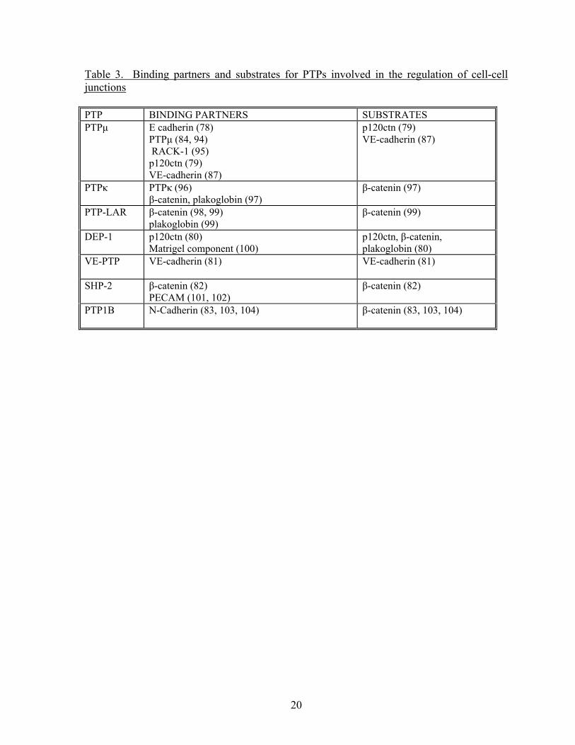
20
Table 3. Binding partners and substrates for PTPs involved in the regulation of cell-cell junctions PTP BINDING PARTNERS SUBSTRATES PTPμ E cadherin (78)
PTPμ (84, 94) RACK-1 (95) p120ctn (79) VE-cadherin (87)
p120ctn (79) VE-cadherin (87)
PTPκ PTPκ (96) β-catenin, plakoglobin (97)
β-catenin (97)
PTP-LAR β-catenin (98, 99) plakoglobin (99)
β-catenin (99)
DEP-1 p120ctn (80) Matrigel component (100)
p120ctn, β-catenin, plakoglobin (80)
VE-PTP VE-cadherin (81) VE-cadherin (81)
SHP-2 β-catenin (82) PECAM (101, 102)
β-catenin (82)
PTP1B N-Cadherin (83, 103, 104) β-catenin (83, 103, 104)
21
PTP acting on VE-cadherin is necessary for maintaining the integrity of endothelial junctions
(81).
Cytosolic PTPs such as PTP1B and Shp-2 have also been implicated in regulating
cell-cell adhesion by controlling phosphorylation of cadherin/catenin proteins (82, 83, 103).
PTP1B binds directly to the cytoplasmic domain of N-cadherin, an interaction that promotes
β-catenin binding and targeting of the cadherin/catenin complex to the cell membrane (104).
Expression of a catalytically inactive mutant of PTP1B disrupts cadherin-mediated adhesion,
with concomitant increases in tyrosine phosphorylation of β-catenin and reduction in the
association of N-cadherin with the actin cytoskeleton, suggesting that the catalytic activity of
PTP1B is important for junctional maintenance (83, 103). Shp-2 is another cytosolic PTP
associated with the cadherin/catenin complex in confluent endothelial cell monolayers,
specifically interacting with β-catenin (82). Thrombin treatment of endothelial cells induces
Shp-2 tyrosine phosphorylation and dissociation from β-catenin followed by junctional
breakdown and the formation of large intercellular gaps, thus supporting the hypothesis that
Shp-2 localization to junctions also helps maintain endothelial junctional strength and
integrity (82). The loss of Shp-2 binding is accompanied by an increase in phosphorylation
of β-catenin, plakoglobin and p120ctn. AJs are not the only adhesive complexes where PTPs
play a role in regulating signal transduction. Shp-2 binds to platelet endothelial cell adhesion
molecule-1 (PECAM-1) (101, 102) and intercellular adhesion molecule 1 (ICAM-1) (105,
106). In response to ICAM-1 engagement, phosphorylated ICAM-1 binds to Shp-2 and this
interaction is necessary for Src activation as well as p38 MAPK activation (106).
The concentration of PTPs, both cytosolic and transmembrane, at AJs indicates the
importance of maintaining low levels of tyrosine phosphorylation at these sites, except when
22
the junctions need to be remodeled or disassembled. In general, the suppression of bulk PTP
activity by inhibitors or the suppression of individual PTPs by siRNA elevates the tyrosine
phosphorylation of cadherins and their associated proteins. In turn, this results in
destabilization of the junctions and diminished epithelial or endothelial barrier functions.
Rho GTPases and cell-cell adhesion
Just as with cell-matrix adhesions (107), there is bidirectional interplay between Rho
family GTPases and cell-cell adhesions. Not only is the assembly of AJs affected by the
activities of Rho proteins, but cell-cell adhesion can stimulate or depress the activities of
these G proteins, suggesting complex feedback loops. Several studies have shown that Rac1
activation promotes assembly of epithelial AJs and inhibition of either Rac1 or RhoA activity
results in disassembly (108-110). Conversely, other studies show that high levels of active
Rac1 or RhoA can actually disrupt both TJs (111) and AJs (112, 113); this might reflect
different cellular contexts or the need for tight regulation of GTPase activity. The formation
of AJs can activate Rac1 and Cdc42 (114, 115) but inhibit RhoA (115). PTPs may play
critical roles in these upstream and downstream pathways by regulating the phosphorylation
and activity of GTPase activating proteins (GAPs) and guanine nucleotide exchange factors
(GEFs) (107). For example, p190RhoGAP tyrosine phosphorylation was implicated in the
depression of RhoA activity downstream from cadherin engagement (116).
With endothelial monolayers, several agents that promote increased permeability also
stimulate Rac1 activity. PTPs are also likely to be important in this response to Rac1
because in these cells Rac1 has been shown to generate reactive oxygen species (ROS) (117).
In turn, ROS are potent inhibitors of PTPs (118) and are responsible for the elevation in
junctional protein tyrosine phosphorylation that results from Rac1 activation (113, 117, 119).
23
Regulation of PTP activity
The resemblance of the extracellular domains of RPTPs to cell adhesion molecules
has stimulated a search for potential binding partners and by extension, for modes of
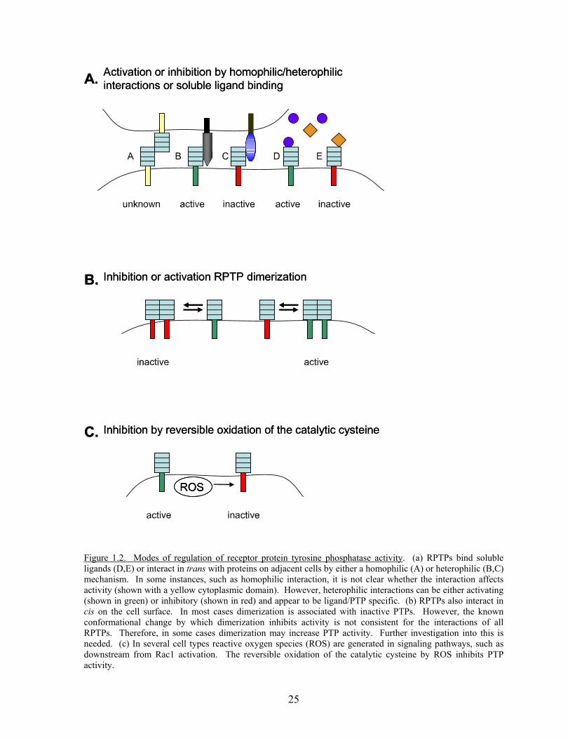
regulation via such interactions. Several mechanisms for modulation of specific PTP activity
exist (Figure 1.2). In some cases, homophilic interactions have been detected, as first shown
with PTPµ (84, 85). Two closely related PTPs, PTPκ and PTPλ, have similarly been shown
to interact in a homophilic manner (96, 120). The extracellular domains of several RPTPs
participate in heterophilic interactions with ECM or other components. For example, LAR
binds the laminin/nidogen complex, which is prominent in many basement membranes (121).
Similarly, PTPσ binds to heparin sulfate proteoglycans (122) and PTPα binds contactin (123
). Although several interactions involving the extracellular domains of RPTPs have been
identified, in most cases no effect on the activity of the PTP has been demonstrated. One
exception is the interaction of PTPβ/ξ with pleiotrophin, which inhibits PTP activity and
results in an increase in β-catenin phosphorylation (124). Pleiotrophin is a heparin binding
growth associated molecule and its interaction with PTPβ/ξ promotes cortical neuron
migration (124). In
contrast, DEP-1 phosphatase activity is up-regulated by the binding of its extracellular
domain to an unknown component of Matrigel® (100). The observation that the activities of
RPTPs can be regulated by the interactions of their extracellular domains will be broadly
important if further work can establish that this is a general characteristic of RPTPs. It is
easy to imagine many scenarios in which tyrosine phosphorylation levels could be regulated
24
by interactions of the extracellular domains of RPTPs with other cells, with matrix molecules
or soluble ligands (Figure 1.2A).
Dimerization of RPTPs provides another potential mode of regulating their activity.
RPTPs have traditionally been considered to be inactive when dimerized and active when
monomers (125). This idea was first proposed when a chimera of the extracellular domain of
EGFR fused to the intracellular domain of CD45 was inhibited by EGF-induced dimerization
(126). Further support was generated when studies revealed RPTPα exists as dimers on the
cell surface, and that this dimerization inhibits the activity of the phosphatase via an
interaction of the tandem catalytic domains, preventing the binding of substrates (127-129).
This suggests a model where RPTPs exist as inactive dimers on the cell surface in the
absence of a ligand and binding of the ligand dissociates the dimers and activates the PTP
(Figure 1.2B). However, dimerization-induced inactivation of RPTPs through blockage of
the active site by an opposing PTP domain may not be a universal mechanism. Structural
analysis of the membrane-proximal catalytic domain of PTPµ and the entire cytoplasmic
domain of PTP-LAR suggests that these PTPs are not inhibited by dimerization (130, 131).
In addition, RPTPs such as DEP-1 and VE-PTP contain a single catalytic domain and
therefore may also not be inhibited by dimerization.
The regulation of PTP activity by ROS is an area developing very rapidly and too
large to review in detail here. The cysteine residue in the catalytic site of classical PTPs has
long been known to be sensitive to oxidation and this has been the basis for broad specificity
inhibitors, such as H202 and pervanadate. Subsequent work has shown that reversible
oxidation of the catalytic cysteine can provide a physiological mechanism for regulating
PTPs (118) (Figure 1.2C). The generation of ROS was originally identified as a defense
25
Figure 1.2. Modes of regulation of receptor protein tyrosine phosphatase activity. (a) RPTPs bind soluble ligands (D,E) or interact in trans with proteins on adjacent cells by either a homophilic (A) or heterophilic (B,C) mechanism. In some instances, such as homophilic interaction, it is not clear whether the interaction affects activity (shown with a yellow cytoplasmic domain). However, heterophilic interactions can be either activating (shown in green) or inhibitory (shown in red) and appear to be ligand/PTP specific. (b) RPTPs also interact in cis on the cell surface. In most cases dimerization is associated with inactive PTPs. However, the known conformational change by which dimerization inhibits activity is not consistent for the interactions of all RPTPs. Therefore, in some cases dimerization may increase PTP activity. Further investigation into this is needed. (c) In several cell types reactive oxygen species (ROS) are generated in signaling pathways, such as downstream from Rac1 activation. The reversible oxidation of the catalytic cysteine by ROS inhibits PTP activity.
26
mechanism in the phagocytic killing of bacteria by leukocytes, but more recently the
generation of low levels of ROS by other cells has been recognized as a widespread
occurrence with important physiological consequences (132). The formation of ROS is
downstream of active Rac1 (17, 117). For example, in endothelial cells, active Rac1
promotes endothelial permeability and is associated with increased tyrosine phosphorylation
of junctional proteins, such as VE-cadherin and β-catenin (117, 119). These effects are
blocked by inhibiting the generation of ROS and are mimicked by pervanadate (117).
Whether these effects are the result of one or a few PTPs being inhibited or reflect broad
inhibition of all PTPs within these cells has not been established. It will be interesting to
determine the degree to which ROS can act locally on one or a few PTPs rather than globally
by inhibiting all PTPs within a cell. Techniques have been developed for analyzing the
reversible oxidation of PTPs (133) and so this area should advance rapidly. There are many
physiological situations in which Rac1 is activated and it will be important to determine
whether the generation of ROS and the consequent inactivation of PTPs is a general signaling
pathway downstream from Rac1 or whether this only occurs in particular situations.
DEP-1 (density enhanced protein tyrosine phosphatase-1)
As mentioned above, there are both cytoplasmic and receptor protein tyrosine
phosphatases. This thesis focuses on the role of RPTPs in general and DEP-1 in particular, in
regulating cell-cell adhesion and signaling events. Also known as PTP-η, PTPRJ, and
CD148, DEP-1 is a single pass transmembrane protein with an extracellular domain
27
consisting of 8 fibronectin type III repeats, a transmembrane domain and a single
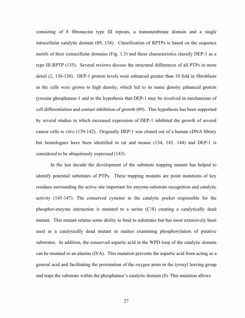
intracellular catalytic domain (89, 134). Classification of RPTPs is based on the sequence
motifs of their extracellular domains (Fig. 1.3) and these characteristics classify DEP-1 as a
type III RPTP (135). Several reviews discuss the structural differences of all PTPs in more
detail (2, 136-138). DEP-1 protein levels were enhanced greater than 10 fold in fibroblasts
as the cells were grown to high density, which led to its name density enhanced protein
tyrosine phosphatase-1 and to the hypothesis that DEP-1 may be involved in mechanisms of
cell differentiation and contact inhibition of growth (89). This hypothesis has been supported
by several studies in which increased expression of DEP-1 inhibited the growth of several
cancer cells in vitro (139-142). Originally DEP-1 was cloned out of a human cDNA library
but homologues have been identified in rat and mouse (134, 143, 144) and DEP-1 is
considered to be ubiquitously expressed (143).
In the last decade the development of the substrate trapping mutant has helped to
identify potential substrates of PTPs. These trapping mutants are point mutations of key
residues surrounding the active site important for enzyme-substrate recognition and catalytic
activity (145-147). The conserved cysteine in the catalytic pocket responsible for the
phosphor-enzyme interaction is mutated to a serine (C/S) creating a catalytically dead
mutant. This mutant retains some ability to bind to substrates but has most extensively been
used as a catalytically dead mutant in studies examining phosphorylation of putative
substrates. In addition, the conserved aspartic acid in the WPD loop of the catalytic domain
can be mutated to an alanine (D/A). This mutation prevents the aspartic acid from acting as a
general acid and facilitating the protonation of the oxygen atom in the tyrosyl leaving group
and traps the substrate within the phosphatase’s catalytic domain (8). This mutation allows
28
Figure 1.3. Structure of receptor protein tyrosine phosphatases. RPTPs are divided into subcategories based on the structure of their extracellular domain. Shown here the 5 most common types. Blue ovals (FN), fibronectin type III repeats; dark blue Ig, Ig like domains; orange circle (CA), carbonyic acid; D1, first catalytic domain, often active, D2, second catalytic domain, often inactive, dark gray, transmembrane domain. Adapted from (2).
29
for the binding of the phosphorylated substrate but prevent its removal, turning a transient
interaction into a
stable one. Several substrates for DEP-1 have been identified using this trapping mutant,
including p120 catenin, β-catenin, the p85 subunit of PI3K, and the HGF receptor Met (80,
148, 149). Physiological effects of these interactions still need to be elucidated, however,
there is a great body of literature demonstrating DEP-1’s regulation of and involvement in
several signaling pathways in different cell types and these will be reviewed in the following
sections.
Role of the extracellular domain of DEP-1
The extracellular domain of DEP-1 is highly N-glycoslyated and believed to be acting
as an adhesion receptor involved in outside-in signaling (89, 134). The intrinsic PTPase
activity of DEP-1 may be regulated or modulated by interacting with other cell surface
molecules (ligand mediated signal transduction) as well as through dimerization of DEP-1
extracellular domains in cis. When cells or recombinant DEP-1 protein were incubated with
Matrigel®, a preparation of extracellular matrix proteins, DEP-1 catalytic activity increased
(100). The exact ligand that causes the increased catalytic activity is not known. This was
the first evidence of increased catalytic activity of a RPTP by an extracellular ligand. A few
years later, a research group developed an antibody to the extracellular domain of DEP-1 and
tested its effect on endothelial cell growth (150). They discovered that antibody-induced
ectodomain oligomeriztion of DEP-1 inhibited cell growth by increasing the catalytic activity
of the cytoplasmic domain (150). This is particularly interesting due to the fact that other
PTPs are believed to be inactivated when dimerized (126-129). Therefore, dimerization of
30
DEP-1 or binding to extracellular proteins can increase PTP activity. One proposed reason
for the difference between other PTPs and DEP-1 is that DEP-1 has a single catalytic
domain, not a tandem set of domains, and is not prone to the inhibitory binding of opposing
PTP domains.
It is also possible that the extracellular domain of DEP-1 is important independent of
its ability to signal to its catalytic domain. Mice with a knock-in mutation of DEP-1, where
GFP replaced the intracellular catalytic domain, resulted in a dramatic embryonic lethal
phenotype in homozygous mutants with mice dying before embryonic day 11.5 (91). These
mice had disorganized vascular structures and cardiac defects (91). Interestingly, when the
entire gene coding for DEP-1, PTPRJ, was deleted from mice they were viable, fertile and
showed no gross anatomical alterations, suggesting PTPRJ is dispensable for normal growth
and development of mice (151). Therefore, loss of the protein had no effect but the presence
of the extracellular domain (without PTP activity) is lethal. This suggests that the
extracellular domain of PTPRJ might act as functional ligand which is able to block
pathways responsible for endothelial cell assembly necessary for correct vascularization
(151).
DEP-1 regulation of growth factor receptors
A number of studies have examined DEP-1’s role in modulating growth factor signaling
by directly acting on the growth factor receptors. Knockdown of endogenous protein or
expression of the catalytically dead mutant of DEP-1 in endothelial cells increased
phosphorylation of VEGFR- 2 as well as p44/42 MAPK in response to VEGF, suggesting
that DEP-1 negatively regulates VEGFR-2 activity (152). Specific residues
31
dephosphorylated by DEP-1 have not yet been fully investigated for this RTK. Biochemical
studies both in vivo and using peptide substrates revealed that DEP-1 is able to directly
dephosphorylate PDGF β-receptor in a site-selective manner (153). Interestingly, the
regulatory Tyr 857 was not found to be a preferred site for DEP-1 dephosphorylation, instead
Tyr 1021 showed the highest affinity for dephosphorylation (153). Biological responses
triggered by PDGF β-receptor and other PTKs is determined by the SH2 domain containing
proteins that associate with receptor in a phosphorylation site selective manner (154, 155).
Therefore, DEP-1 may not be attenuating the RTK signal completely by acting on the
regulatory tyrosine but modulating the signaling output by dephosphorylation of a subset of
SH2-domain binding tyrosines (153). Similarly, HGF receptor kinase Met is also a
substrate for DEP-1 with non-preferred targets in the tyrosines of the activation loop. Instead
DEP-1 dephosphorylates C-terminal tyrosines known to be important for morphogeneis and
Gab1 binding (149). Again, this result supports the role of DEP-1 in controlling the
specificity of signals induced by the RTK rather than a simple “off switch”. For these
reasons, regulated activation and expression of PTPs is a possible mechanism for altering
biological response following ligand stimulation of RTKs. Interestingly, LMW-PTP (a
structurally distinct PTP from DEP-1) is also able to dephosphorylate PDGF β-receptor but
with the reverse preference for tyrosine residues (156). This suggests that the preference for
phospho-peptides of PDGF β-receptor by DEP-1 is not shared by other PTPs involved in
PDGF β-receptor dephosphorylation and can provide an explanation for why several PTPs
are able to act on the same substrate.
Several studies have also investigated the physiological effects of DEP-1 on PDGF
signaling through overexpression and knockdown studies. DEP-1 expression in fibroblasts
32
inhibited PDGF-stimulated cell migration (92, 157), whereas DEP-1 -/- MEFs showed
increased chemotaxis towards PDGF (158). Several PDGF-stimulated signaling events were
negatively modulated by DEP-1 expression, with the most prominent effects being on
phospholipase Cγ1, Ras and ERK1/2 activation (157). Another study also showed that the
Ras-MAPK pathway as well as the PI3-kinase-Akt pathway was negatively regulated by
DEP-1 protein levels (92). Although these studies are in agreement with regards to signaling
effects and migration, contradictory results have been found for the effect of DEP-1 on cell-
matrix adhesion. Jandt et al. found that DEP-1 expression was a positive regulator of cell
adhesion, with DEP-1 expressing cells initially adhering faster but eventually reaching the
same number as control cells over a 4 hr period (157). However, Kellie et al. found DEP-1
fibroblasts displayed a reduced number of cells both adhered and spread compared to control
cells (92). This study supported its claim with reduced FAK phosphorylation compared to
vector controls and reduced FA formation, both associated with decreased adhesion. Both
studies were conducted in NIH3T3 cells and therefore the difference could lie in the matrix
that was used (collagen 1 vs fibronectin) or the extent of over-expression of DEP-1. These
studies enhance the receptor dephosphorylation data, demonstrating that cell adhesion and
migration downstream of PDGF are affected by DEP-1 protein levels.
DEP-1 in cancer
In the last several years, evidence has been published linking DEP-1 with certain cancers.
The chromosomal location of DEP-1 has been mapped to chromosome 11p11.2 on the short
arm of chromosome 11 (134). Previously, loss of heterozygosity or deletion of sequences in
the short arm of human chromosome 11 had been detected in various tumors of epithelial
33
origin, including breast cancer, bladder cancer, and hepatocellular carcinoma (159-161).
Recent studies have looked more specifically at this chromosome, investigating the gene
responsible for DEP-1 expression, PTPRJ. It has been found that human colon, lung, breast
and thyroid cancers frequently have somatic missense mutations, loss of heterozygosity, or
deletions of the PTRRJ gene (DEP-1) (162, 163). In addition, the mouse homologue gene,
Ptprj, is a candidate for the mouse colon cancer susceptibility locus Scc1 (163). DEP-1
expression has been shown to be drastically reduced in multiple cancer cell lines and human
cancers including thyroid, breast, pancreatic and colon cancers. (139-142, 164). Therefore,
DEP-1 has been proposed to be a tumor suppressor. One way in which it may be acting as a
tumor suppressor is by inhibiting cell proliferation. Studies have re-expressed DEP-1 in
cancer cell lines lacking endogenous DEP-1 and found that there was a profound inhibition in
cell proliferation (139-142, 164). In addition, endogenous DEP-1 protein levels have been
reduced with shRNA constructs resulting in enhanced proliferation (164), supporting a
inhibitory role for DEP-1 in regulation of cell growth. In fact, injection of adenovirus
expressing DEP-1 into mice with tumors derived from injected pancreatic cancer cells
blocked the growth of the tumors compared to untreated or control adenovirus treatment
(142). Restoring DEP-1 expression could be a tool for gene therapy in human (pancreatic)
cancers.
A number of mutations in the DEP-1 gene, PTPRJ, have been identified and create
coding polymorphisms often found in human cancers. Gln276Pro, Arg326Gln, and
Asp827Glu are mutations in the extracellular domain, specifically in the 2nd and 8th
fibronection repeats (Fig. 1.4) (142, 163). Sequence alignments, secondary structure
prediction and homology modeling have predicted with high confidence that most amino acid
34
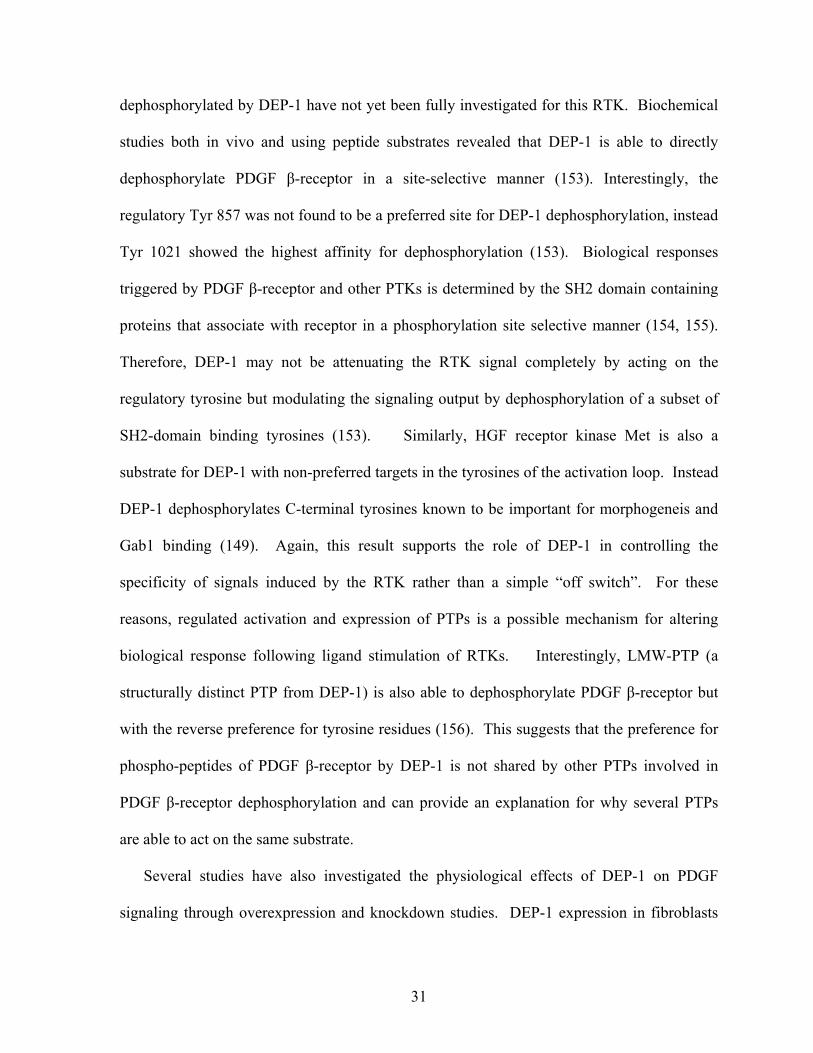
Figure 1.4. Polymorphisms of mouse and human PTPRJ. Schematic representation of polymorphisms that result in amino acid substitutions in both mouse and human. For mice, sequences from Balb/c and STS were compared and positions are from Balb/c sequence. For Human, comparisons were made from PTPRJ gene from colon cancer patients. Prediction of effects of substitution for Human only: The Arg214Cys and Arg326Gln substitutions are predicted to lose positive charges, Gln276Pro results in torsional stress, Pro445Leu releases torsional stress, and the effect of Asp873Glu has not been predicted. The Asp1061Glu substitution is not expected to affect the activity of the catalytic domain, because it is conservative (involving two acidic residues) and affects a residue relatively distant from the active site. FNIII is fibronectin type 3 domain (striped), the transmembrane domain is gray and the catalytic domain is the spotted oval. Modified from Ruivemkamp 2002 (163).
FNIII 1 2 4 5 6 7 83
Mouse
Human
Leu211P
Val217Al Ala533T
Pro622S
Asp1061
Arg214C
Gln276P
Arg326G
Pro445L Asp873G
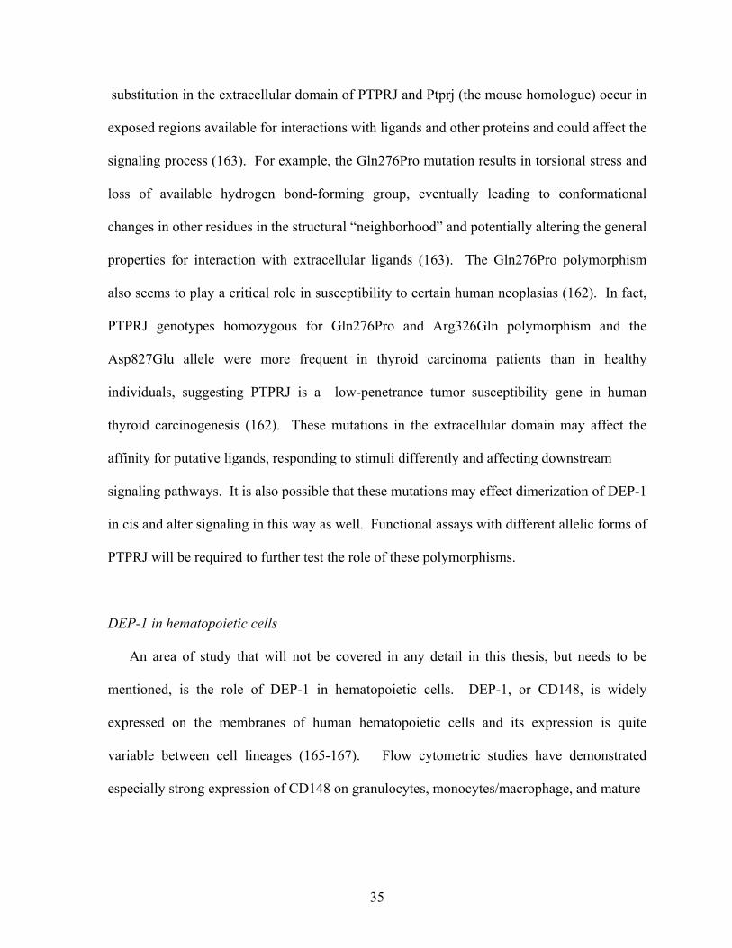
35
substitution in the extracellular domain of PTPRJ and Ptprj (the mouse homologue) occur in
exposed regions available for interactions with ligands and other proteins and could affect the
signaling process (163). For example, the Gln276Pro mutation results in torsional stress and
loss of available hydrogen bond-forming group, eventually leading to conformational
changes in other residues in the structural “neighborhood” and potentially altering the general
properties for interaction with extracellular ligands (163). The Gln276Pro polymorphism
also seems to play a critical role in susceptibility to certain human neoplasias (162). In fact,
PTPRJ genotypes homozygous for Gln276Pro and Arg326Gln polymorphism and the
Asp827Glu allele were more frequent in thyroid carcinoma patients than in healthy
individuals, suggesting PTPRJ is a low-penetrance tumor susceptibility gene in human
thyroid carcinogenesis (162). These mutations in the extracellular domain may affect the
affinity for putative ligands, responding to stimuli differently and affecting downstream
signaling pathways. It is also possible that these mutations may effect dimerization of DEP-1
in cis and alter signaling in this way as well. Functional assays with different allelic forms of
PTPRJ will be required to further test the role of these polymorphisms.
DEP-1 in hematopoietic cells
An area of study that will not be covered in any detail in this thesis, but needs to be
mentioned, is the role of DEP-1 in hematopoietic cells. DEP-1, or CD148, is widely
expressed on the membranes of human hematopoietic cells and its expression is quite
variable between cell lineages (165-167). Flow cytometric studies have demonstrated
especially strong expression of CD148 on granulocytes, monocytes/macrophage, and mature
36
thymocytes (165, 166). Weaker expression was found on peripheral blood lymphocytes, T
cells, B cells, platelets, natural killer cells, and dendritic cells (165, 167), and poor expression
was on transformed lymphoid T- and B- cell lines (165). Upregulation of CD148 occurred
on T cells following activation (165, 166, 168, 169). Experiments in T cells have
demonstrated a role for CD148 in the negative regulation of TCR signaling. Increased
expression of CD148 reduced the activation of the TCR-induced transcription factor NFAT
(nuclear factor of activated T cells) (169-171). In addition, CD148 can also act as a negative
regulator by causing specific dephosphorylation of LAT (linker for activation of T-cell) and
phospholipase Cγ1 (170). Unlike T cells, CD148 expression levels did not change
substantially following stimulation and activation of B cells (167), suggesting that CD148 is
not regulating activity of B cells in a similar manner as T cells.
Cross linking of CD148 can induce increased [Ca2+] and tyrosine phosphorylation of
several proteins in human T cells including phospholipase Cγ1 (165). The increased
phosphotyrosine of intracellular proteins suggests that CD148 might regulate a protein
tyrosine kinase whose activity is dependent on its phosphorylation status. The inhibitory
tyrosine residuce of src has been shown to be a substrate of DEP-1 in malignant thyroid cells
(172) and in B cells (173) and therefore could also be a substrate in T cells and other
leukoctyes. The data above demonstrate that CD148 is involved in signal transduction in T
cells and B cells, however, the significance of its action in immune response is still being
evaluated. It is proposed that CD148 expression could be a marker for immune activation
but further studies will need to be conducted to fully understand its role in hematopoietic
cells.
37
Tight Junctions
Protein components of Tight Junctions
Tight junctions are the most apical of the junctions formed by epithelia and provide a
regulated barrier to paracellular transport of ions, solutes, water as well as cells. They
function as a fence, preventing the mixing of membrane proteins between the apical and
basolateral membranes. Tight junctions consist of 4 transmembrane proteins and several
cytoplasmic proteins which anchor the transmembrane proteins to the actin cytoskeleton (Fig
1.5). Selected protein-protein interactions are depicted in Table 4. In the transmembrane
group there are the junctional adhesion molecules (JAMs), occludin, tricellulin, and claudins
(174-176). My work has focused on occludin, therefore I will not go into great detail about
the other transmembrane proteins but will instead briefly introduce them. Several review
articles cover them in more depth (177-181). The JAMs have 3 family members, A, B, and
C. They localize to TJ and engage in both homophilic and heterophilic adhesions, however
do not induce the formation of TJ strands in cells lacking TJ (182). Tricellulin is very similar
in structure to occludin, but is only located at tricellular junctions and strengthens the barrier
function of epithelial cell sheets (176). The claudin family consists of 24 members with each
exhibiting distinct expression patterns in both tissue- and cell- specific manners (reviews see
(175, 183, 184). Also similar in structure to occludin but not in sequence (185), claudins are
capable of forming TJ strands when expressed in L cells, fibroblasts normally lacking TJs
(186). Claudins are believed to be the main TJ component driving the overall structure of TJ
and the variety in strength, size, and ion specificity of tight junctional barriers in different
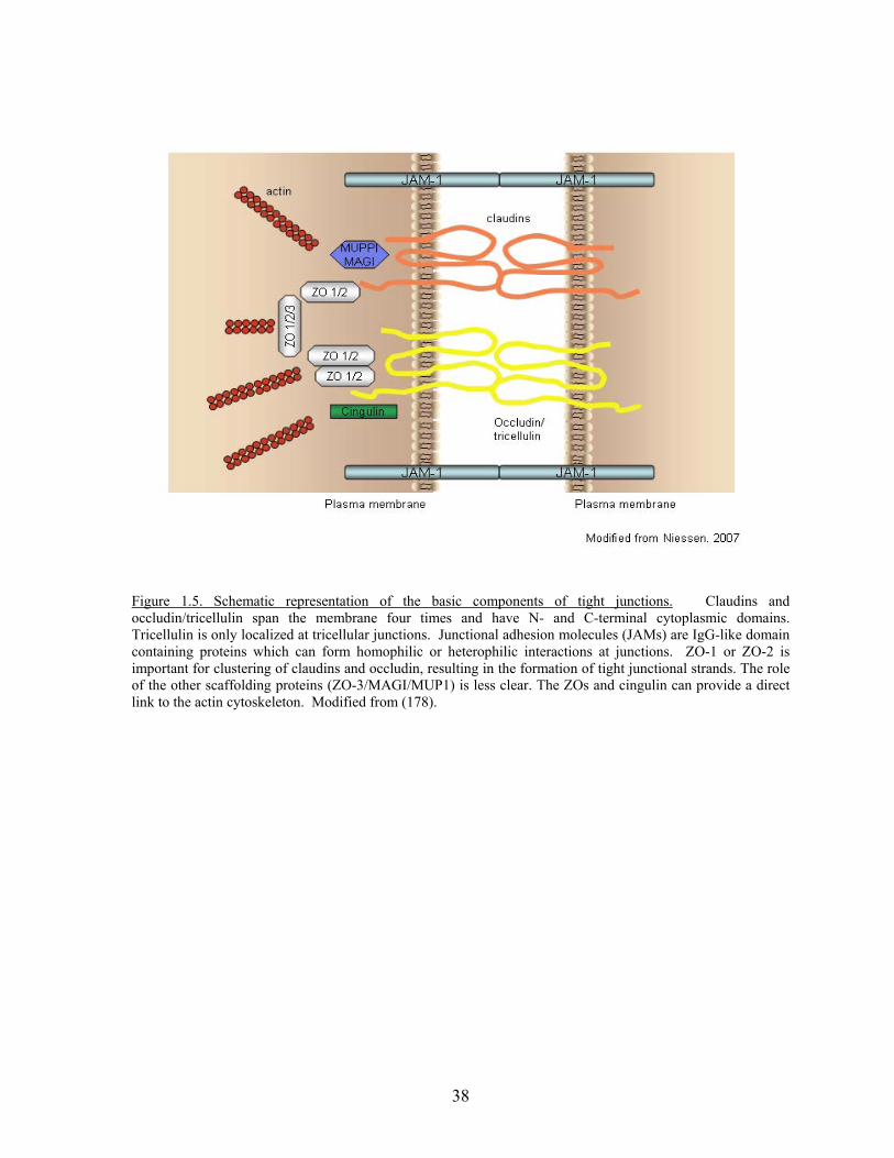
38
Figure 1.5. Schematic representation of the basic components of tight junctions. Claudins and occludin/tricellulin span the membrane four times and have N- and C-terminal cytoplasmic domains. Tricellulin is only localized at tricellular junctions. Junctional adhesion molecules (JAMs) are IgG-like domain containing proteins which can form homophilic or heterophilic interactions at junctions. ZO-1 or ZO-2 is important for clustering of claudins and occludin, resulting in the formation of tight junctional strands. The role of the other scaffolding proteins (ZO-3/MAGI/MUP1) is less clear. The ZOs and cingulin can provide a direct link to the actin cytoskeleton. Modified from (178).
39
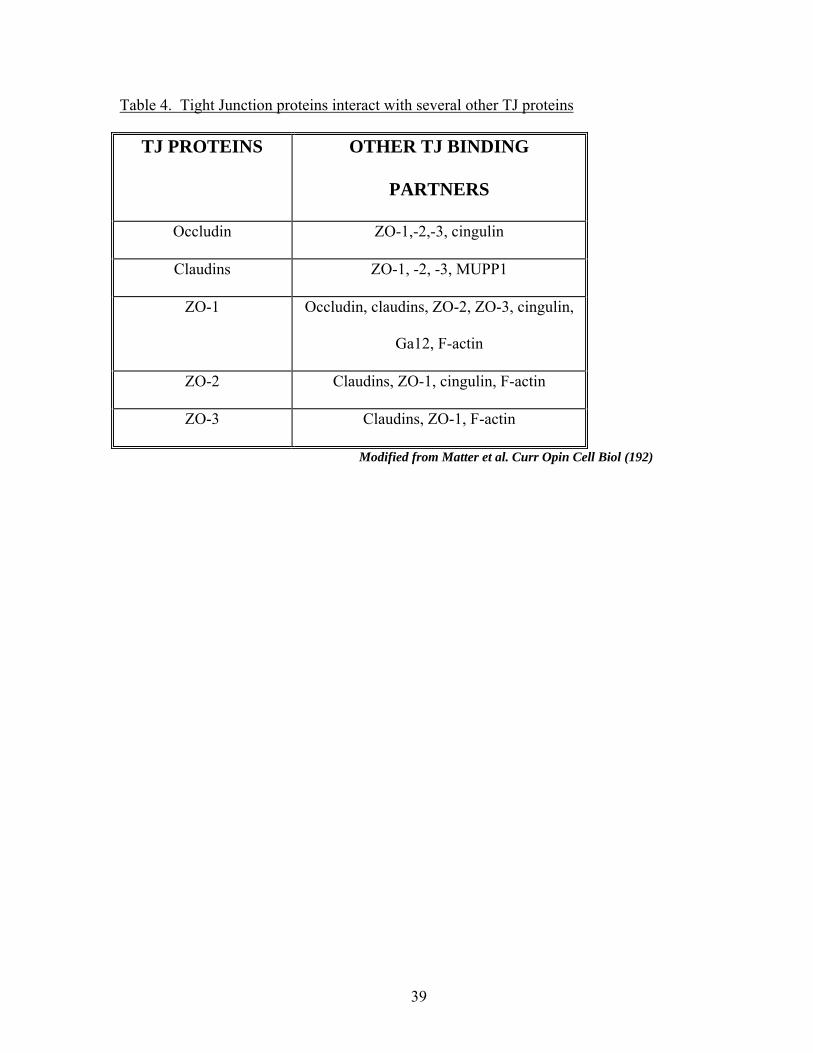
Table 4. Tight Junction proteins interact with several other TJ proteins
TJ PROTEINS OTHER TJ BINDING
PARTNERS
Occludin ZO-1,-2,-3, cingulin
Claudins ZO-1, -2, -3, MUPP1
ZO-1 Occludin, claudins, ZO-2, ZO-3, cingulin,
Ga12, F-actin
ZO-2 Claudins, ZO-1, cingulin, F-actin
ZO-3 Claudins, ZO-1, F-actin
Modified from Matter et al. Curr Opin Cell Biol (192)
40
epithelia and endothelia is largely due to the type of claudin(s) found at specific tight
junctions (175, 187).
Occludin spans the membrane four times creating two extracellular loops with both
an N- and C-terminal cytoplasmic tail and was the first transmembrane component of the TJ
to be identified (188). Early studies of both full length and mutant occludin in epithelial cells
(MDCK) or Xenopus initially suggested a role for occludin in the barrier and fence function
of TJs (189, 190). Truncation and overexpression studies have shown that the N-terminal
domains (intracellular N-terminal tail as well as 2 extracellular loops) are sufficient for
localization of occludin to the the TJ when endogenous occludin is already present (189-
191), and for the maintenance of barrier properties of epithelia (193). The extracellular
domains interact in a homophilic manner and form adhesive contacts with proteins on
adjacent cells (186). Transfection of occludin into fibroblasts lacking TJs was able to induce
Ca2+-independent adhesions. Addition of synthetic peptides or antibodies corresponding to
the extracellular domains of occludin disrupted adhesions, resulting in loss of occludin at the
membrane and perturbing the barrier function of epithelial monolayers (194, 195).
Therefore, interaction of the extracellular domains of occludin may be a method by which
occludin can form an intercellular seal.
There is also data supporting the importance of the C-terminal tail in mediating
occludin localization at TJs. The C-terminal tail can be divided into two distinct subdomains.
The subdomain comprising the 150 amino acids proximal to the membrane is not highly
charged, and is less conserved across species compared to other domains of occludin (196).
In addition, it is not known to interact with other proteins. However, the domain
encompassing the last 150 amino acids (ZO-binding domain) is highly charged, relatively
41
conserved across species, and binds directly to F-actin, cingulin, ZO-1, ZO-2 and ZO-3 (197-
202). Loss of this ZO-binding domain prevents occludin localization to TJs in a bovine
kidney cell line (198) suggesting its importance in targeting to TJs. A connexin-occludin
chimera containing the ZO-binding domain and not the membrane proximal domain of
occludin was able to localize at TJs when ZO-1 is present in cells (203). Combined these
studies suggest the importance of this domain and for the role of cytoplasmic protein binding
(ZO family in particular) in localizing occludin at TJs. Although there is evidence to suggest
that occludin is important for TJ barrier and fence function, occludin knockout mice still had
epithelia with TJ strands and intact barrier function (204). The mice do have mild
phenotypes including growth retardation, male sterility and gastritis suggesting some level of
barrier impairment (204). Perhaps occludin is more of a regulatory player in the TJ and not
essential to its formation. The exact physiological function of occludin remains unclear.
The transmembrane proteins of the TJ do not directly interact with one another;
therefore, there are a number of cytoplasmic proteins that link the transmembrane proteins to
each other and to the actin cytoskeleton. These include the ZO family of proteins, cingulin
and MUPP1/MAGI proteins (Fig. 1.5). I will not go into great detail on cingulin and
MUPP1/MAGI, however it is noteworthy that they are localized at the TJ cytoplasmic plaque
and are scaffolding proteins, linking one or more transmembrane proteins with the actin
cytoskeleton (174). There are 3 ZO proteins, ZO-1, ZO-2, and ZO-3. They are members of
the MAGUK family (membrane-associated guanylate kinase-like homologs). This family is
characterized by N-terminal PDZ domains, a SH3, a GUK domain, an actin binding region,
and a C-terminal proline rich region (205). ZO-1 interacts with JAMs, forms homo- or
heterodimers with ZO-2 and ZO-3 (201), and also directly interacts with claudins via binding
42
of the PDZ domains to the C-terminal tail (200). ZO-1 localization to TJ requires the actin-
binding domain (206) as well as the SH3-U5-GUK-U6 region (207). Point mutants in the
SH3 domain of ZO-1 have showed that the SH3 domain is important in regulating TJ
assembly of epithelial cells (208). ZO-1 and ZO-2 are also necessary for clustering claudins,
TJ strand formation and barrier function (209).
Tyrosine phosphorylation of Tight Junctions
As mentioned previously in this chapter, increased tyrosine phosphorylation of AJ
proteins decreases junctional integrity and decreases the barrier properties of epithelial and
endothelial cells (69-71). Components of the TJ are also regulated by tyrosine
phosphorylation, including claudins, afadin, occludin, and ZO-1 (210, 211, Lohmann, 2004
#560, 212,Van Itallie, 1995 #500). I will discuss phosphorylation with regards to occludin
and ZO-1.
PTP inhibitor studies have demonstrated that phosphorylation of ZO-1 and occludin
and reduced transepithelial electrical resistance (TER) of the monolayer were associated with
reduced PTP activity (212, 213). Oxidative stress and HGF treatment have also been shown
to compromise barrier integrity and increase phosphorylation of both ZO-1 and occludin,
which decreases their association with each other as shown by co-immunprecpitation and
GST pulldown studies (214-216). HGF treatment also prevents ZO-1 from targeting to the
tight junction (215). In addition, c-Src was found to bind to occludin and phosphorylate it.
In vitro binding assays showed that when GST-c-terminal occludin was phosphorylated by c-
Src, significantly less ZO-1, ZO-2, and ZO-3 bound occludin compared to the non-
phosphorylated c-terminal occludin(217). Strengthening of the junctions with cyclic-strain
43
produced opposite effects, with reduced permeability and occludin phosphorylation as well
as increased occludin and ZO-1 localization to cell junctions (218). These studies suggest
that increased tyrosine phosphorylation at TJs is associated with junctional disassembly and
disruption of barrier function. They also support reports suggesting that tyrosine
phosphorylated TJ proteins are often found in the soluble pool and non-phosphorylated forms
are believed to be associated with the insoluble pool of proteins (219). There are a few
conflicting reports dealing with phosphorylation of the TJ. Investigations have demonstrated
that increases in phosphorylation can be associated with TJ assembly following ATP or Ca2+
repletion (219, 220). In the case of the calcium repletion study, removing the calcium
decreased occludin tyrosine phosphorylation within 2 minutes and this correlated with a
reduction in TER. Upon adding back calcium, tyrosine phosphorylation of occludin
increased in parallel with an increase in TER, indicating occludin tyrosine phosphorylation
is linked to TJ formation (220). These differences may have to do with different cell types
used or the possibility that different stimuli may phosphorylate different residues.
Conclusions
PTPs play critical roles in cell-matrix adhesion dynamics as well as a central role
regulating tyrosine phosphorylation in cell-cell adhesions. With respect to RPTPs, the
interactions of their extracellular domains command considerable interest due to their
similarity with cell adhesion molecules. Identifying their ligands is a high priority, as is
determining whether these interactions regulate PTP activity. The large number of PTPs
complexing with, and acting on, cadherins and their associated proteins is striking. Inhibiting
PTP activity and elevating tyrosine phosphorylation at AJs and TJs promotes junctional
44
disassembly and affects TJ permeability. Together these observations demonstrate the
importance of maintaining low tyrosine phosphorylation of junctional components in order to
maintain normal epithelial and endothelial barrier functions. An exciting corollary of this is
that agents that increase permeability or cells that need to cross these junctions, such as
leukocytes or tumor cells, may modulate cell junctions by manipulating PTPs. Exploring
this possibility is one of the challenges facing this field.
The effect of tyrosine phosphorylation on TJ proteins remains unclear. Perhaps
phosphorylation can be involved with both assembly and disassembly, depending on
different tyrosine residues in relevant proteins. This has been shown for focal adhesions,
with phosphorylation of FAK leading to new binding sites for SH2 domain-containing
proteins. When phosphorylated, Tyr 397 interacts with the SH2 domain of Src leading to
increased phosphorylation and the disassembly of focal adhesions. The possibility also exists
that tyrosine phosphorylation may affect junction permeability in a distinct way compared to
its role in regulating assembly and disassembly of junctions. Unfortunately, not much is
known about this. Mapping the phosphorylation sites and correlating changes in the
phosphorylation states of the substrates with regards to assembly, disassembly, and altered
permeability will eventually lead to a better understanding of what tyrosine phosphorylation
is regulating, however this is beyond the scope of this thesis. The next several chapters detail
the finding that tight junction proteins and focal adhesion proteins are substrates for the
RPTP DEP-1. Characterizations of their interactions and consequences of their binding are
discussed.
Chapter 2:
DEP-1 regulates phosphorylation of tight junction proteins and enhances barrier
function of epithelial cells
Summary
Cell-cell adhesion is a dynamic process that can activate several signaling pathways. These
signaling pathways can be regulated through reversible tyrosine phosphorylation events. The
level of tyrosine phosphorylation of junctional proteins reflects the balance between protein
tyrosine kinase and protein tyrosine phosphatase activity. The receptor tyrosine phosphatase
DEP-1 (CD148/PTP-η) has been implicated in cell growth and differentiation as well as in
regulating phosphorylation of junctional proteins. The effect of tyrosine phosphorylation on
the integrity of cell-cell junctions is still under investigation. In this study, we used a
catalytically dead substrate-trapping mutant of DEP-1 to identify potential substrates at cell-
cell junctions. We have shown that in epithelial cells the trapping mutant of DEP-1 interacts
with the tight junction proteins occludin and ZO-1 in a tyrosine phosphorylation-dependent
manner. In contrast, PTP-PEST, Shp2, and PTPµ did not interact with these proteins,
suggesting that the interaction of DEP-1 with occludin and ZO-1 is specific. In addition,
occludin and ZO-1 were dephosphorylated by DEP-1 but not these other phosphatases in
vitro. Over-expression of DEP-1 increased transepithelial electrical resistance in confluent
epithelial monolayers and also reduced paracellular flux of FITC-dextran following a
46
calcium switch. These data suggest that DEP-1 can modify the phosphorylation state of tight
junction proteins and play a role in regulating permeability and junction formation.
Introduction
Tight junctions are the most apical of junctions formed by epithelia and provide a
regulated barrier to paracellular transport of ions, solutes, macromolecules, and even other
cells. In addition, tight junctions act as a “fence” within the plane of the membrane, dividing
the apical and basolateral domains of polarized epithelial cells. These junctions play an
important role in the regulation of multiple cellular processes including cell differentiation,
proliferation and polarity (see reviews (192, 221)). Functional tight junctions are
characterized by the presence of membrane spanning proteins (claudins, occludin, and
JAMs,) as well as cytoplasmic proteins (AF-6 and ZO-1,2,3). Occludin spans the membrane
four times and was the first transmembrane component of the tight junction to be identified
(188). It has two extracellular regions, an intracellular loop, as well as both an N and C-
terminal cytoplasmic tail (188). The C-terminal tail of occludin binds directly to the ZO
family of proteins, which link the protein complex to the actin cytoskeleton (197-199, 201,
203). The long C-terminal domain is rich in serine, threonine, and tyrosine residues, which
can be phosphorylated by various kinases (174). Tyrosine phosphorylation of occludin is
associated with a decrease in transepithelial electrical resistance (TER) (212, 222) and loss of
protein localization at the tight junction (223). Increases in tyrosine phosphorylation of
occludin and ZO-1 result in the dissociation of the occludin-ZO-1 complex and reduces these
proteins localization at the tight junction (214, 217). These data suggest that the
47
phosphorylation state of tight junction proteins can affect the integrity of the tight junction
complex and therefore the integrity of the tight junction itself.
Similarly, the other major junction of epithelia, the adherens junction (AJ), is
regulated by tyrosine phosphorylation. Increased tyrosine phosphorylation of the AJ
decreases the stability of the cadherin-catenin complex, disrupting the association with the
cytoskeleton and reducing junctional integrity (70, 73, 74). Maintenance of junctional
integrity is regulated in part by reversible tyrosine phosphorylation that results from a
competing balance of protein tyrosine kinase (PTK) and protein tyrosine phosphatase (PTP)
activity. Several PTPs have been localized to AJs and shown to bind components of the
cadherin-catenin complex. The PTPs in AJs include receptor-PTPs (PTPµ, DEP-1, and VE-
PTP), as well as cytosolic PTPs (PTP1B and Shp-2) (78-83). The high concentration of
PTPs, both cytosolic and transmembrane, at cell-cell junctions indicates the importance of
maintaining low levels of tyrosine phosphorylation at these sites, except when the junctions
need to be remodeled or disassembled.
DEP-1 (density enhanced phosphatase-1) is a receptor PTP that was first cloned out
of a human cDNA library and whose expression was elevated with increasing cell density
(89). Also known as PTP-η, PTPRJ, and CD148, DEP-1 is comprised of an extracellular
domain of eight fibronectin type III repeats, a transmembrane domain, and a single
cytoplasmic catalytic domain. The protein is ubiquitously expressed (143), suggesting it’s
involvement in a large number of diverse signaling pathways. DEP-1 is involved in
regulating the differentiation of epithelial cells (139, 141, 224, 225), as well as controlling
cell growth and adhesion (139, 141). In addition, DEP-1 is able to attenuate the cellular
response to growth factors through the preferential dephosphorylation of several growth
48
factor receptors, suggesting that DEP-1 can selectively dephosphorylate certain residues to
more finely control signaling (92, 149, 152, 153, 226).
In addition to its role in proliferation and differentiation, DEP-1 can also localize to
areas of cell-cell adhesion in endothelial and epithelial cells, overlapping with the AJ marker
VE-cadherin in endothelia (90). Direct interaction with p120 catenin as well as other
members of the catenin family also supports the idea that DEP-1 plays a role in regulating AJ
protein phosphorylation (80, 149). In the current study, we investigated whether tight
junction proteins are also substrates of DEP-1. We now demonstrate that the substrate-
trapping mutant of DEP-1 interacts with the tight junction proteins occludin and ZO-1. The
association of DEP-1 with occludin and ZO-1 is specific to DEP-1 and not other
phosphatases tested. The binding to the trapping mutant combined with DEP-1’s ability to
dephosphorylate occludin and ZO-1 in vitro indicates that these tight junction proteins are
direct substrates of DEP-1. Furthermore, increased expression of DEP-1 enhances barrier
function as junctions reform following a calcium switch. These results imply a role for DEP-
1 in regulating the phosphorylation state of tight junction proteins and in influencing junction
permeability.
Results
Identification of DEP-1 substrates at the tight junction.
To explore the function of DEP-1 at cell junctions, we sought to identify potential
substrates of DEP-1 at the tight junction. To accomplish this we used a substrate trapping
mutant of DEP-1 in which the conserved aspartic acid in the WPD loop of the catalytic
domain is mutated to an alanine (D1205A), which traps the substrate within the
49
phosphatase’s catalytic domain (8). Cytoplasmic domains of DEP-1 wildtype (DEP-1 WT)
and the substrate-trapping mutant (DEP-1 D/A) were expressed as GST fusion proteins and
used in pulldown assays to identify potential physiological substrates of DEP-1. MCF10A
cells were treated with pervanadate to generate a pool of tyrosine phosphorylated proteins.
As seen in Fig. 2.1A, only the substrate trapping mutant (DEP-1 D/A) was able to bind to
phosphorylated proteins and DEP-1 D/A only bound to a subset of the phosphorylated
proteins found in the lysate. Interestingly, we noticed a band at approx 64-kDa that is able to
bind DEP D/A without the addition of pervanadate. It is possible that this protein is the
64kDa serine/threonine kinase found constitutively associated with DEP-1 (227).
DEP-1 is known to localize and interact with proteins located at adherens junctions
(80, 149). We wanted to examine whether DEP-1 can also bind to and dephosphorylate
neighboring tight junction proteins. To identify potential substrates, we again performed the
GST-DEP-1 pulldown assay and probed with antibodies to known junctional proteins. The
tight junction proteins occludin and ZO-1 were able to bind to DEP-1 D/A in a
phosphorylation-dependent manner (Fig. 2.1B). The PTK src and p120 catenin, substrates
of DEP-1 (172,Holsinger, 2002 #175), were also able to bind to DEP-1 D/A (Fig 2.1B).
However, no interaction was detected between DEP-1 and junctional proteins E-cadherin and
AF-6, or the cytoskeletal protein moesin (Fig. 2.1B). We can detect an interaction between
occludin and DEP-1 D/A with as little as 5 µg of the fusion protein and it increases in a dose-
dependent manner (Fig. 2.1C). However, it requires 20 µg of DEP-1 D/A to detect ZO-1
interaction (Fig. 2.1C).
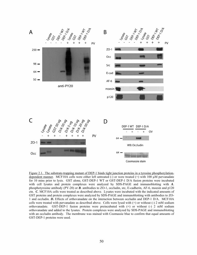
50
Figure 2.1. The substrate-trapping mutant of DEP-1 binds tight junction proteins in a tyrosine phosphorylation-dependent manner. MCF10A cells were either left untreated (-) or were treated (+) with 100 µM pervanadate for 10 mins prior to lysis. GST alone, GST-DEP-1 WT or GST-DEP-1 D/A fusion proteins were incubated with cell lysates and protein complexes were analyzed by SDS-PAGE and immunoblotting with A. phosphotyrosine antibody (PY-20) or B. antibodies to ZO-1, occludin, src, E-cadherin, AF-6, moesin and p120 ctn. C. MCF10A cells were treated as described above. Lysates were incubated with the indicated amounts of GST proteins and protein complexes were analyzed by SDS-PAGE and immunoblotting with antibodies to ZO-1 and occludin. D. Effects of orthovanadate on the interaction between occludin and DEP-1 D/A. MCF10A cells were treated with pervanadate as described above. Cells were lysed with (+) or without (-) 2 mM sodium orthovanadate. GST-DEP-1 fusion proteins were preincubated with (+) or without (-) 2 mM sodium orthovanadate and added to the lysates. Protein complexes were analyzed by SDS-PAGE and immunoblotting with an occludin antibody. The membrane was stained with Coomassie blue to confirm that equal amounts of GST-DEP-1 proteins were used.
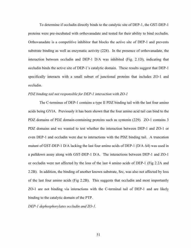
51
To determine if occludin directly binds to the catalytic site of DEP-1, the GST-DEP-1
proteins were pre-incubated with orthovanadate and tested for their ability to bind occludin.
Orthovanadate is a competitive inhibitor that blocks the active site of DEP-1 and prevents
substrate binding as well as enzymatic activity (228). In the presence of orthovanadate, the
interaction between occludin and DEP-1 D/A was inhibited (Fig. 2.1D), indicating that
occludin binds the active site of DEP-1’s catalytic domain. These results suggest that DEP-1
specifically interacts with a small subset of junctional proteins that includes ZO-1 and
occludin.
PDZ binding tail not responsible for DEP-1 interaction with ZO-1
The C-terminus of DEP-1 contains a type II PDZ binding tail with the last four amino
acids being GYIA. Previously it has been shown that the four amino acid tail can bind to the
PDZ domains of PDZ domain-containing proteins such as syntenin (229). ZO-1 contains 3
PDZ domains and we wanted to test whether the interaction between DEP-1 and ZO-1 or
even DEP-1 and occludin were due to interactions with the PDZ binding tail. A truncation
mutant of GST-DEP-1 D/A lacking the last four amino acids of DEP-1 (D/A Δ4) was used in
a pulldown assay along with GST-DEP-1 D/A. The interactions between DEP-1 and ZO-1
or occludin were not affected by the loss of the last 4 amino acids of DEP-1 (Fig 2.2A and
2.2B). In addition, the binding of another known substrate, Src, was also not affected by loss
of the last four amino acids (Fig 2.2B). This suggests that occludin and most importantly
ZO-1 are not binding via interactions with the C-terminal tail of DEP-1 and are likely
binding to the catalytic domain of the PTP.
DEP-1 dephosphorylates occludin and ZO-1.
52
Having shown that occludin and ZO-1 can bind to DEP-1, we wanted to further test
whether they are in fact direct substrates of the phosphatase. First, MCF10A cells were
treated with pervanadate to induce phosphorylation of occludin and ZO-1, and then lysates
were incubated with GST, GST-DEP-1 WT, or GST-DEP-1 D/A. Occludin or ZO-1 was
immunoprecipitated from the lysates and phosphotyrosine levels were revealed by blotting
with an anti-phosphotyrosine antibody. As shown in Fig. 2.3A, wildtype DEP-1
dephosphorylated both occludin and ZO-1, whereas GST and DEP-1 D/A did not; these
results confirm that occludin and ZO-1 are direct substrates of DEP-1 in vitro. Note the
extensive band shift of immunoprecipitated occludin incubated with DEP-1 WT is consistent
with dephosphorylation of occludin by the PTP. Next, we asked whether DEP-1 can
dephosphorylate occludin when both are expressed together in cells. Full length DEP-1 WT
or DEP-1 D/A were co-expressed with occludin in HEK 293ft cells, occludin was
immunoprecipitated and phosphotyrosine levels were analyzed. In cells over expressing
DEP-1 WT, phosphorylation of occludin was dramatically reduced compared to cells
expressing empty vector or DEP-1 D/A (Fig. 2.3B). These combined experiments suggest
that both ZO-1 and occludin are substrates of DEP-1 and that DEP-1 catalytic activity is
required for the dephosphorylation of occludin in vivo.
53
Figure 2.2. The PDZ tail of DEP-1 is not responsible for the interactions with substrates. A truncation mutant of DEP-1 missing the last four amino acids of the C-terminal tail was expressed as a GST fusion protein (GST-DEP-1 D/A Δ4) and tested for interaction with DEP-1 substrates. MCF10A cells were either left untreated (-) or were treated (+) with 100 µM pervanadate for 10 mins prior to lysis. Lysates were incubated with GST, GST-DEP-1 D/A (D/A), or GST-DEP-1 D/A Δ4 (D/A Δ4) and protein complexes were resolved with SDS-PAGE and immunoblotting with A. ZO-1 antibody or B. Occludin and Src antibodies. The smear in the GST lanes of (A) appear to be the non-specific binding of proteins and was found in the entire length of the lane. The GST control was not shown in (B) but previous experiments have never shown occludin binding to GST
.
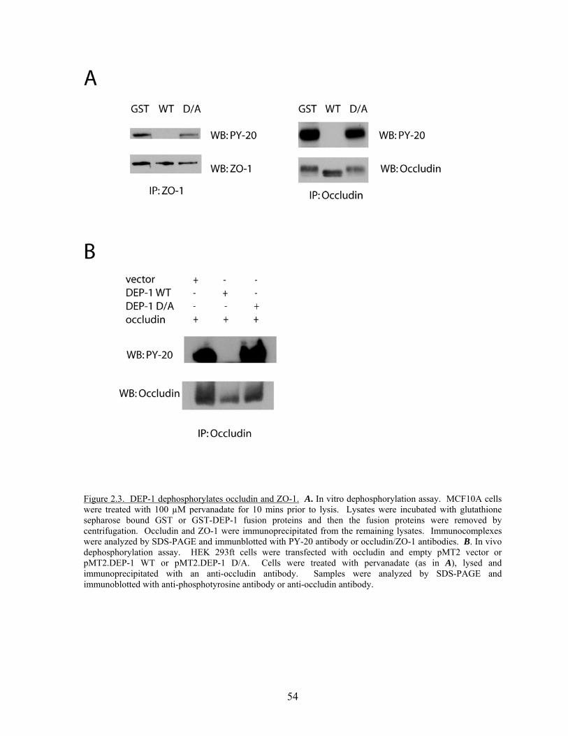
54
Figure 2.3. DEP-1 dephosphorylates occludin and ZO-1. A. In vitro dephosphorylation assay. MCF10A cells were treated with 100 µM pervanadate for 10 mins prior to lysis. Lysates were incubated with glutathione sepharose bound GST or GST-DEP-1 fusion proteins and then the fusion proteins were removed by centrifugation. Occludin and ZO-1 were immunoprecipitated from the remaining lysates. Immunocomplexes were analyzed by SDS-PAGE and immunblotted with PY-20 antibody or occludin/ZO-1 antibodies. B. In vivo dephosphorylation assay. HEK 293ft cells were transfected with occludin and empty pMT2 vector or pMT2.DEP-1 WT or pMT2.DEP-1 D/A. Cells were treated with pervanadate (as in A), lysed and immunoprecipitated with an anti-occludin antibody. Samples were analyzed by SDS-PAGE and immunoblotted with anti-phosphotyrosine antibody or anti-occludin antibody.
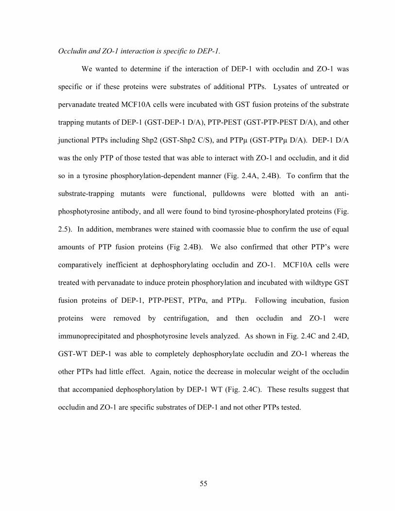
55
Occludin and ZO-1 interaction is specific to DEP-1.
We wanted to determine if the interaction of DEP-1 with occludin and ZO-1 was
specific or if these proteins were substrates of additional PTPs. Lysates of untreated or
pervanadate treated MCF10A cells were incubated with GST fusion proteins of the substrate
trapping mutants of DEP-1 (GST-DEP-1 D/A), PTP-PEST (GST-PTP-PEST D/A), and other
junctional PTPs including Shp2 (GST-Shp2 C/S), and PTPµ (GST-PTPµ D/A). DEP-1 D/A
was the only PTP of those tested that was able to interact with ZO-1 and occludin, and it did
so in a tyrosine phosphorylation-dependent manner (Fig. 2.4A, 2.4B). To confirm that the
substrate-trapping mutants were functional, pulldowns were blotted with an anti-
phosphotyrosine antibody, and all were found to bind tyrosine-phosphorylated proteins (Fig.
2.5). In addition, membranes were stained with coomassie blue to confirm the use of equal
amounts of PTP fusion proteins (Fig 2.4B). We also confirmed that other PTP’s were
comparatively inefficient at dephosphorylating occludin and ZO-1. MCF10A cells were
treated with pervanadate to induce protein phosphorylation and incubated with wildtype GST
fusion proteins of DEP-1, PTP-PEST, PTPα, and PTPµ. Following incubation, fusion
proteins were removed by centrifugation, and then occludin and ZO-1 were
immunoprecipitated and phosphotyrosine levels analyzed. As shown in Fig. 2.4C and 2.4D,
GST-WT DEP-1 was able to completely dephosphorylate occludin and ZO-1 whereas the
other PTPs had little effect. Again, notice the decrease in molecular weight of the occludin
that accompanied dephosphorylation by DEP-1 WT (Fig. 2.4C). These results suggest that
occludin and ZO-1 are specific substrates of DEP-1 and not other PTPs tested.
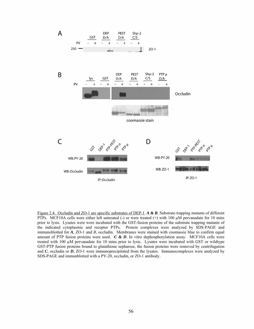
56
Figure 2.4. Occludin and ZO-1 are specific substrates of DEP-1. A & B. Substrate-trapping mutants of different PTPs. MCF10A cells were either left untreated (-) or were treated (+) with 100 µM pervanadate for 10 mins prior to lysis. Lysates were were incubated with the GST-fusion proteins of the substrate trapping mutants of the indicated cytoplasmic and receptor PTPs. Protein complexes were analyzed by SDS-PAGE and immunoblotted for A, ZO-1 and B, occludin. Membranes were stained with coomassie blue to confirm equal amount of PTP fusion proteins were used. C & D. In vitro dephosphorylation assay. MCF10A cells were treated with 100 µM pervanadate for 10 mins prior to lysis. Lysates were incubated with GST or wildtype GST-PTP fusion proteins bound to glutathione sepharose, the fusion proteins were removed by centrifugation and C, occludin or D, ZO-1 were immunoprecipitated from the lysates. Immunocomplexes were analyzed by SDS-PAGE and immunblotted with a PY-20, occludin, or ZO-1 antibody.
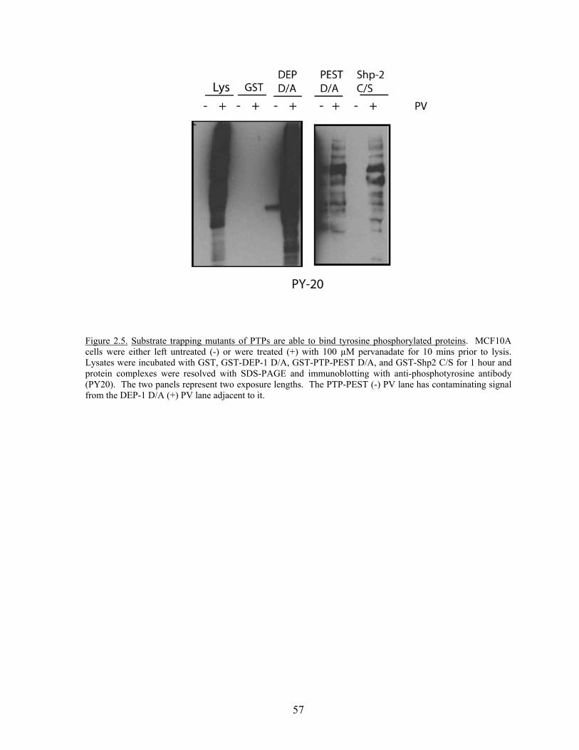
57
Figure 2.5. Substrate trapping mutants of PTPs are able to bind tyrosine phosphorylated proteins. MCF10A cells were either left untreated (-) or were treated (+) with 100 µM pervanadate for 10 mins prior to lysis. Lysates were incubated with GST, GST-DEP-1 D/A, GST-PTP-PEST D/A, and GST-Shp2 C/S for 1 hour and protein complexes were resolved with SDS-PAGE and immunoblotting with anti-phosphotyrosine antibody (PY20). The two panels represent two exposure lengths. The PTP-PEST (-) PV lane has contaminating signal from the DEP-1 D/A (+) PV lane adjacent to it.
58
SAP-1 can dephosphorylate occludin
SAP-1 (stomach cancer-associated protein tyrosine phosphatase-1) is a receptor PTP
which is structurally similar to DEP-1 with an extracellular domain of 8 fibronectin type III
repeats, a transmembrane domain and a single intracellular catalytic domain (230). With its
known structural similarity to DEP-1, we were interested in determining whether the tight
junction proteins occludin and ZO-1 are substrates of SAP-1. We tested the catalytically
inactive mutant of SAP-1 C/S in GST fusion pulldowns and found that occludin did not bind
to this phosphatase compared to DEP-1 D/A (Fig. 2.6A, 2.6B). In addition, ZO-1 was not
able to be pulled down with SAP-1 C/S (Fig. 2.6B).
We also examined the ability of SAP-1 to dephosphorylate occludin in an in vitro
dephosphorylation assay. MCF10A cells were treated with pervanadate and the lysates were
incubated with GST, GST DEP-1 WT or GST SAP-1 WT. Occludin was
immunoprecipitated from the lysates and phosphotyrosine levels were determined by
immunoblotting with a phosphotyrosine antibody. SAP-1 was able to visibly
dephosphorylate occludin and appears to do so even better than DEP-1 (Fig 2.6C). Both
PTPs have a dose dependent affect on phosphotyrosine levels. The preliminary results
suggest that occludin may in fact be a substrate for SAP-1. This would not be all together
surprising based on the similarities between the two PTPs.
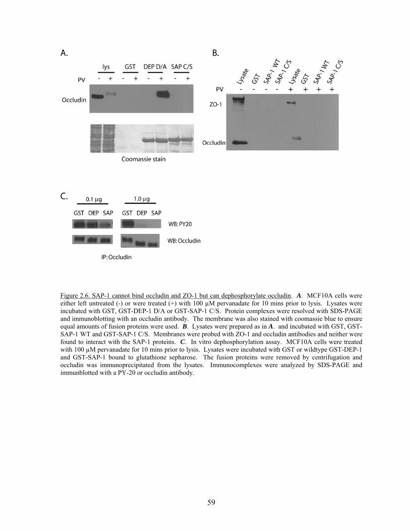
59
Figure 2.6. SAP-1 cannot bind occludin and ZO-1 but can dephosphorylate occludin. A. MCF10A cells were either left untreated (-) or were treated (+) with 100 µM pervanadate for 10 mins prior to lysis. Lysates were incubated with GST, GST-DEP-1 D/A or GST-SAP-1 C/S. Protein complexes were resolved with SDS-PAGE and immunoblotting with an occludin antibody. The membrane was also stained with coomassie blue to ensure equal amounts of fusion proteins were used. B. Lysates were prepared as in A. and incubated with GST, GST-SAP-1 WT and GST-SAP-1 C/S. Membranes were probed with ZO-1 and occludin antibodies and neither were found to interact with the SAP-1 proteins. C. In vitro dephosphorylation assay. MCF10A cells were treated with 100 µM pervanadate for 10 mins prior to lysis. Lysates were incubated with GST or wildtype GST-DEP-1 and GST-SAP-1 bound to glutathione sepharose. The fusion proteins were removed by centrifugation and occludin was immunoprecipitated from the lysates. Immunocomplexes were analyzed by SDS-PAGE and immunblotted with a PY-20 or occludin antibody.
60
DEP-1 binds and dephosphorylates Src-induced phospho-occludin
There are 43 tyrosines in occludin, 23 of which are located in either the intracellular
N- or C-terminus. In the previous experiments we used pervanadate to induce tyrosine
phosphorylation of junctional proteins. Pervanadate is an irreversible PTP-inhibitor that is
102-103 times more potent than vanadate alone (Kadota, Posner 1987; Fatutus and posner
1989). This allows us to increase pools of tyrosine phosphorylated proteins, however, it is
difficult to determine which kinase is acting on our substrates or which residues are
phosphorylated. The identification of kinase(s) acting on the tight junction proteins as well
as the tyrosine residues that DEP-1 is binding would help to clarify how tyrosine
phosphorylation is regulating tight junction function. One potential kinase is Src kinase. Src
localizes to tight junctions (231), binds ZO-1 (232), and can phosphorylate occludin in vitro
leading to disruption of TJs (217). There are several Src consensus sites in the N-and C-
terminal tails of occludin and DEP-1 may be able to bind to one of these sites. First, we
examined whether constitutively active Src (Y527F) was able to phosphorylate occludin in
transfected cells. HEK 293ft cells were co-tranfected with full length occludin and either
empty vector or pCMV-SrcY527F. Cells were lysed in hot SDS sample buffer, occludin was
immunoprecipitated, and phosphotyrosine levels were analyzed. Expression of constitutively
active Src was able to phosphorylate occludin (Fig. 2.7A), supporting data by Kale et al.
(217). Next, we determined whether the Src induced phospho-occludin was able to bind to
the substrate trapping mutant of DEP-1. Again, HEK 293ft cells were transfected with
occludin and either empty vector or SrcY527F. GST-DEP-1 pulldown assays were
performed with the lysates from the transfected cells and the membranes were probed for
occludin. We found that Src-phosphorylated occludin interacted with the substrate trapping
61
mutant of DEP-1 (Fig. 2.7B). Knowing that Src can phosphorylate occludin and these
phosphorylation sites interact with the substrate trapping mutant of DEP-1, we tested whether
wildtype DEP-1 can dephosphorylate Src-induced phospho-occludin. We expressed
wildtype or the catalytically inactive substrate trapping mutant of DEP-1 with occludin and
SrcY527F in HEK 293 cells, immunoprecipitated occludin and analyzed phosphotyrosine
levels. The addition of DEP-1 WT reduced the phosphotyrosine levels of occludin,
suggesting that DEP-1 was able to dephosphoorylate Src phosphorylation sites (Fig. 2.7A).
It is interesting to note that HEK 293ft cells have endogenous DEP-1 and when transfected
with constitutively active Src, endogenous DEP-1 levels increase (Fig. 2.7A).
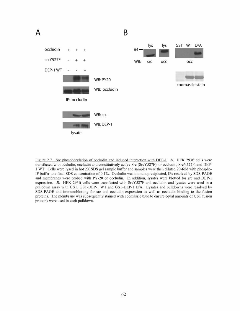
62
Figure 2.7. Src phosphorylation of occludin and induced interaction with DEP-1. A. HEK 293ft cells were transfected with occludin, occludin and constitutively active Src (SrcY527F), or occludin, SrcY527F, and DEP-1 WT. Cells were lysed in hot 2X SDS gel sample buffer and samples were then diluted 20-fold with phospho-IP buffer to a final SDS concentration of 0.1%. Occludin was immunoprecipitated, IPs resolved by SDS-PAGE and membranes were probed with PY-20 or occludin. In addition, lysates were blotted for src and DEP-1 expression. B. HEK 293ft cells were transfected with SrcY527F and occludin and lysates were used in a pulldown assay with GST, GST-DEP-1 WT and GST-DEP-1 D/A. Lysates and pulldowns were resolved by SDS-PAGE and immunoblotting for src and occludin expression as well as occludin binding to the fusion proteins. The membrane was subsequently stained with coomassie blue to ensure equal amounts of GST fusion proteins were used in each pulldown.
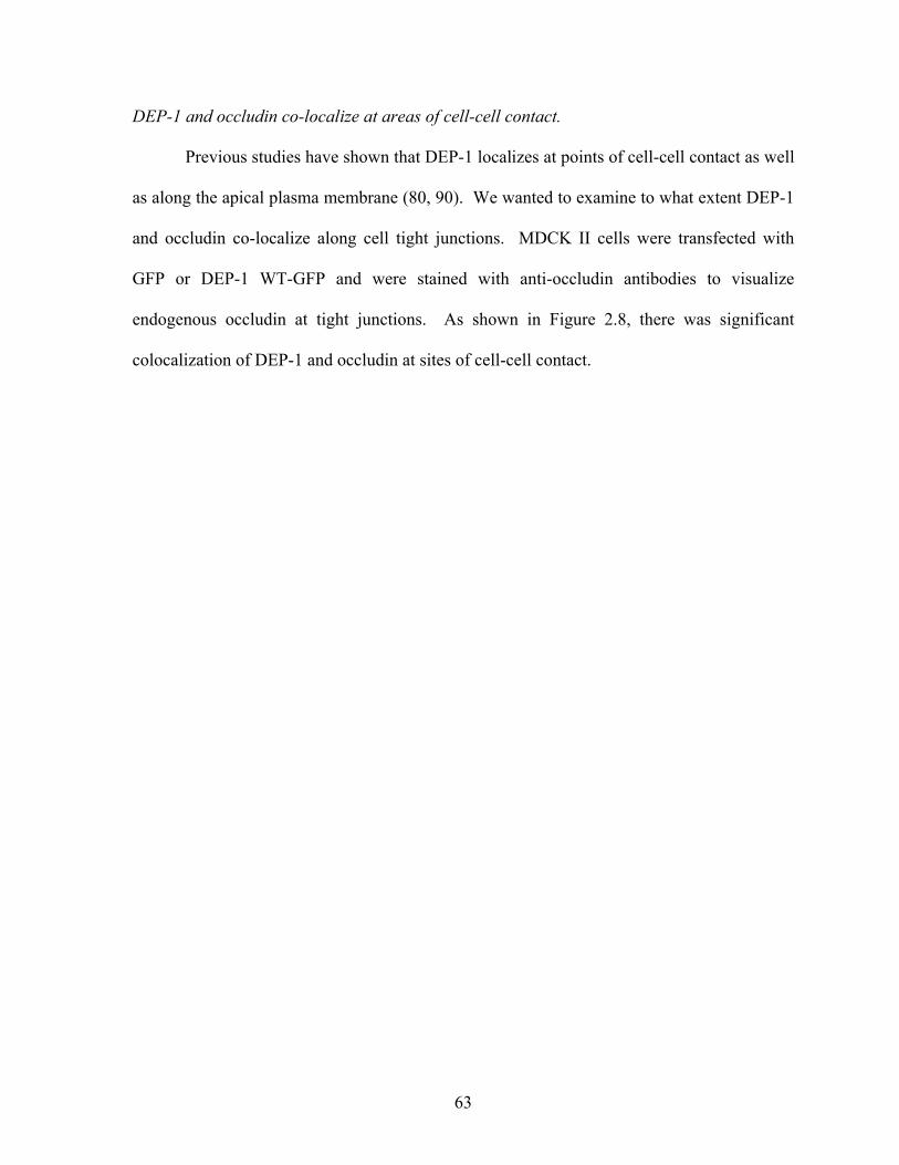
63
DEP-1 and occludin co-localize at areas of cell-cell contact.
Previous studies have shown that DEP-1 localizes at points of cell-cell contact as well
as along the apical plasma membrane (80, 90). We wanted to examine to what extent DEP-1
and occludin co-localize along cell tight junctions. MDCK II cells were transfected with
GFP or DEP-1 WT-GFP and were stained with anti-occludin antibodies to visualize
endogenous occludin at tight junctions. As shown in Figure 2.8, there was significant
colocalization of DEP-1 and occludin at sites of cell-cell contact.
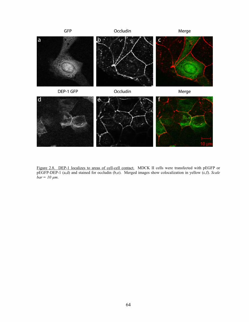
64
Figure 2.8. DEP-1 localizes to areas of cell-cell contact. MDCK II cells were transfected with pEGFP or pEGFP-DEP-1 (a,d) and stained for occludin (b,e). Merged images show colocalization in yellow (c,f). Scale bar = 10 µm.
65
DEP-1 expression enhances barrier function as junction reassemble.
We next wanted to determine whether our previously observed dephosphorylation of
occludin and ZO-1 had functional consequences with respect to tight junction physiology.
MDCK II cells were infected with GFP or DEP-1 WT adenovirus, plated onto transwell
filters, and 4 days later the transepithelial electrical resistance (TER) of the stable junctions
was measured. In epithelial cells overexpressing DEP-1 we saw a small but significant
increase in TER compared to the GFP control (p<0.005) (Fig. 2.9), supporting the hypothesis
that expression of DEP-1 can decrease the permeability of the tight junction.
To better address the ability of DEP-1 to regulate tight junction function, we looked
at FITC-dextran flux across an epithelial monolayer during junctional reassembly following a
calcium switch. MDCK II cells were infected with GFP or DEP-1 WT adenovirus and plated
on transwell filters. After cells had adhered, growth media was replaced with low calcium (5
µM) media overnight. Normal calcium-containing media (1.8 mM) was then added to the
transwells and the ability of FITC-dextran to pass across the monolayer was assessed at 0, 6,
12, 24 and 48 hours. The amount of FITC-dextran that crossed the monolayers was
significantly reduced as junctions reassembled (Fig. 2.10A). At early stages of junction
reassembly (6 hrs), the presence of additional DEP-1 WT decreased permeability indicated
by the further decrease of FITC-dextran flux compared to GFP control cells (Fig. 2.10A,
2.10B.) After 12 and 24 hours in calcium-containing media, the permeability between GFP-
and DEP-1 expressing cells was not significantly different (Fig. 2.10A, 2.10B). In identical
experiments, TER was measured following the calcium switch. Monolayers over-expressing
DEP-1 WT have higher TER than GFP controls at early time points of junction reassembly

66
(Fig. 2.10C). Together the TER and FITC-dextran data reveal that an increase in DEP-1 WT
enhances the barrier function of
67
Figure 2.9. DEP-1 enhances junctional integrity in epithelial cells. MDCK II cells were infected with GFP alone or DEP-1 WT adenovirus, plated on filters (0.4µm pore) and allowed to grow into confluent monolayers. Transepithelial electrical resistance was taken at 4 days post plating. Values are expressed as the mean ± SEM of 4 experiments performed in triplicate. Baseline TER ranges from 45-50 ohms/cm2. Asterisk indicates the value is significantly (p<0.005) different from corresponding values for control group (GFP).
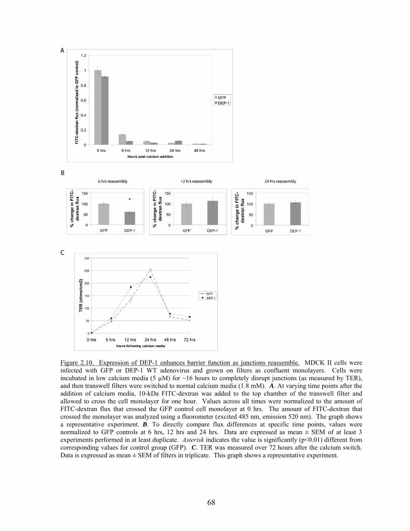
68
Figure 2.10. Expression of DEP-1 enhances barrier function as junctions reassemble. MDCK II cells were infected with GFP or DEP-1 WT adenovirus and grown on filters as confluent monolayers. Cells were incubated in low calcium media (5 µM) for ~16 hours to completely disrupt junctions (as measured by TER), and then transwell filters were switched to normal calcium media (1.8 mM). A. At varying time points after the addition of calcium media, 10-kDa FITC-dextran was added to the top chamber of the transwell filter and allowed to cross the cell monolayer for one hour. Values across all times were normalized to the amount of FITC-dextran flux that crossed the GFP control cell monolayer at 0 hrs. The amount of FITC-dextran that crossed the monolayer was analyzed using a fluorometer (excited 485 nm, emission 520 nm). The graph shows a representative experiment. B. To directly compare flux differences at specific time points, values were normalized to GFP controls at 6 hrs, 12 hrs and 24 hrs. Data are expressed as mean ± SEM of at least 3 experiments performed in at least duplicate. Asterisk indicates the value is significantly (p<0.01) different from corresponding values for control group (GFP). C. TER was measured over 72 hours after the calcium switch. Data is expressed as mean ± SEM of filters in triplicate. This graph shows a representative experiment.
69
epithelial junctions during reassembly. Thus, the presence of DEP-1 is particularly important
at the earliest, most dynamic stages of junctional assembly.
Discussion
Cell-cell junctions can be regulated through reversible tyrosine phosphorylation.
Tyrosine phosphorylation of adherens junction proteins has been shown either to promote the
dissociation of protein complexes at the junction (70, 73, 74, 77) or to promote the
internalization and degradation of junctional proteins (76). Whereas most work in this area
has focused on the tyrosine kinases that act on junctional proteins, it is equally important to
identify the PTPs and their targets within cell-cell junctions. Here we show for the first time
that the prominent tight junction proteins, occludin and ZO-1, interact with and are
dephosphorylated by the PTP DEP-1. Experimentally increasing the levels of DEP-1 reduced
the permeability of epithelial monolayers and this effect was enhanced during the early
stages of junction assembly.
Similar to the adherens junctions, tyrosine phosphorylation has been correlated with
the dissociation of tight junction components, promoting the detachment of ZO-1 from
occludin (214, 215, 217, 233, 234). This increased tyrosine phosphorylation of tight junction
components is paralleled by increased epithelial permeability. Because DEP-1 has
previously been localized to regions of cell-cell contact (80, 90), we examined whether the
phosphorylation of tight junction proteins was regulated by this PTP. We found that the
catalytically dead trapping mutant of DEP-1 binds to phosphorylated forms of occludin and
ZO-1 and that catalytically active DEP-1 dephosphorylates these proteins. Previous work
has shown that DEP-1 interacts with and acts on the adherens junction proteins p120catenin
70
and β-catenin (80, 149), and we have confirmed these interactions (Fig. 2.1B and data not
shown). However, we found that DEP-1 does not bind all tyrosine phosphorylated junctional
proteins (Fig. 2.1B). It is striking that several PTPs are concentrated within epithelial
junctions (235) and it is likely that there is some redundancy with respect to their targets.
However, we were surprised to find that two junctional PTPs, PTPμ and SHP-2, did not
significantly interact with or dephosphorylate occludin and ZO-1 (Fig. 2.4). This result
suggests that the different PTPs within cell-cell junctions have specific targets and that they
may regulate distinct signaling pathways. These pathways may either be initiated within
junctions in response to cell-cell interactions or be triggered by a stimulus such as growth
factors or oxidative stress to regulate junction stability, strength, and permeability. It is
worthy of note that we discovered that the highly homologus PTP SAP-1 is able to
dephosphorylate occludin although no interactions with the substrate trapping mutant (SAP-1
C/S) were found. The C/S mutation in PTP catalytic domains does not have as high an
affinity for substrates since the mutation is of the catalytic cysteine. As a result, the C/S
mutant is not as efficient at binding substrates as compared to the D/A mutant (8). Noguchi
et al. (236) have specifically shown that SAP-1 C/S is less efficient at binding proteins from
pervanadate treated cells than SAP-1 D/A. Therefore, the D/A mutant will be made in the
GST-SAP-1 construct and this experiment will be repeated to clarify whether tight junction
proteins may be substrates. This may provide evidence that although different types of PTPs
may have different substrates, PTPs within the same family type may regulate similar
signaling pathways.
Having found that DEP-1 dephosphorylates occludin and ZO-1, we wanted to explore
DEP-1’s effect on tight junction function in epithelial cells. Mature junctions are thought to
71
exist in a tyrosine dephosphorylated state, with increased tyrosine phosphorylation leading to
increased junction disassembly, increase paracellular permeability and can induce
reorganization of the junctional complex (212, 222). Thus, dephosphorylation of occludin as
well as other junctional proteins, is believed to increase the barrier function of the junction.
Our over-expression of DEP-1 in epithelial cells increased TER (Fig. 2.9), a measure of
increased barrier properties of the junctions, consistent with phosphorylation regulating the
permeability of tight junctions. Although the difference was subtle (5-10% increase), it was
reproducible and statistically significant. We believe the small effect on TER is due to the
fact that the cells had been confluent for several days. At this point, the junctions have
reduced dynamics and the level of tyrosine phosphorylation is very low (essentially
undetectable). Because tyrosine phosphorylation is elevated during disassembly/assembly of
junctions, this led us to explore the effect DEP-1 had on junctional permeability as the
junctions were assembling. Using the established calcium switch technique (237, 238) to
modulate cell-cell adhesion, we measured the permeability of epithelial junctions with both
TER and FITC-dextran flux over a time course of junction reassembly. As shown in Figure
2.10A, as junctions reassemble after calcium addition, the permeability decreases over time.
Importantly, when comparing the permeability of GFP-expressing versus DEP-1-expressing
cells at 6 hrs following calcium restoration, the presence of DEP-1 significantly reduces the
permeability of FITC-dextran flux, suggesting that these junctions reassembled more rapidly
(Fig. 2.10B). By 12 and 24 hours the difference in permeability between GFP- and DEP-1-
expressing cells was not statistically significant (Fig. 2.10B). The TER data shows a slightly
shifted trend, with the cells over-expressing DEP-1 still maintaining an increase in TER at 12
hrs following the initiation of reassembly. This difference may reflect the difference between
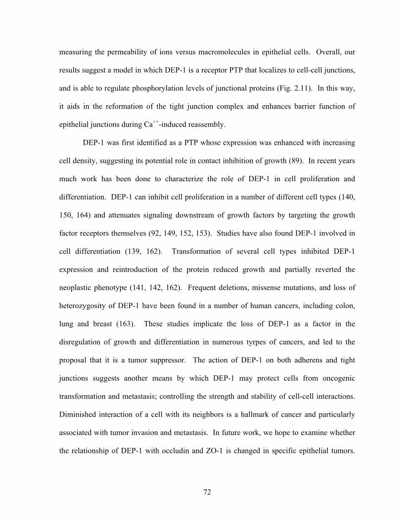
72
measuring the permeability of ions versus macromolecules in epithelial cells. Overall, our
results suggest a model in which DEP-1 is a receptor PTP that localizes to cell-cell junctions,
and is able to regulate phosphorylation levels of junctional proteins (Fig. 2.11). In this way,
it aids in the reformation of the tight junction complex and enhances barrier function of
epithelial junctions during Ca++-induced reassembly.
DEP-1 was first identified as a PTP whose expression was enhanced with increasing
cell density, suggesting its potential role in contact inhibition of growth (89). In recent years
much work has been done to characterize the role of DEP-1 in cell proliferation and
differentiation. DEP-1 can inhibit cell proliferation in a number of different cell types (140,
150, 164) and attenuates signaling downstream of growth factors by targeting the growth
factor receptors themselves (92, 149, 152, 153). Studies have also found DEP-1 involved in
cell differentiation (139, 162). Transformation of several cell types inhibited DEP-1
expression and reintroduction of the protein reduced growth and partially reverted the
neoplastic phenotype (141, 142, 162). Frequent deletions, missense mutations, and loss of
heterozygosity of DEP-1 have been found in a number of human cancers, including colon,
lung and breast (163). These studies implicate the loss of DEP-1 as a factor in the
disregulation of growth and differentiation in numerous tyrpes of cancers, and led to the
proposal that it is a tumor suppressor. The action of DEP-1 on both adherens and tight
junctions suggests another means by which DEP-1 may protect cells from oncogenic
transformation and metastasis; controlling the strength and stability of cell-cell interactions.
Diminished interaction of a cell with its neighbors is a hallmark of cancer and particularly
associated with tumor invasion and metastasis. In future work, we hope to examine whether
the relationship of DEP-1 with occludin and ZO-1 is changed in specific epithelial tumors.

73
The continued identification of junctional substrates of DEP-1 will aid in furthering our
understanding of the role of this PTP in diseases such as cancer.
74
Figure 2.11. Model of how DEP-1 may regulate tight junction integrity. The transmembrane protein occludin binds via its C-terminal tail to the cytoplasmic ZO family of proteins. These ZO proteins link the complex to the actin cytoskeleton creating a stable complex and intact junction. Tyrosine phosphorylation events by Src or other PTKs can phosphorylate both occludin and ZO-1, disrupting their interactions with each other and reducing junction integrity. DEP-1 is a receptor PTP which is able to dephosphorylate both occludin and ZO-1 and may aid in reforming intact junctions.
75
Materials and Methods
Cell Culture and Transfections-- MCF10A cells were grown in a 1:1 mixture of Dulbecco’s
modified Eagle’s medium (DMEM) and F12/Ham medium supplemented with 5% horse
serum, 20 ng/ml EGF, 0.01 mg/ml insulin, 500 ng/ml hydrocortisone, 100 ng/ml cholera
toxin and antibiotics. MDCK II and HEK 293ft cells were cultured in Dulbecco's modified
Eagle's medium-high glucose enriched with 10% fetal bovine serum and antibiotics. Cells
were transfected with Lipofectamine 2000 (Invitrogen) or Fugene6 (Roche Applied Science)
according to the manufacturer’s instructions.
Constructs and Antibodies--pMT2 DEP-1 WT and pMT2 DEP-1 D/A (GenBank TM
accession number U10886) were kindly provided by Nicholas Tonks (Cold Spring Harbor).
pEGFP DEP-1 WT and pEGFP DEP-1 D/A were generated by sub cloning the pMT2
contructs into pEGFP-N2 (Clontech). DEP-1 cytoplasmic domain constructs were generated
using the pMT2 DEP-1 wildtype or D1205A point mutant as templates. A 5’ primer
introduced an EcoR1 before the DEP-1 cytoplasmic sequence at nucleotide 3338 and a
3’primer introduced a Xho1 site after the DEP-1 stop codon. The resulting PCR fragments
(nucleotides 3338-4362) were cloned into the EcoR1/Xho1 sites of the pGEX-4T-1 vector
(GE Healthcare), creating wildtype and point mutant D1205A pGEX-DEP-1 constructs. The
GST-DEP-1 D/A Δ4 construct was generated using the same 5’ primer as above and a 3’
primer which inserted a stop codon after nucleotide 4350. GST-PTP-PEST wildtype and
substrate trapping mutant were obtained from Dr. Sarita Sastry (University of Texas
Galveston), GST-PTP µ wildtype and substrate trapping mutant were kind gifts from Dr.
Susann Brady-Kalnay (Case Western), and GST SAP-1 constructs were given by Takashi
76
Matozaki (Gunma University, Japan). Full length occludin was provided by Alan Fanning
(University of North Carolina Chapel Hill). Constitutively active (Y527F) src was obtained
from Daniel Flynn (West Virginia University). For adenovirus, the full length cDNA of
DEP-1 WT was cloned into pENTR1D (Invitrogen). The entry clone was recombined with
the pAd/CMV/V5 destination vector via LR clonase II. The ViraPower Adenviral expression
vector (Invitrogen Life Technologies) was linearized and then transfected into 293A cells via
Lipofectamine 2000 to generate adenovirus encoding V5-tagged DEP-1. The virus particles
were used to infect MDCK II cells.
Mouse anti-occludin and rabbit anti-ZO-1 antibodies were purchased from Zymed
Laboratories Inc. Mouse anti-human CD148 (DEP-1) antibody was purchased from
Biosource. GFP monoclonal antibody was from Roche. Monoclonal antibodies to E-
cadherin, moesin and p120ctn were purchased from BD Bioscience, anti-Src was purchased
from Millipore, AF-6 rabbit polyclonal was from Novus Biological, and monoclonal PY-20
was purchased from Santa Cruz Biotechnology.
GST fusion proteins-- Expression of the fusion proteins in Escherichia coli were induced with
100 μM IPTG (isopropyl-β-D-thiogalactopyranoside) for 16 hours at room temperature.
Bacterial cells were lysed in buffer containing 50 mM Tris (pH 7.6), 150 mM NaCl, 1%
Triton X-100, 5 mM MgCl2, 1 mM DTT, 10 μg/ml each of aprotinin and leupeptin, and 1
mM phenylmethylsulfonyl fluoride (PMSF) and the fusion proteins were purified by
incubation with glutathione-sepharose 4B beads (GE healthcare) at 4ºC. Catalytic activity of
the fusion proteins was checked by PTP activity assays using pNPP (p-nitrophenylphosphate
(Sigma)).
77
Substrate Trapping Pulldown-- MCF10A cells were either left untreated or treated with 100
μM pervanadate (phosphatase inhibitor) for 15 mins prior to lysis. Cells were rinsed twice in
phosphate-buffered saline and lysed in a modified RIPA buffer (1% Tx-100, 0.5% DOC,
0.2% SDS, 150 mM NaCl, 20 mM Hepes ph 7.4, 2 mM EDTA, 10 μg/ml each aprotinin and
leupeptin, and 1mM PMSF). Insoluble material was removed by centrifugation. MCF10A
lysates (1 mg) were incubated with 10 μg of GST proteins for 1 hour at 4ºC. Beads were
washed in the modified RIPA buffer, resuspended in Laemmli sample buffer and analysed by
SDS-PAGE and western blotting on PVDF membranes (Millipore).
To determine whether the substrates were binding DEP-1 D/A at the PTP active site,
we tested the effect of orthovanadate on complex formation. GST fusion proteins bound to
glutathione sepharose were preincubated in a Hepes lysis buffer (1% Tx-100, 150 mM NaCl,
20 mM Hepes, pH 7.5, 5 mM MgCl2,) with or without 2 mM orthovanadate. MCF10A cells
were treated with 100 μm pervanadate for 20 mins and also lysed in Hepes lysis buffer with
or without 2 mM orthovanadate. Lysates ( 500 μg – 1mg) were incubated with 2 μg of GST-
DEP-1 or GST-DEP-1 D/A for 1.5 hours at 4ºC. Beads were washed in the Hepes Lysis
buffer, resuspended in Laemmli sample buffer, and analysed by SDS-PAGE and western
blotting on PVDF membranes.
Dephosphorylation Assays-- MCF10A cells were treated with 100 µM pervanadate for 15
mins and lysed in the modified RIPA buffer plus 10 μg/ml each aprotinin and leupeptin,
1mM PMSF, and 5 mM iodoacetic acid (IAA). After incubation of the lysates on ice for 10
mins, DTT was added at a final concentration of 10 mM for another 10 mins on ice to
78
inactivate the IAA. Lysates were clarified and 500 μg of total cell lysate was incubated with
1 μg glutathione-sepharose bound GST-PTPs for 20 minutes at room temperature. The
phosphatases were removed by centrifugation, orthovanadate was added to the samples and
occludin was immunoprecipitated for 2 hours at 4 ºC. Beads were washed in the lysis buffer
and processed for SDS PAGE. The immune complexes were analyzed by western blotting
using the PY-20 antibody, occludin monoclonal antibody or ZO-1 polyclonal antibody.
Alternatively, HEK 293ft cells were transfected with pMT2 contructs for 48 hours and lysed
directly in hot 2x SDS gel sample buffer (200mM Tris (pH 6.8), 20% glycerol, 4% SDS, 5%
2-ME), and boiled for 10 min. Samples were then diluted 20-fold with phospho-IP buffer
(150 mM NaCl, 20 mM Tris, pH 7.6, 1% Triton X-100, 1% sodium deoxycholate, 1 mM
sodium orthovanadate, 10 µg/ml leupeptin, 10 µg/ml aprotinin) to a final concentration of
0.1% SDS. One µg of occludin antibody and protein G-sepharose were added and samples
were incubated for 4 hours at 4°C. Samples were then washed in TBS and analyzed by
Western blot using anti-PY20 antibody.
Immunofluorescence-- MDCK II cells were fixed with 3.7% formaldehyde in phosphate
buffered saline and permeabilized in 0.5% Tx-100. Occludin was visualized cells with anti-
occludin monoclonal antibody (Zymed) at 1:200 for 45 mins at 37ºC followed by an anti-
mouse Alexa-594 conjugated secondary antibody. Images were recorded with a Zeiss
LSM510 confocal microscope.
Calcium Switch experiments—MDCK II cells were plated at confluent density (~4 x10⁵
cells/cm2) onto Transwell filters (0.4 µm pore, 12mm diameter; Corning). Growth media was
79
removed from cells 3-4 hours after plating and replaced with DMEM containing low calcium
(5 µM) overnight (~16 hours). Disruption of cell-cell junctions was confirmed by TER
measurements of only 2 ohms/cm2. Junction reassembly was induced with the addition of
calcium containing DMEM (1.8 mM CaCl2) for the indicated length of time.
Permeability assays--TER: MDCK II cells were infected with pAd/CMV/GFP or
pAd/CMV/V5-DEP-1 WT. 24 hours later, cells were plated at confluent density (~4 x10⁵
cells/cm2) onto Transwell filters (0.4 µm pore, 12mm diameter; Corning) and cultured for 4
days, with media replacement daily. TER was measured using an Endohm-12 Transwell
chamber connected to an EVOM voltohmeter according to the manufacturer’s instructions.
For calcium switch experiments, TER was measured at indicated time points following the
re-addition of calcium containing media. The resistance of the filter was subtracted from all
readings.
FITC-Dextran flux: MDCK II cells were infected with DEP-1 or GFP control adenovirus
and plated the same as for the TER measurements above. For the calcium switch, calcium
containing DMEM was added to the chambers for indicated length of time; 1 ml in the outer
chamber and 250 µl in the inner chamber. 10-kDa FITC-dextran (Molecular Probes) was
added to the top chamber in a volume of 50 µl at a final concentration of 1 mg/ml. After 1
hour, a sample was removed from the basolateral (bottom) compartments and read in a
fluorometer (FluroStar) (ex 485nm, em 520 nm).
Statistics—Comparison between two groups was made by the Student’s t test for unpaired
data.
.Chapter 3: Mapping the Region of Occludin Necessary for DEP-1 Interaction
Summary
In the previous chapter novel interactions were discovered between the PTP DEP-1 and
members of the tight junction. Specifically, we were able to characterize occludin as a
substrate of DEP-1 in epithelial cells. Occludin binds to the catalytic domain of DEP-1 and
not to the PDZ tail, however it is not known which region of occludin is responsible for its
interaction with DEP-1. Due to the larger number of intracellular tyrosines (23) in occludin,
this has been difficult to determine. Using truncation mutants we attempted to map the
region in the C-terminal tail of occludin that binds to DEP-1. In pervanadate treated HEK
293ft cells all C-terminal occludin mutants are able to bind the substrate trapping mutant of
DEP-1. This suggests that multiple sites may be important for this interaction. Future
experiments will focus specifically on residues in the ZO-1 binding site.
Introduction
The importance of occludin in tight junctions remains to be definitively resolved.
Mice lacking occludin show no abnormal phenotype and their epithelia still maintained TJ
strands and intact barrier function (204). Nevertheless, over-expression studies of full length
occludin have demonstrated that occludin is a functional component of the TJ (189, 239).
The N-terminal domains of occludin have been implicated in being necessary for correct
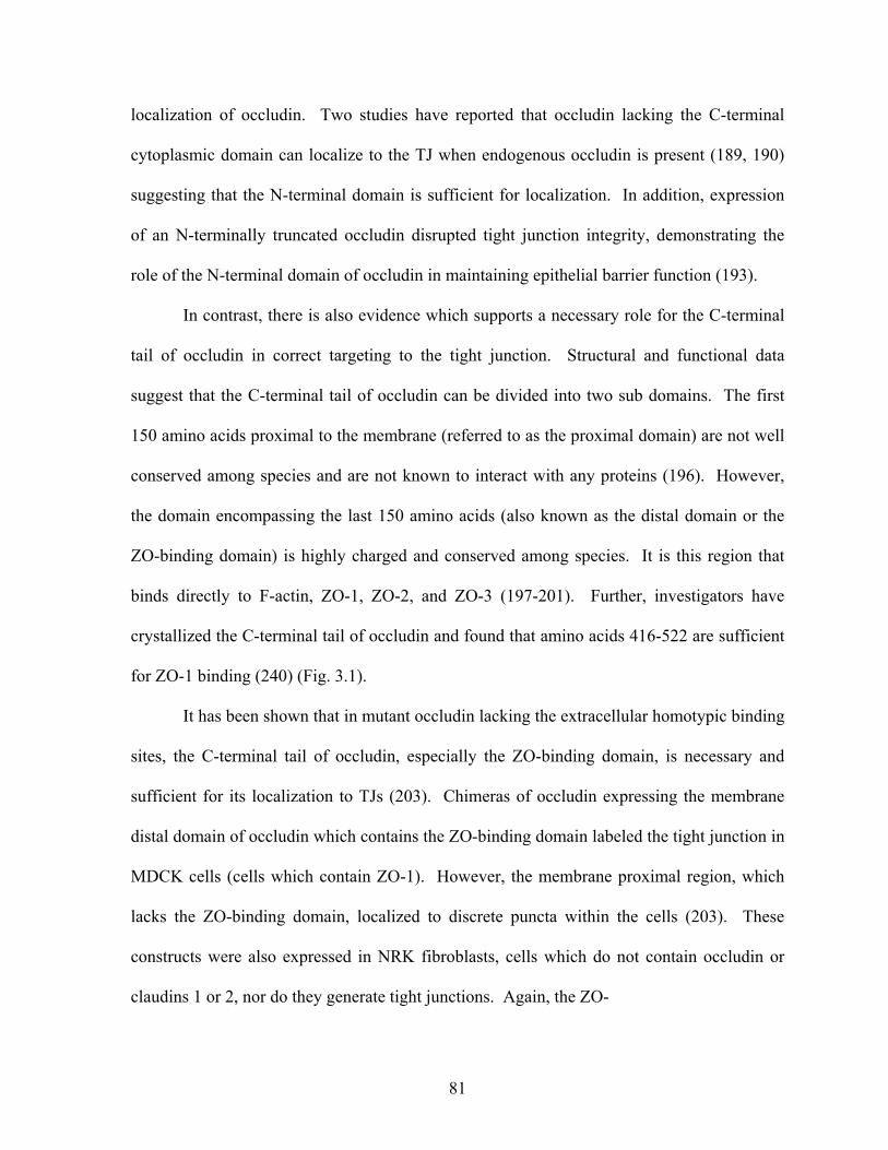
81
localization of occludin. Two studies have reported that occludin lacking the C-terminal
cytoplasmic domain can localize to the TJ when endogenous occludin is present (189, 190)
suggesting that the N-terminal domain is sufficient for localization. In addition, expression
of an N-terminally truncated occludin disrupted tight junction integrity, demonstrating the
role of the N-terminal domain of occludin in maintaining epithelial barrier function (193).
In contrast, there is also evidence which supports a necessary role for the C-terminal
tail of occludin in correct targeting to the tight junction. Structural and functional data
suggest that the C-terminal tail of occludin can be divided into two sub domains. The first
150 amino acids proximal to the membrane (referred to as the proximal domain) are not well
conserved among species and are not known to interact with any proteins (196). However,
the domain encompassing the last 150 amino acids (also known as the distal domain or the
ZO-binding domain) is highly charged and conserved among species. It is this region that
binds directly to F-actin, ZO-1, ZO-2, and ZO-3 (197-201). Further, investigators have
crystallized the C-terminal tail of occludin and found that amino acids 416-522 are sufficient
for ZO-1 binding (240) (Fig. 3.1).
It has been shown that in mutant occludin lacking the extracellular homotypic binding
sites, the C-terminal tail of occludin, especially the ZO-binding domain, is necessary and
sufficient for its localization to TJs (203). Chimeras of occludin expressing the membrane
distal domain of occludin which contains the ZO-binding domain labeled the tight junction in
MDCK cells (cells which contain ZO-1). However, the membrane proximal region, which
lacks the ZO-binding domain, localized to discrete puncta within the cells (203). These
constructs were also expressed in NRK fibroblasts, cells which do not contain occludin or
claudins 1 or 2, nor do they generate tight junctions. Again, the ZO-
82
Figure 3.1. The amino acid sequence for the intracellular C-terminal tail of occludin. Proximal domain (POC) consists of amino acids 265-372. Distal domain (DOC) consists of amino acids 373-522. ZO-1 binding domain consists of amino acids 416-522. Tyrosines are in red, critical lysines in blue, and the amino acids sufficient for ZO-1 binding are bolded and underlined.
83
binding domain of occludin was sufficient to localize occludin to sites containing ZO-1,
suggesting that the interactions with ZO proteins contribute to the targeting occludin at the TJ
(203).
Stable junctions are believed to exist in a dephosphorylated state and increased
tyrosine phosphorylation of both AJ and TJ proteins can affect the stability of these junctions
(69, 70, 212). Src is a tyrosine kinase that localizes to the TJ, binds to ZO-1 (232) and can
phosphorylate occludin in vitro (217). Phosphorylation of the C-terminal tail of occludin by
Src inhibits the binding of ZO-1, ZO-2, and ZO-3 and may be involved in Src-mediated
disruption of TJs (217). Therefore, we propose that the distal domain/ZO-binding domain of
occludin may be the region which interacts with DEP-1. The entire C-terminal tail of
occludin contains 17 tyrosines, with 10 of them in the distal domain (Fig. 3.1). Due to the
large number of tyrosines we decided not to mutate individual tyrosines. Instead, we used
chimeras and truncation mutants of the cytoplasmic C-terminal tail of occludin to narrow
down potential binding domains. We expressed the mutants and conducted pulldown assays
with the substrate-trapping mutant of DEP-1 to determine which domain was important for
the interaction. We found that tyrosines not only in the distal domain but also the proximal
domain of the c-terminal tail are able to interact with DEP-1.
Results
To investigate which domain/tyrosine residue(s) are important for occludin’s
interaction with DEP-1 we used chimeras of the gap junction protein connexin32 and C-
terminal domains of occludin (203). We saw binding of all constructs to GST-DEP-1 D/A,
but not to GST with pervanadate treatment (Fig. 3.2). We were concerned that the chimeras
84
could be binding to DEP-1 via the connexin32 component so we used the GST proteins in a
pulldown assay from pervanadate treated MCF10A cells and probed with an anti-connexin32
antibody. Connexin32 is able to interact with the substrate trapping mutant of DEP-1 (data
not shown) and thus we could not conclude whether the chimeras were binding due to
interactions with connexin32 or occludin.
With this knowledge, we developed truncation mutants of human occludin that were
similar to those used by Mitic et al (203), but without the connexin32 transmembrane
domains. First, we generated these mutants in pEGFP-N2 but found the GFP from
pervanadate-treated cells non-specifically interacted with the substrate-trapping mutant of
DEP-1, again confusing our results (data not shown). As a result, we generated similar myc-
tagged constructs (Fig. 3.3). The myc tag does not contain a tyrosine and should not be
phosphorylated by pervanadate treatment. HEK 293ft cells were transfected with full length
occludin as well as the mutants. 48-hrs post transfection cells were treated with pervanadate
and lysates were used in a substrate trapping mutant pulldown. Full length occludin, as well
as all three mutants, bound to DEP-1 D/A (Fig. 3.4).
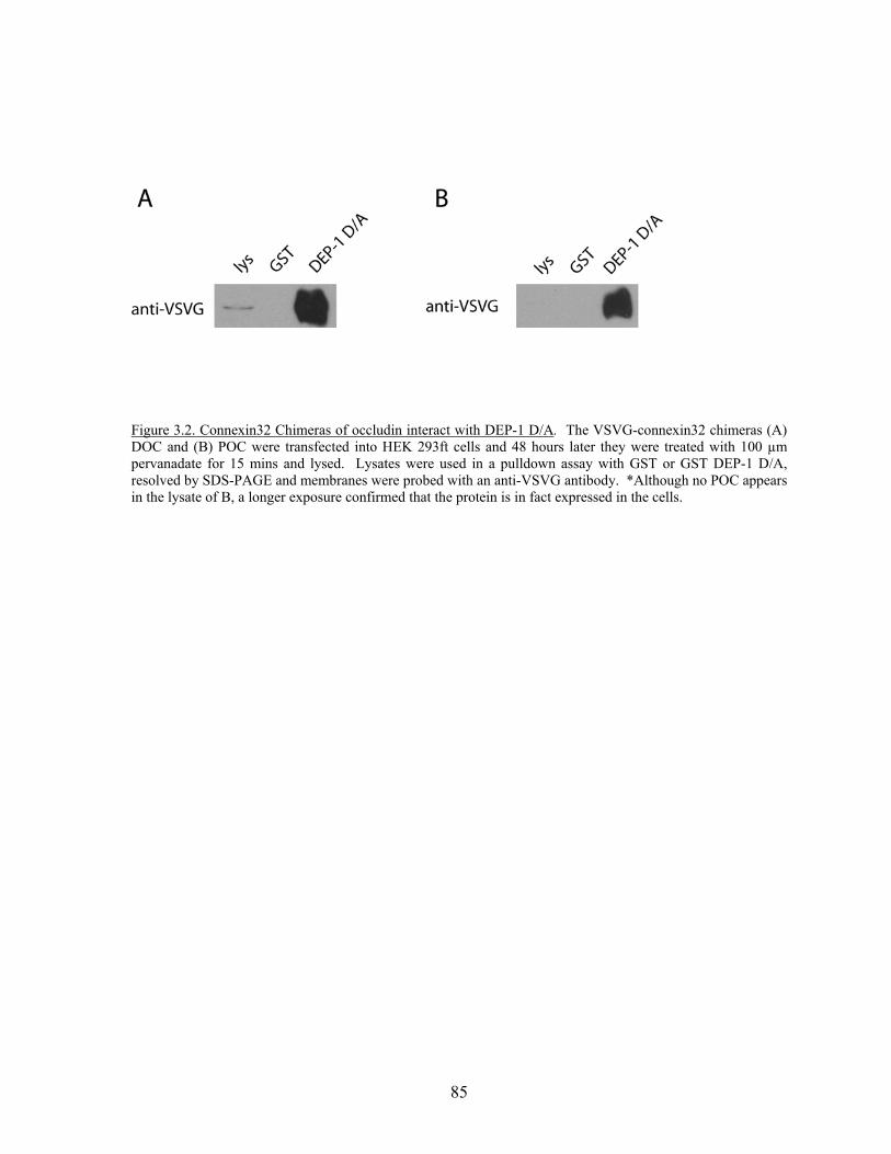
85
Figure 3.2. Connexin32 Chimeras of occludin interact with DEP-1 D/A. The VSVG-connexin32 chimeras (A) DOC and (B) POC were transfected into HEK 293ft cells and 48 hours later they were treated with 100 µm pervanadate for 15 mins and lysed. Lysates were used in a pulldown assay with GST or GST DEP-1 D/A, resolved by SDS-PAGE and membranes were probed with an anti-VSVG antibody. *Although no POC appears in the lysate of B, a longer exposure confirmed that the protein is in fact expressed in the cells.
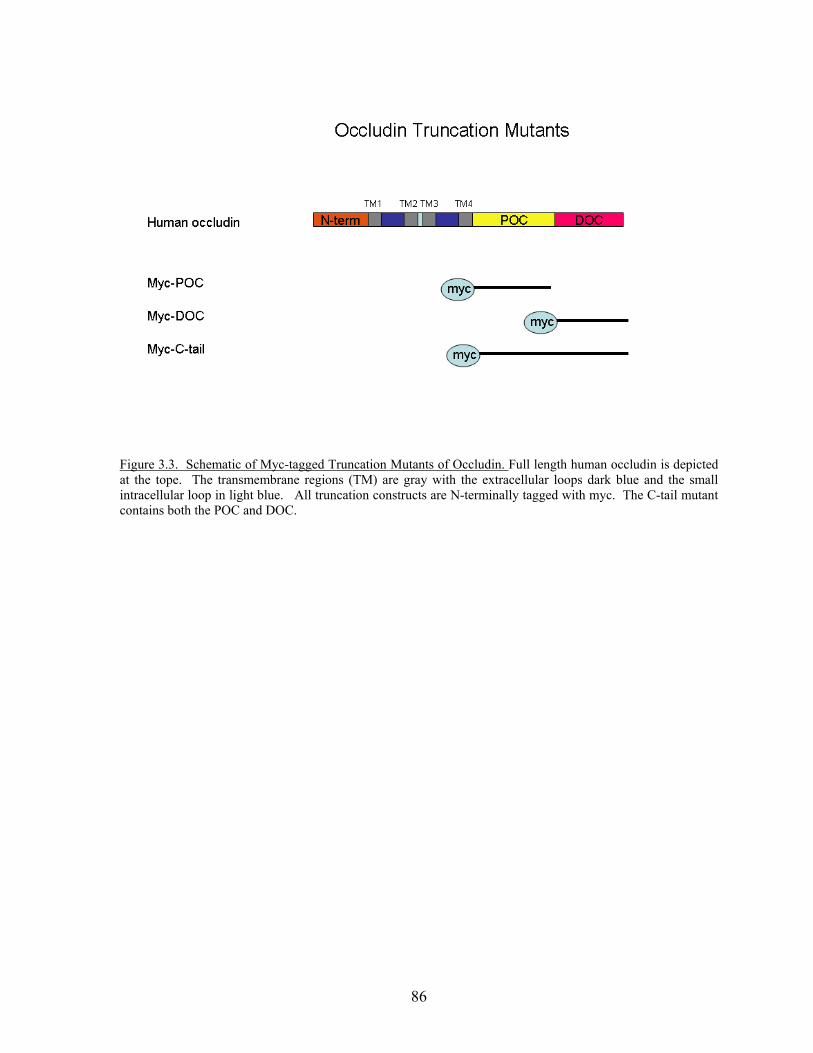
86
Figure 3.3. Schematic of Myc-tagged Truncation Mutants of Occludin. Full length human occludin is depicted at the tope. The transmembrane regions (TM) are gray with the extracellular loops dark blue and the small intracellular loop in light blue. All truncation constructs are N-terminally tagged with myc. The C-tail mutant contains both the POC and DOC.
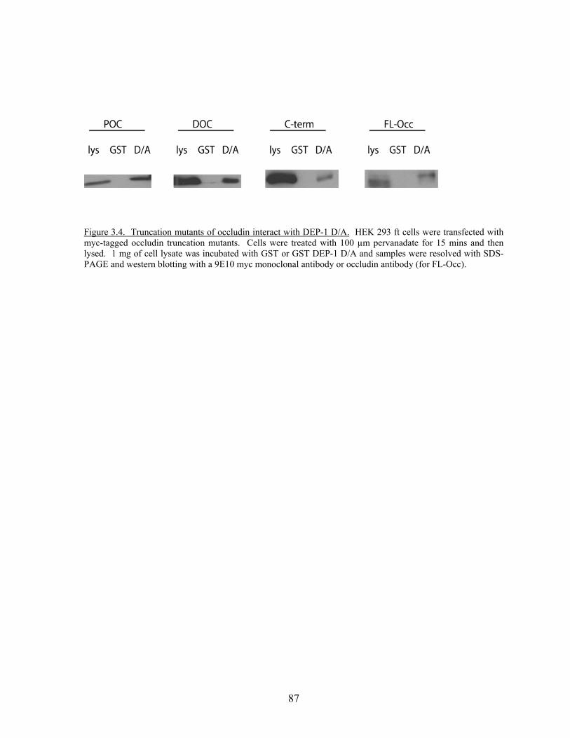
87
Figure 3.4. Truncation mutants of occludin interact with DEP-1 D/A. HEK 293 ft cells were transfected with myc-tagged occludin truncation mutants. Cells were treated with 100 µm pervanadate for 15 mins and then lysed. 1 mg of cell lysate was incubated with GST or GST DEP-1 D/A and samples were resolved with SDS-PAGE and western blotting with a 9E10 myc monoclonal antibody or occludin antibody (for FL-Occ).
88
Discussion This study examined whether an interaction between DEP-1 and occludin occurred by
using truncation mutants of the occludin C-terminal tail. We show that pervanadate
treatment induced binding of all the occludin cytoplasmic truncations to DEP-1. Based on
the literature, we had hypothesized that the distal domain of occludin containing the ZO
binding domain would be the most crucial (203). However, in our experiments, the proximal
domain was also able to interact with DEP-1 following pervanadate treatment. One caveat to
these experiments is that we have not examined binding of these mutants in cells not treated
with pervanadate. Previous work (chapter 2) has never demonstrated an interaction in
untreated cells but we will examine this in the future. In addition, we have not directly
shown that pervanadate treatment induced phosphorylation of these mutants. Since
pervanadate treatment has phosphorylated full length occludin in previous experiments, it is
expected to do so with these mutants but this will be tested. The co-expression of
constitutively active Src and the myc-tagged POC and DOC mutants in HEK 293ft cells
induced phosphorylation of the mutants, demonstrating they are capable of being
phosphorylated (data not shown).
With 17 tyrosines in the C-terminal tail, the possibility exists that DEP-1 may bind to
multiple phosphorylation sites. One major experimental factor that needs to be considered is
the use of pervanadate to induce phosphorylation. Pervanadate inhibits all tyrosine
phosphatases and so causes a global increase in intracellular tyrosine phosphorylation levels.
This treatment may cause the phosphorylation of certain tyrosines that under “normal”
conditions are not phosphorylated, and result in false positive interactions. A better way to
better determine binding sites would be to induce phosphorylation with a known kinase, such
as Src, that phosphorylates the C-terminal tail of occludin (217). There are several Src
89
kinase recognition motifs in both the proximal domain as well as the distal domain which
may be potential DEP-1 binding sites. To test this, constitutively active Src could be co-
transfected with the truncation mutants and used in DEP-1 substrate-trapping mutant
pulldowns to determine which, if any, Src phosphorylated tyrosines can bind to DEP-1.
There are also several EGFR recognition motifs in the cytoplasmic tail of occludin,
suggesting that the addition of EGF may stimulate this pathway and result in site-specific
phosphorylation. Preliminary experiments have been done examining EGF-induced
phosphorylation of occludin; unfortunately, it has been difficult to show occludin
phosphorylation specifically. While generalized tyrosine phosphorylation of lysates
increases with EGF treatment, it may require optimization of immunoprecipiation conditions
in order to determine specific occludin phosphorylation. In addition, EGFR is a substrate of
Src and can be phosphorylated on Y845, an autophosphorylation site (241, 242). As a result,
Src may also be able to induce the phosphorylation of EGFR motifs indirectly. One would
have to keep this in mind when evaluating interaction data. Regardless, these two kinases
would more specifically phosphorylate the C-terminal tail compared to treatment with
pervanadate and future studies will examine their effect on occludin-DEP-1 interactions.
The C-terminal distal domain of occludin has been crystallized and from this study it
was determine that amino acids 416-522 contain the binding site for ZO-1 and are important
for occludin localization to cell-cell junctions.(240). The surface charge profile found a
large midsection of the distal domain which is highly basic and conserved among species.
Double point mutations which changed lysine pairs to aspartic acid and reversed the charge
of this section prevented the binding of ZO-1 to occludin (Lys433/511, Lys444/504, and Lys
485/488). A single point mutation at Lys 433 was also able to disrupt binding of ZO-1. This
90
data suggested that the positive charged surface created by the lysine residues on the alpha
helices of the distal domain of occludin created a binding site for the GUK domain of ZO-1
(240). Interestingly, both Lys 433 and 444 have a tyrosine residue adjacent to them (Fig.
3.1). We propose that because phosphorylation of one or both of these tyrosines will result in
a negative charge in nearly the same location as the Lys->Asp mutation, phosphorylation
might similarly disrupt occludin binding to ZO-1. Therefore, future work should include
mutating these two tyrosines to the phosphomimetic glutamic acid and determining if
interaction with ZO-1 still occurs. If these mutations successfully inhibit ZO-1 binding it
suggests that tyrosine phosphorylation of these residues may be sufficient to attenuate the
occludin and ZO-1 interaction. In addition, mutations of Tyr432 and Tyr443 to
phenylalanine will be used to determine if the occludin-DEP-1 interaction is dependent on
phosphorylation of these residues.
It has been shown previously that DEP-1 can selectively dephosphorylate several
growth factor receptors in a site selective manner to control signaling (149, 152, 153, 226). It
is possible that DEP-1 can bind and dephosphorylate multiple tyrosines on the C-terminal tail
of occludin, which could modulate protein-protein interactions (such as ZO-1), controlling
tight junction protein complex formation and thus the barrier function of tight junctions. The
site selectivity that DEP-1 displayed in PDGF β-receptor dephosphorylation was
predominantly determined by the primary amino acid sequence surrounding the
phosphotyrosine. Poorly dephosphorylated tyrosines have basic residues at positions -4 and
+3 relative to phosphotyrosine (153, 226). Lysine at position +3 seems to be the most crucial
with regards to low affinity for the phosphotyrosine, with a lysine at +1 being better
accepted. For that reason, Tyr432 and Tyr443 are still acceptable potential
91
dephosphorylation sites for DEP-1 on occludin. Tyr443 might be the better of the two
residues since it contains a leucine at +3 and a glycine at -4, both non-polar hydrophobic
residues which have been shown to be favored by DEP-1 (226). It is also possible that the
susceptibility of dephosphorylation of a residue may be affected by the phosphorylation
status of neighboring tyrosine residues. This again emphasizes the role of site specific
phosphorylation by kinases and upstream signaling pathways in deciding which PTP acts on
a substrate at any given time.
Our results suggest that several tyrosines in the C-terminal tail of occludin may be
phosphorylated and consequently bound by the substrate trapping mutant of DEP-1. Further
work needs to examine DEP-1 binding after phosphorylation by specific kinase activity to
avoid excessive, non-physiologic effects of pervanadate. In addition, based on the
identification of residues necessary for ZO-1 binding, specific tyrosines adjacent to critical
lysines in the ZO-1 binding region will also be examined via point mutations studies.
Mapping the residues necessary for DEP-1 interaction will help to clarify potential signaling
pathways and kinases that DEP-1 activity may be counteracting. In addition, we may
determine additional kinases that may be involved in phosphorylation of occludin.
Materials and Methods
Constructs and Antibodies- VSVG-tagged full length human occludin and connexin 32-
occludin chimeras were obtained from Alan Fanning (University of North Carolina Chapel
Hill) and are described previously (194, 203). Myc-tagged truncations of the C-terminal tail
of occludin were made similar to Mitic et al. (203). The proximal C-terminal residues 265-
372 of occludin (POC), the distal c-terminal residues (373-522) (DOC) as well as the entire
92
C-terminal tail (265-522) (C-tail) were first inserted into pEGFP-N2 by amplifying this
region and digesting with BglII and HindIII.. These constructs were then digested with
BglII and KpnI and inserted into pCMV-Myc-J3 (modified Clontech vector from Jia Zhong
at UNC-CH). VSVG monoclonal antibody is from x. 9E10 myc monoclonal was
supernatant produced in the Burridge Lab.
Cell Culture and Transfections- HEK 293ft cells were cultured in Dulbecco's modified
Eagle's medium-high glucose enriched with 10% fetal bovine serum and antibiotics. Cells
were transfected with Fugene6 (Roche Applied Science) according to the manufacturer’s
instructions.
GST fusion proteins and Substrate Trapping Pulldown—GST and GST-DEP-1 D/A were
prepared as described in Chapter 2 material and methods. HEK 293ft cells were transfected
with myc-tagged occludin truncations or full length occludin and processed 48 hours later.
Cells were treated with 100 μM pervanadate for 15 mins to induce phosphorylation prior to
lysis. Cells were rinsed twice in phosphate-buffered saline and lysed in a modified RIPA
buffer (1% Tx-100, 0.5% DOC, 0.2% SDS, 150 mM NaCl, 20 mM Hepes ph 7.4, 2 mM
EDTA, 10 μg/ml each aprotinin and leupeptin, and 1mM PMSF). Insoluble material was
removed by centrifugation. HEK 293ft lysates (1 mg) were incubated with 10 μg of GST or
GST DEP-1 D/A for 1 hour at 4ºC. Beads were washed in the modified RIPA buffer,
resuspended in Laemmli sample buffer and analysed by SDS-PAGE and western blotting on
PVDF membranes (Millipore).
Chapter 4:
Focal adhesion proteins FAK and paxillin are substrates of DEP-1
Summary
FAK and paxillin localize to focal adhesions, points of adhesion between a cell and the
extracellular matrix (ECM). In response to adhesion to ECM proteins such as fibronectin,
both proteins become tyrosine phosphorylated. This tyrosine phosphorylation affects their
activity and recruits binding partners. Consequently, they are potential substrates for PTPs.
In this study, FAK and paxillin bound to the substrate trapping mutant of DEP-1, and were
found to be dephosphorylated by wild type DEP-1. These data suggest that FAK and paxillin
are substrates of DEP-1. However, how DEP-1 regulates these proteins and their
downstream signaling pathways is unknown. Therefore, future studies will need to address
this issue.
Introduction
Post translational modifications such as tyrosine phosphorylation are critical to
several signaling pathways downstream from cell-cell and cell-matrix adhesion.
Phosphotyrosine levels are regulated by the balance between PTK and PTP activity.
Although a number of kinases are known to be involved in increasing phosphorylation at
sites of cell-matrix contact, the PTPs which act there are less understood. In the previous
chapters, substrates and signaling pathways involving DEP-1 and cell-cell adhesions were
94
discussed. Here we examine DEP-1’s role in regulating the phosphorylation of proteins
associated with cell-matrix adhesions.
Focal adhesions are sites of strong adhesion to the ECM in cells grown in tissue
culture. They anchor stress fibers to the plasma membrane via integrins and are multiprotein
signaling complexes for a large number of signaling pathways downstream of integrin
mediated adhesions (60). Both Src and focal adhesion kinase (FAK) are known to localize
here and phosphorylate several of the focal adhesion proteins (4). In addition, paxillin is a
focal adhesion protein that is phosphorylated by several stimuli including integrin dependent
cell adhesion and the tyrosine kinase activity of FAK and CAKβ (243-245). Paxillin is a
signaling molecule that plays an important role as an adaptor protein, recruiting cytoskeletal
and signaling proteins to focal adhesions. FAK and paxillin are known substrates for the
phosphatases Shp-2, PTP-PEST and SAP-1 (18, 31, 236). To test whether FAK and paxillin
might also be substrates for DEP-1 we used the substrate trapping mutant of DEP-1
(described in Chapter 2). We found that they were in fact able to bind the DEP-1 D/A. In
addition, wild type DEP-1 was able to dephosphorylate the proteins suggesting FAK and
paxillin are substrates of DEP-1. We propose that DEP-1 is not only able to act on junctional
proteins but may play a role in the regulation of cell-matrix adhesions as well as adhesion-
mediated signaling pathways.
Results
FAK and paxillin bind to the DEP-1 substrate trapping mutant
We examined whether other known tyrosine phosphorylated proteins important in
cell-matrix interactions are substrates of DEP-1. We treated MCF10A cells with
95
pervanadate, a global PTP inhibitor, and used the lysate in a DEP-1 substrate trapping mutant
pulldown. Both FAK and paxillin were able to bind to DEP-1 D/A in a tyrosine
phosphorylation dependent manner (Fig 4.1A), as well as a dose dependent manner (Fig.
4.1B). We wanted to determine if the interaction of DEP-1 D/A with FAK and paxillin was
specific or if these proteins were substrates of additional PTPs. Lysates of untreated or
pervanadate treated MCF10A cells were incubated with GST fusion proteins of the substrate
trapping mutants of DEP-1 (GST-DEP-1 D/A), PTP-PEST (GST-PTP-PEST D/A), and Shp2
(GST-Shp2 C/S). With a short exposure, DEP-1 D/A was the only PTP of those tested that
was able to interact with FAK and paxillin and as observed previously (Fig. 4.1A) it did so in
a tyrosine phosphorylation-dependent manner (Fig. 4.2A). A longer exposure showed weak
binding of paxillin to PTP-PEST (Fig. 4.2B). Previous studies have shown that paxillin is a
substrate of PTP-PEST (18ref, 246), although interaction with the substrate trapping mutant
(PEST D/A) has not previously been demonstrated.
DEP-1 can dephosphorylate FAK and paxillin in vitro
DEP-1 D/A can bind to both FAK and paxillin as determined by GST fusion protein
pulldowns (Fig. 4.1 and 4.2). We wanted to determine if FAK and paxillin were substrates
for wild type DEP-1 as well as other wild type PTPs in vitro. MCF10A cells were treated
with pervanadate prior to lysis. FAK and paxillin were immunoprecipated from the lysates
and then incubated with purified GST-PTPs. Phosphotyrosine levels of FAK and paxillin
were analyzed by immunoblotting with anti-phosphotyrosine antibodies. Wildtype DEP-1
was able to dephosphorylate FAK and paxillin; where as other PTPs tested had minimal
effects (Fig. 4.3A and 4.3B). All PTPs used were tested for catalytic activity using the
substrate pNPP.
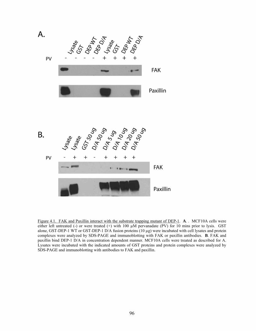
96
Figure 4.1. FAK and Paxillin interact with the substrate trapping mutant of DEP-1. A. . MCF10A cells were either left untreated (-) or were treated (+) with 100 µM pervanadate (PV) for 10 mins prior to lysis. GST alone, GST-DEP-1 WT or GST-DEP-1 D/A fusion proteins (10 µg) were incubated with cell lysates and protein complexes were analyzed by SDS-PAGE and immunoblotting with FAK or paxillin antibodies. B. FAK and paxillin bind DEP-1 D/A in concentration dependent manner. MCF10A cells were treated as described for A. Lysates were incubated with the indicated amounts of GST proteins and protein complexes were analyzed by SDS-PAGE and immunoblotting with antibodies to FAK and paxillin.
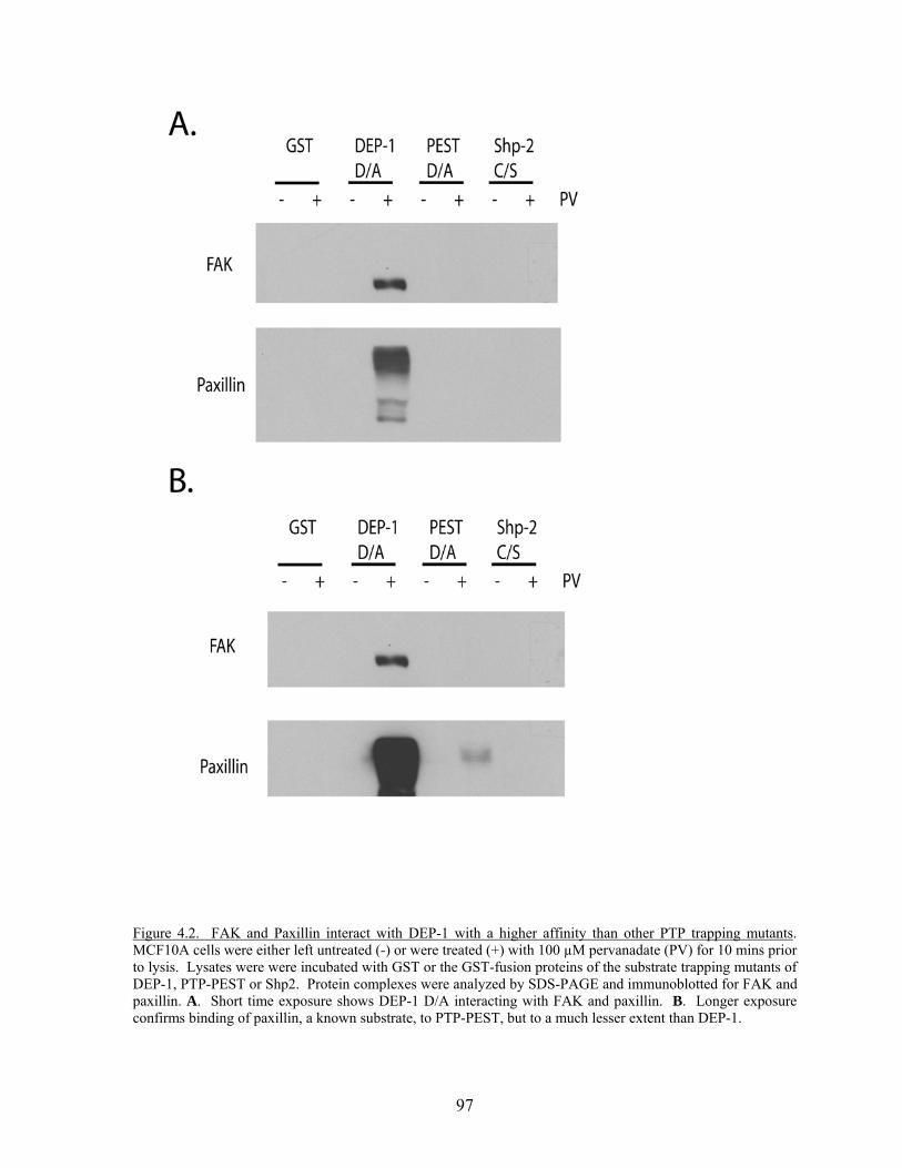
97
Figure 4.2. FAK and Paxillin interact with DEP-1 with a higher affinity than other PTP trapping mutants. MCF10A cells were either left untreated (-) or were treated (+) with 100 µM pervanadate (PV) for 10 mins prior to lysis. Lysates were were incubated with GST or the GST-fusion proteins of the substrate trapping mutants of DEP-1, PTP-PEST or Shp2. Protein complexes were analyzed by SDS-PAGE and immunoblotted for FAK and paxillin. A. Short time exposure shows DEP-1 D/A interacting with FAK and paxillin. B. Longer exposure confirms binding of paxillin, a known substrate, to PTP-PEST, but to a much lesser extent than DEP-1.
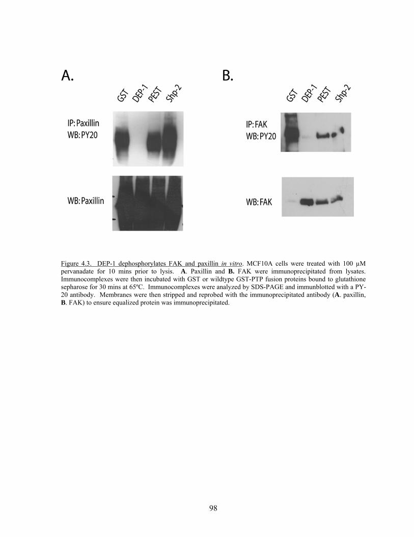
98
Figure 4.3. DEP-1 dephosphorylates FAK and paxillin in vitro. MCF10A cells were treated with 100 µM pervanadate for 10 mins prior to lysis. A. Paxillin and B. FAK were immunoprecipitated from lysates. Immunocomplexes were then incubated with GST or wildtype GST-PTP fusion proteins bound to glutathione sepharose for 30 mins at 65ºC. Immunocomplexes were analyzed by SDS-PAGE and immunblotted with a PY-20 antibody. Membranes were then stripped and reprobed with the immunoprecipitated antibody (A. paxillin, B. FAK) to ensure equalized protein was immunoprecipitated.
99
Discussion
Here we have identified FAK and paxillin as two novel substrates for the receptor
PTP DEP-1. DEP-1 is a single pass transmembrane PTP which localizes to the plasma
membrane and is enhanced at points of cell-cell adhesion. DEP-1 has not been shown to
directly localize at focal adhesions, but neither has PTP-PEST (247), a PTP known to
dephosphorylate paxillin. DEP-1 might be associated with a population of paxillin and/or
FAK not located at focal adhesions. Also, two PTPs related to DEP-1, SAP-1 and GLEPP1,
negatively regulate intergrin-mediated signaling by dephosphorylating p130Cas and
disrupting focal contacts and cytoskeletal components (236, 248). Therefore, DEP-1’s
expression at the plasma membrane as well as its homology to other PTPs with known focal
adhesion substrates suggests that DEP-1 may interact with FAK and paxillin proteins in vivo.
DEP-1 may be both a positive and negative regulator of adhesion-mediated
signaling. DEP-1 binds c-Src and dephosphorylates the inhibitory tyrosine 529, thereby
increasing Src kinase activity (172). Adhesion, along with tyrosine phosphorylation of both
FAK and paxillin, were increased in these cells compared to untransfected or cells expressing
the catalytic inactive mutant (DEP-1 C/S). Inhibition of c-Src activity with PP2 decreased
these responses, suggesting DEP-1 may be positive regulator of Src acvitiy in the context of
integrin-mediated adhesion and may increase levels of phosphotyrosine in FAK and paxillin
(172). Additional data have also confirmed that over expression of DEP-1 increases cell
adhesion (157), strengthening this model.
However, DEP-1 may also negatively affect adhesion-mediated signaling cascades.
When DEP-1 was over-expressed in NIH 3T3 fibroblasts there was a reduction in association
of Src with PDGFR-β concomitant with a decrease in FAK phosphorylation (92).
100
Furthermore, 48 hours after induction of DEP-1 expression, virtually no paxillin containing
focal adhesions were seen in the fibroblasts, however, the control cells appeared normal.
DEP-1 cells were able to make adhesions with the extracellular matrix protein fibronectin,
however, the cells had smaller, dot like peripheral structures which were short lived and
failed to induce phosphorylation of FAK compared to control cells. These focal complexes
were able to form, but did not mature into stable focal adhesions like control cells.
Moreover, DEP-1 cells exhibited decreased migration in response to PDGF and were much
smaller and more rounded demonstrating both a cell spreading and cell migration defect (92).
Therefore, several signaling pathways may be controlled by DEP-1. DEP-1 may be
regulating phosphorylation of FAK indirectly by dephosphorylating PDGFR-β, thereby
attenuating the PDGFR-β-Src interaction, and modulating Src activity and other downstream
signaling pathways. Also, DEP-1 over expression might be inhibiting Rho GTPase activity.
Previous studies found PTP-PEST decreased Rac1 activity and inhibited cell migration by
controlling the phosphorylation levels of GEFs and GAPs (10, 11). Several GEFs and GAPs
for the Rho family are regulated by tyrosine phosphorylation, including the Rac GEF VAV2
and the Rho GAP p190RhoGAP (249, 250). These proteins may be substrates for DEP-1 as
well. Interestingly, our data provides evidence that DEP-1 could also directly be
dephosphorylating FAK and paxillin. In this way, DEP-1 could either prevent focal adhesion
assembly or disassembly by regulating tyrosine phosphorylation of specific residues in FAK.
Modulation of paxillin phosphorylation levels may alter its interactions with downstream
effectors and affect signaling pathways. There are several possible mechanisms and
signaling pathways by which DEP-1 could be exerting an effect on phosphorylation levels of
101
FAK and paxillin. Further studies will need to be conducted to determine if these
mechanisms are influenced by cell type, matrix, or additional stimuli.
Paxillin is a known substrate for PTP-PEST (18, 246). We found that the substrate
trapping mutant of DEP-1 was able to bind to paxillin and wild type DEP-1 can
dephosphorylate it, suggesting paxillin is also a substrate of DEP-1. Although paxillin may
be a substrate for both PTPs, the mechanisms by which the protein-protein interactions occur
between paxillin and the PTPs might be different. PTP-PEST binds paxillin via its C-
terminal non-catalytic domain and paxillin’s C-terminal LIM domains (18, 246, 251).
Therefore, the non catalytic domain interaction recruits paxillin to PTP-PEST and facilitates
paxillin’s dephosphorylation (246). Previous work was unable to show an interaction
between the substrate trapping mutant (C/S) of PTP-PEST with paxillin (251). However, the
substrate trapping mutant (D/A) of PTP-PEST was able to weakly interact with paxillin in
our study. The C/S mutant is known to be a less efficient trapping mutant (8) and so it could
be a combination of the different mutation and a larger amount of GST fusion protein used in
the pulldowns that enabled us to see this interaction. Although we are seeing a small amount
of paxillin binding to GST-PTP-PEST D/A, the primary interaction between these two
proteins is driven independently of the PTP-PEST catalytic domain. On the other hand,
DEP-1 D/A showed a very robust interaction with paxillin even when low levels of GST-
DEP-1 were used (Fig. 4.1), suggesting paxillin may be interacting directly with the catalytic
domain of DEP-1 and not through an accessory binding event. Testing the ability of PTP
inhibitor orthovanadate to block the interaction between DEP-1 and paxillin will help to
determine whether paxillin is exclusively interacting via DEP-1’s catalytic domain.
102
FAK has previously been shown to be a poor substrate for PTP-PEST (41). We have
confirmed this data, demonstrating that FAK binds neither the PTP-PEST trapping mutant,
nor is it dephosphorylated by wild type PTP-PEST in vitro (Fig. 4.2A and 4.3B). However,
FAK is a very strong candidate substrate for DEP-1. FAK not only interacts with the
substrate trapping mutant of DEP-1 after pervanadate treatment, but FAK is
dephosphorylated by recombinant wild type DEP-1 in vitro (Fig. 4.1 and 4.3B). In addition
to the possibility that DEP-1 acts on FAK at focal adhesions, recent work has placed FAK at
cell-cell junctions. FAK has been shown to be important in junction as they form. For
example, when Hela cells were depleted of FAK, N-cadherin mediated cell-cell contacts
were inhibited in subconfluent cultures (252). Expression of constitutively active FAK in
NBT II cells resulted in no change in junctions; however, expression of inactive FAK
drastically diminished cell-cell adhesions at early time points following a calcium switch
(253). During junctional reassembly, active FAK could be seen at junctions in only
approximately 10% of cells and it was never seen in already confluent cultures, suggesting
FAK may only transiently associate at junctions (253). Therefore, FAK may be involved in
the formation and possible stabilization of cell-cell adhesions. It will be interesting to test if
DEP-1 may regulate phosphorylation of Tyr 397 and influence junction formation.
Additionally, identifying other potential tyrosine residues dephosphorylated by DEP-
1 would aid in the delineation of the role of DEP-1 in paxillin and FAK related focal
adhesion and cell-cell junction signaling. There are numerous phospho-specific antibodies
available for FAK and paxillin and it would be very informative to perform DEP-1 substrate
trapping mutant pulldowns to determine which residue(s) is binding to the PTP. Also, DEP-1
could be overexpressed or reduced with siRNA in cells and phosphorylation changes could
103
be examined. To further clarify signaling pathways, phosphorylation could be assessed
following various stimuli. For example, following a calcium switch, phosphorylation levels
of several tyrosine residues in FAK were examined. Little or no change was found with
tyrosines 397, 576, 577, or 925. However, two hours after calcium replenishment Tyr 861
displayed elevated phosphorylation (253). This increase in phosphorylation was dependent
on the interaction with E-cadherin. These findings make tyrosine 861 an attractive candidate
residue DEP-1 may regulate with regards to cell-cell contact formation. Another interesting
residue to examine is Tyr 925. As described in Chapter 1, phosphorylation of Tyr 925 in
FAK is necessary to induce an interaction with dynamin at focal adhesions, a critical step in
focal adhesion disassembly (62). It would be interesting to examine this residue and focal
adhesion disassembly in DEP-1 knockdown cells.
Although we see complete dephosphorylation of FAK and paxillin when incubated
with wildtype DEP-1 it is possible that the pervanadate treatment only phosphorylated certain
tyrosines. Also, DEP-1 may preferentially dephosphorylate certain residues in vivo, but the
use of high levels in the in vitro assay may result in total dephosphorylation. The
experiments presented here were conducted with confluent epithelial cells. These cells do
not contain a large amount of focal adhesions, if any. Future experiments will examine DEP-
1 substrates in fibroblast cell lines.
There is the possibility that there might be cross talk between cell-cell junctions and
focal adhesions. High RhoA is disruptive to AJ formation (110-112). As junctions from,
Rac1 activity is increased and RhoA activity is decreased as direct response to cadherin-
cadherin engagement (115). Cadherin engagement induces src-dependent tyrosine
phosphorylation of p190RhoGAP, thus leading to the observed decrease in RhoA (116).
104
Epithelial cells need low levels of RhoA to reduce their intracellular tension and contractility
and promote the formation of cell-cell adhesions (254). Low RhoA levels also turn off focal
adhesion signaling. DEP-1 may be involved in regulating both cell-cell junction and focal
adhesion signaling at the same time.
Methods and Materials
Cell Culture-- MCF10A cells were grown in a 1:1 mixture of Dulbecco’s modified Eagle’s
medium (DMEM) and F12/Ham medium supplemented with 5% horse serum, 20 ng/ml
EGF, 0.01 mg/ml insulin, 500 ng/ml hydrocortisone, 100 ng/ml cholera toxin and antibiotics.
Constructs and Antibodies—GST DEP-1 constructs were generated as described in Chapter
2. GST PTP-PEST constructs were kindly provided by Sarita Sastry (University of Texas
Galveston). The monoclonal PY-20 was purchased from Santa Cruz Biotechnology.
Monoclonal FAK and paxillin antibodies were from BD Transduction labs.
GST fusion proteins-- Expression of the fusion proteins in Escherichia coli were performed
as previously described in Chapter 2.
Substrate Trapping Pulldown-- MCF10A cells were either left untreated or treated with 100
μM pervanadate (phosphatase inhibitor) for 15 mins prior to lysis. Cells were rinsed twice in
phosphate-buffered saline and lysed in a modified RIPA buffer (1% Tx-100, 0.5% DOC,
0.2% SDS, 150 mM NaCl, 20 mM Hepes ph 7.4, 2 mM EDTA, 10 μg/ml each aprotinin and
leupeptin, and 1mM PMSF). Insoluble material was removed by centrifugation. MCF10A
105
lysates (1 mg) were incubated with 10 μg of GST proteins (Figure 4.1) or 100 µg GST
proteins (Figure 4.2) for 1 hour at 4ºC. Beads were washed in the modified RIPA buffer,
resuspended in Laemmli sample buffer and analysed by SDS-PAGE and western blotting on
PVDF membranes (Millipore).
Dephosphorylation Assays-- MCF10A cells were treated with 100 µM pervanadate for 15
mins and lysed in the modified RIPA buffer plus 10 μg/ml each aprotinin and leupeptin,
1mM PMSF, and 5 mM iodoacetic acid (IAA). After incubation of the lysates on ice for 10
mins, DTT was added at a final concentration of 10 mM for another 10 mins on ice to
inactivate the IAA. Lysates were clarified and FAK or paxillin was immunoprecipitated
from 1 mg lysate with 2 µg antibody for 2 hours at 4ºC. IPs were washed, resupsend in a
succinate buffer (50 mM succinate pH 6.0, 50 mM NaCl, 1 mM EDTA, 1 mM DTT) and
incubated with 1 μg glutathione-sepharose bound GST-PTPs for 30 minutes at 30ºC. The
reaction was quenched washing 1 time in the succinate buffer and resuspending in Laemmli
sample buffer. The immune complexes were run on SDS-PAGE gels and analyzed by
western blotting using the PY-20 antibody, FAK monoclonal antibody or paxillin
monoclonal antibody.
Chapter 5:
Conclusions and Future Directions
A key element regulating cell-cell and cell-matrix adhesion is tyrosine
phosphorylation of proteins at intercellular junctions and focal adhesions. In this
dissertation, DEP-1 has been shown to bind to and dephosphorylate proteins located at both
locations. In chapter 2, occludin and ZO-1 were identified as novel substrates for the RPTP
DEP-1. These proteins appear to be substrates specific for DEP-1 and potentially a related
PTP, SAP-1. DEP-1 and SAP-1 are extremely similar to each other, each with 8 fibronectin
type III repeats and a single catalytic domain (89, 230). Therefore, occludin and ZO-1 might
be substrates specific to this particular family of PTPs. It would be interesting to see if VE-
PTP is also able to act on these TJ proteins. We also provided evidence that the over-
expression of DEP-1 increased transepithelial electrical resistance in confluent monolayers
and reduced paracellular flux of FITC-dextran following a calcium switch. Through
dephosphorylation of junctional components, DEP-1 may enhance the stability of protein
complexes involved with maintaining junctional integrity. Future work will focus on
identifying where DEP-1 interacts with occludin (Chapter 3). This would enhance our
understanding of the specificity of DEP-1’s catalytic domain, including its recognition of the
amino acid sequence surrounding the phospho-tyrosine. It is possible that there are multiple
binding and dephosphorylation sites on occludin, depending on upstream kinase activity. We
propose that Src kinase phosphorylates occludin and disrupts its interaction with DEP-1, but
107
would also like to test growth factor receptors such as EGFR. In addition, Chapter 4
discussed the identification of FAK and paxillin as potential substrates for DEP-1.
Phosphorylation of both FAK and paxillin occurs downstream from integrin-mediated
signaling and DEP-1 may therefore be involved with these pathways as well. Also,
phosphorylation of FAK occurs at cell-cell junctions during junction reassembly (253) and
may be another way in which DEP-1 may control cell-cell adhesion. Experiments still need
to be conducted in order to determine exactly how DEP-1 is controlling physiological
processes associated with dephosphorylation of these focal adhesion proteins.
The work presented here has demonstrated a role for the RPTP in controlling cell
adhesions in epithelial cells. The following chapter will discuss directions for future research
regarding DEP-1 and other RPTPs. First, we will focus on the possibility that RPTPs are
involved in the process of leukocyte transmigration. Next we will examine the importance of
the extracellular domain in regulating PTP activity. Finally, we will end with a look at PTPs
in cancer and the development of PTP inhibitors.
DEP-1 as a regulator of leukocyte transmigration
The vascular endothelium controls the passage of macromolecules and immune cells
across it. There are two known mechanisms for leukocyte migration across the endothelium:
transcellular and paracellular. Direct evidence supports the existence of transcellular
migration (i.e. through a cell), although it is believed to occur in only 5-10% of
transmigratory events in-vitro (255). Thus, the predominant route for leukocyte
transmigration is paracellular migration, where leukocytes migrate between adjacent
endothelial cells through their junctions (256). In the resting state, the endothelium
108
maintains its barrier properties and prevents the passage of cells out of the bloodstream and
into tissues. When an inflammatory response is elicited, however, the properties of the
endothelium change and leukocytes are able to adhere to and migrate through gaps at the
intercellular junctions. The final step in the adhesion cascade of leukocyte transmigration is
diapedesis, or the crawling of the leukocyte between adjacent endothelial cells. In order for
this to occur, the leukocyte must breach a physical barrier of junctional structures including
TJs and AJs (for review see (257)). It has been shown that the adhesive strength of the TJ
and AJ is regulated by the tyrosine phosphorylation levels of their protein components. An
increase in tyrosine phosphorylation dissociates the junctional complexes from the actin
cytoskeleton and reduces junctional integrity (73, 74, 214, 217). Therefore, regulation of
tyrosine phosphorylation levels of junctional components is an important mechanism for
regulating both the stability and permeability of tight and adherens junctions and thus may
affect the ability of leukocytes to transmigrate.
Several PTPs have been shown to localize to junctions as well as interact with
members of the cadherin-catenin complex (235). Specifically, PTPµ, DEP-1, PTP1B, and
VE-PTP are found in endothelial cell junctions (81, 87, 90, 258). Previous studies in our lab
revealed that leukocyte transmigration increased VE-cadherin phosphorylation at residues
specific for p120ctn and β-catenin binding (259). When these residues were mutated to
phenylalanine, transmigration was reduced, directly indicating the involvement of tyrosine
phosphorylation of junctional proteins during leukocyte TEM (259). Several junctional
binding partners for DEP-1 have been discovered such as β-catenin, plakoglobin and p120-
catenin (80) and now occludin and ZO-1. Our preliminary data demonstrates that VE-
cadherin may also be a substrate for DEP-1. With numerous junctional proteins as
109
substrates, it is likely that DEP-1 is involved in modulating phosphorylation of endothelial
junctional proteins. Using the same phospho-specific antibodies used by Allingham et
al.(259), we plan to explore whether DEP-1 interacts with the phosphorylated VE-cadherin
residues necessary for catenin binding. We also plan to evaluate the effect that DEP-1
expression has on leukocyte TEM, and the phosphorylation of other substrates during the
process of transmigration.
One manner by which DEP-1 activity might be altered during TEM is through
binding of its extracellular domain. Receptor PTPs are proposed to act as adhesion receptors,
transducing outside-in signaling. Initially we were interested in exploring the extracellular
binding partners of DEP-1, especially those involved with leukocyte transendothelial
migration (TEM). One possible interaction would be a homophilic, trans interaction of DEP-
1 on adjacent endothelial cells. Other RPTPs are known to interact in homophilic
interactions (84, 96, 120), which stabilize the protein at cell-cell contacts (94). The
clustering of RPTPs at the plasma membrane brings the catalytic domain into close proximity
with junctional protein substrates, promoting a net tyrosine dephosphorylation and potential
strengthening of junctional integrity (78, 80, 94). Another possibility is that DEP-1 is
interacting with itself on a migrating leukocyte. Hematopoietic cells express varying
amounts of DEP-1 (CD148) (165-167), with white blood cells, in particular monocytes and
macrophages, expressing very high levels of DEP-1 (165, 166). Therefore, DEP-1 on
endothelial cells could be binding to DEP-1 on leukocytes and this interaction could affect
the catalytic activity of DEP-1, altering phosphorylation of junctional proteins. A third
hypothesis is that DEP-1 can form heterophilic interactions with other cell adhesion
molecules on endothelium, leukocytes or both. JAMs are not exclusively found on cells that
110
form TJs, but are also on leukocytes (182). As a result, the extracellular domain of JAMs
might interact with the extracellular domain of DEP-1 and aid in controlling TEM. Other
potential targets are the extracellular domains of cadherins and growth factor receptors.
A number of extracellular ligands have been identified for a few RPTPs, including
homophilic and heterophilic interactions (260, 261), however, ligands have not been
identified for the majority of RPTPs. Although no extracellular ligands for DEP-1 have been
identified yet, a recent study suggests that an undefined ligand is found in Matrigel, a
commercial ECM protein preparation (100). Association of DEP-1 with Matrigel increased
the intracellular phosphatase activity of DEP-1 (100). In addition, antibody-induced
ectodomain oligomeriztion of DEP-1 also increased its catalytic activity (150). Therefore,
interaction of DEP-1’s extracellular domain with itself or binding an extracellular protein
may result in an increase in PTP activity. In the model of leukocyte transmigration,
increased activity of DEP-1 due to interactions with proteins on adjacent endothelial cells
would allow for the dephosphorylation of junctional proteins, reformation of TJ and AJ
complexes, and enhanced barrier function of endothelial cells. On the other hand, we would
expect that binding to a leukocyte protein would inhibit PTP activity, allowing for increased
phosphorylation of AJ and TJ proteins and loss of junctional integrity. It is possible that such
a ligand exists and is awaiting discovery. Other attractive PTPs canditates for a role in
leukocyte TEM are PTPµ and the vascular endothelial specific VE-PTP.
The Importance of RPTP Extracellular Domains
Signal transduction from RTKs emanates from the ligand binding extracellular
domain. Ligand binding induces dimerization and autophosphorylation of the intracellular
111
tyrosines leading to activation and recruitment of downstream targets (262). In contrast,
mechanisms controlling RPTP enzyme activity are unknown (263). RPTPs have
extracellular domains that resemble other known cell adhesion molecules and it has been
proposed that they can be involved in homo- and heterotypic intercellular binding (125). As
stated in the introduction, RPTPs have traditionally been considered to be active as
monomers and inactive when dimerized (125). This is known to be true for RPTPα (127-
129), CD45 (264), SAP-1 (265), and PTPBR7 and PTP-SL (266). Other PTPs are able to
dimerize, but it is unclear whether this dimerization affects their catalytic activity. In most
cases, dimerization is independent of the catalytic domain and occurs through the
extracellular domain or transmembrane domain. In fact, the transmembrane domain of PTPλ,
PTP LAR, PTPα, PTPγ, GLEPP1, and DEP-1 are able to homodimerize in Escherichia coli
membranes, albeit to different extents (267). In addition, several studies in eukaryotic cells
have demonstrated that the transmembrane domain of PTPα is necessary and sufficient for
mediating receptor dimerization (127, 129, 268). Furthermore, artificial induction of
dimerization affects the enzyme activity of CD45, RPTPα, and DEP-1 (128, 150, 264).
Inhibition of the catalytic activity of PTPα has been proposed to occur by the catalytic
domain of one PTPα being blocked by specific contacts with an inhibitory wedge domain
from the membrane proximal region of the other PTPα partner (269). The residues that
compose this structure in PTPα are conserved among other RPTPs including PTPµ and PTP-
LAR (130, 131). However, structural analysis of the membrane-proximal catalytic domain
of PTPµ and the entire cytoplasmic domain of PTP-LAR reveal that the inhibitory interaction
between the catalytic domain and the wedge are not observed (130, 131). It remains to be
determined whether the wedge domain can interact with the catalytic domain of other RPTPs,
112
proposing the possibility that this method of dimerization-induced inhibition may not be a
universal mechanism regulating RPTP activity.
Interestingly, introduction of a cysteine residue in the extracellular juxtamembrane
domain of RPTPα results in disulfide bond formation and dimerization (128). Depending on
the exact location of the cysteines, the dimers are either active or inactive (128). This
suggests that the orientation in which the extracellular domain dimerizes affects the
intracellular orientation of the PTP domain and can influence its activity. SAP-1 is a RPTP
which naturally contains an extracellular domain with several cysteine residues capable of
forming disulfide bonds. Altering the redox state of the SAP-1 can reversibly control its
dimerization and its activity (265). Moreover, PTPσ has two major isoforms, differing
slightly in the number of their FNIII repeats. The alternative arrangements of the
extracellular domain alters the ligand binding specificities of this PTP (270), potentially by
changing the rotational conformation of the N-terminal extracellular domain. It is also
possible that modifications of the cytoplasmic domains, such as phosphorylation or
oxidation, also results in changes in the rotational coupling within RPTP dimers and alters
ligand-binding properties. RPTPα-D2 (catalytic domain 2) changes its conformation in
response to the cellular redox state of the cell (268), implying RPTPs may not only have the
capacity for outside-in signaling but also inside-out signaling (261). Collectively, subtle
changes in the relative orientation of RPTP dimers in both the extracellular and intracellular
domains may be able to determine whether the PTP is active or inactive, providing a new
mechanism for regulation.
The increasing complexity of factors that affect dimerization forces us to look at
RPTP dimerization, ligand binding and activity more closely. How exactly are RTPs being
113
turned on and off? Perhaps the answer will never be as simple as it is for RTKs.
Nevertheless, the extracellular domains of PTPs appear to be very important in determining
their activity and downstream signaling pathways. Does the extracellular domain of a RPTP
dictate whether dimerization activates or inhibits it? Or does the dimerization state of RPTPs
affect their ability to bind to extracellular ligands, adding yet another level of complexity?
Too many questions are left unanswered for us to clearly state how RPTP activity is
regulated. In the end, the role of the extracellular domain may be PTP specific or even cell
type specific, determined by the availability of ligands and signaling partners.
PTPs in Cancer
Cell adhesion plays a critical role in the process of metastatic tumor dissemination.
Loss of cadherins expression and other cell adhesion molecules is a trademark sign of
metastatic cells (271). Tumor development is associated with remodeling of the both TJs and
AJs. Neoplastic cells escape from the constraints imposed on them by intercellular junctions
and adopt a migratory behavior. Increased phosphorylation at junctions leads to disassembly
of AJ and TJ protein complexes as well as decreased junctional integrity. Thus, PTP activity
may influence invasion and metastasis in certain cancers.
Loss of function mutations
Loss of function of PTPs has been associated with cancer progression. Less than one
third of point mutations in PTPs occur within the PTP domain, suggesting that loss of
function is indirectly related to the structure of the catalytic domain (272). For example,
RPTPρ is frequently mutated in human cancers including lung, colon, and skin (273). In
colorectal tumor isolates, mutations were found in both the intracellular and extracellular
114
domains (273). Intracellular mutations diminished PTP activity as expected and it was
proposed that mutations in the extracellular domain may alter protein-protein interactions
(273). Five mutations were mapped to residues located in the MAM and immunoglobulin
domains of RPTPρ, an area necessary for adhesion in PTPµ (274-276). Subsequently, these
mutations were introduced into SF9 cells and impaired the homophilic interaction of PTPρ as
demonstrated by the reduction of cell aggregate formation. This suggests that the mutations
found naturally occurring in cancers were loss of function mutations (277). Disruption of
protein-protein interactions may affect several signaling pathways and promote tumor
migration and metastasis. A number of mutations in the DEP-1 gene, PTPRJ, also create
coding polymorphisms and have been found in human cancers. Mutations in the
extracellular domain are located in the 2nd and 8th fibronectin repeats (Fig. 1.4) (142, 163).
These mutations are predicted to occur in exposed regions available for protein-protein
interactions and could affect ligand binding, dimizeration, and downstream signaling
pathways (163). Currently, the effects of these mutations have not been analyzed but it
would be of interest to express them and compare effects of junctional integrity to wildtype
DEP-1. Additionally, point mutations located in the proline-rich domain of PTP-PEST have
been found in breast cancer cell lines (278). Two of these mutations enhance PTP catalytic
activity whereas the third mutation inhibits activity (278). Shp-2 is yet another PTP in
which mutations are associated with certain cancers including juvenile myelomonocytic
leukemia, lung and colon cancer (279, 280). Several mutations in the N-terminal SH2
domain disrupt the inhibitory interaction with its catalytic domain, leading to activation of
Shp-2 (281, 282). Therefore, mutations in non catalytic domains can both activate and
115
inhibit PTP activity, whether through altering localization or interrupting inter- or
intramolecular interactions.
Loss of protein expression
In addition to loss of function mutations, loss of PTP expression is observed in
several cancers. SAP-1 levels were down-regulated in advanced human hepatocellular
carcinoma and (283). Protein expression correlated with the differentiation state of the cells,
with normal tissue or well-differentiated HCC expressing similar or higher levels than non-
cancerous tissue, and poorly differentiated HCC having reduced levels of SAP-1 (283).
DEP-1 expression has been shown to be drastically reduced in multiple cancer cell lines and
human cancers including thyroid, breast, pancreatic and colon cancers (139-142, 164). In
addition, LOH of locus for the DEP-1 gene is frequent in human cancers (163). Therefore,
DEP-1 has been proposed to act as a tumor suppressor by inhibiting cell proliferation.
Studies have re-expressed DEP-1 in cancer cell lines lacking endogenous DEP-1 and found
that there was a profound inhibition in cell proliferation (139-142, 164).
Besides controlling proliferation, DEP-1 may be inhibiting metastasis by reducing
cell motility, as shown in Chapter 4, by means of regulating integrin-mediated signaling
pathways as well as stabilizing cell-cell junctions through dephosphorylation of tight junction
proteins (Chapter 2). Supported by other cancer models such as colon, pancreatic, and
thyroid cancer (140-142, 164), we proposed that breast cancer cell lines would have reduced
DEP-1 levels as well. The protein expression levels of DEP-1 were examined in normal-like
breast epithelial cells (MCF10A) as well as dedifferentiated breast cancer cells (MCF7) and
metastatic breast cancer cells (MDA-MB-231). Cells were lysed and total protein
concentrations were normalized. Lysates were immunoblotted for DEP-1 and junctional
116
proteins β-catenin and E-cadherin. As seen in Fig. 5.1, DEP-1 protein levels increased as
junctional proteins decreased. We anticipated seeing a decrease in junctional proteins in the
metastatic cells, yet we hypothesized that DEP-1 levels would also decrease. One
explanation for our observed increased in DEP-1 is that the cell’s feedback machinery is
trying to counterbalance overgrowth by DEP-1 expression, but perhaps another signaling
pathway is overriding it. Another explanation is that the DEP-1 expressed in the MDA-MB-
231 has disregulated activity possibly due to one of the point mutations found in cancer
(163). To try and determine whether this was a possibility, we compared the catalytic
activity of DEP-1 from MCF10A and MDA-MB-231 cells. In this preliminary experiment,
DEP-1 was immunoprecipitated from an equal amount of total protein from both cell types.
Half of the precipitated protein was run on a gel to determine the amount of DEP-1
immunoprecipitated and the other half was used in a pNPP assay. Significantly more DEP-1
immunoprecipitated from the MDA-MB-231 cells compared to the MCF10A cells (Fig.
5.2A). Interestingly, the catalytic activity of DEP-1 from the MCF10A cells was higher than
the metastatic cells (Fig. 5.2B). Considering that substantially more DEP-1 was
immunoprecipitated and thus used in the MDA-MB-231 activity assay, we can conclude that
DEP-1 in the MDA-MB-231 cells may have a considerable reduction in catalytic activity.
We plan to repeat this as well as to examine mRNA levels of DEP-1 in these cell types to
determine whether transcription is altered. It would also be interesting to compare substrates
for DEP-1 in normal versus breast cancer cell lines.
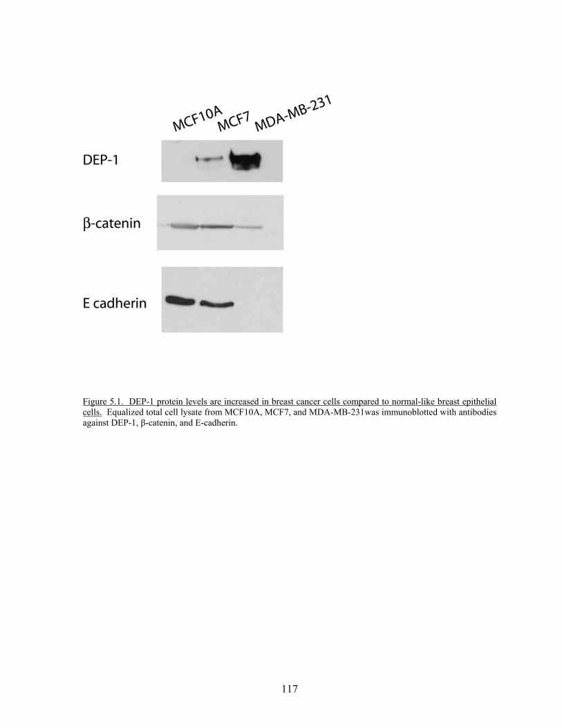
117
Figure 5.1. DEP-1 protein levels are increased in breast cancer cells compared to normal-like breast epithelial cells. Equalized total cell lysate from MCF10A, MCF7, and MDA-MB-231was immunoblotted with antibodies against DEP-1, β-catenin, and E-cadherin.
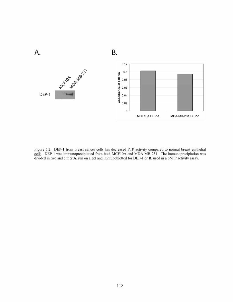
118
Figure 5.2. DEP-1 from breast cancer cells has decreased PTP activity compared to normal breast epithelial cells. DEP-1 was immunoprecipitated from both MCF10A and MDA-MB-231. The immunoprecipiation was divided in two and either A. run on a gel and immunoblotted for DEP-1 or B. used in a pNPP activity assay.
119
Over expression of DEP-1 in Cancer
With our result of increased DEP-1 in breast cancer cells, we were curious if other
PTPs may be up regulated in cancers. One study found an increase in total PTP activity in
breast cancer (284). Another group has shown that DEP-1 levels can vary in breast cancer
cells lines, with MDA-MB-231 being one of the cell lines which exhibited the highest
expression of DEP-1 (227). SAP-1 is overexpressed in human colorectal cancer (285) while
PTP1B overexpression was established in breast tumor tissue (286). In addition, PTP-LAR
mRNA and protein levels were markedly increased in breast cancer cell lines and tissues
(287) while another study found that one third of the primary breast carcinomas tested were
characterized by strongly eleveated RPTPα levels (288). Interestingly, these high protein
levels were associated with low tumor grade (ER positive) and cells experimentally induced
to have high PTPα cells showed delayed tumor growth and metastasis in mice (288).
Increased PTP-LAR and PTP1B levels were found in human breast cancer as a consequence
of expression of the activated neu oncogene. The amount of the PTP directly correlated to
the expression of neu (289). Likewise, knockdown of PTPα induced apoptosis in ER
negative but not ER positive breast cancer cells (290). These results insinuate that the
genetic background of the cancer may regulate the expression level of the PTP as well as the
signaling pathways in which the PTP is involved. Further studies will need to confirm this
but it proposes an intriguing possibility.
PTP inhibitors
Over the last several years, PTP inhibitors have begun to be developed. Researchers
are striving for a highly potent, yet selective small molecular inhibitor. Two main concerns
120
exist. First, PTPs contain a highly conserved active site, therefore obtaining inhibitors that
selectively targets one PTP will be difficult. Second, a large number of side effects could
occur because a single PTP might regulate multiple signaling pathways or a key pathway
might be regulated by several PTPs (291). To address the first concern, researchers are
looking for secondary binding sites in regions adjacent to the catalytic domain. Perhaps
inhibitors targeted to these areas will enhance the inhibitors affinity and selectivity.
There are several PTP1B inhibitors currently being produced. PTP1B counteracts
signaling through the insulin receptor (292), making a small molecule inhibitor of PTP1B a
promising treatment for type 2 diabetes. Recently, mice from the well- established breast
cancer mouse model (NDL2) were crossed with PTP1B deficient mice. Mice lacking PTP1B
in this background displayed a reduced rate of tumor development, number of tumors, and
lung metastases (293). Treatment of PTP1B +/+ mice with a PTP1B specific inhibitor also
delayed the onset of mammary tumors (293). Consequently, PTP1B inhibitors might also
used in cancer treatment, especially in ErbB2-positive breast cancers. Shp-2 has also been
identified an oncogenic PTP and small molecule inhibitors are currently being developed
(294). Although progress is being made for each of these PTPs, inhibitors for PTPs are still
far behind the RTK inhibitor field.
For RPTPs, an alternative to interfering with the catalytic domain would be to target
the extracellular domain and control activity by ligand-ectodomain interactions (137). The
structural diversity of the extracellular domain makes it an attractive target to develop
specific inhibitors. Antibodies to CD45 extracellular domain have already been shown to
have the potential to inhibit CD45 activity (295, 296). Antibodies to additional PTPs may
have a similar effect. Perhaps a compound that stabilized or locked the dimerization state
121
would prevent activity. It would be appealing to develop an inhibitor that would block the
binding of a particular ligand and specifically affect downstream signaling.
Conclusion
The PTP field is experiencing an exciting time. Long gone is the stigma that PTPs
are just “housekeepers” cleaning up after PTK signaling. PTPs are now considered to be as
complex a group of proteins as PTKs, with activity regulated in specific ways. With the
classification of all PTP genes in mice and humans (2), we can ask more questions regarding
the specificity, redundancy, tissue expression and isoforms/mutations of PTPs. The
development of the substrate trapping mutants has led to the identification of more substrates.
Structural information regarding the extracellular and intracellular domains of PTPs is
shedding new light on how catalytic activities are regulated. Now the field needs to focus on
the physiological roles or functions of individual PTPs. One challenge facing researchers is
that PTP expression and function may be regulated in a tissue-specific manner. In which
case, different substrates might be identified in different cell types, resulting in modification
of current signaling pathway models. Several PTPs have been linked to disease states such
as diabetes, cancer and inflammation. Knockout mouse models, siRNA, and the
development of small molecule inhibitors will help to clarify the roles of PTPs in normal
physiological processes and pathological conditions. The future in PTP research possesses
many discoveries for the years to come.
122
References
1. Manning, G., D. B. Whyte, R. Martinez, T. Hunter, and S. Sudarsanam. 2002. The Protein Kinase Complement of the Human Genome. Science 298:1912-1934.
2. Alonso A, Sasin J, Bottini N, Friedberg I, Friedberg I, Osterman A, Godzik A, Hunter
T, Dixon J, and M. T. 2004. Protein Tyrosine Phosphatases in the Human Genome. Cell 117:699-711.
3. Hynes, R. O. 2002. Integrins: Bidirectional, Allosteric Signaling Machines. Cell
110:673-687. 4. Mitra, S. K., D. A. Hanson, and D. D. Schlaepfer. 2005. Focal adhesion kinase: in
command and control of cell motility. Nat Rev Mol Cell Biol 6:56-68. 5. Miyamoto, S., S. K. Akiyama, and K. M. Yamada. 1995. Synergistic roles for
receptor occupancy and aggregation in integrin transmembrane function. Science 267:883-885.
6. Burridge, K., and M. Chrzanowska-Wodnicka. 1996. Focal adhesions, contractility,
and signaling. Annu Rev Cell Dev Biol 12:463-518. 7. Maher, P. 1993. Activation of Phosphotyrosine Phosphatase Activity by Reduction of
Cell- Substrate Adhesion. PNAS 90:11177-11181. 8. Flint, A. J., T. Tiganis, D. Barford, and N. K. Tonks. 1997. Development of
"substrate-trapping" mutants to identify physiological substrates of protein tyrosine phosphatases. Proc Natl Acad Sci U S A 94:1680-1685.
9. Burridge, K., and K. Wennerberg. 2004. Rho and Rac Take Center Stage. Cell
116:167-179. 10. Sastry, S. K., P. D. Lyons, M. D. Schaller, and K. Burridge. 2002. PTP-PEST
controls motility through regulation of Rac1. J Cell Sci 115:4305-4316. 11. Sastry, S. K., Z. Rajfur, B. P. Liu, J.-F. Cote, M. L. Tremblay, and K. Burridge. 2006.
PTP-PEST Couples Membrane Protrusion and Tail Retraction via VAV2 and p190RhoGAP. J. Biol. Chem. 281:11627-11636.
12. Kontaridis, M. I., S. Eminaga, M. Fornaro, C. I. Zito, R. Sordella, J. Settleman, and
A. M. Bennett. 2004. SHP-2 Positively Regulates Myogenesis by Coupling to the Rho GTPase Signaling Pathway. Mol. Cell. Biol. 24:5340-5352.
123
13. O'Reilly, A. M., S. Pluskey, S. E. Shoelson, and B. G. Neel. 2000. Activated mutants of SHP-2 preferentially induce elongation of Xenopus animal caps. Mol Cell Biol 20:299-311.
14. Inagaki, K., T. Noguchi, T. Matozaki, T. Horikawa, K. Fukunaga, M. Tsuda, M.
Ichihashi, and M. Kasuga. 2000. Roles for the protein tyrosine phosphatase SHP-2 in cytoskeletal organization, cell adhesion and cell migration revealed by overexpression of a dominant negative mutant. Oncogene 19:75-84.
15. Schoenwaelder, S. M., L. A. Petch, D. Williamson, R. Shen, G.-S. Feng, and K.
Burridge. 2000. The protein tyrosine phosphatase Shp-2 regulates RhoA activity. Curr Biol 10:1523-1526.
16. Kodama, A., T. Matozaki, A. Fukuhara, M. Kikyo, M. Ichihashi, and Y. Takai. 2000.
Involvement of an SHP-2-Rho small G protein pathway in hepatocyte growth factor/scatter factor-induced cell scattering. Mol Biol Cell 11:2565-2575.
17. Nimnual, A. S., L. J. Taylor, and D. Bar-Sagi. 2003. Redox-dependent
downregulation of Rho by Rac. Nat Cell Biol 5:236-241. 18. Shen, Y., G. Schneider, J. F. Cloutier, A. Veillette, and M. D. Schaller. 1998. Direct
association of protein-tyrosine phosphatase PTP-PEST with paxillin. J Biol Chem 273:6474-6481.
19. Garton, A. J., A. J. Flint, and N. K. Tonks. 1996. Identification of p130(cas) as a
substrate for the cytosolic protein tyrosine phosphatase PTP-PEST. Mol Cell Biol 16:6408-6418.
20. Cary, L. A., D. C. Han, T. R. Polte, S. K. Hanks, and J. L. Guan. 1998. Identification
of p130Cas as a mediator of focal adhesion kinase-promoted cell migration. J Cell Biol 140:211-221.
21. Klemke, R. L., J. Leng, R. Molander, P. C. Brooks, K. Vuori, and D. A. Cheresh.
1998. CAS/Crk coupling serves as a "molecular switch" for induction of cell migration. J Cell Biol 140:961-972.
22. Lamorte, L., S. Rodrigues, V. Sangwan, C. E. Turner, and M. Park. 2003. Crk
associates with a multimolecular Paxillin/GIT2/beta-PIX complex and promotes Rac-dependent relocalization of Paxillin to focal contacts. Mol Biol Cell 14:2818-2831.
23. Cote, J. F., and K. Vuori. 2002. Identification of an evolutionarily conserved
superfamily of DOCK180-related proteins with guanine nucleotide exchange activity. J Cell Sci 115:4901-4913.
24. Brugnera, E., L. Haney, C. Grimsley, M. Lu, S. F. Walk, A. C. Tosello-Trampont, I.
G. Macara, H. Madhani, G. R. Fink, and K. S. Ravichandran. 2002. Unconventional
124
Rac-GEF activity is mediated through the Dock180-ELMO complex. Nat Cell Biol 4:574-582.
25. Turner, C. E. 2000. Paxillin and focal adhesion signalling. Nat Cell Biol 2:E231-236. 26. Bustelo, X. R. 2000. Regulatory and Signaling Properties of the Vav Family. Mol.
Cell. Biol. 20:1461-1477. 27. Angers-Loustau, A., J.-F. Cote, A. Charest, D. Dowbenko, S. Spencer, L. A. Lasky,
and M. L. Tremblay. 1999. Protein Tyrosine Phosphatase-PEST Regulates Focal Adhesion Disassembly, Migration, and Cytokinesis in Fibroblasts. J. Cell Biol. 144:1019-1031.
28. Worthylake, R. A., S. Lemoine, J. M. Watson, and K. Burridge. 2001. RhoA is
required for monocyte tail retraction during transendothelial migration. J. Cell Biol. 154:147-160.
29. Tsuda, M., T. Matozaki, K. Fukunaga, Y. Fujioka, A. Imamoto, T. Noguchi, T.
Takada, T. Yamao, H. Takeda, F. Ochi, T. Yamamoto, and M. Kasuga. 1998. Integrin-mediated tyrosine phosphorylation of SHPS-1 and its association with SHP-2. Roles of Fak and Src family kinases. J Biol Chem 273:13223-13229.
30. Oh, E. S., H. Gu, T. M. Saxton, J. F. Timms, S. Hausdorff, E. U. Frevert, B. B. Kahn,
T. Pawson, B. G. Neel, and S. M. Thomas. 1999. Regulation of early events in integrin signaling by protein tyrosine phosphatase SHP-2. Mol Cell Biol 19:3205-3215.
31. Yu, D. H., C. K. Qu, O. Henegariu, X. Lu, and G. S. Feng. 1998. Protein-tyrosine
phosphatase Shp-2 regulates cell spreading, migration, and focal adhesion. J Biol Chem 273:21125-21131.
32. Zhang, S. Q., W. Yang, M. I. Kontaridis, T. G. Bivona, G. Wen, T. Araki, J. Luo, J.
A. Thompson, B. L. Schraven, M. R. Philips, and B. G. Neel. 2004. Shp2 Regulates Src Family Kinase Activity and Ras/Erk Activation by Controlling Csk Recruitment. Mol Cell 13:341-355.
33. Harder, K. W., N. P. H. Moller, J. W. Peacock, and F. R. Jirik. 1998. Protein-tyrosine
Phosphatase alpha Regulates Src Family Kinases and Alters Cell-Substratum Adhesion. J. Biol. Chem. 273:31890-31900.
34. von Wichert, G., G. Jiang, A. Kostic, K. De Vos, J. Sap, and M. P. Sheetz. 2003.
RPTP-{alpha} acts as a transducer of mechanical force on {alpha}v/{beta}3-integrin-cytoskeleton linkages. J. Cell Biol. 161:143-153.
125
35. Ponniah, S., D. Z. M. Wang, K. L. Lim, and C. J. Pallen. 1999. Targeted disruption of the tyrosine phosphatase PTP[alpha] leads to constitutive downregulation of the kinases Src and Fyn. Curr Biol 9:535-539.
36. Zheng, X. M., Y. Wang, and C. J. Pallen. 1992. Cell transformation and activation of
pp60c-src by overexpression of a protein tyrosine phosphatase. Nature 359:336-339. 37. Liu, F., D. E. Hill, and J. Chernoff. 1996. Direct binding of the proline-rich region of
protein tyrosine phosphatase 1B to the Src homology 3 domain of p130(Cas). J Biol Chem 271:31290-31295.
38. Arregui, C. O., J. Balsamo, and J. Lilien. 1998. Impaired Integrin-mediated Adhesion
and Signaling in Fibroblasts Expressing a Dominant-negative Mutant PTP1B. J. Cell Biol. 143:861-873.
39. Arias-Salgado, E. G., F. Haj, C. Dubois, B. Moran, A. Kasirer-Friede, B. C. Furie, B.
Furie, B. G. Neel, and S. J. Shattil. 2005. PTP-1B is an essential positive regulator of platelet integrin signaling. J Cell Biol 170:837-845.
40. Davidson, D., J.-F. Cloutier, A. Gregorieff, and A. Veillette. 1997. Inhibitory
Tyrosine Protein Kinase p50csk Is Associated with Protein-tyrosine Phosphatase PTP-PEST in Hemopoietic and Non-hemopoietic Cells. J. Biol. Chem. 272:23455-23462.
41. Lyons, P., J. Dunty, E. Schaefer, and M. Schaller. 2001. Inhibition of the catalytic
activity of cell adhesion kinase beta by protein-tyrosine phosphatase-PEST-mediated dephosphorylation. J Biol Chem 276:24422-24431.
42. Lacalle, R. A., E. Mira, C. Gomez-Mouton, S. Jimenez-Baranda, A. C. Martinez, and
S. Manes. 2002. Specific SHP-2 partitioning in raft domains triggers integrin-mediated signaling via Rho activation. J Cell Biol 157:277-289.
43. Sordella, R., W. Jiang, G. C. Chen, M. Curto, and J. Settleman. 2003. Modulation of
Rho GTPase signaling regulates a switch between adipogenesis and myogenesis. Cell 113:147-158.
44. Arthur, W. T., L. A. Petch, and K. Burridge. 2000. Integrin engagement suppresses
RhoA activity via a c-Src-dependent mechanism. Curr Biol 10:719-722. 45. Chiarugi, P., P. Cirri, L. Taddei, E. Giannoni, G. Camici, G. Manao, G. Raugei, and
G. Ramponi. 2000. The low M(r) protein-tyrosine phosphatase is involved in Rho-mediated cytoskeleton rearrangement after integrin and platelet-derived growth factor stimulation. J Biol Chem 275:4640-4646.
46. Clark, E. A., S. J. Shattil, and J. S. Brugge. 1994. Regulation of protein tyrosine
kinases in platelets. Trends Biochem Sci 19:464-469.
126
47. Klinghoffer, R. A., C. Sachsenmaier, J. A. Cooper, and P. Soriano. 1999. Src family
kinases are required for integrin but not PDGFR signal transduction. Embo J 18:2459-2471.
48. Arias-Salgado, E. G., S. Lizano, S. Sarkar, J. S. Brugge, M. H. Ginsberg, and S. J.
Shattil. 2003. Src kinase activation by direct interaction with the integrin beta cytoplasmic domain. Proc Natl Acad Sci U S A 100:13298-13302.
49. Obergfell, A., K. Eto, A. Mocsai, C. Buensuceso, S. L. Moores, J. S. Brugge, C. A.
Lowell, and S. J. Shattil. 2002. Coordinate interactions of Csk, Src, and Syk kinases with [alpha]IIb[beta]3 initiate integrin signaling to the cytoskeleton. J Cell Biol 157:265-275.
50. Su, J., M. Muranjan, and J. Sap. 1999. Receptor protein tyrosine phosphatase [alpha]
activates Src-family kinases and controls integrin-mediated responses in fibroblasts. Current Biology 9:505-511.
51. Zeng, L., X. Si, W. P. Yu, H. T. Le, K. P. Ng, R. M. Teng, K. Ryan, D. Z. Wang, S.
Ponniah, and C. J. Pallen. 2003. PTP alpha regulates integrin-stimulated FAK autophosphorylation and cytoskeletal rearrangement in cell spreading and migration. J Cell Biol 160:137-146.
52. Liu, F., M. A. Sells, and J. Chernoff. 1998. Transformation suppression by protein
tyrosine phosphatase 1B requires a functional SH3 ligand. Mol Cell Biol 18:250-259. 53. Hassid, A., S. Huang, and J. Yao. 1999. Role of PTP-1B in aortic smooth muscle cell
motility and tyrosine phosphorylation of focal adhesion proteins. Am J Physiol Heart Circ Physiol 277:H192-198.
54. Cheng, A., G. S. Bal, B. P. Kennedy, and M. L. Tremblay. 2001. Attenuation of
adhesion-dependent signaling and cell spreading in transformed fibroblasts lacking protein tyrosine phosphatase-1B. J Biol Chem 276:25848-25855.
55. Ilic, D., Y. Furuta, S. Kanazawa, N. Takeda, K. Sobue, N. Nakatsuji, S. Nomura, J.
Fujimoto, M. Okada, and T. Yamamoto. 1995. Reduced cell motility and enhanced focal adhesion contact formation in cells from FAK-deficient mice. Nature 377:539-544.
56. Gilmore, A. P., and L. H. Romer. 1996. Inhibition of focal adhesion kinase (FAK)
signaling in focal adhesions decreases cell motility and proliferation. Mol Biol Cell 7:1209-1224.
57. Webb, D. J., K. Donais, L. A. Whitmore, S. M. Thomas, C. E. Turner, J. T. Parsons,
and A. F. Horwitz. 2004. FAK-Src signalling through paxillin, ERK and MLCK regulates adhesion disassembly. Nat Cell Biol 6:154-161.
127
58. Hughes, P. E., M. W. Renshaw, M. Pfaff, J. Forsyth, V. M. Keivens, M. A. Schwartz,
and M. H. Ginsberg. 1997. Suppression of integrin activation: a novel function of a Ras/Raf-initiated MAP kinase pathway. Cell 88:521-530.
59. Cho, S. Y., and R. L. Klemke. 2002. Purification of pseudopodia from polarized cells
reveals redistribution and activation of Rac through assembly of a CAS/Crk scaffold. J. Cell Biol. 156:725-736.
60. Schaller, M. D. 2001. Biochemical signals and biological responses elicited by the
focal adhesion kinase. Biochimica et Biophysica Acta (BBA) - Molecular Cell Research 1540:1-21.
61. Izaguirre, G., L. Aguirre, Y. P. Hu, H. Y. Lee, D. D. Schlaepfer, B. J. Aneskievich,
and B. Haimovich. 2001. The cytoskeletal/non-muscle isoform of alpha-actinin is phosphorylated on its actin-binding domain by the focal adhesion kinase. J Biol Chem 276:28676-28685.
62. Ezratty, E. J., M. A. Partridge, and G. G. Gundersen. 2005. Microtubule-induced
focal adhesion disassembly is mediated by dynamin and focal adhesion kinase. Nat Cell Biol 7:581-590.
63. Sakisaka, T., and Y. Takai. 2004. Biology and pathology of nectins and nectin-like
molecules. Curr Opin Cell Biol 16:513-521. 64. Kemler, R. 1993. From cadherins to catenins: cytoplasmic protein interactions and
regulation of cell adhesion. Trends Genet. 9:317-321. 65. Lampugnani, M., M. Corada, L. Caveda, F. Breviario, O. Ayalon, B. Geiger, and E.
Dejana. 1995. The molecular organization of endothelial cell to cell junctions: differential association of plakoglobin, beta-catenin, and alpha- catenin with vascular endothelial cadherin (VE-cadherin). J. Cell Biol. 129:203-217.
66. Kam, Z., T. Volberg, and B. Geiger. 1995. Mapping of adherens junction components
using microscopic resonance energy transfer imaging. J Cell Sci 108:1051-1062. 67. Kowalczyk, A. P., and A. B. Reynolds. 2004. Protecting your tail: regulation of
cadherin degradation by p120-catenin. Curr Opin Cell Biol 16:522-527. 68. Yamada, S., S. Pokutta, F. Drees, W. I. Weis, and W. J. Nelson. 2005. Deconstructing
the cadherin-catenin-actin complex. Cell 123:889-901. 69. Daniel, J., and A. B. Reynolds. 1997. Tyrosine phosphorylation and cadherin/catenin
function. Bioessays 19:883-891.
128
70. Lampugnani, M. G., M. Corada, P. Andriopoulou, S. Esser, W. Risau, and E. Dejana. 1997. Cell confluence regulates tyrosine phosphorylation of adherens junction components in endothelial cells. J Cell Sci 110:2065-2077.
71. Volberg, T., Y. Zick, R. Dror, I. Sabanay, C. Gilon, A. Levitzki, and B. Geiger. 1992.
The effect of tyrosine-specific protein phosphorylation on the assembly of adherens-type junctions. Embo J 11:1733-1742.
72. Michalides, R., T. Volberg, and B. Geiger. 1994. Augmentation of adherens junction
formation in mesenchymal cells by co-expression of N-CAM or short-term stimulation of tyrosine-phosphorylation. Cell Adhes Commun 2:481-490.
73. Behrens, J., L. Vakaet, R. Friis, E. Winterhager, F. Van Roy, M. M. Mareel, and W.
Birchmeier. 1993. Loss of epithelial differentiation and gain of invasiveness correlates with tyrosine phosphorylation of the E-cadherin/beta-catenin complex in cells transformed with a temperature-sensitive v-SRC gene. J. Cell Biol. 120:757-766.
74. Matsuyoshi, N., M. Hamaguchi, S. Taniguchi, A. Nagafuchi, S. Tsukita, and M.
Takeichi. 1992. Cadherin-mediated cell-cell adhesion is perturbed by v-src tyrosine phosphorylation in metastatic fibroblasts. J. Cell Biol. 118:703-714.
75. Young, B. A., X. Sui, T. D. Kiser, S. W. Hyun, P. Wang, S. Sakarya, D. J. Angelini,
K. L. Schaphorst, J. D. Hasday, A. S. Cross, L. H. Romer, A. Passaniti, and S. E. Goldblum. 2003. Protein tyrosine phosphatase activity regulates endothelial cell-cell interactions, the paracellular pathway, and capillary tube stability. Am J Physiol Lung Cell Mol Physiol 285:L63-75.
76. Fujita, Y., G. Krause, M. Scheffner, D. Zechner, H. E. Leddy, J. Behrens, T. Sommer,
and W. Birchmeier. 2002. Hakai, a c-Cbl-like protein, ubiquitinates and induces endocytosis of the E-cadherin complex. Nat Cell Biol 4:222-231.
77. Potter, M. D., S. Barbero, and D. A. Cheresh. 2005. Tyrosine Phosphorylation of VE-
cadherin Prevents Binding of p120- and {beta}-Catenin and Maintains the Cellular Mesenchymal State. J. Biol. Chem. 280:31906-31912.
78. Brady-Kalnay S, D. Rimm, and N. Tonks. 1995. Receptor protein tyrosine
phosphatase PTP mu associates with cadherins and catenins in vivo. Journal of Cell Biology 130:977-986.
79. Zondag, G. C. M., A. B. Reynolds, and W. H. Moolenaar. 2000. Receptor Protein-
tyrosine Phosphatase RPTP{micro} Binds to and Dephosphorylates the Catenin p120ctn. J. Biol. Chem. 275:11264-11269.
80. Holsinger, L., K. Ward, B. Duffield, J. Zachwieja, and B. Jallal. 2002. The
transmembrane receptor protein tyrosine phosphatase DEP-1 interacts with p120 catenin. Oncogene 21:7067-7076.
129
81. Nawroth, R., G. Poell, A. Ranft, S. Kloep, U. Samulowitz, G. Fachinger, M. Golding,
D. T. Shima, U. Deutsch, and D. Vestweber. 2002. VE-PTP and VE-cadherin ectodomains interact to faciliate regulation of phosphorylation and cell contacts. EMBO J. 21:4885-4895.
82. Ukropec, J. A., M. K. Hollinger, S. M. Salva, and M. J. Woolkalis. 2000. SHP2
Association with VE-Cadherin Complexes in Human Endothelial Cells Is Regulated by Thrombin. J. Biol. Chem. 275:5983-5986.
83. Balsamo, J., T. Leung, H. Ernst, M. K. Zanin, S. Hoffman, and J. Lilien. 1996.
Regulated binding of PTP1B-like phosphatase to N-cadherin: control of cadherin-mediated adhesion by dephosphorylation of beta-catenin. J. Cell Biol. 134:801-813.
84. Brady-Kalnay, S., A. Flint, and N. Tonks. 1993. Homophilic Binding of PTPmu, a
receptor-type protein tyrosine phosphatase, can mediate cell-cell aggregation. Journal of Cell Biology 122:961-972.
85. Gebbink, M. F., G. C. Zondag, R. W. Wubbolts, R. L. Beijersbergen, I. van Etten, and
W. H. Moolenaar. 1993. Cell-cell adhesion mediated by a receptor-like protein tyrosine phosphatase. J Biol Chem 268:16101-16104.
86. Brady-Kalnay, S. M., T. Mourton, J. P. Nixon, G. E. Pietz, M. Kinch, H. Chen, R.
Brackenbury, D. L. Rimm, R. L. Del Vecchio, and N. K. Tonks. 1998. Dynamic Interaction of PTPµ with Multiple Cadherins In Vivo. J. Cell Biol. 141:287-296.
87. Sui, X. F., T. D. Kiser, S. W. Hyun, D. J. Angelini, R. L. Del Vecchio, B. A. Young,
J. D. Hasday, L. H. Romer, A. Passaniti, N. K. Tonks, and S. E. Goldblum. 2005. Receptor Protein Tyrosine Phosphatase {micro} Regulates the Paracellular Pathway in Human Lung Microvascular Endothelia. Am J Pathol 166:1247-1258.
88. Hellberg, C. B., S. M. Burden-Gulley, G. E. Pietz, and S. M. Brady-Kalnay. 2002.
Expression of the receptor protein-tyrosine phosphatase, PTPmu, restores E-cadherin-dependent adhesion in human prostate carcinoma cells. J Biol Chem 277:11165-11173.
89. Ostman, A., Q. Yang, and N. Tonks. 1994. Expression of DEP-1, A Receptor-Like
Protein-Tyrosine-Phosphatase, is Enhanced with Increasing Cell Density. PNAS 91:9680-9684.
90. Takahashi, T., K. Takahashi, R. MERNAUGH, V. DROZDOFF, C. SIPE, H.
SCHOECKLMANN, B. ROBERT, D. R. ABRAHAMSON, and T. O. DANIEL. 1999. Endothelial Localization of Receptor Tyrosine Phosphatase, ECRTP/DEP-1, in Developing and Mature Renal Vasculature. J Am Soc Nephrol 10:2135-2145.
130
91. Takahashi, T., K. Takahashi, P. L. St. John, P. A. Fleming, T. Tomemori, T. Watanabe, D. R. Abrahamson, C. J. Drake, T. Shirasawa, and T. O. Daniel. 2003. A Mutant Receptor Tyrosine Phosphatase, CD148, Causes Defects in Vascular Development. Moleculare and Cellular Biology 23:1817-1831.
92. Kellie, S., G. Craggs, I. N. Bird, and G. E. Jones. 2004. The tyrosine phosphatase
DEP-1 induces cytoskeletal rearrangements, aberrant cell-substratum interactions and a reduction in cell proliferation. J Cell Sci 117:609-618.
93. Fachinger, G., U. Deutsch, and W. Risau. 1999. Functional interaction of vascular
endothelial-protein-tyrosine-phosphatase with the angiopoietin receptor Tie-2. Oncogene 18:5948-5953.
94. Gebbink, M. F., G. C. Zondag, G. M. Koningstein, E. Feiken, R. W. Wubbolts, and
W. H. Moolenaar. 1995. Cell surface expression of receptor protein tyrosine phosphatase RPTP mu is regulated by cell-cell contact. J. Cell Biol. 131:251-260.
95. Mourton, T., C. B. Hellberg, S. M. Burden-Gulley, J. Hinman, A. Rhee, and S. M.
Brady-Kalnay. 2001. The PTP{micro} Protein-tyrosine Phosphatase Binds and Recruits the Scaffolding Protein RACK1 to Cell-Cell Contacts. J. Biol. Chem. 276:14896-14901.
96. Sap, J., Y.-P. Jiang, D. Friedlander, M. Grumet, and J. Schlessinger. 1994. Receptor
Tyrosine Phosphatase R-PTP-kappa Mediates Homophilic Binding. Mol Cell Biol. 14:1-9.
97. Fuchs, M., T. Muller, M. M. Lerch, and A. Ullrich. 1996. Association of Human
Protein-tyrosine Phosphatase kappa with Members of the Armadillo Family. J. Biol. Chem. 271:16712-16719.
98. Kypta, R. M., H. Su, and L. F. Reichardt. 1996. Association between a
transmembrane protein tyrosine phosphatase and the cadherin-catenin complex. J. Cell Biol. 130:67-77.
99. Muller, T., A. Choidas, E. Reichmann, and A. Ullrich. 1999. Phosphorylation and
Free Pool of beta -Catenin Are Regulated by Tyrosine Kinases and Tyrosine Phosphatases during Epithelial Cell Migration. J. Biol. Chem. 274:10173-10183.
100. Sorby, M., J. Sandstrom, and A. Ostman. 2001. An extracellular ligand increases the
specific activity of the receptor-like protein tyrosine phosphatase DEP-1. Oncogene 20:5219-5224.
101. Masuda, M., M. Osawa, H. Shigematsu, N. Harada, and K. Fujiwara. 1997. Platelet
endothelial cell adhesion molecule-1 is a major SH-PTP2 binding protein in vascular endothelial cells. FEBS Letters 408:331-336.
131
102. Jackson, D. E., K. R. Kupcho, and P. J. Newman. 1997. Characterization of Phosphotyrosine Binding Motifs in the Cytoplasmic Domain of Platelet/Endothelial Cell Adhesion Molecule-1 (PECAM-1) That Are Required for the Cellular Association and Activation of the Protein-tyrosine Phosphatase, SHP-2. J. Biol. Chem. 272:24868-24875.
103. Balsamo, J., C. Arregui, T. Leung, and J. Lilien. 1998. The Nonreceptor Protein
Tyrosine Phosphatase PTP1B Binds to the Cytoplasmic Domain of N-Cadherin and Regulates the Cadherin-Actin Linkage. J. Cell Biol. 143:523-532.
104. Xu, G., C. Arregui, J. Lilien, and J. Balsamo. 2002. PTP1B Modulates the
Association of beta -Catenin with N-cadherin through Binding to an Adjacent and Partially Overlapping Target Site. J. Biol. Chem. 277:49989-49997.
105. Pluskota, E., Y. Chen, and S. E. D'Souza. 2000. Src Homology Domain 2-containing
Tyrosine Phosphatase 2 Associates with Intercellular Adhesion Molecule 1 to Regulate Cell Survival. J. Biol. Chem. 275:30029-30036.
106. Wang, Q., G. R. Pfeiffer, II, and W. A. Gaarde. 2003. Activation of SRC Tyrosine
Kinases in Response to ICAM-1 Ligation in Pulmonary Microvascular Endothelial Cells. J. Biol. Chem. 278:47731-47743.
107. Burridge, K., S. Sastry, and J. Sallee. 2006. Regulation of Cell Adhesion by Protein
Tyrosine Phosphatases 1. Cell-Matrix Adhesion. J Biol Chem In Press. 108. Braga, V. M., L. M. Machesky, A. Hall, and N. A. Hotchin. 1997. The small GTPases
Rho and Rac are required for the establishment of cadherin-dependent cell-cell contacts. J Cell Biol 137:1421-1431.
109. Hordijk, P. L., J. P. ten Klooster, R. A. van der Kammen, F. Michiels, L. C. Oomen,
and J. G. Collard. 1997. Inhibition of invasion of epithelial cells by Tiam1-Rac signaling. Science 278:1464-1466.
110. Zhong, C., M. S. Kinch, and K. Burridge. 1997. Rho-stimulated contractility
contributes to the fibroblastic phenotype of Ras-transformed epithelial cells. Mol Biol Cell 8:2329-2344.
111. Jou, T. S., E. E. Schneeberger, and W. J. Nelson. 1998. Structural and functional
regulation of tight junctions by RhoA and Rac1 small GTPases. J Cell Biol 142:101-115.
112. Braga, V. M., M. Betson, X. Li, and N. Lamarche-Vane. 2000. Activation of the
small GTPase Rac is sufficient to disrupt cadherin-dependent cell-cell adhesion in normal human keratinocytes. Mol Biol Cell 11:3703-3721.
132
113. van Wetering, S., N. van den Berk, J. D. van Buul, F. P. J. Mul, I. Lommerse, R. Mous, J.-P. t. Klooster, J.-J. Zwaginga, and P. L. Hordijk. 2003. VCAM-1-mediated Rac signaling controls endothelial cell-cell contacts and leukocyte transmigration. Am J Physiol Cell Physiol 285:C343-352.
114. Kim, S. H., Z. Li, and D. B. Sacks. 2000. E-cadherin-mediated Cell-Cell Attachment
Activates Cdc42. J. Biol. Chem. 275:36999-37005. 115. Noren, N. K., C. M. Niessen, B. M. Gumbiner, and K. Burridge. 2001. Cadherin
engagement regulates Rho family GTPases. The Journal of Biological Chemistry 276:33305-33308.
116. Noren, N. K., W. T. Arthur, and K. Burridge. 2003. Cadherin Engagement Inhibits
RhoA via p190RhoGAP. J. Biol. Chem. 278:13615-13618. 117. van Wetering, S., J. D. van Buul, S. Quik, F. P. J. Mul, E. C. Anthony, J.-P. t.
Klooster, J. G. Collard, and P. L. Hordijk. 2002. Reactive oxygen species mediate Rac-induced loss of cell-cell adhesion in primary human endothelial cells. J Cell Sci 115:1837-1846.
118. Meng, T., T. Fukada, and N. Tonks. 2002. Reversible oxidation and inactivation of
protein tyrosine phosphatases in vivo. Mol Cell 9:387-399. 119. van Buul, J. D., E. C. Anthony, M. Fernandez-Borja, K. Burridge, and P. L. Hordijk.
2005. Proline-rich Tyrosine Kinase 2 (Pyk2) Mediates Vascular Endothelial-Cadherin-based Cell-Cell Adhesion by Regulating {beta}-Catenin Tyrosine Phosphorylation. J. Biol. Chem. 280:21129-21136.
120. Cheng, J., K. Wu, M. Armanini, N. O'Rourke, D. Dowbenko, and L. A. Lasky. 1997.
A novel protein-tyrosine phosphatase related to the homotypically adhering kappa and mu receptors. J Biol Chem 272:7264-7277.
121. O'Grady, P., T. C. Thai, and H. Saito. 1998. The Laminin-Nidogen Complex is a
Ligand for a Specific Splice Isoform of the Transmembrane Protein Tyrosine Phosphatase LAR. J. Cell Biol. 141:1675-1684.
122. Aricescu, A. R., I. W. McKinnell, W. Halfter, and A. W. Stoker. 2002. Heparan
sulfate proteoglycans are ligands for receptor protein tyrosine phosphatase sigma. Mol Cell Biol 22:1881-1892.
123. Zeng, L., L. D'Alessandri, M. B. Kalousek, L. Vaughan, and C. J. Pallen. 1999.
Protein Tyrosine Phosphatase {alpha} (PTP{alpha}) and Contactin Form a Novel Neuronal Receptor Complex Linked to the Intracellular Tyrosine Kinase fyn. J. Cell Biol. 147:707-714.
133
124. Meng, K., A. Rodriguez-Pena, T. Dimitrov, W. Chen, M. Yamin, M. Noda, and T. F. Deuel. 2000. Pleiotrophin signals increased tyrosine phosphorylation of beta -catenin through inactivation of the intrinsic catalytic activity of the receptor-type protein tyrosine phosphatase beta /zeta. PNAS 97:2603-2608.
125. Weiss, A., and J. Schlessinger. 1998. Switching signals on or off by receptor
dimerization. Cell 94:277-280. 126. Desai, D. M., J. Sap, J. Schlessinger, and A. Weiss. 1993. Ligand-mediated negative
regulation of a chimeric transmembrane receptor tyrosine phosphatase. Cell 73:541-554.
127. Jiang, G., J. den Hertog, and T. Hunter. 2000. Receptor-like protein tyrosine
phosphatase alpha homodimerizes on the cell surface. Mol Cell Biol. 20:5917-5929. 128. Jiang, G., J. den Hertog, J. Su, J. Noel , J. Sap, and T. Hunter. 1999. Dimerization
inhibits the activity of receptor-like protein-tyrosine phosphatase alpha. Nature 401:606-610.
129. Tertoolen, L., C. Blanchetot, G. Jiang, J. Overvoorde, T. Gadella, T. Hunter, and J.
den Hertog. 2001. Dimerization of receptor protein-tyrosine phosphatase alpha in living cells. BMC Cell Biology 2:online.
130. Hoffmann, K. M., N. K. Tonks, and D. Barford. 1997. The crystal structure of domain
1 of receptor protein-tyrosine phosphatase mu. J Biol Chem 272:27505-27508. 131. Nam, H. J., F. Poy, N. X. Krueger, H. Saito, and C. A. Frederick. 1999. Crystal
structure of the tandem phosphatase domains of RPTP LAR. Cell 97:449-457. 132. Bokoch, G. M., and U. G. Knaus. 2003. NADPH oxidases: not just for leukocytes
anymore! Trends Biochem Sci 28:502-508. 133. Meng, T. C., S. F. Hsu, and N. K. Tonks. 2005. Development of a modified in-gel
assay to identify protein tyrosine phosphatases that are oxidized and inactivated in vivo. Methods 35:28-36.
134. Honda, H., J. Inazawa, J. Nishida, Y. Yazaki, and H. Hirai. 1994. Molecular cloning,
characterization, and chromosomal localization of a novel protein-tyrosine phosphatase, HPTP eta. Blood 84:4186-4194.
135. Streuli, M. 1996. Protein tyrosine phosphatases in signalling. Current Opinion in Cell
Biology 8:182-188. 136. Andersen, J. N., P. G. Jansen, S. M. Echwald, O. H. Mortensen, T. Fukada, R. Del
Vecchio, N. K. Tonks, and N. P. H. Moller. 2004. A genomic perspective on protein
134
tyrosine phosphatases: gene structure, pseudogenes, and genetic disease linkage. FASEB J. 18:8-30.
137. Tabernero, L., A. R. Aricescu, E. Y. Jones, and S. E. Szedlacsek. 2008. Protein
tyrosine phosphatases: structure & function relationships. FEBS Journal 275:867-882.
138. Brady-Kalnay, S. M., and N. K. Tonks. 1995. Protein tyrosine phosphatases as
adhesion receptors. Current Opininon in Cell Biology 7:650-657. 139. Keane, M. M., G. A. Lowrey, S. A. Ettenberg, M. A. Dayton, and S. Lipkowitz. 1996.
The protein tyrosine phosphatase DEP-1 is induced during differentiation and inhibits growth of breast cancer cells. Cancer Res 56:4236-4243.
140. Iuliano, R., F. Trapasso, I. Le Pera, F. Schepis, I. Sama, A. Clodomiro, K. R. Dumon,
M. Santoro, L. Chiariotti, G. Viglietto, and A. Fusco. 2003. An Adenovirus Carrying the Rat Protein Tyrosine Phosphatase {eta} Suppresses the Growth of Human Thyroid Carcinoma Cell Lines in Vitro and in Vivo. Cancer Res 63:882-886.
141. Trapasso, F., R. Iuliano, A. Boccia, A. Stella, R. Visconti, P. Bruni, G. Baldassarre,
M. Santoro, G. Viglietto, and A. Fusco. 2000. Rat protein tyrosine phosphatase eta suppresses the neoplastic phenotype of retrovirally transformed thyroid cells through the stabilization of p27(Kip1). Mol Cell Biol 20:9236-9246.
142. Trapasso, F., S. Yendamuri, K. R. Dumon, R. Iuliano, R. Cesari, B. Feig, R. Seto, L.
Infante, H. Ishii, A. Vecchione, M. J. During, C. M. Croce, and A. Fusco. 2004. Restoration of receptor-type protein tyrosine phosphatase {eta} function inhibits human pancreatic carcinoma cell growth in vitro and in vivo. Carcinogenesis 25:2107-2114.
143. Borges, L. G., R. A. Seifert, F. J. Grant, C. E. Hart, C. M. Disteche, S. Edelhoff, F. F.
Solca, M. A. Lieberman, V. Lindner, E. H. Fischer, S. Lok, and D. F. Bowen-Pope. 1996. Cloning and Characterization of Rat Density-Enhanced Phosphatase-1, a Protein Tyrosine Phosphatase Expressed by Vascular Cells. Circ Res 79:570-580.
144. Kuramochi, S., S. Mastuda, Y. Mastuda, T. Saitohb, M. Ohsugia, and T. Yamamoto.
1996. Molecular cloning and characterization of Byp, a murine receptor-type tyrosine phosphatase similar to human DEP-1. FEBS Letters 378:7-14.
145. Barford, D., A. J. Flint, and N. K. Tonks. 1994. Crystal structure of human protein
tyrosine phosphatase 1B. Science 263:1397-1404. 146. Jia, Z., D. Barford, A. Flint, and N. K. Tonks. 1995. Structural basis for
phosphotyrosine peptide recognition by protein tyrosine phosphatase 1B. Science 268:1754-1758.
135
147. Stuckey, J. A., H. L. Schubert, E. B. Fauman, Z. Y. Zhang, J. E. Dixon, and M. A. Saper. 1994. Crystal structure of Yersinia protein tyrosine phosphatase at 2.5 A and the complex with tungstate Nature 370:571-575.
148. Tsuboi, N., T. Utsunomiya, R. L. Roberts, H. Ito, K. Takahashi, M. Noda, and T.
Takahashi. 2008. The Tyrosine Phosphatase CD148 Interacts with p85 Regulatory Subunit of PI 3-kinase. Biochem J 413:193-200.
149. Palka, H. L., M. Park, and N. K. Tonks. 2003. Hepatocyte Growth Factor Receptor
Tyrosine Kinase Met Is a Substrate of the Receptor Protein-tyrosine Phosphatase DEP-1. J. Biol. Chem. 278:5728-5735.
150. Takahashi, T., K. Takahashi, R. L. Mernaugh, N. Tsuboi, H. Liu, and T. O. Daniel.
2006. A monoclonal antibody against CD148, a receptor-like tyrosine phosphatase, inhibits endothelial-cell growth and angiogenesis. Blood 108:1234-1242.
151. Trapasso, F., A. Drusco, S. Costinean, H. Alder, R. Aqeilan, R. Iuliano, E. Guadio, C.
Raso, N. Zanesi, C. Croce, and A. Fusco. 2006. Genetic Ablaion of Ptprj, a Mouse Cancer Susceptibility Gene, Results in Normal Growth and Development and Does Not Predispose to Spontaneous Tumorigenesis. DNA and Cell Biology 25:376-382.
152. Grazia Lampugnani, M., A. Zanetti, M. Corada, T. Takahashi, G. Balconi, F.
Breviario, F. Orsenigo, A. Cattelino, R. Kemler, T. O. Daniel, and E. Dejana. 2003. Contact inhibition of VEGF-induced proliferation requires vascular endothelial cadherin, beta-catenin, and the phosphatase DEP-1/CD148. J Cell Biol 161:793-804.
153. Kovalenko, M., K. Denner, J. Sandstrom, C. Persson, S. GroB, E. Jandt, R. Vilella, F.
Bohmer, and A. Ostman. 2000. Site-selective Dephosphorylation of the Platelet-derived Growth Factor beta -Receptor by the Receptor-like Protein-tyrosine Phosphatase DEP-1. J. Biol. Chem. 275:16219-16226.
154. Pawson, T., and T. M. Saxton. 1999. Signaling Networks—Do All Roads Lead to the
Same Genes? Cell 97:675-678. 155. Heldin, C. H., A. Ostman, and L. Ronnstrand. 1998. Signal transduction via platelet-
derived growth factor receptors. Biochimica & Biophysica Acta 1378:79-113. 156. Bucciantini, M., M. Stefani, N. Taddei, F. Chiti, S. Rigacci, and G. Ramponi. 1998.
Sequence-specific recognition of peptide substrates by the low Mr phosphotyrosine protein phosphatase isoforms. FEBS letters 422:213-217.
157. Jandt, E., D. Karsten, M. Kovalenko, A. Ostman, and F. Bohmer. 2003. The protein
tyrosine phosphatase DEP-1 modulates growth factor-stimulated cell migration and cell-matrix adhesion. Oncogene 22:4175-4185.
136
158. Kappert, K., J. Paulsson, J. Sparwel, O. Leppanen, C. Hellberg, A. Ostman, and P. Micke. 2007. Dynamic changes in the expression of DEP-1 and other PDGF receptor-antagonizing PTPs during onset and termination of neointima formation. FASEB J. 21:523-534.
159. Ali, I., R. Lidereau, C. Theillet, and R. Callahan. 1987. Reduction to homozygosity of
genes on chromosome 11 in human breast neoplasia. Science 238:185. 160. Fearon, E., A. Feinberg, S. Hamilton, and B. Vogelstein. 1985. Loss of genes on the
short arm of chromosome 11 in bladder cancer. Nature 318:377. 161. Wang, H., and C. Rogler. 1988. Deletion in human chromosome arms 11p and 13q in
primary hepatocellular carcinomas. Cytogenet Cell Genet 48:72. 162. Iuliano, R., I. Le Pera, C. Cristofaro, F. Baudi, F. Arturi, P. Pallante, M. L. Martelli,
F. Trapasso, L. Chiariotti, and A. Fusco. 2004. The tyrosine phosphatase PTPRJ/DEP-1 genotype affects thyroid carcinogenesis. Oncogene 23:8432-8438.
163. Ruivenkamp, C. A. L., T. van Wezel, C. Zanon, A. P. M. Stassen, C. Vlcek, T.
Csikos, A. M. Klous, N. Tripodis, A. Perrakis, L. Boerrigter, P. C. Groot, J. Lindeman, W. J. Mooi, G. A. Meijjer, G. Scholten, H. Dauwerse, V. Paces, N. van Zandwijk, G. J. B. van Ommen, and P. Demant. 2002. Ptprj is a candidate for the mouse colon-cancer susceptibility locus Scc1 and is frequently deleted in human cancers. Nat Genet 31:295-300.
164. Balavenkatraman, K. K., E. Jandt, K. Friedrich, T. Kautenburger, B. L. Pool-Zobel,
A. Ostman, and F. D. Bohmer. 2006. DEP-1 protein tyrosine phosphatase inhibits proliferation and migration of colon carcinoma cells and is upregulated by protective nutrients. Oncogene 25:6319-6324.
165. de la Fuente-Garcia, M. A., J. M. Nicolas, J. H. Freed, E. Palou, A. P. Thomas, R.
Vilella, J. Vives, and A. Gaya. 1998. CD148 is a membrane protein tyrosine phosphatase present in all hematopoietic lineages and is involved in signal transduction on lymphocytes. Blood 91:2800-2809.
166. Autschbach, F., E. Palou, G. Mechtersheimer, C. Rohr, F. Pirotto, N. Gassler, H. F.
Otto, B. Schraven, and A. Gaya. 1999. Expression of the membrane protein tyrosine phosphatase CD148 in human tissues. Tissue Antigens 54:485-498.
167. Lin, J., J. W. Zhu, J. E. Baker, and A. Weiss. 2004. Regulated Expression of the
Receptor-Like Tyrosine Phosphatase CD148 on Hemopoietic Cells. J Immunol 173:2324-2330.
168. Palou, E., M. A. de la Fuente-Garcia, J. M. Nicolas, C. Vilardell, J. Vives, and A.
Gaya. 1997. CD148, a membrane protein tyrosine phosphatase, is able to induce tyrosine phosphorylation on human lymphocytes. Immunology Letters 57:101-103.
137
169. Tangye, S. G., J. H. Phillips, L. L. Lanier, J. E. de Vries, and G. Aversa. 1998.
CD148: A Receptor-Type Protein Tyrosine Phosphatase Involved in the Regulation of Human T Cell Activation. J Immunol 161:3249-3255.
170. Baker, J. E., R. Majeti, S. G. Tangye, and A. Weiss. 2001. Protein Tyrosine
Phosphatase CD148-Mediated Inhibition of T-Cell Receptor Signal Transduction Is Associated with Reduced LAT and Phospholipase C{gamma}1 Phosphorylation. Mol. Cell. Biol. 21:2393-2403.
171. Lin, J., and A. Weiss. 2003. The tyrosine phosphatase CD148 is excluded from the
immunologic synapse and down-regulates prolonged T cell signaling. J. Cell Biol. 162:673-682.
172. Pera, I., R. Iuliano, T. Floria, C. Susini, F. Trapasso, M. Santoro, L. Chiariotti, G.
Schettini, G. Viglietto, and A. Fusco. 2005. The rat tyrosine phosphatase eta increases cell adhesion by activating c-Src through dephosphorylation of its inhibitory phosphotyrosine residue. Oncogene.
173. Zhu, J. W., T. Brdicka, T. R. Katsumoto, J. Lin, and A. Weiss. 2008. Structurally
distinct phosphatases CD45 and CD148 both regulate B cell and macrophage immunoreceptor signaling. Immunity 28:183-196.
174. Schneeberger, E. E., and R. D. Lynch. 2004. The tight junction: a multifunctional
complex. Am J Physiol Cell Physiol 286:C1213-1228. 175. Furuse, M., and S. Tsukita. 2006. Claudins in occluding junctions of humans and
flies. Trends Cell Biol 16:181-188. 176. Ikenouchi, J., M. Furuse, K. Furuse, H. Sasaki, S. Tsukita, and S. Tsukita. 2005.
Tricellulin constitutes a novel barrier at tricellular contacts of epithelial cells. J. Cell Biol. 171:939-945.
177. Ebnet, K. 2008. Organization of multiprotein complexes at cell-cell junctions.
Histochem Cell Biol. 31:1-20. 178. Niessen, C. M. 2007. Tight Junctions/Adherens Junctions: Basic Structure and
Function. Journal of Investigative Dermatology 127:2525-2532. 179. Chiba, H., M. Osanai, M. Murata, T. Kojima, and N. Sawada. 2008. Transmembrane
proteins of tight junctions. Biochimica et Biophysica Acta (BBA) - Biomembranes 1778:588-600.
180. González-Mariscal, L., R. Tapia, and D. Chamorro. 2008. Crosstalk of tight junction
components with signaling pathways. Biochimica et Biophysica Acta (BBA) - Biomembranes 1778:729-756.
138
181. Hartsock, A., and W. Nelson. 2008. Adherens and tight junctions: structure, function
and connections to the actin cytoskeleton. Biochim Biophys Acta 1778:660-669. 182. Ebnet, K., A. Suzuki, S. Ohno, and D. Vestweber. 2004. Junctional adhesion
molecules (JAMs): more molecules with dual functions? J Cell Sci 117:19-29. 183. Turksen, K., and T.-C. Troy. 2004. Barriers built on claudins. J Cell Sci 117:2435-
2447. 184. Van Itallie, C. M., and J. M. Anderson. 2006. Claudins and Epithelial Paracellular
Transport. Annual Review of Physiology 68:403-429. 185. Furuse, M., K. Fujita, T. Hiiragi, K. Fujimoto, and S. Tsukita. 1998. Claudin-1 and -
2: Novel Integral Membrane Proteins Localizing at Tight Junctions with No Sequence Similarity to Occludin. J. Cell Biol. 141:1539-1550.
186. Furuse, M., H. Sasaki, K. Fujimoto, and S. Tsukita. 1998. A Single Gene Product,
Claudin-1 or -2, Reconstitutes Tight Junction Strands and Recruits Occludin in Fibroblasts. J. Cell Biol. 143:391-401.
187. Van Itallie, C. M., and J. M. Anderson. 2004. The Role of Claudins in Determining
Paracellular Charge Selectivity. Proc Am Thorac Soc 1:38-41. 188. Furuse, M., T. Hirase, M. Itoh, A. Nagafuchi, S. Yonemura, S. Tsukita, and S.
Tsukita. 1993. Occludin: a novel integral membrane protein localizing at tight junctions. J. Cell Biol. 123:1777-1788.
189. Balda, M. S., J. A. Whitney, C. Flores, S. Gonzalez, M. Cereijido, and K. Matter.
1996. Functional dissociation of paracellular permeability and transepithelial electrical resistance and disruption of the apical- basolateral intramembrane diffusion barrier by expression of a mutant tight junction membrane protein. J. Cell Biol. 134:1031-1049.
190. Chen, Y.-h., C. Merzdorf, D. L. Paul, and D. A. Goodenough. 1997. COOH Terminus
of Occludin Is Required for Tight Junction Barrier Function in Early Xenopus Embryos. J. Cell Biol. 138:891-899.
191. Balda, M. S., C. Flores-Maldonado, M. Cereijido, and K. Matter. 2000. Multiple
domains of occludin are involved in the regulation of paracellular permeability. Journal of Cellular Biochemistry 78:85-96.
192. Matter , K., S. Aijaz, A. Tsapara, and M. Balda. 2005. Mammalian tight junctions in
the regulation of epithelial differentiation and proliferation. Current Opinion in Cell Biology 17:453-458.
139
193. Bamforth, S. D., U. Kniesel, H. Wolburg, B. Engelhardt, and W. Risau. 1999. A dominant mutant of occludin disrupts tight junction structure and function. J Cell Sci 112:1879-1888.
194. Van Itallie, C. M., and J. M. Anderson. 1997. Occludin confers adhesiveness when
expressed in fibroblasts. J Cell Sci 110:1113-1121. 195. Wong, V., and B. M. Gumbiner. 1997. A Synthetic Peptide Corresponding to the
Extracellular Domain of Occludin Perturbs the Tight Junction Permeability Barrier. J. Cell Biol. 136:399-409.
196. Ando-Akatsuka, Y., M. Saitou, T. Hirase, M. Kishi, A. Sakakibara, M. Itoh, S.
Yonemura, M. Furuse, and S. Tsukita. 1996. Interspecies diversity of the occludin sequence: cDNA cloning of human, mouse, dog, and rat-kangaroo homologues. J. Cell Biol. 133:43-47.
197. Fanning, A. S., B. J. Jameson, L. A. Jesaitis, and J. M. Anderson. 1998. The Tight
Junction Protein ZO-1 Establishes a Link between the Transmembrane Protein Occludin and the Actin Cytoskeleton. J. Biol. Chem. 273:29745-29753.
198. Furuse, M., M. Itoh, T. Hirase, A. Nagafuchi, S. Yonemura, and S. Tsukita. 1994.
Direct association of occludin with ZO-1 and its possible involvement in the localization of occludin at tight junctions. J. Cell Biol. 127:1617-1626.
199. Haskins, J., L. Gu, E. S. Wittchen, J. Hibbard, and B. R. Stevenson. 1998. ZO-3, a
Novel Member of the MAGUK Protein Family Found at the Tight Junction, Interacts with ZO-1 and Occludin. J. Cell Biol. 141:199-208.
200. Itoh, M., M. Furuse, K. Morita, K. Kubota, M. Saitou, and S. Tsukita. 1999. Direct
Binding of Three Tight Junction-associated MAGUKs, ZO-1, ZO-2, and ZO-3, with the COOH Termini of Claudins. J. Cell Biol. 147:1351-1363.
201. Wittchen, E. S., J. Haskins, and B. R. Stevenson. 1999. Protein Interactions at the
Tight Junction. Actin has mutlitple binding partners, and ZO-1 forms independent complexes with ZO-2 and ZO-3. J. Biol. Chem. 274:35179-35185.
202. Wittchen, E. S., J. Haskins, and B. R. Stevenson. 2000. Exogenous Expression of the
Amino-terminal Half of the Tight Junction Protein ZO-3 Perturbs Junctional Complex Assembly. J. Cell Biol. 151:825-836.
203. Mitic, L. L., E. E. Schneeberger, A. S. Fanning, and J. M. Anderson. 1999. Connexin-
Occludin Chimeras Containing the ZO-binding Domain of Occludin Localize at MDCK Tight Junctions and NRK Cell Contacts. J. Cell Biol. 146:683-693.
140
204. Saitou, M., M. Furuse, H. Sasaki, J.-D. Schulzke, M. Fromm, H. Takano, T. Noda, and S. Tsukita. 2000. Complex Phenotype of Mice Lacking Occludin, a Component of Tight Junction Strands. Mol. Biol. Cell 11:4131-4142.
205. Willott, E., M. S. Balda, A. S. Fanning, B. Jameson, C. Van Itallie, and J. M.
Anderson. 1993. The tight junction protein ZO-1 is homologous to the Drosophila discs-large tumor suppressor protein of septate junctions. Proceedings of the National Academy of Sciences of the United States of America 90:7834-7838.
206. Fanning, A. S., T. Y. Ma, and J. M. Anderson. 2002. Isolation and functional
characterization of the actin-binding region in the tight junction protein ZO-1. FASEB J. 16:1835-1837.
207. Fanning, A. S., B. P. Little, C. Rahner, D. Utepbergenov, Z. Walther, and J. M.
Anderson. 2007. The Unique-5 and -6 Motifs of ZO-1 Regulate Tight Junction Strand Localization and Scaffolding Properties. Mol. Biol. Cell 18:721-731.
208. McNeil, E., C. T. Capaldo, and I. G. Macara. 2006. Zonula Occludens-1 Function in
the Assembly of Tight Junctions in Madin-Darby Canine Kidney Epithelial Cells. Mol. Biol. Cell 17:1922-1932.
209. Umeda K, Ikenouchi J, Katahira-Tayama S, Furuse K, Sasaki H, Nakayama M,
Matsui T, Tsukita S, Furuse M, and T. S. 2006. ZO-1 and ZO-2 independently determine where claudins are polymerized in tight-junction strand formation. Cell 126:741-754.
210. Nakata, S., N. Fujita, Y. Kitagawa, R. Okamoto, H. Ogita, and Y. Takai. 2007.
Regulation of Platelet-derived Growth Factor Receptor Activation by Afadin through SHP-2: IMPLICATIONS FOR CELLULAR MORPHOLOGY. J. Biol. Chem. 282:37815-37825.
211. Tanaka, M., R. Kamata, and R. Sakai. 2005. EphA2 Phosphorylates the Cytoplasmic
Tail of Claudin-4 and Mediates Paracellular Permeability. J. Biol. Chem. 280:42375-42382.
212. Staddon, J., K. Herrenknecht, C. Smales, and L. Rubin. 1995. Evidence that tyrosine
phosphorylation may increase tight junction permeability. J Cell Sci 108:609-619. 213. Lohmann, C., M. Krischke, J. Wegener, and H. Galla. 2004. Tyrosine phosphatase
inhibition induces loss of blood-brain barrier integrity by matrix metalloproteinase-dependent and -independent pathways. Brain Res 995:184-196.
214. Rao, R. K., S. Basuroy, V. U. Rao, K. Karnaky Jr, and A. Gupta. 2002. Tyrosine
phosphorylation and dissociation of occludin-ZO-1 and E-cadherin-beta-catenin complexes from the cytoskeleton by oxidative stress. Biochem J 368:471-481.
141
215. Hollande, F., E. M. Blanc, J. P. Bali, R. H. Whitehead, A. Pelegrin, G. S. Baldwin, and A. Choquet. 2001. HGF regulates tight junctions in new nontumorigenic gastric epithelial cell line. Am J Physiol Gastrointest Liver Physiol 280:G910-921.
216. Jin, M., E. Barron, S. He, S. J. Ryan, and D. R. Hinton. 2002. Regulation of RPE
Intercellular Junction Integrity and Function by Hepatocyte Growth Factor. Invest. Ophthalmol. Vis. Sci. 43:2782-2790.
217. Kale, G., A. P. Naren, P. Sheth, and R. K. Rao. 2003. Tyrosine phosphorylation of
occludin attenuates its interactions with ZO-1, ZO-2, and ZO-3. Biochemical and Biophysical Research Communications 302:324-329.
218. Collins, N. T., P. M. Cummins, O. C. Colgan, G. Ferguson, Y. A. Birney, R. P.
Murphy, G. Meade, and P. A. Cahill. 2006. Cyclic Strain-Mediated Regulation of Vascular Endothelial Occludin and ZO-1: Influence on Intercellular Tight Junction Assembly and Function. Arterioscler Thromb Vasc Biol 26:62-68.
219. Tsukamoto, T., and S. K. Nigam. 1999. Role of tyrosine phosphorylation in the
reassembly of occludin and other tight junction proteins. Am J Physiol Renal Physiol 276:F737-750.
220. Chen, Y.-H., Q. Lu, D. A. Goodenough, and B. Jeansonne. 2002. Nonreceptor
Tyrosine Kinase c-Yes Interacts with Occludin during Tight Junction Formation in Canine Kidney Epithelial Cells. Mol. Biol. Cell 13:1227-1237.
221. Tsukita, S., M. Furuse, and M. Itoh. 2001. Multifunctional strands in tight junctions.
Nat Rev Mol Cell Biol 2:285-293. 222. Collares-Buzato, C., M. Jepson, N. Simmons, and B. Hirst. 1998. Increased tyrosine
phosphorylation causes redistribution of adherens junction and tight junciton proteins and perturbs paracellular barrier function in MDCK epithelia. European Journal of Cell Biology 76:85-92.
223. Wachtel, M., K. Frei, E. Ehler, A. Fontana, K. Winterhalter, and S. M. Gloor. 1999.
Occludin proteolysis and increased permeability in endothelial cells through tyrosine phosphatase inhibition. J Cell Sci 112:4347-4356.
224. Zhang, L., M. L. Martelli, C. Battaglia, F. Trapasso, D. Tramontano, G. Viglietto, A.
Porcellini, M. Santoro, and A. Fusco. 1997. Thyroid cell transformation inhibits the expression of a novel rat protein tyrosine phosphatase. Exp Cell Res 235:62-70.
225. Martelli, M. L., F. Trapasso, P. Bruni, M. T. Berlingieri, C. Battaglia, M. T. Vento, B.
Belletti, R. Iuliano, M. Santoro, G. Viglietto, and A. Fusco. 1998. Protein Tyrosine Phosphatase-η Expression Is Upregulated by the PKA-Dependent and Is Downregulated by the PKC-Dependent Pathways in Thyroid Cells. Exp Cell Res 245:195-202.
142
226. Persson, C., U. Engstrom, S. L. Mowbray, and A. Ostman. 2002. Primary sequence
determinants responsible for site-selective dephosphorylation of the PDGF β-receptor by the receptor-like protein tyrosine phosphatase DEP-1. FEBS letters 517:27-31.
227. Jallal, B., K. Mossie, G. Vasiloudis, P. Knyazev, J. Zachwieja, F. Clairvoyant, J.
Schilling, and A. Ullrich. 1997. The Receptor-like Protein-tyrosine Phosphatase DEP-1 Is Constitutively Associated with a 64-kDa Protein Serine/ Threonine Kinase. J. Biol. Chem. 272:12158-12163.
228. Huyer, G., S. Liu, J. Kelly, J. Moffat, P. Payette, B. Kennedy, G. Tsaprailis, M. J.
Gresser, and C. Ramachandran. 1997. Mechanism of Inhibition of Protein-tyrosine Phosphatases by Vanadate and Pervanadate. J. Biol. Chem. 272:843-851.
229. Iuliano, R., F. Trapasso, I. SamÃ, I. L. Pera, M. L. Martelli, F. Lembo, M. Santoro,
G. Viglietto, L. Chiariotti, and A. Fusco. 2001. Rat protein tyrosine phosphatase η physically interacts with the PDZ domains of syntenin. FEBS letters 500:41-44.
230. Matozaki, T., T. Suzuki, T. Uchida, J. Inazawa, T. Ariyama, K. Matsuda, K. Horita,
H. Noguchi, H. Mizuno, and C. Sakamoto. 1994. Molecular cloning of a human transmembrane-type protein tyrosine phosphatase and its expression in gastrointestinal cancers. J. Biol. Chem. 269:2075-2081.
231. Anderson, J. M., and C. M. Van Itallie. 1995. Tight junctions and the molecular basis
for regulation of paracellular permeability. Am J Physiol Gastrointest Liver Physiol 269:G467-475.
232. Sabath, E., H. Negoro, S. Beaudry, M. Paniagua, S. Angelow, J. Shah, N.
Grammatikakis, A. S. L. Yu, and B. M. Denker. 2008. G{alpha}12 regulates protein interactions within the MDCK cell tight junction and inhibits tight-junction assembly. J Cell Sci 121:814-824.
233. Antonetti, D. A., A. J. Barber, L. A. Hollinger, E. B. Wolpert, and T. W. Gardner.
1999. Vascular Endothelial Growth Factor Induces Rapid Phosphorylation of Tight Junction Proteins Occludin and Zonula Occluden 1. A POTENTIAL MECHANISM FOR VASCULAR PERMEABILITY IN DIABETIC RETINOPATHY AND TUMORS. J. Biol. Chem. 274:23463-23467.
234. Harhaj, N. S., A. J. Barber, and D. A. Antonetti. 2002. Platelet-derived growth factor
mediates tight junction redistribution and increases permeability in MDCK cells. Journal of Cellular Physiology 193:349-364.
235. Sallee, J. L., E. S. Wittchen, and K. Burridge. 2006. Regulation of Cell Adhesion by
Protein-tyrosine Phosphatases: II. CELL-CELL ADHESION. J. Biol. Chem. 281:16189-16192.
143
236. Noguchi, T., M. Tsuda, H. Takeda, T. Takada, K. Inagaki, T. Yamao, K. Fukunaga, T. Matozaki, and M. Kasuga. 2001. Inhibition of Cell Growth and Spreading by Stomach Cancer-associated Protein-tyrosine Phosphatase-1 (SAP-1) through Dephosphorylation of p130cas. J. Biol. Chem. 276:15216-15224.
237. Cereijido, M., E. S. Robbins, W. J. Dolan, C. A. Rotunno, and D. D. Sabatini. 1978.
Polarized monolayers formed by epithelial cells on a permeable and translucent support. J. Cell Biol. 77:853-880.
238. Gumbiner, B., and K. Simons. 1986. A functional assay for proteins involved in
establishing an epithelial occluding barrier: identification of a uvomorulin-like polypeptide. J. Cell Biol. 102:457-468.
239. McCarthy, K. M., I. B. Skare, M. C. Stankewich, M. Furuse, S. Tsukita, R. A.
Rogers, R. D. Lynch, and E. E. Schneeberger. 1996. Occludin is a functional component of the tight junction. J Cell Sci 109:2287-2298.
240. Li, Y., A. S. Fanning, J. M. Anderson, and A. Lavie. 2005. Structure of the conserved
cytoplasmic C-terminal domain of occludin: identification of the ZO-1 binding surface. Journal Molecular Biology 352:151-164.
241. Wu, W., L. M. Graves, G. N. Gill, S. J. Parsons, and J. M. Samet. 2002. Src-
dependent Phosphorylation of the Epidermal Growth Factor Receptor on Tyrosine 845 Is Required for Zinc-induced Ras Activation. J. Biol. Chem. 277:24252-24257.
242. Sato, K.-i., T. Nagao, T. Iwasaki, Y. Nishihira, and Y. Fukami. 2003. Src-dependent
phosphorylation of the EGF receptor Tyr-845 mediates Stat-p21<sup>waf1</sup> pathway in A431 cells. Genes to Cells 8:995-1003.
243. Turner, C., J. Glenney, and K. Burridge. 1990. Paxillin: a new vinculin-binding
protein present in focal adhesions. J Cell Biol 111:1059-1068. 244. Burridge, K., C. E. Turner, and L. H. Romer. 1992. Tyrosine phosphorylation of
paxillin and pp125FAK accompanies cell adhesion to extracellular matrix: a role in cytoskeletal assembly. J. Cell Biol. 119:893-903.
245. Schaller, M. D. 2001. Paxillin: a focal adhesion-associated adaptor protein. Oncogene
20:6459-6472. 246. Shen, Y., P. Lyons, M. Cooley, D. Davidson, A. Veillette, R. Salgia, J. D. Griffin,
and M. D. Schaller. 2000. The noncatalytic domain of protein-tyrosine phosphatase-PEST targets paxillin for dephosphorylation in vivo. J Biol Chem 275:1405-1413.
247. Charest A, Wagner J, Shen SH, and M. L. Tremblay. 1995. Murine protein tyrosine
phosphatase-PEST, a stable cytosolic protein tyrosine phosphatase. Biochem J 308:425–432.
144
248. Pixley, F. J., P. S. W. Lee, J. S. Condeelis, and E. R. Stanley. 2001. Protein Tyrosine
Phosphatase {phi} Regulates Paxillin Tyrosine Phosphorylation and Mediates Colony-Stimulating Factor 1-Induced Morphological Changes in Macrophages. Mol. Cell. Biol. 21:1795-1809.
249. Chang, J. H., S. Gill, J. Settleman, and S. J. Parsons. 1995. c-Src regulates the
simultaneous rearrangement of actin cytoskeleton, p190RhoGAP, and p120RasGAP following epidermal growth factor stimulation. J. Cell Biol. 130:355-368.
250. Lopez-Lago, M., H. Lee, C. Cruz, N. Movilla, and X. R. Bustelo. 2000. Tyrosine
Phosphorylation Mediates Both Activation and Downmodulation of the Biological Activity of Vav. Mol. Cell. Biol. 20:1678-1691.
251. Cote, J.-F., C. E. Turner, and M. L. Tremblay. 1999. Intact LIM 3 and LIM 4
Domains of Paxillin Are Required for the Association to a Novel Polyproline Region (Pro 2) of Protein-Tyrosine Phosphatase-PEST. J. Biol. Chem. 274:20550-20560.
252. Yano, H., Y. Mazaki, K. Kurokawa, S. K. Hanks, M. Matsuda, and H. Sabe. 2004.
Roles played by a subset of integrin signaling molecules in cadherin-based cell-cell adhesion. J. Cell Biol. 166:283-295.
253. Playford, M. P., K. Vadali, X. Cai, K. Burridge, and M. D. Schaller. 2008. Focal
Adhesion Kinase regulates cell-cell contact formation in epithelial cells via modulation of Rho. Exp Cell Res In press.
254. Arthur, W., N. Noren, and K. Burridge. 2002. Regulation of Rho family GTPases by
cell-cell and cell-matrix adhesion. Biol Res 35:239-246. 255. Carman, C. V., and T. A. Springer. 2004. A transmigratory cup in leukocyte
diapedesis both through individual vascular endothelial cells and between them. J. Cell Biol. 167:377-388.
256. Muller, W. A. 2001. Migration of leukocytes across endothelial junctions:some
concepts and controversies. Microcirculation 8:181-193. 257. Dejana, E. 2004. Endothelial cell-cell junctions: Happy together. Nat Rev Mol Cell
Biol 5:261-270. 258. Nakamura, Y., N. Patrushev, H. Inomata, D. Mehta, N. Urao, H. W. Kim, M. Razvi,
V. Kini, K. Mahadev, B. J. Goldstein, R. McKinney, T. Fukai, and M. Ushio-Fukai. 2008. Role of Protein Tyrosine Phosphatase 1B in Vascular Endothelial Growth Factor Signaling and Cell-Cell Adhesions in Endothelial Cells. Circ Res 102:1182-1191.
145
259. Allingham, M. J., J. D. van Buul, and K. Burridge. 2007. ICAM-1-Mediated, Src- and Pyk2-Dependent Vascular Endothelial Cadherin Tyrosine Phosphorylation Is Required for Leukocyte Transendothelial Migration. J Immunol 179:4053-4064.
260. Beltran, P., and J. Bixby. 2003. Receptor Protein Tyrosine Phosphatases as Mediators
of Cellular Adhesion. Frontiers in Bioscience 8:87-99. 261. den Hertog, J., A. Östman, and F. D. Böhmer. 2008. Protein tyrosine phosphatases:
regulatory mechanisms. FEBS Journal 275:831-847. 262. Schlessinger, J. 2000. Cell signaling by receptor tyrosine kinases. Cell 103:211-225. 263. Stoker, A. W. 2005. Protein tyrosine phosphatases and signalling. J. Endocrinology
185:19-33. 264. Majeti, R., A. M. Bilwes, J. P. Noel, T. Hunter, and A. Weiss. 1998. Dimerization-
induced inhibition of receptor protein tyrosine phosphatase function through an inhibitory wedge. Science 279:88-91.
265. Wälchli, S., X. Espanel, and R. Hooft van Huijsduijnen. 2005. Sap-1/PTPRH activity
is regulated by reversible dimerization. Biochem Biophys Res Commun. 331:497-502. 266. Noordman, Y., E. Augustus, J. Schepens, R. Chirivi, R. Rios, R. Pulido, and W.
Hendriks. 2008. Multimerisation of receptor-type protein tyrosine phosphatases PTPBR7 and PTP-SL attenuates enzymatic activity. Biochimica & Biophysica Acta 1783:275-286.
267. Chin, C.-N., J. N. Sachs, and D. M. Engelman. 2005. Transmembrane
homodimerization of receptor-like protein tyrosine phosphatases. FEBS letters 579:3855-3858.
268. van der Wijk, T., J. Overvoorde, and J. den Hertog. 2004. H2O2-induced
Intermolecular Disulfide Bond Formation between Receptor Protein-tyrosine Phosphatases. J. Biol. Chem. 279:44355-44361.
269. Bilwes, A. M., J. den Hertog, T. Hunter, and J. P. Noel. 1996. Structural basis for
inhibition of receptor protein-tyrosine phosphatase-[alpha] by dimerization. Nature 382:555-559.
270. Lee, S., C. Faux, J. Nixon, D. Alete, J. Chilton, M. Hawadle, and A. W. Stoker. 2007.
Dimerization of Protein Tyrosine Phosphatase {sigma} Governs both Ligand Binding and Isoform Specificity. Mol. Cell. Biol. 27:1795-1808.
271. Nicolson, G. 1988. Cancer metastasis: tumor cell and host organ properties important
in metastasis to specific secondary sites. Biochim Biophys Acta 948:175-224.
146
272. Ostman, A., C. Hellberg, and F. D. Bohmer. 2006. Protein-tyrosine phosphatases and cancer. Nat Rev Cancer 6:307-320.
273. Wang Z, Shen D, Parsons DW, Bardelli A, Sager J, Szabo S, Ptak J, Silliman N,
Peters BA, van der Heijden MS, Parmigiani G, Yan H, Wang TL, Riggins G, Powell SM, Willson JK, Markowitz S, Kinzler KW, Vogelstein B, and V. VE. 2004. Mutational analysis of the tyrosine phosphatome in colorectal cancers. Science 304:1164-1166.
274. Aricescu, A., W. Hon, C. Siebold, W. Lu , P. van der Merwe, and J. EY. 2006.
Molecular analysis of receptor protein tyrosine phosphatase mu-mediated cell adhesion. Embo J 25:701-712.
275. Brady-Kalnay, S. M., and N. K. Tonks. 1994. Identification of the homophilic
binding site of the receptor protein tyrosine phosphatase PTP mu. J. Biol. Chem. 269:28472-28477.
276. Zondag, G. C. M., G. M. Koningstein, Y.-P. Jiang, J. Sap, W. H. Moolenaar, and M.
F. B. G. Gebbink. 1995. Homophilic Interactions Mediated by Receptor Tyrosine Phosphatases µ and [IMAGE]. J. Biol. Chem. 270:14247-14250.
277. Yu, J., S. Becka, P. Zhang, X. Zhang, S. M. Brady-Kalnay, and Z. Wang. 2008.
Tumor-Derived Extracellular Mutations of PTPRT/PTP{rho} Are Defective in Cell Adhesion. Mol Cancer Res 6:1106-1113.
278. Streit S, Ruhe JE, Knyazev P, Knyazeva T, Iacobelli S, Peter S, Hoefler H, and U. A.
2006. PTP-PEST phosphatase variations in human cancer. Cancer Genet Cytogenet 170:48-53.
279. Bentires-Alj, M., J. G. Paez, F. S. David, H. Keilhack, B. Halmos, K. Naoki, J. M.
Maris, A. Richardson, A. Bardelli, D. J. Sugarbaker, W. G. Richards, J. Du, L. Girard, J. D. Minna, M. L. Loh, D. E. Fisher, V. E. Velculescu, B. Vogelstein, M. Meyerson, W. R. Sellers, and B. G. Neel. 2004. Activating Mutations of the Noonan Syndrome-Associated SHP2/PTPN11 Gene in Human Solid Tumors and Adult Acute Myelogenous Leukemia. Cancer Res 64:8816-8820.
280. Tartaglia, M., S. Martinelli, I. Iavarone, G. Cazzaniga, M. Spinelli, E. Giarin, V.
Petrangeli, C. Carta, R. Masetti, M. Aricò, F. Locatelli, G. Basso, M. Sorcini, A. Pession, and A. Biondi. 2005. Somatic PTPN11 mutations in childhood acute myeloid leukaemia. British Journal of Haematology 129:333-339.
281. O'Reilly, A. M., S. Pluskey, S. E. Shoelson, and B. G. Neel. 2000. Activated Mutants
of SHP-2 Preferentially Induce Elongation of Xenopus Animal Caps. Mol. Cell. Biol. 20:299-311.
147
282. Keilhack, H., F. S. David, M. McGregor, L. C. Cantley, and B. G. Neel. 2005. Diverse Biochemical Properties of Shp2 Mutants: IMPLICATIONS FOR DISEASE PHENOTYPES. J. Biol. Chem. 280:30984-30993.
283. Nagano, H., T. Noguchi, K. Inagaki, S. Yoon, T. Matozaki, H. Itoh, M. Kasuga, and
Y. Hayashi. 2003. Downregulation of stomach cancer-associated protein tyrosine phosphatase-1 (SAP-1) in advanced human hepatocellular carcinoma. Oncogene 22:4656-4663.
284. Ottenhoff-Kalff, A. E., B. A. van Oirschot, A. Hennipman, R. A. de Weger, G. E.
Staal, and G. Rijksen. 1995. Protein tyrosine phosphatase activity as a diagnostic parameter in breast cancer. Breast Cancer Res Treat 33:245-256.
285. Seo, Y., T. Matozaki, M. Tsuda, Y. Hayashi, H. Itoh, and M. Kasuga. 1997.
Overexpression of SAP-1, a Transmembrane-Type Protein Tyrosine Phosphatase, in Human Colorectal Cancers. Biochemical and Biophysical Research Communications 231:705-711.
286. Wiener, J. R., B.-J. M. Kerns, E. L. Harvey, M. R. Conaway, J. D. lglehart, A.
Berchuck, and R. C. Bast, Jr. 1994. Overexpression of the Protien Tyrosine Phosphatase PTP1B in Human Breast Cancer: Assocation With p185c-erbB-2 Protein Expression. J. Natl. Cancer Inst. 86:372-378.
287. Yang, T., J. S. Zhang, S. M. Massa, X. Han, and F. M. Longo. 1999. Leukocyte
common antigen-related tyrosine phosphatase receptor: Increased expression and neuronal-type splicing in breast cancer cells and tissue. Molecular Carcinogenesis 25:139-149.
288. Ardini, E., R. Agresti, E. Tagliabue, M. Greco, P. Aiello, L. Yang, S. Ménard, and J.
Sap. 2000. Expression of protein tyrosine phosphatase alpha (RPTPalpha) in human breast cancer correlates with low tumor grade, and inhibits tumor cell growth in vitro and in vivo. Oncogene 19:4979-4987.
289. Zhai, Y.-F., H. Beittenmiller, B. Wang, M. N. Gould, C. Oakley, W. J. Esselman, and
C. W. Welsch. 1993. Increased Expression of Specific Protein Tyrosine Phosphatases in Human Breast Epithelial Cells Neoplastically Transformed by the neu Oncogene. Cancer Res 53:2272-2278.
290. Zheng, X., R. J. Resnick, and D. Shalloway. 2008. Apoptosis of estrogen-receptor
negative breast cancer and colon cancer cell lines by PTPalpha and src RNAi. International Journal of Cancer 122:1999-2007.
291. Zhang, Z. 2001. Protein tyrosine phosphatases: prospects for therapeutics. Curr Opin
Chem Biology 5:416-423.
148
292. Elchebly M, P. P., Michaliszyn E, Cromlish W, Collins S, Loy AL, Normandin D, Cheng A, Himms-Hagen J, Chan CC, Ramachandran C, Gresser MJ, Tremblay ML, Kennedy BP. 1999. Increased insulin sensitivity and obesity resistance in mice lacking the protein tyrosine phosphatase-1B gene. Science 283:1544-1548.
293. Julien, S., N. Dubé, M. Read, J. Penney, M. Paquet, Y. Han, B. Kennedy, W. Muller,
and M. Tremblay. 2007. Protein tyrosine phosphatase 1B deficiency or inhibition delays ErbB2-induced mammary tumorigenesis and protects from lung metastasis. Nature Genetics 39:338-346.
294. Lawrence, H. R., R. Pireddu, L. Chen, Y. Luo, S.-S. Sung, A. M. Szymanski, M. L.
Yip, W. C. Guida, S. M. Sebti, J. Wu, and N. J. Lawrence. 2008. Inhibitors of Src Homology-2 Domain Containing Protein Tyrosine Phosphatase-2 (Shp2) Based on Oxindole Scaffolds. J. Med. Chem. 51:4948-4956.
295. Kiener, P. A., and R. S. Mittler. 1989. CD45-protein tyrosine phosphatase cross-
linking inhibits T cell receptor CD3-mediated activation in human T cells. J Immunol 143:23-28.
296. Ledbetter, J. A., G. L. Schieven, F. M. Uckun, and J. B. Imboden. 1991. CD45 cross-
linking regulates phospholipase C activation and tyrosine phosphorylation of specific substrates in CD3/Ti-stimulated T cells. J Immunol 146:1577-1583.