Embed Size (px)
Citation preview
JOURNAL OF VIROLOGY, Apr. 2009, p. 3175–3186 Vol. 83, No. 70022-538X/09/$08.00�0 doi:10.1128/JVI.01907-08Copyright © 2009, American Society for Microbiology. All Rights Reserved.
Dendritic Cells Are Required for Optimal Activation of Natural KillerFunctions following Primary Infection with Herpes Simplex
Virus Type 1�
Sadik H. Kassim,1,4† Naveen K. Rajasagi,1,4‡ Barry W. Ritz,2 Stephen B. Pruett,3§Elizabeth M. Gardner,2¶ Robert Chervenak,1 and Stephen R. Jennings4*
Department of Microbiology and Immunology, Louisiana State University Health Sciences Center, Shreveport, Louisiana 711301;Department of Bioscience and Biotechnology, Drexel University, Philadelphia, Pennsylvania 191042; Department of
Cell Biology and Anatomy, Louisiana State University Health Sciences Center, Shreveport, Louisiana 711303; andDepartment of Microbiology and Immunology, Drexel University College of Medicine,
Philadelphia, Pennsylvania 191294
Received 10 September 2008/Accepted 7 January 2009
Natural killer (NK) cells play an important role in the optimal clearance of herpes simplex virus type 1(HSV-1) infection in mice. Activated NK cells function via cytokine secretion or direct cytolysis of target cells;dendritic cells (DCs) are thought to make critical contributions in the activation of both of these functions. Yet,the magnitude and physiological relevance of DC-mediated NK cell activation in vivo is not completelyunderstood. To examine the contribution of DC help in regulating NK cell functions after infection with HSV-1,we utilized a transgenic mouse model that allows the transient ablation of DCs. Using this approach, it wasfound that the gamma interferon (IFN-�) expression potential of NK cells is quantitatively and qualitativelyimpaired in the absence of DCs. With regard to priming of NK cytolytic functions, the ablation of DCs did notsignificantly affect cytotoxic protein expression by NK cells. An in vivo cytolytic assay did, however, revealimpairments in the magnitude of NK cell cytotoxicity. Overall, this study provides direct evidence thatfunctional DCs are required for optimal IFN-� expression and cytolytic function by NK cells following infectionwith HSV-1.
Natural killer (NK) cells are powerful innate effectors of theimmune system that play an especially important role in thedevelopment of optimal antiviral, antibacterial, and antitumorimmunity. Activated NK cells operate through direct cytolysisof target cells and through cytokine secretion (16). Dendriticcells (DCs) make crucial contributions affecting the overallmagnitude of NK cell activation (13). Recent studies usingintravital microscopy show that DCs interact with NK cells instrategic areas of secondary lymphoid tissues (7). The func-tional DC/NK interactions that take place in these tissues arethought to be particularly important in the regulation of bothinnate resistance (3, 19, 31) and adaptive immunity to patho-gens (4) and tumors (17).
DCs activate NK cells via a combination of direct cell con-tact and the secretion of inflammatory cytokines (23, 62). Invitro coculture studies have shown gamma interferon (IFN-�)
production by NK cells to be dependent on the synergistic inter-action of DC-derived interleukin-12 (IL-12) and IL-18 followingstimulation with lipopolysaccharide (18) and poly(I:C) (19). Barret al. (9), by contrast, have shown herpes simplex virus type 1(HSV-1)-induced IFN-� expression by NK cells in vitro to bedependent on IL-18 but not IL-12. Type I IFN production by DCsplays a central role in the induction of in vitro NK cell cytotoxicityfollowing activation by viruses or by Toll-like receptor (TLR)ligands (19, 23).
In vivo studies have demonstrated that the activation of NKcell responses to viral and bacterial pathogens requires thepresence of CD11chigh DCs (31, 39, 48). In the absence of DCs,HSV-1-infected mice were found to have significantly lowerlevels of activated NK cells and succumbed to mortality within4 days of infection (31). Although it was speculated thatDC/NK interactions were important for innate resistance toHSV-1 infection, the nature of this interaction was not exam-ined (31). With regard to the protozoan parasite Leishmaniainfantum, the induction of NK cell responses was found to bedependent on DCs via a TLR-9- and IL-12-linked mechanism(48). Lucas et al., by contrast, found that NK cell responses toviral and bacterial pathogens in vivo were dependent on DCsvia a type I IFN- and IL-15-dependent mechanism and thatIL-12 played a limited role in DC priming of NK cells (39).
NK cell functions are highly regulated by a balance of acti-vating and inhibitory receptors (38). Many of these inhibitoryreceptors are specific for major histocompatibility complexclass I (MHC-I) molecules—making NK cell lysis, in large part,a function of MHC-I expression on target cells (42), with target
* Corresponding author. Mailing address: Department of Microbiologyand Immunology (Room 18107), Drexel University College of Medicine,245 N. 15th Street, Philadelphia, PA 19106. Phone: (215) 762-3719. Fax:(215) 762-1955. E-mail: [email protected].
† Present address: Gene Therapy Program, University of Pennsyl-vania School of Medicine, Philadelphia, PA 19104.
‡ Present address: Department of Microbiology, University of Ten-nessee, Knoxville, TN 37996.
§ Present address: Department of Basic Sciences, College of Veter-inary Medicine, Mississippi State University, Mississippi State, MS39762.
¶ Present address: Department of Food Sciences and Human Nu-trition, Michigan State University, East Lansing, MI 48824.
� Published ahead of print on 14 January 2009.
3175
on May 29, 2018 by guest
http://jvi.asm.org/
Dow
nloaded from
cells expressing low levels of MHC being more susceptible toNK lysis than cells expressing high levels of MHC. Cells lackingthe beta-2 microglobulin (�2 m�/�) gene, which encodes thesmall subunit of the MHC-I molecule and is generally requiredfor the transport of class I heavy chains from the endoplasmicreticulum to the cell surface, are particularly sensitive to NKcell lysis both in vitro (42) and in vivo (29). Most studies of therole of DCs in regulating NK cell cytotoxicity have been per-formed using the traditional chromium-51 (51Cr) release assay,a method limited to the in vitro and ex vivo analysis of end-stage lysis of target cells; thus, the in vivo magnitude of DC-mediated activation of NK cell cytotoxicity and the physiolog-ical relevance of this phenomenon following viral infectionremain unknown (5, 39, 48).
In this study, we took advantage of a transgenic mousemodel in which DCs can be transiently ablated (30) to examinethe in vivo contribution of DCs to the induction of NK cellactivation following subcutaneous foot pad (FP) infection withHSV-1. It was found that the depletion of DCs leads to aprofound impediment in the acquisition of important activa-tion markers but does not affect the granule-forming abilitiesof NK cells. Functionally, intracellular staining revealed thatonly a specific subset of NK cells, limited to the popliteal lymphnodes (PLN) of mice, had the ability to produce IFN-� andthat the absence of DCs led to a marked decrease in IFN-�production by NK cells. These observations indicate that DChelp is essential for IFN-� expression. An in vivo flow cyto-metric carboxyfluorescein succinimidyl ester (CFSE)-based as-say was used to study NK cell-mediated cytolysis followinginfection. Using this approach, the highest levels of NK cytol-ysis were found in non-DC-depleted HSV-1-infected mice. Theabsence of DCs, however, did not lead to a complete abolish-ment of NK cell-mediated cytolysis; similar levels of activitywere observed in naïve, DC-depleted, and DC-depleted in-fected mice. Collectively, these results suggest that DC help isrequired for the optimal priming of NK cell cytotoxicity. More-over, they indicate the potential existence of two NK cell pop-ulations in terms of having a DC requirement for the activationof cytotoxicity, with one population being dependent on DCpriming and the other being independent of DC priming fol-lowing viral infection. Finally, the role of NK cells in innateresistance against HSV-1 was examined. Depletion of NK cellsdid not significantly affect viral titers or viral spread. Likewise,NK cell depletion had no marked effect on HSV-1-specificCD8� T-cell responses. Collectively, the data presented hereinprovide new insights into the relevance of DC-mediated regu-lation of NK cell functions in vivo within the context of HSV-1infections.
MATERIALS AND METHODS
Mice. All studies have been reviewed and approved by the Drexel UniversityCollege of Medicine Institutional Animal Care and Use Committee. A breedingpair of B6.FVB-Tg Itgax-DTR/EGFP 57Lan/J (DTR) mice was purchased fromJackson Laboratories (Bar Harbor, ME) at 4 to 5 weeks of age (15th-generationbackcross to C57BL/6) (30). Mice were bred in house and were genotyped fromtail DNA using published primer sequences (30). Male B6 (H-2b) and B6.129P2-B2mtm1Unc/J (�2 m�/�) were purchased from Jackson Laboratories (Bar Har-bor, ME). All mice were used between 6 and 12 weeks of age.
DT treatment and HSV-1 immunization of DTR mice. DTR transgenic micewere injected intraperitoneally (i.p.) with 100 ng diphtheria toxin (DT) (Sigma-Aldrich, St. Louis, MO) suspended in phosphate-buffered saline (PBS) or given
PBS alone (36). Sixteen to 24 h posttreatment with DT or PBS, mice wereanesthetized by i.p. injection of 60 mg/kg of bodyweight of sodium pentobarbital(Butler, Columbus, OH). Mice were then given 5 � 106 PFU HSV-1 in 50 �l ofIscove’s modified Dulbecco’s medium (IMDM) in each hind FP. Twenty-four to30 h postinfection (p.i.), mice were killed. The spleen, draining PLN, and ingui-nal lymph nodes (ILN) were harvested for phenotypic and cytotoxicity analysesusing fluorescence-activated cell sorting (FACS).
NK cell depletion. Male B6 mice were depleted of NK cells by the adminis-tration of an anti-NK1.1 immunoglobulin G (IgG) antibody (PK136) (32, 33).Mice were given i.p. injections of 600 �g of antibody diluted to 0.5 ml in normalsaline (32, 33). Control mice were injected with saline alone.
Virus. HSV-1 strain KOS, obtained from S. L. Wechsler, University of Cali-fornia, Irvine, previously plaque purified three times on Vero cell monolayers,was established as a stock by infection of CV-1 cells at a multiplicity of infectionof 0.01.
Poly(I:C) treatment. B6 mice were injected i.p. with poly(I:C) (0.1 mg/mouse)(Sigma Chemical Co., St. Louis, MO) diluted to 0.5 ml in normal saline. Controlmice were injected with saline alone.
CFSE-based in vivo cytolytic assay. A 1:1 mixture of target cells labeled withdifferential concentrations of CFSE was used to detect in vivo cytotoxic activity.Briefly, spleens were harvested from �2 m�/� and B6 mice, the latter groupserving as a control target cell population. Erythrocytes were removed from thegenerated splenocyte suspensions by osmotic lysis. The cells were then washedand split into two equal populations. The �2 m�/� target cell population wasincubated at 37°C for 45 min and labeled with 5.0 �M CFSE (CFSEhi cells). TheB6 control target population was pulsed and labeled with 0.5 �M CFSE (CFSElo
cells). For intravenous (i.v.) injection, an equal number of cells from eachpopulation was mixed together such that each mouse received a total of 8 to 10million cells in 300 �l of saline solution. Cells were injected into mice that hadpreviously been infected with HSV-1 or treated with poly(I:C). After 5 h, micewere killed for their lymph nodes and spleens. Harvested organs were homoge-nized into single-cell suspensions, washed, and suspended in PBS containing 1%fetal calf serum. A FACSCalibur instrument (Becton-Dickinson [BD], San Jose,CA) was used for event acquisition. Up to 3 � 104 CFSE-positive cells werecollected for analysis, which was performed using Flow Jo software (Tree Star,Ashland, OR) and Cell Quest (BD). To calculate specific lysis, the followingformula was used: ratio � (percent CFSElo/percent CFSEhi). Percent specificlysis � [1 � (ratio unprimed/ratio primed)] � 100.
Antibodies and reagents. The following reagents were used in this study:phycoerythrin (PE) or biotin-conjugated anti-NK1.1 (clone PK136; eBio-sciences), allophycocyanin (APC) anti-B220 (clone RA3-6B2; eBiosciences),APC anti-CD16 (clone 93), and biotin anti-CD11c (clone N418; eBiosciences).To analyze biotinylated reagents, streptavidin-PerCP (BD) was used for pheno-typic analysis of cell populations in the spleen. For the intracellular staining ofperforin and granzyme B, cells were fixed and permeabilized after surface stain-ing by using a fixation and permeabilization kit (eBiosciences). Cells were intra-cellularly stained with a PE-conjugated anti-mouse perforin antibody (cloneeBioOMAK-D; eBiosciences) and fluorescein isothiocyanate-conjugated anti-mouse granzyme B antibody (clone 16G6; eBiosciences) or the appropriateisotype controls (PE rat IgG2a, catalog no. 12-4321, and fluorescein isothiocya-nate rat IgG2b, catalog no. 11-4031; eBiosciences). For the intracellular analysisof ex vivo IFN-� production by NK cells, single-cell splenocyte suspensions werecultured for 5 h in a 96-well U-bottom microtiter plate (Costar, Cambridge, MA)at a concentration of 1 � 106 cells/well in 0.2 ml IMDM containing 10% fetal calfserum, 20 mM HEPES, 2 mM L-glutamine, 50 �M �-mercaptoethanol, and 20 �ggentamicin sulfate (complete medium) with 1 �l/ml brefeldin A (GolgiPlug; BDBiosciences). With respect to the positive control group, cells were cultured inthe presence of 500 ng/ml ionomycin (Sigma) and 5 ng/ml phorbol myristateacetate (PMA). At the end of the culture period, cells were surface stained withPE anti-NK1.1 (clone PK136; eBiosciences, San Diego, CA), fixed and perme-abilized by using a fixation and permeabilization kit (eBiosciences), and intra-cellularly stained with APC anti-IFN-� (clone XMG1.2; eBiosciences). Cellswere collected for analysis by using a FACSCalibur instrument (BD, San Jose,CA). The data were analyzed using Flow Jo software (Tree Star, Ashland, OR)and CellQuest (BD). The same protocol was utilized for intracellular IFN-� andtumor necrosis factor alpha (TNF-�) detection of HSV-1-specific CD8� T cellsfrom the PLN of mice, with the following modifications. Lymph node cells werecultured at a concentration of 1 � 106 cells/well in 0.2 ml complete medium with1 �l/ml GolgiPlug, HSV-1 glycoprotein B (498SSIEFARL505) peptide, or vesic-ular stomatitis virus nucleoprotein (52RGYGYQGL59) as a control (31).
IFN bioassay. Mice were bled at the times indicated, and serum was separatedby high-speed centrifugation. Total IFN bioactivity from the serum was mea-sured by inhibition of the cytopathic effect of encephalomyocarditis (EMC) virus
3176 KASSIM ET AL. J. VIROL.
on May 29, 2018 by guest
http://jvi.asm.org/
Dow
nloaded from
on mouse L929 cells. Briefly, serial twofold dilutions of 50 �l of IFN-containingserum were made in IMDM without serum in a 96-well plate, and 0.1 ml of L929cells (5 � 104 cells) was added per well. Following overnight incubation, cellswere challenged with 50 �l of EMC virus at a multiplicity of infection of 1. Afterincubation for 24 h, the degree of cytopathy was quantified. Individual wells wereranked on a scale of 1 to 4, with 1 being all alive and 4 being all dead. One unitof IFN activity is defined as the inverse of the dilution that gave 50% protectionagainst the cytopathic effect of EMC virus in L929 cells.
Quantitation of HSV-1 in FP and SC tissues. The level of infectious HSV-1 inthe FP and spinal cord (SC) was determined as described previously (31). Briefly,individual FPs and the SC were removed and stored in complete medium at�80°C. Tissues were disrupted by homogenization in 1-ml glass grinders(Wheaton, Millville, NJ) and centrifuged at high speed (400 � g). The resultantcell-free homogenate was assayed at various dilutions on Vero cell monolayers in12-well tissue culture plates overlaid with 0.5% methylcellulose. Plaques werevisualized following fixation of the monolayers with 10% buffered formalin andstaining with 0.5% crystal violet.
Statistical analyses. When the experimental design involved comparison oftwo groups, the Mann-Whitney t test was used. When experimental designsinvolved multiple treatments, analysis of variance (ANOVA) followed byTukey’s multiple comparison test was used. Statistical analyses were performedusing GraphPad 4.0 software (GraphPad, Inc., San Diego, CA). The P value ofsignificant differences is reported. Unless otherwise stated, plotted data repre-sent the means standard errors of the means (SEMs) of the results.
RESULTS
DC depletion abolishes type I IFN production during pri-mary HSV-1 infection. The CD11c-DTR-Tg mouse model uti-lizes a DT-based system that allows for the inducible, transientablation of CD11c� DCs following treatment with DT (50).This model has successfully been used to study the role of DCsin microbial immunity (30, 31, 36, 43) and in the developmentof airway hypersensitivity (60). We therefore took advantage ofthe CD11c-DTR-Tg model to examine whether DCs are
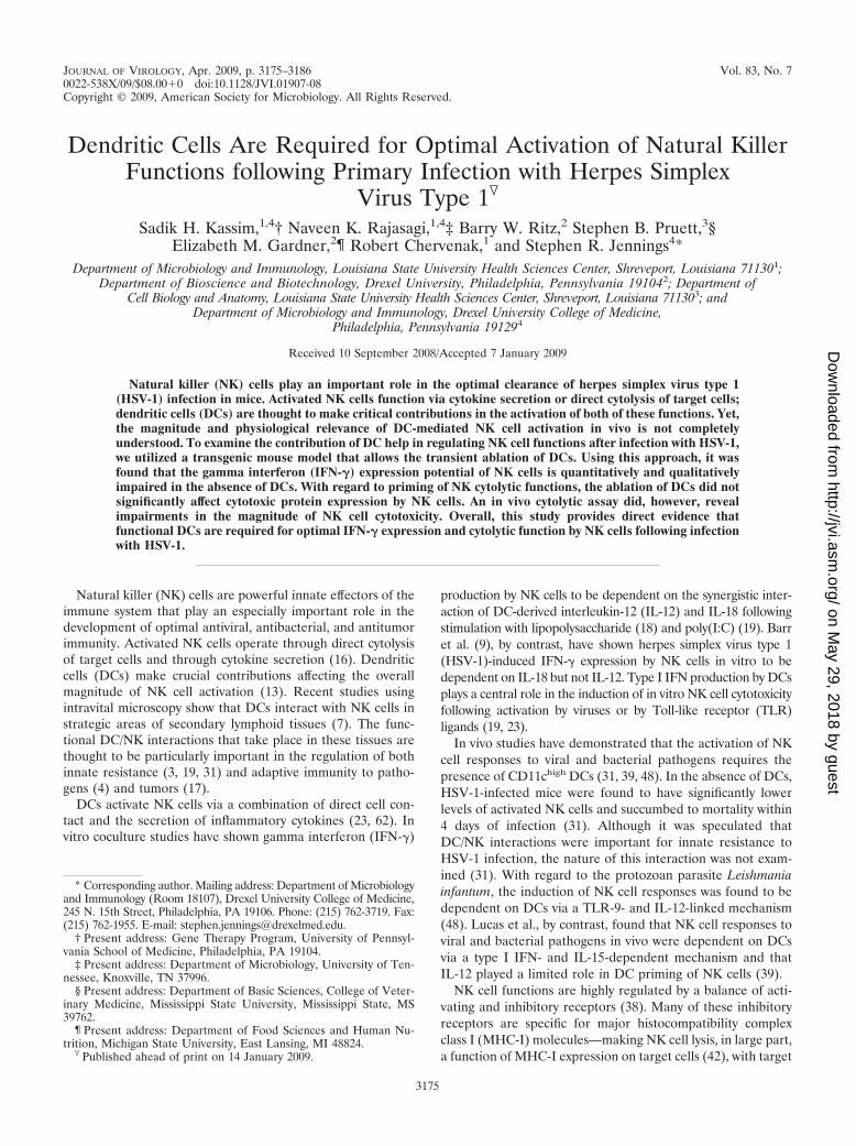
important for triggering optimal NK cell activation and cy-totoxicity following infection with HSV-1. The efficiency ofDC depletion was verified in mice 30 h p.i. Non-DC-de-pleted HSV-1-infected mice had a higher frequency of DCsthan DT-treated HSV-1-infected mice and naïve controlmice (Fig. 1A).
We next examined the functional implications of DC abla-tion, specifically with regard to cytokine production, followingHSV-1 infection. DC-derived cytokines, including IFN-�/�,IL-12, IL-15, and IL-18, have been implicated in NK cell acti-vation in vitro (62). Of these, IFN-�/� seem to be especiallyimportant for NK cell cytotoxicity (62). We therefore exam-ined serum type I IFN levels in DC-ablated and non-DC-ablated mice 30 h after HSV-1 infection. DC ablation led to asignificant decrease in the production of functional type I IFN(P 0.05) (Fig. 1B); no significant differences were foundbetween DC-ablated HSV-1-infected and non-HSV-1-infectedcontrol mice (Fig. 1B). The depletion of macrophages did notaffect HSV-1 pathogenesis (31), nor did it affect type I IFNproduction (data not shown). These data confirm that, as ob-served for other viral infections (12), DCs are the principalsource of type I IFN during the early immune response toHSV-1. We next examined the implications of reduced type IIFN production for NK cell activation.
DC depletion impairs NK cell activation. Several cell sur-face molecules, including B220, Thy-1, CD2, CD16, LFA-1,CD69, CD45RA, and CD45RO, are known to be upregulatedon activated murine and human NK cells (63). Of these, B220and CD16 are thought to be particularly important determi-nants of NK cytotoxicity, with B220 being a marker of non-
FIG. 1. DC depletion abolishes type I IFN production during primary HSV-1 infection. CD11c/DTR-Tg mice were i.p. injected with PBS (0.5ml/mouse) or DT (100 ng/mouse) 24 h prior to hind-FP infection with HSV-1 at 5 � 106 PFU per FP. Thirty hours p.i. with HSV-1, spleens wereharvested for FACS analysis of DC frequencies. (A) Representative FACS contour plots depicting the percentages of total CD11c� MHC-II� DCs.(B) Thirty hours p.i. with HSV-1, serum samples were harvested via cardiac blood withdrawal. Type I IFN levels were determined by virusneutralization assay. Values represent the means standard deviations of pooled data from two independent experiments with two to three miceper group. DT � HSV-1, DT-treated HSV-1-infected mice (n � 5); PBS � HSV-1, PBS-treated HSV-1-infected mice (n � 6); naïve mice (n �4). Statistical analyses using the Mann-Whitney test revealed P values of 0.05 (*) between the PBS-treated HSV-1-infected group and naïvecontrol group and between the PBS-treated HSV-1-infected group and the DT-treated HSV-1-infected group.
VOL. 83, 2009 DENDRITIC CELL-NK CELL INTERACTIONS IN HSV INFECTIONS 3177
on May 29, 2018 by guest
http://jvi.asm.org/
Dow
nloaded from
MHC-restricted killers (8, 31) and CD16 being a marker ofantibody-dependent cellular cytotoxicity but not other modesof NK cell-mediated cytotoxicity (34).
To initially determine the cytotoxic potential of NK cells inthe absence of DCs, the frequency of B220� NK and CD16�
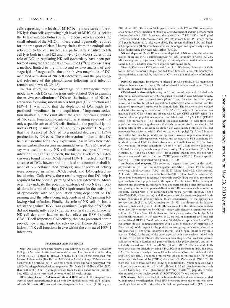
NK cells was examined 30 h after subcutaneous FP infectionwith HSV-1 in DT-treated and PBS-treated CD11c-DTR-Tgmice. Because secondary lymphoid organs are important sitesof DC/NK interaction (7, 16), a specific emphasis was placedon the levels of activated NK cells in the spleens of mice. Aspreviously reported (39, 48), DT treatment does not appear toimpair the total frequency of splenic NK1.1� CD3� cells (Fig.2A). DC-intact mice were found to have a higher frequency ofNK1.1� CD3� B220hi (Fig. 2B, left panel) and NK1.1� CD3�
CD16hi cells (Fig. 2B, right panel) than DC-ablated and naïvemice. DC-ablated and naïve mice had comparable levels ofactivated NK cells (Fig. 2A and B). These data extend previ-ously published results (31) and suggest that the absence ofDCs impedes the optimal activation, and perhaps the prolifer-ation or recruitment, of NK cells to secondary lymphoid organsfollowing infection with HSV-1. We next examined the effectof DC depletion on NK cell function.
DCs are not required for granule formation by NK cells.Like CD8� cytotoxic T lymphocytes, NK cells utilize the mech-anism of granule exocytosis in cell death. Lytic granules, whichcontain a mixture of potent proteins, release their content intothe intercellular space between the NK cell and target cell,whereby the action of these enzymes results in target cell death
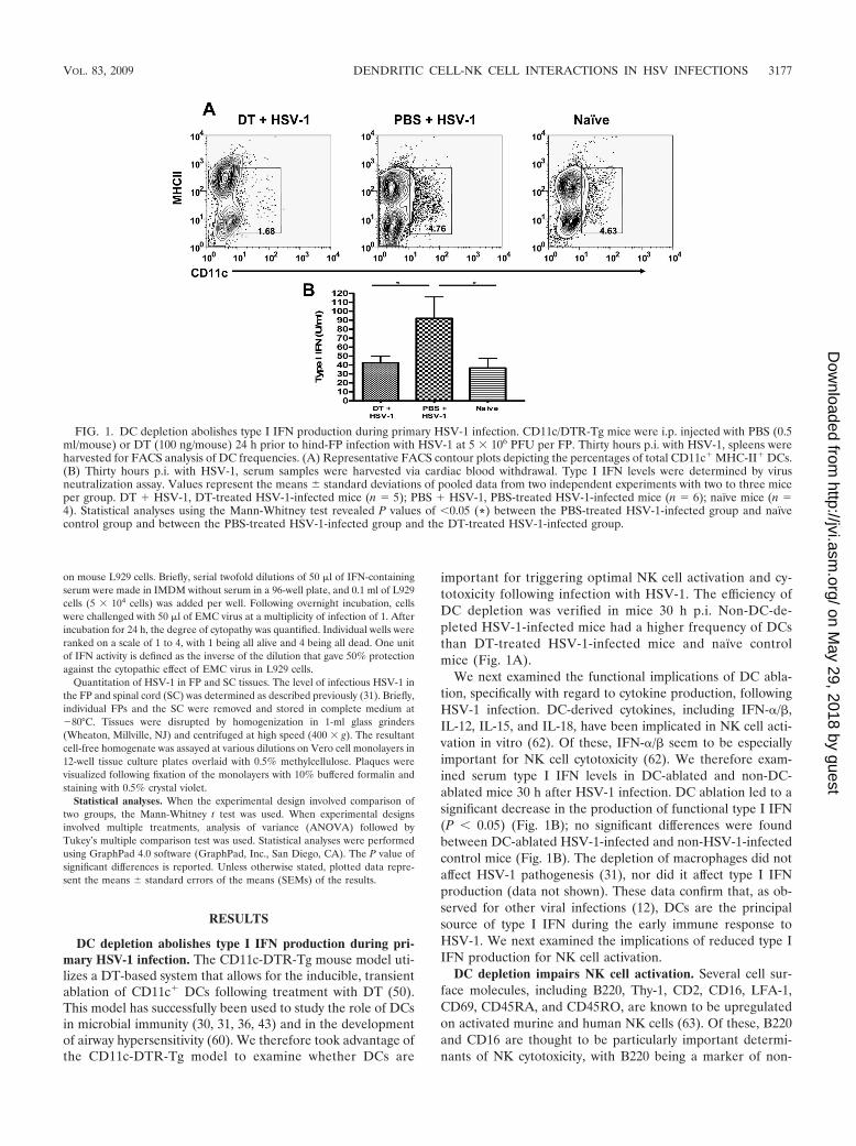
(35, 56). Major granule proteins include perforin (Prf), a cellmembrane disruptor, and granzymes (Grz), a family of struc-turally related serine proteases with various substrate specific-ities (53). To date, 11 granzymes have been identified (54).GrzB, found in both humans and mice, appears to play aparticularly important role in triggering target cell death (53).Prf and GrzB function synergistically by forming a single mac-romolecular complex at the target cell surface, which triggersnuclear damage and results in cell death (41, 50). The signalsthat regulate granule formation and polarization upon activa-tion are not completely understood. Recent studies, however,suggest that DCs are important for the optimal expression ofGrzB and Prf by activated CD8� cytotoxic T lymphocytes andNK cells (15, 39). Whether DCs play a similar role in NK cellgranule formation following HSV-1 infection is not known. Exvivo intracellular staining and flow cytometric analysis wastherefore undertaken to analyze the effect of DC ablation onGrzB and Prf expression by activated NK cells 24 to 30 h p.i.with HSV-1. Using this approach, comparable levels of Prf(Fig. 3A and C) and GrzB (Fig. 3B and D) were found forDC-depleted and non-DC-depleted mice. Infected mice, re-gardless of the presence of DCs, expressed higher levels of Prf(Fig. 3A and C) and GrzB (Fig. 3B and D) after infection thannaïve mice. B220� NK1.1� CD3� cells expressed significantlyhigher levels of Prf (Fig. 3A and C) (P 0.05) and GrzB (Fig.3C and D) (P 0.001) than did B220� NK1.1� CD3� cells inboth DC-ablated and non-DC-ablated mice; no significant dif-ferences were observed between these NK cell subsets in naïve
FIG. 2. DC depletion impairs optimal NK cell activation. (A) Spleens were harvested from DT-treated (DT � HSV-1), PBS-treated (PBS �HSV-1), and naïve DTR-Tg mice 30 h p.i. with HSV-1 and stained for NK1.1 and CD3. (B) B220 or CD16 expression by NK1.1� CD3� cells fromnaïve, DT-treated HSV-1-infected mice (DT � HSV-1), and PBS-treated HSV-1-infected mice (PBS � HSV-1) was analyzed. Representativehistograms for one mouse are displayed. These data are representative of the results of two separate experiments using a minimum of four miceper group per experiment.
3178 KASSIM ET AL. J. VIROL.
on May 29, 2018 by guest
http://jvi.asm.org/
Dow
nloaded from
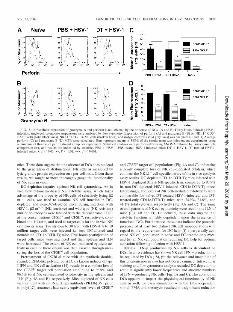
mice. These data suggest that the absence of DCs does not leadto the generation of dysfunctional NK cells as measured bylytic-granule protein expression on a per-cell basis. Given theseresults, we sought to more thoroughly gauge the functionalityof NK cells in vivo.
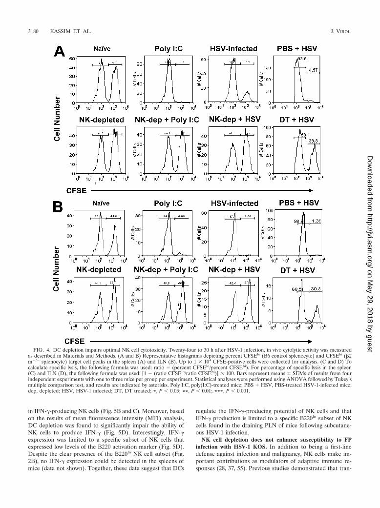
DC depletion impairs optimal NK cell cytotoxicity. An invivo flow cytometric-based NK cytolytic assay, which takesadvantage of the property of NK cells of selectively lysing �2m�/� cells, was used to examine NK cell function in DC-depleted and non-DC-depleted mice during infection withHSV-1. �2 m�/� (NK sensitive) and wild-type (NK resistant)murine splenocytes were labeled with the fluorochrome CFSEat the concentrations CFSEhi and CFSElo, respectively, com-bined at a 1:1 ratio, and used as target cells for the in vivo NKcytotoxicity assay. Twenty-four to 30 h p.i. with HSV-1, 8 to 10million target cells were injected i.v. into DC-ablated andnonablated CD11c-DTR-Tg mice. Five hours postinjection oftarget cells, mice were sacrificed and their spleens and ILNwere harvested. The extent of NK cell-mediated cytolytic ac-tivity in each of these organs was then assayed through mea-suring the loss of the CFSEhi cell population.
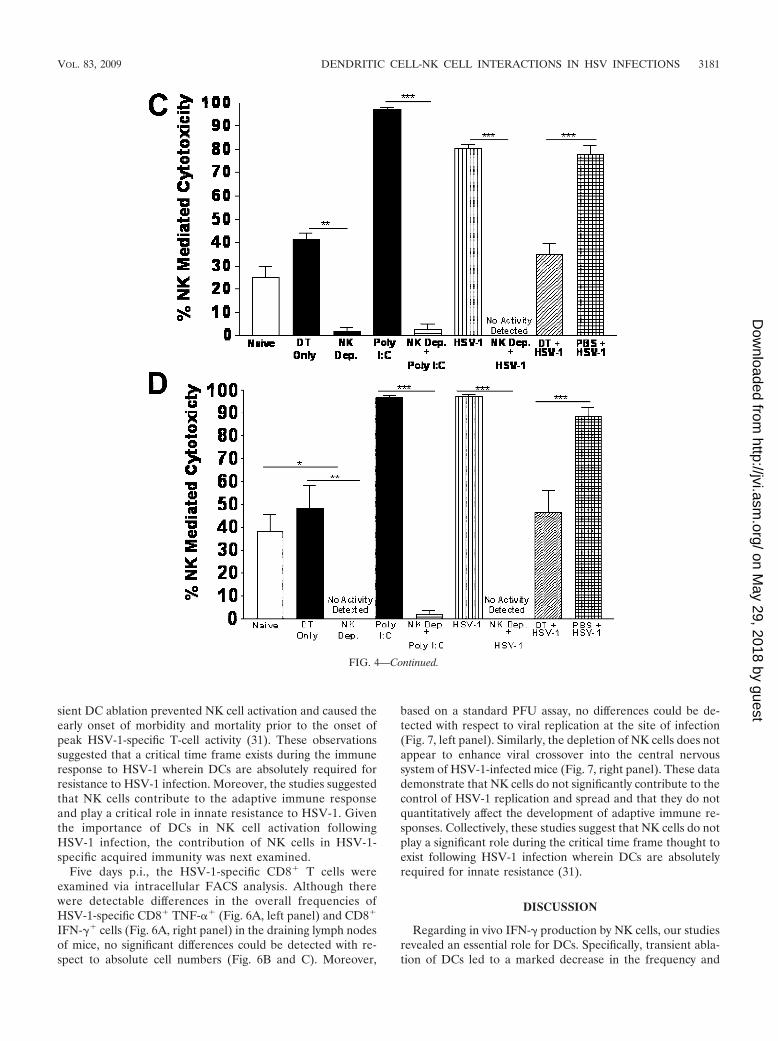
Pretreatment of C57BL/6 mice with the synthetic double-stranded RNA-like polymer poly(I:C), a known inducer of typeI IFN and NK cell activation (14), results in a complete loss ofthe CFSEhi target cell population amounting to 96.9% and96.6% total NK cell-mediated cytotoxicity in the spleens andILN (Fig. 4A and B), respectively. Mice depleted of NK cellsvia treatment with anti-NK1.1 IgG antibody (PK136) 36 h priorto poly(I:C) treatment had nearly equivalent levels of CFSEhi
and CFSElo target cell populations (Fig. 4A and C), indicatinga nearly complete loss of NK cell-mediated cytolysis, whichconfirms the NK1.1� cell-specific nature of the in vivo cytolysisassay results. DC-depleted CD11c-DTR-Tg mice infected withHSV-1 displayed 31.8% NK-specific lysis, compared to 80.9%in non-DC-depleted HSV-1-infected CD11c-DTR-Tg mice.Interestingly, the levels of NK cell-mediated cytotoxicity werecomparable for naïve, DT-treated HSV-1-infected, and DT-treated-only CD11c-DTR-Tg mice, with 24.9%, 31.8%, and41.1% total cytolysis, respectively (Fig. 4A and C). The sameoverall patterns of NK cell cytotoxicity were seen in the ILN ofmice (Fig. 4B and D). Collectively, these data suggest thatcytolytic function is highly dependent upon the presence offunctional DCs. Furthermore, these data indicate the potentialpresence of at least two distinct NK cell subpopulations withregard to the requirement for DC help: (i) a perpetually acti-vated NK cell population in naïve and DT-treated-only mice;and (ii) an NK cell population requiring DC help for optimalactivation following infection with HSV-1.
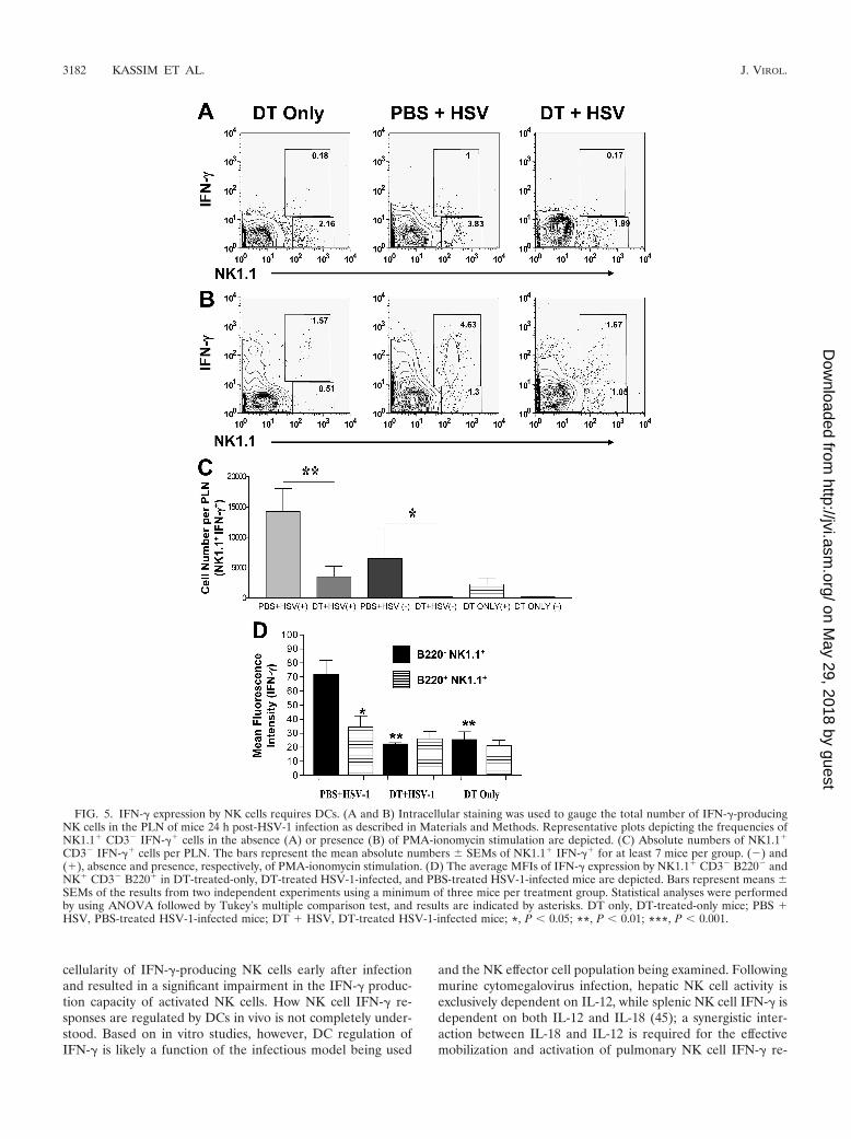
Optimal IFN-� production by NK cells is dependent onDCs. In vitro evidence has shown NK cell IFN-� production tobe regulated by DCs (18), yet the relevance and magnitude ofthis phenomenon in vivo has not been examined. Intracellularstaining and flow cytometric analysis revealed DC depletion toresult in significantly lower frequencies and absolute numbersof IFN-�-producing NK cells (Fig. 5A and C). The ablation ofDCs appears to impact the physiological functionality of NKcells as well, for even stimulation with the DC-independentstimuli PMA and ionomycin resulted in a significant reduction
FIG. 3. Intracellular expression of granzyme B and perforin is not affected by the presence of DCs. (A and B) Thirty hours following HSV-1infection, single-cell splenocyte suspensions were analyzed by flow cytometry. Expression of perforin (A) and granzyme B (B) on NK1.1� CD3�
B220� cells (solid black lines), NK1.1� CD3� B220� cells (broken lines), and isotype controls (solid gray lines) was analyzed. (C and D) Averageperforin (C) and granzyme B (D) MFIs were calculated. Bars represent means SEMs of the results from two independent experiments usinga minimum of three mice per treatment group per experiment. Statistical analyses were performed by using ANOVA followed by Tukey’s multiplecomparison test, and results are indicated by asterisks. PBS � HSV-1, PBS-treated HSV-1-infected mice; DT � HSV-1, DT-treated HSV-1-infected mice; *, P 0.05; **, P 0.01; ***, P 0.001.
VOL. 83, 2009 DENDRITIC CELL-NK CELL INTERACTIONS IN HSV INFECTIONS 3179
on May 29, 2018 by guest
http://jvi.asm.org/
Dow
nloaded from
in IFN-�-producing NK cells (Fig. 5B and C). Moreover, basedon the results of mean fluorescence intensity (MFI) analysis,DC depletion was found to significantly impair the ability ofNK cells to produce IFN-� (Fig. 5D). Interestingly, IFN-�expression was limited to a specific subset of NK cells thatexpressed low levels of the B220 activation marker (Fig. 5D).Despite the clear presence of the B220lo NK cell subset (Fig.2B), no IFN-� expression could be detected in the spleens ofmice (data not shown). Together, these data suggest that DCs
regulate the IFN-�-producing potential of NK cells and thatIFN-� production is limited to a specific B220lo subset of NKcells found in the draining PLN of mice following subcutane-ous HSV-1 infection.
NK cell depletion does not enhance susceptibility to FPinfection with HSV-1 KOS. In addition to being a first-linedefense against infection and malignancy, NK cells make im-portant contributions as modulators of adaptive immune re-sponses (28, 37, 55). Previous studies demonstrated that tran-
FIG. 4. DC depletion impairs optimal NK cell cytotoxicity. Twenty-four to 30 h after HSV-1 infection, in vivo cytolytic activity was measuredas described in Materials and Methods. (A and B) Representative histograms depicting percent CFSElo (B6 control splenocyte) and CFSEhi (�2m�/� splenocyte) target cell peaks in the spleen (A) and ILN (B). Up to 1 � 104 CFSE-positive cells were collected for analysis. (C and D) Tocalculate specific lysis, the following formula was used: ratio � (percent CFSElo/percent CFSEhi). For percentage of specific lysis in the spleen(C) and ILN (D), the following formula was used: [1 � (ratio CFSElo/ratio CFSEhi)] � 100. Bars represent means SEMs of results from fourindependent experiments with one to three mice per group per experiment. Statistical analyses were performed using ANOVA followed by Tukey’smultiple comparison test, and results are indicated by asterisks. Poly I:C, poly(I:C)-treated mice; PBS � HSV, PBS-treated HSV-1-infected mice;dep, depleted; HSV, HSV-1 infected; DT, DT treated; *, P 0.05; **, P 0.01; ***, P 0.001.
3180 KASSIM ET AL. J. VIROL.
on May 29, 2018 by guest
http://jvi.asm.org/
Dow
nloaded from
sient DC ablation prevented NK cell activation and caused theearly onset of morbidity and mortality prior to the onset ofpeak HSV-1-specific T-cell activity (31). These observationssuggested that a critical time frame exists during the immuneresponse to HSV-1 wherein DCs are absolutely required forresistance to HSV-1 infection. Moreover, the studies suggestedthat NK cells contribute to the adaptive immune responseand play a critical role in innate resistance to HSV-1. Giventhe importance of DCs in NK cell activation followingHSV-1 infection, the contribution of NK cells in HSV-1-specific acquired immunity was next examined.
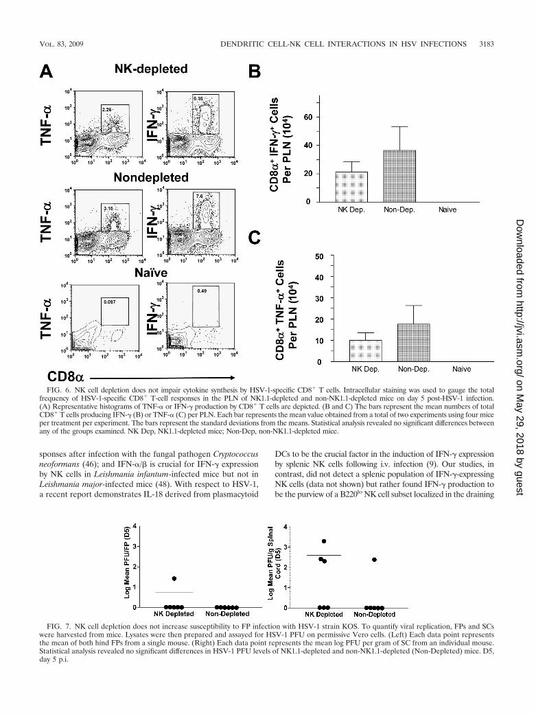
Five days p.i., the HSV-1-specific CD8� T cells wereexamined via intracellular FACS analysis. Although therewere detectable differences in the overall frequencies ofHSV-1-specific CD8� TNF-�� (Fig. 6A, left panel) and CD8�
IFN-�� cells (Fig. 6A, right panel) in the draining lymph nodesof mice, no significant differences could be detected with re-spect to absolute cell numbers (Fig. 6B and C). Moreover,
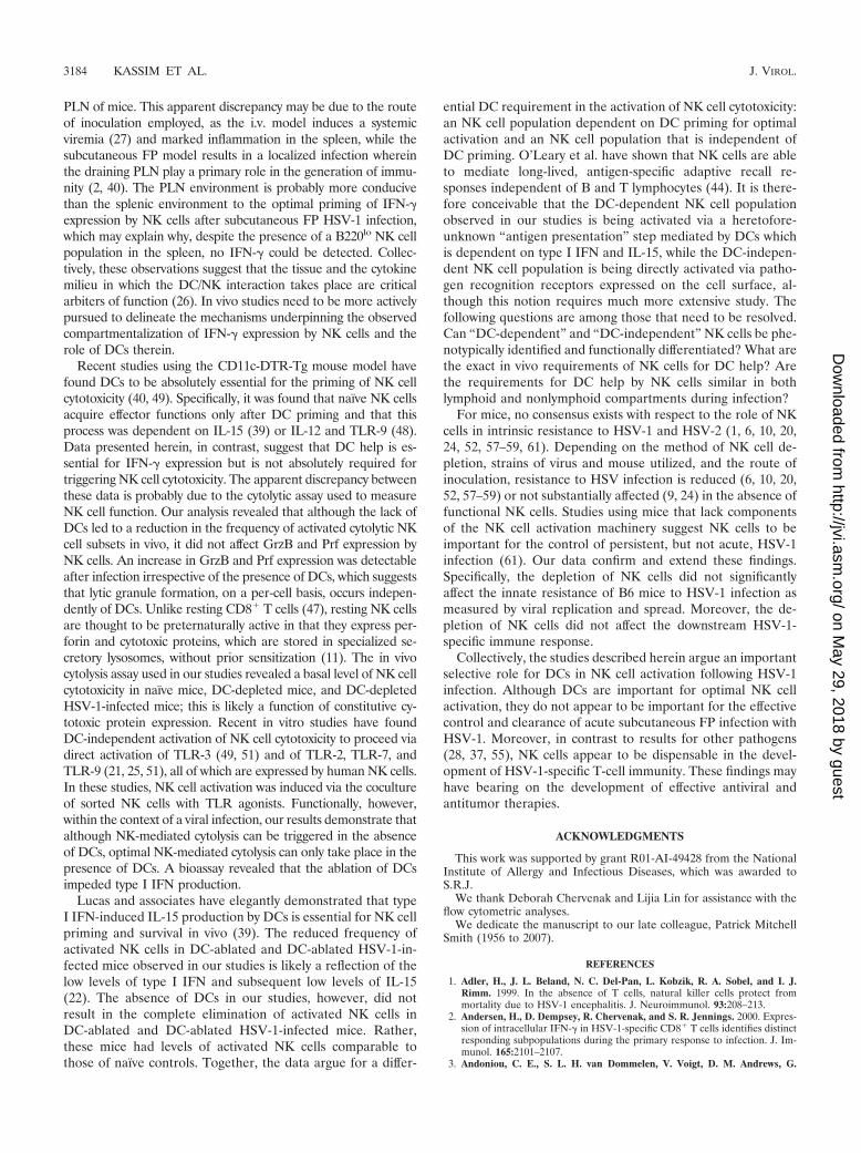
based on a standard PFU assay, no differences could be de-tected with respect to viral replication at the site of infection(Fig. 7, left panel). Similarly, the depletion of NK cells does notappear to enhance viral crossover into the central nervoussystem of HSV-1-infected mice (Fig. 7, right panel). These datademonstrate that NK cells do not significantly contribute to thecontrol of HSV-1 replication and spread and that they do notquantitatively affect the development of adaptive immune re-sponses. Collectively, these studies suggest that NK cells do notplay a significant role during the critical time frame thought toexist following HSV-1 infection wherein DCs are absolutelyrequired for innate resistance (31).
DISCUSSION
Regarding in vivo IFN-� production by NK cells, our studiesrevealed an essential role for DCs. Specifically, transient abla-tion of DCs led to a marked decrease in the frequency and
FIG. 4—Continued.
VOL. 83, 2009 DENDRITIC CELL-NK CELL INTERACTIONS IN HSV INFECTIONS 3181
on May 29, 2018 by guest
http://jvi.asm.org/
Dow
nloaded from
cellularity of IFN-�-producing NK cells early after infectionand resulted in a significant impairment in the IFN-� produc-tion capacity of activated NK cells. How NK cell IFN-� re-sponses are regulated by DCs in vivo is not completely under-stood. Based on in vitro studies, however, DC regulation ofIFN-� is likely a function of the infectious model being used
and the NK effector cell population being examined. Followingmurine cytomegalovirus infection, hepatic NK cell activity isexclusively dependent on IL-12, while splenic NK cell IFN-� isdependent on both IL-12 and IL-18 (45); a synergistic inter-action between IL-18 and IL-12 is required for the effectivemobilization and activation of pulmonary NK cell IFN-� re-
FIG. 5. IFN-� expression by NK cells requires DCs. (A and B) Intracellular staining was used to gauge the total number of IFN-�-producingNK cells in the PLN of mice 24 h post-HSV-1 infection as described in Materials and Methods. Representative plots depicting the frequencies ofNK1.1� CD3� IFN-�� cells in the absence (A) or presence (B) of PMA-ionomycin stimulation are depicted. (C) Absolute numbers of NK1.1�
CD3� IFN-�� cells per PLN. The bars represent the mean absolute numbers SEMs of NK1.1� IFN-�� for at least 7 mice per group. (�) and(�), absence and presence, respectively, of PMA-ionomycin stimulation. (D) The average MFIs of IFN-� expression by NK1.1� CD3� B220� andNK� CD3� B220� in DT-treated-only, DT-treated HSV-1-infected, and PBS-treated HSV-1-infected mice are depicted. Bars represent means SEMs of the results from two independent experiments using a minimum of three mice per treatment group. Statistical analyses were performedby using ANOVA followed by Tukey’s multiple comparison test, and results are indicated by asterisks. DT only, DT-treated-only mice; PBS �HSV, PBS-treated HSV-1-infected mice; DT � HSV, DT-treated HSV-1-infected mice; *, P 0.05; **, P 0.01; ***, P 0.001.
3182 KASSIM ET AL. J. VIROL.
on May 29, 2018 by guest
http://jvi.asm.org/
Dow
nloaded from
sponses after infection with the fungal pathogen Cryptococcusneoformans (46); and IFN-�/� is crucial for IFN-� expressionby NK cells in Leishmania infantum-infected mice but not inLeishmania major-infected mice (48). With respect to HSV-1,a recent report demonstrates IL-18 derived from plasmacytoid
DCs to be the crucial factor in the induction of IFN-� expressionby splenic NK cells following i.v. infection (9). Our studies, incontrast, did not detect a splenic population of IFN-�-expressingNK cells (data not shown) but rather found IFN-� production tobe the purview of a B220lo NK cell subset localized in the draining
FIG. 6. NK cell depletion does not impair cytokine synthesis by HSV-1-specific CD8� T cells. Intracellular staining was used to gauge the totalfrequency of HSV-1-specific CD8� T-cell responses in the PLN of NK1.1-depleted and non-NK1.1-depleted mice on day 5 post-HSV-1 infection.(A) Representative histograms of TNF-� or IFN-� production by CD8� T cells are depicted. (B and C) The bars represent the mean numbers of totalCD8� T cells producing IFN-� (B) or TNF-� (C) per PLN. Each bar represents the mean value obtained from a total of two experiments using four miceper treatment per experiment. The bars represent the standard deviations from the means. Statistical analysis revealed no significant differences betweenany of the groups examined. NK Dep, NK1.1-depleted mice; Non-Dep, non-NK1.1-depleted mice.
FIG. 7. NK cell depletion does not increase susceptibility to FP infection with HSV-1 strain KOS. To quantify viral replication, FPs and SCswere harvested from mice. Lysates were then prepared and assayed for HSV-1 PFU on permissive Vero cells. (Left) Each data point representsthe mean of both hind FPs from a single mouse. (Right) Each data point represents the mean log PFU per gram of SC from an individual mouse.Statistical analysis revealed no significant differences in HSV-1 PFU levels of NK1.1-depleted and non-NK1.1-depleted (Non-Depleted) mice. D5,day 5 p.i.
VOL. 83, 2009 DENDRITIC CELL-NK CELL INTERACTIONS IN HSV INFECTIONS 3183
on May 29, 2018 by guest
http://jvi.asm.org/
Dow
nloaded from
PLN of mice. This apparent discrepancy may be due to the routeof inoculation employed, as the i.v. model induces a systemicviremia (27) and marked inflammation in the spleen, while thesubcutaneous FP model results in a localized infection whereinthe draining PLN play a primary role in the generation of immu-nity (2, 40). The PLN environment is probably more conducivethan the splenic environment to the optimal priming of IFN-�expression by NK cells after subcutaneous FP HSV-1 infection,which may explain why, despite the presence of a B220lo NK cellpopulation in the spleen, no IFN-� could be detected. Collec-tively, these observations suggest that the tissue and the cytokinemilieu in which the DC/NK interaction takes place are criticalarbiters of function (26). In vivo studies need to be more activelypursued to delineate the mechanisms underpinning the observedcompartmentalization of IFN-� expression by NK cells and therole of DCs therein.
Recent studies using the CD11c-DTR-Tg mouse model havefound DCs to be absolutely essential for the priming of NK cellcytotoxicity (40, 49). Specifically, it was found that naïve NK cellsacquire effector functions only after DC priming and that thisprocess was dependent on IL-15 (39) or IL-12 and TLR-9 (48).Data presented herein, in contrast, suggest that DC help is es-sential for IFN-� expression but is not absolutely required fortriggering NK cell cytotoxicity. The apparent discrepancy betweenthese data is probably due to the cytolytic assay used to measureNK cell function. Our analysis revealed that although the lack ofDCs led to a reduction in the frequency of activated cytolytic NKcell subsets in vivo, it did not affect GrzB and Prf expression byNK cells. An increase in GrzB and Prf expression was detectableafter infection irrespective of the presence of DCs, which suggeststhat lytic granule formation, on a per-cell basis, occurs indepen-dently of DCs. Unlike resting CD8� T cells (47), resting NK cellsare thought to be preternaturally active in that they express per-forin and cytotoxic proteins, which are stored in specialized se-cretory lysosomes, without prior sensitization (11). The in vivocytolysis assay used in our studies revealed a basal level of NK cellcytotoxicity in naïve mice, DC-depleted mice, and DC-depletedHSV-1-infected mice; this is likely a function of constitutive cy-totoxic protein expression. Recent in vitro studies have foundDC-independent activation of NK cell cytotoxicity to proceed viadirect activation of TLR-3 (49, 51) and of TLR-2, TLR-7, andTLR-9 (21, 25, 51), all of which are expressed by human NK cells.In these studies, NK cell activation was induced via the cocultureof sorted NK cells with TLR agonists. Functionally, however,within the context of a viral infection, our results demonstrate thatalthough NK-mediated cytolysis can be triggered in the absenceof DCs, optimal NK-mediated cytolysis can only take place in thepresence of DCs. A bioassay revealed that the ablation of DCsimpeded type I IFN production.
Lucas and associates have elegantly demonstrated that typeI IFN-induced IL-15 production by DCs is essential for NK cellpriming and survival in vivo (39). The reduced frequency ofactivated NK cells in DC-ablated and DC-ablated HSV-1-in-fected mice observed in our studies is likely a reflection of thelow levels of type I IFN and subsequent low levels of IL-15(22). The absence of DCs in our studies, however, did notresult in the complete elimination of activated NK cells inDC-ablated and DC-ablated HSV-1-infected mice. Rather,these mice had levels of activated NK cells comparable tothose of naïve controls. Together, the data argue for a differ-
ential DC requirement in the activation of NK cell cytotoxicity:an NK cell population dependent on DC priming for optimalactivation and an NK cell population that is independent ofDC priming. O’Leary et al. have shown that NK cells are ableto mediate long-lived, antigen-specific adaptive recall re-sponses independent of B and T lymphocytes (44). It is there-fore conceivable that the DC-dependent NK cell populationobserved in our studies is being activated via a heretofore-unknown “antigen presentation” step mediated by DCs whichis dependent on type I IFN and IL-15, while the DC-indepen-dent NK cell population is being directly activated via patho-gen recognition receptors expressed on the cell surface, al-though this notion requires much more extensive study. Thefollowing questions are among those that need to be resolved.Can “DC-dependent” and “DC-independent” NK cells be phe-notypically identified and functionally differentiated? What arethe exact in vivo requirements of NK cells for DC help? Arethe requirements for DC help by NK cells similar in bothlymphoid and nonlymphoid compartments during infection?
For mice, no consensus exists with respect to the role of NKcells in intrinsic resistance to HSV-1 and HSV-2 (1, 6, 10, 20,24, 52, 57–59, 61). Depending on the method of NK cell de-pletion, strains of virus and mouse utilized, and the route ofinoculation, resistance to HSV infection is reduced (6, 10, 20,52, 57–59) or not substantially affected (9, 24) in the absence offunctional NK cells. Studies using mice that lack componentsof the NK cell activation machinery suggest NK cells to beimportant for the control of persistent, but not acute, HSV-1infection (61). Our data confirm and extend these findings.Specifically, the depletion of NK cells did not significantlyaffect the innate resistance of B6 mice to HSV-1 infection asmeasured by viral replication and spread. Moreover, the de-pletion of NK cells did not affect the downstream HSV-1-specific immune response.
Collectively, the studies described herein argue an importantselective role for DCs in NK cell activation following HSV-1infection. Although DCs are important for optimal NK cellactivation, they do not appear to be important for the effectivecontrol and clearance of acute subcutaneous FP infection withHSV-1. Moreover, in contrast to results for other pathogens(28, 37, 55), NK cells appear to be dispensable in the devel-opment of HSV-1-specific T-cell immunity. These findings mayhave bearing on the development of effective antiviral andantitumor therapies.
ACKNOWLEDGMENTS
This work was supported by grant R01-AI-49428 from the NationalInstitute of Allergy and Infectious Diseases, which was awarded toS.R.J.
We thank Deborah Chervenak and Lijia Lin for assistance with theflow cytometric analyses.
We dedicate the manuscript to our late colleague, Patrick MitchellSmith (1956 to 2007).
REFERENCES
1. Adler, H., J. L. Beland, N. C. Del-Pan, L. Kobzik, R. A. Sobel, and I. J.Rimm. 1999. In the absence of T cells, natural killer cells protect frommortality due to HSV-1 encephalitis. J. Neuroimmunol. 93:208–213.
2. Andersen, H., D. Dempsey, R. Chervenak, and S. R. Jennings. 2000. Expres-sion of intracellular IFN-� in HSV-1-specific CD8� T cells identifies distinctresponding subpopulations during the primary response to infection. J. Im-munol. 165:2101–2107.
3. Andoniou, C. E., S. L. H. van Dommelen, V. Voigt, D. M. Andrews, G.
3184 KASSIM ET AL. J. VIROL.
on May 29, 2018 by guest
http://jvi.asm.org/
Dow
nloaded from
Brizardi, C. Asselin-Paturel, T. Delale, K. J. Stacey, G. Trinchieri, and M. A.Degli-Esposti. 2005. Interaction between conventional dendritic cells andnatural killer cells is integral to the activation of effective antiviral immunity.Nat. Immunol. 6:1011–1019.
4. Andrews, D. M., C. E. Andoniou, A. A. Scalzo, S. L. H. Van Dommelen, M. E.Wallace, M. J. Smyth, and M. A. Degli-Esposti. 2005. Cross-talk betweendendritic cells and natural killer cells in viral infection. Mol. Immunol.42:547–555.
5. Andrews, D. M., A. A. Scalzo, W. M. Yokoyama, M. J. Smyth, and M. A.Degli-Esposti. 2003. Functional interactions between dendritic cells and NKcells during viral infection. Nat. Immunol. 4:175–181.
6. Ashkar, A. A., and K. L. Rosenthal. 2003. Interleukin-15 and natural killerand NKT cells play a critical role in innate protection against genital herpessimplex virus type 2 infection. J. Virol. 77:10168–10171.
7. Bajenoff, M., B. Breart, A. Y. C. Huang, H. Qi, J. Cazareth, V. M. Braud,R. N. Germain, and N. Glaichenhaus. 2006. Natural killer cell behavior inlymph nodes revealed by static and real-time imaging. J. Exp. Med. 203:619–631.
8. Ballas, Z. K., and W. Rasmussen. 1990. Lymphokine-activated killer (LAK)cells. IV. Characterization of murine LAK effector subpopulations. J. Im-munol. 144:386–395.
9. Barr, D. P., G. T. Belz, P. C. Reading, M. Wojtasiak, P. G. Whitney, W. R.Heath, F. R. Carbone, and A. G. Brooks. 2007. A role for plasmacytoiddendritic cells in the rapid IL-18-dependent activation of NK cells followingHSV-1 infection. Eur. J. Immunol. 37:1334–1342.
10. Brandt, C. R., and C. A. Salkowski. 1992. Activation of NK cells in micefollowing corneal infection with herpes simplex virus type-1. Invest. Oph-thalmol. Vis. Sci. 33:113–120.
11. Bryceson, Y. T., M. E. March, D. F. Barber, H. G. Ljunggren, and E. O.Long. 2005. Cytolytic granule polarization and degranulation controlled bydifferent receptors in resting NK cells. J. Exp. Med. 202:1001–1012.
12. Ciavarra, R. P., A. Stephens, S. Nagy, M. Sekellick, and C. Steel. 2006.Evaluation of immunological paradigms in a virus model: are dendritic cellscritical for antiviral immunity and viral clearance? J. Immunol. 177:492–500.
13. Colonna, M., G. Trinchieri, and Y. J. Liu. 2004. Plasmacytoid dendritic cellsin immunity. Nat. Immunol. 5:1219–1226.
14. Djeu, J. Y., J. A. Heinbaugh, H. T. Holden, and R. B. Herberman. 1979.Augmentation of mouse natural killer cell activity by interferon and inter-feron inducers. J. Immunol. 122:175–181.
15. Dubsky, P., H. Saito, M. Leogier, C. Dantin, J. E. Connolly, J. Banchereau,and A. K. Palucka. 2007. IL-15-induced human DC efficiently prime mela-noma-specific naïve CD8(�) T cells to differentiate into CTL. Eur. J. Im-munol. 37:1678–1690.
16. Ferlazzo, G., and C. Munz. 2004. NK cell compartments and their activationby dendritic cells. J. Immunol. 172:1333–1339.
17. Fernandez, N. C., A. Lozier, C. Flament, P. Ricciardi-Castagnoli, D. Bellet,M. Suter, M. Perricaudet, T. Tursz, E. Maraskovsky, and L. Zitvogel. 1999.Dendritic cells directly trigger NK cell functions: cross-talk relevant in innateand anti-tumor immune responses in vivo. Nat. Med. 5:405–411.
18. Gerosa, F., B. Baldanni-Guerra, C. Nisii, V. Marchesini, G. Carra, and G.Trinchieri. 2002. Reciprocal activating interaction between natural killercells and dendritic cells. J. Exp. Med. 195:327–333.
19. Gerosa, F., A. Gobbi, P. Zorzi, S. Burg, F. Briere, G. Carra, and G. Trinch-ieri. 2005. The reciprocal interaction of NK cells with plasmacytoid or my-eloid dendritic cells profoundly affects innate resistance functions. J. Immu-nol. 174:727–734.
20. Ghiasi, H., S. Cai, G. C. Perng, A. B. Nesburn, and S. L. Wechsler. 2000. Therole of natural killer cells in protection of mice against death and cornealscarring following ocular HSV-1 infection. Antivir. Res. 45:33–45.
21. Gorski, K. S., E. L. Waller, J. Bjornton-Severson, J. A. Hanten, C. L. Riter,W. C. Kieper, K. B. Gorden, J. S. Miller, J. P. Vasilakos, M. A. Tomai, andS. S. Alkan. 2006. Distinct indirect pathways govern human NK-cell activa-tion by TLR-7 and TLR-8 agonists. Int. Immunol. 18:1115–1126.
22. Gosselin, J., A. Tomoiu, R. C. Gallo, and L. Flamand. 1999. Interleukin-15as an activator of natural killer cell-mediated antiviral response. Blood 94:4210–4219.
23. Granucci, F., I. Zanoni, N. Pavelka, S. L. H. van Dommelen, C. E. Andoniou,F. Belardelli, M. A. Degli Esposti, and P. Ricciardi-Castagnoli. 2004. Acontribution of mouse dendritic cell-derived IL-2 for NK cell activation. J.Exp. Med. 200:287–295.
24. Halford, W. P., J. L. Maender, and B. M. Gebhardt. 2005. Re-evaluating therole of natural killer cells in innate resistance to herpes simplex virus type 1.Virol. J. 2:56.
25. Hart, O. M., V. Athie-Morales, G. M. O’Connor, and C. M. Gardiner. 2005.TLR7/8-mediated activation of human NK cells results in accessory cell-dependent IFN-gamma production. J. Immunol. 175:1636–1642.
26. Hayakawa, Y., N. D. Huntington, S. L. Nutt, and M. J. Smyth. 2006. Func-tional subsets of mouse natural killer cells. Immunol. Rev. 214:47–55.
27. Hill, T. J., D. L. Yirrell, and W. A. Blyth. 1986. Infection of the adrenal glandas a route to the central nervous system after viraemia with herpes simplexvirus in the mouse. J. Gen. Virol. 67:309–320.
28. Hussell, T., and P. J. Openshaw. 1998. Intracellular IFN-gamma expression
in natural killer cells precedes lung CD8� T cell recruitment during respi-ratory syncytial virus infection. J. Gen. Virol. 79:2593–2601.
29. Johansson, S. E., H. Hall, J. Bjorklund, and P. Hoglund. 2004. Broadlyimpaired NK cell function in non-obese diabetic mice is partially restored byNK cell activation in vivo and by IL-12/IL-18 in vitro. Int. Immunol. 16:1–11.
30. Jung, S., D. Unutmaz, P. Wong, G. Sano, K. De los Santos, T. Sparwasser,S. Wu, S. Vuthoori, K. Ko, F. Zavala, E. G. Pamer, D. R. Littman, and R. A.Lang. 2002. In vivo depletion of CD11c(�) dendritic cells abrogates primingof CD8(�) T cells by exogenous cell-associated antigens. Immunity 17:211–220.
31. Kassim, S. H., N. K. Rajasagi, X. Zhao, R. Chervenak, and S. R. Jennings.2006. In vivo ablation of CD11c-positive dendritic cells increases suscepti-bility to herpes simplex virus type I infection and diminishes NK and T-cellresponses. J. Virol. 80:3985–3993.
32. Koo, G. C., and J. R. Peppard. 1984. Establishment of monoclonal anti-NK1.1 antibody. Hybridoma 3:301–303.
33. Koo, G. C., J. R. Peppard, and E. C. Lattime. 1986. Characterization ofcytotoxic cells generated from bone marrow culture. Cell. Immunol. 98:172–180.
34. Lanier, L. L. 1998. NK cell receptors. Annu. Rev. Immunol. 16:359–393.35. Lee, R. K., and E. R. Podack. 1996. Perforin, Fas ligand, and tumor necrosis
factor are the major cytotoxic molecules used by lymphokine-activated killercells. J. Immunol. 157:1919–1925.
36. Liu, C. H., Y. T. Fan, A. Dias, L. Esper, R. A. Corn, A. Bafica, F. S. Machado,and J. Aliberti. 2006. Cutting edge: dendritic cells are essential for in vivoIL-12 production and development of resistance against Toxoplasma gondiiinfection in mice. J. Immunol. 177:31–35.
37. Liu, Z. X., S. Govindarajan, S. Okamoto, and G. Dennert. 2000. NK cellscause liver injury and facilitate the induction of T cell-mediated immunity toa viral liver infection. J. Immunol. 164:6480–6486.
38. Lodoen, M. B., and L. L. Lanier. 2005. Viral modulation of NK cell immu-nity. Nat. Rev. Microbiol. 3:59–69.
39. Lucas, M., W. Schachterle, K. Oberle, P. Aichele, and A. Diefenbach. 2007.Dendritic cells prime natural killer cells by trans-presenting interleukin 15.Immunity 26:503–517.
40. McNally, J. M., D. Dempsey, R. M. Wolcott, R. Chervenak, and S. R.Jennings. 1999. Phenotypic identification of antigen-dependent and antigen-independent CD8 CTL precursors in the draining lymph node during acutecutaneous herpes simplex virus type 1 infection. J. Immunol. 163:675–681.
41. Metkar, S. S., B. Wang, M. Aguilar-Santelises, S. M. Raja, L. Uhlin-Hansen,E. Podack, J. A. Trapani, and C. J. Froelich. 2002. Cytotoxic cell granule-mediated apoptosis: perforin delivers granzyme B-serglycin complexes intotarget cells without plasma membrane pore formation. Immunity 16:417–428.
42. Mullbacher, A., and N. J. King. 1989. Target cell lysis by natural killercells is influenced by beta 2-microglobulin expression. Scand. J. Immunol.30:21–29.
43. Ohteki, T., H. Tada, K. Ishida, T. Sato, C. Maki, T. Yamada, J. Hamuro, andS. Koyasu. 2006. Essential roles of DC-derived IL-15 as a mediator ofinflammatory responses in vivo. J. Exp. Med. 203:2329–2338.
44. O’Leary, J. G., M. Goodarzi, D. L. Drayton, and U. H. von Andrian. 2006. Tcell- and B cell-independent adaptive immunity mediated by natural killercells. Nat. Immunol. 7:507–516.
45. Pien, G. C., A. R. Satoksar, K. Takeda, S. Akira, and C. A. Biron. 2000.Cutting edge: selective IL-18 requirements for induction of compartmentalIFN-gamma responses during viral infection. J. Immunol. 165:4787–4791.
46. Qureshi, M. H., T. Zhang, Y. Koguchi, K. Nakashima, H. Okamura, M.Kurimoto, and K. Kawakami. 1999. Combined effects of IL-12 and IL-18 onthe clinical course and local cytokine production in murine pulmonary in-fection with Cryptococcus neoformans. Eur. J. Immunol. 29:643–649.
47. Radoja, S., A. B. Frey, and S. Vukmanovic. 2006. T-cell receptor signalingevents triggering granule exocytosis. Crit. Rev. Immunol. 26:265–290.
48. Schleicher, U., J. Liese, I. Knippertz, C. Kurzmann, A. Hesse, A. Heit,J. A. A. Fischer, S. Weiss, U. Kalinke, S. Kunz, and C. Bogdan. 2007. NK cellactivation in visceral leishmaniasis requires TLR9, myeloid DCs, and IL-12,but is independent of plasmacytoid DCs. J. Exp. Med. 204:893–906.
49. Schmidt, K. N., B. Leung, M. Kwong, K. A. Zarember, S. Satyal, T. A. Navas,F. Wang, and P. J. Godowski. 2004. APC-independent activation of NK cellsby the Toll-like receptor 3 agonist double-stranded RNA. J. Immunol. 172:138–143.
50. Shi, L., C. M. Kam, J. C. Powers, R. Aebersold, and A. H. Greenberg. 1992.Purification of three cytotoxic lymphocyte granule serine proteases thatinduce apoptosis through distinct substrate and target cell interactions. J.Exp. Med. 176:1521–1529.
51. Sivori, S., M. Falco, M. Della Chiesa, S. Carlomagno, M. Vitale, L. Moretta,and A. Moretta. 2004. CpG and double-stranded RNA trigger human NKcells by Toll-like receptors: induction of cytokine release and cytotoxicityagainst tumors and dendritic cells. Proc. Natl. Acad. Sci. USA 101:10116–10121.
52. Smith, J. R., A. M. Thackray, and R. Bujdoso. 2001. Reduced herpessimplex virus type 1 latency in Flt-3 ligand-treated mice is associated with
VOL. 83, 2009 DENDRITIC CELL-NK CELL INTERACTIONS IN HSV INFECTIONS 3185
on May 29, 2018 by guest
http://jvi.asm.org/
Dow
nloaded from
enhanced numbers of natural killer and dendritic cells. Immunology102:352–358.
53. Smyth, M. J., E. Cretney, J. M. Kelly, J. A. Westwood, S. E. Street, H. Yagita,K. Takeda, S. L. van Dommelen, M. A. Degli-Esposti, and Y. Hayakawa.2005. Activation of NK cell cytotoxicity. Mol. Immunol. 42:501–510.
54. Smyth, M. J., M. D. O’Connor, and J. Trapani. 1996. Granzymes: a varietyof serine protease specificities encoded by genetically distinct subfamilies.J. Leukoc. Biol. 60:555–562.
55. Su, H. C., K. B. Nguyen, T. P. Salazar-Mather, M. C. Ruzek, M. Y. Dalod,and C. A. Biron. 2001. NK cell functions restrain T cell responses during viralinfections. Eur. J. Immunol. 31:3048–3055.
56. Sutton, V. R., J. E. Davis, M. Cancilla, R. W. Johnstone, A. A. Ruefli, K.Sedelies, K. A. Browne, and J. A. Trapani. 2000. Initiation of apoptosis bygranzyme B requires direct cleavage of bid, but not direct granzyme B-mediated caspase activation. J. Exp. Med. 192:1403–1414.
57. Tanigawa, M., J. E. Bigger, M. Y. Kanter, and S. S. Atherton. 2000. Naturalkiller cells prevent direct anterior-to-posterior spread of herpes simplex virustype 1 in the eye. Invest. Opthalmol. Vis. Sci. 41:132–137.
58. Thapa, M., W. A. Kuziel, and D. J. Carr. 2007. Susceptibility of CCR5-
deficient mice to genital herpes simplex virus type 2 is linked to NK cellmobilization. J. Virol. 81:3704–3713.
59. Tsunobuchi, H., H. Nishimura, F. Goshima, T. Daikoku, H. Suzuki, I. Na-kashima, Y. Nishiyama, and Y. Yoshikai. 2000. A protective role of inter-leukin-15 in a mouse model for systemic infection with herpes simplex virus.Virology 275:57–66.
60. van Rijt, L. S., S. Jung, A. Kleinjan, N. Vos, M. Willart, C. Duez, H. C.Hoogsteden, and B. N. Lambrecht. 2005. In vivo depletion of lung CD11c�dendritic cells during allergen challenge abrogates the characteristic featuresof asthma. J. Exp. Med. 201:981–991.
61. Vollstedt, S., S. Arnold, C. Schwerdel, M. Franchini, G. Alber, J. P. Di Santo,M. Ackermann, and M. Suter. 2004. Interplay between alpha/beta andgamma interferons with B, T, and natural killer cells in the defense againstherpes simplex virus type 1. J. Virol. 78:3846–3850.
62. Walzer, T., M. Dalod, S. H. Robbins, L. Zitvogel, and E. Vivier. 2005.Natural-killer cells and dendritic cells: l’union fait la force. Blood 106:2252–2258.
63. Yokoyama, W. M. 1995. Natural killer cell receptors. Curr. Opin. Immunol.7:110–120.
3186 KASSIM ET AL. J. VIROL.
on May 29, 2018 by guest
http://jvi.asm.org/
Dow
nloaded from